Listeria monocytogenes Delivery of HPV16 Major Capsid Protein L1 Induces Systemic and Mucosal...
10
Listeria monocytogenes Delivery of HPV-16 Major Capsid Protein L1 Induces Systemic and Mucosal Cell-Mediated CD4 + and CD8 + T-Cell Responses After Oral Immunization Waleed Mustafa, 1 Paulo Cesar Maciag, 1 Zhen-kun Pan, 1 Jessica R. Weaver, 2 Yuhong Xiao, 2 Stuart N. Isaacs, 2 and Yvonne Paterson 1 Abstract Neutralizing antibodies are thought to be required at mucosal surfaces to prevent human papillomavirus (HPV) transmission. However, the potential for cell-mediated immunity in mediating protection against HPV infection has not been well explored. We generated recombinant Listeria monocytogenes (Lm) constructs that secrete lis- teriolysin O (LLO) fused with overlapping N-terminal (LLO-L1 1–258 ) or C-terminal (LLO-L1 238–474 ) fragments of HPV type 16 major capsid protein L1 (HPV-16-L1). Oral immunization of mice with either construct induced IFN-g-producing CD8 + and CD4 + T cells in the spleen and in the Peyer’s patches with the C-terminal construct. Oral immunization with both constructs resulted in diminished viral titers in the cervix and uterus of mice after intravaginal challenge with vaccinia virus expressing HPV-16-L1. Introduction O ncogenic human papillomaviruses (HPVs) infect the genital tract and are associated with human anogenital cancers, particularly cervical cancer (1–3). HPV is the primary causative agent in more than 98% of cases, with HPV type 16 being linked with 50% of cervical cancers and high-grade cervical intraepithelial neoplasias (4). The discovery that ex- pression of the major capsid protein L1 of papillomaviruses in different expression systems (e.g., insect cells or yeast) leads to self-assembly of virus-like particles (VLPs) was a break- through in the development of prophylactic vaccines against HPV infection (5). Several studies demonstrated that systemic immunization of animals with VLPs induces the generation of neutralizing antibodies and L1-specific cytotoxic T lympho- cytes (CTLs), resulting in protection against challenge with the corresponding papillomavirus type (6–8). In humans, VLP-based HPV vaccines have been shown to be highly ef- fective in protecting against HPV types covered by the vaccine (9–11). However, VLP-based HPV vaccines are not able to eliminate viral infections in already infected women in whom neutralizing antibodies, even if produced at mucosal surfaces, cannot eliminate already infected cells. In addition, high pro- duction costs are a major drawback of these vaccines, which can affect their distribution in developing countries, where over 80% of cervical cancer cases occur. An economically advantageous alternative to VLP-based vaccines might be the use of L1 pentamers (capsomeres), which can be purified from Escherichia coli and have been shown to induce a protective systemic immune response after parenteral administration (12–14). Furthermore, mucosal immunization has been in- vestigated as an alternative immunization route that elicits an efficient immune response for several antigens (15–17). In- deed, intranasal as well as oral administration of VLPs in- duces neutralizing antibodies and L1-specific CTLs (18–21). An additional advantage of mucosal immunization is the in- duction of secretory IgA antibodies at the site of infection. It is not known how efficient the parenteral administration of L1 particles is in provoking the generation of secretory anti- bodies in the genital tract (22–24). Listeria monocytogenes (Lm) is an intracellular pathogen that has direct access to the cytoplasm of antigen-presenting cells (APCs) due to the hemolytic activity of listeriolysin O (LLO) (25). LLO, a 529-aa protein, is secreted by Lm, allowing the bacterium to escape the vacuole and enter the cytosol. The hemolytic domain of LLO resides in the C-terminus of the protein. Proteins secreted by Lm during this intracellular phase of its life cycle are effectively targeted by the cellular immune system (26). We have taken advantage of Lm to target proteins to the immune system by engineering the bacterium to secrete fragments of the major capsid L1 protein of HPV16 by fusing it to LLO. We have previously shown the 1 Department of Microbiology and 2 Division of Infectious Diseases, Department of Medicine, University of Pennsylvania School of Medicine, Philadelphia, Pennsylvania. VIRAL IMMUNOLOGY Volume 22, Number 3, 2009 ª Mary Ann Liebert, Inc. Pp. 195–204 DOI: 10.1089=vim.2008.0071 195
-
Upload
independent -
Category
Documents
-
view
0 -
download
0
Transcript of Listeria monocytogenes Delivery of HPV16 Major Capsid Protein L1 Induces Systemic and Mucosal...

Listeria monocytogenes Delivery of HPV-16 Major CapsidProtein L1 Induces Systemic and Mucosal Cell-Mediated
CD4+ and CD8+ T-Cell Responses After Oral Immunization
Waleed Mustafa,1 Paulo Cesar Maciag,1 Zhen-kun Pan,1 Jessica R. Weaver,2 Yuhong Xiao,2
Stuart N. Isaacs,2 and Yvonne Paterson1
Abstract
Neutralizing antibodies are thought to be required at mucosal surfaces to prevent human papillomavirus (HPV)transmission. However, the potential for cell-mediated immunity in mediating protection against HPV infectionhas not been well explored. We generated recombinant Listeria monocytogenes (Lm) constructs that secrete lis-teriolysin O (LLO) fused with overlapping N-terminal (LLO-L11–258) or C-terminal (LLO-L1238–474) fragments ofHPV type 16 major capsid protein L1 (HPV-16-L1). Oral immunization of mice with either construct inducedIFN-g-producing CD8+ and CD4+ T cells in the spleen and in the Peyer’s patches with the C-terminal construct.Oral immunization with both constructs resulted in diminished viral titers in the cervix and uterus of mice afterintravaginal challenge with vaccinia virus expressing HPV-16-L1.
Introduction
Oncogenic human papillomaviruses (HPVs) infect thegenital tract and are associated with human anogenital
cancers, particularly cervical cancer (1–3). HPV is the primarycausative agent in more than 98% of cases, with HPV type 16being linked with 50% of cervical cancers and high-gradecervical intraepithelial neoplasias (4). The discovery that ex-pression of the major capsid protein L1 of papillomavirusesin different expression systems (e.g., insect cells or yeast) leadsto self-assembly of virus-like particles (VLPs) was a break-through in the development of prophylactic vaccines againstHPV infection (5). Several studies demonstrated that systemicimmunization of animals with VLPs induces the generation ofneutralizing antibodies and L1-specific cytotoxic T lympho-cytes (CTLs), resulting in protection against challenge withthe corresponding papillomavirus type (6–8). In humans,VLP-based HPV vaccines have been shown to be highly ef-fective in protecting against HPV types covered by the vaccine(9–11). However, VLP-based HPV vaccines are not able toeliminate viral infections in already infected women in whomneutralizing antibodies, even if produced at mucosal surfaces,cannot eliminate already infected cells. In addition, high pro-duction costs are a major drawback of these vaccines, whichcan affect their distribution in developing countries, whereover 80% of cervical cancer cases occur. An economically
advantageous alternative to VLP-based vaccines might be theuse of L1 pentamers (capsomeres), which can be purified fromEscherichia coli and have been shown to induce a protectivesystemic immune response after parenteral administration(12–14). Furthermore, mucosal immunization has been in-vestigated as an alternative immunization route that elicits anefficient immune response for several antigens (15–17). In-deed, intranasal as well as oral administration of VLPs in-duces neutralizing antibodies and L1-specific CTLs (18–21).An additional advantage of mucosal immunization is the in-duction of secretory IgA antibodies at the site of infection. Itis not known how efficient the parenteral administration ofL1 particles is in provoking the generation of secretory anti-bodies in the genital tract (22–24).
Listeria monocytogenes (Lm) is an intracellular pathogenthat has direct access to the cytoplasm of antigen-presentingcells (APCs) due to the hemolytic activity of listeriolysin O(LLO) (25). LLO, a 529-aa protein, is secreted by Lm, allowingthe bacterium to escape the vacuole and enter the cytosol.The hemolytic domain of LLO resides in the C-terminus ofthe protein. Proteins secreted by Lm during this intracellularphase of its life cycle are effectively targeted by the cellularimmune system (26). We have taken advantage of Lm totarget proteins to the immune system by engineering thebacterium to secrete fragments of the major capsid L1 proteinof HPV16 by fusing it to LLO. We have previously shown the
1Department of Microbiology and 2Division of Infectious Diseases, Department of Medicine, University of Pennsylvania School ofMedicine, Philadelphia, Pennsylvania.
VIRAL IMMUNOLOGYVolume 22, Number 3, 2009ª Mary Ann Liebert, Inc.Pp. 195–204DOI: 10.1089=vim.2008.0071
195
efficacy of Lm as a vaccine vector for combating viral infec-tion at mucosal surfaces, with recombinant Lm expressingeither the influenza nucleoprotein (27), SIV antigens (28–30),or HIV Gag (31). We have also shown that the treatment ofmice bearing tumors expressing NP (32,33) or HPV16-E7(34,35) with Lm-LLO-NP or Lm-LLO-E7, respectively, re-sulted in the eradication of tumors expressing these antigens.A further advantage of Listeria is that it can be cheaplyproduced in broth free of animal products and can be de-livered by the ‘‘needle-free’’ oral route, which makes it anattractive vaccine for use in developing countries.
Because the L1 protein is too large to be expressed in asecretable form by L. monocytogenes, we have constructedtwo recombinant L. monocytogenes strains that express andsecrete overlapping N-terminal (Lm-PL2-LLO-L11–258) andC-terminal (Lm-PL2-LLO-L1238–474) fragments of HPV16-L1protein. The expression system used in these constructs wassimilar in that they both integrate a single copy of the fusiongene into the Lm chromosome, but differed in the fragmentof L1 expressed. Oral immunization with both constructsinduced systemic cell-mediated immune responses, whereasa mucosal cell-mediated immune response to L1 was in-duced only by the Lm-PL2-LLO-L1238–474 construct. Induc-tion of cell-mediated immune responses to L1 in vaccinatedmice resulted in evidence of protection after vaginal chal-lenge with a recombinant vaccinia virus engineered to ex-press the HPV16 L1 protein.
Materials and Methods
Construction of Lm-PL2-LLO-L11–258
and Lm-PL2-LLO-L1238–474 strains
We have often experienced difficulty in attempting theexpression and secretion by Listeria of proteins greater thanabout 50 kD (28,36), thus we were not surprised that our firstattempt in constructing a Listeria vaccine that expressed full-length L1 was unsuccessful. We thus used a previously suc-cessful strategy of expressing the protein as two overlappingpeptide fragments, residues 1–258 and 238–474 (28). The L1 N-terminus fragment encoded by 3–774 bp was amplified byPCR from pGEX (kindly provided by Dr. Richard Roden,Johns Hopkins University) using the primers 50-CTCGAGTCTCTTTGGCTGCCTAGTGAG G–30 (XhoI site is in bold)and 50-ACTAGTTTACTTGTCATCGTCGTCCTTGTAGTCTCTAACAAACATTTGTTCCC-30 (SpeI site is in bold, and theflag sequence is underlined). The L1 C-terminus fragmentencoded by 714–1422 bp was truncated by insertion of a stopcodon at position 1422 bp (37), and amplified by PCR frompGEX using the primers 50-CTCGAGTCAGAACCATATGGCGACAGC-30 (XhoI site is in bold) and 50-ACTAGTTTACTTGTCATCGTCGTCCTTGTAGTCCAATCCTGCTTGTAGTAAAAATTTGC-30 (SpeI site is in bold, and the flag sequenceis underlined). The amplified fragments were cloned intopCR2.1 (Invitrogen, San Diego, CA). These fragments wereexcised from pCR2.1 and ligated into an expression plasmidderived from the integration plasmid pPL2 (38).
Initially, we created a cassette containing the transcrip-tional terminator rrnBT1, the listerial actA promoter (PactA),and a gene encoding the first 420 residues of the listeriolysinO virulence factor (hly) fused to the gene encoding theHPV16 E7 protein to be inserted into the pPL2 plasmid.Briefly, a fragment of the origin of replication p15 including
the AlwN I site was amplified from pTV3 plasmid (39) usingthe forward primer 50-ACTGGCAG CAGCCACTGG-30 (theAlwN I site is underlined), and the reverse primer 50-TTGCGGCCGC GCTAGA AATATTTTATCTG ATT-30 (theNot I site is underlined). The PCR product was cloned intothe pCR2.1 plasmid. The PactA was amplified using theforward primer 50-GGTAACC GCGGCCGC TGATTAACAATGTTAGAGAA-30 (the Not I site is underlined), and the re-verse primer 50-TTATACTCCC TCCTCGTG-30. A fragmentof the hly gene including its HpaI site was amplified by PCRfrom pTV3 (39) using the forward primer 50-GTATCACGAGGAGGGAGTATAAAT GGAAAAAATAATGCTAGTTTT-30 (the PactA sequence is in italics), and the reverse pri-mer 50-GTTAACGTTTGATTTAGTGGC-30 (the Hpa I site isunderlined). The PactA and hly PCR products were joined ina second PCR reaction and cloned into the pCR2.1 plasmid.The PactA-hly insert was subsequently excised from thepCR2.1 by double digestion with NotI and SpeI and ligatedinto the pCR2.1-oriP15 plasmid, which was previously line-arized with Not I and Xba I. The resultant insert containedthe p15 fragment, and the PactA and the hly gene fragmentwas finally digested as a cassette from pCR2.1 by doubledigestion with AlwN I and Hpa I and cloned in frame withthe hly gene in the pTV3 plasmid (39) linearized with thesame enzymes. The transcription terminator rrnBT1 wasexcised from the pL1V1 plasmid by double digestion withEco0109I and EcoRI and the overhangs were filled by incu-bation with the Klenow fragment DNA polymerase for bluntligation. The rrnBT1 terminator was cloned into the pTV3-PactA plasmid upstream the PactA in the Not I site, whichwas blunted by incubation with the Klenow polymerase. Thecassette created in the pTV3 comprised the rrnBT1 termina-tor, PactA, and the hly gene fused to the HPV16 gene forthe E7 protein. This rrnBT1-PactA-hly-e7 cassette was excisedby double digestion with Not I and Spe I and then ligated tothe pPL2 plasmid linearized using the same enzymes. The e7gene was excised by double digestion with the XhoI and SpeIenzymes and replaced by the gene encoding either L11–258 orL1238–474.
The pPL2-LLO-L11–258 and pPL2-LLO-L238–474 were used totransform SM10 E. coli by electroporation and the resultantstrains were mated with the L. monocytogenes strain 10403S forconjugation. A single colony of SM10-pPL2-LLO-L11–258 andSM10-pPL2-LLO-L1238–474 (donor strain) were individuallygrown in 5-mL LB medium with 34mg=mL chloramphenicol.The 10403S (recipient strain) was grown in BHI with strepto-mycin 50mg=mL. All cultures were incubated overnight at378C on a shaker. The overnight culture was used to inoculatenew media and the bacteria were grown to mid-log phase. Thedonor and recipient were then mixed at a 5:3 ratio, pelleteddown, and then washed twice with 30 mL BHI. The bacteriawere then resuspended in 200mL and applied to the center of aBHI agar plate. The plate was incubated for 4 h at 308C, and thesemi-dry central area of the plate was washed with 900mL BHI.Aliquots of 50 and 100mL were plated onto BHI agar plateswith 6.8mg=mL chloramphenicol and 50mg=mL streptomycinand incubated at 308C. After overnight incubation the plateswere transferred to 378C and incubated overnight. Trans-formed colonies with the plasmid integrated into the chro-mosome were selected by growth on BHI with 6.8mg=mLchloramphenicol and 100mg=mL streptomycin. The resul-tant L. monocytogenes strains expressed either the N-terminus
196 MUSTAFA ET AL.
(Lm-PL2-LLO-L11–258) or C-terminus (Lm-PL2-LLO-L1238–474)fragments of the HPV16 L1 protein. A previously describedstrain, Lm-NP (27), which is a similarly attenuated strain ofListeria that expresses the influenza nucleoprotein gene NPfused to LLO, was used as a control vaccine for the vacciniachallenge experiments.
Western blotting
Lm constructs were grown in LB media (Difco Labs, De-troit, MI) to stationary phase and supernatants were collectedfor evaluation of secreted proteins. The secreted proteins wereprecipitated using 10% TCA and resuspended in SDS loadingsample buffer supplemented with 0.1 N NaOH. Identicalamounts of each TCA-precipitated supernatant were loadedand separated on 4–12% Tris-glycine SDS-PAGE gels (Novex,San Diego, CA). The gel was transferred to polyvinylidenedifluoride membrane (Nen Life Sciences, Boston, MA). Blotswere blocked with 4% nonfat milk in PBS with 0.1% Tween 20and probed with either an anti-HPV-mAb (HPV-16 Ab-3;NeoMarkers, Fremont, CA), or a polyclonal rabbit serumraised to the first 30 residues of the LLO protein (36). Subse-quently the blot was incubated with HRP-conjugated anti-mouse secondary Ab (Amersham Pharmacia Biotech, LittleChalfont, U.K.) and HRP-conjugated anti-rabbit secondaryAb. Blots were developed with Amersham ECL detection re-agents and exposed to Hyperfilm (Amersham PharmaciaBiotech).
Bacterial growth
Bacteria were grown in brain heart infusion mediumsupplemented with 6.8 mg=mL chloramphenicol. The bacteriawere frozen in aliquots and stored at �808C.
Generation of recombinant vaccinia virus Vac-L1
Similar to a prior recombinant vaccinia virus expressingL1 (40), a recombinant vaccinia virus was generated bytransfection with pSC65-based plasmid construct (41). pSC65is a modified shuttle vector that drives foreign gene expres-sion with the synthetic vaccinia virus early-late promoter.However, unlike the previously described virus (40), wemade a silent mutation within a cryptic termination signalthat appears in the HPV L1 coding sequence to prevent ex-pression of a truncated form of the protein at early times ofinfection. To accomplish this, we designed the following ol-igonucleotides and performed SOEing PCR. The oligonu-cleotides synthesized for this modification and amplificationwere as follows: 50-CGCGTCGAC ATGTCTCTTTGGCTGCCTAGTGAGGCCACTG-30, where the underlined sequencerepresents an introduced Sal I site; 50-CCTTCGTAAATAAAAGAATAAGCTGTCG CC-30 and 50-GGCGACAGCTTATTCTTTTATTTACGAAGG-30, where the bold nucleotiderepresents the base change to alter the termination signal(this would result in a codon change from TTT to TTC, whichboth represent a phenylalanine), and 50-GGACCCGGG TTACAATCCTGCTTGTAGTAAAAATTTGCG- 30, where theunderlined sequence represents an introduced Sma I site.After amplification, the resultant fragment was digested withSal I and Sma I and then ligated into pSC-65. Successful in-troduction of this modification was confirmed by sequencing.The recombinant virus was isolated by standard procedure.
Briefly, pSC65-L1 was used to transfect CV1 cells thathad been infected with wild-type vaccinia virus strain WR(Western Reserve). Cell lysates obtained with this infection-transfection step contained vaccinia virus recombinantthat was plaque purified three times on a thymidine kinase–deficient cell line in the presence of bromodeoxyuridine alongwith staining with X-Gal. Expression of L1 product by plaque-purified vaccinia virus was verified by Western blotting usingBSC-1 cells.
Mice
Female C57BL=6 mice were purchased from Charles RiverBreeding Laboratories (Wilmington, MA) or The JacksonLaboratory (Bar Harbor, ME). All mice were maintained in apathogen-free environment. The mice used in this studywere 6–12 wk old.
Immunization of mice
C57BL=6 mice were orally immunized and boosted 3 wklater, as previously described (31). Briefly, the oral immuni-zation was performed by intragastric inoculation usingfeeding needles to deliver 109 CFU of bacteria in 200mL PBS.The animals were not fasted before immunization. Controlmice received the same treatment using Lm-LLO-NP (27).Seven days after the boost the mice were sacrificed andspleens and Peyer’s patches were harvested for evaluation ofthe immune responses.
Bacterial translocation studies
Spleen and Peyer’s patches (PPs) were removed on days 2,3, and 5 after oral immunization with 109 Lm constructs. Fourmice were used for each time point for each group. Eachtissue was homogenized in 1 mL PBS, and bacterial growthwas determined by plating serial 10-fold dilutions on BHIagar supplemented with the appropriate concentration ofchloramphenicol. The detection limit was set to 102 CFU perorgan. The colonies were counted after 24–48 h of incubationat 378C. Bacterial levels in each tissue were calculated as CFUper tissue homogenate for each mouse at each time point andplotted individually. Statistical analyses were performed us-ing a non-parametric Mann-Whitney test.
Preparation of tissue for T-cell analysis
A single-cell suspension was prepared from the spleenand PP by homogenization of the tissues in RPMI mediumand filtration of the homogenate through a nylon mesh. Cellswere pelleted gently and washed three times with RPMIsupplemented with 10% FCS, 2 mM L-glutamine, 100 U=mLpenicillin, 100mg=mL streptomycin, 100mM non-essentialamino acids, and 1 mM sodium pyruvate.
Oligonucleotide primers
Primers were synthesized by Operon Technologies (Ala-meda, CA) and were resuspended in Tris-EDTA and storedat �208C.
Synthetic peptide and VLP
Synthetic L1 (165–173) peptide was synthesized by EZ-Biolab (Westfield, IN) and purified by HPLC to 97.2% purity.
ORAL LISTERIA-BASED VACCINE TARGETING HPV-16 L1 197
A stock solution was prepared in PBS at a concentration of2 mg=mL. VLP was a kind gift from Dr. Richard Roden(Department of Pathology, Johns Hopkins University), andDr. Robert Rose (Department of Microbiology, University ofRochester).
Flow cytometric analysis
Because the immunodominant peptide for H-2b mice isknown (14) and is an epitope in the N-terminal construct,C57BL=6 mice were immunized orally with 1�109 CFU ofLm-PL2-LLO-L11–258 and boosted 3 wk later. Splenocytesharvested 7 d after the boost were stimulated in vitro with theL1165–173 peptide (AGVRNERCI) (2 mM), PMA (25 ng=mL),and ionomycin (1mM), or left unstimulated for 5 h at 378C.L1165–173 has been identified as the immunodominant epi-tope for L1 recognition by CTL in the H-2b mouse (14).Three-color flow cytometry for CD8b (53-6.7, PE conjugated;BD Pharmingen, San Diego, CA), CD62L (MEL-14, APC con-jugated; BD Pharmingen), and IFN-g (PE conjugated XMG1.2; BD Pharmingen) was performed using a FACS Caliburflow cytometer and analyzed with CellQuest software (Bec-ton Dickinson, Mountain View, CA).
ELISPOT assay
Multiscreen ninety-six-well plates (Millipore, Bedford,MA) were coated with 15 mg=mL rat anti-mouse IFN-g anti-body (clone AN18; Mabtech, Mariemont, OH) in 100 mL ofPBS and incubated overnight at 48C. The wells were washedand blocked with culture medium containing 10% fetal bo-vine serum. Serial dilutions of 4�105, 2�105, and 1�105=wellof pooled splenocytes and Peyer’s patch lymphocytes fromeach vaccinated mouse group were applied to the wells intriplicate. The cells were incubated at 378C for 24 h with IL-2(5 U=mL) and stimulated with VLP (10mg=mL), concanava-lin A (2.5mg=mL; Sigma) or left unstimulated. The plates werethen washed and followed by incubation with 1mg=mLbiotinylated IFN-g antibody (clone R4-6A2; Mabtech) in 100mLPBS at 48C overnight. After washing, 1:100 streptavidin-horseradish peroxidase in 100mL PBS were added and incu-bated for 1 h at room temperature. The spots were developedafter washing, the addition of 100mL of substrate, and incu-bation at room temperature for 15 min. Color developmentwas stopped by washing extensively in tap water. The spotswere counted on an ELISPOT reader. The results were nor-malized by subtraction of background spots in the corre-sponding unstimulated wells from the VLP-stimulated wells,and the results were expressed as the mean number andstandard deviation of IFN-g-secreting cells per 4�105 sple-nocytes or PP lymphocytes.
Depletion experiment
Splenocytes were harvested by physical disaggregation,passed through a 100-micron sieve, and incubated with ratantibodies designed to deplete all cells except CD4 or CD8 Tcells (per the instructions for use of the Dynal negative iso-lation kits). Subsequently, washed Dynal beads (coated withanti-rat Ig) were added to the cells in 14-mL tubes, and thetubes were placed on a magnet until the labeled cells ad-hered to the magnet side of the tube. Supernatant containingeither purified CD4 or CD8 T cells was removed from the
tubes and the cell concentration was counted on a hemocy-tometer. The purity of the CD4 or CD8 T cells was verified as�90% by flow cytometry analysis using a combination ofconjugated CD3, CD4, and CD8 antibodies.
Recombinant vaccinia virus Vac-L1 challenge
Mice were immunized as described above. Nine days afterthe boost, the mice were subcutaneously injected with 2 mgprogesterone (Depo-Provera; Pharmacia & Upjohn, Kala-mazoo, MI) to thin the vaginal mucosa (42). Five days laterthe mice were anesthetized via IP injection of ketamine(100 mg=kg body weight; Wyeth, Madison, NJ) and xylazine(10 mg=kg; Butler Co.) in 100mL 0.9% NaCl, and infectedintravaginally with a total dose of 4�107 PFU of recombinantVac-L1 (given as two 10-uL instillations of 2�107 PFU over a1-h period). Six days after the challenge, the mice were sac-rificed and the uterus and cervix were dissected from eachmouse. The tissues were then homogenized in nylon meshand viral titers determined on BSC-1 cell monolayers by se-rial dilutions. The resulting plaques were stained with 0.1%crystal violet. The number of PFU of virus in each tissue wasplotted for individual mice. Statistical analyses of the aver-age viral titer within each group of mice were performedusing a non-parametric Mann-Whitney test.
Results
Construction of chromosomal rL. monocytogenesthat express and secrete HPV-16-L1 protein
We have designed and constructed two Lm strains thatexpress and secrete LLO as a fusion protein with overlapp-ing fragments of HPV-16 L1. In Lm-PL2-LLO-L11–258 andLm-PL2-LLO-L1238–474 constructs, a site-specific single copyphage integration vector (38) was used to express and secretea fusion protein consisting of a non-functional LLO joinedwith either the L11–258 or L1238–474 fragment. Typically, largeproteins are not well tolerated by prokaryotic organisms andwe have frequently resorted to expressing proteins greaterthan 50 kD as overlapping polypeptide fragments (28,36).Since the immune response generated by Listeria is pre-dominantly cell-mediated, any impact this vaccine designmay have on the proper folding of the foreign protein, andthus its ability to generate neutralizing antibodies, is not ofconsequence.
To evaluate the expression of the recombinant fusionproteins by these constructs, we used Western blot analysisof the protein secreted by these constructs. As shown in Fig.1A, Lm-PL2-LLO-L11–258 secreted a 76-kDa LLO-L11–258 re-combinant protein that was detected when probed with anantibody that is thought to recognize a linear internal epitopeof L1 (NeoMarkers data sheet, Fremont, CA). Lm-PL2-LLO-L1238–474 did not show a secreted L1 product when probedwith anti-L1 antibody in this blot, as presumably the epitoperecognized is not within this fragment. However, when ananti-LLO antibody was used, a 72-kDa protein correspond-ing to the secreted LLO-L1238–474 fusion protein was detectedin Lm-PL2-LLO-L1238–474 culture supernatant (Fig. 1B). Asexpected, the anti-LLO antibody also detected the LLO-L11–258 secreted by the Lm-PL2-LLO-L11–258, and indicatesthat there is poorer expression of the C-terminal fusion protein(Fig. 1B). In addition to the fusion proteins (72 and 76 kDa)
198 MUSTAFA ET AL.

the anti-LLO antibody (Fig. 1B) also detects endogenous full-length LLO (58 kDa) and a degradation product of LLO(48 kDa), which we commonly detect in expression systemsof this type.
Gut translocation of Lm recombinant constructs
Mucosal immune responses induced by oral immuniza-tion could be dependent on antigen presentation at themucosal induction sites by infected cells. Induction of CD8þ
T cells strongly depends on the presence of Listeria in theinduction sites for successful T-cell-mediated immunity (43).Therefore, we investigated the kinetics of bacterial translo-cation into the spleen and PP after primary oral immuniza-tion. When bacteria are given orally, they are challenged bythe acidic environment of the stomach, the commensal floraof the intestinal tract, and the mucus and epithelial layers asphysical barriers. To induce immunity, the bacteria mustovercome these considerable challenges to translocate toimmune-inductive sites. As shown in Fig. 2A and B, oralimmunization resulted in transient colonization of the spleenand PP. The colonization profile of the recombinant Lmconstructs differed among the tissues evaluated. Higherlevels of Lm-PL2-LLO-L11–258 were detectable in the spleenuntil day 3 post-inoculation (Fig. 2A), whereas Lm-PL2-LLO-L1238–474 had somewhat lower levels that were detectable foronly 2 d. In contrast, in the PP, Lm-PL2-LLO-L1238–474 hadhigh levels in the Peyer’s patches that were detectable untilday 3 post-inoculation, while Lm-PL2-LLO-L11–258 was rap-idly cleared (Fig. 2B). The number of bacteria in the spleenand PP of Lm-PL2-LLO-L11–258 compared to Lm-PL2-LLO-L1238–474 on days 2 and 3 was statistically significant( p< 0.05). All mice in both groups immunized with the twoconstructs successfully cleared the bacteria 5 d after immu-nization in both tissues, and levels of bacteria were below thelowest detection limit (Fig. 2A and B).
Induction of cell-mediated immunity after oralimmunization with Lm-L1 constructs
To determine whether oral immunization with Lm-PL2-LLO-L11–258, which expresses the N-terminus of HPV16 L1protein, could generate CD8þ T cells specific to L1, wemeasured the immune response to an MHC class I-restrictedepitope previously identified in the C57BL=6 mouse thatcomprises the residues 165–173 of the L1 protein (14). IFN-gproduction in spleen and PP from immunized mice afterin-vitro stimulation with the L1 peptide was measured byintracellular cytokine staining. Fig. 3 shows the induction ofactivated IFN-g-producing CD8þ T cells in the spleens of miceimmunized with Lm-PL2-LLO-L11–258, corresponding to ap-proximately 3% of the total CD8þ T cells in these mice (Fig.3B). In the PP, however, L1-specific CD8þ IFN-g-producing Tcells were not detected by intracellular cytokine staining inmice immunized with Lm-PL2-LLO-L11–258 (data not shown).Mice immunized with the L1 C-terminus-expressing con-struct (Lm-PL2-LLO-L1238–474) were not tested in this assay, asno CTL epitope has been mapped out in the C-terminusfragment that can be used for in-vitro restimulation.
To further test the induction of T-cell responses and de-termine whether oral immunization could generate func-tional CD8þ and CD4þ T cells in the spleen and PP frommice immunized and boosted orally by both Lm strains, we
FIG. 1. LLO-L11–258 and LLO-L1238–474 secretion wereanalyzed by Western blots. Lm-PL2-LLO-L1238–474 (lane 1),wild-type Listeria monocytogenes 10403s (lane 2), molecularweight marker (lane 3), HPV-16 VLP (lane 4), and Lm-PL2-LLO-L11–258 (lane 5) were grown on a shaker overnight at378C in Lauria-Bertoni broth. Equivalent numbers of bacte-ria, as determined by OD600-nm absorbance, were pelletedand 18 mL of each supernatant was TCA precipitated. Theblots were probed with anti-HPV-16-L1 monoclonal anti-body (Neomarker) (A) and anti-LLO polyclonal antibody(B), followed by HRP-conjugated anti-mouse or anti-rabbitsecondary antibody (Amersham). The blots were developedusing ECL detection reagents (Amersham). Note that inFig. 1B some lanes have been removed from the originalWestern blots, but all the lanes shown came from a singleWestern blot.
ORAL LISTERIA-BASED VACCINE TARGETING HPV-16 L1 199

measured antigen-specific IFN-g production by ELISpot as-say. Splenocytes and PP lymphocytes from immunized micewere restimulated in vitro with HPV16 VLPs. After in-vitrostimulation with VLPs for 24 h, L1-specific IFN-g-secretingcells were detected in the spleens of mice immunized witheither Lm-PL2-LLO-L11–258 or Lm-PL2-LLO-L1238–474 (Fig. 4).The individual contributions of L1-specific CD8þ and CD4þ
T cells were evaluated after depletion of each subset ofthese T lymphocytes. Upon immunization, both CD8þ andCD4þ T-cell responses were induced in the spleen, and L1-specfic CD4þ T cells outnumbered CD8þ T cells (Fig. 4). In-terestingly, L1-specific IFN-g-producing cells (122� 40 per4�105 cells) were observed in the PP from mice immu-nized with the Lm-PL2-LLO-L1238–474 (data not shown). Incontrast, T-cell responses were undetectable in the PP af-ter oral immunization with Lm-PL2-LLO-L11–258 (data notshown). These findings are consistent with our data ongut translocation for each construct, since only the Lm-PL2-LLO-L1238–474 but not the Lm-PL2-LLO-L11–258 construct
could be detected in the PP 3 d after primary immunization.Depletion of CD4þ and CD8þ T cells was not performed onPP lymphocytes, as we recovered insufficient number of cellsto perform these experiments.
Protection against mucosal virus challenge
We determined that cell-mediated immunity can be in-duced by oral immunization with either Lm-PL2-LLO-L11–258
or Lm-PL2-LLO-L1238–474 vaccines. To evaluate whetherthese responses are protective, we challenged immunizedmice in the vaginal mucosa with a recombinant vaccinia vi-rus that expresses the HPV16-L1 protein (Vac-L1). Mice wereorally immunized twice with either Lm-PL2-LLO-L11–258
or Lm-PL2-LLO-L1238–474 vaccines and then progesterone-treated animals were challenged intravaginally with Vac-L1.Six days after the challenge the mice were sacrificed, the cer-vix and uterus harvested, and viral titers determined. Becauseof the degree of variance of viral titers between individual
FIG. 2. Time course of translocation to the spleen (A) and Peyer’s patches (B) after a single oral immunization of C57BL=6mice with 109 CFU of the recombinant L. monocytogenes strains Lm-PL2-LLO-L11–258 (solid triangles) and Lm-PL2-LLO-L1238–
474 (open triangles).. Data are shown as colony-forming units (CFU) per organ homogenate after immunization as describedin materials and methods. Individual data are shown for each of four mice per group per time point.
FIG. 3. The induction of L1-specific CD8þ T cells in the spleen after oral immunization and boost with 109 CFU Lm-PL2-LLO-L11–258. Four C57BL=6 mice were immunized and boosted 3 wk after the primary immunization. Spleens were removedon day 7 after the boost, and splenocytes were analyzed for the presence of activated IFN-g-producing CD8þ T cells.Splenocytes were restimulated ex vivo for 5 h with (A) PMA and ionomycin, (B) L1165–173-specific peptide, or (C) left withoutstimulation. The plots show CD8þ CD62Llow cells gated using CellQuest software. These data are representative of threedifferent experiments.
200 MUSTAFA ET AL.
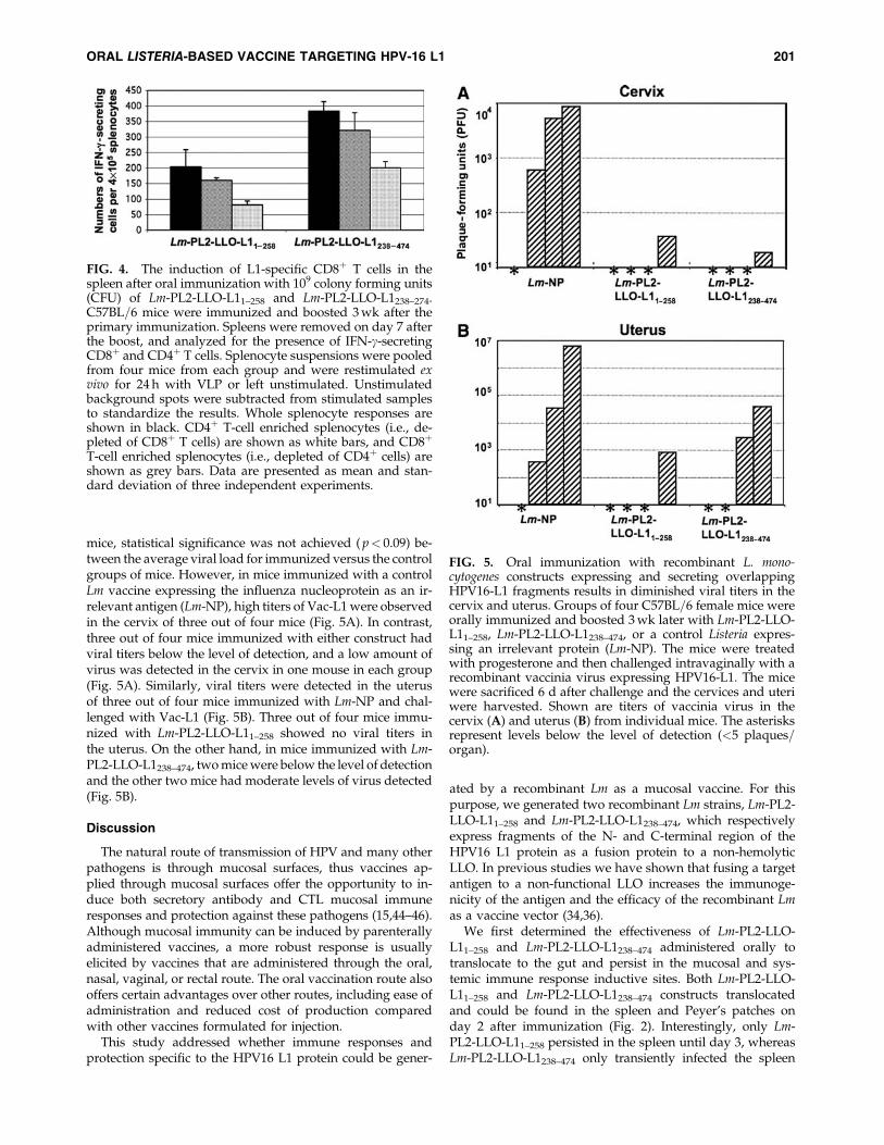
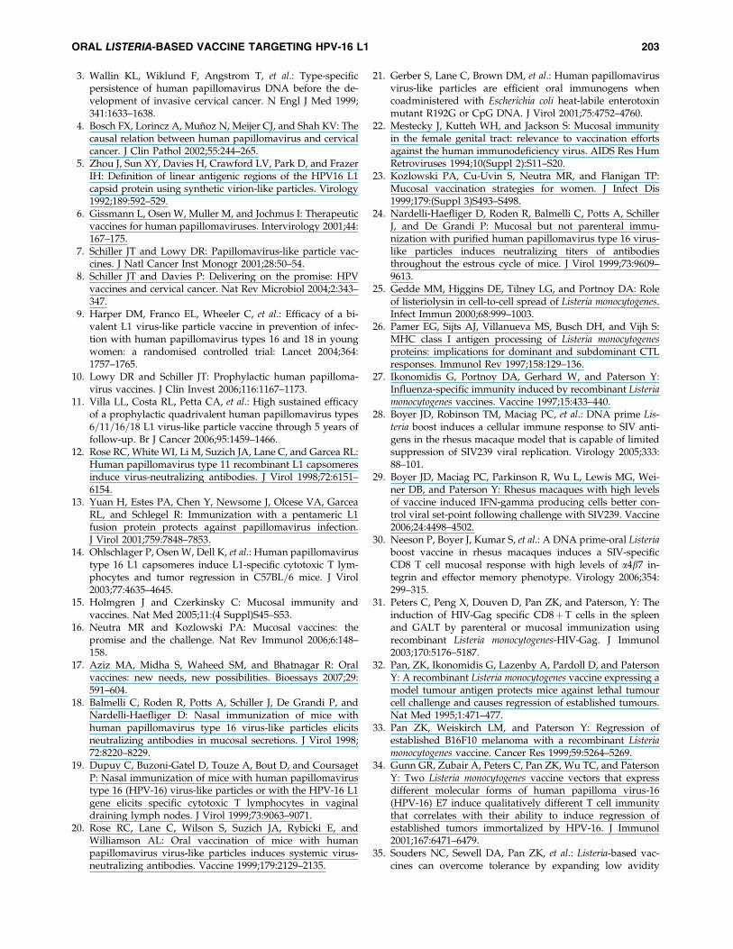
mice, statistical significance was not achieved ( p< 0.09) be-tween the average viral load for immunized versus the controlgroups of mice. However, in mice immunized with a controlLm vaccine expressing the influenza nucleoprotein as an ir-relevant antigen (Lm-NP), high titers of Vac-L1 were observedin the cervix of three out of four mice (Fig. 5A). In contrast,three out of four mice immunized with either construct hadviral titers below the level of detection, and a low amount ofvirus was detected in the cervix in one mouse in each group(Fig. 5A). Similarly, viral titers were detected in the uterusof three out of four mice immunized with Lm-NP and chal-lenged with Vac-L1 (Fig. 5B). Three out of four mice immu-nized with Lm-PL2-LLO-L11–258 showed no viral titers inthe uterus. On the other hand, in mice immunized with Lm-PL2-LLO-L1238–474, two mice were below the level of detectionand the other two mice had moderate levels of virus detected(Fig. 5B).
Discussion
The natural route of transmission of HPV and many otherpathogens is through mucosal surfaces, thus vaccines ap-plied through mucosal surfaces offer the opportunity to in-duce both secretory antibody and CTL mucosal immuneresponses and protection against these pathogens (15,44–46).Although mucosal immunity can be induced by parenterallyadministered vaccines, a more robust response is usuallyelicited by vaccines that are administered through the oral,nasal, vaginal, or rectal route. The oral vaccination route alsooffers certain advantages over other routes, including ease ofadministration and reduced cost of production comparedwith other vaccines formulated for injection.
This study addressed whether immune responses andprotection specific to the HPV16 L1 protein could be gener-
ated by a recombinant Lm as a mucosal vaccine. For thispurpose, we generated two recombinant Lm strains, Lm-PL2-LLO-L11–258 and Lm-PL2-LLO-L1238–474, which respectivelyexpress fragments of the N- and C-terminal region of theHPV16 L1 protein as a fusion protein to a non-hemolyticLLO. In previous studies we have shown that fusing a targetantigen to a non-functional LLO increases the immunoge-nicity of the antigen and the efficacy of the recombinant Lmas a vaccine vector (34,36).
We first determined the effectiveness of Lm-PL2-LLO-L11–258 and Lm-PL2-LLO-L1238–474 administered orally totranslocate to the gut and persist in the mucosal and sys-temic immune response inductive sites. Both Lm-PL2-LLO-L11–258 and Lm-PL2-LLO-L1238–474 constructs translocatedand could be found in the spleen and Peyer’s patches onday 2 after immunization (Fig. 2). Interestingly, only Lm-PL2-LLO-L11–258 persisted in the spleen until day 3, whereasLm-PL2-LLO-L1238–474 only transiently infected the spleen
FIG. 4. The induction of L1-specific CD8þ T cells in thespleen after oral immunization with 109 colony forming units(CFU) of Lm-PL2-LLO-L11–258 and Lm-PL2-LLO-L1238–274.C57BL=6 mice were immunized and boosted 3 wk after theprimary immunization. Spleens were removed on day 7 afterthe boost, and analyzed for the presence of IFN-g-secretingCD8þ and CD4þ T cells. Splenocyte suspensions were pooledfrom four mice from each group and were restimulated exvivo for 24 h with VLP or left unstimulated. Unstimulatedbackground spots were subtracted from stimulated samplesto standardize the results. Whole splenocyte responses areshown in black. CD4þ T-cell enriched splenocytes (i.e., de-pleted of CD8þ T cells) are shown as white bars, and CD8þ
T-cell enriched splenocytes (i.e., depleted of CD4þ cells) areshown as grey bars. Data are presented as mean and stan-dard deviation of three independent experiments.
FIG. 5. Oral immunization with recombinant L. mono-cytogenes constructs expressing and secreting overlappingHPV16-L1 fragments results in diminished viral titers in thecervix and uterus. Groups of four C57BL=6 female mice wereorally immunized and boosted 3 wk later with Lm-PL2-LLO-L11–258, Lm-PL2-LLO-L1238–474, or a control Listeria expres-sing an irrelevant protein (Lm-NP). The mice were treatedwith progesterone and then challenged intravaginally with arecombinant vaccinia virus expressing HPV16-L1. The micewere sacrificed 6 d after challenge and the cervices and uteriwere harvested. Shown are titers of vaccinia virus in thecervix (A) and uterus (B) from individual mice. The asterisksrepresent levels below the level of detection (<5 plaques=organ).
ORAL LISTERIA-BASED VACCINE TARGETING HPV-16 L1 201

but persisted in Peyer’s Patches until day 3 (Fig. 2). Giventhat colonization of Listeria by the oral route first results inits appearance in Peyer’s patches followed by spread to themesenteric lymph nodes and then the spleen (31), this sug-gests that Lm-PL2-LLO-L1238–474 is less able to spread thanLm-PL2-LLO-L11–258, due to lower virulence. The mecha-nism of spread is not well known but probably involves thecolonization of macrophages that then carry the bacteriathrough the lymphatics to distant organs. Although bothrecombinant strains are isogenic, except for the HPV-16 L1fragment they express, differences in virulence and the abil-ity to persist in macrophages can vary substantially amongisogenic strains of bacteria associated with foreign antigenexpression (47).
We further found that oral immunization with both con-structs induced splenic IFN-g-producing CD8þ T cells (Figs.3 and 4). Both vaccines induced substantial numbers of thesecells, although an immunodominant epitope for HPV-16 L1has been identified only in the 1–258 fragment of the mole-cule (14). However, we have previously shown that deliveryof a foreign antigen by Listeria and also fusion to LLO canenhance antigen processing and reveal sub-dominant epi-topes that do not emerge using other vaccine approaches(36). Importantly, in this study we also show the inductionof splenic L1-specific CD4þ T cells after oral immunizationwith our Lm recombinant constructs (Fig. 4). The IFN-gproduced by CD4þ T cells may be a necessary supplementfor protection against antigenic challenge; however, they arealso likely to be playing a role in the induction of CTL.Interestingly, IFN-g-producing cells in the PP were inducedonly after oral immunization with Lm-PL2-LLO-L1238–474.Our finding that only Lm-PL2-LLO- L1238–474 persisted in thePP until day 3 after oral immunization (Fig. 2B) is consistentwith the notion that persistent antigenic exposure by re-combinant Lm is a prerequisite for induction of antigen-specific T cells (43).
The efficacy of vaccines depends on their generation of apotent pool of Ag-specific T cells ready to expand rapidly toexpress killing function and protect against re-exposure tothe antigen. The induced L1-specific CD8þ and CD4þ T cellsafter immunization should provide protection against HPVchallenge and constitute an effective line of defense againstantigenic challenge. As indicated above, the L1-specificCD8þ and CD4þ T cells induced after oral immunizationwith the two recombinant Lm constructs showed evidence ofprotection against Vac-L1 challenge in mice immunized withLm-PL2-LLO-L11–258 and Lm-PL2-LLO-L1238–474 (Fig. 5).Previous studies have shown that parenteral immuniza-tion with VLP or capsomeres induces potent systemic cell-mediated immune responses resulting in protection orregression of established C3 tumors, which express HPV16L1, in mice (14). Intranasal and oral vaccination with differ-ent recombinant strains of Salmonella have shown inductionof neutralizing antibodies in the serum and genital tract, aswell as protection from experimental tumor challenge withC3 cells (48,49). Mucosal intranasal immunization with VLPhas also been shown to induce serum and genital antibodyresponses (50). In humans, VLP vaccination induces neu-tralizing antibody responses that results in protection againstHPV infection (9,11). In fact, studies in animal models andhumans have clearly shown the importance of neutralizingantibody responses in protection from or prevention of HPV
transmission. In this study we were not able to detect anti-body titers against L1 in the serum or vaginal secretions(data not shown). This is in agreement with previous ob-servations showing that Lm in not a potent inducer of hu-moral immune responses since it is an intracellular pathogen(32). Thus the protection conferred by the Lm vaccines in thisstudy is clearly cell mediated. This is consistent with stud-ies performed in the cotton-tailed rabbit papillomavirusmodel of papilloma infections, where cell-mediated immuneresponses against L1 have been shown to be effective at lim-iting papilloma formation in both a prophylactic (51) andtherapeutic (52) setting.
Although VLP vaccination has shown clear success inpreventing HPV infection, vaccination with VLPs is ineffec-tive against established HPV infections (53). In this casemucosal cell-mediated immune responses to HPV antigensmay be more effective for viral clearance, instead of neu-tralizing antibodies. However, targeting L1 by cellular im-munity is limited to active HPV infections and low-gradesquamous intraepithelial lesions, because L1 is generally lostin high-grade squamous intraepithelial lesions and invasivecancers (54). For these advanced lesions, vaccines targetingthe oncoproteins E6 and E7 from HPV have a clear advan-tage, as these genes are required in the malignant transfor-mation process and are retained in cervical carcinomas(55,56).
Conclusion
In conclusion, this study describes new oral Lm-basedHPV-L1-expressing vaccines, which induce potent antigen-specific CD8þ and CD4þ T-cell responses and show evidenceof protection against a heterologous challenge. Clinical trialshave shown the efficiency of parenteral VLP vaccines in pre-vention of cervical infection with certain HPV types andsubsequent cervical cancer. This work is an initial step towardexamining the contribution of cellular immune responses toL1 generated by mucosally administered Lm constructs ex-pressing L1.
Acknowledgments
This work was supported by CA69632 (Y.P.) andAI066333 (S.N.I.). W.M. was partially supported by traininggrant T32CA09140.
Disclosure Statement
Yvonne Paterson wishes to disclose that she has a financialinterest in Advaxis, Inc., a vaccine and therapeutic companythat has licensed or has an option to license all patents fromthe University of Pennsylvania that concern the use of Listeriamonocytogenes or listerial products as vaccines.
References
1. Ho GY, Burk RD, Klein S, et al.: Persistent genital humanpapillomavirus infection as a risk factor for persistent cer-vical dysplasia. J Natl Cancer Inst 1995;87:1365–1371.
2. Nobbenhuis MA, Walboomers JM, Helmerhorst TJ, et al.:Relation of human papillomavirus status to cervical lesionsand consequences for cervical-cancer screening: a prospec-tive study. Lancet 1999;354:20–25.
202 MUSTAFA ET AL.
3. Wallin KL, Wiklund F, Angstrom T, et al.: Type-specificpersistence of human papillomavirus DNA before the de-velopment of invasive cervical cancer. N Engl J Med 1999;341:1633–1638.
4. Bosch FX, Lorincz A, Munoz N, Meijer CJ, and Shah KV: Thecausal relation between human papillomavirus and cervicalcancer. J Clin Pathol 2002;55:244–265.
5. Zhou J, Sun XY, Davies H, Crawford LV, Park D, and FrazerIH: Definition of linear antigenic regions of the HPV16 L1capsid protein using synthetic virion-like particles. Virology1992;189:592–529.
6. Gissmann L, Osen W, Muller M, and Jochmus I: Therapeuticvaccines for human papillomaviruses. Intervirology 2001;44:167–175.
7. Schiller JT and Lowy DR: Papillomavirus-like particle vac-cines. J Natl Cancer Inst Monogr 2001;28:50–54.
8. Schiller JT and Davies P: Delivering on the promise: HPVvaccines and cervical cancer. Nat Rev Microbiol 2004;2:343–347.
9. Harper DM, Franco EL, Wheeler C, et al.: Efficacy of a bi-valent L1 virus-like particle vaccine in prevention of infec-tion with human papillomavirus types 16 and 18 in youngwomen: a randomised controlled trial: Lancet 2004;364:1757–1765.
10. Lowy DR and Schiller JT: Prophylactic human papilloma-virus vaccines. J Clin Invest 2006;116:1167–1173.
11. Villa LL, Costa RL, Petta CA, et al.: High sustained efficacyof a prophylactic quadrivalent human papillomavirus types6=11=16=18 L1 virus-like particle vaccine through 5 years offollow-up. Br J Cancer 2006;95:1459–1466.
12. Rose RC, White WI, Li M, Suzich JA, Lane C, and Garcea RL:Human papillomavirus type 11 recombinant L1 capsomeresinduce virus-neutralizing antibodies. J Virol 1998;72:6151–6154.
13. Yuan H, Estes PA, Chen Y, Newsome J, Olcese VA, GarceaRL, and Schlegel R: Immunization with a pentameric L1fusion protein protects against papillomavirus infection.J Virol 2001;759:7848–7853.
14. Ohlschlager P, Osen W, Dell K, et al.: Human papillomavirustype 16 L1 capsomeres induce L1-specific cytotoxic T lym-phocytes and tumor regression in C57BL=6 mice. J Virol2003;77:4635–4645.
15. Holmgren J and Czerkinsky C: Mucosal immunity andvaccines. Nat Med 2005;11:(4 Suppl)S45–S53.
16. Neutra MR and Kozlowski PA: Mucosal vaccines: thepromise and the challenge. Nat Rev Immunol 2006;6:148–158.
17. Aziz MA, Midha S, Waheed SM, and Bhatnagar R: Oralvaccines: new needs, new possibilities. Bioessays 2007;29:591–604.
18. Balmelli C, Roden R, Potts A, Schiller J, De Grandi P, andNardelli-Haefliger D: Nasal immunization of mice withhuman papillomavirus type 16 virus-like particles elicitsneutralizing antibodies in mucosal secretions. J Virol 1998;72:8220–8229.
19. Dupuy C, Buzoni-Gatel D, Touze A, Bout D, and CoursagetP: Nasal immunization of mice with human papillomavirustype 16 (HPV-16) virus-like particles or with the HPV-16 L1gene elicits specific cytotoxic T lymphocytes in vaginaldraining lymph nodes. J Virol 1999;73:9063–9071.
20. Rose RC, Lane C, Wilson S, Suzich JA, Rybicki E, andWilliamson AL: Oral vaccination of mice with humanpapillomavirus virus-like particles induces systemic virus-neutralizing antibodies. Vaccine 1999;179:2129–2135.
21. Gerber S, Lane C, Brown DM, et al.: Human papillomavirusvirus-like particles are efficient oral immunogens whencoadministered with Escherichia coli heat-labile enterotoxinmutant R192G or CpG DNA. J Virol 2001;75:4752–4760.
22. Mestecky J, Kutteh WH, and Jackson S: Mucosal immunityin the female genital tract: relevance to vaccination effortsagainst the human immunodeficiency virus. AIDS Res HumRetroviruses 1994;10(Suppl 2):S11–S20.
23. Kozlowski PA, Cu-Uvin S, Neutra MR, and Flanigan TP:Mucosal vaccination strategies for women. J Infect Dis1999;179:(Suppl 3)S493–S498.
24. Nardelli-Haefliger D, Roden R, Balmelli C, Potts A, SchillerJ, and De Grandi P: Mucosal but not parenteral immu-nization with purified human papillomavirus type 16 virus-like particles induces neutralizing titers of antibodiesthroughout the estrous cycle of mice. J Virol 1999;73:9609–9613.
25. Gedde MM, Higgins DE, Tilney LG, and Portnoy DA: Roleof listeriolysin in cell-to-cell spread of Listeria monocytogenes.Infect Immun 2000;68:999–1003.
26. Pamer EG, Sijts AJ, Villanueva MS, Busch DH, and Vijh S:MHC class I antigen processing of Listeria monocytogenesproteins: implications for dominant and subdominant CTLresponses. Immunol Rev 1997;158:129–136.
27. Ikonomidis G, Portnoy DA, Gerhard W, and Paterson Y:Influenza-specific immunity induced by recombinant Listeriamonocytogenes vaccines. Vaccine 1997;15:433–440.
28. Boyer JD, Robinson TM, Maciag PC, et al.: DNA prime Lis-teria boost induces a cellular immune response to SIV anti-gens in the rhesus macaque model that is capable of limitedsuppression of SIV239 viral replication. Virology 2005;333:88–101.
29. Boyer JD, Maciag PC, Parkinson R, Wu L, Lewis MG, Wei-ner DB, and Paterson Y: Rhesus macaques with high levelsof vaccine induced IFN-gamma producing cells better con-trol viral set-point following challenge with SIV239. Vaccine2006;24:4498–4502.
30. Neeson P, Boyer J, Kumar S, et al.: A DNA prime-oral Listeriaboost vaccine in rhesus macaques induces a SIV-specificCD8 T cell mucosal response with high levels of a4b7 in-tegrin and effector memory phenotype. Virology 2006;354:299–315.
31. Peters C, Peng X, Douven D, Pan ZK, and Paterson, Y: Theinduction of HIV-Gag specific CD8þT cells in the spleenand GALT by parenteral or mucosal immunization usingrecombinant Listeria monocytogenes-HIV-Gag. J Immunol2003;170:5176–5187.
32. Pan, ZK, Ikonomidis G, Lazenby A, Pardoll D, and PatersonY: A recombinant Listeria monocytogenes vaccine expressing amodel tumour antigen protects mice against lethal tumourcell challenge and causes regression of established tumours.Nat Med 1995;1:471–477.
33. Pan ZK, Weiskirch LM, and Paterson Y: Regression ofestablished B16F10 melanoma with a recombinant Listeriamonocytogenes vaccine. Cancer Res 1999;59:5264–5269.
34. Gunn GR, Zubair A, Peters C, Pan ZK, Wu TC, and PatersonY: Two Listeria monocytogenes vaccine vectors that expressdifferent molecular forms of human papilloma virus-16(HPV-16) E7 induce qualitatively different T cell immunitythat correlates with their ability to induce regression ofestablished tumors immortalized by HPV-16. J Immunol2001;167:6471–6479.
35. Souders NC, Sewell DA, Pan ZK, et al.: Listeria-based vac-cines can overcome tolerance by expanding low avidity
ORAL LISTERIA-BASED VACCINE TARGETING HPV-16 L1 203

CD8þT cells capable of killing a solid tumor in a transgenicmouse model of cancer. Cancer Immunity 2007;7:2.
36. Singh R, Dominiecki ME, Jaffee EM, and Paterson Y: Fusionto listeriolysin O and delivery by Listeria monocytogenes en-hances the immunogenicity of HER-2=neu and reveals sub-dominant epitopes in the FVB=N mouse. J Immunol2005;175:3663–3673.
37. Touze A, Mahe D, El Mehdaoui S, et al.: The nine C-terminalamino acids of the major capsid protein of the human pap-illomavirus type 16 are essential to DNA binding and genetransfer capacity. FEMS Microbiol Lett 2000;189:121–127.
38. Laure P, Chow MY, Loessner MJ, Portnoy DA, and CalendarR: Construction, characterization, and use of two Listeriamonocytogenes site-specific phage integration vectors. J Bac-teriol 2002;184:4177–4186.
39. Verch T, Pan ZK, and Paterson Y: Listeria monocytogenes-based antibiotic resistance gene-free antigen delivery systemapplicable to other bacterial vectors and DNA vaccines. In-fect Immun 2004;72:6418–6425.
40. Marais D, Passmore JA, Maclean J, Rose R, and WilliamsonAL: A recombinant human papillomavirus (HPV) type 16L1-vaccinia virus murine challenge model demonstrates cell-mediated immunity against HPV virus-like particles. J GenVirol 1999;80:2471–2475
41. Chakrabarti S, Sisler JR, and Moss B: Compact, synthetic,vaccinia virus early=late promoter for protein expression.Biotechniques 1997;23:1094–1097.
42. Jiang JQ, Patrick A, Moss RB, and Rosenthal KL:CD8þT-cell-mediated cross-clade protection in the genitaltract following intranasal immunization with inactivatedhuman immunodeficiency virus antigen plus CpG oligo-deoxynucleotides. J Virol 2005;79:393–400.
43. North RJ, Berche PA, and Newborg MF: Immunologic con-sequences of antibiotic-induced abridgement of bacterial in-fection: effect on generation and loss of protective T cells andlevel of immunologic memory. J Immunol 1981;127:342–346.
44. Guerrero RA, Ball JM, Krater SS, Pacheco SE, Clements JD,and Estes MK: Recombinant Norwalk virus-like particlesadministered intranasally to mice induce systemic and mu-cosal (fecal and vaginal) immune responses. J Virol 2001;75:9713–9722.
45. Shi W, Liu J, Huang Y, and Qiao L: Papillomavirus pseudo-virus: a novel vaccine to induce mucosal and systemic cyto-toxic T-lymphocyte responses. J Virol 2001;75:10139–10148.
46. Niikura M, Takamura S, Kim G, et al.: Chimeric recombinanthepatitis E virus-like particles as an oral vaccine vehiclepresenting foreign epitopes. Virology 2002;293:273–280.
47. Galen JE, and Levine, MM: Can a ‘‘flawless’’ live vectorvaccine strain be engineered? Trends Microbiol 2001;9:372–376.
48. Revaz V, Benyacoub J, Kast WM, Schiller JT, De Grandi P,and Nardelli-Haefliger D: Mucosal vaccination with a re-
combinant Salmonella typhimurium expressing human papil-lomavirus type 16 (HPV16) L1 virus-like particles (VLPs) orHPV16 VLPs purified from insect cells inhibits the growth ofHPV16-expressing tumor cells in mice. Virology 2001;279:354–360.
49. Fraillery D, Baud D, Pang SY, et al: Salmonella enterica serovartyphi Ty21a expressing human papillomavirus type 16 L1 asa potential live vaccine against cervical cancer and typhoidfever. Clin Vaccine Immunol 2007;14:1285–1295.
50. Sasagawa T, Tani M, Basha W, et al.: A human papilloma-virus type 16 vaccine by oral delivery of L1 protein. VirusRes 2005;110:81–90.
51. Hu J, Cladel NM, Budgeon LR, Reed CA, Pickel MD, andChristensen ND: Protective cell-mediated immunity byDNA vaccination against papillomavirus L1 capsid proteinin the cottontail rabbit papillomavirus model. Viral Im-munol 2006;19:492–507.
52. Govan VA, Rybicki EP, and Williamson AL: Therapeuticimmunisation of rabbits with cottontail rabbit papillomavi-rus (CRPV) virus-like particles (VLP) induces regression ofestablished papillomas. Virol J 2008;5:45.
53. Hildesheim A, Herrero R, Wacholder S, et al., and the CostaRican HPV Vaccine Trial Group: Effect of human papillo-mavirus 16=18 L1 viruslike particle vaccine among youngwomen with preexisting infection: a randomized trial.JAMA 2007;298:743–753.
54. Stoler MH, Rhodes CR, Whitbeck A, Wolinsky SM, ChowLT, and Broker TR: Human papillomavirus type 16 and 18gene expression in cervical neoplasias. Hum Pathol 1992;23:117–128.
55. Choo KB, Pan CC, and Han SH: Integration of humanpapillomavirus type 16 into cellular DNA of cervical carci-noma: preferential deletion of the E2 gene and invariableretention of the long control region and the E6=E7 openreading frames. Virology 1987;161:259–261.
56. Vousden KH, Doniger J, DiPaolo JA, and Lowy DR: The E7open reading frame of human papillomavirus type 16 en-codes a transforming gene. Oncogene Res 1988;3:167–175.
Address reprint requests to:Dr. Yvonne Paterson
Department of MicrobiologyUniversity of Pennsylvania School of Medicine
Room 323, Johnson PavilionPhiladelphia, PA 19104
E-mail: [email protected]
Received September 12, 2008; accepted January 21, 2009.
204 MUSTAFA ET AL.