
Detection of HIV1 Tat and JCV capsid protein, VP1, in AIDS brain with progressive multifocal...
8
Short Communication Detection of HIV-1 Tat and JCV capsid protein, VP1, in AIDS brain with progressive multifocal leukoencephalopathy Luis Del Valle 1 , Sidney Croul 1,2 , Susan Morgello 3 , Shohreh Amini 1 , Jay Rappaport 1 and Kamel Khalili* ,1 1 Center for Neurovirology and Cancer Biology, College of Science and Technology, Temple University, Philadelphia, PA 19122, USA; 2 Department of Pathology and Laboratory Medicine, MCP Hahnemann University School of Medicine, Philadelphia, PA 19102, USA and 3 Department of Pathology, Mount Sinai Medical Center, New York, NY 10029, USA HIV-1 infection can lead to severe central nervous system (CNS) clinical syndromes in more than 50% of HIV-1 positive individuals. Progressive multifocal leukoencephalopathy (PML) is the frequent opportunistic infection of the CNS which is seen in as high as 5% of AIDS patients. Results from previous cell culture studies showed that the HIV-1 regulatory protein, Tat can potentiate transcription of the human neurotropic virus, JCV, the causative agent for PML in cells derived from the human CNS. In this communication we examine the presence of the HIV-1 regulatory protein, Tat, as well as the HIV-1 and JCV structural proteins, p24 and VP1, respectively in AIDS/PML clinical samples. We demonstrate high level expression of the JCV capsid protein, VP1, in oligodendrocytes and to some degree in astrocytes of AIDS with PML. In HIV- 1+ samples expression of HIV-1 core protein, p24 was detected in perivascular monocytic cells and to a lesser extent in astrocytes and endothelial cells. A lack of p24 expression in oligodendrocytes suggested no infection of these cells with HIV-1. Interestingly, HIV-1 Tat was detected in various infected cells as well as in uninfected oligodendrocytes from HIV-1+ tissue, supporting the earlier in vitro findings that secreted Tat from the infected cells can be localized in the neighboring uninfected cells. The presence of Tat in oligodendrocytes was particularly interesting as this protein can up-modulate JCV gene transcription and several key cell cycle regulatory proteins including cyclin E, Cdk2, and pRb. The data presented here provide in vivo evidence for a role of HIV-1 Tat in the pathogenesis of AIDS/PML by acting as a positive regulatory protein that affects the expression of JCV and other cell regulatory proteins in the CNS. Journal of NeuroVirology (2000) 6, 221 – 228 Keywords: PML; AIDS; JC virus; central nervous system; immunohistochemistry HIV-1 infection of the central nervous system (CNS) induces a variety of clinical manifestations (Price et al, 1991). Progressive Multifocal Leukoencephalo- pathy (PML) is among the most common HIV-1 associated CNS complications (Berger and Concha 1995). PML is a subacute CNS demyelinating disease that results from cytolytic infection of oligodendrocytes with the human neurotropic polyomavirus, JCV (Walker, 1985). Although PML is recognized to occur in patients with a variety of immunodeficient conditions due to altered T-cell surveillance, the most frequent immunodeficiency which results in reactivation of JCV is the Acquired Immunodeficiency Syndrome (AIDS). PML is found in 4 – 8% of AIDS patients and is now considered an AIDS defining illness (Berger and Concha 1995; Berger et al, 1987). The relatively high rate of PML in AIDS patients in comparison with patients suffering from other severe forms of T-cell suppres- sive illnesses suggests that HIV-1 infection of the CNS may contribute to the increased frequency of PML. Previously, a direct role for HIV-1 in JCV reactivation was investigated by in vitro cell culture system and the results showed that the HIV-1 *Correspondence: K Khalili, Center for Neurovirology and Cancer Biology, College of Science and Technology, Temple University, 1900 North 12th Street, 015-96, Philadelphia, PA 19122, USA Accepted 24 March 2000 Journal of NeuroVirology (2000) 6, 221 – 228 ª 2000 Journal of NeuroVirology, Inc. www.jneurovirol.com
-
Upload
independent -
Category
Documents
-
view
3 -
download
0
Transcript of Detection of HIV1 Tat and JCV capsid protein, VP1, in AIDS brain with progressive multifocal...

Short Communication
Detection of HIV-1 Tat and JCV capsid protein, VP1, inAIDS brain with progressive multifocalleukoencephalopathy
Luis Del Valle1, Sidney Croul1,2, Susan Morgello3, Shohreh Amini1, Jay Rappaport1 and Kamel Khalili*,1
1Center for Neurovirology and Cancer Biology, College of Science and Technology, Temple University, Philadelphia,PA 19122, USA; 2Department of Pathology and Laboratory Medicine, MCP Hahnemann University School of Medicine,Philadelphia, PA 19102, USA and 3Department of Pathology, Mount Sinai Medical Center, New York, NY 10029, USA
HIV-1 infection can lead to severe central nervous system (CNS) clinicalsyndromes in more than 50% of HIV-1 positive individuals. Progressivemultifocal leukoencephalopathy (PML) is the frequent opportunistic infectionof the CNS which is seen in as high as 5% of AIDS patients. Results fromprevious cell culture studies showed that the HIV-1 regulatory protein, Tat canpotentiate transcription of the human neurotropic virus, JCV, the causativeagent for PML in cells derived from the human CNS. In this communication weexamine the presence of the HIV-1 regulatory protein, Tat, as well as the HIV-1and JCV structural proteins, p24 and VP1, respectively in AIDS/PML clinicalsamples. We demonstrate high level expression of the JCV capsid protein, VP1,in oligodendrocytes and to some degree in astrocytes of AIDS with PML. In HIV-1+ samples expression of HIV-1 core protein, p24 was detected in perivascularmonocytic cells and to a lesser extent in astrocytes and endothelial cells. A lackof p24 expression in oligodendrocytes suggested no infection of these cells withHIV-1. Interestingly, HIV-1 Tat was detected in various infected cells as well asin uninfected oligodendrocytes from HIV-1+ tissue, supporting the earlier invitro ®ndings that secreted Tat from the infected cells can be localized in theneighboring uninfected cells. The presence of Tat in oligodendrocytes wasparticularly interesting as this protein can up-modulate JCV gene transcriptionand several key cell cycle regulatory proteins including cyclin E, Cdk2, andpRb. The data presented here provide in vivo evidence for a role of HIV-1 Tat inthe pathogenesis of AIDS/PML by acting as a positive regulatory protein thataffects the expression of JCV and other cell regulatory proteins in the CNS.Journal of NeuroVirology (2000) 6, 221 ± 228
Keywords: PML; AIDS; JC virus; central nervous system; immunohistochemistry
HIV-1 infection of the central nervous system (CNS)induces a variety of clinical manifestations (Price etal, 1991). Progressive Multifocal Leukoencephalo-pathy (PML) is among the most common HIV-1associated CNS complications (Berger and Concha1995). PML is a subacute CNS demyelinatingdisease that results from cytolytic infection ofoligodendrocytes with the human neurotropicpolyomavirus, JCV (Walker, 1985). Although PML
is recognized to occur in patients with a variety ofimmunode®cient conditions due to altered T-cellsurveillance, the most frequent immunode®ciencywhich results in reactivation of JCV is the AcquiredImmunode®ciency Syndrome (AIDS). PML is foundin 4 ± 8% of AIDS patients and is now consideredan AIDS de®ning illness (Berger and Concha 1995;Berger et al, 1987). The relatively high rate of PMLin AIDS patients in comparison with patientssuffering from other severe forms of T-cell suppres-sive illnesses suggests that HIV-1 infection of theCNS may contribute to the increased frequency ofPML. Previously, a direct role for HIV-1 in JCVreactivation was investigated by in vitro cell culturesystem and the results showed that the HIV-1
*Correspondence: K Khalili, Center for Neurovirology and CancerBiology, College of Science and Technology, Temple University,1900 North 12th Street, 015-96, Philadelphia, PA 19122, USAAccepted 24 March 2000
Journal of NeuroVirology (2000) 6, 221 ± 228ã 2000 Journal of NeuroVirology, Inc.www.jneurovirol.com

Table 1 Clinical data and histopathological ®ndings
Case Age and gender Diagnosis Associated diseases Demyelination Astrocytosis Inclusions In¯ammation
1
234
5
678
9
44 years, Male
6 years, Male48 years, Male33 years, Male
44 years, Female
41 years, Male41 years, Female56 years, Male
47 years, Male
PML (HIV+)
PML (HIV+)PML (HIV+)PML (HIV+)
No pathology
Cerebral infarctionsCreutzfeld Jacob DiseaseNo pathology
No pathology
Tuberculosis, lungadenocarcinoma
HIV+, pneumoniaHIV+, pneumoniaCommon variable
immunode®ciencypseudomonas, pneumonia
HIV+, B cell lymphoma,cirrhosis
NoneNoneCardiac transplant,
renal failureHodgkin's disease,
endocarditis
++++
+++++++++
N/F
N/FN/FN/F
N/F
++++
++++++++++
N/F
++++++++N/F
N/F
++++
+++++++++
N/F
N/FN/FN/F
N/F
+++
+++++++
N/F
++N/FN/F
N/F
N/F, not found.
Figure 1 Histological evaluation of AIDS/PML. (A,B) display demyelinated lesions from AIDS/PML samples as examined by H & E(A) and Luxol Fast Blue (B). The areas of demyelination are shown by arrows and an arrowhead. (C) Detection of oligodendrocyteswith glassy intranuclear inclusion (arrowhead) and atypical astrocytes with eccentric eosinophilic cytoplasm and speckled, notchednuclei (arrow). (D) Perivascular in¯ammatory cells (arrow). Original magni®cations, A and B 206, C 1006, and D 406.
HIV-1 Tat and JCV VP1 in AIDS/PMLL Del Valle et al
222
Journal of NeuroVirology

regulatory protein, Tat, stimulates transcription ofthe JCV promoter in glial cells (Chowdhury et al,1990; 1992; 1993; Tada et al, 1990).
Tat is a 14 kDa protein which is produced at theearly stage of HIV-1 infection and potentiatestranscription and replication of the HIV-1 genome(Liu et al, 1999). To exert its activity, Tat localizesitself in close proximity to the transcription startsite by binding to the TAR RNA sequence posi-tioned in the leader of HIV-1 transcripts and/orinteracting with various cellular regulatory proteins(Hottiger and Nabel, 1998; Xiao et al, 1998).Interestingly, a region similar to HIV-1 TAR hasbeen detected in the JCV genome which confers Tatresponsiveness to the transcription of JCV in glialcells (Chowdhury et al, 1992). Furthermore, expres-sion of Tat in glial cells, in the absence of HIV-1,stimulated transcription of the JCV late genomewhich is responsible for the viral capsid proteinsincluding VP1 (Tada et al, 1990). These observa-tions have provided a molecular model for commu-nication of HIV-1 and JCV via the Tat transcriptionfactor. However, the caveat of this model stemmedfrom studies ruling out HIV-1 infection of oligoden-drocytes, the cell type which is infected by JCV. Onthe other hand, other studies, showed that HIV-1 Tat
can be secreted from the infected cells and be takenup by the neighboring uninfected cells (Frankel andPabo, 1988) supporting a model in which secretionof Tat from the HIV-1 infected brain cells such asmicroglia and astrocytes can be localized in non-infected cells such as oligodendrocytes. Function-ally, results from several cell culture studiesshowed that soluble Tat is active after uptake andcan induce transcription of Tat responsive genes inthe cells (Nath et al, 1999; Sawaya et al, 1998). Inthis study, we have examined the presence of Tatand JCV late gene product in brain tissue from AIDSpatients with PML and demonstrated accumulationof Tat in oligodendrocytes infected with JCV.Further, we demonstrated expression of severalTat-responsive cell cycle regulatory proteins invarious CNS cells.
The most common histological features of PMLinclude extensive demyelinated plaques, the pre-sence of atypical astrocytes within these plaquesand eosinophilic intranuclear inclusions in oligo-dendrocytes. Other features such as microglialnodules and perivascular lymphocytic cuf®ng areless frequently observed in PML. Table 1 sum-marizes the clinical data and presents the patholo-gic features of the tissues which were used in this
Figure 2 Immunohistochemical evaluation of AIDS/PML with anti-JCV VP1 antibody. (A, B, C) Detection of VP1 positive stainedoligodendrocytes within and adjacent to demyelinated lesions (arrows). (A, B, D) Low, but detectable, levels of staining was observedin smaller numbers of astrocytes (arrowheads). Original magni®cations, A and B 406, C and D 1006.
HIV-1 Tat and JCV VP1 in AIDS/PMLL Del Valle et al
223
Journal of NeuroVirology
study. In general, all four cases displayed plaques ofdemyelination with atypical reactive astrocytestypi®ed by eccentric eosinophilic cytoplasm. Figure1 illustrates representative data depicting demyeli-nated plaques (Figure 1A,B), reactive astrocytes andoligodendrocytes with viral inclusions withinnuclei (Figure 1C). Also, in accord with previousobservations (Aksamit et al, 1990), perivascularin®ltration of in¯ammatory cells was observed inHIV-1 positive PML (Figure 1D).
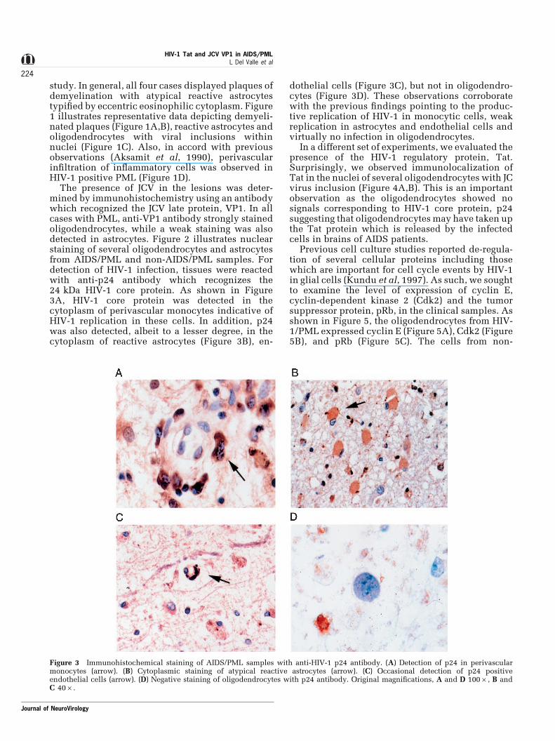
The presence of JCV in the lesions was deter-mined by immunohistochemistry using an antibodywhich recognized the JCV late protein, VP1. In allcases with PML, anti-VP1 antibody strongly stainedoligodendrocytes, while a weak staining was alsodetected in astrocytes. Figure 2 illustrates nuclearstaining of several oligodendrocytes and astrocytesfrom AIDS/PML and non-AIDS/PML samples. Fordetection of HIV-1 infection, tissues were reactedwith anti-p24 antibody which recognizes the24 kDa HIV-1 core protein. As shown in Figure3A, HIV-1 core protein was detected in thecytoplasm of perivascular monocytes indicative ofHIV-1 replication in these cells. In addition, p24was also detected, albeit to a lesser degree, in thecytoplasm of reactive astrocytes (Figure 3B), en-
dothelial cells (Figure 3C), but not in oligodendro-cytes (Figure 3D). These observations corroboratewith the previous ®ndings pointing to the produc-tive replication of HIV-1 in monocytic cells, weakreplication in astrocytes and endothelial cells andvirtually no infection in oligodendrocytes.
In a different set of experiments, we evaluated thepresence of the HIV-1 regulatory protein, Tat.Surprisingly, we observed immunolocalization ofTat in the nuclei of several oligodendrocytes with JCvirus inclusion (Figure 4A,B). This is an importantobservation as the oligodendrocytes showed nosignals corresponding to HIV-1 core protein, p24suggesting that oligodendrocytes may have taken upthe Tat protein which is released by the infectedcells in brains of AIDS patients.
Previous cell culture studies reported de-regula-tion of several cellular proteins including thosewhich are important for cell cycle events by HIV-1in glial cells (Kundu et al, 1997). As such, we soughtto examine the level of expression of cyclin E,cyclin-dependent kinase 2 (Cdk2) and the tumorsuppressor protein, pRb, in the clinical samples. Asshown in Figure 5, the oligodendrocytes from HIV-1/PML expressed cyclin E (Figure 5A), Cdk2 (Figure5B), and pRb (Figure 5C). The cells from non-
Figure 3 Immunohistochemical staining of AIDS/PML samples with anti-HIV-1 p24 antibody. (A) Detection of p24 in perivascularmonocytes (arrow). (B) Cytoplasmic staining of atypical reactive astrocytes (arrow). (C) Occasional detection of p24 positiveendothelial cells (arrow). (D) Negative staining of oligodendrocytes with p24 antibody. Original magni®cations, A and D 1006, B andC 406.
HIV-1 Tat and JCV VP1 in AIDS/PMLL Del Valle et al
224
Journal of NeuroVirology

infected tissue showed no sign for expression ofthese proteins. Also, astrocytes from the AIDS/PMLshowed strong positive signals for those regulatoryproteins (Figure 5D ± F). These observations alto-gether demonstrate the presence of HIV-1 Tat inglial cells of HIV-1 infected brain and de-regulatedexpression of the cellular proteins which areinvolved in the control of glial cell function. Table2 summarizes the results from the immunohisto-chemical evaluations of the clinical samples used inthis study.
In summary, in this study we present data which,along with the previous in vitro ®ndings, support amodel in which HIV-1 Tat, in the absence ofinfection of oligodendrocytes may participate inthe reactivation of JCV and development of PML.Our results show that in PML lesions, HIV-1 coreprotein, p24, is present predominantly in perivas-cular monocytes and to a lesser degree in astrocytes.On the other hand, the JCV capsid protein, VP1, wasdetected mainly in oligodendrocytes and seconda-rily in astrocytes. Tat immunostaining of oligoden-drocytes is of particular interest suggesting theproposition that Tat is taken up by these cells.Thus, based on our previous in vitro observationsand the data presented here, it is likely that the
presence of Tat in oligodendrocytes results inincreased transcription of the JCV late gene whichis responsible for VP1 production in these cells. It isimportant to note that the brain tissue from case 5(shown in Table 1), an HIV-1 positive patient withno neurologic disease showed no immunoreactivityfor HIV-1 and JCV related proteins indicating thattheir expression is closely related to the pathologicprocess. Two immunosuppressed cases, 8 and 9,showed no staining for JCV related proteins furthersupporting the notion that their expression isdisease related.
Tat can also affect expression of several cellulargenes such as the regulators of cell growth anddifferentiation. The previous data from cell culturestudies revealed that Tat may alter astrocytic cellgrowth in vitro by altering expression of cyclin Eand its partner Cdk2, and their downstream target,pRb. Our data show expression of cyclin E, Cdk2,and pRb in oligodendrocytes and astrocytes of allPML cases, i.e. HIV-1 positive and HIV-1 negative.This result is not surprising since the JCV regulatoryprotein, T-antigen, by itself can alter the expressionand activity of these proteins (Krynska et al, 1997).Interestingly, we observed an increase in theexpression of cyclin E, Cdk2, and pRb in monocytes
Figure 4 Detection of Tat in AIDS/PML samples. (A, B) Immunoreactivity of oligodendrocyte nuclei with anti-Tat antibody. (C)Detection of Tat in perivascular monocytic cells. (D) Weak staining of astrocytes with Tat antibody. Original magni®cations, A, B andD 1006, C 406.
HIV-1 Tat and JCV VP1 in AIDS/PMLL Del Valle et al
225
Journal of NeuroVirology

and microglia of HIV-1 positive PML cases (data notshown) suggesting that these alterations are HIV-1related and may be induced by Tat protein.
Taken together, these observations, which areconsistent with previous ®ndings (Bonwetsch et al,1999), provide in vivo evidence for the presence ofHIV-1 Tat in various CNS cells including thosewhich are HIV-1 negative and provide supporting invivo evidence for the earlier cell culture studiespointing to the role of Tat in the pathogenesis of PMLin AIDS patients.
Materials and methods
Tissue samplesThree autopsy samples of HIV-1 positive PMLpatients and one autopsy of HIV-1 negative PMLpatient were obtained from the Manhattan HIVBrain Bank (R24 MH59724) at Mount Sinai MedicalCenter (New York, NY, USA) and utilized in thesestudies. All samples were formalin-®xed andparaf®n-embedded. Formalin-®xed and paraf®n-embedded control brain tissue was obtained fromthe Hahnemann University Hospital archives whichincludes autopsy frontal cortex and white matterfrom a patient with HIV-1 with no CNS pathology,two samples of normal cortex and white matter frompatients without history or pathologic evidence ofHIV-1 and two samples of cortex and white matterfrom patients with CNS lesions in the absence of
HIV-1. Table 1 illustrates clinical data for thepatient samples.
Histologic and immunohistochemical analysisFour micron thick sections were stained withhematoxylin and eosin to con®rm tissue diag-noses. Immunohistochemistry was performed bydeparaf®nizing four micron sections in xylene,followed by rehydration and nonenzymatic anti-gen retrieval in 0.01 M sodium citrate buffer(pH 6.0) at 958C for 40 min. After cooling toroom temperature (approximately 20 min), slideswere rinsed and endogenous peroxide wasquenched by incubation in MeOH/3% H2O2 for20 min. This was followed by a wash inphosphate buffer saline (PBS) and blocking inPBS plus 1% BSA and 5% normal horse serumfor 2 h at room temperature. Slides were thenincubated overnight with primary antibodies. Todetect JC virus VP1 protein, a mouse monoclonalantibody to recombinant JCV VP1 (a generous giftfrom Dr Eugene O Major, NINDS/NIH, Bethesda,MD, USA) was used at 1 : 1000 dilution. HIV-1p24 core protein was detected with a rabbitpolyclonal antibody against baculovirus derivedwith IIIB p24 at a 1 : 1000 dilution (Intracel Corp.,Rockville, MD, USA). Tat was detected withrabbit polyclonal antiserum HIV-1BH10 generatedagainst recombinant HIV-1 Tat at 1 : 2000 dilution(a generous gift from Dr Avindra Nath, Depart-
Figure 5 Detection of cell cycle regulators in glial cells of AIDS/PML samples. (A, B, C) The nuclei of oligodendrocytes in thedemyelinated lesions showed positive staining to cyclin E (A), Cdk2 (B), and pRb (C). (D, E, F) Depict positive nuclear staining ofastrocytes with cyclin E, cdk2, and pRb (D ± F, respectively). Original magni®cations, all panels 1006.
HIV-1 Tat and JCV VP1 in AIDS/PMLL Del Valle et al
226
Journal of NeuroVirology

ment of Neurology, University of KentuckyCollege of Medicine, Lexington, KY, USA). CyclinE was detected with a rabbit polyclonal antibody,C-19 at 1 : 1000 dilution, Cdk2 was detected witha rabbit polyclonal antibody, SC-163 1 : 500dilution, and pRb was detected with a mousemonoclonal antibody to the human protein,14001A 1 : 100 dilution (Pharmigen). Positivecontrols for Cyclin E, Cdk2, and pRb includedhuman medulloblastoma specimens. Negativecontrols consisted of tissues incubated in bufferfor monoclonal antibodies or normal rabbit serumfor polyclonal antibodies without primary anti-body. Primary antibodies were detected withbiotinylated anti-mouse or anti-rabbit secondaryantibodies, avidin biotin peroxidase complex anddiaminobenzidine chromagen according to manu-facturer's instructions (Vectastain Elite ABC Per-oxidase Kit, Vector Laboratories, Burlingame, CA,USA). Following a light counterstain with hema-toxylin, sections were dehydrated and cover-slipped with Permount.
Evaluation of stained slidesEach slide was examined independently by twomicroscopists for the characteristics of thelesions found with H & E staining. In addition,they estimated the presence of immunohisto-
chemical stain product with the various anti-bodies and the percentage of positive cells.Percentages were converted into a four levelscale where +=1 ± 25%, ++=26 ± 50%, +++=51 ±75%, and ++++=76 ± 100% positive nuclei im-munohistochemically. For the antibodies used inthis study only nuclear localization of stainingwas accepted as positive. All differences be-tween the two examiners were resolved byexamining the tissues together to achieve con-sensus.
Acknowledgments
We wish to thank Dr Eugene O Major and AvindraNath for their generous gifts, and past and presentmembers of the Center for Neurovirology andCancer Biology for their insightful discussion andsharing of ideas. The HIV-1 and PML tissues wereobtained from the Manhattan HIV Brain Bank(R24 MH59724) at Mount Sinai Medical Center,New York, NY, USA. We would also like to thankCynthia Schriver for editorial assistance andpreparation of the manuscript. This work wasmade possible by grants awarded by the NIH to JRappaport, S Amini and K Khalili.
References
Aksamit AJ, Gendelman HE, Orenstein JM, PezeshkpourGH (1990). AIDS-associated progressive multifocalleukoencephalopathy (PML): comparison to non-AIDSPML with in situ hybridization and immunohisto-chemistry. Neurology 40: 1073 ± 1078.
Berger JR, Concha M (1995). Progressive multifocalleukoencephalopathy: the evolution of a disease onceconsidered rare. J NeuroVirology 1: 5 ± 18.
Berger JR, Kaszovitz D, Post JD, Dickinson G (1987).Progressive multifocal leukoencephalopathy asso-ciated with human immunode®ciency virus infection:a review of the literature with a report of sixteencases. Ann Intern Med 107: 87.
Table 2 Immunohistochemical ®ndings
Case VP-1 HIV p24 Tat Cyclin E Cdk2 pRb
1
2
3
4
56789
++++ Oligo++Astro
++++ Oligo++ Astro
++++ Oligo++ Astro
++++ OLigo++ AstroNoneNoneNoneNoneNone
+ Astro+++ Mono++ Endo++ Astro++ Mono
++ Astro+++ Mono+++ EndoNone
+ Endo+ Endo+ Endo+ Endo+ Endo
+++ Oligo+ Astro++ Mono+++ Oligo+++ Micro
++++ Oligo++ Astro+ MicroNone
NoneNoneNoneNoneNone
++ Oligo+ Astro
+ Oligo+ Micro
+++ Oligo++ Astro
+ Oligo
NoneNoneNoneNoneNone
++ Oligo
++ Oligo+ Astro
++ Oligo+ Mono
++ Oligo+ AstroNoneNoneNoneNoneNone
++ Oligo++ Astro+ Mono+++ Oligo+ Astro+ Micro+++ Oligo
+++ Oligo+++ AstroNoneNoneNoneNoneNone
Abbreviations: Oligo, oligodendrocyte; Astro, astrocyte; Mono, monocyte; Micro, microglia; Endo, endothelium; N/D, not determined.
HIV-1 Tat and JCV VP1 in AIDS/PMLL Del Valle et al
227
Journal of NeuroVirology

Bonwetsch R, Croul S, Richardson MW, Lorenzana C,Del Valle L, Sverstiuk AE, Amini S, Morgello S,Khalili K, Rappaport J (1999). Role of HIV-1 Tat andCC chemokine MIP-1a in the pathogenesis of HIVassociated central nervous system disorders. J Neuro-virology 5: 685 ± 694.
Chowdhury M, Taylor JP, Tada H, Rappaport J, Wong-Staal F, Amini S, Khalili K (1990). Regulation of thehuman neurotropic virus promoter by JCV T-antigenand HIV-1 Tat protein. Oncogene 5: 1737 ± 1742.
Chowdhury M, Taylor JP, Chang C-F, Rappaport J,Khalili K (1992). Evidence that a sequence similar toTAR is important for induction of the JC virus latepromoter by human immunode®ciency virus type 1Tat. J Virol 66: 7355 ± 7361.
Chowdhury M, Kundu M, Khalili K (1993). GC/GA richsequence confers Tat-responsiveness to human neuro-tropic virus promoter, JCVL, in cells derived fromCNS. Oncogene 8: 887 ± 892.
Frankel AD, Pabo CO (1988). Cellular uptake of the tatprotein from human immunode®ciency virus. Cell 55:1189 ± 1193.
Hottiger MO, Nabel GJ (1998). Interaction of humanimmunode®ciency virus type 1 Tat with the transcrip-tional coactivators p300 and CREB binding protein. JVirol 72: 8252 ± 8256.
Krynska B, Gordon J, Otte J, Franks R, Knobler R,Giordano A, DeLuca A, Khalili K (1997). Role of cellcycle regulators in tumor formation in transgenic miceexpressing the human neurotropic virus, JCV, earlyprotein. J Cell Biochem 67: 223 ± 230.
Kundu M, Guermah M, Roeder RG, Amini S, Khalili K(1997). Interaction between cell cycle regulator, E2F-1,and NF-kappa B mediates repression of HIV-1 genetranscription. J Biol Chem 272: 29468 ± 29474.
Liu Y, Sune C, Garcia-Blanco MA (1999). Humanimmunode®ciency virus type 1 Tat-dependent activa-tion of an arrested RNA polymerase II elongationcomplex. Virology 255: 337 ± 346.
Nash A, Conant K, Chen P, Scott C, Major EO (1999).Transient exposure to HIV-1 Tat protein results incytokine production in macrophages and astrocytes. Ahit and run phenomenon. J Biol Chem 274: 17098 ±17102.
Price RW, Sidtis JJ, Brew BJ (1991). AIDS dementiacomplex and HIV-1 infection: a view from the clinic.Brain Pathol 1: 155 ± 162.
Sawaya BE, Thatikunta P, Denisova L, Brady J, Khalili K,Amini S (1998). Regulation of TNFa and TGFb-1 genetranscription by HIV-1 Tat in CNS cells. J Neuroim-munol 87: 33 ± 42.
Tada H, Rappaport J, Amini S, Lashgari M, Wong-StahlF, Khalili K (1990). Cell type transactivation of humanneurotropic virus JCV promoter by HIV-TAT protein.Proc Natl Acad Sci USA 87: 3479 ± 3483.
Walker DL (1985). Progressive multifocal leukoencepha-lopathy. In: Handbook of Clinical Neurology, vol. 47,PJ Vinken et al, (eds). Elsevier Science Publishing,Inc., New York, pp 503 ± 524.
Xiao H, Tao Y, Greenblatt J, Roeder RG (1998). Acofactor, TIP30, speci®cally enhances HIV-1 Tat-activated transcription. Proc Natl Acad Sci USA 95:2146 ± 2151.
HIV-1 Tat and JCV VP1 in AIDS/PMLL Del Valle et al
228
Journal of NeuroVirology