Genetic diversity in the VP1 gene of foot-and-mouth disease virus serotype Asia 1
Upload
independentCategory
view
1download
0
Virology 296, 241–250 (2002)
Birnavirus VP1 Proteins Form a Distinct Subgroup of RNA-Dependent RNA PolymerasesLacking a GDD Motif1
Philip S. Shwed,* Peter Dobos,* Lynne A. Cameron,* Vikram N. Vakharia,† and Roy Duncan‡,2
*Department of Microbiology, University of Guelph, Guelph, Ontario, Canada N1G 2W1; †Center for Agricultural Biotechnology,University of Maryland Biotechnology Institute, College Park, Maryland 20742; and ‡Department of Microbiology
and Immunology, Dalhousie University, Halifax, Nova Scotia, Canada B3H 4H7
Received June 27, 2001; returned to author for revision August 22, 2001; accepted November 28, 2001
We have cloned and characterized the Drosophila X virus (DXV) genome segment B and its encoded VP1, the putativeRNA-dependent RNA polymerase (RdRp) present in the virion. The 2991-bp open reading frame encodes the largestbirnavirus VP1 at 977 aa, with a calculated Mr of 112.8 kDa. As with the VP1 proteins of the type species of the other twogenera in the family Birnaviridae, namely, infectious pancreatic necrosis virus (genus Aquabirnavirus) and infectious bursaldisease virus (genus Avibirnavirus), the DXV (genus Entomobirnavirus) VP1 protein contains a consensus GTP-binding siteand appears to possess self-guanylylation activity. All of the birnavirus VP1 proteins contain conserved RdRp motifs thatreside in the catalytic “palm” domain of all classes of polymerases. However, the birnavirus RdRps lack the highly conservedGly-Asp-Asp (GDD) sequence, a component of the proposed catalytic site of this enzyme family that exists in the conservedmotif VI of the palm domain of other RdRps. All three birnavirus RdRps do contain downstream DD motifs that could functionas part of the catalytic triad. These motifs are, however, located in spatially distinct regions of the various birnavirus VP1
INTRODUCTION
All RNA viruses, irrespective of replication and codingstrategies, encode an RNA-dependent RNA polymerase(RdRp) (EC 2.7.7.48), which carries out multiple functionsfrom replication to capping activities (Spies et al., 1987;Ishihama and Nagata, 1988). The genes encoding RdRpsare among the most conserved of all viral genes and forthis reason have been used extensively for phylogeneticanalysis (Koonin and Dolja, 1993). Crystal structures andsequence-predicted structural analysis of RdRps, as wellas those of DNA-dependent DNA polymerases and re-verse transcriptases, suggest a conserved polymerasetertiary structure resembling a right hand with finger,thumb, and palm domains (Ollis et al., 1985; Kohlstaedt etal., 1992; Hansen et al., 1997; O’Reilly and Kao, 1998;Lesburg et al., 1999; Butcher et al., 2001). Amino acidsequence analysis has also highlighted eight conservedRdRp motifs, four of which (denoted IV–VII or A–D) residein the catalytic palm domain (Poch et al., 1989; Koonin,1991; O’Reilly and Kao, 1998). Although the functions ofthese motifs are not entirely understood, they are be-lieved to correspond to RNA polymerase functions such
1 The nucleotide sequence data reported in this paper have beensubmitted to the GenBank nucleotide sequence database and havebeen assigned Accession No. AF196645.
2
241
as RNA polymerization, nucleotide triphosphate binding,and template and product binding. In particular, a Gly-Asp-Asp (GDD) sequence found in motif VI is conservedin almost all polymerases. In a few cases, such as withthe RdRps of infectious bronchitis virus and the dsRNAphage �6, the glycine residue is substituted in this motifbut the di-aspartate sequence is conserved (Boursnell etal., 1987; Mindich et al., 1988). These two aspartateresidues, in conjunction with a third conserved aspartatein motif IV, are believed to represent the catalytic site ofthe polymerase by functioning to coordinate the position-ing of two metal ions, water, the free 3�-hydroxyl of thenascent strand, and the incoming ribonucleosidetriphosphate (Kamer and Argos, 1984; Argos, 1988; Gor-balenya and Koonin, 1988; Steitz, 1993, 1998).
In view of the highly conserved nature of the RdRpGDD motif and its postulated role as the catalytic site ofthis enzyme family, it is surprising that the presumptiveRdRps of the dsRNA virus family Birnaviridae apparentlylack the GDD motif (Morgan et al., 1988; Duncan et al.,1991). The bisegmented dsRNA genomes of the mem-bers of the Birnaviridae family are found in non-envel-oped icosohedral nucleocapsids of 60 nm in diameter(reviewed in Dobos, 1995a). Both genome segments ofgenus Aquabirnavirus (type species IPNV; Duncan andDobos, 1986; Duncan et al., 1991) and genus Avibirnavi-
proteins. These results suggest that the VP1 proteins of birlacking the conserved RdRp motif VI or have repositioned t
To whom reprint requests should be addressed. Fax: (902)494-6770. E-mail: [email protected].
doi:10.1006/viro.2001.1334
es form a defined subgroup of polymerases that either aretif to different structural regions. © 2002 Elsevier Science (USA)
rus (type species IBDV; Azad et al., 1985; Morgan et al.,1988; Kibenge et al., 1990; Kibenge and Qian, 1994) have
0042-6822/02 $35.00
navirushis mo
© 2002 Elsevier Science (USA)All rights reserved.
been cloned and sequenced. The larger one, segment A,encodes the major structural proteins of the viral particleand a proteolytic enzyme involved in precursor proteinprocessing. Genome segment A of Drosophila X virus(DXV), the type species of the third and final genus of theBirnaviridae (genus Entomobirnavirus), has also beensequenced and shows a genetic organization similar tothat of the other birnaviruses (Chung et al., 1996). Thesmaller segment B of IPNV and IBDV encodes a minorstructural protein, VP1, the putative RdRp. To date, nosequence information has been available for DXV ge-nome segment B.
The birnavirus VP1 protein has a low copy number andfeatures several conserved motifs found in the RdRps ofother viruses (Bruenn, 1991; Koonin, 1991, 1992; Ribasand Wickner, 1992). Birnavirus RdRps, however, possessa number of attributes that distinguish them from mostRdRps. Similar to the reverse transcriptase of hepatitis Bvirus, they are found in both free and genome-linkedforms (Revet and Delain, 1982; Nagy and Dobos, 1984a,b;Calvert et al., 1991; Weber et al., 1994). Rather than beingprimer-independent or using the free hydroxyl groups ofRNA as primers for initiation of RNA synthesis, birnavirusRdRps use hydroxyl groups of amino acid residueswithin the protein itself. This situation is similar to theexample of the reverse transcriptase of the hepadnavirusthat primes RNA-directed DNA synthesis from an internalsite of the polypeptide (Wang and Seeger, 1992). In con-trast to reovirus and similar to the dsRNA phage �6, thein vitro synthesis of IPNV mRNA appears to follow asemiconservative strand displacement mechanism(Mertens et al., 1982; Dobos, 1995b). More importantly,the RdRps of IPNV and IBDV lack the GDD sequence, thehallmark feature of this enzyme family. In the case ofIBDV, VP1 contains a LDD motif in place of the GDD inmotif VI, and the flanking amino acid residues are hydro-philic, not hydrophobic as with most RdRps (Morgan etal., 1988; Duncan et al., 1991). For IPNV, the GDD motif iscompletely absent from the region corresponding to thepresumptive motif VI of IBDV (Duncan et al., 1991). Onepossible explanation for the absence of the GDD motif inthe IPNV RdRp sequence is that a downstream LDDmotif (residues 653–655) might function as the catalyticsite, possibly reflecting a repositioning of motif VI (Koo-nin, 1992). Based on the VP1 sequences obtained fromtwo of the three genera, it appears that birnavirusescontain a novel catalytic site and represent a new classof RdRps or that the birnavirus XDD motif is spatiallyrearranged relative to the conserved motifs found inother RdRps.
In order to clarify the relationship between the GDDmotif, the birnavirus polymerase, and other RdRps, wecloned and sequenced the VP1-encoding genome seg-ment B of DXV, the type species of the only remaininggenus of birnaviruses, the Entomobirnaviruses. Our re-sults indicate that the 113-kDa DXV VP1 protein, the
putative RdRp, exhibits self-guanylylation activity and likeIPNV, it lacks a GDD sequence. Sequence comparisonsand secondary structure predictions reveal that the RdRpsequences of the three genera of Birnaviridae are moresimilar to one another than to other viral RdRps. Further-more, the sequence analysis suggests that motif VI ei-ther is absent altogether or has been repositioned todistinct locations in each of the birnavirus VP1 proteins.
RESULTS AND DISCUSSION
Cloning and sequence analysis of DXV segment B
In order to more fully evaluate the relationship be-tween the GDD motif and birnavirus RdRp function, wecloned and characterized the DXV segment B and itsencoded VP1. Two overlapping cDNA clones that hybrid-ized to a segment B RNA probe were identified (data notshown). The nucleotide sequences of these clones, aswell as separate amplifications of the 5�- and 3�-ends ofthe segment, were compiled to generate the contiguoussegment B sequence (Accession No. AF196645). Therewere no sequence discrepancies in the 464-nucleotideoverlap region of these two cDNA clones or with thesequence of the 5�-terminal 650 nucleotides and thesequence from nucleotides 1300 to 1900 of a third inde-pendent cDNA clone, suggesting that the presented se-quence is a reasonable estimate of the quasispecies.
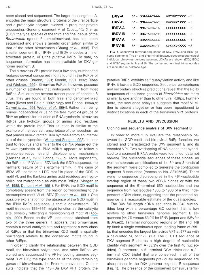
The DXV full-length cDNA sequence is 3243 nucleo-tides long with a percentage GC content that is lowrelative to other birnavirus genome segment B se-quences (44.7% versus 53.9% for IPNV jasper and 53% forIBDVser2). Terminal noncoding regions of 104 and 148bp flank a single continuous open reading frame of 2991bp that encodes the largest birnavirus VP1 at 977 aa witha calculated M r of 112.8 kDa. The 5� UTR sequence ofDXV segment B shares a high degree of nucleotideidentity with segment A (83.3% over the first 40 nucleo-tides). Furthermore, a 5�-terminal GGA triplet and a 3�-terminal CCC triplet that are conserved in all of thebirnavirus genome segments previously sequenced arealso present in the DXV genome segment B sequence(Fig. 1). The presence of the conserved birnavirus termi-
FIG. 1. Conserved terminal sequences of DXV, IPNV, and IBDV ge-nome segments. The 5�- and 3�-terminal deoxynucleotide sequences ofindividual birnavirus genome segment cDNAs are shown (DXV, IBDV,and IPNV segments A and B). The conserved terminal trinucleotidesare indicated in boldface type.
242 SHWED ET AL.

nal trinucleotides indicates that the reported sequencerepresents the entire cDNA sequence of DXV segment B,thereby completing the sequence analysis of this ento-mobirnavirus genome.
In vitro self-guanylylation of DXV VP1
A consensus NTP-binding motif, G/AXXXXGKS/T(Walker et al., 1982), is present in ras-type GTP-bindingproteins (Argos and Leberman, 1985) and in viral pro-teins with a role in RNA replication (Kaariainen et al.,1987). A GXXXXGR/KT motif is present in the IPNV andIBDV VP1 sequences (Fig. 2) and may represent a po-tential GTP-binding site involved in the self-guanylylationactivity associated with these birnavirus VP1s (Dobos,1993). Self-guanylylation of the IPNV and IBDV VP1 pro-teins results in the formation of a VP1pG complex linkedby a serine–5�GMP phosphodiester bond to the 5�-end ofboth genome segments (Dobos, 1993). The VP1pG com-plex acts as a primer during RNA synthesis both in vitro(Dobos, 1995a) and in vivo (Magyar et al., 1998), and theVP1 polypeptide remains covalently linked to the 5�-endof the RNA and thus becomes the genome-linked proteinVPg.
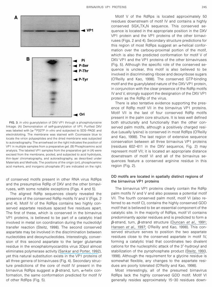
The region of the DXV VP1 sequence that correspondsto the putative GTP-binding site present in the IPNV andIBDV VP1 proteins contains a GXXXXGKK sequence (res-idues 256–263 in Fig. 2) that could function as a GTP-binding site. We therefore determined whether the VP1polypeptide encoded by DXV genome segment B is gua-nylylated in vitro. As shown in Fig. 3, VP1 present inpurified DXV virions was labeled in the presence of[�-32P]GTP. None of the other viral proteins were labeledwith [�-32P]GTP (data not shown). The identification ofthe radiolabeled protein as VP1 was determined bystaining the blot; no other DXV structural protein thatstains with Coomassie blue migrates with an electro-phoretic mobility similar to that of VP1 (VP1 is 113 kDa;the next largest detectable DXV structural protein is VP2,with a molecular mass of approximately 55 kDa). Thelabeling was specific for GTP since VP1 was not labeledby similar incubations with [�-32P]UTP, [�-32P]CTP, or[�-32P]ATP (data not shown).
The radiolabeled VP1 protein obtained from multiplelanes of a preparative gel (Fig. 3A) was subjected tophosphoamino acid analysis. The radiolabeled moietiesdetected by thin-layer chromatography following partialacid hydrolysis of the [�-32P]GTP-labeled VP1 proteincorresponded to liberated free phosphate (Pi), partialhydrolysis products (spots below the phosphotyrosine),and phosphotyrosine (P-Tyr). There was no evidence ofradiolabel associated with phosphoserine or phospho-threonine, which were clearly resolved from phosphoty-rosine by chromatography, as indicated by the migrationof phosphoamino acid markers (shown on the right inFig. 3B). Therefore, in contrast to other birnaviruses (Do-
bos, 1993), the GMP associated with the DXV VP1 proteinfollowing in vitro guanylylation is linked to a tyrosineresidue.
Although no tyrosine in the DXV VP1 protein alignswith a serine in the IPNV sequence to suggest a con-served guanylylation site, we noted the close proximity ofTyr48 in DXV with Ser 44 of IPNV and Ser 46 of IBDV (Fig.2). Since the specific site of serine guanylylation in theIPNV VP1 protein is unknown, the significance of thisobservation is not apparent. If the guanylylation site isspatially conserved in the birnavirus VP1 proteins, thisrepresents a potential site for GMP addition. However, itis also conceivable that the extensive sequence diver-gence of the birnavirus VP1 proteins may contribute toaltered protein folding and the localization of a tyrosineresidue from the DXV VP1 protein in a similar position asa serine residue of IPNV in the tertiary structure of thebirnavirus VP1 proteins. Regardless of the actual site ofVP1 guanylylation, our results suggest that the DXV VP1protein also possesses self-guanylylating activity, con-sistent with its presumptive designation as the RdRp ofthe virus and suggesting that it likely serves a similarrole in priming RNA replication, as occurs with the otherbirnaviruses.
Identification of conserved RdRp motifs in the DXVVP1 protein
The DXV VP1 protein was used as the query in aBLASTP search of the nonredundant Swiss-Protein da-tabase (version 39.0, May 2000). Only other birnavirusVP1 genes produced biologically relevant alignments. Acomparison of the aligned birnavirus VP1 proteins re-vealed significant sequence divergence with only 25%sequence identity between the VP1 protein of DXV andthose of IPNV and IBDV (Fig. 2). The sequence hetero-geneity was most apparent in the amino- and carboxy-terminal 250–300 amino acids of the birnavirus VP1 pro-teins. In particular, the carboxy-proximal one-third of theproteins display almost no sequence identity between allthree proteins and contain numerous insertions/dele-tions required to maintain the alignment. Extensiveamino acid identity between the three birnavirus VP1proteins was observed in the central one-third of theproteins (residues 338–618 in the DXV sequence fromFig. 2). This central region of VP1 contains the consen-sus GTP-binding motif discussed above. Several of theconserved motifs found in the catalytic palm domain ofother polymerases were also identified in this centralregion (Poch et al., 1989; Bruenn, 1991; Koonin, 1991,1992; Koonin and Dolja, 1993).
The conserved RdRp motifs from several RNA viruseswere aligned with similar motifs present in the birnavirusVP1 proteins (Fig. 4). Obvious sequence similarity andconserved predicted secondary structures and spatialarrangements were observed between the organization
243BIRNAVIRUS VP1 PROTEINS
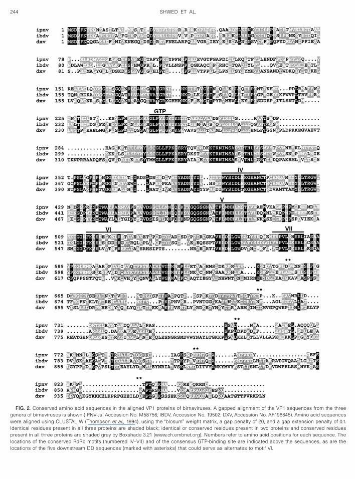
FIG. 2. Conserved amino acid sequences in the aligned VP1 proteins of birnaviruses. A gapped alignment of the VP1 sequences from the threegenera of birnaviruses is shown (IPNV-Ja, Accession No. M58756; IBDV, Accession No. 19502; DXV, Accession No. AF196645). Amino acid sequenceswere aligned using CLUSTAL W (Thompson et al., 1994), using the “blosum” weight matrix, a gap penalty of 20, and a gap extension penalty of 0.1.Identical residues present in all three proteins are shaded black; identical or conserved residues present in two proteins and conserved residuespresent in all three proteins are shaded gray by Boxshade 3.21 (www.ch.embnet.org). Numbers refer to amino acid positions for each sequence. Thelocations of the conserved RdRp motifs (numbered IV–VII) and of the consensus GTP-binding site are indicated above the sequences, as are thelocations of the five downstream DD sequences (marked with asterisks) that could serve as alternates to motif VI.
244 SHWED ET AL.
of conserved motifs present in other RNA virus RdRpsand the presumptive RdRp of DXV and the other birnavi-ruses, with some notable exceptions (Figs. 4 and 5).
The aligned RdRp sequences clearly revealed thepresence of the conserved RdRp motifs IV and V (Figs. 2and 4). Motif IV of the RdRps contains two highly con-served aspartate residues spaced five residues apart.The first of these, which is conserved in the birnavirusVP1 proteins, is believed to be part of a catalytic triadinvolved in metal ion coordination during the nucleotidyltransfer reaction (Steitz, 1998). The second conservedaspartate may be involved in the discrimination betweennucleotides and deoxynucleotides. Interestingly, conver-sion of this second aspartate to the larger glutamateresidue in the encephalomyocarditis virus 3Dpol almosteliminates polymerase activity (Sankar and Porter, 1992),yet this natural substitution exists in the VP1 proteins ofall three genera of birnaviruses (Fig. 4). Secondary struc-ture predictions of the core of motif IV present in thebirnavirus RdRps suggest a �-strand, turn, �-helix con-formation, the same conformation predicted for motif IVof other RdRps (Fig. 5).
Motif V of the RdRps is located approximately 50residues downstream of motif IV and contains a highlyconserved SGX3TX3N sequence. This conserved se-quence is located in the appropriate position in the DXVVP1 protein and the VP1 proteins of the other birnavi-ruses (Figs. 2 and 4). Secondary structure predictions forthis region of most RdRps suggest an �-helical confor-mation over the carboxy-proximal portion of the motif,which is also the predicted conformation for motif V ofDXV VP1 and the VP1 proteins of the other birnaviruses(Fig. 5). Although the specific role of the conserved se-quence is unclear, this motif is also believed to beinvolved in discriminating ribose and deoxyribose sugars(O’Reilly and Kao, 1998). The conserved GTP-bindingmotif and the guanylylated nature of the DXV VP1 protein,in conjunction with the clear presence of the RdRp motifsIV and V, strongly support the designation of the DXV VP1protein as the RdRp of the virus.
There is also tentative evidence supporting the pres-ence of RdRp motif VII in the birnavirus VP1 proteins.Motif VII is the last of four conserved RdRp motifspresent in the palm core structure. It is less well definedboth structurally and functionally than the other con-served palm motifs, although a positively charged resi-due (usually lysine) is conserved in most RdRps (O’Reillyand Kao, 1998). The last region of extensive sequenceconservation between all three birnavirus VP1 proteins(residues 602–611 in the DXV sequence, Fig. 2) mayrepresent motif VII; it is located an appropriate distancedownstream of motif VI and all of the birnavirus se-quences feature a conserved arginine residue in thisregion (Fig. 2).
DD motifs are located in spatially distinct regions ofthe birnavirus VP1 proteins
The birnavirus VP1 proteins clearly contain the RdRppalm motifs IV and V and also possess a potential motifVII. The fourth conserved palm motif, motif VI (also re-ferred to as motif C), contains the highly conserved GDDmotif that is believed to be an essential component of thecatalytic site. In the majority of RdRps, motif VI containspredominantly apolar residues and is predicted to form a�-strand, turn, �-strand structure (Duncan et al., 1991;Hansen et al., 1997; O’Reilly and Kao, 1998). This con-served structure serves to position the two aspartateresidues close to the conserved aspartate in motif IV,forming a catalytic triad that coordinates two divalentcations for the nucleophilic attack of the 3�-hydroxyl andstabilization of the pyrophosphate product (Steitz, 1993,1998). Although the requirement for a glycine residue issomewhat flexible, any changes to the aspartate resi-dues are poorly tolerated (O’Reilly and Kao, 1998).
Most interestingly, all of the presumed birnavirusRdRps lack the highly conserved GDD motif. Motif VIgenerally resides approximately 15–30 residues down-
FIG. 3. In vitro guanylylation of DXV VP1 through a phosphotyrosinelinkage. (A) Demonstration of self-guanylylation of VP1. Purified DXVwas labeled with [�-32P]GTP in vitro and subjected to SDS–PAGE andelectroblotting. The membrane was stained with Coomassie blue tolocate the virion polypeptides and the dried membrane was subjectedto autoradiography. The arrowhead on the right indicates the position ofVP1 in multiple samples from a preparative gel. (B) Phosphoamino acidanalysis. The labeled VP1 samples from the preparative gel in (A) wereexcised from the membrane, pooled, and subjected to acid hydrolysis,thin-layer chromatography, and autoradiography, as described underMaterials and Methods. The positions of the origin (ori), phosphoaminoacid markers, and inorganic phosphate (Pi) are indicated on the right.
245BIRNAVIRUS VP1 PROTEINS

stream of motif V in most RdRps (Fig. 4). This region ofthe IBDV VP1 protein contains an IDD sequence, similarto the SDD sequence present in the �6 RdRp, that couldserve as the functional equivalent of the GDD motif. IBDVmotif VI, however, contains an unusual preponderance ofcharged residues compared to the more hydrophobicenvironment surrounding the GDD motif in most otherRdRps (Fig. 4). Secondary structure predictions for RdRpmotif VI of IBDV also suggest potential differences in thestructure of this region compared to motif VI of otherRdRps. A preferred �-helical conformation is predictedfor the region encompassing the IBDV IDD motif, unlikethe predicted �-strand, turn, �-strand structure sug-gested for motif VI of most RdRps (Fig. 5). However, asimilar �-helical structure is predicted for motif VI of �6,yet the crystal structure of �6 VP2 indicates that thismotif exists in a �-strand, turn, �-strand conformation,the same conformation observed for motif VI in the crys-tal structures of hepatitis C virus and poliovirus RdRps(Hansen et al., 1997; Lesburg et al., 1999; Butcher et al.,2001). The �-helical prediction for this region of IBDVmust, therefore, be interpreted with caution. However,
this region in all three birnavirus VP1 proteins is pre-dicted to exist as an extended �-helix (Fig. 5). The con-served structural prediction of clearly homologous pro-teins containing sequence heterogeneity lends addedconfidence to the predicted �-helical conformation ofthis region in all three birnavirus VP1 proteins. The onlyother downstream DD sequence (PDD) exists at position761 in the IBDV VP1 protein. This di-aspartate sequenceexists in a region of limited sequence similarity to theother birnavirus VP1 proteins (Fig. 2), it is surrounded bycharged residues, and secondary structure predictionsshow high scores for an extended loop region (data notshown). Therefore, if IBDV VP1 does contain a homologof RdRp motif VI, then the first DD motif is more likely toserve this function.
Striking alterations were noted when comparing thesequences of the DXV and IPNV regions correspondingto the tentative motif VI of IBDV. This region in both theDXV and the IPNV VP1 proteins lacks any DD sequenceand displays more extensive sequence heterogeneitythan observed for the conserved motifs IV and V presentin the birnavirus VP1 proteins (Fig. 2). The absence of a
FIG. 4. Sequence alignments of the conserved RdRp motifs. The aligned amino acid sequences from conserved domains IV–VI of representativeplus-stranded animal RNA viruses, plus-stranded plant RNA viruses, and double-stranded RNA viruses are shown. The generalized consensussequence, displayed at the top of the figure, is expressed with the following groups: J, apolar (LIVMFYWCAGP); O, polar (STNQH); B, apolar or polar(J � O); Z, polar or charged (O � KDER); U, aromatic (FYW); (—), any amino acid. Uppercase letters indicate that the amino acid conforms to thegeneralized consensus sequence. Highly conserved residues in the consensus are indicated with an asterisk, and residues conforming to the highlyconserved consensus residues are indicated in boldface type. Arabic numbers represent the number of amino acids upstream or between the motifs.The virus abbreviations are as follows: POLIO, poliovirus type 1 (Accession No. P03299); EMC, encephalomyocarditis virus (Accession No. GNNYE);FMDV, foot and mouth disease virus (Accession No. P03305); SBV, Sindbis virus (Accession No. AAA96974); YFV, yellow fever virus (Accession No.POLG_TEFV2); IBV, infectious bronchitis virus (Accession No. CAC39112); TMV, tobacco mosaic virus (Accession No. RRPO_TOML); BMV, bromemosaic virus (Accession No. P2BVA); TEV, tobacco etch virus (Accession No. GNBVEV); CNV, cucumber necrosis virus (Accession No. RRVGCN);MCMV, maize chlorotic mottle virus (Accession No. CAB55589); BTV, bluetongue virus serotype 10 (Accession No. RRXRBT); ROTA, simian rotavirusSA11 (Accession No. P1XRSR); REO, mammalian reovirus strain Dearing (Accession No. MWXR33); PHI6, bacteriophage �6 (Accession No.VP2_BPPH6); IBDV, infectious bursal disease virus serotype P2 (Accession No. AF240687); IPNV, infectious pancreatic necrosis virus serotype jasper(Accession No. M58756); DXV, Drosophila X virus (Accession No. AF196645).
246 SHWED ET AL.
DD sequence in the DXV and IPNV sequences indicatesthat the tentative motif VI of IBDV is not conserved in asimilar location in these related birnavirus RdRps.
We examined the IPNV and DXV VP1 sequences forthe presence of downstream DD motifs that might rep-resent the missing motif VI. In the case of IPNV, it hasbeen suggested that a downstream LDD sequence, res-idues 653–655 (Fig. 2), may represent a repositionedmotif VI (Koonin, 1992). However, the alignment of thethree birnavirus VP1 proteins indicates that there is noconservation of aspartate residues in the region corre-sponding to the LDD sequence of IPNV as might beexpected if a structural rearrangement of the birnavirusRdRp resulted in repositioning of motif VI (Fig. 2). Thereare no other downstream DD sequences in the IPNV VP1protein that could serve a similar catalytic function as theGDD sequence present in other RdRps. If the down-stream DD sequence in IPNV VP1 does represent themissing motif VI, then the event leading to repositioningof this motif did not occur in a similar fashion with theother birnavirus RdRps.
A similar situation exists for DXV, which also lacks aDD sequence in the region corresponding to the tenta-tive motif VI of IBDV. DXV VP1 does contain three down-stream DD sequences (residues 755/56, 895/96, and957/958) that could function as homologs of motif VI (Fig.2). As with IPNV, however, the downstream DD residuesof DXV do not align with any of the DD sequences
present in the other birnavirus RdRps, and they occur inregions of extensive birnavirus VP1 sequence heteroge-neity (Fig. 2). The last two DXV DD sequences occur inpredicted loop regions (data not shown), while the firstdownstream DD sequence (residues 755/56) of DXV re-sides in a region with high-probability �-helix scores,similar to the predicted secondary structures of the ten-tative motif VI in IBDV and the downstream DD sequencein IPNV (Fig. 5). As a result, all of the birnavirus VP1proteins contain a DD sequence that could function in asimilar fashion as the GDD motif present in the majorityof RdRps. However, the downstream DD motifs in thebirnavirus VP1 proteins all exist in predicted �-helical orloop conformations, these sequences are located in spa-tially distinct regions of the birnavirus RdRps, and theyoccur in regions of extensive sequence heterogeneity.
The absence of significant sequence similarity be-tween the RdRps of the dsRNA totiviruses and other RNAviruses, and the identification of additional conservedmotifs present in the RdRps of different totiviruses, led tothe suggestion that totivirus RdRps represent a distinctsubgroup within this enzyme family (Bruenn, 1993;Routhier and Bruenn, 1998). Our results suggest that theVP1 proteins of the birnaviruses form a second definedsubgroup of RdRps with significant differences in thearrangement of the predicted catalytic domain of thisenzyme family. Based on the clear conservation of motifsIV and V in the birnavirus VP1 proteins, it seems reason-
FIG. 5. Predicted secondary structures in the conserved region of the RdRp core palm structure (motifs IV, V, and VI). The palm motifs of poliovirus(POLIO) and hepatitis C virus (HCV) are presented along with the same motifs identified in RdRps derived from a selection of dsRNA viruses (REO,mammalian reovirus strain Dearing; PHI6, bacteriophage �6; IBDV, infectious bursal disease virus serotype P2; IPNV, infectious pancreatic necrosisvirus serotype jasper; DXV, Drosophila X virus). The predicted consensus secondary structures were derived using the PHD method of Rost andSander (1994) using PredictProtein software (www.embl-heidelberg.de/predictprotein/). h, alpha helix; b, beta strand; dot, no prediction. The locationsof the highly conserved residues characteristic of each motif are indicated with an asterisk and the corresponding residues are shown in boldfacetype.
247BIRNAVIRUS VP1 PROTEINS
able to assume that these RdRps function in a mannersimilar to that predicted for all other RdRps. If so, then thebirnavirus RdRps should be dependent on the formationof a catalytic triad of aspartate residues. The only way toenvision such a conserved catalytic activity requires ac-cepting that the birnavirus RdRps have repositioned mo-tif VI to three separate downstream locations in regionsof extensive sequence and predicted structural hetero-geneity. Conversely, the only other possibility is that ahomolog of motif VI and its highly conserved GDD motifmay not function as the catalytic core of the birnaviruspolymerases. The absence or repositioning of motif VI toseparate locations in the birnavirus VP1 proteins is, asfar as we are aware, a unique situation for a viral RdRp.Regardless of the actual catalytic mechanism of thebirnavirus RdRps, it seems likely that the structures ofthe birnavirus RdRps may exhibit significant variationfrom other RdRps. The potential differences in the ar-rangement of catalytic motifs and the structure of thissubgroup of RdRps may account for the distinct at-tributes of birnavirus RdRps, namely, their role as bothpolymerase and primer for semiconservative genomereplication (Mertens et al., 1982; Nagy and Dobos1984a,b; Calvert et al., 1991; Dobos, 1995a). More de-tailed structural and mutational analyses of the birnavi-rus RdRp proteins should reveal important insights intothe conformation, precise location, and features of thecatalytic site of this important subgroup of viral poly-merases.
MATERIALS AND METHODS
Cells and virus
DXV was propagated in Schneider’s Drosophila line 1cells, WR strain (Friesen and Rueckert, 1982) (WR strainkindly provided by Dr. Dasgupta). Plaque-purified viruswas used to infect cell cultures at a multiplicity of infec-tion of 0.01 plaque-forming units per cell. Purification ofDXV was carried out by freon extraction and sucrose andCsCl gradient centrifugation in TNE buffer (0.1 M Tris, 0.1M NaCl, 1 mM EDTA, pH 7.3) as described previously(Dobos and Rowe, 1977). The final purified virus bandfrom CsCl gradients was dialyzed against 10 mM Tris–HCl, pH 8.0, 0.3 mM MgCl2 and stored in aliquots at�80°C at a concentration of approximately 1 mg ml�1.
Cloning strategy
cDNA clones of segment B of DXV were constructedby denaturing genomic RNA and poly(A) tailing it withEscherichia coli poly(A) polymerase (Cashdollar et al.,1982). Briefly, 20 �g of virion RNA in 2.5 �l of water wasincubated with 22.5 �l of dimethyl sulfoxide for 45 min at60°C. The polyadenylation reaction mixture consisted of50 mM Tris, pH 7.9, 10 mM MgCl2, 250 mM NaCl, 2.5 mMMnCl2, 0.25 mM ATP, 250 g BSA, and 1 U of poly(A)
polymerase (Gibco BRL). The reaction was incubated for10 min at 37°C. The RNA was then extracted with phe-nol–chloroform and recovered by ethanol precipitation.Polyadenylated RNA was resuspended in 10 �l of dieth-ylpyrocarbonate-treated water.
First-strand synthesis was carried out by denaturing20 �g of virion RNA consisting of segment A and B in 9�l of 10 mM Tris–HCl, pH 7.5, by incubation at 100°C for2 min, followed by quick freezing in a dry ice–ethanolbath. The thawed suspension was then made 10 mMwith respect to methyl mercury hydroxide. Denaturationproceeded for 45–60 min at 23°C in the presence of 10 Uof RNasin (Promega, Canada). An additional 10 U ofRNasin was added with �-mercaptoethanol to a concen-tration of 187 mM and the reaction was incubated at23°C for another 15 min. Viral cDNA was synthesized byreverse-transcribing denatured dsRNA using randomprimers as described by Gubler and Hoffman (1983).
Double-stranded cDNA was tailed with dCTP usingTdT, cloned into the dGTP tailed PstI site of pTZ18R(Mead et al., 1986), and then subcloned into competent E.coli HB101 or DH5 cells according to Sambrook et al.(1989). Two positive recombinants were identified byrestriction endonuclease mapping, as described byCameron (1991).
Amplification of 5� and 3� noncoding regions
The nucleotide sequences of the 5� and 3� noncodingregions of DXV segment B were determined by the meth-ods described for IBDV (Mundt and Muller, 1995) and IPNV(Yao and Vakharia, 1998). To determine the 3�-termini ofboth strands of segments A and B, the viral RNA waspolyadenylated and reverse transcribed with a poly(dT)primer containing a NotI site, and resulting cDNAs wereamplified by PCR with virus-specific primers DXV5�R(5�-GTTGGTATGTGCAGGCCAGAG-3� (nucleotides 410–430, antisense orientation) and DXV3�F (5�-GCAGCACCCT-GTTGTCAAAC-3� at nucleotides 2828–2846, sense orienta-tion). The reverse transcription products were separated byagarose gel electrophoresis, purified by a QIAquick gelextraction kit (Qiagen Inc.), and directly sequenced by thedideoxy chain termination method. The 5� terminus of seg-ment B was determined by rapid amplification of cDNAends using the 5� RACE System (Life Technologies Inc.).
Sequence analysis and secondary structureprediction
The sequence of DXV genome segment B was ob-tained from two independent clones representing the 5�-and 3�-proximal regions of the genome segment. Bothclones were sequenced in their entirety in both direc-tions by automated sequencing and compiled usingGenerunner version 3.0 software. The clones overlappedby 464 nucleotides, which allowed a contiguous se-quence to be obtained. The clones were truncated by 19
248 SHWED ET AL.
nucleotides at the 5�-end and by 193 nucleotides at the3�-end. The terminal sequences were obtained by sep-arate amplification of the 5�- and 3�-termini, as describedabove, to generate the complete DXV genome segment BcDNA sequence. There were no sequence discrepan-cies in the 464-nucleotide overlap region of the twoindependent clones, suggesting that the presented se-quence is a reasonable estimate of the quasispecies.This was further confirmed by sequencing of the 5�-terminal 650 nucleotides and an additional 600 nucleo-tides from the central region (nucleotides 1300–1900) ofa third independent cDNA clone; there were no discrep-ancies with the presented sequence.
The nonredundant coding sequence databases weresearched using the BLAST 2.0 program blastp (www.ncbi.nlm.nih.gov; Altschul et al., 1997). Multiple align-ments of amino acid sequences were made with theprogram CLUSTALW (Thompson et al., 1994). RdRp motifsearches were carried out using PHI-BLAST. Secondarystructure consensus predictions were carried out usingthe PHD method of Rost and Sander (1994) using Pre-dictProtein software (http://www.embl-heidelberg.de/predictprotein/).
In vitro guanylylation and characterization of the DXVVP1 guanylate linkage
A 25-�l reaction mixture in 10 mM Tris–HCl, pH 8.0,contained 5 �g of purified virus, 0.3 mM MgCl2, and 20�Ci [�-32P]GTP (sp act 3000 Ci/mmol, ICN). After incuba-tion in a 37°C water bath for 15 min, the reaction wasstopped by the addition of an equal volume of double-concentration electrophoresis sample buffer (1� ESB: 50mM Tris–HCl, pH 6.7, 2% SDS, 5% 2-ME, 10% glycerol,and a trace of bromophenol blue), heated in a boilingwater bath, and subjected to a 10% SDS–PAGE resolvinggel, 5% stacking gel (Laemmli, 1970), followed by Coo-massie blue staining and autoradiography. For phos-phoamino acid analysis, PAGE was followed by electro-blotting onto PVDF membranes (Immobilon, MilliporeCorp.) and the labeled VP1 was detected by autoradiog-raphy, excised from the membrane, cut into small pieces,and placed in 5.6 M HCl for hydrolysis at 110°C for 90min in a tightly capped microfuge tube. The HCl wasevaporated under vacuum, resuspended in 200 �l water,and evaporated again. The resulting pellet was resus-pended in 10 �l water, to which 10 �g each of phospho-serine, phosphothreonine, and phosphotyrosine wasadded and subjected to thin-layer electrophoresis in 5%acetic acid, 0.5% pyridine, pH 3.5, at 400 V for 100 min.The phosphoamino acid standards were located byspraying the plate with a 0.1% solution of ninhydrin inethanol followed by brief heating in an oven at 60°C. Thelabeled phosphoamino acid was detected by autoradiog-raphy.
ACKNOWLEDGMENTS
This research was supported by the Natural Sciences and Engineer-ing Research Council of Canada (P.D.), and by a grant from the Cana-dian Institutes of Health Research (CIHR) to R.D.
REFERENCES
Altschul, S. F., Madden, T. L., Schaffer, A. A., Zhang, J., Zhang, Z., Miller,W., and Lipman, D. J. (1997). Gapped BLAST and PSI-BLAST: A newgeneration of protein database search programs. Nucleic Acids Res.25, 3389–3402.
Argos, P. (1988). A sequence motif in many polymerases. Nucleic AcidsRes. 16, 9909–9915.
Argos, P., and Leberman, R. (1985). Homologies and anomalies inprimary structural patterns of nucleotide binding proteins. Eur. J. Bio-chem. 152, 651–656.
Azad, A. A., Barrett, S. A., and Fahey, K. J. (1985). The characterizationand molecular cloning of the double-stranded RNA genome of anAustralian strain of infectious bursal disease virus. Virology 143,35–44.
Boursnell, M. E., Brown, T. D., Foulds, I. J., Green, P. F., Tomley, F. M., andBinns, M. M. (1987). Completion of the sequence of the genome ofthe coronavirus avian infectious bronchitis virus. J. Gen. Virol. 68,57–77.
Bruenn, J. A. (1991). Relationships among the positive strand anddouble-strand RNA viruses as viewed through their RNA-dependentRNA polymerase. Nucleic Acids Res. 19, 217–226.
Bruenn, J. A. (1993). A closely related group of RNA-dependent RNApolymerases from double stranded viruses. Nucleic Acids Res. 21,5667–5669.
Butcher, S. J., Grimes, J. M., Makeyev, E. V., Bamford, D. H., and Stuart,D. I. (2001). A mechanism for initiating RNA polymerization. Nature410, 235–240.
Calvert, J. G., Nagy, E., Soler, M., and Dobos, P. (1991). Characterizationof the VPg-dsRNA linkage of infectious pancreatic necrosis virus.J. Gen. Virol. 72, 2563–2567.
Cameron, L. A. (1991). “Cloning of the Drosophila X Virus Genome andPartial Sequencing of segment B Encoding the Putative Polymerase,”M.Sc. Thesis, University of Guelph.
Cashdollar, L. W., Esparaz, J., Hudson, G. R., Chimelo, R., Lee, P. W. K.,and Joklik, W. K. (1982). Cloning the double-stranded RNA genes ofreovirus: The sequence of the cloned S2 gene. Proc. Natl. Acad. Sci.USA 79, 7644–7648.
Chung, H. K., Kordyban, S., Cameron, L., and Dobos, P. (1996). Se-quence analysis of the bicistronic Drosophila X virus genome seg-ment A and its encoded polypeptide. Virology 225, 359–368, doi:10.1006/viro.1996.0610.
Dobos, P. (1993). In vitro guanylylation of infectious pancreatic necrosisvirus polypeptide VP1. Virology 193, 403–413, doi:10.1006/viro.1993.1137.
Dobos, P. (1995a). The molecular biology of infectious pancreatic ne-crosis virus (IPNV). Annu. Rev. Fish Dis. 5, 25–54.
Dobos, P. (1995b). Protein primed RNA synthesis in vitro by the virion-associated RNA polymerase of infectious pancreatic necrosis virus.Virology 208, 19–25, doi:10.1006/viro.1995.1125.
Dobos, P., and Rowe, D. (1977). Peptide map comparison of infectiouspancreatic necrosis virus-specific polypeptides. J. Virol. 24, 805–820.
Duncan, R., and Dobos, P. (1986). The nucleotide sequence of infec-tious pancreatic necrosis virus (IPNV) segment A reveals one largeORF encoding a precursor polyprotein. Nucleic Acids Res. 14, 5934.
Duncan, R., Mason, C. L., Nagy, E., Leong, J. A., and Dobos, P. (1991).Sequence analysis of the infectious pancreatic necrosis virus ge-nome segment B and its encoded VP1 protein: A putative RNA-dependent RNA polymerase lacking the Gly-Asp-Asp motif. Virology181, 541–552.
Friesen, P. D., and Rueckert, R. R. (1982). Black beetle virus: Messengerfor protein B is a subgenomic viral RNA. J. Virol. 421, 986–995.
249BIRNAVIRUS VP1 PROTEINS

Gorbalenya, A. E., and Koonin, E. V. (1988). Birnavirus RNA polymeraseis related to polymerases of positive strand viruses. Nucleic AcidsRes. 16, 7735.
Gubler, U., and Hoffman, B. J. (1983). A simple and very efficient methodfor generating cDNA libraries. Gene 25, 263–269.
Hansen, J. L., Long, A. M., and Schultz, S. C. (1997). Structure of theRNA-dependent RNA polymerase of polio virus. Structure 5, 1109–1122.
Ishihama, A., and Nagata, K. (1988). Viral RNA polymerases. CRC Crit.Rev. Biochem. 23, 27–76.
Kaariainen, L., Takkinen, K., Keranan, S., and Soderlund, H. (1987).Replication of the genome of alphaviruses. J. Cell Sci. 7, 231–250.
Kamer, G., and Argos, P. (1984). Primary structural comparison ofRNA-dependent polymerases from plant, animal and bacterial vi-ruses. Nucleic Acids Res. 12, 7269–7282.
Kibenge, F. S., Jackwood, D. J., and Mercado, C. C. (1990). Nucleotidesequence analysis of genome segment A of infectious bursal dis-ease virus. J. Gen. Virol. 71, 569–577.
Kibenge, F. S., and Qian, B. (1994). Sequence conservation in the RNApolymerase gene of infectious bursal disease viruses. Arch. Virol.134, 441–449.
Kohlstaedt, L. A., Wang, J., Friedman, J. M., Rice, P. A., and Steitz, T. A.(1992). Crystal structure at 3.5 Å resolution of HIV-1 reverse tran-scriptase complexed with an inhibitor. Science 256, 1783–1790.
Koonin, E. V. (1991). The phylogeny of RNA dependent RNA poly-merases of positive-stranded RNA viruses. J. Gen. Virol. 72, 2197–2206.
Koonin, E. V. (1992). Evolution of double-stranded RNA viruses: A caseof polyphyletic origin from different groups of positive-stranded RNAviruses. Semin. Virol. 3, 327–339.
Koonin, E. V., and Dolja, V. V. (1993). Evolution and taxonomy of positive-strand viruses: Implications and analysis of amino acid sequences.Crit. Rev. Biochem. Mol. Biol. 28, 375–430.
Laemmli, U. K. (1970). Cleavage of structural proteins during the as-sembly of the head of bacteriophage T4. Nature 227, 680–685.
Lesburg, C. A., Cable, M. B., Ferrari, E., Hong, Z., Mannarino, A. F., andWeber, P. C. (1999). Crystal structure of the RNA-dependent RNApolymerase from hepatitis C virus reveals a fully encircled active site.Nat. Struct. Biol. 6, 937–943.
Magyar, G., Chung, H. K., and Dobos, P. (1998). Conversion of VP1 toVPg in cells infected by infectious pancreatic necrosis virus. Virology245, 142–150.
Mead, D., Szczesna-Skorupa, E., and Kemper, B. (1986). Single-stranded DNA “blue” T7 promoter plasmids: A versatile tandempromoter system for cloning and protein engineering. Protein Eng. 1,67–74.
Mertens, P. P., Jamison, P. B., and Dobos, P. (1982). In vitro RNAsynthesis of infectious pancreatic necrosis virus-associated RNApolymerase. J. Gen. Virol. 59, 47–56.
Mindich, L., Nemhauser, I., Gottlieb, P., Romatschuk, M., Carton, J.,Frucht, S., Strassman, J., Bamford, D. H., and Kalkkinen, N. (1988).Nucleotide sequence of the large double-stranded RNA segment ofbacteriophage phi 6: Genes specifying the viral replicase and tran-scriptase. J. Virol. 62, 1180–1185.
Morgan, M. M., Macreadie, I. G., Harley, V. R., Hudson, P. J., and Azad,A. A. (1988). Sequence of the small double-stranded RNA genomicsegment of infectious bursal disease virus and its deduced 90-kDaproduct. Virology 163, 240–242.
Mundt, E., and Muller, H. (1995). Complete nucleotide sequences of 5�and 3� noncoding regions of both genome segments of different
strains of infectious bursal disease virus. Virology 209, 10–18, doi:10.1006/viro.1995.1226.
Nagy, E., and Dobos, P. (1984a). Synthesis of Drosophila X virus pro-teins in cultured Drosophila cells. Virology 134, 358–367.
Nagy, E., and Dobos, P. (1984b). Coding assignments of Drosophila Xvirus genome segments: In vitro translation of native and denaturedvirion dsRNA. Virology 137, 58–66.
Ollis, D. L., Brick, P., Hamlin, R., Xuong, N. G., and Steitz, T. A. (1985).Structure of the large fragment of Escherichia coli DNA polymeraseI complexed with dNTP. Nature 313, 762–766.
O’Reilly, E. K., and Kao, C. C. (1998). Analysis of RNA-dependent RNApolymerase structure and function as guided by known polymerasestructures and computer predictions of secondary structure. Virology252, 287–303.
Poch, O., Sauvaget, I., Delarue, M., and Tordo, N. (1989). Identificationof four conserved motifs among RNA-dependent polymerase encod-ing elements. EMBO J. 8, 3867–3874.
Revet, B., and Delain, E. (1982). Drosophila X virus contains a 1-�mdouble-stranded RNA circularized by a 67-kDa terminal protein: Highresolution mapping of its genome. Virology 123, 29–44.
Rost, B., and Sander, C. (1994). Combining evolutionary information andneural networks to predict protein secondary structure. Protein 19,55–77.
Routhier, E., and Bruenn, J. A. (1998). Functions of conserved motifs inthe RNA-dependent RNA polymerase of a yeast double-strandedRNA virus. J. Virol. 72, 4427–4429.
Ribas, J. C., and Wickner, R. B. (1992). RNA-dependent RNA polymeraseconsensus sequence of the L-A double-stranded RNA virus: Defini-tion of essential domains. Proc. Natl. Acad. Sci. USA 89, 2185–2189.
Sambrook, J., Fritsch, E. F., and Maniatis, T. (1989). “Molecular Cloning:A Laboratory Manual,” 2nd ed., Cold Spring Harbor Laboratory, ColdSpring Harbor, NY.
Sankar, S., and Porter, A. G. (1992). Point mutations which drasticallyaffect the polymerization activity of encephalomyocarditis virus-RNAdependent RNA polymerase correspond to the active site of Esche-richia coli DNA polymerase I. J. Biol. Chem. 267, 10168–10176.
Spies, U., Muller, H., and Becht, H. (1987). Properties of RNA polymer-ase activity associated with infectious bursal disease virus andcharacterization of its reaction products. Virus Res. 8, 127–140.
Steitz, T. A. (1993). DNA and RNA-dependent DNA polymerases. Curr.Opin. Struct. Biol. 3, 31.
Steitz, T. A. (1998). A mechanism for all polymerases. Nature 391,231–232.
Thompson, J. D., Higgins, D. G., and Gibson, T. J. (1994). CLUSTAL W:Improving the sensitivity of progressive multiple sequence alignmentthrough sequence weighting, position specific gap penalties andweight matrix choice. Nucleic Acids Res. 22, 4673–4680.
Walker, J. E., Saraste, M., Runswick, M. J., and Gay, N. J. (1982). Distantlyrelated sequences in the �- and �-subunits of ATP synthase, myosin,kinases and other ATP-requiring enzymes and a common nucleotidebinding fold. EMBO J. 1, 945–951.
Wang, G. H., and Seeger, C. (1992). The reverse transcriptase of hep-atitis B virus acts as a protein primer for viral DNA synthesis. Cell 71,663–670.
Weber, M., Bronsema, V., Bartos, H., Bosserhoff, A., Bartenschlager, R.,and Schaller, H. (1994). Hepadnavirus P protein utilizes a tyrosineresidue in the TP domain to prime reverse transcription. J. Virol. 68,2994–2999.
Yao, K., and Vakharia, V. N. (1998). Generation of infectious pancreaticnecrosis virus from cloned cDNA. J. Virol. 72, 8913–8920.
250 SHWED ET AL.
Copyright © 2022 FDOKUMEN