Characterization of a monoclonal anti-capsid antibody that cross-reacts with three major primate...
11
Characterization of a monoclonal anti-capsid antibody that cross-reacts with three major primate lentivirus lineages Brigitte E. Sanders-Beer a, ⁎, Magdalena Eschricht b , Janna Seifried b , Vanessa M. Hirsch c , Jonathan S. Allan d, 1 , Stephen Norley b a BIOQUAL, Inc., 9600 Medical Center Drive, Rockville, MD 20850, USA b Robert-Koch-Institut, Nordufer 20, 13353 Berlin, Germany c Laboratory of Molecular Microbiology, National Institute of Allergy and Infectious Diseases, National Institutes of Health, 4 Memorial Dr., Bethesda, MD 20892-0460, USA d Texas Biomedical Research Institute, Department of Virology and Immunology, 7620 NW Loop 410 at Military Drive, San Antonio, TX 78227, USA abstract article info Article history: Received 13 August 2011 Accepted 4 November 2011 Available online 5 December 2011 Keywords: AG3.0 Epitope Gag Capsid p24 p27 Human immunodeficiency virus Simian immunodeficiency virus Mouse monoclonal antibodies with varying specificities against the Gag capsid of simian and human immu- nodeficiency virus (SIV/HIV) were generated by immunizing mice with whole inactivated SIVagmTYO-1. Monoclonal antibody AG3.0 showed the broadest reactivity recognizing the Gag capsid protein (p24–27) and Gag precursors p38, p55, and p150 of HIV-1, HIV-2, SIVmac, and SIVagm. Using overlapping peptides, the AG3.0 epitope was mapped in capsid to a sequence (SPRTLNA) conserved among HIV-1, HIV-2, SIVrcm, SIVsm/mac, and SIVagm related viruses. Because of its broad cross-reactivity, AG3.0 was used to develop an antigen capture assay with a lower detection limit of 100 pg/ml HIV-1 Gag p24. Interestingly, AG3.0 was found to have a faster binding on/off rate for SIVagmVer and SIVmac Gag than for SIVagmSab Gag, possibly due to differences outside the SPRTLNA motif. In addition, the ribonucleic acid (RNA) coding for AG3.0 was sequenced to facilitate the development of humanized monoclonal antibodies. © 2011 Elsevier Inc. All rights reserved. Introduction Since the discovery of HIV-1 and HIV-2 (Barre-Sinoussi et al., 1983; Clavel et al., 1986; Gallo et al., 1983) an increasing number of related SIVs have been isolated from nonhuman primates. Serological evidence of SIV infection has been shown for at least 40 of the 69 dif- ferent primate species in Africa and has been confirmed by sequence analysis in 32 (Aghokeng et al., 2010; Locatelli et al., 2008a). Com- plete SIV genome sequences are available for 20 species (Liegeois et al., 2009; Van Heuverswyn and Peeters, 2007; VandeWoude and Apetrei, 2006). The primate lentiviruses can presently be classified into 10 distinct lineages based upon phylogenetic relationships of full-length sequences: 1) SIVcpz from chimpanzees (Pan troglodytes) including HIV-1 and SIVgor from western gorillas (Gorilla gorilla) (Santiago et al., 2003; Van Heuverswyn et al., 2006, 2007); 2) SIVsm from sooty mangabeys (Cercocebus atys) including HIV-2 and SIVmac from macaques (Macaca spp.) (Clavel et al., 1986; Franchini et al., 1987; Hirsch et al., 1989); 3) SIVagm from African green monkeys (members of the Chlorocebus aethiops superspecies) (Allan et al., 1991; Hirsch et al., 1993b; Johnson et al., 1990); 4) SIVsyk from Sykes' monkeys (Cercopithecus albogularis)(Hirsch et al., 1993a); 5) SIVlho from L'Hoest monkeys (C. lhoesti) including SIVsun from sun- tailed monkeys (C. solatus) and SIVmnd-1 from mandrills (Mandrillus sphinx)(Beer et al., 1999; Hirsch et al., 1999; Tsujimoto et al., 1989); 6) SIVrcm from red-capped mangabeys (Cercocebus torquatus) in- cluding SIVdrl from drills (Mandrillus leucophaeus), and SIVmnd-2 from mandrills (Beer et al., 2001; Hu et al., 2003); 7) SIVdeb from DeBrazza monkeys (C. neglectus)(Bibollet-Ruche et al., 2004) includ- ing SIVden from Dent's mona monkeys (C. mona denti)(Dazza et al., 2005); 8) SIVgsn from greater spot-nosed monkeys (C. nictitans) in- cluding SIVmon from mona monkeys (C. mona), and SIVmus-1 and mus-2 from mustached monkeys (C. cephus)(Aghokeng et al., 2007; Courgnaud et al., 2003a); 9) SIVtal from talapoin monkeys (Miopithecus ogouensis)(Liegeois et al., 2006); and 10) SIVcol from Colobus monkeys (Colobus guereza)(Courgnaud et al., 2001). Some lineages, such as SIVcpz, are presumed to be the result of recombination between other lineages (SIVrcm and SIVgsn) (Bailes et al., 2003). Smaller fragments from other SIV-infected African primate species have also been se- quenced and may indicate the existence of either separate lineages Virology 422 (2012) 402–412 ⁎ Corresponding author. Fax: + 1 301 251 1260. E-mail addresses: [email protected] (B.E. Sanders-Beer), [email protected] (M. Eschricht), [email protected] (J. Seifried), [email protected] (V.M. Hirsch), [email protected] (S. Norley). 1 Jonathan Allan passed away in 2009; please contact Jean Patterson (jpatters@sfbr. org). 0042-6822/$ – see front matter © 2011 Elsevier Inc. All rights reserved. doi:10.1016/j.virol.2011.11.003 Contents lists available at SciVerse ScienceDirect Virology journal homepage: www.elsevier.com/locate/yviro
-
Upload
independent -
Category
Documents
-
view
4 -
download
0
Transcript of Characterization of a monoclonal anti-capsid antibody that cross-reacts with three major primate...

Virology 422 (2012) 402–412
Contents lists available at SciVerse ScienceDirect
Virology
j ourna l homepage: www.e lsev ie r .com/ locate /yv i ro
Characterization of a monoclonal anti-capsid antibody that cross-reacts with threemajor primate lentivirus lineages
Brigitte E. Sanders-Beer a,⁎, Magdalena Eschricht b, Janna Seifried b, Vanessa M. Hirsch c,Jonathan S. Allan d,1, Stephen Norley b
a BIOQUAL, Inc., 9600 Medical Center Drive, Rockville, MD 20850, USAb Robert-Koch-Institut, Nordufer 20, 13353 Berlin, Germanyc Laboratory of Molecular Microbiology, National Institute of Allergy and Infectious Diseases, National Institutes of Health, 4 Memorial Dr., Bethesda, MD 20892-0460, USAd Texas Biomedical Research Institute, Department of Virology and Immunology, 7620 NW Loop 410 at Military Drive, San Antonio, TX 78227, USA
⁎ Corresponding author. Fax: +1 301 251 1260.E-mail addresses: [email protected] (B.E. Sande
(M. Eschricht), [email protected] (J. Seifried), vhirsch@[email protected] (S. Norley).
1 Jonathan Allan passed away in 2009; please contactorg).
0042-6822/$ – see front matter © 2011 Elsevier Inc. Alldoi:10.1016/j.virol.2011.11.003
a b s t r a c t
a r t i c l e i n f oArticle history:Received 13 August 2011Accepted 4 November 2011Available online 5 December 2011
Keywords:AG3.0EpitopeGagCapsidp24p27Human immunodeficiency virusSimian immunodeficiency virus
Mouse monoclonal antibodies with varying specificities against the Gag capsid of simian and human immu-nodeficiency virus (SIV/HIV) were generated by immunizing mice with whole inactivated SIVagmTYO-1.Monoclonal antibody AG3.0 showed the broadest reactivity recognizing the Gag capsid protein (p24–27)and Gag precursors p38, p55, and p150 of HIV-1, HIV-2, SIVmac, and SIVagm. Using overlapping peptides,the AG3.0 epitope was mapped in capsid to a sequence (SPRTLNA) conserved among HIV-1, HIV-2, SIVrcm,SIVsm/mac, and SIVagm related viruses. Because of its broad cross-reactivity, AG3.0 was used to developan antigen capture assay with a lower detection limit of 100 pg/ml HIV-1 Gag p24. Interestingly, AG3.0was found to have a faster binding on/off rate for SIVagmVer and SIVmac Gag than for SIVagmSab Gag,possibly due to differences outside the SPRTLNA motif. In addition, the ribonucleic acid (RNA) coding forAG3.0 was sequenced to facilitate the development of humanized monoclonal antibodies.
© 2011 Elsevier Inc. All rights reserved.
Introduction
Since the discovery of HIV-1 and HIV-2 (Barre-Sinoussi et al.,1983; Clavel et al., 1986; Gallo et al., 1983) an increasing number ofrelated SIVs have been isolated from nonhuman primates. Serologicalevidence of SIV infection has been shown for at least 40 of the 69 dif-ferent primate species in Africa and has been confirmed by sequenceanalysis in 32 (Aghokeng et al., 2010; Locatelli et al., 2008a). Com-plete SIV genome sequences are available for 20 species (Liegeoiset al., 2009; Van Heuverswyn and Peeters, 2007; VandeWoude andApetrei, 2006). The primate lentiviruses can presently be classifiedinto 10 distinct lineages based upon phylogenetic relationships offull-length sequences: 1) SIVcpz from chimpanzees (Pan troglodytes)including HIV-1 and SIVgor from western gorillas (Gorilla gorilla)(Santiago et al., 2003; Van Heuverswyn et al., 2006, 2007); 2) SIVsmfrom sooty mangabeys (Cercocebus atys) including HIV-2 and SIVmac
rs-Beer), [email protected] (V.M. Hirsch),
Jean Patterson (jpatters@sfbr.
rights reserved.
from macaques (Macaca spp.) (Clavel et al., 1986; Franchini et al.,1987; Hirsch et al., 1989); 3) SIVagm from African green monkeys(members of the Chlorocebus aethiops superspecies) (Allan et al.,1991; Hirsch et al., 1993b; Johnson et al., 1990); 4) SIVsyk fromSykes' monkeys (Cercopithecus albogularis) (Hirsch et al., 1993a); 5)SIVlho from L'Hoest monkeys (C. lhoesti) including SIVsun from sun-tailed monkeys (C. solatus) and SIVmnd-1 from mandrills (Mandrillussphinx) (Beer et al., 1999; Hirsch et al., 1999; Tsujimoto et al., 1989);6) SIVrcm from red-capped mangabeys (Cercocebus torquatus) in-cluding SIVdrl from drills (Mandrillus leucophaeus), and SIVmnd-2from mandrills (Beer et al., 2001; Hu et al., 2003); 7) SIVdeb fromDeBrazza monkeys (C. neglectus) (Bibollet-Ruche et al., 2004) includ-ing SIVden from Dent's mona monkeys (C. mona denti) (Dazza et al.,2005); 8) SIVgsn from greater spot-nosed monkeys (C. nictitans) in-cluding SIVmon from mona monkeys (C. mona), and SIVmus-1 andmus-2 from mustached monkeys (C. cephus) (Aghokeng et al., 2007;Courgnaud et al., 2003a); 9) SIVtal from talapoinmonkeys (Miopithecusogouensis) (Liegeois et al., 2006); and 10) SIVcol from Colobus monkeys(Colobus guereza) (Courgnaud et al., 2001). Some lineages, such asSIVcpz, are presumed to be the result of recombination between otherlineages (SIVrcm and SIVgsn) (Bailes et al., 2003). Smaller fragmentsfrom other SIV-infected African primate species have also been se-quenced and may indicate the existence of either separate lineages

403B.E. Sanders-Beer et al. / Virology 422 (2012) 402–412
[SIVbkm from blackmangabeys (Lophocebus aterrimus) (Takemura et al.,2005), SIV-ASC-Qu from Schmidt's guenon (C. ascanius schmidti)(Verschoor et al., 2004)], or reveal genetic relationships to the lineagesmentioned above (SIVwrc, SIVolc to the SIVlho lineage) (Courgnaudet al., 2003b; Locatelli et al., 2008b).
For diagnostic purposes, monoclonal antibodies (mAbs) are usefulto characterize immunodominant antigenic sites or functional do-mains on viral components. In addition, they are needed to developa variety of virological assays, most importantly antigen capture as-says for HIV and SIV. Moreover, measurements of SIV antigen incells and tissues, such as kinetic studies, virus titrations, immunohis-tochemistry, and neutralization tests, require a reliable and fast meth-od for measuring virus production. HIV/SIV capsid protein (p24–27)is the most abundant protein produced during virus replication(Veronese et al., 1988) and is found both inside infected cells and invirus particles released by infected cells forming a protective shellfor viral RNA. Antigen-capture enzyme-linked immunosorbent assays(ELISAs) that determine the p24–27 amount in plasma or cell culturesamples are frequently used in HIV/SIV research as endpoint for neu-tralization as well as antiviral assays. Besides antigen capture ELISA,there are other assays available for quantitating HIV/SIV in body ortissue culture fluids, such as viral RNA quantification or the measure-ments of reverse transcriptase (RT) activity. Compared to the antigencapture ELISA, those other methods have both advantages and disad-vantages. The measurement of viral RNA is highly sensitive, but re-quires exact knowledge of the underlying sequence and necessitatesstorage of the samples at very low temperatures to prevent degrada-tion of the viral RNA. On the other hand, quantification of RT enzymeactivity is less sensitive than that of p24–27 protein determinationbecause Gag is the major structural protein and is present at about5000 copies per virion (Briggs et al., 2004). In addition, inappropriatesample storage can also interfere with sensitivity of RT quantitationsince enzymes are more sensitive to temperature changes than areproteins. Measurement of RT activity is also not specific for HIV/SIVsince all retroviruses code for this enzyme. Compared to viral RNAand RT, p24–27 is a fairly stable protein, is specific for and also anti-genically cross-reactive with a wide variety of HIVs and SIVs, andlarge numbers of samples can be processed on a single ELISA plate.Antigen capture assays for HIV and SIV are commercially available,but are expensive and can have limited utility for detecting a widerange of SIV and HIV types. Neutralization tests or virus titrationsusually require processing of high quantities of samples, and thus itis desirable to develop cost-effective in-house assays that incorporatemonoclonal antibodies broadly reactive to SIV and HIV. Furthermore,determinations can be made by detecting p24–27 expression ininfected cells by means of immunohistochemical staining methodolo-gies or western blot/radioimmunoprecipitation.
In this study, the generation and characterization of several mousemonoclonal antibodies against SIVagm are presented; one antibody(AG3.0) was broadly reactive to HIV-1, HIV-2/SIVsm/SIVmac andSIVagmbywestern blotting and radioimmunoprecipitation. The epitoperecognized by AG3.0 mapped to a 7-mer peptide within the N-terminalportion of the Gag capsid, a highly conserved region among primate len-tiviruses (HIV-1, HIV-2/SIVmac, SIVagm, and SIVrcm). The antibodywasdonated to the NIH AIDS Research and Reference Reagent Program(ARRRP; www.aidsreagent.org, catalog number 4121) and has beenused in experimental studies by a number of investigators (Leblancet al., 2008; Potash et al., 1998; Sakuma et al., 2007; Schiavoni et al.,2004). As a next step, a p24–27 antigen capture assay comparable insensitivity to immunostaining of infected cells was developed. Testingof representative virus strains from six primate lentiviral lineages byantigen capture assay revealed the broad spectrum binding of mAbAG3.0 in recognizing p24–27 species from HIV-1, HIV-2, SIVmac,SIVagm, and SIVrcm, but not SIVlho, SIVsun, and SIVsyk. The lowerlimit of quantitation of the assay was determined to be 100 pg/ml p24Gag. Subsequently, both the heavy and light chains of AG3.0 were
sequenced and annotated to facilitate humanized anti-p24 antibodydevelopment.
Results
Generation of three monoclonal antibodies that react with SIV Gag
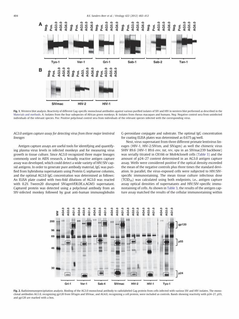
Mousemonoclonal antibodies againstwhole disrupted SIVagmTYO-1were produced using a standard mouse-cell hybridoma technique.Hybridomas were screened for reactivity to SIVagmTYO-1 by wholevirus ELISA. The specificity of positive hybridomas was further evaluatedby immunoblotting and three hybridomas were selected, whose super-natants detected SIV Gag p27. The hybridomas were cloned by singlecell endpoint dilution and were designated AG3.0, AG5.0, and AG6.0.All three monoclonal antibodies were determined to be of the immuno-globulin G type 1 (IgG1) isotype. As shown in Fig. 1, AG3.0 showed thebroadest reactivity by immunoblotting and detected Gag p24–27 fromall four SIVagm isolates (ver, gri, sab, tan) as well as from SIVmac, HIV-2 and HIV-1, AG5.0 reacted with all SIVagm subtypes, and HIV-1 andHIV-2 Gag proteins, but not with SIVmac Gag while AG6.0 only reactedwith SIVagmGag from vervet and tantalusmonkeys. Additionally, radio-immunoprecipitation analysis (RIPA) was performed with both cell andvirus lysates (Fig. 2). RIPA differs from immunoblot analysis in that radi-olabeled native proteins in solution react with antibody whereas inimmunoblotting viral proteins aremostly denatured due to the presenceof sodiumdodecyl sulfate (SDS). Themonoclonal antibodyAG3.0 reactedwith p24–27 and the Gag precursor proteins p38, p55, and p160 fromSIV- and HIV-infected cell lysates. Monoclonal antibody AG1.0, recogniz-ing gp120 from SIVagm and SIVmac, and antibody AG4.0, an anti-cellularantibody, served as controls. Overall, AG3.0 was the most broadly cross-reacting monoclonal antibody recognizing the Gag capsid and precursorproducts of at least three different primate lentivirus lineages.
AG3.0 reacts with a 7-mer Gag peptide that is conserved between severallentivirus lineages
In order to determine the epitope recognized by AG3.0, we initiallyused a series of overlapping Gag p26 peptides provided by the EU AIDSVaccine Integrated Project's Programme EVA for epitope mapping.Twenty-two 20-mer peptides with a 10 amino acid overlap were testedfor reactivity with AG3.0 (Table 1). AG3.0 reacted with a single peptidespanning amino acid 11 to 30 from SIVmac251Gag (HLPLSPRTLNAWVKLIEEKK), but not with any of the surrounding peptides, indicating thatthe minimal AG3.0 epitope is located in the center of the peptide at theN-terminus of Gag capsid. Subsequent fine mapping using 13-mers with12-mer overlaps covering the sequence GGNYVHLPLSPRTLNAWVKLIEEKK (SIVmac251Gag aa 141–165) revealed a minimal epitope withthe sequence SPRTLNA. This was concluded from the fact that two ofthe peptides missing one amino acid at the beginning or the end of theepitope (NYVHLPLSPRTLN and PRTLNAVWKLIEE, Fig. 3) were not recog-nized by AG3.0. A sequence alignment between representatives of tenprimate lentiviral lineages is shown in Table 2A and demonstrates thatthe epitope SPRTLNA is conserved between most SIVcpz, SIVsm, SIVagm,and SIVrcm related viruses. Only theHIV-1 Ngroup and SIVcpz-ANTdifferby one amino acid at the N-terminus (threonine instead of serine). Thispeptide also differs in the following viruses (conserved aminoacids underlined): SIVlho (SPRTIQT), SIVsun (SPRTVQT), SIVmndGB1(SPRIIQT), SIVcol (SPRTLGA), SIVsyk (PVRTLKT), SIVmon (EPRLLKT),SIVmus-1 (NARILKT), SIVmus-2 (SPRLLKT), SIVgsn (NSRILKT), SIVdeb(SPRIVKT), SIVden (STRVLKT), and SIVtal (SPRLLKT). Generally, theN-terminus of the epitope was more conserved than the C-terminusamong the ten primate lentivirus lineages. The only conserved aminoacid among all simian and human immunodeficiency viruses describedso far was arginine in position 3.
Neg
.
Po
s.
AG
3.0
AG
5.0
AG
6.0
Neg
.
Po
s.
AG
3.0
AG
5.0
AG
6.0
Neg
.
Po
s.
AG
3.0
AG
5.0
AG
6.0
Neg
.
Po
s.A
G3.
0
AG
5.0
AG
6.0
Neg
.
Po
s.
AG
3.0
AG
5.0
AG
6.0
Neg
.
Po
s.
AG
3.0
AG
5.0
AG
6.0
Neg
.P
os.
AG
3.0
AG
5.0
AG
6.0
Neg
.
Po
s.A
G3.
AG
5.0
AG
6.0
Neg
.P
os.
AG
3.0
AG
5.0
AG
6.0
Gri-1Tyo-1 Ver-1 Sab-1 Sab-2 Tan-1
SIVmac HIV-2 HIV-1
A
B
Fig. 1.Western blot analysis. Reactivity of different Gag-specific monoclonal antibodies against various purified isolates of SIV and HIV in western blot performed as described in theMaterials and methods. A: Isolates from the four subspecies of African green monkeys. B: Isolates from rhesus macaques and humans. Neg: Negative control sera from uninfectedindividuals of the relevant species. Pos: Positive polyclonal control sera from individuals of the relevant species infected with the corresponding virus.
404 B.E. Sanders-Beer et al. / Virology 422 (2012) 402–412
AG3.0 antigen capture assay for detecting virus from three major lentivirallineages
Antigen capture assays are useful tools for identifying and quantify-ing plasma virus levels in infected monkeys and for measuring virusgrowth in tissue culture. Since AG3.0 recognized three major lineagescommonly used in AIDS research, a broadly reactive antigen captureassay was developed, which could detect a wide variety of HIV/SIV cap-sid antigens. In order to generate pure antibody material, IgG was puri-fied from hybridoma supernatants using Protein G sepharose columns,and the optimal AG3.0 IgG concentration was determined as follows:An ELISA plate coated with two-fold dilutions of AG3.0 was reactedwith 0.2% Tween20 disrupted SIVagmVER.DE.x.AGM3 supernatant.Captured protein was detected using a polyclonal antibody from anSIV-infected monkey followed by goat anti-human immunoglobulin
MW
AG
4.0
AG
3.0
AG
1.0
AG
4.0
AG
3.0
AG
1.0
AG
4.0
AG
3.0
AG
1.0
AG
4.0
AG
3.0
Gri-1 Ver-1 Sab-4 SIVm
200
96
69
46
30
Fig. 2. Radioimmunoprecipitation analysis. Binding of the AG3.0 monoclonal antibody to radclonal antibodies AG1.0, recognizing gp120 from SIVagm and SIVmac, and AG4.0, recognizinand gp120 are marked with a box.
G-peroxidase conjugate and substrate. The optimal IgG concentrationfor coating ELISA plates was determined as 0.675 μg/well.
Next, virus supernatant from three different primate lentivirus lin-eages (HIV-1, HIV-2/SIVsm, and SIVagm) as well the chimeric virusSHIV 89.6 (HIV-1 89.6 env, tat, rev, vpu in an SIVmac239 backbone)was serially titrated in C8166 or Molt4clone8 cells (Table 3) and theamount of p24–27 content determined in an AG3.0 antigen captureassay. Wells were considered positive if the optical density exceededthe mean of the negative controls plus three times the standard devi-ation. In parallel, the virus-exposed cells were subjected to HIV/SIV-specific immunostaining. The mean tissue culture infectious dose(TCID50) was calculated using both endpoints, i.e., antigen captureassay optical densities of supernatants and HIV/SIV-specific immu-nostaining of cells. As shown in Table 3, the results of the antigen cap-ture assay matched the results of the cellular immunostaining within
AG
1.0
AG
4.0
AG
3.0
AG
1.0
AG
4.0
AG
3.0
AG
1.0
AG
4.0
AG
3.0
AG
1.0
ac HIV-2 HIV-1 Tyo-1
gp120
p55
p24
iolabeled Gag protein from cells infected with various SIV and HIV isolates. The mono-g a cell protein, were included as controls. Bands showing reactivity with p24–27, p55,

Table 1Rough epitope mapping of AG3.0.
SIVmac251 Gag Capsid Sequence
001 PVQQIGGNYV HLPLSPRTLN AWVKLIEEKK FGAEVVPGFQ ALSEGCTPYD051 INQMLNCVGD HQAAMQIIRD IINEEAADWD LQHPQPAPQQ GQLREPSGSD101 IAGTTSSVDE QIQWMYRQQN PIPVGNIYRR WIQLGLQKCV RMYNPTNILD151 VKQGPKEPFQ SYVDRFYKSL RAEQTDAAVK NWMTQTLLIQ NANPDCKLVL201 KLGLVNPTLE EMLTACQGVG GPGQKARL
Peptidea Sequence Mean OD StDev
1 01–20 0.01 0.002 11–30 0.37 0.053 21–40 0.01 0.004 31–50 0.01 0.005 41–60 0.01 0.006 51–70 0.01 0.007 61–80 0.01 0.008 71–90 0.01 0.009 81–100 0.01 0.0010 91–110 0.01 0.0011 101–120 0.01 0.0012 111–130 0.01 0.0013 121–140 0.01 0.0014 131–150 0.01 0.0015 141–160 0.01 0.0016 151–170 0.01 0.0017 161–180 0.01 0.0018 171–190 0.01 0.0019 181–200 0.01 0.0020 191–210 0.01 0.0021 201–220 0.01 0.0022 211–228 0.01 0.00
a Peptides were ordered from the European AIDS Reagent Programme, referenceARP714.1-22; peptides 15, 16, and 18 have an additional N-terminal cysteine.
GGNYVHLPLSPRT1
GNYVHLPLSPRTL2
NYVHLPLSPRTLN3
YVHLPLSPRTLNA4
VHLPLSPRTLNAW5
HLPLSPRTLNAWV6
LPLSPRTLNAWVK7
PLSPRTLNAWVKL8
LSPRTLNAWVKLI9
SPRTLNAWVKLIE10
PRTLNAWVKLIEE11
RTLNAWVKLIEEK12
TLNAWVKLIEEKK13
A B
Fig. 3. Fine epitope mapping of SIVmac Gag. Binding of the AG3.0 monoclonal antibodyto synthetic peptides spanning the putative epitope in Gag within the sequenceGGNYVHLPLSPRTLNAWVKLIEEKK (SIVmac251 Gag aa 141–165). The peptides, 13-merswith a 12 amino acid overlap were synthesized directly onto a cellulose membrane andbinding of AG3.0 visualized by enhanced chemiluminescence (A). A density plot alongthe membrane was carried out to aid analysis or reactivity (B).
405B.E. Sanders-Beer et al. / Virology 422 (2012) 402–412
the expected range of assay variability. These results suggested thatthe antigen capture assay is equivalent to immunostaining as end-point analysis for virus titrations and virus neutralization assays.
Third, the reactivity of AG3.0 to HIVs and SIVs from five primatelentivirus lineages was tested by antigen capture ELISA. As shown inFig. 4, the AG3.0-based antigen capture assay was able to detect Gagcapsid from HIV-1, HIV-2, SIVmac, SIVagm, and SIVrcm, but notfrom SIVsun, SIVlho, and SIVsyk. For this experiment, two-fold dilu-tions of virus supernatants were assayed, and the optical densitiesdecreased proportionally to the virus dilutions. At about a 100-folddilution of supernatant, the signal became undetectable. Overall, thecross-reactivity of the AG3.0 antibody was consistent with theamino acid conservation of the Gag epitope (see Table 2A). Asdescribed above and shown in Table 2A, the epitope SPRTLNA ismostly conserved among the HIV-1/SIVcpz, HIV-2/SIVsmm, andSIVagm lineages, and SIVrcm. In SIVsyk, SIVsun, and SIVlho, it differsby 3–4 amino acids, which prohibit binding to the monoclonal anti-body. Viruses from the SIVdeb, SIVgsn/mon/mus, and SIVtal lineageswere not tested, but the epitope differs by 2–5 amino acids fromSPRTLNA, and it would be unlikely that the AG3.0 antibody cross-reacts with Gag from these viruses. In contrast, the conservation of theepitope in SIVdrl and SIVmnd-2, which are phylogenetically related toSIVrcm. likely predicts cross-reactivity.
Affinity determination of the interaction between AG3.0 and Gag proteinsfrom SIVagm and SIVmac
Surface plasmon resonance (SPR) technology was used to evaluatethe binding affinity of the monoclonal antibody AG3.0 to different SIVGag proteins. Although the calculated maximal response (Rmax) was100 response units (RU), the observed Rmax was more than double,likely due to multimerization of the Gag protein (data not shown).The highest affinity was obtained using SIVagmVer (3.7 nM) whilethe affinities for SIVmac Gag and SIVagmSab Gag were lower but sim-ilar with 20 nM and 35 nM, respectively (Fig. 5A). Interestingly, while
the dissociation of SIVagmVer Gag and SIVmac Gag was fairly rapid,the binding between the AG3.0 mAb and SIVagmSab Gag was foundto be very stable, with very little dissociation detectable (Fig. 5B).
Sequencing of the AG3.0 antigen binding site
cDNAs generated from RNA coding for the variable fragments of theAG3.0 heavy and light chains were sequenced and analyzed using theVbase2 application (http://www.vbase2.org) to identify the conservedframework regions (FR1-FR4) andvariable complementarity determiningregions (CDR1–CDR3) according to the International ImMunoGeneTics(IMGT) nomenclature (Fig. 6). The three variable heavy chain regionswere found to consist of 8, 8 and 16 amino acids for CDR1, CDR2 andCDR3, respectively, whereas the light chain had corresponding CDRlengths of 11, 3, and 9.
Discussion
Monoclonal antibodies against SIVagm Gag p27 were developed toallow the Gag capsid expression in African greenmonkeys and in patho-genic macaque models of SIV infection to be compared. Gag is an impor-tant structural protein and is often referred to as a precursor because it issubject to cleavage by the viral protease, which yields the internal struc-tural proteins of the mature virion (Freed, 1998; Swanstrom and Wills,1997; Vogt, 1997; Wills and Craven, 1991). Three of the Gag cleavageproducts, matrix, capsid, and nucleocapsid, are common to all retrovi-ruses and are always arranged in this order within the Gag precursor,with matrix being at the N-terminus. Additionally, the Gag precursors

Table 2AAlignment of Gag capsid N-terminus (B.FR.83.HXB2_LAI_IIIB aa 160–176).
Table 3Comparison of TCID50/ml calculations using AG3.0 antigen capture assay or HIV/SIV-specific immunostaining.
Virus Titrated in Log10 Titer (TCID50/ml)
Endpoint analysis
AG3.0 antigen captureassay of supernatants
HIV/SIV-specificimmunostainingof cells
SHIV 89.6 C8166 cells 5.52 5.64HIV-2 UC3 C8166 cells 4.99 4.79SIVsmmPBj 1.9 C8166 cells 4.87 5.04SIVagm3 C8166 cells 6.54 6.48SIVagm3 Molt4clone8 cells 4.81 4.63SIVmac 32H C8166 cells 6.71 6.71HIV-1 LAI C8166 cells 4.69 4.39
406 B.E. Sanders-Beer et al. / Virology 422 (2012) 402–412
of HIV and SIV possess a C-terminal domain termed p6 that is unique toprimate lentiviruses, as well as two “spacer” regions that separate capsidfrom nucleocapsid, and nucleocapsid from p6 (Henderson et al., 1992;Mervis et al., 1988).
Capsid, which directly follows matrix in the Gag precursor, has cru-cial roles in particle assembly and after entry into a new target cell.However, the function of capsid in the early phase of the replicationcycle is not well understood. In the mature virion, capsid forms theshell of the core, which is occasionally tubular but most often conical,a feature that distinguishes lentiviruses, such as HIV-1, from mostother retroviruses (Gelderblom, 1991). Capsid has two predominantlyα-helical domains that are connected through a flexible linker region
Table 2BAlignment of amino acid sequence adjacent to SPRTLNA.
VER . KE . x . TYO1 AQQQGNAWVHVPLSPRTLNAWVKAVEEKKFGAEIVPMVER . DE . x . AGM3 --------I--SPRTLNA----------------SAB . SN . x . SAB1C IVSVN-Q---Q-SPRTLNA---VI-----S--V---MAC . US . x . 239 V--I-GNY--L-SPRTLNA---LI--------V--G
(Gamble et al., 1997; Gitti et al., 1996;Momany et al., 1996) and has dif-ferent roles in virus morphogenesis. The N-terminal domain, whichcomprises two thirds of HIV-1 capsid, is required for the formation ofthe mature core but is dispensable for the assembly of immature virusparticles (Borsetti et al., 1998; Dorfman et al., 1994; Reicin et al., 1995,1996; Srinivasakumar et al., 1995;Wang and Barklis, 1993). In contrast,the C-terminal capsid domain is crucial both for particle assembly andfor core formation (Dorfman et al., 1994; Mammano et al., 1994;McDermott et al., 1996; Reicin et al., 1995). The N-terminal domain ofHIV-1 capsid interacts with the human peptidyl-prolyl cis-trans isomer-ase cyclophilin A (CyPA), which leads to the specific incorporation of thisubiquitous cytosolic host protein into virions (Franke et al., 1994; Lubanet al., 1993; Thali et al., 1994). This feature is unique for HIV-1 and absentin other primate lentiviruses andHIV-2. Based on the regular appearanceof the synthetic cones, Sundquist et al. propose that retroviral cores arecomposed of hexagonal lattices that are closed through the incorporationof a total of 12 pentagons (Ganser et al., 2003).
In the study presented here, three monoclonal antibodies againstSIVagm from the vervet subspecies (SIVagmTYO-1) were generatedand characterized. AG6.0 cross-reacted with two subspecies of Africangreenmonkeys (vervet and tantalus), whereas AG5.0 showed a broadercross-reactivity, reacting with SIVagm Gag p26 from vervet, grivet,sabaeus, and tantalus, HIV-1 and HIV-2. Interestingly, although HIV-2and SIVmac have similar ancestries, having both arisen by transmissionfrom sooty mangabeys (SIVsm), only the p26 from HIV-2ST was recog-nized. AG3.0 showed the broadest cross-reactivity and recognized notonly the SIVagm lineage, but also members of the HIV-2 lineage (HIV-2, SIVmac), HIV-1 lineage, and SIVrcm. In addition, AG3.0 reacted withthe Gag precursors p55 and p38 and the Gag-Pol precursor in infectedcell lysates. This broadly cross-reactive antibody (AG3.0) is availablefrom the NIH ARRRP as “Monoclonal antibody to HIV-1 p24” under cat-alog no. 4121 (Simm et al., 1995). In addition, the AG3.0 producing hy-bridoma cells are also available. A number of monoclonal antibodiesspecific for HIV and SIV Gag are deposited in the NIH ARRRP but, withthe exception of AG3.0, none reacts with SIVagm or is broadly cross-reactive with SIV and HIV. So far, only Otteken et al. (1992) successfullyproduced monoclonal antibodies to SIVagm Gag p27 (strain TYO-7).However, these monoclonal antibodies (1.17.3, 1A7 and 1F6) are pri-marily type-specific, reacting with only certain types of SIVagm andtheHIV-2 lineage, but notwith HIV-1 or SIVmndGB1. Thesemonoclonalantibodies mapped to a peptide spanning amino acids 152 to 172 inSIVmac251, which is conserved in the HIV-2 lineage, but not inSIVagmTYO-1, HIV-1 IIIB, and SIVmndGB1. One particular monoclonalantibody, FM18, generated against SIVmac, also reacted with SIVagm,but not with HIV-1 (Lairmore et al., 1993). Since this antibody has notbeen mapped, the full extent of cross-reactivity is unknown. However,our broadest reactive monoclonal antibody, AG3.0, is able to detectp24–27 from all of the main primate lentivirus lineages including HIV-1, SIVmac/HIV-2, SIVagm, and SIVrcm.

SIVsun.GA.98.L14
0.0
0.5
1.0
1.5
2.0
2.5
3.0
3.5
SIVlho.LST.CD.88.447
SIVsyk.KE.x.SYK173
0.0
0.5
1.0
1.5
2.0
2.5
3.0 SIVrcm.NG.x.NG411
HIV-2.A.CI.88.UC1
0.0
0.5
1.0
1.5
2.0
2.5
3.0 SIVagm.VER.DE.x.AGM3
HIV-1.DE.HAN-2
0.0
0.5
1.0
1.5
2.0
2.5
3.0
1 10 100 1000
SIVmac.GB.x.251_32
1 10 100 1000 10000
AG3.0 p24-27 ELISA
Supernatant Dilution
OD
492/577
Fig. 4. Antigen capture ELISA using various isolates. Supernatants from cells infected with various isolates of SIV and HIV were tested at different dilutions for reactivity in the AG3.0based antigen capture assay as described in the material and methods.
407B.E. Sanders-Beer et al. / Virology 422 (2012) 402–412
Rough and fine mapping revealed the minimal epitope recognizedby AG3.0 to be SPRTLNA (Table 1, Fig. 3). This epitope is conservedthroughout the HIV-1 (including SIVrcm), HIV-2 and SIVagm lineages,and is partially conserved throughout the other SIV lineages (SIVlho,SIVsyk, SIVcol, SIVdeb, SIVtal, SIVgsn), although it is completely absentin non-primate lentiviruses. However, despite this partial conservation,AG3.0 does not recognize SIVlho, SIVsun, or SIVsyk Gag, indirectly con-firming theminimal epitope needed for binding (Fig. 4). Von Schwedleret al. (2003) showed that viruses with a mutated arginine and aspara-gine in the SPRTLNA motif failed to form a cone-shaped core and werenot infectious, indirectly demonstrating the importance of this epitopefor viral assembly. The Los Alamos Immunology database contains 21HIV-1 cytotoxic T-lymphocyte (CTL) epitopes restricted by HLA-A, -B,and -C haplotypes spanning the SPRTLNA peptide (http://www.hiv.lanl.gov/content/immunology/ctl_search) suggesting immunogenicdominance of the region. For example, a dominant CTL epitope ISPRTL-NAWhas beendetected inHIV-1 positive individuals having the protec-tive haplotype HLA B*5701 (Bailey et al., 2006), which is found in a veryhigh frequency in HIV-1 infected non-progressors (11/13 or 85% versus19/200 or 9.5% of progressors). Non-progressors tended to have an im-mune response that was highly focused on four p24 epitopes that werepresented by B*5701, ISPRTLNAW, KAFSPEVIPMF, TSTLQEQIGW, andQASQEVKNW (Migueles and Connors, 2001). THE SPRTLNA epitope isalso part of three human CD4+ T helper cell HIV-1 epitopes and twomouse monoclonal antibody HIV-1 epitopes (AISPRTLNAW for the F5-2antibody and VHQAISPRTLNAWVK for the ID8F6 antibody) (http://www.hiv.lanl.gov/content/immunology/ctl_search).
After mapping the AG3.0 epitope, we sought to develop an antigencapture ELISA using the AG3.0 monoclonal antibody since it showedthe broadest cross-reactivity against HIV/SIV (Binninger-Schinzel
et al., 2008). Currently, there is only one commercially available anti-gen capture assay that cross-reacts with SIVmac, SIVagm, SIVdrl, andSIVmnd-2 (from ZeptoMetrix Corp.) (Hu et al., 2003; Pandrea et al.,2006). However this assay is expensive and the epitope recognizedby themonoclonal antibody that forms the basis of the test is not publicknowledge. Therefore, the degree of cross-reactivity with different HIV/SIV isolates can only be determined empirically. Most in-house antigencapture assays using monoclonal antibodies developed to date are ei-ther optimized for the HIV-1/SIVcpz or HIV-2/SIVmac/SIVsm lineage(Lohman et al., 1991; Thorstensson et al., 1991; Wehrly and Chesebro,1997). Only one assay, based on a polyclonal sandwich system, wasalso able to detect SIVmnd to an appreciable degree (Beirnaert et al.,1998). Our AG3.0 in-house antigen capture assay had a lower limit ofdetection of 100 pg/ml, comparable to that of Zeptometrix's SIV p27 an-tigen ELISA (62.5 pg/ml). Optimization of the reagents used in theAG3.0 antigen capture assay should further enhance its sensitivity.
The binding affinity of AG3.0 to SIVagm and SIVmac Gag purifiedfrom native virus was determined using SPR technology. SIVagmSab2Gag was found to bind slowly to AG3.0, whereas the overall bindingaffinity was more similar to SIVmac than to SIVagmVer. In addition,the dissociation curve was markedly different from both SIVmac andSIVagmVer and indicated a very stable complex between SIVagmSab2and AG3.0. The same results were obtained with two different Gag con-centrations (data not shown). Although all three Gag proteins share thesame minimal epitope, the binding structures could be influenced byresidues flanking the SPRTLNA epitope, resulting in different tertiarystructures of the whole Gag protein and therefore different binding af-finities to AG3.0 (Table 2B). To our knowledge, crystallization of theSIV capsid protein, needed to address this hypothesis, has not yet beenachieved. Finally, the AG3.0 monoclonal antibody RNA was sequenced

-100
-80
-60
-40
-20
0
20
1000 1500 2000 2500 3000 3500 4000 4500Time (s)
Res
pons
e (R
U) SIVagmSab2
SIVmac239
SIVagmVer
A. Binding Affinity of SIVagmVer Gag, SIVagmSab2 Gag and SIVmac Gag
B. Adjusted dissociation curves of the three Gag proteins
Fig. 5. Surface plasmon resonance (SPR) analysis. A: Binding affinity profiles of Gag proteins derived from different SIVs to AG3.0 measured by BIAcore as described in the materialsand methods. Sensor chips coated in AG3.0 were exposed to purified preparations of the different Gag proteins at various concentrations. Binding was measured as a change in RU atequilibrium. The apparent kD values were determined using the BiaEvaluation software (GE Healthcare). B: Dissociation curves for the different Gag proteins (1.5 μg/ml) using aninjection period of 1080 s, a dissociation time of 3000 s, and a flow rate of 5 μl/min.
408 B.E. Sanders-Beer et al. / Virology 422 (2012) 402–412
and annotated to facilitate creating a human- ormonkey-adaptedmono-clonal antibody specific for the SPRTLNA epitope. This would allow anti-capsid antibodies for modulation of SIV/HIV infection to be evaluated.
In summary, we generated three mouse monoclonal antibodiesagainst SIVagmTYO-1 (Ver) Gag capsid, each with varying degrees ofcross-reactivity to SIV/HIV. The broadest cross-reactivity was found withAG3.0, which recognized p24–27 from the threemajor primate lentiviruslineages (HIV-1/SIVcpz/SIVrcm, HIV-2/SIVsm/SIVmac, and SIVagm) andmapped to a conserved 7 amino acid motif in Gag, SPRTLNA (aa156–162 of SIVagmTYO-1 Gag). An antigen capture assay was devel-oped, which could detect as little as 100 pg/ml of p24 Gag.
Materials and methods
Monoclonal antibody production
BALB/c mice were immunized by intraperitoneal injection of 10 μgsucrose gradient purified SIVagm(TYO-1) in phosphate buffered sa-line (days 0 and 21) in addition to 10 μg of 1% Triton X-100-treated
SIVagm(TYO-1) (day 14). The mice were sacrificed at day 24 and thespleen removed for fusion of splenic cells with SP-2 mouse myelomacells. The hybridomas were grown in 96-well plates using DMEM (LifeTechnologies) supplemented with 20 mM 4-(2-hydroxyethyl)-1-piperazineethanesulfonic acid (HEPES), 20% fetal bovine serum, with10 mM hypoxanthine and 2 μg/ml of azaserine (Sigma-Aldrich) forselection. Supernatant fluid (100 μl) from each well showing visiblehybridoma growth was evaluated for reactivity to SIVagm(TYO-1) bywhole virus ELISA and positive samples screened by western blotting.Clones of interest were subcloned twice. The isotype of each monoclo-nal antibody was determined with the Sigma ImmunoType™ Kitaccording to the manufacturer's instructions.
Virus purification
Culture supernatant fluid (5–10 l) from SIVagm(TYO-1) infectedMOLT4clone8 cells was concentrated 50–100 fold with a Minitan ul-trafiltration system (Millipore) containing 5 filters with a 300,000molecular weight pore size. The retentate (50–100 ml) was layered

Fig. 6. Amino acid sequences of the heavy and light chains of the AG3.0 monoclonal an-tibody. The four conserved framework regions (FR1-FR4) and the three hypervariablecomplementarity determining regions (CDR1–CDR3) are displayed according to theIMGT nomenclature (Lefranc, 2007). Unoccupied amino acid positions are markedwith an en dash (–). X=positions in the conserved FR1 region of the heavy chain, atwhich the identity of the amino acid could not be unequivocally determined fromthe sequence.
409B.E. Sanders-Beer et al. / Virology 422 (2012) 402–412
onto 20/70% discontinuous sucrose gradients, and the virus was re-covered from the interface after centrifugation for 2 h at 71,500×g.The virus fraction was diluted 2× with TEN (0.05 M Tris–HCl pH 7.2,0.15 M NaCl, and 1 mM Ethylenediaminetetraacetic acid (EDTA)),layered over a 20–70% continuous sucrose gradient and centrifugedovernight at 71,500×g. One milliliter fractions were assessed with arefractometer, the virus fractions (1.16–1.18 g/cm3) diluted 5-fold withTEN buffer, and the virus was pelleted by centrifugation at 71,500×gfor 2 h at 4 °C. The virus pellets were resuspended in a volume of1.0 ml phosphate buffered saline (PBS), and the protein concentrationwas determined using the Bio-Rad Protein Assay according to themanu-facturer's instructions (Bio-Rad Life Sciences). The virus was further an-alyzed for purity by SDS-PAGE with the protein bands visualized byimmunoblotting.
Whole virus ELISA
Purified SIVagm(TYO-1) was diluted to 2 μg/ml in borate bufferedsaline (BBS) and used to coat Immulon II plates (Dynex TechnologiesInc., Chantilly, VA) overnight at 4 °C. Wells were blocked with 10% nor-mal goat serum in BBS for 30 min at 37 °C, and the plates washed 3times with 1% Tween20 in phosphate buffered saline. One hundredmi-croliters of hybridoma supernatant fluidwas then added, and the plateswere incubated for 60 min at 37 °C, andwashed three times prior to ad-dition of biotinylated anti-mouse antibody. Binding was determinedcolorimetrically by incubation with streptavidin-horseradish peroxi-dase (Amersham) before washing and developing with 2,2′-Azinobis(3-ethylbenzthiazoline-sulfonic acid) (ABTS).
Cells and viruses
All human CD4+ T cell lines were maintained in complete mediumconsisting of RPMI 1640 supplemented with 15% fetal bovine serum(Life Technologies), HEPES, 2 mM L-glutamine, 50 μg/ml streptomycinand 50 U/ml of penicillin. SIVagm strains VER.KE.x.TYO1, GRI_677,SAB.SN.x.SAB1C, SAB.SN.x.SAB2, SAB.SN.x.SAB4, TAN.UG.x.TAN1, andHIV-2ST (Broussard et al., 2001; Fomsgaard et al., 1991; Jin et al.,1994; Kumar et al., 1990; Shibata et al., 1990; Soares et al., 1997)were grown in Molt4clone8 cells, SIVagm(Ver-1) (Jin et al., 1994) inSupT1 cells, SIVmac251 in Hut78 cells, and HIV-1/IIIB in Molt3 cells.
Western blot analysis
Western blotting was performed as previously described (Allanet al., 1991). Virus in culture supernatants taken from infected celllines at 48–72 h was purified through a 20% sucrose cushion, separat-ed by SDS-polyacrylamide gel electrophoresis, and then blotted ontonitrocellulose sheets. The nitrocellulose paper was blocked with 3% bo-vine serum albumin and subsequently incubated with a 1:8 dilution ofmonoclonal antibody supernatant fluid or a 1:80 dilution of seroposi-tive and seronegative controls. Viral proteins were detected using thestreptavidin–biotin system (Amersham) with diaminobenzidine asthe substrate for color development.
Radioimmunoprecipitation analysis
Radioimmunoprecipitation analysis (RIPA) of virus lysates wasconducted as described previously (Allan et al., 1992). Infectedcells were metabolically labeled for 16 h with 35-S cysteine andmethionine (Translabel, ICN Biomedicals, Inc., Costa Mesa, CA) at1 mCi/107 cells in cysteine–methionine free RPMI 1640 with 15%FBS. The cells were resuspended in RIPA buffer (0.08 M Tris–HClpH 7.4, 0.15 M NaCl) containing 0.1% SDS, 1% Triton X-100, and 1%deoxycholate with protease inhibitors (1.0 mM phenylmethyl sulfo-nyl fluoride (Pierce) and 2 μg/ml of leupeptin (Sigma-Aldrich)) andcentrifuged at 100,000×g to remove protein aggregates. Proteinswere then immunoprecipitated with monoclonal antibodies bound toprotein A sepharose. The complexes were washed 5 times with com-plete RIPA lysing buffer, the viral proteins solubilized by resuspensionin electrophoresis sample buffer (0.08 M Tris–HCl pH 6.8, 2% SDS,0.1 M dithiothreitol, 10% glycerol, and 1 mg/ml of bromophenol blue),and the viral proteins separated by 11.3% SDS-PAGE. Proteins werevisualized by impregnating the gel with EnHance (DuPont NEN, Boston,MA) before drying and exposure to Kodak SB5 radiographic film(Eastman Kodak).
Epitope mapping
A panel of overlapping SIVmac Gag synthetic peptides (20-mers,10 overlap; kindly provided by the European AIDS Reagent Pro-gramme, reference ARP714.1-22) was used in a standard ELISA forrough epitope mapping of the AG3.0 monoclonal antibody. For fineepitope mapping, a series of 13-mer synthetic peptides with an over-lap of 12 aa covering the sequence GGNYVHLPLSPRTLNAWVKLIEEKK(SIVmac251 Gag 141–165) was synthesized directly onto a cellulosemembrane (Jerini Bio Tools GmbH, Berlin, Germany). Briefly, the mem-brane was washed 3 times in Tris-buffered saline containing 0.05%Tween20 (T-TBS) and blocked overnight at 4 °C with T-TBS plus 2%milk powder and 10% sucrose (blocking buffer). After washing 3 timeswith T-TBS, the membrane was incubated overnight at 4 °C with a 1:50dilution of purified AG3.0 mAb in blocking buffer. The membrane wasthen washed and incubated for 1 h at room temperature with a1:10,000 dilution of goat anti-mouse IgG peroxidase conjugate (Sigma-Aldrich). After three final washes, spots were visualized using the

410 B.E. Sanders-Beer et al. / Virology 422 (2012) 402–412
enhanced chemiluminescence kit (Amersham), followed by exposure tophotographic film (RPN 2103H Hyperfilm, Amersham).
Antigen capture ELISA
Protein G sepharose column (Pharmacia) 4 Fast Flow (25 ml) wasused to purify IgG from cell culture supernatants according to themanufacturer's instructions. For the antigen capture ELISA, 50 μl of a13.5 μg/ml AG3.0 solution in distilled water was dried onto 96-wellflat-bottom polystyrene assay plates (PRO BIND, BD Biosciences)overnight at 37 °C. The next day, the monoclonal antibody layer wasblocked with 100 μl PBS/2% nonfat milk powder for 1 h and subse-quently washed 3 times with PBS/0.05% Tween20. SIV/HIV superna-tant or SIV/HIV infected cells were lysed with 0.2% Tween20 for30 min., 100 μl lysate plus 50 μl PBS/2% nonfat milk powder/0.05%Tween20 added to the AG3.0 coated wells, and the plates incubatedovernight at 4 °C and the next day for 2 h at 37 °C. The plates werewashed 3 times with PBS/0.05% Tween20, 50 μl of specific anti-SIVplasma from an infected monkey or plasma pool from HIV-1 infectedindividuals, diluted 1:1000 in PBS/2% nonfat milk powder/0.05%Tween20, were added to the wells, and the plates incubated at37 °C for 1 h. The plates were then washed 5 times with PBS/0.05%Tween20 and under the stream of tab water for five seconds. Finally,10 μg o-phenylenediaminedihydrochloride (Sigma-Aldrich) in 50 μlPBS pH 6.0 and 0.012 μl H2O2 were added as substrate, and the reac-tion stopped with 20 μl 2.5 M H2SO4. The optical density of eachwell was read at 492 nm using a microplate reader.
Immunostaining of cells
Flat bottom 96-well plates were incubated with 5 μg of poly-L-lysine (Sigma) in 100 μl of distilled water overnight at 4 °C. Plateswere washed twice with PBS, and 200 μl of cell suspension wasadded to the wells. Plates were incubated for 1 h at 37 °C to allowcells to settle, and then the supernatant was removed. Plates weresubmerged in −20 °C methanol and fixed for 30 min. at −20 °Cbefore carefully washing three times with PBS and blocking with100 μl of PBS/2% nonfat milk powder for 1 h at room temperature.The blocking solution was then removed, and 50 μl of the appropri-ate anti-SIV/HIV plasma diluted in PBS/2% nonfat milk powderadded for 1 h at 37 °C. Wells were washed five times with PBS andincubated with 50 μl of 1:500 anti-human IgG (Sigma-Aldrich) inPBS/2% nonfat milk powder for 1 h at 37 °C. Wells were then washedagain five times with PBS and finally the substrate added (10 μg 3-amino-9-ethylcarbazol (Sigma-Aldrich), 2.5 μl dimethylformamide,47.5 μl 20 mM sodium acetate, pH 5.0, and 0.025 μl H2O2 per well).After 10 to 20 min of incubation with the substrate, SIV/HIV infectedcells were stained red. The wells were washed once with PBS, andthe substrate reaction stopped with 20% glycerol.
Virus titrations
To determine the TCID50 of virus stocks, supernatants were initiallydiluted 1:10 and then 11 dilutions steps with 1:3 dilutions wereperformed. Row 12 served as negative control (medium only). Fiftymicroliters of virus dilutions or media were added to 2×10³ C8166 orMolt4clone8 cells in 8 replicates. The plates were incubated for 7 days,and the SIV/HIV replication was monitored by antigen capture assayor cellular immunostaining.
Surface plasmon resonance (SPR) analysis
To determine the binding affinity of Gag proteins derived from thedifferent SIVs to the monoclonal AG3.0 mAb, samples were analyzedin duplicate with a BIAcore X100 instrument (GE Healthcare) at25 °C in HBS-EP+buffer (10 mM Hepes, 150 mM NaCl, 3 mM EDTA,
0.05% Surfactant P20, pH 7.4) using the mouse antibody capture kit(GE Healthcare). The final immobilization level of anti-mouse IgGantibody was 8500 RU on both flow cells (Fc1 and Fc2) as recom-mended by the manufacturer. The antibody was captured at a finalconcentration of 20 μg/ml for 20 s at a flow rate of 10 μl/min and aconstant level of 500 RU and 120 RU for measuring SIV Gag binding.The different analytes were injected under the following conditions:The association/dissociation times for SIVagmVer Gag and SIVmacGag were 720/1200 s and for SIVagmSab Gag 1080/5400 s. Further-more, association and dissociation curves were estimated for all 3Gag proteins using an injection period of 1080 s and a dissociationtime of 3000 s at a flow rate of 5 μl/min. The regeneration bufferconsisted of 10 mM glycine/HCl, pH 1.7. The apparent kD valueswere determined using the BiaEvaluation software (GE Healthcare).All three Gag preparations (SIVagmVer, SIVagmSab, and SIVmac)were produced from native purified virus.
Sequencing of the AG3.0 heavy and light chain variable regions
RNA was isolated from 1×10⁷ AG3.0 hybridoma cells using theRNeasy plus kit (Qiagen) according to the manufacturer's instructionsand eluted in 30 μl DEPC-treated H2O. cDNA was generated with theCloned AMV cDNA synthesis kit (Invitrogen) using oligodT primersand a temperature of 50 °C for 1 h. Amplification of variable regionfragments was performed with 1.25U Pfu DNA polymerase (Fermen-tas) in the supplied PCR buffer containing 2 mM MgSO4, 0.2 mMdNTPs, and 25 pmol of each primer per 50 μl reaction. Forward andreverse primers from a mouse IgG library primer set (Progen) wereused in all possible combinations for light and heavy chain fragments.Amplification cycles were as follows: initial denaturation for 5 min at95 °C, followed by 35 cycles of 30 s/95 °C, 30 s/55 °C, 60 s/72 °C and afinal elongation of 10 min at 72 °C. Amplified PCR products of approx-imately 400 kb were purified using a PCR purification kit (Fermentas)or, if more than one product was generated, separated on a 1.5% aga-rose gel and subsequently extracted using a gel extraction kit(Qiagen). Sequencing was performed on an ABI 3500DX sequencer.Sequences obtained were analyzed using Vbase2 (http://www.vbase2.org) to identify the complementarity determining regions ofthe heavy and light chain variable fragments.
Acknowledgments
This paper is dedicated to the memory of Jonathan S. Allan whogenerated and initially characterized the AG3.0 monoclonal antibodyin his laboratory. We thank Evelyn M. Whitehead, Manuela Schütze,and Nicole Norley for their excellent technical assistance. We wouldalso like to thank Phyllis Kanki and Soulemayne M'Boup for theHIV-2 serum, and the U.S. Air Force Wilford Hall AIDS program forthe HIV-1 serum. This work was funded in part from NIH grants AI-28273 and AI-41396 to JSA.
Nucleotide sequence accession numbersThe DNA sequence for AG3.0 has been submitted to GenBank under
accession numbers JN252707 (heavy chain CDR) and JN252708 (lightchain CDR).
References
Aghokeng, A.F., Bailes, E., Loul, S., Courgnaud, V., Mpoudi-Ngolle, E., Sharp, P.M.,Delaporte, E., Peeters, M., 2007. Full-length sequence analysis of SIVmus in wildpopulations of mustached monkeys (Cercopithecus cephus) from Cameroon pro-vides evidence for two co-circulating SIVmus lineages. Virology 360 (2), 407–418.
Aghokeng, A.F., Ayouba, A., Mpoudi-Ngole, E., Loul, S., Liegeois, F., Delaporte, E., Peeters,M., 2010. Extensive survey on the prevalence and genetic diversity of SIVs inprimate bushmeat provides insights into risks for potential new cross-speciestransmissions. Infect. Genet. Evol. 10 (3), 386–396.
Allan, J.S., Short, M., Taylor, M.E., Su, S., Hirsch, V.M., Johnson, P.R., Shaw, G.M., Hahn,B.H., 1991. Species-specific diversity among simian immunodeficiency virusesfrom African green monkeys. J. Virol. 65 (6), 2816–2828.

411B.E. Sanders-Beer et al. / Virology 422 (2012) 402–412
Allan, J.S., Whitehead, E.M., Strout, K., Short, M., Kanda, P., Hart, T.K., Bugelski, P.J., 1992.Strong association of simian immunodeficiency virus (SIVagm) envelope glycopro-tein heterodimers: possible role in receptor-mediated activation. AIDS Res. Hum.Retroviruses 8 (12), 2011–2020.
Bailes, E., Gao, F., Bibollet-Ruche, F., Courgnaud, V., Peeters, M., Marx, P.A., Hahn, B.H.,Sharp, P.M., 2003. Hybrid origin of SIV in chimpanzees. Science 300 (5626), 1713.
Bailey, J.R., Williams, T.M., Siliciano, R.F., Blankson, J.N., 2006. Maintenance of viral suppres-sion in HIV-1-infected HLA-B*57+ elite suppressors despite CTL escape mutations. J.Exp. Med. 203 (5), 1357–1369.
Barre-Sinoussi, F., Chermann, J.C., Rey, F., Nugeyre, M.T., Chamaret, S., Gruest, J., Dauguet,C., Axler-Blin, C., Vezinet-Brun, F., Rouzioux, C., Rozenbaum, W., Montagnier, L.,1983. Isolation of a T-lymphotropic retrovirus from a patient at risk for acquiredimmune deficiency syndrome (AIDS). Science 220 (4599), 868–871.
Beer, B.E., Bailes, E., Goeken, R., Dapolito, G., Coulibaly, C., Norley, S.G., Kurth, R., Gautier,J.P., Gautier-Hion, A., Vallet, D., Sharp, P.M., Hirsch, V.M., 1999. Simian immunodefi-ciency virus (SIV) from sun-tailed monkeys (Cercopithecus solatus): evidence forhost-dependent evolution of SIV within the C. lhoesti superspecies. J. Virol. 73 (9),7734–7744.
Beer, B.E., Foley, B.T., Kuiken, C.L., Tooze, Z., Goeken, R.M., Brown, C.R., Hu, J., St Claire, M.,Korber, B.T., Hirsch, V.M., 2001. Characterization of novel simian immunodeficiencyviruses from red-capped mangabeys from Nigeria (SIVrcmNG409 and -NG411). J.Virol. 75 (24), 12014–12027.
Beirnaert, E., Willems, B., Peeters, M., Bouckaert, A., Heyndrickx, L., Zhong, P.,Vereecken, K., Coppens, S., Davis, D., Ndumbe, P., Janssens, W., van der Groen, G.,1998. Design and evaluation of an in-house HIV-1 (group M and O), SIVmnd andSIVcpz antigen capture assay. J. Virol. Methods 73 (1), 65–70.
Bibollet-Ruche, F., Bailes, E., Gao, F., Pourrut, X., Barlow, K.L., Clewley, J.P., Mwenda, J.M.,Langat, D.K., Chege, G.K., McClure, H.M., Mpoudi-Ngole, E., Delaporte, E., Peeters,M., Shaw, G.M., Sharp, P.M., Hahn, B.H., 2004. New simian immunodeficiencyvirus infecting De Brazza's monkeys (Cercopithecus neglectus): evidence for acercopithecus monkey virus clade. J. Virol. 78 (14), 7748–7762.
Binninger-Schinzel, D., Muller, D., Wolf, T., Krause, B., Meye, B., Winskowsky, G., Raupp,S., Norley, S., Brodt, R., Werner, A., 2008. Characterization of a chemokine receptorCCR5-negative T cell line and its use in determining human immunodeficiencyvirus type 1 phenotype. J. Med. Virol. 80 (2), 192–200.
Borsetti, A., Ohagen, A., Gottlinger, H.G., 1998. The C-terminal half of the humanimmunodeficiency virus type 1 Gag precursor is sufficient for efficient particleassembly. J. Virol. 72 (11), 9313–9317.
Briggs, J.A., Simon, M.N., Gross, I., Krausslich, H.G., Fuller, S.D., Vogt, V.M., Johnson, M.C.,2004. The stoichiometry of Gag protein in HIV-1. Nat. Struct. Mol. Biol. 11 (7),672–675.
Broussard, S.R., Staprans, S.I., White, R., Whitehead, E.M., Feinberg, M.B., Allan, J.S.,2001. Simian immunodeficiency virus replicates to high levels in naturally infectedAfrican green monkeys without inducing immunologic or neurologic disease. J.Virol. 75 (5), 2262–2275.
Clavel, F., Guetard, D., Brun-Vezinet, F., Chamaret, S., Rey,M.A., Santos-Ferreira,M.O., Laurent,A.G., Dauguet, C., Katlama, C., Rouzioux, C., et al., 1986. Isolation of a new humanretrovirus fromWest African patients with AIDS. Science 233 (4761), 343–346.
Courgnaud, V., Pourrut, X., Bibollet-Ruche, F., Mpoudi-Ngole, E., Bourgeois, A., Delaporte,E., Peeters, M., 2001. Characterization of a novel simian immunodeficiency virusfrom guereza colobus monkeys (Colobus guereza) in Cameroon: a new lineage inthe nonhuman primate lentivirus family. J. Virol. 75 (2), 857–866.
Courgnaud, V., Formenty, P., Akoua-Koffi, C., Noe, R., Boesch, C., Delaporte, E., Peeters, M.,2003a. Partial molecular characterization of two simian immunodeficiency viruses(SIV) from African colobids: SIVwrc from Western red colobus (Piliocolobus badius)and SIVolc from olive colobus (Procolobus verus). J. Virol. 77 (1), 744–748.
Courgnaud, V., Abela, B., Pourrut, X., Mpoudi-Ngole, E., Loul, S., Delaporte, E., Peeters,M., 2003b. Identification of a new simian immunodeficiency virus lineage with avpu gene present among different Cercopithecus monkeys (C. mona, C. cephus,and C. nictitans) from Cameroon. J. Virol. 77 (23), 12523–12534.
Dazza,M.C., Ekwalanga, M., Nende, M., Shamamba, K.B., Bitshi, P., Paraskevis, D., Saragosti,S., 2005. Characterization of a novel vpu-harboring simian immunodeficiency virusfrom a Dent's Mona monkey (Cercopithecus mona denti). J. Virol. 79 (13), 8560–8571.
Dorfman, T., Bukovsky, A., Ohagen, A., Hoglund, S., Gottlinger, H.G., 1994. Functionaldomains of the capsid protein of human immunodeficiency virus type 1. J. Virol.68 (12), 8180–8187.
Fomsgaard, A., Hirsch, V.M., Allan, J.S., Johnson, P.R., 1991. A highly divergent proviralDNA clone of SIV from a distinct species of African green monkey. Virology 182(1), 397–402.
Franchini, G., Gurgo, C., Guo, H.G., Gallo, R.C., Collalti, E., Fargnoli, K.A., Hall, L.F., Wong-Staal, F., Reitz Jr., M.S., 1987. Sequence of simian immunodeficiency virus and its rela-tionship to the human immunodeficiency viruses. Nature 328 (6130), 539–543.
Franke, E.K., Yuan, H.E., Luban, J., 1994. Specific incorporation of cyclophilin A into HIV-1virions. Nature 372 (6504), 359–362.
Freed, E.O., 1998. HIV-1 gag proteins: diverse functions in the virus life cycle. Virology251 (1), 1–15.
Gallo, R.C., Sarin, P.S., Gelmann, E.P., Robert-Guroff, M., Richardson, E., Kalyanaraman,V.S., Mann, D., Sidhu, G.D., Stahl, R.E., Zolla-Pazner, S., Leibowitch, J., Popovic, M.,1983. Isolation of human T-cell leukemia virus in acquired immune deficiencysyndrome (AIDS). Science 220 (4599), 865–867.
Gamble, T.R., Yoo, S., Vajdos, F.F., von Schwedler, U.K., Worthylake, D.K., Wang, H.,McCutcheon, J.P., Sundquist, W.I., Hill, C.P., 1997. Structure of the carboxyl-terminaldimerization domain of the HIV-1 capsid protein. Science 278 (5339), 849–853.
Ganser, B.K., Cheng, A., Sundquist, W.I., Yeager, M., 2003. Three-dimensional structureof the M-MuLV CA protein on a lipid monolayer: a general model for retroviralcapsid assembly. EMBO J. 22 (12), 2886–2892.
Gelderblom, H.R., 1991. Assembly and morphology of HIV: potential effect of structureon viral function. AIDS 5 (6), 617–637.
Gitti, R.K., Lee, B.M., Walker, J., Summers, M.F., Yoo, S., Sundquist, W.I., 1996. Structureof the amino-terminal core domain of the HIV-1 capsid protein. Science 273(5272), 231–235.
Henderson, L.E., Bowers, M.A., Sowder II, R.C., Serabyn, S.A., Johnson, D.G., Bess Jr., J.W.,Arthur, L.O., Bryant, D.K., Fenselau, C., 1992. Gag proteins of the highly replicativeMN strain of human immunodeficiency virus type 1: posttranslational modifica-tions, proteolytic processings, and complete amino acid sequences. J. Virol. 66(4), 1856–1865.
Hirsch, V.M., Olmsted, R.A., Murphey-Corb,M., Purcell, R.H., Johnson, P.R., 1989. AnAfricanprimate lentivirus (SIVsm) closely related to HIV-2. Nature 339 (6223), 389–392.
Hirsch, V.M., Dapolito, G.A., Goldstein, S., McClure, H., Emau, P., Fultz, P.N., Isahakia, M.,Lenroot, R., Myers, G., Johnson, P.R., 1993a. A distinct African lentivirus from Sykes'monkeys. J. Virol. 67 (3), 1517–1528.
Hirsch, V.M., McGann, C., Dapolito, G., Goldstein, S., Ogen-Odoi, A., Biryawaho, B.,Lakwo, T., Johnson, P.R., 1993b. Identification of a new subgroup of SIVagm intantalus monkeys. Virology 197 (1), 426–430.
Hirsch, V.M., Campbell, B.J., Bailes, E., Goeken, R., Brown, C., Elkins, W.R., Axthelm, M.,Murphey-Corb, M., Sharp, P.M., 1999. Characterization of a novel simian immunode-ficiency virus (SIV) fromL'Hoestmonkeys (Cercopithecus l'hoesti): implications for theorigins of SIVmnd and other primate lentiviruses. J. Virol. 73 (2), 1036–1045.
Hu, J., Switzer,W.M., Foley, B.T., Robertson, D.L., Goeken, R.M., Korber, B.T., Hirsch, V.M., Beer,B.E., 2003. Characterization and comparison of recombinant simian immunodeficiencyvirus from drill (Mandrillus leucophaeus) and mandrill (Mandrillus sphinx) isolates. J.Virol. 77 (8), 4867–4880.
Jin, M.J., Hui, H., Robertson, D.L., Muller, M.C., Barre-Sinoussi, F., Hirsch, V.M., Allan, J.S.,Shaw, G.M., Sharp, P.M., Hahn, B.H., 1994. Mosaic genome structure of simianimmunodeficiency virus from west African green monkeys. EMBO J. 13 (12),2935–2947.
Johnson, P.R., Fomsgaard, A., Allan, J., Gravell, M., London, W.T., Olmsted, R.A., Hirsch,V.M., 1990. Simian immunodeficiency viruses from African green monkeys displayunusual genetic diversity. J. Virol. 64 (3), 1086–1092.
Kumar, P., Hui, H.X., Kappes, J.C., Haggarty, B.S., Hoxie, J.A., Arya, S.K., Shaw, G.M., Hahn,B.H., 1990. Molecular characterization of an attenuated human immunodeficiencyvirus type 2 isolate. J. Virol. 64 (2), 890–901.
Lairmore, M.D., Hofheinz, D.E., Letvin, N.L., Stoner, C.S., Pearlman, S., Toedter, G.P., 1993.Detection of simian immunodeficiency virus and human immunodeficiency virustype 2 capsid antigens by a monoclonal antibody-based antigen capture assay.AIDS Res. Hum. Retroviruses 9 (6), 565–571.
Leblanc, J.J., Perez, O., Hope, T.J., 2008. Probing the structural states of human immuno-deficiency virus type 1 pr55gag by using monoclonal antibodies. J. Virol. 82 (5),2570–2574.
Lefranc, M.P., 2007. WHO-IUIS Nomenclature Subcommittee for immunoglobulins andT cell receptors report. Immunogenetics 59 (12), 899–902.
Liegeois, F., Courgnaud, V., Switzer, W.M., Murphy, H.W., Loul, S., Aghokeng, A., Pourrut,X., Mpoudi-Ngole, E., Delaporte, E., Peeters, M., 2006. Molecular characterization ofa novel simian immunodeficiency virus lineage (SIVtal) from northern talapoins(Miopithecus ogouensis). Virology 349 (1), 55–65.
Liegeois, F., Lafay, B., Formenty, P., Locatelli, S., Courgnaud, V., Delaporte, E., Peeters, M.,2009. Full-length genome characterization of a novel simian immunodeficiencyvirus lineage (SIVolc) from olive Colobus (Procolobus verus) and new SIVwrcPbbstrains from Western Red Colobus (Piliocolobus badius badius) from the Tai Forestin Ivory Coast. J. Virol. 83 (1), 428–439.
Locatelli, S., Lafay, B., Liegeois, F., Ting, N., Delaporte, E., Peeters, M., 2008a. Fullmolecular characterization of a simian immunodeficiency virus, SIVwrcpbt fromTemminck's red colobus (Piliocolobus badius temminckii) from Abuko NatureReserve, The Gambia. Virology 376 (1), 90–100.
Locatelli, S., Liegeois, F., Lafay, B., Roeder, A.D., Bruford, M.W., Formenty, P., Noe, R.,Delaporte, E., Peeters, M., 2008b. Prevalence and genetic diversity of simian immu-nodeficiency virus infection in wild-living red colobus monkeys (Piliocolobusbadius badius) from the Tai forest, Cote d'Ivoire SIVwrc in wild-living westernred colobus monkeys. Infect. Genet. Evol. 8 (1), 1–14.
Lohman, B.L., Higgins, J., Marthas, M.L., Marx, P.A., Pedersen, N.C., 1991. Development ofsimian immunodeficiency virus isolation, titration, and neutralization assays whichuse whole blood from rhesus monkeys and an antigen capture enzyme-linkedimmunosorbent assay. J. Clin. Microbiol. 29 (10), 2187–2192.
Luban, J., Bossolt, K.L., Franke, E.K., Kalpana, G.V., Goff, S.P., 1993. Human immunodeficiencyvirus type 1 Gag protein binds to cyclophilins A and B. Cell 73 (6), 1067–1078.
Mammano, F., Ohagen, A., Hoglund, S., Gottlinger, H.G., 1994. Role of the majorhomology region of human immunodeficiency virus type 1 in virion morphogene-sis. J. Virol. 68 (8), 4927–4936.
McDermott, J., Farrell, L., Ross, R., Barklis, E., 1996. Structural analysis of humanimmunodeficiency virus type 1 Gag protein interactions, using cysteine-specificreagents. J. Virol. 70 (8), 5106–5114.
Mervis, R.J., Ahmad, N., Lillehoj, E.P., Raum, M.G., Salazar, F.H., Chan, H.W., Venkatesan, S.,1988. The gag gene products of human immunodeficiency virus type 1: alignmentwithin the gag open reading frame, identification of posttranslational modifications,and evidence for alternative gag precursors. J. Virol. 62 (11), 3993–4002.
Migueles, S.A., Connors, M., 2001. Frequency and function of HIV-specific CD8(+) Tcells. Immunol. Lett. 79 (1–2), 141–150.
Momany, C., Kovari, L.C., Prongay, A.J., Keller, W., Gitti, R.K., Lee, B.M., Gorbalenya, A.E.,Tong, L., McClure, J., Ehrlich, L.S., Summers, M.F., Carter, C., Rossmann, M.G., 1996.Crystal structure of dimeric HIV-1 capsid protein. Nat. Struct. Biol. 3 (9), 763–770.
Otteken, A., Nick, S., Bergter, W., Voss, G., Faisst, A.C., Stahl-Hennig, C., Hunsmann, G.,1992. Identification of a Gag protein epitope conserved among all four groups of

412 B.E. Sanders-Beer et al. / Virology 422 (2012) 402–412
primate immunodeficiency viruses by using monoclonal antibodies. J. Gen. Virol.73 (Pt 10), 2721–2724.
Pandrea, I., Apetrei, C., Dufour, J., Dillon, N., Barbercheck, J., Metzger, M., Jacquelin, B.,Bohm, R., Marx, P.A., Barre-Sinoussi, F., Hirsch, V.M., Muller-Trutwin, M.C., Lackner,A.A., Veazey, R.S., 2006. Simian immunodeficiency virus SIVagm.sab infection ofCaribbean African green monkeys: a new model for the study of SIV pathogenesisin natural hosts. J. Virol. 80 (10), 4858–4867.
Potash, M.J., Bentsman, G., Muir, T., Krachmarov, C., Sova, P., Volsky, D.J., 1998. Peptideinhibitors of HIV-1 protease and viral infection of peripheral blood lymphocytesbased on HIV-1 Vif. Proc. Natl. Acad. Sci. U. S. A. 95 (23), 13865–13868.
Reicin, A.S., Paik, S., Berkowitz, R.D., Luban, J., Lowy, I., Goff, S.P., 1995. Linker insertionmutations in the human immunodeficiency virus type 1 gag gene: effects on virionparticle assembly, release, and infectivity. J. Virol. 69 (2), 642–650.
Reicin, A.S., Ohagen, A., Yin, L., Hoglund, S., Goff, S.P., 1996. The role of Gag in humanimmunodeficiency virus type 1 virion morphogenesis and early steps of the virallife cycle. J. Virol. 70 (12), 8645–8652.
Sakuma, R., Noser, J.A., Ohmine, S., Ikeda, Y., 2007. Rhesus monkey TRIM5alpha restrictsHIV-1 production through rapid degradation of viral Gag polyproteins. Nat. Med.13 (5), 631–635.
Santiago, M.L., Lukasik, M., Kamenya, S., Li, Y., Bibollet-Ruche, F., Bailes, E., Muller, M.N.,Emery, M., Goldenberg, D.A., Lwanga, J.S., Ayouba, A., Nerrienet, E., McClure, H.M.,Heeney, J.L., Watts, D.P., Pusey, A.E., Collins, D.A., Wrangham, R.W., Goodall, J.,Brookfield, J.F., Sharp, P.M., Shaw, G.M., Hahn, B.H., 2003. Foci of endemic simianimmunodeficiency virus infection in wild-living eastern chimpanzees (Pantroglodytes schweinfurthii). J. Virol. 77 (13), 7545–7562.
Schiavoni, I., Trapp, S., Santarcangelo, A.C., Piacentini, V., Pugliese, K., Baur, A., Federico,M., 2004. HIV-1 Nef enhances both membrane expression and virion incorporationof Env products. A model for the Nef-dependent increase of HIV-1 infectivity. J.Biol. Chem. 279 (22), 22996–23006.
Shibata, R., Miura, T., Hayami, M., Sakai, H., Ogawa, K., Kiyomasu, T., Ishimoto, A., Adachi, A.,1990. Construction and characterization of an infectious DNA clone and of mutants ofsimian immunodeficiency virus isolated from the African green monkey. J. Virol. 64(1), 307–312.
Simm, M., Shahabuddin, M., Chao, W., Allan, J.S., Volsky, D.J., 1995. Aberrant Gagprotein composition of a human immunodeficiency virus type 1 vif mutant pro-duced in primary lymphocytes. J. Virol. 69 (7), 4582–4586.
Soares, M.A., Robertson, D.L., Hui, H., Allan, J.S., Shaw, G.M., Hahn, B.H., 1997. A full-length and replication-competent proviral clone of SIVAGM from tantalus mon-keys. Virology 228 (2), 394–399.
Srinivasakumar, N., Hammarskjold, M.L., Rekosh, D., 1995. Characterization of deletionmutations in the capsid region of human immunodeficiency virus type 1 that affectparticle formation and Gag-Pol precursor incorporation. J. Virol. 69 (10),6106–6114.
Swanstrom, R., Wills, J.W., 1997. Synthesis, Assembly, and Processing of Viral Proteins.
Takemura, T., Ekwalanga, M., Bikandou, B., Ido, E., Yamaguchi-Kabata, Y., Ohkura, S.,Harada, H., Takehisa, J., Ichimura, H., Parra, H.J., Nende, M., Mubwo, E., Sepole, M.,Hayami, M., Miura, T., 2005. A novel simian immunodeficiency virus from blackmangabey (Lophocebus aterrimus) in the Democratic Republic of Congo. J. Gen.Virol. 86 (Pt 7), 1967–1971.
Thali, M., Bukovsky, A., Kondo, E., Rosenwirth, B., Walsh, C.T., Sodroski, J., Gottlinger,H.G., 1994. Functional association of cyclophilin A with HIV-1 virions. Nature 372(6504), 363–365.
Thorstensson, R., Walther, L., Putkonen, P., Albert, J., Biberfeld, G., 1991. A captureenzyme immunoassay for detection of HIV-2/SIV antigen. J. Acquir. ImmuneDefic. Syndr. 4 (4), 374–379.
Tsujimoto, H., Hasegawa, A., Maki, N., Fukasawa, M., Miura, T., Speidel, S., Cooper, R.W.,Moriyama, E.N., Gojobori, T., Hayami, M., 1989. Sequence of a novel simianimmunodeficiency virus from a wild-caught African mandrill. Nature 341 (6242),539–541.
Van Heuverswyn, F., Peeters, M., 2007. The origins of HIV and implications for the globalepidemic. Curr. Infect. Dis. Rep. 9 (4), 338–346.
Van Heuverswyn, F., Li, Y., Neel, C., Bailes, E., Keele, B.F., Liu, W., Loul, S., Butel, C., Liegeois,F., Bienvenue, Y., Ngolle, E.M., Sharp, P.M., Shaw, G.M., Delaporte, E., Hahn, B.H.,Peeters, M., 2006. Human immunodeficiency viruses: SIV infection in wild gorillas.Nature 444 (7116), 164.
Van Heuverswyn, F., Li, Y., Bailes, E., Neel, C., Lafay, B., Keele, B.F., Shaw, K.S., Takehisa, J.,Kraus, M.H., Loul, S., Butel, C., Liegeois, F., Yangda, B., Sharp, P.M., Mpoudi-Ngole, E.,Delaporte, E., Hahn, B.H., Peeters, M., 2007. Genetic diversity and phylogeographicclustering of SIVcpzPtt in wild chimpanzees in Cameroon. Virology 368 (1), 155–171.
VandeWoude, S., Apetrei, C., 2006. Going wild: lessons from naturally occurringT-lymphotropic lentiviruses. Clin. Microbiol. Rev. 19 (4), 728–762.
Veronese, F.D., Copeland, T.D., Oroszlan, S., Gallo, R.C., Sarngadharan, M.G., 1988.Biochemical and immunological analysis of human immunodeficiency virus gaggene products p17 and p24. J. Virol. 62 (3), 795–801.
Verschoor, E.J., Fagrouch, Z., Bontjer, I., Niphuis, H., Heeney, J.L., 2004. A novel simianimmunodeficiency virus isolated from a Schmidt's guenon (Cercopithecus ascaniusschmidti). J. Gen. Virol. 85 (Pt 1), 21–24.
Vogt, V.M., 1997. Retroviral virions and genomes. In: Coffin, J.M., Hughes, S.H., Varmus, H.E.(Eds.), Retroviruses. Cold Spring Harbor Laboratory Press, Cold Spring Harbor (NY).
Von Schwedler, U.K., Stray, K.M., Garrus, J.E., Sundquist, W.I., 2003. Functional surfaces of thehuman immunodeficiency virus type 1 capsid protein. J. Virol. 77 (9), 5439–5450.
Wang, C.T., Barklis, E., 1993. Assembly, processing, and infectivity of human immuno-deficiency virus type 1 gag mutants. J. Virol. 67 (7), 4264–4273.
Wehrly, K., Chesebro, B., 1997. p24 antigen capture assay for quantification of humanimmunodeficiency virus using readily available inexpensive reagents. Methods12 (4), 288–293.
Wills, J.W., Craven, R.C., 1991. Form, function, and use of retroviral gag proteins. AIDS 5(6), 639–654.