The cysteine residues of HIV-1 capsid regulate oligomerization and cyclophilin A-induced changes
11
The Cysteine Residues of HIV-1 Capsid Regulate Oligomerization and Cyclophilin A-Induced Changes Marjorie Bon Homme,* Carol Carter,* y and Suzanne Scarlata* *Department of Physiology and Biophysics, and y Department of Microbiology, State University of New York at Stony Brook, New York ABSTRACT Assembly of the HIV-1 virus involves, in part, strong interactions between the capsid (CA) domains of the Gag polyprotein. During maturation, the core of HIV-1 virions undergoes profound morphological changes due primarily to proteolysis of the CA domain from other Gag domains which may allow for more efficient disassembly of the viral core in the early stages of infection. The host protein cyclophilin A (CypA), a cis-trans prolyl isomerase, in some way seems to assist in this assembly/disassembly process. Using an unproteolyzed construct of CA, we show that binding of CypA induces a large-scale conformational change in CA that is independent of its cis-trans prolyl isomerase activity. This change appears to be mediated by Cys-198 of CA since mutation to Ala renders CypA unable to induce this change and alters the kinetics and stability of protein cores that may ultimately result in inefficient disassembly of viral cores. Alternately, mutation of the second CA Cys (C218A) allows for CypA-induced conformational changes but alters the kinetics and morphology of the protein cores that may ultimately result in inefficient assembly of viral cores. These studies show the importance of the CA Cys residues in mediating the contacts needed for viral assembly and disassembly. INTRODUCTION Assembly of human immunodeficiency virus type 1 (HIV-1) in host cells involves the oligomerization of the structural precursor proteins, Pr55 Gag and Pr160 Gag-Pol , the binding of genomic RNA, the association of the complex with viral envelope glycoproteins on the plasma membrane, and budding and release of the assembled virus particle (Scarlata and Carter, 2003). The Pr55 Gag precursor is sufficient by itself to direct membrane-binding, association with RNA, budding, and release. Expression of Gag or some of its constituent domains results in formation of particles that are morphologically very similar to the native immature virus (Gross et al., 1998; Campbell and Rein, 1999; Ehrlich et al., 2001). Gag oligomerization is mediated through several points in its matrix (MA), capsid (CA), and nucleocapsid (NC) domains (von Poblotzki et al., 1993; Mammano et al., 1994; Recin et al., 1995; Dorfman et al., 1997). Subsequent to assembly and release of the virion from host cells, Gag is proteolytically cleaved by the protease (PR) in Gag-Pol into the MA, CA, NC, and p6 products. This processing results in profound morphological rearrangements that change the spherical immature particle to a structure with a conical core composed entirely of the CA protein. The structural basis for this change is proposed to mechanistically result from cleavage at the N-terminus of the CA domain, releasing a Pro residue (Pro1) which subsequently forms a salt bridge with residue Asp-51 creating an intramolecular loop (Gamble et al., 1996; Gitti et al., 1996; Gross et al., 1998; von Schwedler et al., 1998). The mechanism through which this process alters CA-CA contacts, which ultimately result in a change in virus morphology, is not clear. It is clear that the strong CA interactions that occur in the context of Gag must be significantly weakened to allow for disassembly of the virus upon entry into host cells in the early stages of infection. Besides providing structural integrity to HIV-1 virions, the CA domain in Gag is critical for the incorporation of the host protein cyclophilin A (CypA) into the virus. CypA is a cis-trans prolyl isomerase and in some types of cells, HIV-1 requires CypA for full infectivity, but other closely related viruses, such as SIV, do not (Braaten et al., 1996). CypA binds to a site in the CA domain, and interestingly, the requirement for cyclophilin can be transferred to other viruses by inserting the CypA-binding site into their CA domains (Bukovsky et al., 1997). The function of CypA in the HIV-1 life cycle is unclear. It has been suggested that CypA serves as a point of first contact between the virion and T cells that possess cyclophilin receptors on their membrane (Sherry et al., 1998; Saphire et al., 1999). However, other studies suggest a role for cyclophilin in post-entry events (Steinkasserer et al., 1995; Braaten et al., 1996; Ackerson et al., 1998). Under some conditions, CypA has been shown to protect HIV-1 from dominant host restriction factors that block infection by targeting incoming viral CA. In other cases CypA appears to be essential for host restriction (Towers et al., 2003). CypA also appears to be necessary for the functional expression of viral protein R (Vpr) (Zander et al., 2003). Vpr participates in nuclear localization of the viral preintegration complex and induces G 2 cell-cycle arrest in infected, proliferating-T cells (Bukinsky and Adzhubei, 1999; Henklein et al., 2000). Also, since CypA is a cis-trans prolyl isomerase, it may affect viral assembly/dissembly Submitted September 20, 2004, and accepted for publication December 28, 2004. Address reprint requests to Suzanne Scarlata, Tel.: 631-444-3071; Fax: 631-444-3432; E-mail: [email protected]. Ó 2005 by the Biophysical Society 0006-3495/05/03/2078/11 $2.00 doi: 10.1529/biophysj.104.053298 2078 Biophysical Journal Volume 88 March 2005 2078–2088
-
Upload
independent -
Category
Documents
-
view
0 -
download
0
Transcript of The cysteine residues of HIV-1 capsid regulate oligomerization and cyclophilin A-induced changes
The Cysteine Residues of HIV-1 Capsid Regulate Oligomerization andCyclophilin A-Induced Changes
Marjorie Bon Homme,* Carol Carter,*y and Suzanne Scarlata**Department of Physiology and Biophysics, and yDepartment of Microbiology, State University of New York at Stony Brook, New York
ABSTRACT Assembly of the HIV-1 virus involves, in part, strong interactions between the capsid (CA) domains of the Gagpolyprotein. During maturation, the core of HIV-1 virions undergoes profound morphological changes due primarily toproteolysis of the CA domain from other Gag domains which may allow for more efficient disassembly of the viral core in theearly stages of infection. The host protein cyclophilin A (CypA), a cis-trans prolyl isomerase, in some way seems to assist in thisassembly/disassembly process. Using an unproteolyzed construct of CA, we show that binding of CypA induces a large-scaleconformational change in CA that is independent of its cis-trans prolyl isomerase activity. This change appears to be mediatedby Cys-198 of CA since mutation to Ala renders CypA unable to induce this change and alters the kinetics and stability ofprotein cores that may ultimately result in inefficient disassembly of viral cores. Alternately, mutation of the second CA Cys(C218A) allows for CypA-induced conformational changes but alters the kinetics and morphology of the protein cores that mayultimately result in inefficient assembly of viral cores. These studies show the importance of the CA Cys residues in mediatingthe contacts needed for viral assembly and disassembly.
INTRODUCTION
Assembly of human immunodeficiency virus type 1 (HIV-1)
in host cells involves the oligomerization of the structural
precursor proteins, Pr55Gag and Pr160Gag-Pol, the binding of
genomic RNA, the association of the complex with viral
envelope glycoproteins on the plasma membrane, and
budding and release of the assembled virus particle (Scarlata
and Carter, 2003). The Pr55Gag precursor is sufficient by
itself to direct membrane-binding, association with RNA,
budding, and release. Expression of Gag or some of its
constituent domains results in formation of particles that are
morphologically very similar to the native immature virus
(Gross et al., 1998; Campbell and Rein, 1999; Ehrlich et al.,
2001). Gag oligomerization is mediated through several
points in its matrix (MA), capsid (CA), and nucleocapsid
(NC) domains (von Poblotzki et al., 1993; Mammano et al.,
1994; Recin et al., 1995; Dorfman et al., 1997). Subsequent
to assembly and release of the virion from host cells, Gag is
proteolytically cleaved by the protease (PR) in Gag-Pol into
the MA, CA, NC, and p6 products. This processing results in
profound morphological rearrangements that change the
spherical immature particle to a structure with a conical core
composed entirely of the CA protein. The structural basis for
this change is proposed to mechanistically result from
cleavage at the N-terminus of the CA domain, releasing a Pro
residue (Pro1) which subsequently forms a salt bridge with
residue Asp-51 creating an intramolecular loop (Gamble
et al., 1996; Gitti et al., 1996; Gross et al., 1998; von
Schwedler et al., 1998). The mechanism through which this
process alters CA-CA contacts, which ultimately result in
a change in virus morphology, is not clear. It is clear that the
strong CA interactions that occur in the context of Gag must
be significantly weakened to allow for disassembly of the
virus upon entry into host cells in the early stages of
infection.
Besides providing structural integrity to HIV-1 virions,
the CA domain in Gag is critical for the incorporation of the
host protein cyclophilin A (CypA) into the virus. CypA is a
cis-trans prolyl isomerase and in some types of cells, HIV-1
requires CypA for full infectivity, but other closely related
viruses, such as SIV, do not (Braaten et al., 1996). CypA
binds to a site in the CA domain, and interestingly, the
requirement for cyclophilin can be transferred to other
viruses by inserting the CypA-binding site into their CA
domains (Bukovsky et al., 1997). The function of CypA in
the HIV-1 life cycle is unclear. It has been suggested that
CypA serves as a point of first contact between the virion and
T cells that possess cyclophilin receptors on their membrane
(Sherry et al., 1998; Saphire et al., 1999). However, other
studies suggest a role for cyclophilin in post-entry events
(Steinkasserer et al., 1995; Braaten et al., 1996; Ackerson
et al., 1998). Under some conditions, CypA has been shown
to protect HIV-1 from dominant host restriction factors that
block infection by targeting incoming viral CA. In other
cases CypA appears to be essential for host restriction
(Towers et al., 2003). CypA also appears to be necessary for
the functional expression of viral protein R (Vpr) (Zander
et al., 2003). Vpr participates in nuclear localization of the
viral preintegration complex and induces G2 cell-cycle arrest
in infected, proliferating-T cells (Bukinsky and Adzhubei,
1999; Henklein et al., 2000). Also, since CypA is a cis-transprolyl isomerase, it may affect viral assembly/dissembly
Submitted September 20, 2004, and accepted for publication December 28,2004.
Address reprint requests to Suzanne Scarlata, Tel.: 631-444-3071; Fax:
631-444-3432; E-mail: [email protected].
� 2005 by the Biophysical Society
0006-3495/05/03/2078/11 $2.00 doi: 10.1529/biophysj.104.053298
2078 Biophysical Journal Volume 88 March 2005 2078–2088
through conformation changes that perturb diminish strong
CA-CA interactions.
CypA binds to residues 87–92 in the HIV-1 CA domain of
Gag (i.e., HAGPIA). The CA protein consists of two distinct
predominantly a-helical domains linked by a short, un-
structured region (Berthet-Colominas et al., 1999). The
N-terminal domain, which is connected to the MA protein in
the context of Gag, contains the CypA-binding site (Franke
et al., 1994; Liang et al., 2003). The C-terminal domain,
which is connected to the NC domain and p6 region in the
context of Gag, plays an essential role in CA-CA interactions
(Colgan et al., 1996; Kattendbeck et al., 1997; Lanman et al.,
2002). Previous studies have shown that the binding of
CypA to this site alters the structure in distal regions of the
protein (Bosco et al., 2002), and we found that binding of
CypA to the N-terminus of a immature form of capsid
resulted in pronounced changes in the environment of a
fluorescent probe covalently linked to one of the two Cys
residues in the C-terminal domain (BonHomme et al., 2003).
In this study, we have used the changes in fluorescence of
probes covalently attached to one of the two CA Cys
residues as a read-out of the ability of CypA to modulate
conformational changes in aminohexahistidine-tagged CA
(His6-CA), which should mimic the immature form of the
protein. To better understand the mechanism underlying the
changes induced by CypA-binding, we mutated each of
the Cys residues into Ala or Ser and studied the behavior of
these mutants. The CA Cys residues lie close to each other,
but do not form a disulfide linkage, as indicated by their
accessibility to Cys-interactive probes (Colgan et al., 1996;
McDermott et al., 1996). Single Cys mutants of capsid have
been previously studied. Cellular studies have shown that
mutation of Cys-198 interferes with viral particle disassem-
bly, whereas mutation of Cys-218 blocks virus assembly
(McDermott et al., 1996). Our studies show that CypA-
binding induces conformational changes in the C-terminal
domain of His6-CA oligomers which were mediated through
Cys-198. CypA-binding may thus facilitate the transition of
the virus to a mature, disassembly-competent form.
MATERIALS AND METHODS
Constructs
The HIV-1 CA sequence (derived from pBH10, GenBank accession number
M15654) was subcloned into the pB6 vector, a derivative of pET 11d
(Novagen, Madison, WI) that expresses aminohexahistidine-tagged pro-
teins. Mutants of aminohexahistidine-tagged CA protein, abbreviated His6-
CA, were made using Gene Editor In Vitro Site-Directed Mutagenesis
System (Promega, Madison, WI). Insert regions were confirmed by DNA
sequencing. Glutathione S-transferase-fused CypA, wild-type, and H54Q,
was a gift from Dr. D. Braaten and Dr. J. Luban from the Dept. of
Microbiology, College of Physicians and Surgeons of Columbia University,
New York. Two antibodies were used in this study. The first, NEN-NEA
9306 (NEN-DuPont, DuPont, Wilmington, DE), is a monoclonal antibody
that we found to binds to the region that encompasses amino acids 75–98 in
the CA domain as shown in Fig. 1 a. The second is a monospecific antibody
raised against a peptide whose sequence was residues 151–160 in the CA C-
terminal domain shown in Fig. 1 a (International Enzymes, Fallbrook, CA).
The amount of antibody used in this study was based on the supplier’s
recommendation and was adjusted to give similar potencies.
Recombinant protein expression and purification
His6-CA protein was expressed in Escherichia coli-strain BL21 (DE3)
(Novagen) and purified by gravity chromatography using Ni-NTA Super-
flow agarose (Qiagen, Valencia, CA). His6-CA was eluted with 250 mM
imidazole and the protein was found to be .95% pure by 15%-sodium
dodecyl sulfate-polyacrylamide gel electrophoresis and Coomassie staining.
His6-CA was dialyzed into 30 mM MES (2-(N-morpholino)ethanesulfonic
acid) pH 6, 1 mM EDTA, 1 mM DTT and 20% glycerol, and stored at
�70�C.Glutathione S-transferase-fused CypA was expressed in Escherichia coli-
strain c600 and purified by gravity chromatography using Glutathione
Sepharose 4B resin (Amersham Biosciences, Piscataway, NJ). The
glutathione S-transferase moiety was cleaved using thrombin. CypA was
found to be.95% pure by 15%-sodium dodecyl sulfate-polyacrylamide gel
electrophoresis (SDS-PAGE) and Coomassie staining. CypA was dialyzed
into 50 mM Tris pH 8, 30 mM NaCl and 0.5 mM DTT. After dialysis,
sodium azide (0.004%) was added to CypA and the protein was stored
at 4�C.
Labeling with acrylodan
The Cys residues of His6-CA were modified using the thiol-reactive probe
acrylodan (Molecular Probes). To avoid protein aggregation during the
labeling procedure, His6-CA (0.13 mmoles) was immobilized on Ni-NTA
agarose (500 mL, Qiagen) after dilution (103) of the protein so that DTT,
present in the storage buffer, would not reduce the nickel ions. The column
was then washed with three-column volumes of 100 mM NaH2PO4 pH 7.
Agarose-bound His6-CA (0.13 mmoles) was incubated with acrylodan
(1.3 mmoles) in 100 mM NaH2PO4, pH 7, for 2 h at room temperature in a
rotary shaker. The reactionwas stopped by the addition ofb-mercaptoethanol
(13mmoles).Agarose-boundHis6-CAwaswashed three timeswith10-column
volumes of 100 mM NaH2PO4 pH 7 (5 mL), to remove excess acrylodan.
Acrylodan-labeled His6-CA was eluted with two-column volumes of
500 mM imidazole in 50 mM NaH2PO4, pH 7. Unreacted acrylodan was
removed using size exclusion chromatography (Bio-Gel P-2 Gel, Bio-Rad
Laboratories, Hercules, CA) and 50 mM NaH2PO4, pH 7 as the eluant.
Typically 40% to 80% of wild-type CA molecules were labeled with two
acrylodan molecules. Protein concentration was determined using Bio-Rad
Protein Assay Dye Reagent Concentrate (Bio-Rad Laboratories). The extent
of labeling was estimated by absorption.
CypA was labeled with acrylodan by first dialyzing, three times for
20 min against 1000 volumes of 100 mM NaH2PO4 pH 7, to remove DTT.
After, a 10-fold molar excess of acrylodan was added to CypA and the
mixture was incubated for 2 h at room temperature in a rotary shaker. The
rest of the procedure was the same as that followed for CA labeling.
Measurement of apparent-interaction constantsand dissociation constants usingfluorescence spectroscopy
All fluorescent measurements were made on an ISS PC1 photon counting
spectrofluorometer (ISS, Champaign, IL) using quartz microcuvettes with
a path length of 3 mm. An excitation wavelength was determined for each
sample and was typically close to 368 nm. The emission wavelength was
scanned from 400 nm to 600 nm. To determine apparent-interaction
constants (KI, APP), acrylodan-labeled His6-CA (;2 mM) was titrated with
CypA (up to 3 mM) in 100 mMNaH2PO4 at either pH 8, 7, or 6. The control
Role of HIV-1 Capsid Cys Residues 2079
Biophysical Journal 88(3) 2078–2088
sample was acrylodan-labeled His6-CA (;2 mM) titrated with an equivalent
volume of buffer. To determine dissociation constants (KD) acrylodan-
labeled CypA (0.020mMor 0.2 mM) was titrated with His6-capsid (0.6 mM–
3.8 mM) in 100 mM NaH2PO4 pH 8, 7, or 6. Control studies substituted
buffer for His6-capsid. For studies that used cyclosporin A (CsA), CypA was
preincubated with a fivefold molar excess of CsA for 1 h. Binding constants
were calculated using either the running integral or the median of the
emission spectra (see Data analysis).
Rate of core formation
To form cores, His6-CA (113 mM) was dialyzed three times against 1000
volumes of 50 mM Tris pH 8 and 1 M NaCl at 4�C for 1 h. Studies were
performed by placing His6-CA in a 10 mm-quartz cuvette with constant
stirring and the temperature set to 37�C. The rate of core formation was
monitored at an angle of 90� to the incident light in an ISS spectrofluorometer
with the excitation and emission wavelengths set at 350 nm. The rate of core
formation was found by fitting the change in scattering intensity with time to
a first order rate constant (see Data analysis).
Hydrostatic-pressure dissociation of cores asmonitored by single-angle light scatter
Cores made of His6-CA protein (113 mM) were loaded into a silanized
quartz cuvette topped with a collapsible-polyethylene lid for the trans-
mission of pressure. The cuvette was placed into a high-pressure cell made
of heat-treated Vascomax, which was connected to a pressure generator
(High Pressure Equipment, Erie, PA) through a high-pressure line also made
of Vascomax. Pressure was generated through the compression of 100%
ethanol. The windows of the cell are made of sapphire to allow for light
measurement. The pressure cell was placed into the sample chamber of the
fluorometer. Single-angle light-scattering measurements were collected at an
angle 90� of the incident light (350 nm). Pressure was increased in steps of
250 bars allowing 5 min for equilibrium to be reached.
Data analysis
To obtain apparent-interaction constants (KI, APP) and dissociation constants
(KD), spectral parameters, such as the running integral or the median value of
the emission peak, were calculated for experimental and control spectra. If
the running integral was chosen, then the parameters were corrected for
dilution. If the median value was chosen, then the wavelengths were
converted to a unit of energy (e.g., wavenumbers). The control parameters
were subtracted from the experimental parameters and the control-corrected
parameters were normalized. The normalized-spectral parameters represent
the degree of association.
We have viewed the interaction between His6-CA and CypA either
directly by labeling CypA with a probe close to the capsid interaction site
and obtaining an apparent dissociation constant, or indirectly by measuring
the change of a fluorescence probe in the C-terminal domain of capsid when
CypA binds to the N-terminal domain to obtain and apparent interaction
constant.
We note that the fluorescence spectrum of the fluorophore used in these
studies, acrylodan, was constant within error from pH 6 to8 as measured
using a b-mercaptoethanol adduct.
Determination of KD, the dissociation constants
To obtain dissociation constants (KD values) for His6-CA-CypA-binding,
CypA was labeled with acrylodan. CypA has four cysteines distributed
throughout its structure. Cysteines 52, 62, 115, and 161 are located 14.8,
10.6, 7.1, and 19.6 A, respectively, away from the CypA-binding site (Pro-
90) in CA the protein based on the structure of Berthet-Colominas and co-
workers (Protein Data Bank (PDB) 1E6J (Berthet-Colominas et al., 1999)).
These distances were obtained using WebLab ViewerPro 3.7 (Accelrys, San
Diego, CA). Use of acrylodan-labeled CypA allows monitoring of CA-
CypA-binding directly as opposed to monitoring CypA-induced changes in
the CA C-terminal domain. In this case, a bimolecular-dissociation model
can be assumed because acrylodan-labeled CypA titrated with capsid
behaves with 1:1 stoichiometry.
The data are presented as the change in fluorescence intensity of
acrylodan-CypA as a function of total capsid concentration. We assume that
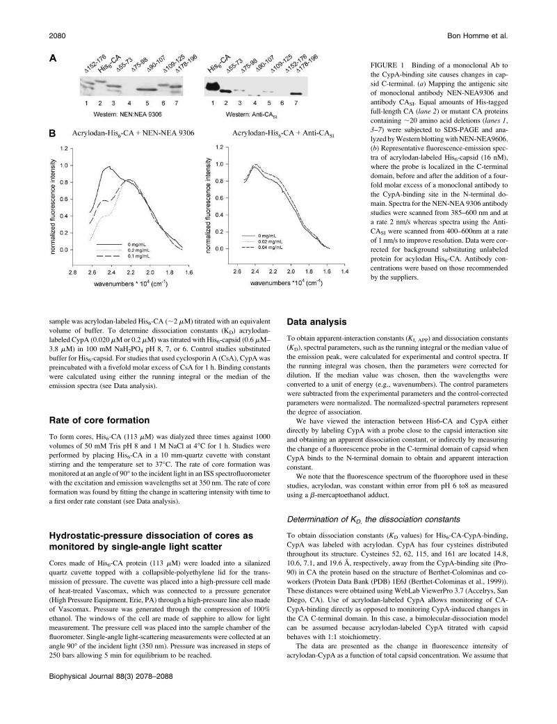
FIGURE 1 Binding of a monoclonal Ab to
the CypA-binding site causes changes in cap-
sid C-terminal. (a) Mapping the antigenic site
of monoclonal antibody NEN-NEA9306 and
antibody CASI. Equal amounts of His-tagged
full-length CA (lane 2) or mutant CA proteins
containing ;20 amino acid deletions (lanes 1,
3–7) were subjected to SDS-PAGE and ana-
lyzed byWestern blottingwithNEN-NEA9606.
(b) Representative fluorescence-emission spec-
tra of acrylodan-labeled His6-capsid (16 nM),
where the probe is localized in the C-terminal
domain, before and after the addition of a four-
fold molar excess of a monoclonal antibody to
the CypA-binding site in the N-terminal do-
main. Spectra for the NEN-NEA 9306 antibody
studies were scanned from 385–600 nm and at
a rate 2 nm/s whereas spectra using the Anti-
CASI were scanned from 400–600nm at a rate
of 1 nm/s to improve resolution. Data were cor-
rected for background substituting unlabeled
protein for acylodan His6-CA. Antibody con-
centrations were based on those recommended
by the suppliers.
2080 Bon Homme et al.
Biophysical Journal 88(3) 2078–2088
all of the proteins are associated when the titration curve reaches saturation.
From the change in intensity, we calculate the degree of protein association
at each point along the titration curve (see BonHomme et al., 2003).
The reported Kd values were determined using
Kd ¼ ½CA-CypA�=ð½CAT � ½CA-CypA�Þ3 ð½CypAT � ½CA-CypA�ÞÞ;
where the total concentration of the two proteins, given as subscript T, is
known and the concentration of the CA-CypA complex was determined
from the fraction associated multiplied by the concentration of total capsid.
The Kd values were calculated of each experimental point on the titration
curve where the fraction associated was between 0.1 and 0.9. The values for
3–6 titration studies were pooled and averaged and the standard error is
given.
Determination of KI, APP, the apparent-interaction constant
To calculate apparent-interaction constants (KI, APP), acrylodan-labeled
CypA concentrations at each titration point and the degree of association
were fit to a following hyperbolic equation using SigmaPlot 2001 version
7.101. The reported apparent-interaction constant (KI, APP) and error
represent the mean and mean 6 SE of the individual experiments.
The CA protein exists as a tetramer or higher oligomer at pH 8 and pH 7,
and as a monomer-dimer at pH 6 (Ehrlich et al., 2001). Distance
measurements of the crystal structure using WebLabViewerPro 3.7
(Accelrys) indicate that the distance (63.5 A) between the CypA-binding
site and acrylodan does not allow for direct monitoring of binding (PDB
1E6J; Berthet-Colominas et al., 1999). For these reasons, any changes
detected using this experimental design, resulting from a binding interac-
tion, will most likely represent a change in conformation, a change in
oligomerization state or a combination of these events. The exact nature
of the change being monitored is unclear. However, because the change
follows a rectangular hyperbolic, then this interaction may be thought to be
specific to one site and the constant of the hyperbolic equation is defined as
the apparent-interaction constant (KI, APP). The tiration curves present the
change in fluorescence as a function of the amount of total protein added.
Determination of the rate constants
To obtain initial rates of core formation, the exponential-rise portion of the
curves were fit to the following equation using SigmaPlot 2001 version
7.101,
ISALS ¼ ½capsid� 3 ð1� e�ktÞwhere ISALS is the intensity of single-angle light scatter, [capsid] is the His6-
CA protein concentration, k is the constant of the equation in units of events
per second, and t is time in seconds.
RESULTS
CypA induces conformational changes in theC-terminal domain of His6-CA
We previously demonstrated that the oligomerization state
of CA increases from monomer-dimer at pH 6 to dimer-
tetramer at higher pH values (BonHomme et al., 2003). We
have also found that the binding of CypA to the N-terminal
domain of CA produces large changes in the environment
of Cys-reactive probes in the C-terminal domain, suggesting
that CypA induces a conformational change (BonHomme
et al., 2003). This conformational change does not occur at
pH 6 but is apparent at pH 7 and 8. It is unclear whether this
pH dependence results from the inability of CypA to bind to
monomeric CA at pH 6 or whether CypA-binding is unable
to transmit conformational changes to the C-terminal domain
at the lower pH.
In initial studies, we used previously described His6-CA
mutants that contained deletions throughout the protein
(Dietrich et al., 2001) to map the antigenic site of several
monoclonal antibodies. One of these, NEN-NEA 9306,
recognized an antigenic site within the CypA interaction
domain in the N-terminal domain of His6-CA. In Fig. 1 a, weshow that NEN-NEA 9306 recognized WT His6-CA (lane 2)and all of the His6-CAdeletionmutants (lanes 1, 3, 5, 6, and 7)except the mutant lacking residues 75–98, which includes
H87AGPIA92 (lane 4). Interestingly, we found that addition
of NEN-NEA 9306 to acrylodan-labeled His6-CA could
confer changes in the C-terminal domain as visualized by
changes in the fluorescence of the probe acrylodan co-
valently attached to one or both of the Cys residues located in
the C-terminal domain (Fig. 1 b).The effect of binding NEN-NEA 9306 was compared to
that of a second antibody, anti-CASI (International En-
zymes), which was raised against residues 151–160 in the
CA C-terminal domain. In Fig. 1 a, we show that this
antibody recognized WT His6-CA (lane 1) and all of the
His6-CA deletion mutants (lanes 2–5 and 7) except the
mutant lacking residues 152–176 (lane 6). In contrast to
the results obtained with NEN-NEA 9306, no change was
detectedusing the antibodyagainst residues151–160 (Fig. 1 b).These results suggest that perturbation of CA through
interaction with the region encompassing the CypA-binding
site induces conformational changes in the CA C-terminal
domain.
To further characterize the ability of CypA to bind and
transduce conformational changes in capsid, we compared
the binding of acrylodan-labeled CypA to unlabeled His6-
CA. Since this association is visualized directly by changes
in acrylodan emission on the labeled CypA due to changes in
the local dielectric environment as unlabeled His6-CA
associates, we can follow the changes in fluorescence to
obtain an apparent dissociation constant (KD). Alternately,
we can follow the association indirectly by changes in the
fluorescence of acrylodan-labeled His6-CA that result from
conformation changes brought about by the binding to
unlabeled CypA to a distal site on capsid to obtain an
apparent interaction constant (KI, APP)] (see Methods).
In Fig. 2, we present results for KD and KI, APP evaluated at
pH 8, 7, and 6. We note that the data in Fig. 2, a–c, havepreviously been reported (BonHomme et al., 2003).
Although the data are identical to these previous results,
they have been analyzed using a hyperbolic fit rather than
a bimolecular association, which, as discussed in Methods, is
inappropriate for this association. We present these data
again to allow for easy comparison between the wild-type
proteins and the mutants discussed below.
Role of HIV-1 Capsid Cys Residues 2081
Biophysical Journal 88(3) 2078–2088
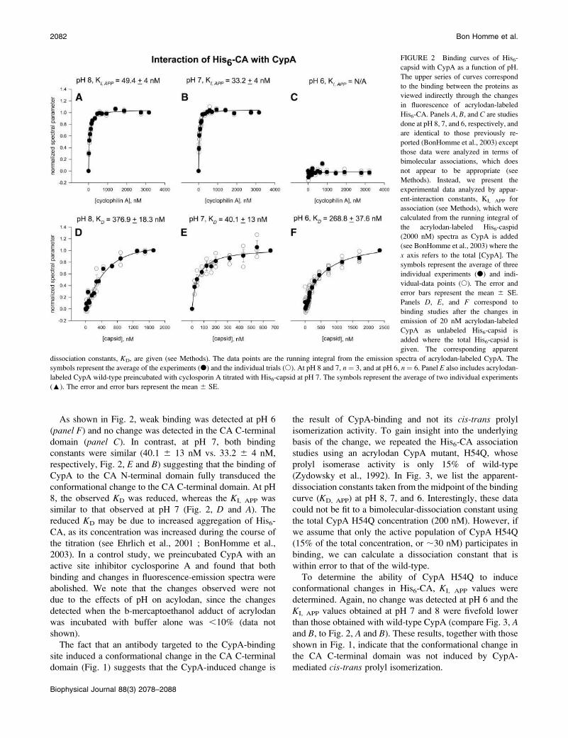
As shown in Fig. 2, weak binding was detected at pH 6
(panel F) and no change was detected in the CA C-terminal
domain (panel C). In contrast, at pH 7, both binding
constants were similar (40.1 6 13 nM vs. 33.2 6 4 nM,
respectively, Fig. 2, E and B) suggesting that the binding of
CypA to the CA N-terminal domain fully transduced the
conformational change to the CA C-terminal domain. At pH
8, the observed KD was reduced, whereas the KI, APP was
similar to that observed at pH 7 (Fig. 2, D and A). Thereduced KD may be due to increased aggregation of His6-
CA, as its concentration was increased during the course of
the titration (see Ehrlich et al., 2001 ; BonHomme et al.,
2003). In a control study, we preincubated CypA with an
active site inhibitor cyclosporine A and found that both
binding and changes in fluorescence-emission spectra were
abolished. We note that the changes observed were not
due to the effects of pH on acylodan, since the changes
detected when the b-mercaptoethanol adduct of acrylodan
was incubated with buffer alone was ,10% (data not
shown).
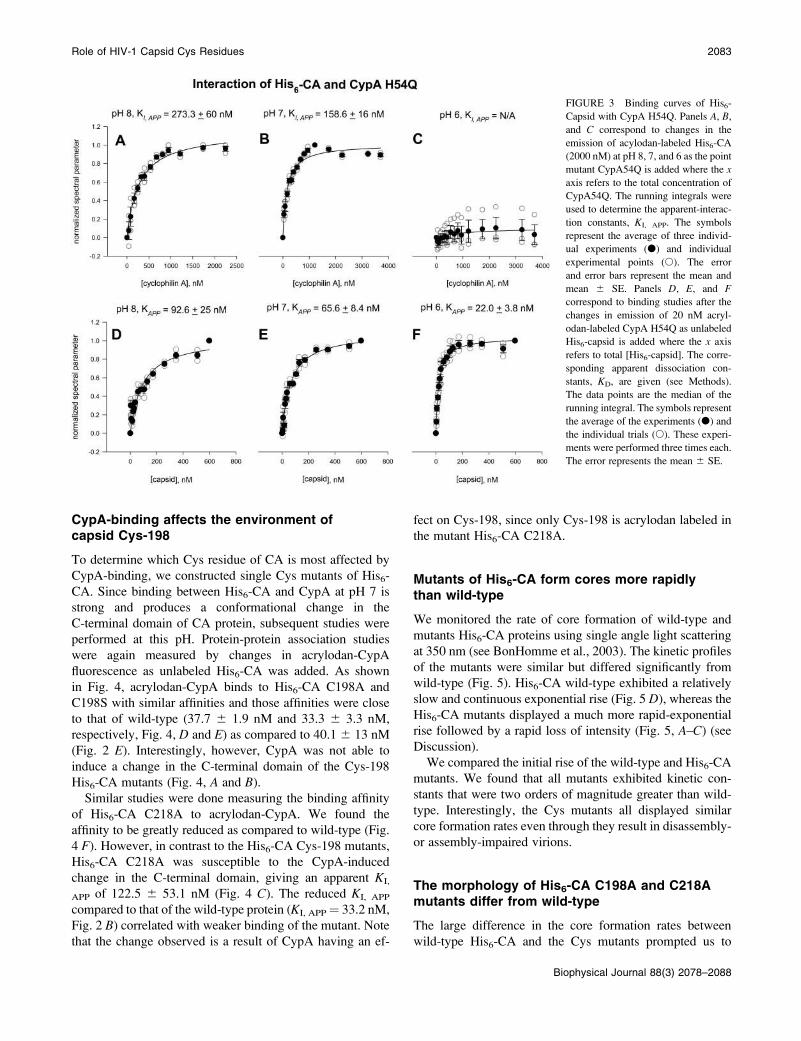
The fact that an antibody targeted to the CypA-binding
site induced a conformational change in the CA C-terminal
domain (Fig. 1) suggests that the CypA-induced change is
the result of CypA-binding and not its cis-trans prolyl
isomerization activity. To gain insight into the underlying
basis of the change, we repeated the His6-CA association
studies using an acrylodan CypA mutant, H54Q, whose
prolyl isomerase activity is only 15% of wild-type
(Zydowsky et al., 1992). In Fig. 3, we list the apparent-
dissociation constants taken from the midpoint of the binding
curve (KD, APP) at pH 8, 7, and 6. Interestingly, these data
could not be fit to a bimolecular-dissociation constant using
the total CypA H54Q concentration (200 nM). However, if
we assume that only the active population of CypA H54Q
(15% of the total concentration, or ;30 nM) participates in
binding, we can calculate a dissociation constant that is
within error to that of the wild-type.
To determine the ability of CypA H54Q to induce
conformational changes in His6-CA, KI, APP values were
determined. Again, no change was detected at pH 6 and the
KI, APP values obtained at pH 7 and 8 were fivefold lower
than those obtained with wild-type CypA (compare Fig. 3, Aand B, to Fig. 2, A and B). These results, together with those
shown in Fig. 1, indicate that the conformational change in
the CA C-terminal domain was not induced by CypA-
mediated cis-trans prolyl isomerization.
FIGURE 2 Binding curves of His6-
capsid with CypA as a function of pH.
The upper series of curves correspond
to the binding between the proteins as
viewed indirectly through the changes
in fluorescence of acrylodan-labeled
His6-CA. Panels A, B, and C are studies
done at pH 8, 7, and 6, respectively, and
are identical to those previously re-
ported (BonHomme et al., 2003) except
those data were analyzed in terms of
bimolecular associations, which does
not appear to be appropriate (see
Methods). Instead, we present the
experimental data analyzed by appar-
ent-interaction constants, KI, APP for
association (see Methods), which were
calculated from the running integral of
the acrylodan-labeled His6-caspid
(2000 nM) spectra as CypA is added
(see BonHomme et al., 2003) where the
x axis refers to the total [CypA]. The
symbols represent the average of three
individual experiments (d) and indi-
vidual-data points (s). The error and
error bars represent the mean 6 SE.
Panels D, E, and F correspond to
binding studies after the changes in
emission of 20 nM acrylodan-labeled
CypA as unlabeled His6-capsid is
added where the total His6-capsid is
given. The corresponding apparent
dissociation constants, KD, are given (see Methods). The data points are the running integral from the emission spectra of acrylodan-labeled CypA. The
symbols represent the average of the experiments (d) and the individual trials (s). At pH 8 and 7, n ¼ 3, and at pH 6, n ¼ 6. Panel E also includes acrylodan-
labeled CypA wild-type preincubated with cyclosporin A titrated with His6-capsid at pH 7. The symbols represent the average of two individual experiments
(:). The error and error bars represent the mean 6 SE.
2082 Bon Homme et al.
Biophysical Journal 88(3) 2078–2088
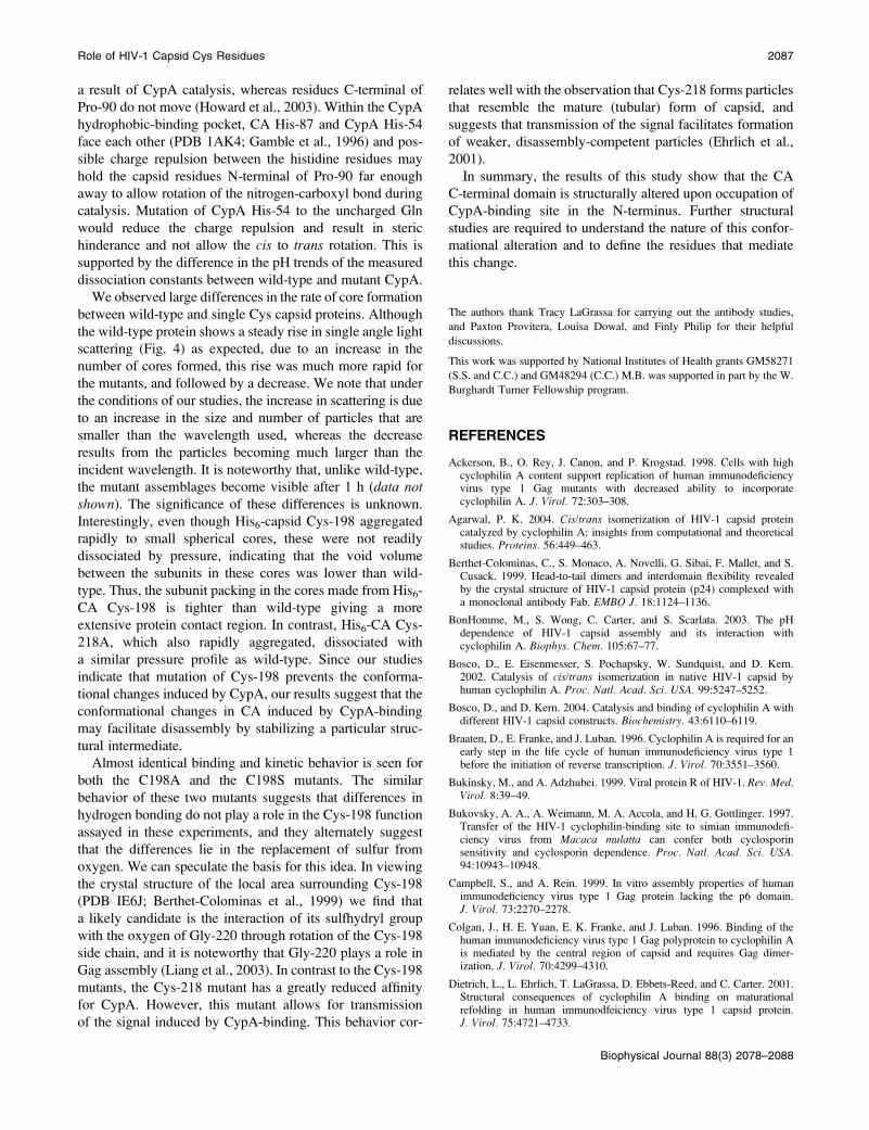
CypA-binding affects the environment ofcapsid Cys-198
To determine which Cys residue of CA is most affected by
CypA-binding, we constructed single Cys mutants of His6-
CA. Since binding between His6-CA and CypA at pH 7 is
strong and produces a conformational change in the
C-terminal domain of CA protein, subsequent studies were
performed at this pH. Protein-protein association studies
were again measured by changes in acrylodan-CypA
fluorescence as unlabeled His6-CA was added. As shown
in Fig. 4, acrylodan-CypA binds to His6-CA C198A and
C198S with similar affinities and those affinities were close
to that of wild-type (37.7 6 1.9 nM and 33.3 6 3.3 nM,
respectively, Fig. 4, D and E) as compared to 40.1 6 13 nM
(Fig. 2 E). Interestingly, however, CypA was not able to
induce a change in the C-terminal domain of the Cys-198
His6-CA mutants (Fig. 4, A and B).Similar studies were done measuring the binding affinity
of His6-CA C218A to acrylodan-CypA. We found the
affinity to be greatly reduced as compared to wild-type (Fig.
4 F). However, in contrast to the His6-CA Cys-198 mutants,
His6-CA C218A was susceptible to the CypA-induced
change in the C-terminal domain, giving an apparent KI,
APP of 122.5 6 53.1 nM (Fig. 4 C). The reduced KI, APP
compared to that of the wild-type protein (KI, APP¼ 33.2 nM,
Fig. 2 B) correlated with weaker binding of the mutant. Note
that the change observed is a result of CypA having an ef-
fect on Cys-198, since only Cys-198 is acrylodan labeled in
the mutant His6-CA C218A.
Mutants of His6-CA form cores more rapidlythan wild-type
We monitored the rate of core formation of wild-type and
mutants His6-CA proteins using single angle light scattering
at 350 nm (see BonHomme et al., 2003). The kinetic profiles
of the mutants were similar but differed significantly from
wild-type (Fig. 5). His6-CA wild-type exhibited a relatively
slow and continuous exponential rise (Fig. 5 D), whereas theHis6-CA mutants displayed a much more rapid-exponential
rise followed by a rapid loss of intensity (Fig. 5, A–C) (seeDiscussion).
We compared the initial rise of the wild-type and His6-CA
mutants. We found that all mutants exhibited kinetic con-
stants that were two orders of magnitude greater than wild-
type. Interestingly, the Cys mutants all displayed similar
core formation rates even through they result in disassembly-
or assembly-impaired virions.
The morphology of His6-CA C198A and C218Amutants differ from wild-type
The large difference in the core formation rates between
wild-type His6-CA and the Cys mutants prompted us to
FIGURE 3 Binding curves of His6-
Capsid with CypA H54Q. Panels A, B,
and C correspond to changes in the
emission of acylodan-labeled His6-CA
(2000 nM) at pH 8, 7, and 6 as the point
mutant CypA54Q is added where the x
axis refers to the total concentration of
CypA54Q. The running integrals were
used to determine the apparent-interac-
tion constants, KI, APP. The symbols
represent the average of three individ-
ual experiments (d) and individual
experimental points (s). The error
and error bars represent the mean and
mean 6 SE. Panels D, E, and Fcorrespond to binding studies after the
changes in emission of 20 nM acryl-
odan-labeled CypA H54Q as unlabeled
His6-capsid is added where the x axis
refers to total [His6-capsid]. The corre-
sponding apparent dissociation con-
stants, KD, are given (see Methods).
The data points are the median of the
running integral. The symbols represent
the average of the experiments (d) and
the individual trials (s). These experi-
ments were performed three times each.
The error represents the mean 6 SE.
Role of HIV-1 Capsid Cys Residues 2083
Biophysical Journal 88(3) 2078–2088

determine whether these proteins formed morphologically
distinct complexes. The morphology of the resulting cores
was investigated by electron microscopy (Fig. 6). His6-CA
C198A (Fig. 6 A) forms spherical structures similar to the
wild-type protein (BonHomme et al., 2003), although the
cores formed by the mutant protein were much smaller in
size. In contrast, His6-CA C218A formed mainly tubular
structures in addition to small spherical structures (Fig. 6 B).We note that mature (i.e., untagged) CA has been shown to
form tubular structures that are larger in size than the ones
observed here (Gross et al., 1998; Ehrlich et al., 2001)
His6-CA C198A forms cores whose stability isconcentration dependent
We tested the stability of the structures formed by wild-type
and Cys mutated His6-CA using high pressure. In general,
the application of hydrostatic pressure results in the dis-
sociation of protein oligomers due to the penetration of sol-
vent into the void volume in the subunit interfaces. The
more extensive the subunit contacts, the smaller the void
volume and the greater the pressure needed for dissociation
(see Heremans, 1992).
Using single-angle light scattering, we monitored the
dissociation of the CA proteins in real time as a function of
hydrostatic pressure. We have previously shown that the
application of hydrostatic pressure on His6-CA results in
a concentration-dependent reduction in light scattering that is
directly related to dissociation of virion cores (BonHomme
et al., 2003). Here,wefind thatwild-typeHis6-CA andC218A
at 113 mM showed significant dissociation in the first 1750
bars (426 5% and 386 2%, respectively, Fig. 7 A). Releaseof pressure allowed the values for the light scattering to return
to within 10% of their original value. In contrast, the
FIGURE 4 Binding of the single Cys mutants C198A, C198S, and C218 A of His6-capsid with CypA. Panels A, B, and C show the change in emission of the
acylodan-labeled His6-capsid point mutants, C198A-, C198S-, and C218A at 2200 nM as CypA is added where the x axis refers to the total [CypA]. The
running integral of the spectra were used to determine the apparent-interaction constants, KI, APP (see Methods). The symbols represent the average of
individual experiments (d) and individual-data points (s), where n ¼ 3 for His6-CA C198A and C198S, and n ¼ 4 for C218A. The error and error bars
represent the mean6 SE. Panels D, E, and F correspond to binding studies after the changes in emission of 20 nM acrylodan–labeled CypA as the unlabeled
His6-CA C198A (n¼ 3), C198S (n¼ 3) and C218A (n¼ 5) were added at pH 7.KD values were calculated as described in the Methods. The symbols represent
the average of the experiments (d) and the individual trials (s). The errors and error bars represent the mean 6 SE.
2084 Bon Homme et al.
Biophysical Journal 88(3) 2078–2088

complexes of C198A His6-CA formed at the same concen-
tration using were relatively insensitive to pressure dissoci-
ation; only 9 6 2% of His6-capsid C198A dissociated in the
same 1750 bar range. This result correlates well with previous
studies indicating that viruses containingCys-198mutants are
impaired in disassembly (McDermott et al., 1996).
DISCUSSION
The assembly of HIV-1 is due in part to strong interactions in
the CA domain of Gag. During or after viral assembly and
release from the host cell, the immature capsid undergoes
drastic morphological changes induced by proteolytic
maturation. These changes allow the virus to be effectively
disassembled upon its entry into host cells in the early stage
of infection. Here, we have linked two factors that have been
implicated in the assembly/disassembly process: the host-
protein CypA and the CA C-terminal domain Cys residues.
We monitored changes in the C-terminal region of an
immature form of CA through changes in the emission
properties of the fluorescent probe, acrylodan, located in this
domain, and have found that both Cys-198 and Cys-218 of
CA play key roles in modulating the tertiary and quaternary
structures needed for efficient assembly and disassembly of
the virus. Although it is impossible to state unequivocally
whether the spectral changes we are viewing are due to
structural changes or conformational fluctuations, the extent
of the changes in acrylodan intensity and energy suggests
that binding of a molecule to the CypA-binding site
stabilizes a set of CA conformations. Some of these proposed
conformational changes appear to be mediated by CypA, but
additional factors must be involved since addition of CypA
did not detectably affect the dissociation of the wild-type
immature CA in our previous studies (BonHomme et al.,
2003).
An important role of the CA protein in the HIV-1 life cycle
is to promote multimerization during assembly. We have
previously found that oligomerization of CA in vitro can
be induced by the combination of pH values above 6 and
high ionic strength (Ehrlich et al., 2001). Additionally, the
N-terminal domain of CA incorporates CypA into virions
(Franke et al., 1994; Gamble et al., 1996). We have found
that binding of CypA causes a change in the environment of
an acrylodan probe covalently linked to the one of the two
FIGURE 5 Kinetic profiles of His6-capsid
core formation. Panels A, B, C, and D show the
rate at which His6-capsid C198A, C198S,
C218A form cores at 60 mM, 50 mM Tris pH
8, and 1 M NaCl, 37�C with constant stirring as
monitored by single-angle light scattering.
Role of HIV-1 Capsid Cys Residues 2085
Biophysical Journal 88(3) 2078–2088

Cys in the C-terminal domain. Monitoring the association
between CypA and His6-CA, either by viewing changes in
a probe attached to CA or a probe attached to CypA, shows
that binding is weaker to monomeric His6-CA (i.e., at pH 6)
and that this binding does not transmit changes to the CA
C-terminal domain. However, the binding of CypA to
oligomerized His6-CA (i.e., . pH 6) allows for this change.
Together with the observation that changes in the C-terminal
domain on His6-CA can be induced by the binding of
a monoclonal antibody to the N-terminal domain CypA-
binding site, makes it unlikely that the effect of CypA on
HIV-1 Gag is due to its cis-trans prolyl isomerase activity.
Binding of CypA to CA, and catalysis of Pro-90
isomerization, has been previously characterized using
a number of biophysical methods including fluorescence,
NMR, crystallographic, and theoretical (Endrich et al., 1996;
Gamble et al., 1996; Bosco et al., 2002; BonHomme et al.,
2003; Agarwal, 2004; Bosco and Kern, 2004). Taken
together, these studies show that binding of CypA to the
single site encompassing Pro-90 promotes cis/trans isomer-
ization. However, our data suggests that Pro-90 isomeriza-
tion may not play a role in the conformational changes in the
C-terminal domain observed here since binding of antibody
to this site also confers these changes (Fig. 1). It is possible,
but unlikely, that the antibody binding accelerates prolyl
isomerization. A comprehensive NMR study by Bosco and
Kern (2004) shows that CypA binds to both mature and
immature forms of CA and that binding and catalysis is
not linked to maturational processing. Interestingly, these
authors did not observe structural changes in the C-terminal
domain upon CypA-binding, but noted that CypA may alter
the structure of this region when CA is in an aggregated
form. This idea is well supported by these studies showing
that no changes in the C-terminal domain could be detected
when CypA binds to CA at pH 6 where CA should be
monomeric (Fig. 2).
We could only fit the CypA H54Q-CA titration curves if
we assume that a fraction (i.e., 15%) of the CypA mutant is
capable of binding, and the affinity turns out to be similar to
wild-type. We could not fit the data by simply assuming that
H54Q binds to CA with a weaker affinity. This result
suggests that although the CypA H54Q mutant has mainly
lost the ability to induce changes in the CA C-terminal
domain, a small subpopulation of the mutant may retain the
ability to bind immature and mature forms of CA. To
understand how the CypA H54Q mutant can bind to capsid
as tight or tighter than wild-type and still have a low level of
isomerase activity, we propose reaction scheme based on
previous work (Howard et al., 2003). Here, the peptide
backbone loses double character due to formation of
a hydrogen bond between the nitrogen (h1) on CypA’s
Arg55 and the nitrogen (a) on the CA Pro-90 residue
(Howard et al., 2003). In the ground state, the distance
between these groups ranges from 3.3 to 4.4 A (Howard
et al., 2003). Formation of this hydrogen bond stabilizes
a pyramidal sp3-hybridization state for capsid’s Pro-90
resulting in localized single-bond character. During the
transition state, increasing single-bond character allows for
both the lengthening and ultimately the free rotation of
the bond between the CA Pro-90 nitrogen (a) and the
neighboring carboxyl carbon. In addition, the hydrogen bond
between the nitrogen (h1) on CypA’s Arg-55 and the
nitrogen (a) on CA Pro-90 is expected to shorten. It has been
demonstrated that the two residues immediately N-terminal
of Pro-90 in CA move from the cis to the trans position as
FIGURE 6 Morphology of His6-capsid mutant cores as imaged by
electron microscopy. Electron-microscopy images of His6-CA C198A
(panel A) and C218A (panel B). His6-CA (113 mM) cores were deposited
(5 mL) onto an electron-microscopy grid and stained with a saturated solu-
tion of uranyl acetate.
FIGURE 7 Core disassembly induced by high hydrostatic pressure.
Hydrostatic pressure was used to disassemble cores made of His6-CA
(113 mM) wild-type, C198A, and C218A. To obtain a range of core
association, single-angle light scatter measurements of the proteins were
taken before oligomerization and subtracted from measurements taken after
oligomerization. The extent of disassembly was normalized within the range
of core association. For His6-CA wild-type, the data points represent the
average of three experiments whereas the others were performed five times.
The error bars represent the mean 6 SE.
2086 Bon Homme et al.
Biophysical Journal 88(3) 2078–2088
a result of CypA catalysis, whereas residues C-terminal of
Pro-90 do not move (Howard et al., 2003). Within the CypA
hydrophobic-binding pocket, CA His-87 and CypA His-54
face each other (PDB 1AK4; Gamble et al., 1996) and pos-
sible charge repulsion between the histidine residues may
hold the capsid residues N-terminal of Pro-90 far enough
away to allow rotation of the nitrogen-carboxyl bond during
catalysis. Mutation of CypA His-54 to the uncharged Gln
would reduce the charge repulsion and result in steric
hinderance and not allow the cis to trans rotation. This is
supported by the difference in the pH trends of the measured
dissociation constants between wild-type and mutant CypA.
We observed large differences in the rate of core formation
between wild-type and single Cys capsid proteins. Although
the wild-type protein shows a steady rise in single angle light
scattering (Fig. 4) as expected, due to an increase in the
number of cores formed, this rise was much more rapid for
the mutants, and followed by a decrease. We note that under
the conditions of our studies, the increase in scattering is due
to an increase in the size and number of particles that are
smaller than the wavelength used, whereas the decrease
results from the particles becoming much larger than the
incident wavelength. It is noteworthy that, unlike wild-type,
the mutant assemblages become visible after 1 h (data notshown). The significance of these differences is unknown.
Interestingly, even though His6-capsid Cys-198 aggregated
rapidly to small spherical cores, these were not readily
dissociated by pressure, indicating that the void volume
between the subunits in these cores was lower than wild-
type. Thus, the subunit packing in the cores made from His6-
CA Cys-198 is tighter than wild-type giving a more
extensive protein contact region. In contrast, His6-CA Cys-
218A, which also rapidly aggregated, dissociated with
a similar pressure profile as wild-type. Since our studies
indicate that mutation of Cys-198 prevents the conforma-
tional changes induced by CypA, our results suggest that the
conformational changes in CA induced by CypA-binding
may facilitate disassembly by stabilizing a particular struc-
tural intermediate.
Almost identical binding and kinetic behavior is seen for
both the C198A and the C198S mutants. The similar
behavior of these two mutants suggests that differences in
hydrogen bonding do not play a role in the Cys-198 function
assayed in these experiments, and they alternately suggest
that the differences lie in the replacement of sulfur from
oxygen. We can speculate the basis for this idea. In viewing
the crystal structure of the local area surrounding Cys-198
(PDB IE6J; Berthet-Colominas et al., 1999) we find that
a likely candidate is the interaction of its sulfhydryl group
with the oxygen of Gly-220 through rotation of the Cys-198
side chain, and it is noteworthy that Gly-220 plays a role in
Gag assembly (Liang et al., 2003). In contrast to the Cys-198
mutants, the Cys-218 mutant has a greatly reduced affinity
for CypA. However, this mutant allows for transmission
of the signal induced by CypA-binding. This behavior cor-
relates well with the observation that Cys-218 forms particles
that resemble the mature (tubular) form of capsid, and
suggests that transmission of the signal facilitates formation
of weaker, disassembly-competent particles (Ehrlich et al.,
2001).
In summary, the results of this study show that the CA
C-terminal domain is structurally altered upon occupation of
CypA-binding site in the N-terminus. Further structural
studies are required to understand the nature of this confor-
mational alteration and to define the residues that mediate
this change.
The authors thank Tracy LaGrassa for carrying out the antibody studies,
and Paxton Provitera, Louisa Dowal, and Finly Philip for their helpful
discussions.
This work was supported by National Institutes of Health grants GM58271
(S.S. and C.C.) and GM48294 (C.C.) M.B. was supported in part by the W.
Burghardt Turner Fellowship program.
REFERENCES
Ackerson, B., O. Rey, J. Canon, and P. Krogstad. 1998. Cells with highcyclophilin A content support replication of human immunodeficiencyvirus type 1 Gag mutants with decreased ability to incorporatecyclophilin A. J. Virol. 72:303–308.
Agarwal, P. K. 2004. Cis/trans isomerization of HIV-1 capsid proteincatalyzed by cyclophilin A: insights from computational and theoreticalstudies. Proteins. 56:449–463.
Berthet-Colominas, C., S. Monaco, A. Novelli, G. Sibai, F. Mallet, and S.Cusack. 1999. Head-to-tail dimers and interdomain flexibility revealedby the crystal structure of HIV-1 capsid protein (p24) complexed witha monoclonal antibody Fab. EMBO J. 18:1124–1136.
BonHomme, M., S. Wong, C. Carter, and S. Scarlata. 2003. The pHdependence of HIV-1 capsid assembly and its interaction withcyclophilin A. Biophys. Chem. 105:67–77.
Bosco, D., E. Eisenmesser, S. Pochapsky, W. Sundquist, and D. Kern.2002. Catalysis of cis/trans isomerization in native HIV-1 capsid byhuman cyclophilin A. Proc. Natl. Acad. Sci. USA. 99:5247–5252.
Bosco, D., and D. Kern. 2004. Catalysis and binding of cyclophilin A withdifferent HIV-1 capsid constructs. Biochemistry. 43:6110–6119.
Braaten, D., E. Franke, and J. Luban. 1996. Cyclophilin A is required for anearly step in the life cycle of human immunodeficiency virus type 1before the initiation of reverse transcription. J. Virol. 70:3551–3560.
Bukinsky, M., and A. Adzhubei. 1999. Viral protein R of HIV-1. Rev. Med.Virol. 8:39–49.
Bukovsky, A. A., A. Weimann, M. A. Accola, and H. G. Gottlinger. 1997.Transfer of the HIV-1 cyclophilin-binding site to simian immunodefi-ciency virus from Macaca mulatta can confer both cyclosporinsensitivity and cyclosporin dependence. Proc. Natl. Acad. Sci. USA.94:10943–10948.
Campbell, S., and A. Rein. 1999. In vitro assembly properties of humanimmunodeficiency virus type 1 Gag protein lacking the p6 domain.J. Virol. 73:2270–2278.
Colgan, J., H. E. Yuan, E. K. Franke, and J. Luban. 1996. Binding of thehuman immunodeficiency virus type 1 Gag polyprotein to cyclophilin Ais mediated by the central region of capsid and requires Gag dimer-ization. J. Virol. 70:4299–4310.
Dietrich, L., L. Ehrlich, T. LaGrassa, D. Ebbets-Reed, and C. Carter. 2001.Structural consequences of cyclophilin A binding on maturationalrefolding in human immunodfeiciency virus type 1 capsid protein.J. Virol. 75:4721–4733.
Role of HIV-1 Capsid Cys Residues 2087
Biophysical Journal 88(3) 2078–2088
Dorfman, T., A. Weimann, A. Borsetti, C. T. Walsh, and H. G. Gottlinger.1997. Active-site residues of cyclophilin A are crucial for itsincorporation into human immunodeficiency virus type 1 virions.J. Virol. 71:7110–7113.
Ehrlich, L. S., T. Liu, S. Scarlata, B. Chu, and C. A. Carter. 2001. HIV-1capsid protein forms spherical (immature-like) and tubular (mature-like)particles in vitro: structure switching by pH-induced conformationalchanges. Biophys. J. 81:586–594.
Endrich, M., P. Gehrig, and H. Gehring. 1996. Maturation-inducedconformational changes of HIV-1 capsid protein and identification oftwo high affinity sites for cyclophilin in the C-terminal domain. J. Biol.Chem. 274:5326–5332.
Franke, E. K., H. E. Yuan, and J. Luban. 1994. Specific incorporation ofcyclophilin A into HIV-1 virions. Nature. 372:359–362.
Gamble, T. R., F. F. Vajdos, S. Yoo, D. K. Worthylake, M. Houseweart,W. I. Sundquist, and C. P. Hill. 1996. Crystal structure of human cyclo-philin A bound to the amino-terminal domain of HIV-1 capsid. Cell. 87:1285–1294.
Gitti, R., B. Lee, J. Walker, M. K. Summers, S. Yoo, and W. Sundquist.1996. Structure of the N-terminal core domain of the HIV-1 capsidprotein. Science. 273:231–235.
Gross, I., H. Hohenberg, C. Huckhagel, and H.-G. Krausslich. 1998.N-terminal extension of human immunodeficiency virus capsid proteinconverts the in vitro assembly phenotype from tubular to sphericalparticles. J. Virol. 72:4798–4810.
Henklein, P., K. Bruns, M. P. Sherman, U. Tessmer, K. Licha, J. Kopp,C. M. de Noronha, W. C. Greene, V. Wray, and U. Schubert. 2000.Functional and structural characterization of synthetic HIV-1 vpr thattransduces cells, localizes to the nucleus, and induces G2 cell cyclearrest. J. Biol. Chem. 275:32016–32026.
Heremans, K. A. H. 1992. High pressure effects upon proteins and otherbiomolecules. Annu. Rev. Biophys. Bioeng. 11:1–21.
Howard, B. R., F. F. Vajdos, S. Li, W. I. Sundquist, and C. P. Hill. 2003.Structural insights into the catalytic mechanism of cyclophilin A. Nat.Struct. Biol. 475–481.
Kattendbeck, B., A. von Poblotzki, A. Rohrhofer, H. Wolf, and S. Modrow.1997. Inhibition of human immunodeficiency virus type 1 particleformation by alteration of defined amino acids within the C-terminus ofcapsid. J. Gener. Virol. 78:2489–2496.
Lanman, J., J. Sexton, M. Sakalian, and P. E. Prevelige Jr. 2002. Kineticanalysis of the role of intersubunit interactions in human immunodefi-ciency virus type 1 capsid protein assembly in vitro. J. Virol. 76:6900–6908.
Liang, C., J. Hu, J. B. Whitney, L. Kleiman, and M. A. Wainberg. 2003. Astructurally disordered region at the C terminus of capsid plays essential
roles in multimerization and membrane binding of the Gag protein ofhuman immunodeficiency virus type 1. J. Virol. 77:1772–1783.
Mammano, F., A. Ohagen, S. Hoglund, and H. Gottlinger. 1994. Role ofthe major homology region of human immunodeficiency virus type 1 invirion morphogenesis. J. Virol. 68:4927–4936.
McDermott, G., L. Farrell, R. Ross, and E. Barklise. 1996. Structuralanalysis of human immunodeficiency virus type 1 Gag protein inter-actions using cysteine-specific reagents. J. Virol. 70:5106–5114.
Recin, A. S., S. R. Paik, R. D. Berkowitz, J. Luban, I. Lowy, and S. P. Goff.1995. Linker insertion mutation in the human immunodeficeincy virustype 1 gag gene: effects on virion particle assembly, release andinfectivity. J. Virol. 69:642–650.
Saphire, A., M. Bobardt, and P. Gallay. 1999. Host cyclophilin A mediatesHIV-1 attachment to target cells via heparans. EMBO J. 18:6771–6785.
Scarlata, S., and C. Carter. 2003. Role of HIV-1 gag domains in viralassembly. Biochim. Biophys. Acta. 1614:62–72.
Sherry, B., G. Zybarth, M. Alfano, L. Dubrobsky, R. Mitchell, D. Rich, P.Ulrich, R. Bucala, A. Cerami, andM. Bukrinsky. 1998. Role of cyclophilinA in the uptake of HIV-1 by macrophages and T lymphocytes. Proc. Natl.Acad. Sci. USA. 95:1758–1763.
Steinkasserer, A., R. Harrison, A. Billich, F. Hammerschmid, G. Werner, B.Wolff, P. Peichl, G. Palfi, W. Schnitzel, E. Mlynar, and others. 1995.Mode of action of SDZ NIM 811, a nonimmunosuppressive cyclosporinA analog with activity against human immunodeficiency virus type 1(HIV-1): interference with early and late events in HIV-1 replication.J. Virol. 69:814–824.
Towers, G. J., T. Hatziioannou, S. Cowan, S. P. Goff, J. Luban, and P. D.Bieniasz. 2003. Cyclophilin A modulates the sensitivity of HIV-1 to hostrestriction factors. Nat. Med. 9:1138–1143.
von Poblotzki, A., R. Wagner, M. Niedrig, G. Wanner, H. Wolf, and S.Modrow. 1993. Identification of a region of Pr55gag-polyproteinessential for HIV-1 particle formation. Virology. 193:981–985.
von Schwedler, U., T. Stemmler, V. Klishko, S. Li, K. Albertine, D. Davis,and W. Sundquist. 1998. Proteolytic refolding of the HIV-1 capsidprotein amino -terminus facilitates viral core assembly. EMBO J. 17:1555–1568.
Zander, K., M. P. Sherman, U. Tessmer, K. Bruns, V. Wray, A. T. Prechtel,E. Schubert, P. Henklein, J. Luban, J. Neidleman, W. C. Greene, and U.Schubert. 2003. Cyclophilin A interacts with HIV-1 Vpr and is requiredfor its functional expression. J. Biol. Chem. 278:43202–43213.
Zydowsky, L., F. Etzkorn, H. Y. Change, S. B. Ferguson, L. A. Stolz, S. I.Ho, and C. T. Walsh. 1992. Active site mutants of human cyclophilin Aseparate peptidyl-prolyl isomerase activity from cyclosporin A bindingand calcineurin inhibition. Protein Sci. 1:1092–1099.
2088 Bon Homme et al.
Biophysical Journal 88(3) 2078–2088