Regulation of c-Fes tyrosine kinase and biological activities by N-terminal coiled-coil...
9
MOLECULAR AND CELLULAR BIOLOGY, 0270-7306/99/$04.0010 Dec. 1999, p. 8335–8343 Vol. 19, No. 12 Copyright © 1999, American Society for Microbiology. All Rights Reserved. Regulation of c-Fes Tyrosine Kinase and Biological Activities by N-Terminal Coiled-Coil Oligomerization Domains HAIYUN CHENG, 1,2 JIM A. ROGERS, 1 NANCY A. DUNHAM, 1 AND THOMAS E. SMITHGALL 1,2,3 * Eppley Institute for Research in Cancer 1 and Department of Pharmacology, 2 University of Nebraska Medical Center, Omaha, Nebraska, and Department of Molecular Genetics and Biochemistry, University of Pittsburgh School of Medicine, Pittsburgh, Pennsylvania 3 Received 9 July 1999/Accepted 19 August 1999 The cytoplasmic protein-tyrosine kinase Fes has been implicated in cytokine signal transduction, hemato- poiesis, and embryonic development. Previous work from our laboratory has shown that active Fes exists as a large oligomeric complex in vitro. However, when Fes is expressed in mammalian cells, its kinase activity is tightly repressed. The Fes unique N-terminal sequence has two regions with strong homology to coiled-coil- forming domains often found in oligomeric proteins. Here we show that disruption or deletion of the first coiled-coil domain upregulates Fes tyrosine kinase and transforming activities in Rat-2 fibroblasts and enhances Fes differentiation-inducing activity in myeloid leukemia cells. Conversely, expression of a Fes truncation mutant consisting only of the unique N-terminal domain interfered with Rat-2 fibroblast transfor- mation by an activated Fes mutant, suggesting that oligomerization is essential for Fes activation in vivo. Coexpression with the Fes N-terminal region did not affect the transforming activity of v-Src in Rat-2 cells, arguing against a nonspecific suppressive effect. Taken together, these findings suggest a model in which Fes activation may involve coiled-coil-mediated interconversion of monomeric and oligomeric forms of the kinase. Mutation of the first coiled-coil domain may activate Fes by disturbing intramolecular coiled-coil interaction, allowing for oligomerization via the second coiled-coil domain. Deletion of the second coiled-coil domain blocks fibroblast transformation by an activated form of c-Fes, consistent with this model. These results provide the first evidence for regulation of a nonreceptor protein-tyrosine kinase by coiled-coil domains. The c-fes proto-oncogene is the normal cellular homolog of the transforming oncogenes found in several avian and feline retroviruses (13, 15, 16, 19, 22, 38). The human c-fes locus encodes a cytoplasmic protein-tyrosine kinase (Fes) primarily expressed in hematopoietic cells of the myeloid lineage (7, 28, 40). Distinct from Src and other nonreceptor tyrosine kinases, Fes has a long N-terminal unique region, followed by a central SH2 domain and a C-terminal kinase domain with a total molecular mass of 93 kDa. Previous studies have shown that Fes is activated by numerous hematopoietic growth factors (17, 18, 20, 30), implicating Fes as a downstream component of cytokine receptor signaling. Other work has shown that Fes suppresses the growth and induces the differentiation of the myeloid leukemia cell line, K-562 (46). Taken together, these results strongly suggest that Fes is involved in the regulation of hematopoietic cell proliferation, differentiation and function. Despite the important roles that Fes plays in hematopoietic development, little is known about the mechanisms that regu- late its tyrosine kinase activity. In the absence of activating signals, Fes tyrosine kinase activity is strongly repressed in cells (12). Indeed, high-level overexpression of Fes is required to induce kinase activation in vivo and to release transforming activity in fibroblasts (8). Recent work from our laboratory has shown that Fes autophosphorylation occurs by an intermolec- ular mechanism, suggesting that oligomerization may be an important initial event in Fes activation (36, 37). We also observed that active Fes elutes from a gel filtration column as a large oligomer and that the unique N-terminal domain is required for oligomerization. Taken together, these data sug- gest that oligomerization may be required for kinase activation and that suppression of oligomerization may represent a neg- ative regulatory mechanism for Fes in vivo. Computer analysis of the Fes N-terminal region revealed two strong consensus sequences for the formation of coiled- coil oligomerization domains (36). Coiled-coil domains are comprised of amphipathic a-helices with a characteristic hep- tad repeat in which the first and fourth residues are hydropho- bic and pack against each other to form a hydrophobic core (26). The remainder of the amino acids are often hydrophilic, helping to solvate the oligomeric structure. In this study, we investigated the contribution of the Fes coiled-coil homology domains to the regulation of Fes tyrosine kinase activity and biological function. We observed that disruption or deletion of the more N-terminal coiled-coil domain (CC1) released Fes tyrosine kinase activity in fibroblasts, leading to transforma- tion. In contrast to CC1, deletion of the second coiled-coil domain (CC2) inhibited transformation by an activated form of Fes, suggesting that CC2 may be required for oligomeriza- tion. In addition, a truncation mutant of Fes bearing only the unique N-terminal domain strongly suppressed the transform- ing and tyrosine kinase activity of an activated Fes mutant in fibroblasts, providing further evidence that Fes is active as an oligomer in living cells. These findings are consistent with a mechanism of Fes kinase regulation that involves interconver- sion of monomeric and oligomeric forms of the protein. MATERIALS AND METHODS Fes coiled-coil domain mutants. The nucleotide sequence encoding the first N-terminal coiled-coil homology domain of Fes (CC1; amino acids 128 to 169) was deleted by using the Gene Editor oligonucleotide-directed mutagenesis system according to the manufacturer’s instructions (Promega). A similar ap- proach was utilized to insert the b-turn motif Leu-Pro-Ala-Gly-Ser between N-terminal amino acids 148 and 149, which form the boundary between the third and fourth heptad repeats of the predicted coiled-coil structure. The coding * Corresponding author. Mailing address: Department of Molecular Genetics and Biochemistry, University of Pittsburgh School of Medi- cine, E1240 Biomedical Science Tower, Pittsburgh, PA 15261. Phone: (412) 648-9495. Fax: (412) 624-1401. E-mail: tsmithga1@pitt.edu. 8335 on April 24, 2016 by guest http://mcb.asm.org/ Downloaded from
-
Upload
independent -
Category
Documents
-
view
1 -
download
0
Transcript of Regulation of c-Fes tyrosine kinase and biological activities by N-terminal coiled-coil...
MOLECULAR AND CELLULAR BIOLOGY,0270-7306/99/$04.0010
Dec. 1999, p. 8335–8343 Vol. 19, No. 12
Copyright © 1999, American Society for Microbiology. All Rights Reserved.
Regulation of c-Fes Tyrosine Kinase and Biological Activities byN-Terminal Coiled-Coil Oligomerization Domains
HAIYUN CHENG,1,2 JIM A. ROGERS,1 NANCY A. DUNHAM,1 AND THOMAS E. SMITHGALL1,2,3*
Eppley Institute for Research in Cancer1 and Department of Pharmacology,2 University of Nebraska Medical Center,Omaha, Nebraska, and Department of Molecular Genetics and Biochemistry, University of Pittsburgh
School of Medicine, Pittsburgh, Pennsylvania3
Received 9 July 1999/Accepted 19 August 1999
The cytoplasmic protein-tyrosine kinase Fes has been implicated in cytokine signal transduction, hemato-poiesis, and embryonic development. Previous work from our laboratory has shown that active Fes exists as alarge oligomeric complex in vitro. However, when Fes is expressed in mammalian cells, its kinase activity istightly repressed. The Fes unique N-terminal sequence has two regions with strong homology to coiled-coil-forming domains often found in oligomeric proteins. Here we show that disruption or deletion of the firstcoiled-coil domain upregulates Fes tyrosine kinase and transforming activities in Rat-2 fibroblasts andenhances Fes differentiation-inducing activity in myeloid leukemia cells. Conversely, expression of a Festruncation mutant consisting only of the unique N-terminal domain interfered with Rat-2 fibroblast transfor-mation by an activated Fes mutant, suggesting that oligomerization is essential for Fes activation in vivo.Coexpression with the Fes N-terminal region did not affect the transforming activity of v-Src in Rat-2 cells,arguing against a nonspecific suppressive effect. Taken together, these findings suggest a model in which Fesactivation may involve coiled-coil-mediated interconversion of monomeric and oligomeric forms of the kinase.Mutation of the first coiled-coil domain may activate Fes by disturbing intramolecular coiled-coil interaction,allowing for oligomerization via the second coiled-coil domain. Deletion of the second coiled-coil domain blocksfibroblast transformation by an activated form of c-Fes, consistent with this model. These results provide thefirst evidence for regulation of a nonreceptor protein-tyrosine kinase by coiled-coil domains.
The c-fes proto-oncogene is the normal cellular homolog ofthe transforming oncogenes found in several avian and felineretroviruses (13, 15, 16, 19, 22, 38). The human c-fes locusencodes a cytoplasmic protein-tyrosine kinase (Fes) primarilyexpressed in hematopoietic cells of the myeloid lineage (7, 28,40). Distinct from Src and other nonreceptor tyrosine kinases,Fes has a long N-terminal unique region, followed by a centralSH2 domain and a C-terminal kinase domain with a totalmolecular mass of 93 kDa. Previous studies have shown thatFes is activated by numerous hematopoietic growth factors (17,18, 20, 30), implicating Fes as a downstream component ofcytokine receptor signaling. Other work has shown that Fessuppresses the growth and induces the differentiation of themyeloid leukemia cell line, K-562 (46). Taken together, theseresults strongly suggest that Fes is involved in the regulation ofhematopoietic cell proliferation, differentiation and function.
Despite the important roles that Fes plays in hematopoieticdevelopment, little is known about the mechanisms that regu-late its tyrosine kinase activity. In the absence of activatingsignals, Fes tyrosine kinase activity is strongly repressed in cells(12). Indeed, high-level overexpression of Fes is required toinduce kinase activation in vivo and to release transformingactivity in fibroblasts (8). Recent work from our laboratory hasshown that Fes autophosphorylation occurs by an intermolec-ular mechanism, suggesting that oligomerization may be animportant initial event in Fes activation (36, 37). We alsoobserved that active Fes elutes from a gel filtration column asa large oligomer and that the unique N-terminal domain isrequired for oligomerization. Taken together, these data sug-
gest that oligomerization may be required for kinase activationand that suppression of oligomerization may represent a neg-ative regulatory mechanism for Fes in vivo.
Computer analysis of the Fes N-terminal region revealedtwo strong consensus sequences for the formation of coiled-coil oligomerization domains (36). Coiled-coil domains arecomprised of amphipathic a-helices with a characteristic hep-tad repeat in which the first and fourth residues are hydropho-bic and pack against each other to form a hydrophobic core(26). The remainder of the amino acids are often hydrophilic,helping to solvate the oligomeric structure. In this study, weinvestigated the contribution of the Fes coiled-coil homologydomains to the regulation of Fes tyrosine kinase activity andbiological function. We observed that disruption or deletion ofthe more N-terminal coiled-coil domain (CC1) released Festyrosine kinase activity in fibroblasts, leading to transforma-tion. In contrast to CC1, deletion of the second coiled-coildomain (CC2) inhibited transformation by an activated formof Fes, suggesting that CC2 may be required for oligomeriza-tion. In addition, a truncation mutant of Fes bearing only theunique N-terminal domain strongly suppressed the transform-ing and tyrosine kinase activity of an activated Fes mutant infibroblasts, providing further evidence that Fes is active as anoligomer in living cells. These findings are consistent with amechanism of Fes kinase regulation that involves interconver-sion of monomeric and oligomeric forms of the protein.
MATERIALS AND METHODS
Fes coiled-coil domain mutants. The nucleotide sequence encoding the firstN-terminal coiled-coil homology domain of Fes (CC1; amino acids 128 to 169)was deleted by using the Gene Editor oligonucleotide-directed mutagenesissystem according to the manufacturer’s instructions (Promega). A similar ap-proach was utilized to insert the b-turn motif Leu-Pro-Ala-Gly-Ser betweenN-terminal amino acids 148 and 149, which form the boundary between the thirdand fourth heptad repeats of the predicted coiled-coil structure. The coding
* Corresponding author. Mailing address: Department of MolecularGenetics and Biochemistry, University of Pittsburgh School of Medi-cine, E1240 Biomedical Science Tower, Pittsburgh, PA 15261. Phone:(412) 648-9495. Fax: (412) 624-1401. E-mail: [email protected].
8335
on April 24, 2016 by guest
http://mcb.asm
.org/D
ownloaded from
sequence for the second coiled-coil domain of Fes (CC2; amino acids 310 to 344)was deleted by using a standard PCR-based strategy. The N-terminal myristyla-tion signal sequence of v-Src was added to the N-terminal region of Fes and thecoiled-coil domain mutants by using a PCR-based approach. The 59 end of theFes cDNA was amplified by using a forward primer encoding a unique HindIIIsite and the v-Src myristylation signal sequence Met-Gly-Ser-Ser-Lys-Ser-Lysfused to Fes homologous sequences beginning with codon 3 and a reverse primerthat maps to the 59 end of the Fes unique N-terminal domain. The resulting PCRproduct was digested with HindIII and AccI and swapped with the equivalentrestriction fragment in wild-type Fes or the coiled-coil domain mutants to gen-erate the full-length Myr-Fes cDNAs. The coding sequence for the Fes N-terminal region (Fes-NT), the myristylated form of the N-terminal region (Myr-Fes-NT), and related N-terminal constructs lacking the first or second coiled-coilhomology domains (DCC1-NT, DCC2-NT, Myr-DCC1-NT, and Myr-DCC2-NT)were amplified by PCR from the corresponding full-length Fes cDNAs. All FescDNAs used in these studies bear a C-terminal FLAG epitope tag (37).
Expression of Fes proteins in 293T cells. 293T human embryonic kidney cells(35) were maintained in Dulbecco modified Eagle’s medium (DMEM) supple-mented with 5% fetal bovine serum (FBS). Cells were transfected with FescDNAs in the expression vector pCDNA3 (InVitrogen) by using a modifiedcalcium phosphate method described in detail elsewhere (37). Forty-eight hourslater, whole-cell protein extracts were prepared by heating equal numbers of cellsdirectly in sodium dodecyl sulfate-polyacrylamide gel electrophoresis (SDS-PAGE) sample buffer. Proteins were resolved by SDS-PAGE, transferred topolyvinylidene difluoride (PVDF) membranes, and probed with antibodies toFLAG (M2; Sigma) to detect Fes protein expression or with antiphosphotyrosineantibodies (PY20; Transduction Laboratories) to detect autophosphorylation.
Production of recombinant retroviruses and infection of Rat-2 fibroblasts.Wild-type and coiled-coil mutant Fes cDNAs were subcloned into the retroviralvector pSRaMSVtkneo (33). Retroviruses were produced by cotransfecting 293Tcells with the pSRa-Fes constructs and an ecotropic packaging vector describedelsewhere (2, 24). The retroviral supernatant was collected every 12 h for 3 days,and the supernatants were pooled and stored at 280°C.
Rat-2 cells were maintained in DMEM supplemented with 5% FBS. For viralinfection, cells (2.5 3 105) were plated in 60-mm tissue culture dishes andincubated overnight at 37°C. Retroviral supernatants (5 ml) were thawed on ice,supplemented with Polybrene to 4 mg/ml, and added to the cells. After incuba-tion for 4 h at room temperature, the viral supernatants were removed andreplaced with 4 ml of fresh medium. The infected cells were then incubated for48 h at 37°C prior to further analysis. Replicate plates of cells were lysed andanalyzed for Fes protein expression as described above for the 293T cells.
Transformation assays. Rat-2 fibroblast transformation was assessed by usinga focus-forming assay. Retrovirally infected Rat-2 cells (2 3 104) were plated in60-mm tissue culture dishes in the presence of 800 mg of G418 per ml. The cellswere incubated at 37°C for 2 weeks, at which time they were Wright-Giemsastained and observed by light microscopy. For some experiments, stained fociwere enumerated from a scanned image by using colony-counting software (Bio-Rad GS-710 Calibrated Imaging Densitometer and Quantity One software).
For coexpression experiments, Rat-2 cells were first infected with recombinantretroviruses carrying the various Fes N-terminal constructs or empty virus as anegative control as described above. At 48 h postinfection, the cells were splitand reseeded in 6-well plates at 2 3 105 cells/well. The following day, the cellswere reinfected with retroviruses carrying activated forms of Fes or v-Src, incu-bated for 48 h, and replated at 2 3 104 cells/60-mm culture dish in the presenceof G418. Foci were stained and counted 2 weeks later as described above.
In vitro tyrosine kinase assay for Fes. Populations of Rat-2 cells stably ex-pressing wild-type and mutant forms of Fes were lysed in Fes lysis buffer (50 mMTris-HCl, pH 7.4; 50 mM NaCl; 1 mM EDTA; 1 mM MgCl2; 0.1% Triton X-100)supplemented with 25 mg of aprotinin per ml, 50 mg of leupeptin per ml, 1 mMphenylmethylsulfonyl fluoride, 20 mM NaF, 1 mM Na3VO4, and 50 mMNa2MoO4. Fes proteins were immunoprecipitated from clarified cell lysates withthe M2 anti-FLAG monoclonal antibody resin. The immunoprecipitates werewashed with radioimmune precipitation assay buffer (50 mM Tris-HCl, 150 mMNaCl, 1% Triton X-100, 0.1% SDS, 1 mM EDTA, 1% sodium deoxycholate),followed by kinase assay buffer (50 mM HEPES, pH 7.4; 10 mM MgCl2). Thefinal pellets were incubated with [g-32P]ATP (10 mCi; Dupont-New EnglandNuclear) and 2 mg of a glutathione S-transferase (GST) fusion protein containingamino acids 162 to 413 of the human Bcr protein. Previous studies have shownthat this protein is strongly phosphorylated by Fes in vitro (23). After incubationfor 15 min at 30°C, the reactions were stopped by heating in SDS-PAGE samplebuffer. Phosphorylated proteins were resolved on SDS-polyacrylamide gels andvisualized by using storage phosphorimaging.
Chemical cross-linking. Fes N-terminal domain constructs were expressed asFLAG fusion proteins in 293T cells. Cells were lysed by sonication in Fes lysisbuffer, and clarified cell lysates were incubated with the bifunctional cross-linkingreagent disuccinimidyl suberate (DSS) at a final concentration of 1.0 mM asdescribed previously (36). The reactions were incubated for 5 min at roomtemperature and stopped by heating in SDS-PAGE sample buffer. Cross-linkedFes proteins were visualized by immunoblotting with anti-FLAG antibodies.
Hematopoietic differentiation assay. The human erythroleukemia cell lineK-562 (25) was obtained from the American Type Culture Collection and grownin RPMI 1640 medium containing 10% FCS. K-562 cells were infected with
recombinant Fes retroviruses by using a coculture approach. Cultures of virus-producing 293T cells were initiated by cotransfection with retroviral and pack-aging plasmids as described above. At 2 days posttransfection, 4 ml of DMEMcontaining 5% FCS and 3 3 105 K-562 cells were added to the 293T culturesalong with 4 mg of polybrene per ml. After incubation for 2 days at 37°C, theK-562 cells were removed from the 293T cell culture by aspiration. InfectedK-562 cells were replated on new culture dishes and incubated for an additional2 days at 37°C, allowing residual 293T cells to readhere. The infected K-562 cellswere reharvested, counted by trypan blue exclusion, and plated at 105 cells/60-mm tissue culture dish in 4 ml of RPMI containing 10% FCS and 800 mg ofG418 per ml. Four days later, cells were fixed for 20 min in 1% paraformaldehydeand stored at 4°C in phosphate-buffered saline (PBS) prior to staining and flowcytometry. For single-cell analysis of Fes expression, 105 fixed cells were perme-abilized with 0.05% saponin in RPMI containing 5% FBS (RPMI-FBS) for 20min. Cells were resuspended in a minimal volume of RPMI-FBS containing theM2 anti-FLAG monoclonal antibody (20 mg/ml) for 1 h. The cells were washedtwice with RPMI-FBS and then incubated with a goat anti-mouse immunoglob-ulin G-fluorescein isothiocyanate (FITC) conjugate (20 mg/ml in RPMI-FBS;Molecular Probes) for 1 h. The cells were then washed three more times prior tofluorescence-activated cell sorter analysis. Analysis of CD13 and CD33 expres-sion was performed with direct FITC-conjugated antibodies for these myeloiddifferentiation markers (Southern Biotechnology Associates) (6). Staining wasperformed as described above for the M2 antibody, except the saponin wasomitted. A population of K-562 cells cocultured with 293T cells producing aretrovirus carrying only the neo selection marker served as a negative control inall experiments.
RESULTS
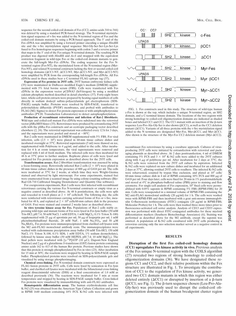
Disruption of the first Fes coiled-coil homology domain(CC1) upregulates Fes kinase activity in vivo. Previous analysisof the Fes unique N-terminal region with the COILS algorithm(27) revealed two regions of strong homology to coiled-coiloligomerization domains (36). We have designated these re-gions CC1 and CC2, and their relative positions within the Fesstructure are illustrated in Fig. 1. To investigate the contribu-tion of CC1 to the regulation of Fes kinase activity, we gener-ated two CC1 domain mutants in which this region was eitherdeleted entirely (DCC1) or disrupted by insertion of a b-turn(bCC1; see Fig. 1). The b-turn sequence chosen (Leu-Pro-Ala-Gly-Ser) was previously used to disrupt the coiled-coil oli-gomerization domain of the human breakpoint cluster region
FIG. 1. Fes constructs used in this study. The structure of wild-type humanc-Fes is shown at the top, which includes a unique N-terminal region, an SH2domain, and a C-terminal kinase domain. The locations of the two regions withstrong homology to coiled-coil oligomerization domains are indicated as shadedboxes and labeled CC1 and CC2. The CC1 mutant with an insertion of the b-turnsequence Leu-Pro-Ala-Gly-Ser is also shown (bCC1), as well as the CC1 deletionmutant (DCC1). Variants of all three proteins with the v-Src myristylation signaladded to the N terminus are designated Myr-Fes, Myr-bCC1, and Myr-DCC1.Also shown is the structure of the Myr-Fes CC2 deletion mutant (Myr-DCC2).
8336 CHENG ET AL. MOL. CELL. BIOL.
on April 24, 2016 by guest
http://mcb.asm
.org/D
ownloaded from
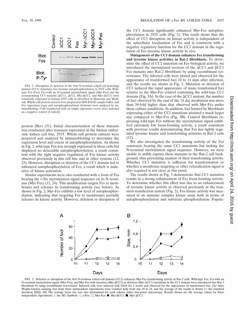
protein (Bcr) (31). Initial characterization of these mutantswas conducted after transient expression in the human embry-onic kidney cell line, 293T. Whole-cell protein extracts wereprepared and analyzed by immunoblotting to determine theexpression level and extent of autophosphorylation. As shownin Fig. 2, wild-type Fes was strongly expressed in these cells butdisplayed no detectable autophosphorylation, a result consis-tent with the tight negative regulation of Fes kinase activityobserved previously in this cell line and in other systems (12,23). However, disruption or deletion of the CC1 domain led toenhanced autophosphorylation of Fes, a result which is indic-ative of kinase activation.
Similar experiments were also conducted with a form of Fesbearing the v-Src myristylation signal sequence on its N termi-nus (Myr-Fes) (11, 24). This modification targets Fes to mem-branes and releases its transforming activity (see below). Asshown in Fig. 2, Myr-Fes exhibits a low level of autophosphor-ylation, indicating that targeting Fes to membranes partiallyreleases its kinase activity. However, deletion or disruption of
the CC1 domain significantly enhanced Myr-Fes autophos-phorylation in 293T cells (Fig. 2). This result shows that theeffect of CC1 disruption on kinase activity is independent ofthe subcellular localization of Fes and is consistent with anegative regulatory function for the CC1 domain in the regu-lation of Fes tyrosine kinase activity in vivo.
Mutagenesis of the CC1 domain enhances Fes transformingand tyrosine kinase activities in Rat-2 fibroblasts. To deter-mine the effect of CC1 mutation on Fes biological activity, weintroduced the myristylated versions of the DCC1 and bCC1Fes mutants into Rat-2 fibroblasts by using recombinant ret-roviruses. The infected cells were plated and observed for theappearance of transformed foci 10 to 14 days after infection,and the results are shown in Fig. 3. Mutation or deletion ofCC1 induced the rapid appearance of many transformed focirelative to the Myr-Fes control containing the wild-type CC1domain (Fig. 3A). In the case of the DCC1 mutant, the numberof foci observed by the end of the 14 day incubation was morethan 50-fold higher than that observed with Myr-Fes underthese culture conditions. In addition, foci formed by fibroblastsexpressing either of the CC1 mutations attained a much largersize compared to Myr-Fes (Fig. 3B). Control fibroblasts ex-pressing wild-type Fes without the myristylation signal exhib-ited extremely low focus-forming activity, a result consistentwith previous results demonstrating that Fes has tightly regu-lated tyrosine kinase and transforming activities in Rat-2 cells(12).
We also investigated the transforming activity of the Fesconstructs bearing the same CC1 mutations but lacking theN-terminal myristylation signal sequence. However, we wereunable to stably express these mutants in the Rat-2 cell back-ground, thus preventing analysis of their transforming activity.Whether CC1 mutation is sufficient for transformation orwhether a membrane-targeting or other relocalization signal isalso required is not clear at this point.
The results shown in Fig. 3 demonstrate that CC1 mutationresults in a strong enhancement of Fes focus-forming activity.To determine whether this effect was due to an enhancementof tyrosine kinase activity as observed previously in the tran-sient-transfection system (Fig. 2), Fes kinase activity was mea-sured in an immune complex kinase assay both in terms ofautophosphorylation and substrate phosphorylation. Popula-
FIG. 2. Disruption or deletion of the first N-terminal coiled-coil homologydomain (CC1) stimulates Fes tyrosine autophosphorylation in 293T cells. Wild-type Fes (Fes), Fes with an N-terminal myristylation signal (Myr-Fes) and thecorresponding CC1 mutants (bCC1, DCC1, Myr-bCC1, and Myr-DCC1) weretransiently expressed in human 293T cells as described in Materials and Meth-ods. Whole-cell protein extracts were prepared in SDS-PAGE sample buffer, andFes expression (top) and autophosphorylation (bottom) were analyzed by im-munoblotting. Cells transfected with an empty expression vector were includedas a negative control (Control).
FIG. 3. Deletion or disruption of the first N-terminal coiled-coil domain (CC1) enhances Myr-Fes transforming activity in Rat-2 cells. Wild-type Fes, Fes with anN-terminal myristylation signal (Myr-Fes), and Myr-Fes with insertion (Myr-bCC1) or deletion (Myr-DCC1) mutations in the CC1 domain were introduced into Rat-2fibroblasts by using recombinant retroviruses. Infected cells were selected with G418 for 2 weeks and observed for the appearance of transformed foci. (A) AfterWright-Giemsa staining, foci from three independent experiments were counted daily from day 10 to 14, and the average of the results is shown (6 the standarddeviation [SD]). (B) The average focus size was also determined for each culture under low-power microscopy. Results shown are the average values for threeindependent experiments 6 the SD. Symbols: E, c-Fes; h, Myr-Fes; F, Myr-bCC1; ■, Myr-DCC1.
VOL. 19, 1999 REGULATION OF c-Fes BY COILED COILS 8337
on April 24, 2016 by guest
http://mcb.asm
.org/D
ownloaded from

tions of Rat-2 cells expressing Fes, Myr-Fes, Myr-bCC1, andMyr-DCC1 were lysed, and the Fes proteins were recovered byimmunoprecipitation. After washing, the immunoprecipitateswere incubated with [g-32P]ATP and the substrate protein,GST-Bcr. This recombinant GST fusion protein contains hu-man Bcr amino acids 162-413, which include the major tyrosinephosphorylation sites for this Fes substrate (23, 29). As shownin Fig. 4, both of the CC1 mutants exhibited increased tyrosinekinase activity relative to the wild-type Fes and Myr-Fes con-trols. This result suggests that the increased transforming ac-tivity of the CC1 mutants is due to enhancement of theirintrinsic tyrosine kinase activity and is consistent with a role forthe CC1 domain in the negative regulation of tyrosine kinaseactivity in vivo.
The CC1 domain is not required for oligomerization of theFes N-terminal region. Previous work from our laboratory hasshown that the active form of Fes is oligomeric (36). Datashown above indicate that deletion or mutation of the firstcoiled-coil domain leads to kinase activation and fibroblasttransformation. These findings suggest that the Fes CC1 do-main is not required for formation of the active oligomer andthat CC2 or another portion of the N-terminal domain medi-ates oligomerization. To test this possibility directly, chemicalcross-linking experiments were performed on Fes N-terminalproteins with or without the CC1 deletion. The N-terminalproteins were transiently expressed in 293T cells, and cell ly-sates were incubated in the presence or absence of the bifunc-tional cross-linking reagent, DSS. The reactions were analyzedfor the presence of cross-linked products by SDS-PAGE andimmunoblotting, and the result is shown in Fig. 5A. The rela-tive levels of the monomeric proteins and the major oligomericcross-linked products were determined by densitometry andare expressed as a ratio in Fig. 5B. All of the N-terminalproteins tested yielded higher-molecular-weight products inthe presence of the cross-linking reagent, including those bear-ing the N-terminal myristyl modification. Thus, the CC1 do-main is not required for oligomerization of the N-terminaldomain.
Transformation of Rat-2 fibroblasts by Myr-Fes is blockedby coexpression with the N-terminal unique domain. Recentstudies from our laboratory have shown that Fes autophos-
phorylation can be suppressed by a kinase-inactive form of Fesin vitro, suggesting that oligomerization may be an essentialpart of the activation mechanism in vivo (36). To test this idea,we coexpressed a truncated form of Fes consisting of only themyristylated N-terminal region together with Myr-Fes in Rat-2fibroblasts and scored the changes in focus-forming activity
FIG. 4. Deletion or disruption of the first N-terminal coiled-coil domain (CC1) enhances Myr-Fes tyrosine kinase activity in Rat-2 cells. Fes, Myr-Fes, Myr-DCC1,or Myr-bCC1 proteins were immunoprecipitated from lysates of Rat-2 fibroblasts and incubated in vitro with [g-32P]ATP and a recombinant GST-Bcr fusion proteinsubstrate. Phosphorylated GST-Bcr, as well as autophosphorylated Fes proteins, was resolved by SDS-PAGE, and the relative levels of 32P incorporation weredetermined by storage phosphorimaging. Equivalent levels of Fes proteins were present in the immune complexes, as shown by immunoblot analysis (data not shown).(A) Autophosphorylation. (B) Substrate phosphorylation. Results shown are the average of two separate determinations. The entire experiment was repeated threetimes with comparable results.
FIG. 5. The CC1 domain is not required for cross-linking of Fes N-terminalproteins. The N-terminal region of wild-type Fes (WT), the myristylated form ofthe N-terminal region (Myr), the N-terminal region lacking the first coiled-coilhomology domain (DCC1), and the myristylated version of the N-terminal CC1deletion mutant (Myr-DCC1) were transiently expressed in 293T cells. Clarifiedcell extracts were incubated in the presence (1) or absence (2) of the bifunc-tional cross-linking reagent DSS as described in Materials and Methods. (A)Cross-linked products were visualized by immunoblotting. The locations of themonomeric and oligomeric forms of the N-terminal proteins are indicated. (B)The relative levels of the monomeric and major oligomeric forms of the FesN-terminal proteins were determined by densitometry and then plotted as theratio shown. Four independent cross-linking experiments produced comparableresults.
8338 CHENG ET AL. MOL. CELL. BIOL.
on April 24, 2016 by guest
http://mcb.asm
.org/D
ownloaded from

after 2 weeks. As shown in Fig. 6, coexpression of Myr-Fes withthe myristylated N-terminal domain led to a 75% reduction inthe number of transformed foci compared to control Rat-2cells coinfected with Myr-Fes and parental retroviruses. Im-munoblots show equivalent expression of Myr-Fes in bothcases, indicating that suppression of transforming activity bythe N-terminal domain is not due to differences in the expres-sion of Myr-Fes.
The activity of the wild-type Fes N-terminal region (withoutthe myristylation signal) was also tested in this suppressionassay. As shown in Fig. 6, removal of the myristyl group re-duced the ability of the N-terminal region to induce the sup-pression of Myr-Fes transforming activity. This difference sug-gests that targeting of the N-terminal domain to the samecompartment as Myr-Fes is important for suppression. In ad-dition, fusion to the N-terminal myristylation signal may dis-turb intramolecular interaction between the CC1 and CC2domains, promoting the trans-interaction with Myr-Fes that isrequired for the dominant-negative effect (see Discussion).
We also tested the suppressive activity of Fes N-terminalproteins with deletions of either CC1 or CC2. Figure 6 showsthat both CC1 and CC2 N-terminal protein deletion mutantsproduced approximately 50% suppression of Myr-Fes focus-forming activity, indicating that the presence of either CC1 orCC2 is sufficient for suppression. In this case, suppression wasindependent of the presence of the myristylation signal se-quence. These results suggest that deletion of one coiled-coil
domain may free the remaining coiled-coil to interact withMyr-Fes to produce the dominant-inhibitory effect.
We also used the suppression assay to determine the rangeof possible coiled-coil interactions. For these experiments,transformation was induced by using the strongly transformingMyr-bCC1-Fes mutant, which has a single functional coiled-coil domain (CC2). As shown in Fig. 7, the wild-type andmyristylated forms of the N-terminal domain inhibited Myr-bCC1-Fes transforming activity by approximately 30 and 75%,respectively, as observed for Myr-Fes (Fig. 6). Interestingly,the Fes N-terminal proteins lacking either the CC1 or the CC2domain also produced strong inhibitory effects. These datasuggest that suppression of Myr-bCC1-Fes transforming activ-ity by DCC1 N-terminal proteins is mediated by CC2-CC2interaction, while suppression by DCC2 is mediated by CC1-CC2 interaction. Thus, homomeric interactions between CC2domains, as well as heteromeric interactions between CC1 andCC2, appear to be possible. A very similar pattern of suppres-sion was observed upon coexpression of the same panel ofN-terminal proteins with the strongly transforming Myr-DCC1Fes mutant as well (data not shown).
To control for specificity in the N-terminal suppression stud-ies, the Fes N-terminal proteins used for the experimentsshown in Fig. 6 and 7 were also coexpressed with v-Src in thefocus-forming assay. v-Src tyrosine kinase was chosen becauseit is dependent upon myristylation for fibroblast transforma-tion (21) but is regulated by mechanisms that do not involve
FIG. 6. Suppression of Myr-Fes transforming activity by coexpression withFes N-terminal domain proteins. Rat-2 fibroblasts were infected with recombi-nant retroviruses carrying the wild-type Fes N-terminal sequence (WT N-term),the N-terminal sequence lacking the first coiled-coil homology domain (DCC1N-term), or the N-terminal sequence lacking the second coiled-coil homologydomain (DCC2 N-term). A parallel series of N-terminal constructs bearing thev-Src myristylation sequence was also tested (indicated as 1 Myr). Cells infectedwith a retrovirus carrying only the neo selection marker served as a negativecontrol (Con). Forty-eight hours later, the cells were reinfected with a Myr-Fesretrovirus and selected with G418 for 2 weeks as described in Materials andMethods. Foci were visualized by Wright-Giemsa staining and counted by usinga Bio-Rad Model GS-710 Scanning Densitometer and colony-counting software.Foci from three independent cultures were counted and normalized to thenegative control average value; the bar graph shows the average normalizedvalue 6 the SD. Expression of Myr-Fes and the N-terminal proteins was verifiedby immunoblotting with the anti-FLAG antibody, which recognizes the FLAGepitope fused to the C terminus of each protein (lower two panels). This entireexperiment was performed twice and produced the same pattern of inhibitioneach time.
FIG. 7. Suppression of Myr-bCC1 Fes transforming activity by coexpressionwith Fes N-terminal domain proteins. Rat-2 fibroblasts were infected with re-combinant retroviruses carrying the wild-type Fes N-terminal sequence (N-term), the N-terminal sequence lacking the first coiled-coil homology domain(DCC1 N-term), or the N-terminal sequence lacking the second coiled-coil ho-mology domain (DCC2 N-term). A parallel series of N-terminal constructs bear-ing the v-Src myristylation sequence was also tested (indicated as 1 Myr). Cellsinfected with a retrovirus carrying only the neo selection marker served as anegative control (Con). Forty-eight hours later, the cells were reinfected with aMyr-bCC1 Fes retrovirus and selected with G418 for 2 weeks as described inMaterials and Methods. Foci were visualized by Wright-Giemsa staining andcounted by using a Bio-Rad Model GS-710 Scanning Densitometer and colony-counting software. Foci from three independent cultures were counted andnormalized to the negative control average value; the bar graph shows theaverage normalized value 6 the SD. Expression of Myr-bCC1 and the N-termi-nal proteins was verified by immunoblotting with the anti-FLAG antibody, whichrecognizes the FLAG epitope fused to the C terminus of each protein (lower twopanels). This entire experiment was performed twice and produced the samepattern of inhibition each time.
VOL. 19, 1999 REGULATION OF c-Fes BY COILED COILS 8339
on April 24, 2016 by guest
http://mcb.asm
.org/D
ownloaded from

coiled-coil domains. As shown in Fig. 8, none of the c-FesN-terminal proteins affected v-Src-induced transformation,supporting the conclusion that the suppressive actions of theN-terminal proteins are specific to Fes-mediated transforma-tion.
Data presented in Fig. 6 to 8 suggest that the ability of FesN-terminal proteins to suppress Myr-Fes-induced transform-ing activity involves direct interaction between the proteins,resulting in the formation of inactive mixed oligomers. To testthis idea further, we performed kinase assays on Myr-Fes im-munoprecipitates from cultures coexpressing the Myr-Fes N-terminal region or matched controls. As shown in Fig. 9, theextent of autophosphorylation of both Myr-Fes and the Myr-DCC1 Fes mutant were inhibited by more than 50% in thepresence of the N-terminal protein. These results support di-rect inhibition of kinase activity in vivo as a mechanism for thesuppressive action of the N-terminal region and agree with ourprevious results showing that a purified recombinant N-termi-nal protein binds directly to Fes and inhibits its kinase activityin vitro (36).
The Fes CC2 domain is required for Fes transforming andkinase activities. Data presented so far point to the CC1 do-main as a negative regulator of Fes kinase activity that is notrequired for oligomerization and activation and implicate theCC2 domain in oligomer formation. To test this possibilitydirectly, we created a CC2 deletion in the context of Myr-Fesand compared its transforming activity with wild-type Fes,Myr-Fes, and Myr-DCC1 Fes, as well as v-Src. As shown in Fig.10, deletion of the CC2 domain completely inhibited the trans-
forming activity of Myr-Fes. This result is in sharp contrast tothe effect of the CC1 mutation, which releases strong trans-forming activity. Deletion of CC2 also resulted in greatly re-duced kinase activity compared to Myr-Fes or Myr-DCC1 (Fig.9). These results show that CC2 is required for both biologicaland kinase activities and suggest that it may contribute tooligomerization in vivo.
Fes CC1 domain mutants retain the ability to induce differ-entiation in K-562 cells. Previous studies have shown thatexpression of Fes is sufficient to induce differentiation of themyeloid leukemia cell line, K-562 (6, 46). However, studies ofFes-transfected K-562 cell populations suggest that only a frac-tion of the cells undergo differentiation after transfection withFes (6). This result suggests that a threshold of Fes tyrosinekinase activity must be reached in order for differentiation tooccur. Therefore, we tested the differentiation-inducing activ-ity of the Fes mutants lacking the CC1 domain which demon-strated elevated tyrosine kinase activity and strong transform-ing potential in fibroblasts. Both the myristylated andnonmyristylated forms of wild-type Fes and the DCC1 mutantwere tested. Each of these proteins was introduced into K-562cells by using recombinant retroviruses, and differentiation wasassessed as the percentage of cells that express the cell surfacemyeloid differentiation antigens CD13 and CD33 as deter-mined by flow cytometry. The percentage of cells expressingFes was also evaluated by this method. As shown in Fig. 11, theratio of Fes protein to differentiation marker expression wasapproximately equal for wild-type Fes, Myr-Fes, and the CC1deletion mutant without the myristylation signal sequence. In-terestingly, the myristylated form of Fes with the CC1 deletionexhibited enhanced differentiation-inducing activity, as re-flected by the higher ratio of CD13 and CD33 to Fes proteinexpression. These results show that deletion of the CC1 do-main together with a membrane targeting signal leads to robust
FIG. 8. Coexpression with Fes N-terminal domain proteins does not inhibitRat-2 cell transformation by v-Src. Rat-2 fibroblasts were infected with recom-binant retroviruses carrying the wild-type Fes N-terminal sequence (N-term), theN-terminal sequence lacking the first coiled-coil homology domain (DCC1 N-term), or the N-terminal sequence lacking the second coiled-coil homologydomain (DCC2 N-term). A parallel series of N-terminal constructs bearing thev-Src myristylation sequence was also tested (indicated as 1 Myr). Cells infectedwith a retrovirus carrying only the neo selection marker served as a negativecontrol (Con). Forty-eight hours later, the cells were reinfected with a v-Srcretrovirus and selected with G418 for 2 weeks as described in Materials andMethods. Foci were visualized by Wright-Giemsa staining and counted by usinga Bio-Rad Model GS-710 Imaging Densitometer and colony-counting software.Foci from three independent cultures were counted and normalized to thenegative control average value; the bar graph shows the average normalizedvalue 6 the SD. Expression of the N-terminal proteins was verified by immu-noblotting with the anti-FLAG antibody, which recognizes the FLAG epitopefused to the C terminus of each protein (lower panel). This entire experimentwas performed twice and produced the same result each time.
FIG. 9. Coexpression with the Fes N-terminal domain inhibits autophos-phorylation of Myr-Fes and the Myr-Fes CC1 domain deletion mutant. Rat-2fibroblasts expressing Myr-Fes, Myr-DCC1, and Myr-DCC2 in the presence (1)or absence (2) of the myristylated N-terminal domain protein (N-term) werelysed, and the Fes proteins were immunoprecipitated with the anti-FLAG anti-body. The immunoprecipitates were washed, incubated with [g-32P]ATP, andseparated by SDS-PAGE. The radiolabeled proteins were transferred to PVDFmembranes to allow determination of protein levels by immunoblotting anddensitometry. Incorporation of 32P into Fes proteins was determined by storagephosphorimaging, and the resulting values were corrected for protein levels andplotted as shown. This experiment was repeated three times with comparableresults.
8340 CHENG ET AL. MOL. CELL. BIOL.
on April 24, 2016 by guest
http://mcb.asm
.org/D
ownloaded from

differentiation signaling in K-562 cells and are consistent withthe fibroblast transformation data. Thus, CC1 also appears tofunction as a negative regulator of Fes biological activity in aphysiologically relevant cellular context.
DISCUSSION
Previous studies have established that Fes tyrosine kinaseactivity is tightly regulated both in physiological sites of expres-sion such as myeloid hematopoietic cells and after overexpres-sion in rodent fibroblasts and human embryonic kidney cells(12, 23, 28). Efficient regulation of Fes kinase activity in thesediverse cell types implies that the mechanism of negative reg-ulation may be intrinsic to the structure of the kinase. Datapresented in this report show that the coiled-coil domains ofFes may have an important function in the suppression of Festyrosine kinase activity in both the fibroblast transformationmodel and in hematopoietic cells. Mutations in the more N-terminal coiled-coil domain, including deletion, insertion of ab-turn, or even a single point mutation of a conserved leucineresidue (data not shown), all released Fes tyrosine kinase andtransforming activities in fibroblasts and enhanced differenti-ation-inducing activity in K-562 cells. These data strongly sup-port an important negative regulatory function for this coiled-coil motif and provide the first example of a coiled-coil domain
contributing to the negative regulation of a cellular tyrosinekinase.
Previous data from our laboratory have established that theactive form of Fes exists as a large oligomeric complex, at leastin vitro (36). In addition, we found that Fes autophosphoryla-tion, which is a critical first step in the activation mechanism,occurs by a trans mechanism reminiscent of growth factor re-ceptor tyrosine kinases (37). Similar results have been reportedfor the Fes-related tyrosine kinase, v-Fps (42, 43). These pre-vious data suggested that oligomerization is the key to Fesactivation in vivo. Thus, suppression of Fes kinase activity mayrequire maintenance of the monomeric state as a mechanismto prevent the trans phosphorylation required for kinase acti-vation. This may be achieved by intramolecular interactionbetween the first and second coiled-coil domains. Evidence forCC1-CC2 interaction is provided by in vivo suppression exper-iments in which Fes N-terminal proteins lacking CC2 areshown to suppress transformation by Myr-bCC1 (Fig. 7); thisFes mutant lacks a functional CC1 domain. We have alsoobtained additional evidence for CC1-CC2 interaction by usingthe yeast two-hybrid system (data not shown). This model alsoexplains why mutations in the more N-terminal coiled-coildomain release tyrosine kinase activity. Such mutations wouldprevent CC1-CC2 interaction and promote constitutive oli-gomerization. The formation of active Fes oligomers appearsto require CC2, since deletion of the CC2 domain almostcompletely inhibited transformation by Myr-Fes and alsogreatly reduced its kinase activity. Interestingly, mutations inthe CC2-homologous region of v-Fps have also been shown toreduce its kinase and transforming activities (34). Determina-tion of the three-dimensional structure of the Fes N-terminalregion will provide an important test of this hypotheticalmodel.
Data presented here also provide new evidence that oli-gomerization is critical to Fes activation in living cells. The FesN-terminal domain serves as an effective dominant-negative
FIG. 10. Deletion of the second coiled-coil homology domain inhibits thetransforming activity of Myr-Fes. Rat-2 fibroblasts were infected with recombi-nant retroviruses carrying wild-type c-Fes, Myr-Fes, Myr-DCC1, Myr-DCC2, orv-Src. Cells infected with a retrovirus carrying only the neo selection markerserved as a negative control (Con). Infected cells were cultured for 2 weeks in thepresence of G418 and were visualized by Wright-Giemsa staining. The experi-ment was performed in triplicate; representative scanned images of the stainedfoci are shown.
FIG. 11. Differentiation of K-562 leukemia cells by Fes coiled-coil domainmutants. K-562 myeloid leukemia cells were incubated with 293T cells producingrecombinant retroviruses carrying either wild-type Fes, Myr-Fes, DCC1, Myr-DCC1, or only the G418 resistance marker as a negative control (Con). After 48 hof coculture, K-562 cells were separated from the 293T cells and replated in thepresence of G418. Four days later, infected cells were analyzed by flow cytometryfor expression of Fes proteins by using the M2 anti-FLAG monoclonal antibodyand for cell surface CD13 and CD33 by using direct FITC-conjugated antibodiesto these myeloid cell-surface antigens. The percentage of cells positive for Fes(open bars), CD13 (gray bars), and CD33 (solid bars) are shown. Enhancementof differentiation by Myr-DCC1 was observed in two independent experiments.
VOL. 19, 1999 REGULATION OF c-Fes BY COILED COILS 8341
on April 24, 2016 by guest
http://mcb.asm
.org/D
ownloaded from

mutant since it was able to suppress Rat-2 cell transformationby Myr-Fes (Fig. 6). This result suggests that the N-terminaldomain of Fes is capable of interacting with full-length Myr-Fes molecules, resulting in the formation of nonproductiveoligomers that are unable to undergo trans phosphorylation.These results are consistent with earlier work from our labo-ratory showing that both the Fes N-terminal domain and afull-length kinase-inactive Fes mutant can suppress wild-typeFes autophosphorylation in vitro (36). Importantly, both theisolated N-terminal region and the kinase-dead Fes mutant canform physical complexes with wild-type Fes in vitro (36).
Although our data are consistent with a model in which CC1may regulate Fes kinase activity via intramolecular interactionwith CC2, alternative hypotheses should be considered as well.For example, the CC1 domain or other features of the FesN-terminal region may interact in trans with inhibitory proteinswhich suppress Fes oligomerization and activation. A numberof trans-inhibitory proteins have been proposed as regulatorsof c-Abl, another nonreceptor tyrosine kinase with tightly reg-ulated tyrosine kinase and transforming activities (41). In thecase of c-Abl, negative regulation seems to require an intactSH3 domain, since mutations in this domain are strongly acti-vating in vivo. Whether or not similar interacting proteins existthat suppress the tyrosine kinase activity of Fes will requirefurther investigation.
Although data presented here provide some of the firstevidence demonstrating regulation of a cellular tyrosine kinaseby coiled-coil domains in vivo, previous work has shown thatcoiled coils contribute to the constitutive activation of theBcr-Abl and TEL-Abl oncoproteins associated with variousleukemias (9, 31). In each case, translocations result in thefusion of the c-Abl tyrosine kinase to a coiled-coil oligomer-ization function provided by Bcr or TEL. In the case of Bcr-Abl, N-terminal Bcr-encoded sequences form a tetrameriza-tion motif that is required for constitutive activation of the Ablkinase, as well as for localization to the actin cytoskeleton (31).Interestingly, coexpression of a peptide containing the Bcroligomerization domain is sufficient to reverse the transformedphenotype of Bcr-Abl-transformed myeloid leukemia cells(14). These data suggest that suppression results from theformation of nonfunctional mixed oligomers and are consistentwith our results demonstrating that coexpression with the FesN-terminal region blocks transformation by activated forms offull-length Fes.
Intramolecular interactions are emerging as a commontheme in the regulation of nonreceptor protein-tyrosine ki-nases. Perhaps the best-studied example involves kinases of theSrc family. The X-ray crystal structures of c-Src (44, 45) andthe closely related kinase Hck (39) strongly suggest that neg-ative regulation involves two intramolecular interactions. First,tyrosine phosphorylation of the conserved tail tyrosine residueinduces intramolecular interaction with the SH2 domain, aninteraction long suspected because mutations in either of theseregions induce kinase activation (4). A second interaction re-vealed by the crystal structure involves the SH3 domain and anintramolecular ligand formed by the linker region connectingthe SH2 and kinase domains. This intramolecular interaction isalso critical for effective negative regulation of Src family ki-nases, since mutations in the SH3 domain or the linker regionalso release the tyrosine kinase and transforming activities ofc-Src and Hck (3, 5, 10). Activation of Src family kinases canalso result from interactions with other proteins that bind tothe SH2 or SH3 domain or both, presumably by displacementof these intramolecular interactions (1, 2, 32). These findingswith Src-related kinases suggest that association of Fes withother proteins may also lead to activation by disturbing in-
tramolecular, negative-regulatory interactions. For example,previous work from our laboratory has shown that Fes associ-ates with and phosphorylates Bcr, the normal breakpoint-clus-ter region protein (23, 29). Fes-Bcr interaction is mediated, inpart, by the Fes N-terminal region (29). More recently, weobserved that coexpression with Bcr induces Fes autophos-phorylation (23). Association of Fes with Bcr may disrupt CC1-CC2 interaction, leading to kinase activation. Bcr is also anoligomeric protein (31), suggesting that it is capable of inter-acting with several molecules of Fes simultaneously. Recruit-ment of multiple Fes molecules into close proximity may con-tribute to activation via trans phosphorylation.
ACKNOWLEDGMENT
This work was supported by grant CA58667 from the National In-stitutes of Health.
REFERENCES
1. Alexandropoulos, K., and D. Baltimore. 1997. Coordinate activation of c-Srcby SH3 and SH2 binding sites on a novel, p130Cas-related protein, Sin.Genes Dev. 10:1341–1355.
2. Briggs, S. D., M. Sharkey, M. Stevenson, and T. E. Smithgall. 1997. SH3-mediated Hck tyrosine kinase activation and fibroblast transformation by theNef protein of HIV-1. J. Biol. Chem. 272:17899–17902.
3. Briggs, S. D., and T. E. Smithgall. 1999. SH2-kinase linker mutations releaseHck tyrosine kinase and transforming activities in rat-2 fibroblasts. J. Biol.Chem. 274:26579–26583.
4. Brown, M. T., and J. A. Cooper. 1996. Regulation, substrates, and functionsof Src. Biochim. Biophys. Acta 1287:121–149.
5. Erpel, T., G. Superti-Furga, and S. A. Courtneidge. 1995. Mutational anal-ysis of the Src SH3 domain: the same residues of the ligand binding surfaceare important for intra- and intermolecular interactions. EMBO J. 14:963–975.
6. Fang, F., S. Ahmad, J. Lei, R. W. Klecker, J. B. Trepel, T. E. Smithgall, andR. I. Glazer. 1993. The effect of mutation of tyrosine 713 in p93c-fes on itscatalytic activity and ability to promote myeloid differentiation in K-562 cells.Biochemistry 32:6995–7001.
7. Feldman, R. A., J. L. Gabrilove, J. P. Tam, M. A. S. Moore, and H. Hanafusa.1985. Specific expression of the human cellular fps/fes-encoded proteinNCP92 in normal and leukemic myeloid cells. Proc. Natl. Acad. Sci. USA82:2379–2383.
8. Feldman, R. A., D. R. Lowy, W. C. Vass, and T. J. Velu. 1989. A highlyefficient retroviral vector allows detection of the transforming activity of thehuman c-fps/fes proto-oncogene. J. Virol. 63:5469–5474.
9. Golub, T. R., A. Goga, G. F. Barker, D. E. H. Afar, J. McLaughlin, S. K.Bohlander, J. D. Rowley, O. N. Witte, and D. G. Gilliland. 1996. Oligomer-ization of the ABL tyrosine kinase by the Ets protein TEL in human leuke-mia. Mol. Cell. Biol. 16:4107–4116.
10. Gonfloni, S., J. C. Williams, K. Hattula, A. Weijland, R. K. Wierenga, and G.Superti-Furga. 1997. The role of the linker between the SH2 domain andcatalytic domain in the regulation and function of Src. EMBO J. 16:7261–7271.
11. Greer, P., J. Haigh, G. Mbamalu, W. Khoo, A. Bernstein, and T. Pawson.1994. The Fps/Fes protein-tyrosine kinase promotes angiogenesis in trans-genic mice. Mol. Cell. Biol. 14:6755–6763.
12. Greer, P. A., K. Meckling-Hansen, and T. Pawson. 1988. The human c-fps/fesgene product expressed ectopically in rat fibroblasts is nontransforming andhas restrained protein-tyrosine kinase activity. Mol. Cell. Biol. 8:578–587.
13. Groffen, J., N. Heisterkamp, M. Shibuya, H. Hanafusa, and J. R. Stephen-son. 1983. Transforming genes of avian (v-fps) and mammalian (v-fes) ret-roviruses correspond to a common cellular locus. Virology 125:480–486.
14. Guo, X. Y., J. M. Cuillerot, T. Wang, Y. Wu, R. Arlinghaus, D. Claxton, C.Bachier, J. Greenberger, I. Colombowala, and A. B. Deisseroth. 1998. Pep-tide containing the BCR oligomerization domain (AA 1–160) reverses thetransformed phenotype of p210bcr-abl positive 32D myeloid leukemia cells.Oncogene 17:825–833.
15. Hampe, A., I. Laprevotte, F. Galibert, L. A. Fedele, and C. J. Sherr. 1982.Nucleotide sequences of feline retroviral oncogenes (v-fes) provide evidencefor a family of tyrosine-specific protein kinase genes. Cell 30:775–785.
16. Hanafusa, T., L.-H. Wang, S. M. Anderson, R. E. Karess, W. S. Hayward,and H. Hanafusa. 1980. Characterization of the transforming gene of Fuji-nami sarcoma virus. Proc. Natl. Acad. Sci. USA 77:3009–3013.
17. Hanazono, Y., S. Chiba, K. Sasaki, H. Mano, A. Miyajima, K. Arai, Y.Yazaki, and H. Hirai. 1993. c-fps/fes protein-tyrosine kinase is implicated ina signaling pathway triggered by granulocyte-macrophage colony-stimulatingfactor and interleukin-3. EMBO J. 12:1641–1646.
18. Hanazono, Y., S. Chiba, K. Sasaki, H. Mano, Y. Yazaki, and H. Hirai. 1993.
8342 CHENG ET AL. MOL. CELL. BIOL.
on April 24, 2016 by guest
http://mcb.asm
.org/D
ownloaded from

Erythropoietin induces tyrosine phosphorylation and kinase activity of thec-fps/fes proto-oncogene product in human erythropoietin-responsive cells.Blood 81:3193–3196.
19. Huang, C.-C., C. Hammond, and J. M. Bishop. 1984. Nucleotide sequence ofv-fps in the PRC II strain of avian sarcoma virus. J. Virol. 50:125–131.
20. Izuhara, K., R. A. Feldman, P. Greer, and N. Harada. 1994. Interaction ofthe c-fes proto-oncogene product with the interleukin-4 receptor. J. Biol.Chem. 269:18623–18629.
21. Kamps, M. P., J. E. Buss, and B. M. Sefton. 1986. Rous sarcoma virustransforming protein lacking myristic acid phosphorylates known polypep-tide substrates without inducing transformation. Cell 45:105–112.
22. Lee, W.-H., K. Bister, A. Pawson, T. Robins, C. Moscovici, and P. H. Dues-berg. 1980. Fujinami sarcoma virus: an avian RNA tumor virus with a uniquetransforming gene. Proc. Natl. Acad. Sci. USA 77:2018–2022.
23. Li, J., and T. E. Smithgall. 1996. Co-expression with Bcr induces activationof the Fes tyrosine kinase and phosphorylation of specific N-terminal Bcrtyrosine residues. J. Biol. Chem. 271:32930–32936.
24. Li, J., and T. E. Smithgall. 1998. Fibroblast transformation by Fps/Festyrosine kinases requires Ras, Rac and Cdc42 and induces extracellularsignal-regulated and c-Jun N-terminal kinase activation. J. Biol. Chem. 273:13828–13834.
25. Lozzio, B. B., C. B. Lozzio, E. G. Bamberger, and A. S. Feliu. 1981. Amultipotential leukemia cell line (K-562) of human origin. Proc. Soc. Exp.Biol. Med. 166:546–550.
26. Lupas, A. 1996. Prediction and analysis of coiled-coil structures. MethodsEnzymol. 266:513–525.
27. Lupas, A., M. Van Dyke, and J. Stock. 1991. Predicting coiled coils fromprotein sequences. Science 252:1162–1164.
28. MacDonald, I., J. Levy, and T. Pawson. 1985. Expression of the mammalianc-fes protein in hematopoietic cells and identification of a distinct fes-relatedprotein. Mol. Cell. Biol. 5:2543–2551.
29. Maru, Y., K. L. Peters, D. E. H. Afar, M. Shibuya, O. N. Witte, and T. E.Smithgall. 1995. Tyrosine phosphorylation of BCR by FPS/FES protein-tyrosine kinases induces association of BCR with GRB-2/SOS. Mol. Cell.Biol. 15:835–842.
30. Matsuda, T., T. Fukada, M. Takahashi-Tezuka, Y. Okuyama, Y. Fujitani, Y.Hanazono, H. Hirai, and T. Hirano. 1995. Activation of Fes tyrosine kinaseby gp130, an interleukin-6 family cytokine signal transducer, and their asso-ciation. J. Biol. Chem. 270:11037–1109.
31. McWhirter, J. R., D. L. Galasso, and J. Y. J. Wang. 1993. A coiled-coiloligomerization domain of bcr is essential for the transforming function ofbcr-abl oncoproteins. Mol. Cell. Biol. 13:7587–7595.
32. Moarefi, I., M. LaFevre-Bernt, F. Sicheri, M. Huse, C.-H. Lee, J. Kuriyan,and W. T. Miller. 1997. Activation of the Src-family tyrosine kinase Hck by
SH3 domain displacement. Nature 385:650–653.33. Muller, A. J., J. C. Young, A. M. Pendergast, M. Pondel, R. N. Landau, D. R.
Littman, and O. N. Witte. 1991. BCR first exon sequences specifically acti-vate the BCR/ABL tyrosine kinase oncogene of Philadelphia chromosome-positive human leukemias. Mol. Cell. Biol. 11:1785–1792.
34. Park, W.-Y., and J.-S. Seo. 1995. Leucine zipper-like domain regulates theautophosphorylation and the transforming activity of P130gag-fps. Biochem.Biophys. Res. Commun. 211:447–453.
35. Pear, W. S., G. P. Nolan, M. L. Scott, and D. Baltimore. 1993. Production ofhigh-titer helper-free retroviruses by transient transfection. Proc. Natl. Acad.Sci. USA 90:8392–8396.
36. Read, R. D., J. M. Lionberger, and T. E. Smithgall. 1997. Oligomerization ofthe Fes tyrosine kinase: evidence for a coiled-coil domain in the uniqueN-terminal region. J. Biol. Chem. 272:18498–18503.
37. Rogers, J. A., R. D. Read, J. Li, K. L. Peters, and T. E. Smithgall. 1996.Autophosphorylation of the Fes tyrosine kinase: evidence for an intermo-lecular mechanism involving two kinase domain tyrosine residues. J. Biol.Chem. 271:17519–17525.
38. Shibuya, M., and H. Hanafusa. 1982. Nucleotide sequence of Fujinamisarcoma virus: evolutionary relationship of its transforming gene with trans-forming genes of other sarcoma viruses. Cell 30:787–795.
39. Sicheri, F., I. Moarefi, and J. Kuriyan. 1997. Crystal structure of the Srcfamily tyrosine kinase Hck. Nature 385:602–609.
40. Smithgall, T. E., G. Yu, and R. I. Glazer. 1988. Identification of the differ-entiation-associated p93 tyrosine protein kinase of HL-60 leukemia cells asthe product of the human c-fes locus and its expression in myelomonocyticcells. J. Biol. Chem. 263:15050–15055.
41. Van Etten, R. A. 1999. Cycling, stressed-out and nervous: cellular functionsof c-Abl. Trends Cell Biol. 9:179–186.
42. Weinmaster, G., M. J. Zoller, and T. Pawson. 1986. A lysine in the ATP-binding site of P130gag-fps is essential for protein-tyrosine kinase activity.EMBO J. 5:69–76.
43. Weinmaster, G., M. J. Zoller, M. Smith, E. Hinze, and T. Pawson. 1984.Mutagenesis of Fujinami sarcoma virus: evidence that tyrosine phosphory-lation of p130gag-fps modulates its biological activity. Cell 37:559–568.
44. Williams, J. C., A. Weijland, S. Gonfloni, A. Thompson, S. A. Courtneidge,G. Superti-Furga, and R. K. Wierenga. 1997. The 2.35 Å crystal structure ofthe inactivated form of chicken Src: a dynamic molecule with multiple reg-ulatory interactions. J. Mol. Biol. 274:757–775.
45. Xu, W., S. C. Harrison, and M. J. Eck. 1997. Three-dimensional structure ofthe tyrosine kinase c-Src. Nature 385:595–602.
46. Yu, G., T. E. Smithgall, and R. I. Glazer. 1989. K562 leukemia cells trans-fected with the human c-fes gene acquire the ability to undergo myeloiddifferentiation. J. Biol. Chem. 264:10276–10281.
VOL. 19, 1999 REGULATION OF c-Fes BY COILED COILS 8343
on April 24, 2016 by guest
http://mcb.asm
.org/D
ownloaded from