Microinjection of Anti-coilin Antibodies Affects the Structure of Coiled Bodies
14
The Rockefeller University Press, 0021-9525/98/08/899/14 $2.00 The Journal of Cell Biology, Volume 142, Number 4, August 24, 1998 899–912 http://www.jcb.org 899 Microinjection of Anti-coilin Antibodies Affects the Structure of Coiled Bodies Fátima Almeida,* Rainer Saffrich, ‡ Wilhelm Ansorge, ‡ and Maria Carmo-Fonseca* *Institute of Histology and Embryology, Faculty of Medicine, University of Lisbon, 1699 Lisboa Codex, Portugal; and ‡ European Molecular Biology Laboratory, D-69117 Heidelberg, Germany Abstract. The coiled body is a distinct subnuclear do- main enriched in small nuclear ribonucleoprotein parti- cles (snRNPs) involved in processing of pre-mRNA. Although the function of the coiled body is still un- known, current models propose that it may have a role in snRNP biogenesis, transport, or recycling. Here we describe that anti-coilin antibodies promote a specific disappearance of the coiled body in living human cells, thus providing a novel tool for the functional analysis of this structure. Monoclonal antibodies (mAbs) were raised against recombinant human coilin, the major structural protein of the coiled body. Four mAbs are shown to induce a progressive disappearance of coiled bodies within z6 h after microinjection into the nucleus of HeLa cells. After their disappearance, coiled bodies are not seen to re-form, although injected cells remain viable for at least 3 d. Epitope mapping reveals that the mAbs recognize distinct amino acid motifs scattered along the complete coilin sequence. By 24 and 48 h af- ter injection of antibodies that promote coiled body dis- appearance, splicing snRNPs are normally distributed in the nucleoplasm, the nucleolus remains unaffected, and the cell cycle progresses normally. Furthermore, cells devoid of coiled bodies for z24 h maintain the ability to splice both adenoviral pre-mRNAs and tran- siently overexpressed human b-globin transcripts. In conclusion, within the time range of this study, no ma- jor nuclear abnormalities are detected after coiled body disappearance. Key words: coiled body • p80–coilin • splicing • splice- osomal snRNPs • nucleolus he intranuclear structure that is now known as the coiled body was first described in 1903 by the neuro- cytologist Ramon-y-Cajal. Cajal observed that neu- rons stained with silver contained spherical structures z0.5 mm in diameter which were often associated with nu- cleoli, and called them nucleolar accessory bodies. Later, electron microscopists have rediscovered the accessory body of Cajal and introduced the name coiled body be- cause when this structure is viewed with the electron micro- scope it resembles a tangle of coiled threads (Hardin et al., 1969; Monneron and Bernhard, 1969; Hervás et al., 1980). The next major advance in the study of coiled bodies came with the discovery of patient autoimmune sera that selectively stain these structures and react with a protein of z80 kD termed p80–coilin (Raska et al., 1990, 1991; Andrade et al., 1991). Anti-coilin antibodies proved to be a very convenient marker for identifying coiled bodies, and data from numerous laboratories indicate that similar or equivalent structures are present in nuclei from plants (Moreno Diaz de la Espina et al., 1980; Beven et al., 1995), flies (Yannoni and White, 1997), frogs (Gall et al., 1995; Roth, 1995), birds (Ochs et al., 1995), and mammals (for review see Bohmann et al., 1995b). The gene encoding p80–coilin has been cloned and se- quenced (Andrade et al., 1991; Chan et al., 1994; Wu et al., 1994; Carmo-Fonseca et al., 1994). The deduced protein sequence shows no prominent homology to other known proteins, except for the Xenopus protein SPH-1 (Tuma et al., 1993). This protein is highly homologous to coilin at both its amino and carboxy termini, but shows much less homology in the internal domain (see Carmo-Fonseca et al., 1994). In the nucleus of amphibian oocytes SPH-1 is lo- calized in spheres that are thought to be equivalent to coiled bodies (for review see Roth, 1995; Gall et al., 1995). The coilin sequence includes two motifs at amino acid po- sitions 107–112 and 181–198 that closely match the consen- sus sequence of the simple and bipartite nuclear localiza- tion sequence (NLS) 1 , respectively (Bohmann et al., 1995a). Mutational analysis data are consistent with both Address all correspondence to M. Carmo-Fonseca, Institute of Histology and Embryology, Faculty of Medicine, Av. Prof. Egas Moniz, 1699 Lisboa Codex, Portugal. Tel.: (351) 1 7934340. Fax: (351) 1 7951780. E-mail: [email protected] 1. Abbreviations used in this paper: Ad2, adenovirus type 2; BrdU, bro- modeoxyuridine; BrUTP, 5-bromo-29-uridine-59-triphosphate; Cy5, in- dodicarbocyanine; MLP, major late promotor; NLS, nuclear localization signal; snoRNA, small nucleolar RNA; snRNP, small nuclear ribonucleo- protein. T on December 17, 2015 jcb.rupress.org Downloaded from Published August 24, 1998
-
Upload
independent -
Category
Documents
-
view
4 -
download
0
Transcript of Microinjection of Anti-coilin Antibodies Affects the Structure of Coiled Bodies
The Rockefeller University Press, 0021-9525/98/08/899/14 $2.00The Journal of Cell Biology, Volume 142, Number 4, August 24, 1998 899–912http://www.jcb.org 899
Microinjection of Anti-coilin Antibodies Affects the Structure of Coiled Bodies
Fátima Almeida,* Rainer Saffrich,
‡
Wilhelm Ansorge,
‡
and Maria Carmo-Fonseca*
*Institute of Histology and Embryology, Faculty of Medicine, University of Lisbon, 1699 Lisboa Codex, Portugal;
and
‡
European Molecular Biology Laboratory, D-69117 Heidelberg, Germany
Abstract.
The coiled body is a distinct subnuclear do-main enriched in small nuclear ribonucleoprotein parti-cles (snRNPs) involved in processing of pre-mRNA. Although the function of the coiled body is still un-known, current models propose that it may have a role in snRNP biogenesis, transport, or recycling. Here we describe that anti-coilin antibodies promote a specific disappearance of the coiled body in living human cells, thus providing a novel tool for the functional analysis of this structure. Monoclonal antibodies (mAbs) were raised against recombinant human coilin, the major structural protein of the coiled body. Four mAbs are shown to induce a progressive disappearance of coiled
bodies within
z
6 h after microinjection into the nucleus of HeLa cells. After their disappearance, coiled bodies are not seen to re-form, although injected cells remain
viable for at least 3 d. Epitope mapping reveals that the mAbs recognize distinct amino acid motifs scattered along the complete coilin sequence. By 24 and 48 h af-ter injection of antibodies that promote coiled body dis-appearance, splicing snRNPs are normally distributed in the nucleoplasm, the nucleolus remains unaffected, and the cell cycle progresses normally. Furthermore, cells devoid of coiled bodies for
z
24 h maintain the ability to splice both adenoviral pre-mRNAs and tran-
siently overexpressed human
b
-globin transcripts. In conclusion, within the time range of this study, no ma-jor nuclear abnormalities are detected after coiled body disappearance.
Key words: coiled body • p80–coilin • splicing • splice-osomal snRNPs • nucleolus
he
intranuclear structure that is now known as thecoiled body was first described in 1903 by the neuro-cytologist Ramon-y-Cajal. Cajal observed that neu-
rons stained with silver contained spherical structures
z
0.5
m
m in diameter which were often associated with nu-cleoli, and called them nucleolar accessory bodies. Later,electron microscopists have rediscovered the accessorybody of Cajal and introduced the name coiled body be-cause when this structure is viewed with the electron micro-scope it resembles a tangle of coiled threads (Hardin et al.,1969; Monneron and Bernhard, 1969; Hervás et al., 1980).
The next major advance in the study of coiled bodiescame with the discovery of patient autoimmune sera thatselectively stain these structures and react with a proteinof
z
80 kD termed p80–coilin (Raska et al., 1990, 1991;Andrade et al., 1991). Anti-coilin antibodies proved to bea very convenient marker for identifying coiled bodies,and data from numerous laboratories indicate that similaror equivalent structures are present in nuclei from plants(Moreno Diaz de la Espina et al., 1980; Beven et al., 1995),
flies (Yannoni and White, 1997), frogs (Gall et al., 1995;Roth, 1995), birds (Ochs et al., 1995), and mammals (forreview see Bohmann et al., 1995
b
).The gene encoding p80–coilin has been cloned and se-
quenced (Andrade et al., 1991; Chan et al., 1994; Wu et al.,1994; Carmo-Fonseca et al., 1994). The deduced proteinsequence shows no prominent homology to other knownproteins, except for the
Xenopus
protein SPH-1 (Tuma etal., 1993). This protein is highly homologous to coilin atboth its amino and carboxy termini, but shows much lesshomology in the internal domain (see Carmo-Fonseca etal., 1994). In the nucleus of amphibian oocytes SPH-1 is lo-calized in spheres that are thought to be equivalent tocoiled bodies (for review see Roth, 1995; Gall et al., 1995).The coilin sequence includes two motifs at amino acid po-sitions 107–112 and 181–198 that closely match the consen-sus sequence of the simple and bipartite nuclear localiza-
tion sequence (NLS)
1
, respectively (Bohmann et al.,1995
a
). Mutational analysis data are consistent with both
Address all correspondence to M. Carmo-Fonseca, Institute of Histologyand Embryology, Faculty of Medicine, Av. Prof. Egas Moniz, 1699 LisboaCodex, Portugal. Tel.: (351) 1 7934340. Fax: (351) 1 7951780. E-mail:[email protected]
1.
Abbreviations used in this paper
: Ad2, adenovirus type 2; BrdU, bro-modeoxyuridine; BrUTP, 5-bromo-2
9
-uridine-5
9
-triphosphate; Cy5, in-dodicarbocyanine; MLP, major late promotor; NLS, nuclear localizationsignal; snoRNA, small nucleolar RNA; snRNP, small nuclear ribonucleo-protein.
T
on Decem
ber 17, 2015jcb.rupress.org
Dow
nloaded from
Published August 24, 1998
The Journal of Cell Biology, Volume 142, 1998 900
NLS motifs having a function in nuclear import. However,they may contribute differentially to the transport and arenot sufficient to target coilin to the coiled body (Bohmannet al., 1995
a
). The protein sequence of p80-coilin include ahigh percentage of serine residues, several of which arephosphorylated in vivo
(Carmo-Fonseca et al., 1993). In amore recent mutational analysis, a single amino acidchange at position 202, converting the wild-type sequencefrom serine to aspartate (which simulates a constitutivelyphosphorylated serine), was shown to cause the disappear-ance of coiled bodies and a redistribution of coilin to intra-nucleolar domains (Lyon et al., 1997). Similar results wereobserved after exposure of cells to the specific Ser/Thrprotein phosphatase inhibitor, okadaic acid, suggestingthat protein dephosphorylation is required for normalcoiled body assembly (Lyon et al., 1997).
In addition to p80-coilin, other components of the coiledbody include the U1, U2, U4/U6, and U5 small nuclear ri-bonucleoprotein particles (snRNPs) involved in splicing ofpre-mRNA (Carmo-Fonseca et al., 1991
a
, 1992); the U7snRNA required for 3
9
end processing of histone mRNA(Frey and Matera, 1995); the nucleolar protein fibrillarin(Raska et al., 1990, 1991) and the fibrillarin-associated U3and U8 small nucleolar RNAs (snoRNAs) involved inprocessing of pre-rRNA (Wu et al., 1993; Bauer et al.,1994; Jiménez-Garcia et al., 1994); nucleolar proteinsNopp140 and NAP57 (Meier and Blobel, 1994) and ribo-somal protein S6 (Jiménez-Garcia et al., 1994). Finally, thecoiled body contains a number of kinases, namely the cdk-activating kinase associated with the transcription factorTFIIH, the cAMP-dependent protein kinase, and the dou-ble-stranded RNA-activated protein kinase, DAI (Jordanet al., 1997).
After the initial discovery that coiled bodies are highlyenriched in spliceosomal snRNPs, it was proposed thatthey may be functionally related to splicing (Carmo-Fon-seca et al., 1991
a
). However, the coiled body does not con-tain essential non-snRNP protein-splicing factors, such asSR proteins and U2AF65 (Carmo-Fonseca et al., 1991
b
,Gama-Carvalho et al., 1997), and does not accumulate na-scent transcripts (Huang et al., 1994; Rebelo et al., 1996,Jordan et al., 1997). Thus, it is unlikely that coiled bodiesrepresent major sites of pre-mRNA splicing. Conse-quently, it was suggested that the coiled body may be in-volved in some aspect of snRNP biogenesis, transport, orrecycling (Lamond and Carmo-Fonseca, 1993; Bohmannet al., 1995
b
; Gall et al., 1995).In the present work we have raised a family of mAbs
that react with distinct amino acid motifs scattered alongthe coilin protein. Upon microinjection of HeLa cells intothe nucleus, four distinct antibodies are shown to promotethe disappearance of coiled bodies, thus providing a noveltool to investigate the function of this subnuclear compart-ment.
Materials and Methods
Production of mAbs
Two partial human p80-coilin cDNAs were cloned into the His6–tag vec-tors pRSET B and pRSET A (Invitrogen, Leek, The Netherlands). Thecorresponding His–coilin fusion proteins were expressed in
Escherichia
coli
and purified using an Ni-NTA agarose affinity column (Quiagen,Hilden, Germany), as previously described (Bohmann et al., 1995
a
). Forimmunizations, 6-wk-old female BALB/c mice were used. Five animalswere injected with the amino-terminal 127 amino acids of coilin, and an-other five animals were immunized with the carboxy-terminal 389 aminoacids. The fusion proteins were diluted in 0.1 M sodium phosphate buffer,pH 8, to a concentration of 0.2 mg/ml, emulsified with Freund’s adjuvant(Difco Laboratories, Detroit, MI), and then injected intraperitoneally.Mice were boosted twice at 3-wk intervals and test bleeds were taken. Thesera were screened by immunofluorescence and immunoblotting. Thespleen cells from the two mice with higher antisera titers were fused withAg8.653 myeloma cells and hybrids were selected as described by Harlowand Lane (1988). After 10 d, hybridoma supernatants were tested for reac-tivity by immunofluorescence and immunoblotting. Of 456 fusion wells,216 tested positive and six were successfully cloned by limiting dilution onmicrotiter plates. These hybridoma cell lines were used to induce a perito-neal tumor in pristane (Carl Roth, Karlsruhe, Germany) primed adult fe-male mice (Harlow and Lane, 1988). Ascitic fluid was collected and immu-noglobulin class, subclass, and light chain type was determined using amouse-hybridoma subtyping kit (Boehringer Mannheim, Mannheim, Ger-many). All mAbs were purified from ascitic fluid by DEAE Affi-Gel bluechromatography (Bio-Rad Laboratories, Munich, Germany) as described(Bruck et al., 1982), and subsequently concentrated by ultrafiltration onUltrafree-15 membranes (Millipore Corporation, Waters Chromatogra-phy, Bedford, MA). Purified mAbs were conjugated to lissaminerhodamine B200 sulfonylchloride (Polysciences, Warrington, PA) by us-ing standard procedures.
Generation and Expression of Full-length and Mutant p80–Coilin Constructs
Deletion mutants were derived from pKH8 and pBS751.2A (Bohmann etal. 1995
a
) by using appropriate restriction enzyme sites. Plasmid pKH8encodes the carboxy-terminal 389 amino acids of coilin, whereaspBS751.2A contains the full-length coding region of the protein. Both thefull-length and truncated forms of p80–coilin were expressed in
E
.
coli
andpurified as His–tag fusion proteins by Ni-NTA affinity chromatography.
Cell Culture
HeLa cells were grown as monolayers in minimum essential medium(MEM) with Earle’s Salts supplemented with 2 mM
l-
glutamine, 1%MEM nonessential amino acids, 50 IU/ml penicillin and 50 mg/ml strepto-mycin, and 10% fetal calf serum (GIBCO BRL, Paisley, Scotland, UK).Infection with adenovirus type 2 (Ad2) was performed as described (Re-belo et al., 1996).
SDS-PAGE and Immunoblotting
HeLa cells were harvested by trypsinization and crude nuclei were iso-lated as described (Jordan et al., 1996). Cells and isolated nuclei weremixed with SDS-PAGE sample buffer (62.5 mM Tris-HCl pH 6.8, 2%SDS, 5%
b
-mercaptoethanol, 10% glycerol, and 0.01% bromophenolblue) containing 200 U/ml benzonase (Sigma Chemical Co., St. Louis,MO), incubated for 15 min at room temperature (to digest DNA) andthen boiled for 5 min. Purified His–coilin fusion proteins were directlyboiled in SDS-PAGE sample buffer. Proteins were separated on either 8,10, 12, or 15% acrylamide gels and transferred to nitrocellulose mem-branes using a semi-dry electrotransfer apparatus (Bio-Rad Laboratories,Hercules, CA). The membranes were blocked and washed with 2% nonfatmilk powder in PBS. The blots were incubated for 1 h with primary anti-bodies diluted in washing buffer, washed three times for 10 min each inthe same buffer, incubated for 1 h with secondary antibodies conjugatedto horseradish peroxidase (Bio-Rad Laboratories) and developed using achemiluminescence reaction (enhanced chemiluminescence; AmershamInt., Buckinghamshire, England, UK).
Immunofluorescence
For indirect immunofluorescence cells were grown on 10
3
10-mm glasscoverslips. The cells were washed twice in PBS, fixed with 3.7% formalde-hyde (freshly prepared from paraformaldehyde) in PBS for 10 min atroom temperature, and subsequently permeabilized with 0.5% Triton X-100in PBS for 15 min at room temperature. Alternatively, cells were first per-
on Decem
ber 17, 2015jcb.rupress.org
Dow
nloaded from
Published August 24, 1998
Almeida et al.
Microinjection of Anti-coilin Antibodies
901
meabilized with 0.5% Triton X-100 in CSK buffer (100 mM NaCl, 300 mMsucrose, 10 mM Pipes, 3 mM MgCl
2
, 1 mM EGTA, pH 6.8; Fey et al.,1986) containing 0.1 mM PMSF for 1 min on ice, and subsequently fixedwith 3.7% formaldehyde in CSK buffer, for 10 min at room temperature.After fixation and permeabilization the cells were rinsed in PBS contain-ing 0.05% Tween-20 (PBS-Tw), incubated for 30 min with primary anti-bodies diluted in PBS, washed in PBS-Tw, and then incubated for 30 minwith the appropriate secondary antibodies conjugated to fluorescein(FITC), Texas red, or indodicarbocyanine (Cy5) (Jackson Immuno-Research Laboratories, West Grove, PA). Finally, the coverslips weremounted in VectaShield (Vector Laboratories, Peterborough, UK) andsealed with nail polish.
Visualization of Replication and Transcription Sites
For the visualization of replication sites, 50
m
M bromodeoxyuridine(BrdU; Boehringer Mannheim) was added to the culture medium for ei-ther 2 or 24 h. To detect the incorporated BrdU, the cells were permeabi-lized with 0.5% Triton X-100 and fixed in formaldehyde as describedabove. The cellular DNA was denatured by incubation in 1.5 M HCl for10 min at room temperature. Detection of BrdU-DNA was performed byindirect immunofluorescence using purified sheep anti-BrdU polyclonalantibody (Biodesign International, Kennebunk, ME) and a secondary an-tibody coupled to fluorescein.
For the detection of nucleoplasmic transcription 5-bromo-2
9
-uridine-5
9
-triphosphate (BrUTP, 5
m
M of solution; Sigma Chemical Co.) was mi-croinjected into the cytoplasm of HeLa cells. The cells were further incu-bated for 10 min at 37
8
C, permeabilized with 0.5% Triton X-100, and fixedin formaldehyde as described above. The incorporated bromo-uridine wasdetected using a monoclonal antibody directed against BrdU (BoehringerMannheim) and a secondary antibody labeled with fluorescein.
In Situ Hybridization
For in situ hybridization, cells were fixed with 3.7% formaldehyde in CSKbuffer for 10 min at room temperature and subsequently permeabilizedwith 0.05% SDS in 100 mM Tris-HCl, pH 7.5, 150 mM NaCl, 12.5 mMEDTA, for 5 min. Detection of spliced transcripts was performed usingsynthetic oligonucleotide probes, as previously described (Zhang et al.,1994; Bridge et al., 1996). Detection of U2 snRNA, U3 snoRNA, and 28SrRNA was performed using biotinylated 2
9
-O-allyl oligoribonucleotideprobes, as previously reported (Carmo-Fonseca et al., 1992).
Confocal Microscopy
Samples were analyzed with the laser-scanning microscope LSM410 (CarlZeiss, Inc., Thornwood, NY). Argon ion (48-nm), HeNe (54-nm), and Kr(63-nm) lasers were used to excite FITC, Texas red, and Cy5 fluores-cences, respectively.
Microinjections and Live Cell Observations
For microinjection experiments, cells were grown on either 10
3
10-mmglass coverslips or CELlocate coverslips (Eppendorf Scientific, Hamburg,Germany). Micropipettes obtained from Clark Electromedical Instru-ments (Pangbourne, UK) were freshly prepared on a P-87 puller (SutterInstruments, Novato, CA) and microinjections were performed using ei-ther an Eppendorf microinjector 5242 (Eppendorf Scientific, Inc., Ham-burg, Germany) or the AIS system (Ansorge and Pepperkok, 1988). Puri-fied mAbs were microinjected at concentrations ranging between 2.5 and5 mg/ml. Plasmid DNA was microinjected at 0.1 mg/ml. For live cell ob-servations, the cells were grown in glass bottom microwell dishes (MatTekCorporation, Ashland, MA) placed in a temperature-controlled chamberon the stage of an inverted microscope (Carl Zeiss, Inc., Oberkochen,Germany). The medium in the dish was overlaid with mineral oil to slowevaporation. Images were acquired with a Photometrics (Tucson, AZ)cooled slow scan charge-coupled device camera CH250 with a CE200Acamera control unit, using a 63
3
, 1.4 NA Plan Apo oil immersion lens(Carl Zeiss, Inc.). The system included motorized shutters and filterwheels with a SUN Sparcstation 10/41 providing automated time-lapsedata collection using the KHOROS software, as previously described(Herr et al., 1993).
Results
Specificity of Anti-coilin mAbs
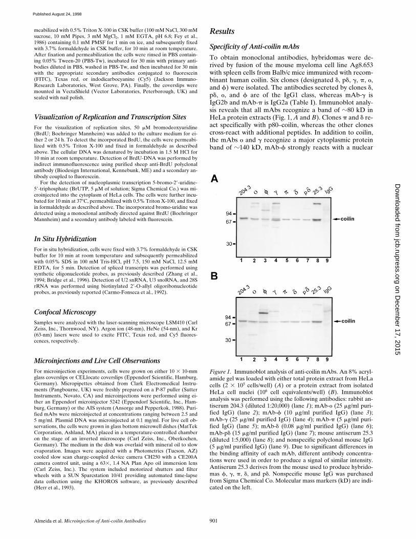
To obtain monoclonal antibodies, hybridomas were de-rived by fusion of the mouse myeloma cell line Ag8.653with spleen cells from Balb/c mice immunized with recom-binant human coilin. Six clones (designated
d
, p
d
,
g
,
p
,
o
,and
f
) were isolated. The antibodies secreted by clones
d
,p
d
,
o
, and
f
are of the IgG1 class, whereas mAb-
g
isIgG2b and mAb-
p
is IgG2a (Table I). Immunoblot analy-sis reveals that all mAbs recognize a band of
z
80 kD inHeLa protein extracts (Fig. 1,
A
and
B
). Clones
p
and
d
re-act specifically with p80–coilin, whereas the other clonescross-react with additional peptides. In addition to coilin,the mAbs
o
and
g
recognize a major cytoplasmic proteinband of
z
140 kD, mAb-
f
strongly reacts with a nuclear
Figure 1. Immunoblot analysis of anti-coilin mAbs. An 8% acryl-amide gel was loaded with either total protein extract from HeLacells (2 3 105 cells/well) (A) or a protein extract from isolatedHeLa cell nuclei (106 cell equivalents/well) (B). Immunoblotanalysis was performed using the following antibodies: rabbit an-tiserum 204.3 (diluted 1:20,000) (lane 1); mAb-o (25 mg/ml puri-fied IgG) (lane 2); mAb-f (10 mg/ml purified IgG) (lane 3);mAb-g (25 mg/ml purified IgG) (lane 4); mAb-p (5 mg/ml puri-fied IgG) (lane 5); mAb-d (0.08 mg/ml purified IgG) (lane 6);mAb-pd (15 mg/ml purified IgG) (lane 7); mouse antiserum 25.3(diluted 1:5,000) (lane 8); and nonspecific polyclonal mouse IgG(5 mg/ml purified IgG) (lane 9). Due to significant differences inthe binding affinity of each mAb, different antibody concentra-tions were used in order to produce a signal of similar intensity.Antiserum 25.3 derives from the mouse used to produce hybrido-mas f, g, p, d, and pd. Nonspecific mouse IgG was purchasedfrom Sigma Chemical Co. Molecular mass markers (kD) are indi-cated on the left.
on Decem
ber 17, 2015jcb.rupress.org
Dow
nloaded from
Published August 24, 1998

The Journal of Cell Biology, Volume 142, 1998 902
protein of
z
110 kD, and mAb-p
d
reveals a minor nuclearprotein band of
z
115 kD. To map the epitopes recognizedby each mAb, a series of His–coilin deletion mutants wasgenerated and probed by immunoblot analysis (Fig. 2).From these data we conclude that the different clones re-act with epitopes distributed along the entire protein se-quence. By immunofluorescence all mAbs labeled coiledbodies (Fig. 3
A
and data not shown), as previously re-ported for clone
d
(Rebelo et al., 1996).
Microinjection of mAbs Affect the CoiledBody Structure
To determine whether the mAbs interfere with coiledbodies in the native environment of the living cell nucleus,each antibody was purified from ascitic fluid and microin-
jected into the nuclei of interphase HeLa cells. The cellswere immediately removed from the microscope stage,permeabilized with detergent, fixed, and then incubatedwith fluorescent secondary antibodies. The time betweeninjection and fixation was
z20 min. As shown in Fig. 3 B,injected mAb-d binds to coiled bodies within this time pe-riod, producing a labeling pattern similar to that obtainedby indirect immunofluorescence on fixed cells (Fig. 3 A).In contrast, when cells are observed 24 h after injection ofmAb-d the coiled bodies are no longer visible (Fig. 3 C),suggesting that binding of mAb-d to coilin affects thestructure of the coiled body. To further confirm this idea,similar experiments were performed using mAb-d prein-cubated with recombinant coilin protein. Purified mAb-dwas incubated for 1 h with increasing concentrations ofcoilin and then used for indirect immunofluorescence onfixed cells. As shown in Fig. 3 D, no signal was detected af-ter incubation of mAb-d with a 1:2 molar excess of coilin,indicating that all antigen binding sites had been blocked.The same preparation of neutralized mAb-d was then mi-croinjected into the nucleus of HeLa cells. As expectedfrom its inability to bind to endogenous coilin, immedi-ately after injection mAb-d appears diffuse throughout thenucleoplasm (Fig. 3 E, also see B). However, there is anadditional faint staining of coiled bodies (Fig. 3 E, arrow),implying that in vivo a small fraction of IgG molecules isstill able to bind to endogenous coilin. This could be dueto a progressive exchange of recombinant coilin by endo-
Figure 2. Epitope mapping of mAbs. (A) Dia-gram showing the reactivity of each mAb with thefull-length coilin and different deletion mutantsof the protein, as determined by immunoblotanalysis. The data show that the epitopes recog-nized by mAbs o, f, and g map between aminoacids 1–127, 187–291, and 292–362, respectively.The mAbs d and pd react with epitopes locatedbetween acids 363 and 481. The mAb-p recog-nizes either an epitope localized around aminoacids 481–482, or a conformational epitopepresent in the coilin mutant encompassing aminoacids 363–576, but absent from the mutants en-compassing amino acids 363–481 and 482–576.(B) Diagram showing the mapped epitopes (Y) inrelation to the complete coilin sequence. Hatchedregions depict the two motifs that closely matchthe consensus sequence of simple and bipartitenuclear localization sequences, NLSa and NLSb,respectively (Bohmann et al., 1995a). The dia-gram also depicts the position of serine 202. Con-version of this amino acid residue into aspartatecauses the disappearance of coiled bodies and aredistribution of coilin to intranucleolar domains(Lyon et al., 1997).
Table I. Immunoglobulin Class, Subclass, and Light Chain Typing of Anti-coilin mAbs
mAb Abbreviated name Ig subtype
1D4-d d IgG1/k5P11-pd pd IgG1/k1G3-g g IgG2b/k5P10-p p IgG2a/k3W8-o o IgG1/k4F9-f f IgG1/k
on Decem
ber 17, 2015jcb.rupress.org
Dow
nloaded from
Published August 24, 1998
Almeida et al. Microinjection of Anti-coilin Antibodies 903
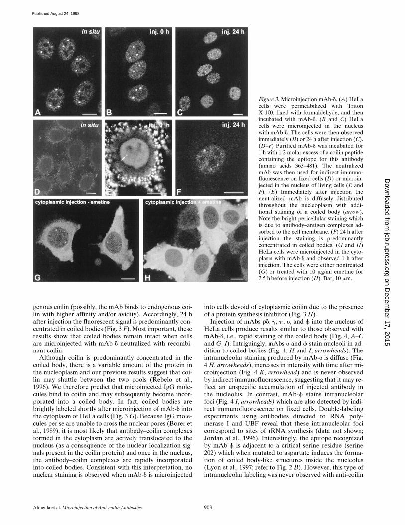
genous coilin (possibly, the mAb binds to endogenous coi-lin with higher affinity and/or avidity). Accordingly, 24 hafter injection the fluorescent signal is predominantly con-centrated in coiled bodies (Fig. 3 F). Most important, theseresults show that coiled bodies remain intact when cellsare microinjected with mAb-d neutralized with recombi-nant coilin.
Although coilin is predominantly concentrated in thecoiled body, there is a variable amount of the protein inthe nucleoplasm and our previous results suggest that coi-lin may shuttle between the two pools (Rebelo et al.,1996). We therefore predict that microinjected IgG mole-cules bind to coilin and may subsequently become incor-porated into a coiled body. In fact, coiled bodies arebrightly labeled shortly after microinjection of mAb-d intothe cytoplasm of HeLa cells (Fig. 3 G). Because IgG mole-cules per se are unable to cross the nuclear pores (Borer etal., 1989), it is most likely that antibody–coilin complexesformed in the cytoplasm are actively translocated to thenucleus (as a consequence of the nuclear localization sig-nals present in the coilin protein) and once in the nucleus,the antibody–coilin complexes are rapidly incorporatedinto coiled bodies. Consistent with this interpretation, nonuclear staining is observed when mAb-d is microinjected
into cells devoid of cytoplasmic coilin due to the presenceof a protein synthesis inhibitor (Fig. 3 H).
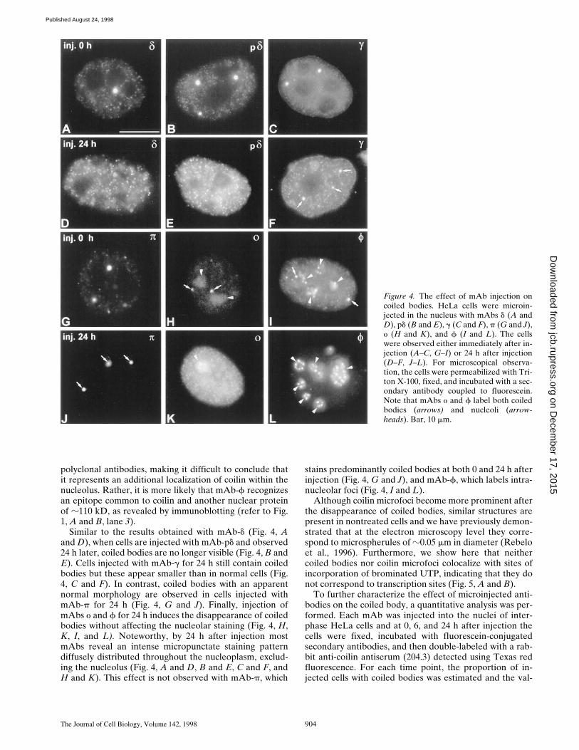
Injection of mAbs pd, g, p, o, and f into the nucleus ofHeLa cells produce results similar to those observed withmAb-d, i.e., rapid staining of the coiled body (Fig. 4, A–Cand G–I). Intriguingly, mAbs o and f stain nucleoli in ad-dition to coiled bodies (Fig. 4, H and I, arrowheads). Theintranucleolar staining produced by mAb-o is diffuse (Fig.4 H, arrowheads), increases in intensity with time after mi-croinjection (Fig. 4 K, arrowhead) and is never observedby indirect immunofluorescence, suggesting that it may re-flect an unspecific accumulation of injected antibody inthe nucleolus. In contrast, mAb-f stains intranucleolarfoci (Fig. 4 I, arrowheads) which are also detected by indi-rect immunofluorescence on fixed cells. Double-labelingexperiments using antibodies directed to RNA poly-merase I and UBF reveal that these intranucleolar focicorrespond to sites of rRNA synthesis (data not shown;Jordan at al., 1996). Interestingly, the epitope recognizedby mAb-f is adjacent to a critical serine residue (serine202) which when mutated to aspartate induces the forma-tion of coiled body-like structures inside the nucleolus(Lyon et al., 1997; refer to Fig. 2 B). However, this type ofintranucleolar labeling was never observed with anti-coilin
Figure 3. Microinjection mAb-d. (A) HeLacells were permeabilized with TritonX-100, fixed with formaldehyde, and thenincubated with mAb-d. (B and C) HeLacells were microinjected in the nucleuswith mAb-d. The cells were then observedimmediately (B) or 24 h after injection (C).(D–F) Purified mAb-d was incubated for1 h with 1:2 molar excess of a coilin peptidecontaining the epitope for this antibody(amino acids 363–481). The neutralizedmAb was then used for indirect immuno-fluorescence on fixed cells (D) or microin-jected in the nucleus of living cells (E andF). (E) Immediately after injection theneutralized mAb is diffusely distributedthroughout the nucleoplasm with addi-tional staining of a coiled body (arrow).Note the bright pericellular staining whichis due to antibody–antigen complexes ad-sorbed to the cell membrane. (F) 24 h afterinjection the staining is predominantlyconcentrated in coiled bodies. (G and H)HeLa cells were microinjected in the cyto-plasm with mAb-d and observed 1 h afterinjection. The cells were either nontreated(G) or treated with 10 mg/ml emetine for2.5 h before injection (H). Bar, 10 mm.
on Decem
ber 17, 2015jcb.rupress.org
Dow
nloaded from
Published August 24, 1998
The Journal of Cell Biology, Volume 142, 1998 904
polyclonal antibodies, making it difficult to conclude thatit represents an additional localization of coilin within thenucleolus. Rather, it is more likely that mAb-f recognizesan epitope common to coilin and another nuclear proteinof z110 kD, as revealed by immunoblotting (refer to Fig.1, A and B, lane 3).
Similar to the results obtained with mAb-d (Fig. 4, Aand D), when cells are injected with mAb-pd and observed24 h later, coiled bodies are no longer visible (Fig. 4, B andE). Cells injected with mAb-g for 24 h still contain coiledbodies but these appear smaller than in normal cells (Fig.4, C and F). In contrast, coiled bodies with an apparentnormal morphology are observed in cells injected withmAb-p for 24 h (Fig. 4, G and J). Finally, injection ofmAbs o and f for 24 h induces the disappearance of coiledbodies without affecting the nucleolar staining (Fig. 4, H,K, I, and L). Noteworthy, by 24 h after injection mostmAbs reveal an intense micropunctate staining patterndiffusely distributed throughout the nucleoplasm, exclud-ing the nucleolus (Fig. 4, A and D, B and E, C and F, andH and K). This effect is not observed with mAb-p, which
stains predominantly coiled bodies at both 0 and 24 h afterinjection (Fig. 4, G and J), and mAb-f, which labels intra-nucleolar foci (Fig. 4, I and L).
Although coilin microfoci become more prominent afterthe disappearance of coiled bodies, similar structures arepresent in nontreated cells and we have previously demon-strated that at the electron microscopy level they corre-spond to microspherules of z0.05 mm in diameter (Rebeloet al., 1996). Furthermore, we show here that neithercoiled bodies nor coilin microfoci colocalize with sites ofincorporation of brominated UTP, indicating that they donot correspond to transcription sites (Fig. 5, A and B).
To further characterize the effect of microinjected anti-bodies on the coiled body, a quantitative analysis was per-formed. Each mAb was injected into the nuclei of inter-phase HeLa cells and at 0, 6, and 24 h after injection thecells were fixed, incubated with fluorescein-conjugatedsecondary antibodies, and then double-labeled with a rab-bit anti-coilin antiserum (204.3) detected using Texas redfluorescence. For each time point, the proportion of in-jected cells with coiled bodies was estimated and the val-
Figure 4. The effect of mAb injection oncoiled bodies. HeLa cells were microin-jected in the nucleus with mAbs d (A andD), pd (B and E), g (C and F), p (G and J),o (H and K), and f (I and L). The cellswere observed either immediately after in-jection (A–C, G–I) or 24 h after injection(D–F, J–L). For microscopical observa-tion, the cells were permeabilized with Tri-ton X-100, fixed, and incubated with a sec-ondary antibody coupled to fluorescein.Note that mAbs o and f label both coiledbodies (arrows) and nucleoli (arrow-heads). Bar, 10 mm.
on Decem
ber 17, 2015jcb.rupress.org
Dow
nloaded from
Published August 24, 1998

Almeida et al. Microinjection of Anti-coilin Antibodies 905
ues at time zero were taken as reference (Fig. 6). The re-sults show that immediately after injection, the proportionof injected cells containing coiled bodies identified by anti-serum 204.3 was similar to that of noninjected cells. At 6and 24 h after injection, the proportion of noninjected cellscontaining coiled bodies remained unaltered, whereas in-jected cells showed significant changes. The mAbs d, o,and f induce a drastic disappearance of coiled bodies. ThemAb pd also reduces the proportion of cells with coiledbodies, although less efficiently than the previous ones.The mAb-g induces a slight decrease in the proportion ofinjected cells containing coiled bodies at 6 h after injec-tion, but this value remains roughly unchanged by 24 h af-ter injection. In striking contrast with these results, mAb-pcauses an increase in the relative numbers of injected cellscontaining coiled bodies.
Importantly, the morphological patterns produced byeach mAb at 24 h after injection are maintained up to 3 d,indicating that within this time period, the antibodies arenot lethal to the cells and remain stable in the nucleus.Furthermore, addition of BrdU to the culture medium ofcells injected for 24–48 h with either mAb-d or mAb-p in-dicates that DNA synthesis and mitosis are occurring atapproximately the same rates among injected and nonin-jected cells (data not shown).
In conclusion, we have produced monoclonal antibodieswhich, depending on their binding sites on coilin, are ableto differentially interfere with the structure of the coiledbody in vivo.
Visualization of Coiled Bodies in Living Cells
To clarify the mechanisms by which the anti-coilin anti-bodies are affecting the coiled body, images of live cellswere recorded at 20–30-min intervals after injection ofmAbs coupled to rhodamine.
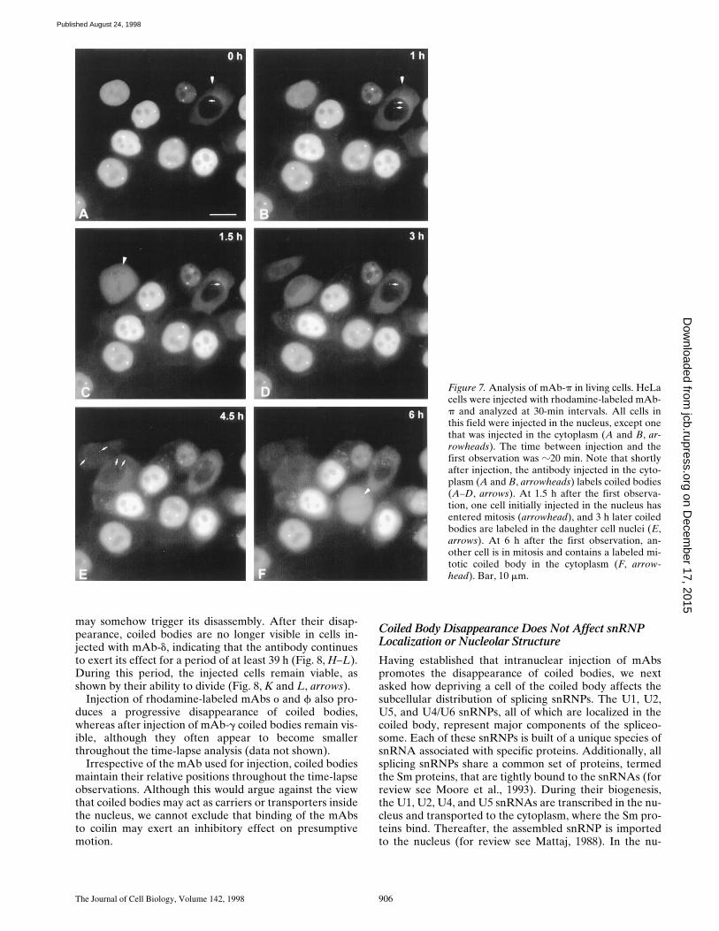
Injection of rhodamine-labeled mAb-p into the nucleus
reveals intensely labeled coiled bodies, which do not ap-pear to change their relative positions during the time-lapse observations (Fig. 7, A–F). Injection of the antibodyin the cytoplasm is rapidly followed by transport to the nu-cleus and incorporation into coiled bodies (Fig. 7, A–D, ar-rows). Importantly, microinjected cells undergo mitosis(Fig. 7, C and F, arrowheads) and labeled coiled bodies areseen in the daughter cells (Fig. 7 E, arrows). This demon-strates that, first, microinjection is not perturbing normalcell physiology and, second, binding of mAb-p to coilin isnot preventing de novo assembly of coiled bodies.
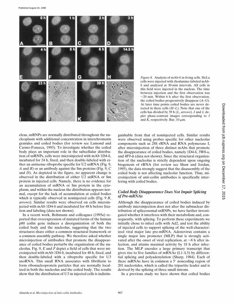
In contrast with the above observations, injection ofrhodamine-labeled mAb-d into the nucleus shows a pro-gressive disappearance of coiled bodies over a period ofz6 h (Fig. 8, A–G). However, coiled bodies are readily vis-ible immediately after injection into the nucleus (Fig. 8 A)or shortly after injection into the cytoplasm (refer to Fig. 3G). Thus, binding of mAb-d is not preventing the assemblyof coilin–antibody complexes into coiled bodies. Possibly,when complexes of mAb-d with coilin are incorporatedinto a coiled body they block the subsequent assembly ofnew protein, leading to a progressive disappearance of thestructure due to its normal turnover rate. Alternatively,the presence of mAb-d/coilin complexes in a coiled body
Figure 5. Coilin does not colocalize with transcription sites. (A)HeLa cells were microinjected with Br-UTP in the cytoplasm,fixed, and then immunolabeled with mAb-p. The sites of incor-porated Br-uridine are stained green and the sites containing coi-lin are stained red. Note that coilin is present both in coiledbodies (arrows) and in numerous nucleoplasmic microfoci (ar-rowheads). (B) HeLa cells were injected in the nucleus withmAb-d. 24 h later the cells were reinjected in the cytoplasm withBr-UTP. Note that the coilin microfoci (stained red) do not colo-calize with transcription sites (stained green). Bar, 10 mm.
Figure 6. Quantitative analysis of the effects of mAb injection oncoiled bodies. Cells were microinjected in the nucleus with mAbsd, pd, g, p, o, and f. The cells were either immediately permeabi-lized and fixed (t0), or further incubated for 6 or 24 h. The in-jected mAb was detected using fluorescein-conjugated secondaryantibodies. Cells were double-labeled with a rabbit anti-coilin an-tiserum (204.3) detected using Texas red fluorescence. For eachmAb, the percentage of injected cells with coiled bodies was esti-mated at each time point (CB[tn]), and the values at time zero(CB[t0]) were taken as reference. For each mAb, at least three in-dependent microinjection experiments were performed. The to-tal number of cells counted per mAb at each time point rangedbetween 100 and 600. on D
ecember 17, 2015
jcb.rupress.orgD
ownloaded from
Published August 24, 1998
The Journal of Cell Biology, Volume 142, 1998 906
may somehow trigger its disassembly. After their disap-pearance, coiled bodies are no longer visible in cells in-jected with mAb-d, indicating that the antibody continuesto exert its effect for a period of at least 39 h (Fig. 8, H–L).During this period, the injected cells remain viable, asshown by their ability to divide (Fig. 8, K and L, arrows).
Injection of rhodamine-labeled mAbs o and f also pro-duces a progressive disappearance of coiled bodies,whereas after injection of mAb-g coiled bodies remain vis-ible, although they often appear to become smallerthroughout the time-lapse analysis (data not shown).
Irrespective of the mAb used for injection, coiled bodiesmaintain their relative positions throughout the time-lapseobservations. Although this would argue against the viewthat coiled bodies may act as carriers or transporters insidethe nucleus, we cannot exclude that binding of the mAbsto coilin may exert an inhibitory effect on presumptivemotion.
Coiled Body Disappearance Does Not Affect snRNP Localization or Nucleolar Structure
Having established that intranuclear injection of mAbspromotes the disappearance of coiled bodies, we nextasked how depriving a cell of the coiled body affects thesubcellular distribution of splicing snRNPs. The U1, U2,U5, and U4/U6 snRNPs, all of which are localized in thecoiled body, represent major components of the spliceo-some. Each of these snRNPs is built of a unique species ofsnRNA associated with specific proteins. Additionally, allsplicing snRNPs share a common set of proteins, termedthe Sm proteins, that are tightly bound to the snRNAs (forreview see Moore et al., 1993). During their biogenesis,the U1, U2, U4, and U5 snRNAs are transcribed in the nu-cleus and transported to the cytoplasm, where the Sm pro-teins bind. Thereafter, the assembled snRNP is importedto the nucleus (for review see Mattaj, 1988). In the nu-
Figure 7. Analysis of mAb-p in living cells. HeLacells were injected with rhodamine-labeled mAb-p and analyzed at 30-min intervals. All cells inthis field were injected in the nucleus, except onethat was injected in the cytoplasm (A and B, ar-rowheads). The time between injection and thefirst observation was z20 min. Note that shortlyafter injection, the antibody injected in the cyto-plasm (A and B, arrowheads) labels coiled bodies(A–D, arrows). At 1.5 h after the first observa-tion, one cell initially injected in the nucleus hasentered mitosis (arrowhead), and 3 h later coiledbodies are labeled in the daughter cell nuclei (E,arrows). At 6 h after the first observation, an-other cell is in mitosis and contains a labeled mi-totic coiled body in the cytoplasm (F, arrow-head). Bar, 10 mm.
on Decem
ber 17, 2015jcb.rupress.org
Dow
nloaded from
Published August 24, 1998
Almeida et al. Microinjection of Anti-coilin Antibodies 907
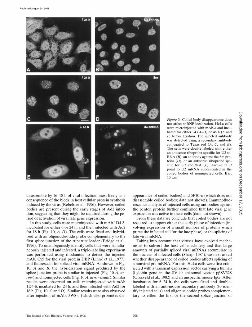
cleus, snRNPs are normally distributed throughout the nu-cleoplasm with additional concentration in interchromatingranules and coiled bodies (for review see Lamond andCarmo-Fonseca, 1993). To investigate whether the coiledbody plays an important role in the subcellular distribu-tion of snRNPs, cells were microinjected with mAb 1D4-d,incubated for 24 h, fixed, and then double-labeled with ei-ther an antisense riboprobe specific for U2 snRNA (Fig. 9,A and B) or an antibody against the Sm proteins (Fig. 9, Cand D). As depicted in the figure, no apparent change isobserved in the distribution of either U2 snRNA or Smprotein in injected cells. Namely, there is no evidence foran accumulation of snRNA or Sm protein in the cyto-plasm, and within the nucleus the distribution appears nor-mal, except for the lack of accumulation at coiled bodieswhich is typically observed in noninjected cells (Fig. 9 B,arrows). Similar results were observed on cells microin-jected with mAb 1D4-d and incubated for 48 h before fixa-tion and labeling (data not shown).
In a recent work, Bohmann and colleagues (1995a) re-ported that overexpression of mutated forms of the humanp80 coilin gene induces a disorganization of both thecoiled body and the nucleolus, suggesting that the twostructures share either a common structural framework ora common assembly pathway. We therefore asked whethermicroinjection of antibodies that promote the disappear-ance of coiled bodies perturbs the organization of the nu-cleolus. Fig. 9, E and F depict a field of cells that were mi-croinjected with mAb 1D4-d, incubated for 48 h, fixed, andthen double-labeled with a riboprobe specific for U3snoRNA. This small RNA associates with fibrillarin toform ribonucleoprotein particles that are normally local-ized in both the nucleolus and the coiled body. The resultsshow that the distribution of U3 in injected cells is indistin-
guishable from that of noninjected cells. Similar resultswere observed using probes specific for other nucleolarcomponents such as 28S rRNA and RNA polymerase I,after microinjection of three distinct mAbs that promotethe disappearance of coiled bodies, namely 1D4-d, 3W8-o,and 4F9-f (data not shown). Since the structural organiza-tion of the nucleolus is strictly dependent upon ongoingbiogenesis of rRNA (for review see Shaw and Jordan,1995), the data strongly suggest that the disassembly of thecoiled body is not affecting nucleolar function. Thus, mi-croinjection of anti-coilin antibodies is specifically inter-fering with coiled bodies.
Coiled Body Disappearance Does Not Impair Splicingof Pre-mRNAs
Although the disappearance of coiled bodies induced byantibody microinjection does not alter the subnuclear dis-tribution of spliceosomal snRNPs, we have further investi-gated whether it interferes with their metabolism and, con-sequently, with splicing. To perform these experiments weinitially chose to infect cells with Ad2, and test the abilityof injected cells to support splicing of the well-character-ized viral major late pre-mRNA. Adenovirus contains asingle major late promoter (MLP) that is strongly acti-vated after the onset of viral replication, at z8 h after in-fection, and attains maximal activity by 18 h after infec-tion. The MLP encodes a large primary transcript thatgives rise to five families of mRNAs (L1–L5) by differen-tial splicing and polyadenylation (Sharp, 1984). Each ofthese mRNAs have in common a 59 noncoding region of201 nucleotides, which is called the tripartite leader and isderived by the splicing of three small introns.
In a previous study we have shown that coiled bodies
Figure 8. Analysis of mAb-d in living cells. HeLacells were injected with rhodamine-labeled mAb-d and analyzed at 30-min intervals. All cells inthis field were injected in the nucleus. The timebetween injection and the first observation wasz20 min. Within 6 h after the first observation,the coiled bodies progressively disappear (A–G).At later time points coiled bodies are never de-tected in these cells (H–L). Note that one of thecells has divided by 39 h (L, arrows). J and L de-pict phase-contrast images corresponding to Iand K, respectively. Bar, 10 mm.
on Decem
ber 17, 2015jcb.rupress.org
Dow
nloaded from
Published August 24, 1998
The Journal of Cell Biology, Volume 142, 1998 908
disassemble by 16–18 h of viral infection, most likely as aconsequence of the block in host cellular protein synthesisinduced by the virus (Rebelo et al., 1996). However, coiledbodies are present during the early stages of Ad2 infec-tion, suggesting that they might be required during the pe-riod of activation of viral late gene expression.
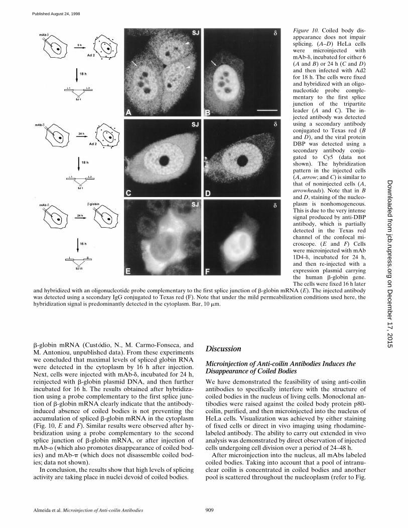
In this study, cells were microinjected with mAb 1D4-d,incubated for either 6 or 24 h, and then infected with Ad2for 18 h (Fig. 10, A–D). The cells were fixed and hybrid-ized with an oligonucleotide probe complementary to thefirst splice junction of the tripartite leader (Bridge et al.,1996). To unambiguously identify cells that were simulta-neously injected and infected, a triple-labeling experimentwas performed using rhodamine to detect the injectedmAb, Cy5 for the viral protein DBP (Linné et al., 1977),and fluorescein for spliced viral mRNA. As shown in Fig.10, A and B, the hybridization signal produced by thesplice junction probe is similar in injected (Fig. 10 A, ar-row) and noninjected cells (Fig. 10 A, arrowheads). Similarresults were observed on cells microinjected with mAb1D4-d, incubated for 24 h, and then infected with Ad2 for18 h (Fig. 10, C and D). Similar results were also observedafter injection of mAbs 3W8-o (which also promotes dis-
appearance of coiled bodies) and 5P10-p (which does notdisassemble coiled bodies; data not shown). Immunofluo-rescence analysis of injected cells using antibodies againstthe penton protein further confirmed that late viral geneexpression was active in these cells (data not shown).
From these data we conclude that coiled bodies are notrequired to support either the early phase of infection (in-volving expression of a small number of proteins whichprime the infected cell for the late phase) or the splicing oflate viral mRNA.
Taking into account that viruses have evolved mecha-nisms to subvert the host cell machinery and that largeamounts of partially spliced viral mRNAs accumulate inthe nucleus of infected cells (Sharp, 1984), we next askedwhether disappearance of coiled bodies affects splicing ofa nonviral pre-mRNA. For this, HeLa cells were first coin-jected with a transient expression vector carrying a humanb-globin gene in the SV-40 epissomal vector pbSV328(Grosveld et al., 1982) and an unspecific mouse IgG. Afterincubation for 6–24 h, the cells were fixed and double-labeled with an anti-mouse secondary antibody (to iden-tify injected cells) and oligonucleotide probes complemen-tary to either the first or the second splice junction of
Figure 9. Coiled body disappearance doesnot affect snRNP localization. HeLa cellswere microinjected with mAb-d and incu-bated for either 24 (A–D) or 48 h (E andF) before fixation. The injected antibodywas detected using a secondary antibodyconjugated to Texas red (A, C, and E).The cells were double-labeled with eitheran antisense riboprobe specific for U2 sn-RNA (B), an antibody against the Sm pro-teins (D), or an antisense riboprobe spe-cific for U3 snoRNA (F). Arrows in Bpoint to U2 snRNA concentrated in thecoiled bodies of noninjected cells. Bar,10 mm.
on Decem
ber 17, 2015jcb.rupress.org
Dow
nloaded from
Published August 24, 1998
Almeida et al. Microinjection of Anti-coilin Antibodies 909
b-globin mRNA (Custódio, N., M. Carmo-Fonseca, andM. Antoniou, unpublished data). From these experimentswe concluded that maximal levels of spliced globin RNAwere detected in the cytoplasm by 16 h after injection.Next, cells were injected with mAb-d, incubated for 24 h,reinjected with b-globin plasmid DNA, and then furtherincubated for 16 h. The results obtained after hybridiza-tion using a probe complementary to the first splice junc-tion of b-globin mRNA clearly indicate that the antibody-induced absence of coiled bodies is not preventing theaccumulation of spliced b-globin mRNA in the cytoplasm(Fig. 10, E and F). Similar results were observed after hy-bridization using a probe complementary to the secondsplice junction of b-globin mRNA, or after injection ofmAb-o (which also promotes disappearance of coiled bod-ies) and mAb-p (which does not disassemble coiled bod-ies; data not shown).
In conclusion, the results show that high levels of splicingactivity are taking place in nuclei devoid of coiled bodies.
Discussion
Microinjection of Anti-coilin Antibodies Induces the Disappearance of Coiled Bodies
We have demonstrated the feasibility of using anti-coilinantibodies to specifically interfere with the structure ofcoiled bodies in the nucleus of living cells. Monoclonal an-tibodies were raised against the coiled body protein p80-coilin, purified, and then microinjected into the nucleus ofHeLa cells. Visualization was achieved by either stainingof fixed cells or direct in vivo imaging using rhodamine-labeled antibody. The ability to carry out extended in vivoanalysis was demonstrated by direct observation of injectedcells undergoing cell division over a period of 24–48 h.
After microinjection into the nucleus, all mAbs labeledcoiled bodies. Taking into account that a pool of intranu-clear coilin is concentrated in coiled bodies and anotherpool is scattered throughout the nucleoplasm (refer to Fig.
Figure 10. Coiled body dis-appearance does not impairsplicing. (A–D) HeLa cellswere microinjected withmAb-d, incubated for either 6(A and B) or 24 h (C and D)and then infected with Ad2for 18 h. The cells were fixedand hybridized with an oligo-nucleotide probe comple-mentary to the first splicejunction of the tripartiteleader (A and C). The in-jected antibody was detectedusing a secondary antibodyconjugated to Texas red (Band D), and the viral proteinDBP was detected using asecondary antibody conju-gated to Cy5 (data notshown). The hybridizationpattern in the injected cells(A, arrow; and C) is similar tothat of noninjected cells (A,arrowheads). Note that in Band D, staining of the nucleo-plasm is nonhomogeneous.This is due to the very intensesignal produced by anti-DBPantibody, which is partiallydetected in the Texas redchannel of the confocal mi-croscope. (E and F) Cellswere microinjected with mAb1D4-d, incubated for 24 h,and then re-injected with aexpression plasmid carryingthe human b-globin gene.The cells were fixed 16 h later
and hybridized with an oligonucleotide probe complementary to the first splice junction of b-globin mRNA (E). The injected antibodywas detected using a secondary IgG conjugated to Texas red (F). Note that under the mild permeabilization conditions used here, thehybridization signal is predominantly detected in the cytoplasm. Bar, 10 mm.
on Decem
ber 17, 2015jcb.rupress.org
Dow
nloaded from
Published August 24, 1998
The Journal of Cell Biology, Volume 142, 1998 910
3 A), this result may reflect either binding of the injectedantibody to coilin molecules present in coiled bodies, orbinding to nucleoplasmic coilin with subsequent assemblyof antibody–coilin complexes into coiled bodies. The find-ing that coiled bodies become labeled upon microinjectionof the antibody into the cytoplasm supports the latter in-terpretation, and it is possible that both mechanisms mayoccur when the antibody is injected into the nucleus.
Within 24 h after injection, two distinct antibody-induced phenotypes are clearly identified. The mAbs d, o,and f cause the disappearance of coiled bodies (as con-firmed by double-labeling using an anti-coilin rabbit se-rum) in .90% of injected cells. In contrast, mAb-p has noapparent effect on the integrity of the coiled body andmAb-g induces only a slight decrease (,20%) in the num-ber of cells with coiled bodies. Most probably, the firstphenotype is caused by the fact that antibody-coilin com-plexes incorporated in a coiled body impair the subse-quent assembly of new coilin molecules. This may eitherdirectly trigger disassembly of the structure or simply leadto its disappearance as a consequence of its high turnoverrate. In fact, two lines of evidence suggest that the coiledbody goes through a rapid cycle of assembly/disassemblyin the nucleus. First, exogenous coilin is able to correctlylocalize in coiled bodies, although overexpression of theprotein fails to increase their size or number (Wu et al.,1994; Bohmann et al., 1995a). Second, treatment of cellswith inhibitors of protein synthesis causes a progressivedisappearance of coiled bodies (Lafarga et al., 1994; Re-belo et al., 1996).
Remarkably, the mAbs capable of interfering with thecoiled body bind to epitopes spread throughout the coilinprotein (refer to Fig. 2). In good agreement with this, itwas previously reported that removal of either amino- orcarboxy-terminal sequences of p80–coilin prevented inter-action with coiled bodies (Bohmann et al., 1995a). Takentogether, these data suggest that assembly of coilin intocoiled bodies requires multiple sequence motifs scatteredalong the protein.
Coiled Body Disappearance Does Not Impair Splicing
Currently proposed hypothesis for the function of thecoiled body are based on the idea that this structure issomehow involved in snRNP metabolism (see Bohmannet al., 1995b). According to the model that snRNPs mustcycle through the coiled body in order to be recycled, as-sembled or modified de novo, one prediction would bethat in the absence of coiled bodies either preexisting ornewly synthesized snRNPs would become unavailable forspliceosome assembly. Although slow proliferating cellssuch as primary human fibroblasts are normally devoid ofcoiled bodies (Huang and Spector, 1992), they contain coi-lin and may assemble coiled bodies (Carmo-Fonseca et al.,1993). Possibly, in the presence of low levels of splicing ac-tivity the requirements for snRNP recycling or de novosynthesis are so low that coiled bodies form only tran-siently or are too small to be clearly identified with thelight microscope. Consistent with this view, coiled bodiesare conspicuous in metabolically activate cells and becomemore numerous upon stimulation of gene expression(Ochs et al., 1995). Therefore, to determine whether the
disappearance of coiled bodies affects snRNP function, itis important to overload the nucleus with highly abundantintron-containing transcripts. Furthermore, taking into ac-count that the half-lives of spliceosomal snRNPs are z20 h(Moore et al., 1993), it is crucial to analyze cells devoid ofcoiled bodies for long periods of time.
Here we have microinjected mAbs that induce disap-pearance of coiled bodies, incubated the cells for 24 h, andthen infected them with adenovirus. The human Ad2causes a productive infection of HeLa cells that proceedsthrough an infectious cycle of z36 h (for review see Hor-witz, 1990). This cycle is conventionally divided into earlyand late stages, separated by the onset of viral replicationwhich occurs at z8 h after infection. After the onset of vi-ral DNA replication, z90% of the genome is expressedand the amount of RNA made increases up to 10-fold.During this phase a single MLP is strongly activated andencodes a large primary transcript that gives rise to fivefamilies of mRNAs (L1–L5) by differential splicing andpolyadenylation (Sharp, 1984). Each of these mRNAshave in common a 59 noncoding region of 201 nucleotides,which is called the tripartite leader and is derived by thesplicing of three small introns. Transcription from MLP at-tains a maximum level at 18 h after infection and then re-mains constant for at least 10 h, yielding large amounts ofstructural polypetides that comprise the virion particle orare involved in packaging viral genomic DNA. During thisperiod, the viruses compete with the host cell for controlof the translational apparatus and Ad2 has evolved multi-ple mechanisms that suppress cellular protein synthesisand enhance translation of late viral mRNAs. Probably asa consequence of the block of host protein synthesis, mostcoiled bodies disassemble by 16–18 h of viral infection(Rebelo et al., 1996). However, coiled bodies are presentduring the early stages of Ad2 infection and it remained anopen issue whether they are required during that period.The data presented here indicate that disappearance ofcoiled bodies does not impair either early or late phases ofadenoviral infection. In fact, cells that were infected aslong as 24 h after microinjection of mAbs contain highamounts of spliced tripartite leader mRNAs in the cyto-plasm and express normal levels of late phase proteins.
In another set of experiments we analyzed the effect ofantibody-induced disassembly of coiled bodies on splicingof transiently overexpressed b-globin transcripts. Controlexperiments show that within 16 h after plasmid injectionthere is an intense cytoplasmic hybridization signal pro-duced by a splice junction probe, indicating that these cellsare splicing large amounts of b-globin pre-mRNAs. Re-markably, cells that were injected with mAb-d, incubatedfor 24 h, and then reinjected with b-globin expression vec-tor continue to splice and export b-globin mRNA to thecytoplasm. Thus, highly abundant pre-mRNAs continue tobe spliced in the absence of coiled bodies. However, dueto the long half-lives of spliceosomal snRNPs, it is possiblethat during the time course of our experiments the level ofpreexisting snRNPs available in the nucleus does not dropbelow the threshold required to maintain splicing activity.
Although the data presented here do not exclude thehypothesis that coiled bodies play a basic role in snRNPbiogenesis or recycling, alternative ideas should be consid-ered concerning the function of this structure. First, the
on Decem
ber 17, 2015jcb.rupress.org
Dow
nloaded from
Published August 24, 1998
Almeida et al. Microinjection of Anti-coilin Antibodies 911
coiled body may simply represent a storage site for sn-RNPs in the nucleus. However, we find this hypothesis un-likely taking into account that coiled bodies are very dy-namic structures strictly dependent upon ongoing RNAand protein synthesis (Carmo-Fonseca et al., 1992; Lafargaet al., 1994; Rebelo et al., 1996). Second, coiled bodies maybe involved in some type of protein or snRNA modifica-tion that is not essential for splicing. For instance, it isknown that snRNAs undergo extensive posttranscrip-tional modifications on base and sugar residues, and it wasrecently proposed that these modifications could be intro-duced into snRNA at the coiled body, by the same (or sim-ilar) enzymatic machinery that is used in the nucleolus tomodify rRNA (Bohmann et al., 1995b). Very little isknown about the functional significance of these modifica-tions in the U1, U2, U5, and U4/U6 snRNPs, and there-fore it is conceivable that at least some may not be essen-tial for spliceosome assembly. Third, coiled bodies may bepreferentially involved in the metabolism of specializedRNAs in differentiated tissue cells such as neurons. Fi-nally, it is important to consider that coiled bodies, like nu-cleoli, may represent the morphological consequence of anactivity that does not necessarily require a dedicated com-partment in the nucleus. In fact, one may argue that thenucleolus as an organized preassembled structure is notessential for ribosome biogenesis, since processing of ribo-somal RNA occurs normally when expressed from a plas-mid containing a single copy of the rRNA gene (Nierras etal., 1997). Here we observe that upon disassembly of thecoiled body by antibody injection, the pool of coilin dis-persed throughout the nucleoplasm is intensified (refer toFig. 4). Thus, it is possible that whatever activity is takingplace at the coiled body it may also occur in associationwith nucleoplasmic coilin. Consistent with this view, it wasrecently reported that coiled bodies tend to associate pref-erentially with tandem repeated or tightly clustered genes,suggesting that, like nucleoli and rRNA genes, the coiledbody may arise as a local concentration of p80–coilin de-termined by expression of these clustered genes (Gao etal., 1997). In conclusion, it will be important for future ex-periments addressing the function of the coiled body to fo-cus on suppressing the expression of p80–coilin at both celland organism level.
We wish to acknowledge D. Ferreira (University of Lisbon, Lisbon, Por-tugal) and A. Lamond (University of Dundee, Dundee, UK) for support.We are also grateful to A. Sawyer (European Molecular Biological Labo-ratory [EMBL], Heidelberg, Germany) for extensive technical advice onmonoclonal antibody production, to J. Romão (Gulbenkian Institute, Qei-ras, Portugal) for animal care facilities, and to our colleagues M. Carvalho,N. Custódio, and L. Teixeira (all three from University of Lisbon) for helpin some experiments and for critical discussions. We thank the followinggroups for generously providing materials used in this study: A. Lamondand K. Bohmann (EMBL) for p80–coilin cDNAs, anti-coilin rabbit serum204.3, and U2 snRNA riboprobe; E. Bridge (University of Uppsala, Upp-sala, Sweden) for adenoviruses, splice-junction probe, anti-DBP, and anti-penton antibodies; M. Antoniou (Guy’s Hospital, London, UK) forb-globin expression vector and splice-junction probes; and W. van Ven-rooij (University of Nijmengen, Nijmengen, The Netherlands) for anti-Smautoimmune serum C45.
This study was supported by grants from Junta Nacional de Investi-gação Científica e Tecnológica/Program PRAXIS XXI and from the Eu-ropean Union. F. Almeida was supported by a PRAXIS XXI doctoral fel-
lowship and a short-term fellowship from the European MolecularBiology Organization.
Received for publication 2 March 1998 and in revised form 26 June 1998.
References
Andrade, L.E.C., E.K.L. Chan, I. Raska, C.L. Peebles, G. Roos, and E.M. Tan.1991. Human autoantibody to a novel protein of the nuclear coiled body: im-munological characterization and cDNA cloning of p80-coilin. J. Exp. Med.173:1407–1419.
Ansorge, W., and R. Pepperkok. 1988. Performance of an automated system forcapillary microinjection into living cells. J. Biochem. Biophys. Methods. 16:283–292.
Bauer, D.W., C. Murphy, Z. Wu, C.-H.H. Wu, and J.G. Gall. 1994. In vitro as-sembly of coiled bodies in Xenopus egg extracts. Mol. Biol. Cell. 5:633–644.
Beven, A.F., G.G. Simpson, J.W.S. Brown, and P.J. Shaw. 1995. The organiza-tion of spliceosomal components in the nuclei of higher plants. J. Cell Sci.108:509–518.
Bohmann, K., J. Ferreira, and A.I. Lamond. 1995a. Mutational analysis of p80coilin indicates a functional interaction between coiled bodies and the nucle-olus. J. Cell Biol. 131:817–831.
Bohmann, K., J. Ferreira, N. Santama, K. Weis, and A.I. Lamond. 1995b. Mo-lecular analysis of the coiled body. J. Cell Sci. 19(Suppl.):107–113.
Borer, R.A., C.F. Lehner, H.M. Eppenberger, and E.A. Nigg. 1989. Major nu-cleolar proteins shuttle between nucleus and cytoplasm. Cell. 56:379–390.
Bridge, E., K.-U. Riedel, B.-M. Johansson, and U. Pettersson. 1996. Spliced ex-ons of adenovirus late RNAs colocalize with snRNP in a specific nuclear do-main. J. Cell Biol. 135:303–314.
Bruck, C., D. Portetelle, C. Glineur, and A. Bollen. 1982. One-step purificationof mouse monoclonal antibodies from ascitic fluid by DEAD Affi-Gel Bluechromatography. J. Immunol. Methods. 53:313–319.
Carmo-Fonseca, M., D. Tollervey, R. Pepperkok, S.M.L. Barabino, A. Merdes,C. Brunner, P.D. Zamore, M.R. Green, E.C. Hurt, and A.I. Lamond. 1991a.Mammalian nuclei contain foci which are highly enriched in components ofthe pre-mRNA splicing machinery. EMBO (Eur. Mol. Biol. Organ.) J. 10:195–206.
Carmo-Fonseca, M., R. Pepperkok, B.S. Sproat, W. Ansorge, M.S. Swanson,and A.I. Lamond. 1991b. In vivo detection of snRNP-organelles in the nucleiof mammalian cells. EMBO (Eur. Mol. Biol. Organ.) J. 10:1863–1873.
Carmo-Fonseca, M., R. Pepperkok, M.T. Carvalho, and A.I. Lamond. 1992.Transcription-dependent colocalization of the U1, U2, U4/U6, and U5 sn-RNPs in coiled bodies. J. Cell Biol. 117:1–14.
Carmo-Fonseca, M., J. Ferreira, and A.I. Lamond. 1993. Assembly of snRNP-containing coiled bodies is regulated in interphase and mitosis: evidence thatthe coiled body is a kinetic nuclear structure. J. Cell Biol. 120:841–852.
Carmo-Fonseca, M., K. Bohmann, M.T. Carvalho, and A.I. Lamond. 1994. P80-coilin. In Manual of Biological Markers of Disease. W.J. van Venrooij andR.N. Maini, editors. Kluwer Academic Publishers. Norwell, MA. B8.2:1–11.
Chan, E.K.L., S. Takano, L.E.C. Andrade, J.C. Hamel, and A.G. Matera. 1994.Structure, expression, and chromosomal localization of human p80-coilingene. Nucl. Acids Res. 22:4462–4469.
Fey, E.G., G. Krochmalnic, and S. Penman. 1986. The nonchromatin substruc-tures of the nucleus: the ribonucleoprotein (RNP)-containing and RNP-depleted matrices analyzed by sequential fractionation and resinless sectionelectron microscopy. J. Cell Biol. 102:1654–1665.
Frey, M.R., and A.G. Matera. 1995. Coiled bodies contain U7 small nuclearRNA and associate with specific DNA sequences in interphase human cells.Proc. Natl. Acad. Sci. USA. 92:5915–5919.
Gall, J.G., A. Tsvetkov, Z. Wu, and C. Murphy. 1995. Is the sphere organelle/coiled body a universal nuclear component? Dev. Genet. 16:25–35.
Gama-Carvalho, M., R.D. Kraus, L. Chiang, J. Valcárcel, M.R. Green, and M.Carmo-Fonseca. 1997. Targeting of U2AF65 to sites of active splicing in thenucleus. J. Cell Biol. 137:975–987.
Gao, L., M.R. Frey, and A.G. Matera. 1997. Human genes encoding U3 snRNAassociate with coiled bodies in interphase cells and are clustered on chromo-some 17p11.2 in a complex inverted repeat structure. Nucleic Acids Res. 25:4740–4747.
Grosveld, F., E. de Boer, C.K. Shewkmaker, and R.A. Flavell. 1982. DNA se-quences necessary for transcription of the rabbit beta-globin gene in vivo.Nature. 295:120–126.
Harlow, E., and D. Lane. 1988. Antibodies: A Laboratory Manual. Cold SpringHarbor Laboratory Press, Cold Spring Harbor, NY. 726 pp.
Hardin, J.W., S.S. Spicer, and W.B. Green, editors. 1969. The paranucleolarstructure, accessory body of Cajal, sex chromatin, and related structures innuclei of rat trigeminal neurons: a cytochemical and ultrastructural study.Anat. Rec. 164:403–432.
Hervás, J.P., J. Villegas, D. Crespo, and M. Lafargs. 1980. Coiled bodies in su-praoptic nucleus of the rat hypothalamus during the postnatal period. Am. J.Anat. 159:447–454.
Herr, S., T. Bastian, R. Pepperkok, C. Boulin, and W. Ansorge. 1993. A fullyautomated image acquisition and analysis system for low light level fluores-cence microscopy. Methods Mol. Cell. Biol. 4:164–170.
on Decem
ber 17, 2015jcb.rupress.org
Dow
nloaded from
Published August 24, 1998
The Journal of Cell Biology, Volume 142, 1998 912
Horwitz, M.S. 1990. Adenoviridae and Their Replication. In Virology. Vol. 1,B.N. Fields and D.M. Knipe, editors. Raven Press, New York. 1679–1721.
Huang, S., and D.L. Spector. 1992. U1 and U2 small nuclear RNAs are presentin nuclear speckles. Proc. Natl. Acad. Sci. USA. 89:305–308.
Huang, S., T.J. Deerinck, M.H. Ellisman, and D.L. Spector. 1994. In vivo analy-sis of the stability and transport of nuclear poly(A)1 RNA. J. Cell Biol. 126:877–899.
Jiménez-Garcia, L.F., M.L. Segura-Valdez, R.L. Ochs, L.I. Rothblum, R. Han-nan, and D.L. Spector. 1994. Nucleogenesis: U3 snRNA-containing prenu-cleolar bodies move to sites of active pre-rRNA transcription after mitosis.Mol. Biol. Cell. 5:955–966.
Jordan, P., M. Mannervik, L. Tora, and M. Carmo-Fonseca. 1996. In vivo evi-dence that TATA-binding protein/SL1 colocalizes with UBF and RNApolymerase I when rRNA synthesis is either active or inactive. J. Cell Biol.133:225–234.
Jordan, P., C. Cunha, and M. Carmo-Fonseca. 1997. The cdk7-cyclinH-MAT1complex associated with TFIIH is localized in coiled bodies. Mol. Biol. Cell.8:1207–1217.
Lafarga, M., M.T. Berciano, M.A. Andres, and P.S. Testillano. 1994. Effects ofcycloheximide on the structural organization of the nucleolus and the coiledbody in normal and stimulated supraoptic neurons of the rat. J. Neurocytol.23:500–513.
Lamond, A.I., and M. Carmo-Fonseca. 1993. The coiled body. Trends Cell Biol.3:198–204.
Linné, T.H., H. Jornvall, and L. Philipson. 1977. Purification and characteriza-tion of the phosphorylated DNA binding protein from adenovirus type 2 in-fected cells. Eur. J. Biochem. 76:481–490.
Lyon, C.E., K. Bohmann, J. Sleeman, and A.I. Lamond. 1997. Inhibition of pro-tein dephosphorylation results in the accumulation of splicing snRNPs andcoiled bodies within the nucleolus. Exp. Cell Res. 230:84–93.
Mattaj, I.W. 1988. U snRNP assembly and transport. In Structure and Functionof Major and Minor Small Nuclear Ribonucleoprotein Particles. M. Birn-stiel, editor. Springer Verlag, New York. 100–114.
Meier, U.T., and G. Blobel. 1994. NAP57, a mammalian nucleolar protein witha putative homolog in yeast and bacteria. J. Cell Biol. 127:1505–1514.
Monneron, A., and W. Bernhard. 1969. Fine structural organization of the in-terphase nucleus in some mammalian cells. J. Ultrastruct. Res. 27:266–288.
Moore, M.J., C.C. Query, and P. Sharp. 1993. Splicing of precursors to mRNAby the spliceosome. In The RNA World. Cold Spring Harbor Laboratory
Press, Cold Spring Harbor, NY. 303–357.Moreno Diaz de la Espina, S., A. Sanchez Pina, M.C. Risueño, F.J. Medina, and
M.E. Fernandez-Gomes. 1980. The role of plant coiled bodies in the nuclearRNA metabolism. Electron Microsc. 2:240–241.
Nierras, C.R., S.W. Liebman, and J.R. Warner. 1997. Does Saccharomyces needan organized nucleolus? Chromosoma. 105:444–451.
Ochs, R.L., T.W. Stein, Jr., L.E.C. Andrade, D. Gallo, E.K.L. Chan, E.M. Tan,and K. Brasch. 1995. Formation of nuclear bodies in hepatocytes of estro-gen-treated roosters. Mol. Biol. Cell. 6:345–356.
Ramón y Cajal, S.R. 1903. Un sencillo método de coloracion selectiva del reti-culo protoplasmico y sus efectos en los diversos organos nerviosos de verte-brados e invertebrados. Trab. Lab. Invest. Biol. 2:129–221.
Raska, I., R.L. Ochs, L.E.C. Andrade, E.K.L. Chan, R. Burlingame, C. Peebles,D. Gruol, and E.M. Tan. 1990. Association between the nucleolus and thecoiled body. J. Struct. Biol. 104:120–127.
Raska, I., L.E.C. Andrade, R.L. Ochs, E.K.L. Chan, C.-M. Chang, G. Roos, andE.M. Tan. 1991. Immunological and ultrastructural studies of the nuclearcoiled body with autoimmune antibodies. Exp. Cell Res. 195:27–37.
Rebelo, L., F. Almeida, C. Ramos, K. Bohmann, A.I. Lamond, and M. Carmo-Fonseca. 1996. The dynamics of coiled bodies in the nucleus of adenovirus-infected cells. Mol. Biol. Cell. 7:1137–1151.
Roth, M.B. 1995. Spheres, coiled bodies and nuclear bodies. Curr. Opin. CellBiol. 7:325–328.
Sharp, P.A. 1984. Adenovirus transcription. In The Adenovirus. H.S Ginsberg,editor. Plenum Press, New York. 173–204.
Shaw, P.J., and E.G. Jordan. 1995. The nucleolus. Annu. Rev. Cell Dev. Biol. 11:93–121.
Tuma, R.S., J.A. Stolk, and M.B. Roth. 1993. Identification and characteriza-tion of a sphere organelle protein. J. Cell Biol. 122:767–773.
Wu, Z., C. Murphy, C.-H.H. Wu, A. Tsvetkov, and J.G. Gall. 1993. Snurpo-somes and coiled bodies. Cold Spr. Harb. Symp. Quant. Biol. 58:747–754.
Wu, Z., C. Murphy, and J.G. Gall. 1994. Human p80-coilin is targeted to sphereorganelles in the amphibian germinal vesicle. Mol. Biol. Cell. 5:1119–1127.
Yannoni, Y.M., and K. White. 1997. Association of the neuron-specific RNAbinding domain-containing protein ELAV with the coiled body in Drosoph-ila neurons. Chromosoma. 105:332–341.
Zhang, G., K.L. Taneja, R.H. Singer, and M.R. Green. 1994. Localization ofpre-mRNA splicing in mammalian nuclei. Nature. 372:809–812.
on Decem
ber 17, 2015jcb.rupress.org
Dow
nloaded from
Published August 24, 1998