Multiple active states and oligomerization of CCR5 revealed by functional properties of monoclonal...
56
1 Multiple active states and oligomerization of CCR5 revealed by the functional properties of monoclonal antibodies Cédric Blanpain 1 , Jean-Marie Vanderwinden 2 , Josef Cihak 3 , Valérie Wittamer 1 , Emmanuel Le Poul 4 , Hassan Issafras 5 , Manfred Stangassinger 3 , Gilbert Vassart 1,6 , Stefano Marullo 5 , Detlef Schlöndorff 7 , Marc Parmentier 1, 8 , Matthias Mack 7 . 1 Institut de Recherche Interdisciplinaire en Biologie Humaine et Nucléaire, 2 Laboratoire de Neurophysiologie, 6 Service de Génétique Médicale and 8 Laboratoire de Cytologie et Cancérologie Expérimentale, Université Libre de Bruxelles, B-1070 Bruxelles, Belgium. 3 Institute for Animal Physiology, University of Munich, 80539 Munich, Germany. 4 Euroscreen s.a., B-1070 Bruxelles, Belgium. 5 Département de Biologie Cellulaire, Institut Cochin de Génétique Moléculaire, 75014 Paris, France. 7 Medizinische Poliklinik, University of Munich, 80336 Munich, Germany. Running title: Functional anti-CCR5 antibodies Keywords: G protein-coupled receptors / CCR5 / monoclonal antibodies / oligomerization / internalization Corresponding author: Marc Parmentier IRIBHN, ULB Campus Erasme 808 route de Lennik B-1070 Bruxelles Belgium. Phone: 32-2-555 41 71 Fax: 32-2-555 46 55 E-mail: mparment@ ulb.ac.be MBC in Press, published on February 4, 2002 as 10.1091/mbc.01-03-0129
Transcript of Multiple active states and oligomerization of CCR5 revealed by functional properties of monoclonal...
1
Multiple active states and oligomerization of CCR5 revealed by
the functional properties of monoclonal antibodies
Cédric Blanpain1, Jean-Marie Vanderwinden2, Josef Cihak3, Valérie Wittamer1, Emmanuel
Le Poul4, Hassan Issafras5, Manfred Stangassinger3, Gilbert Vassart1,6, Stefano Marullo5,
Detlef Schlöndorff7, Marc Parmentier1, 8, Matthias Mack7.
1Institut de Recherche Interdisciplinaire en Biologie Humaine et Nucléaire, 2Laboratoire de
Neurophysiologie, 6Service de Génétique Médicale and 8Laboratoire de Cytologie et
Cancérologie Expérimentale, Université Libre de Bruxelles, B-1070 Bruxelles, Belgium.
3Institute for Animal Physiology, University of Munich, 80539 Munich, Germany.
4Euroscreen s.a., B-1070 Bruxelles, Belgium. 5Département de Biologie Cellulaire, Institut
Cochin de Génétique Moléculaire, 75014 Paris, France. 7Medizinische Poliklinik, University
of Munich, 80336 Munich, Germany.
Running title: Functional anti-CCR5 antibodies
Keywords: G protein-coupled receptors / CCR5 / monoclonal antibodies / oligomerization /
internalization
Corresponding author: Marc Parmentier IRIBHN, ULB Campus Erasme 808 route de Lennik B-1070 Bruxelles Belgium. Phone: 32-2-555 41 71 Fax: 32-2-555 46 55 E-mail: mparment@ ulb.ac.be
MBC in Press, published on February 4, 2002 as 10.1091/mbc.01-03-0129

2
Abstract
CC-chemokine receptor 5 (CCR5) is the principal coreceptor for macrophage-tropic strains
of human immunodeficiency virus type I (HIV-1). We have generated a set of anti-CCR5
monoclonal antibodies (MAbs) and characterized them in terms of epitope recognition,
competition with chemokine binding, receptor activation and trafficking, and coreceptor
activity. MC-4, MC-5 and MC-7 mapped to the amino-terminal domain, MC-1 to the second
extracellular loop, and MC-6 to a conformational epitope covering multiple extracellular
domains. MC-1 and MC-6 inhibited RANTES, MIP-1β and Env binding, whereas MC-5
inhibited MIP-1β and Env but not RANTES binding. MC-6 induced signaling in different
functional assays, suggesting that this MAb stabilizes an active conformation of CCR5. Flow
cytometry and real time confocal microscopy showed that MC-1 promoted strong CCR5
endocytosis. MC-1 but not its monovalent isoforms induced an increase in the transfer of
energy between CCR5 molecules. Also, its monovalent isoforms bound efficiently, but did
not internalize the receptor. In contrast, MC-4 did not prevent RANTES binding or
subsequent signaling, but inhibited its ability to promote CCR5 internalization. These results
suggest the existence of multiple active conformations of CCR5 and indicate that CCR5
oligomers are involved in an internalization process which is distinct from that induced by the
receptor's agonists.

3
Introduction
Chemokines constitute a large family of proteins that regulate leukocyte recruitment to
sites of inflammation and coordinate their trafficking throughout the body. They mediate
these functions through the binding and activation of seven-transmembrane domains G
protein-coupled receptors (GPCRs) specifically expressed by various populations of
leukocytes (Murphy et al., 2000; Baggiolini, 1998). CCR5 is a functional receptor for the
inflammatory CC-chemokines MIP-1α, MIP-1β, RANTES, MCP-2 and MCP-4 (Samson et
al., 1996a; Murphy et al., 2000). It is expressed on memory T lymphocytes, macrophages,
dendritic cells, thymocytes, hematopoietic progenitors, and other cell populations (Murphy et
al., 2000). CCR5 is thought to be involved in the recruitment of leukocytes in a growing
number of inflammatory diseases, such as rheumatoid arthritis, multiple sclerosis and asthma,
and it plays a major role in AIDS pathogenesis (Berger et al., 1999; Gerard and Rollins,
2001). Cellular entry of HIV is initiated by the interaction between the gp120 subunit of the
viral Env glycoprotein, CD4 and a coreceptor that belongs to the chemokine receptor family.
CCR5 is the principal coreceptor for macrophage tropic (M-tropic or R5) HIV strains, which
are responsible for viral transmission and predominate during the asymptomatic phase of the
disease. The essential role of CCR5 in HIV pathogenesis was demonstrated by the strong
resistance towards HIV infection of individuals homozygous for a non-functional allele
(CCR5∆32) of the coreceptor gene (Samson et al., 1996b; Liu et al., 1996). These individuals
have no obvious pathological phenotype, making CCR5 an attractive candidate for therapeutic
intervention. Agents proposed as potential viral entry blockers include chemokines or
chemokine analogues, monoclonal antibodies and chemical antagonists (Moore and
Stevenson, 2000). The first described inhibitors of CCR5 coreceptor function were its natural

4
ligands MIP-1α, MIP-1β and RANTES (Cocchi et al., 1995). Synthetic derivatives of natural
chemokines, such as AOP-RANTES were later shown to display enhanced HIV suppressive
activities (Simmons et al., 1997). Two complementary mechanisms have been proposed to
account for the ability of chemokines to inhibit HIV infection. First, chemokines compete for
gp120 binding (Trkola et al., 1996). Second, agonists induce CCR5 internalization, resulting
in a prolonged reduction of the coreceptor number at the cell surface (Mack et al., 1998;
Amara et al., 1997; Alkhatib et al., 1997), a parameter found to be critical for HIV infection
(Wu et al., 1997a). The relative contribution of these two mechanisms remains to be clarified
in each case. The pronounced antiviral activity of AOP-RANTES has been attributed to the
efficient induction of endocytosis and the inability of CCR5 to re-accumulate on the cell
surface after removal of the ligand (Mack et al., 1998; Signoret et al., 2000). It has also been
suggested that CCR5 endocytosis induced by antibodies may account for HIV resistance of
exposed-uninfected individuals (Lopalco et al., 2000).
Monoclonal antibodies blocking chemokine and gp120 binding to CCR5 have been
described (Wu et al., 1997b; Lee et al., 1999; Olson et al., 1999; Hill et al., 1998), as well as a
first set of chemical CCR5 antagonists (Baba et al., 1999), that are presently regarded as
candidate therapeutic inhibitors of viral entry. However, from a theoretical point of view,
compounds able to inhibit gp120 binding and promote efficient and prolonged internalization
of CCR5, without triggering the intracellular signaling cascades, would constitute ideal anti-
viral agents. Monoclonal antibodies interacting with various epitopes of a receptor constitute
interesting tools to study the relationship between stabilization of an "active" receptor
conformation and the various consequences of this activation (G protein coupling,
internalization, …). Moreover, the availability of dimeric or monomeric isoforms of the same
monoclonal allows to determine the contribution of receptor dimerization in the studied

5
processes.
In this study, we have characterized a novel set of anti-CCR5 MAbs, and have determined
their functional properties. Their epitopes were characterized by using a large panel of
chimeric and mutant receptors. We correlated these findings with the ability of the MAbs to
inhibit chemokine and/or gp120 binding, to modulate activation of intracellular cascades, and
to influence receptor trafficking.

6
Materials and Methods
Generation of monoclonal antibodies against CCR5
BALB/c mice were immunized at four weeks intervals by at least 6 intraperitoneal
injections of CHO cells stably transfected with CCR5. Four days after the last injection, the
spleens were removed and the splenocytes fused with P3X63-Ag8 plasmocytoma cells.
Culture supernatants were screened by flow cytometry on CHO cells expressing CCR5, or
CXCR4 as control. In addition to MC-1 (Mack et al., 1998) and MC-5 (Mack et al., 2000),
three monoclonal antibodies (MC-4, MC-6 and MC-7) were obtained, that specifically
recognize CCR5 and do not cross-react with CHO cells overexpressing CCR1 to 4 or CXCR4.
None of the MAbs stained CCR5-deficient (CCR5 ∆32/∆32) PBMCs. Apart from the clone
MC-5 (IgG-2a), all other clones were of IgG-1 isotype.
Generation of the monovalent MAbs isoforms
A plasmid encoding an MC-1 single-chain fragment (ScFv-MC-1) was performed by
reverse transcription on total RNA extracted from the αCCR5 hybridoma MC-1 with random
hexamers and the SuperScript reverse transcriptase (Gibco). The light and heavy variable
domains were cloned by PCR amplification using Pfu polymerase (Orlandi et al., 1989). As
described previously, the two domains were joined by a linker coding for (Gly4Ser1)3 and a C-
terminal tail encoding 6 histidines was attached to facilitate purification. The single-chain
fragment was expressed in the periplasmic space of E. coli and purified by Ni-NTA (Qiagen)
as described (Mack et al., 1995).
The F(ab) fragments were obtained by digesting MC-1, MC-4 and MC-6 mAbs (2 mg/ml
each) respectively for 2 h with 0.02 mg/ml, for 4 h with 0.02 mg/ml, and for 2 h with 0.1
7
mg/ml papain (Sigma) in PBS containing 20 mM EDTA and 20 mM cysteine (Sigma). After
stopping the reaction with 30 mM iodocetamide, the antibodies were dialyzed overnight
against PBS. Fc fragments and undigested mAbs were removed with Protein A Sepharose
CL-4B (Pharmacia). Aliquots of each digestion were checked by SDS-PAGE and Coomassie
blue staining. Using FITC-labeled secondary antibodies specific for F(ab) fragments (Jackson
ImmunoResearch), similar mean channel fluorescence values were obtained with F(ab)
fragments and half-equimolar amounts of the parental monoclonal antibodies, indicating that
the binding properties were not affected by the digestion procedure.
CCR5 constructs
All CCR5 constructs used in this study have been previously described (Samson et al.,
1997; Lee et al., 1999; Blanpain et al., 1999a; Blanpain et al., 2000, Blanpain et al., 2001 ).
The constructs were sequenced and subcloned into the bicistronic expression vector pEFIN3
for the generation of stable cell lines as previously described (Samson et al., 1997).
Cell culture and expression of mutant receptors in CHO-K1 cells
CHO-K1 cells stably expressing apoaequorin, Gα16 and wild-type or mutant CCR5
receptor (Blanpain et al., 1999b) were cultured in HAM’s F12 medium supplemented with
10% fetal calf serum (Life Technologies), 100 units/ml penicillin, 100 µg/ml streptomycin
(Life Technologies), 250 µg/ml Zeocin (Invitrogen) and 400 µg/ml G418 (Life Technologies).
FACS analysis
Binding affinities of the different MAbs for CCR5 were determined by flow cytometry.
CCR5-expressing CHO-K1 cells were incubated with MAbs for 30 min on ice, washed,
stained with different anti-mouse IgG secondary Abs conjugated with phycoerythrin (Sigma),
8
FITC (Sigma) or Texas Red (Jackson Immunoresearch Laboratories) and analyzed on a
FACScan (Becton-Dickinson). The binding parameters were determined by non-linear
regression using the PRISM Software.
Epitopes were mapped by flow cytometry using a panel of about 70 CHO-K1 cell lines
stably expressing chimeric and point mutant receptors (Samson et al., 1997; Lee et al., 1999;
Blanpain et al., 1999a; Blanpain et al., 2000). Cells were incubated for 30 minutes on ice with
saturating concentrations of anti-CCR5 MAbs, washed and stained with PE-conjugated anti-
mouse Ig Ab (Sigma). CHO-K1 cells expressing CCR2b were used as negative control.
For endocytosis experiments, 5 x 10⁵ CHO cells expressing wtCCR5 were incubated for 30
minutes at 37° with chemokines or MAbs. Cells were then placed on ice, incubated with 15
-1 or MC-4 or their respective F(ab) fragments for 1 hour, washed with cold PBS, and
stained with FITC-conjugated anti-mouse Ig Ab (F313, Dako). Cells were washed and
analyzed on a FACSCalibur (Becton Dickinson). For experiments using ScFv-MC-1, cells
were incubated for 45 min with 3 µg/ml ScFv-MC-1, on ice or 37° as indicated. Cells were
washed twice with cold PBS, and incubated with 4 µg/ml anti-His Ab (Dianova) for 45 min,
on ice or at 37° as indicated. Cells were washed again with cold PBS and stained with PE-
labeled rabbit anti-mouse F(ab)2 (Dako), and analyzed on a FACSCalibur using CellQuest
software.
For inhibition of chemokine-induced CCR5 endocytosis by MAbs, CHO cells expressing
CCR5 were incubated on ice with medium or MC-4 or MC-4 F(ab) for 30 min. Cells were
then incubated with RANTES or AOP-RANTES for 30 min at 37°. The cells were washed,
incubated with 15 µg/ml MC-4 or MC-4 F(ab) followed by FITC-conjugated anti-mouse
F(ab) Ig Ab (Dako).

9
Binding assays
Competition binding assays were performed with CCR5 expressing CHO-K1 cells as
described (Blanpain et al., 1999a), using 0.08 nM [125I]-MIP-1β or [125I]-RANTES (2000
Ci/mmol, Amersham-Pharmacia) as tracer, variable concentrations of MAbs and 40,000 cells.
Total binding was measured in the absence of competitor and non-specific binding was
measured in the presence of a 100-fold excess of unlabelled chemokine. Soluble JRFL gp120
was iodinated as described (Lee et al., 1999). Env binding assays were performed on CCR5
expressing CHO-K1 cells with [125I]-JRFL gp120 as tracer, in the presence of 100 nM sCD4
and competitors. Total binding was measured in the absence of competitor and non-specific
binding was measured in the presence of a 100 fold excess of unlabelled gp120.
Aequorin-based functional assay
The functional response to chemokines and monoclonals was analyzed with an aequorin-
based assay as described (Blanpain et al., 1999b). Inhibition of chemokine signaling by anti-
CCR5 MAbs was analyzed with the same assay. MAbs were incubated for 30 min at room
temperature with 50,000 CHO-K1 cells expressing CCR5, then RANTES or MIP-1β (1 nM
final concentration) were added to the cell suspension, and the luminescence was recorded for
30 s in a luminometer. 100 % stimulation was defined as the response to 1 nM chemokine in
the absence of MAbs, and 0 % as the luminescence in the absence of ligand. A chemokine
dose-response curve was used as positive control in each experiment.
GTPγγS binding assay
Membranes of CHO-K1 cells expressing CCR5 (20 µg/ml), prepared from cells treated or
not with 100 ng/ml Pertussis toxin (PTX) for 18 hours, were incubated in 20 mM HEPES pH

10
7.4, 100 mM NaCl, 3 mM MgCl2, 3 µM GDP and 10µg/ml saponin, with different
concentrations of RANTES or MAbs, in 96-well microplates (Basic FlashPlates, New
England Nuclear), for 15 minutes at room temperature. After addition of [35S]-GTPγS (0.1
nM, Amersham-Pharmacia), the microplates were shaken for 1 min and incubated further for
30 min at 30°. The incubation was stopped by centrifugation of the plates for 10 min at 800 g
and 4°, and aspiration of the supernatant. Microplates were counted in a TopCount (Packard)
for 1 min per well. Neither RANTES nor MAbs did affect [35S]-GTPγS binding to membranes
of CHO-K1 cells expressing other related (CCR8) or unrelated (CRF2) GPCRs. Functional
parameters were determined with the PRISM software (Graphpad Software) using nonlinear
regression applied to a sigmoidal dose-response model.
Inhibition of cAMP accumulation
Inhibition of cAMP accumulation by chemokines and monoclonals was performed on
CCR5 expressing cells spread on Petri dishes (25,000 cells per well) containing cultured
overnight. Cells were preincubated for 15 min in Krebs-Ringer HEPES (KRH) buffer and 1
mM IBMX (Calbiochem), and then incubated for 20 min in the same medium supplemented
with 5 µM forskolin and variable concentrations of RANTES or 10 µg/ml MAbs. The cAMP
content was measured by ELISA (cAMP-screen, CS100, Tropix) according to the procedure
specified by the manufacturer.
In vivo cellular assays for receptor trafficking and oligomerization
For confocal microscopy in living cells, clonal cell lines expressing CCR5-GFP were seeded
on 22 mm round glass coverslips, and grown for 18 hours. Coverslips were rinsed in
DMEM/F12 and placed in the observation chamber (maintained at 37°) of a MRC 1024 confocal
microscope (Bio-Rad Laboratories) fitted on an Axiovert 100 inverted microscope (Zeiss)
11
equipped with a Plan-Neofluar 40x/1.3 oil immersion objective (Zeiss). The 488 nm excitation
beam of an Argon-Krypton laser and a 522-532 nm band-pass emission filter were used for
viewing EGFP. The 568 nm excitation beam and a 605-632 nm band-pass emission filter were
used for viewing transferrin alexafluor 564. The beam power was kept below 10% of maximal
power, in order to reduce photobleaching and phototoxicity. Pinhole was set to generate 1 µm-
thick optical sections. Fields of interest (512 x 512 pixels) were selected visually. Data were
sequentially collected for each fluorochrome (approximate collection time: 4 sec), every 3 min
for the indicated time. Cells expressing CCR5-GFP were incubated with 50 µg/ml alexafluor-
transferrin (Molecular Probes) alone or together with ligands for 15 to 45 min, and washed three
times before viewing.
For induction of CCR5-GFP endocytosis, MAbs (10 µg/ml) or RANTES (100 nM) were
added to the cells, and images were acquired every 3 min for 45 min. For ScFv- MC-1
experiments, cells were first incubated for 45 min with ScFv-MC-1, washed three times,
incubated with anti-His (4 µg/ml) for 30 min, and images were collected every 3 min. No
endocytosis was seen with control IgG or anti-His Ab. For inhibition of CCR5-GFP
endocytosis, the cells were first incubated with MAbs (10 µg/ml) for 45 minutes, then with
100 nM RANTES, and frames were acquired every 3 min for 45 min.
For determining the endocytic pathways, CCR5-GFP expressing cells were incubated with
100 ng/ml Pertussis toxin (Sigma) for 18 hours, 0.45M Sucrose for 1 hour , 2.5 µg/ml filipin
III (Sigma) for 45 minutes, or 10 mM β-methyl-cyclodextrine (Sigma) for 45 minutes at 37°,
and then tested for endocytosis in DMEM/F12 containing the inhibitor.
Arrestin translocation assays were performed by transfecting 100 ng of β-arrestin 2-EGFP
(gift of Mark Scott) into CCR5-expressing cells. The day after, cells were analyzed by

12
confocal microscopy as described above following addition of RANTES or MAbs.
The bioluminescence resonance energy transfer (BRET) assay was performed as described
by Angers et al. (2000). Briefly, humanized Renilla luciferase (Packard) and the yellow
variant of GFP (Clontech) were fused to the last C-terminal residue of CCR5 and expressed in
HEK293 cells. Fusion proteins were expressed at the plasma membrane and were internalized
upon agonist stimulation (as determined by FACS analysis). In stable clones expressing either
wild-type CCR5 or the fusion proteins, RANTES and MIP-1β resulted in the inhibition of
forskolin-induced cAMP production. Antibody-promoted changes of BRET-ratio were
calculated by subtracting the basal BRET ratio, measured in the absence of antibodies, from
the BRET ratios observed in the presence of the indicated antibodies. The details of the
application of the BRET assay to CCR5 will be described elsewhere (Issafras and colleagues,
manuscript in preparation).
13
Results
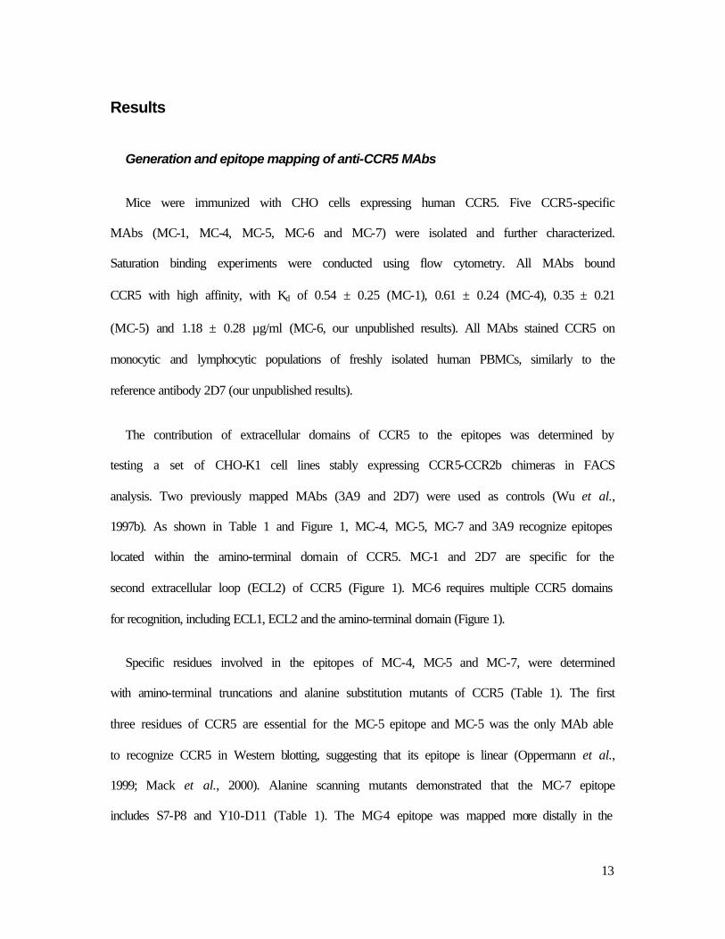
Generation and epitope mapping of anti-CCR5 MAbs
Mice were immunized with CHO cells expressing human CCR5. Five CCR5-specific
MAbs (MC-1, MC-4, MC-5, MC-6 and MC-7) were isolated and further characterized.
Saturation binding experiments were conducted using flow cytometry. All MAbs bound
CCR5 with high affinity, with Kd of 0.54 ± 0.25 (MC-1), 0.61 ± 0.24 (MC-4), 0.35 ± 0.21
(MC-5) and 1.18 ± 0.28 µg/ml (MC-6, our unpublished results). All MAbs stained CCR5 on
monocytic and lymphocytic populations of freshly isolated human PBMCs, similarly to the
reference antibody 2D7 (our unpublished results).
The contribution of extracellular domains of CCR5 to the epitopes was determined by
testing a set of CHO-K1 cell lines stably expressing CCR5-CCR2b chimeras in FACS
analysis. Two previously mapped MAbs (3A9 and 2D7) were used as controls (Wu et al.,
1997b). As shown in Table 1 and Figure 1, MC-4, MC-5, MC-7 and 3A9 recognize epitopes
located within the amino-terminal domain of CCR5. MC-1 and 2D7 are specific for the
second extracellular loop (ECL2) of CCR5 (Figure 1). MC-6 requires multiple CCR5 domains
for recognition, including ECL1, ECL2 and the amino-terminal domain (Figure 1).
Specific residues involved in the epitopes of MC-4, MC-5 and MC-7, were determined
with amino-terminal truncations and alanine substitution mutants of CCR5 (Table 1). The first
three residues of CCR5 are essential for the MC-5 epitope and MC-5 was the only MAb able
to recognize CCR5 in Western blotting, suggesting that its epitope is linear (Oppermann et al.,
1999; Mack et al., 2000). Alanine scanning mutants demonstrated that the MC-7 epitope
includes S7-P8 and Y10-D11 (Table 1). The MC-4 epitope was mapped more distally in the

14
CCR5 amino-terminus (residues 14-17 and E18). MC-1 and MC-6, like 2D7, were not
affected by mutations in CCR5 N-terminus (Table 1).
Chimeras and point mutants involving ECL2 were used to specify the epitopes of MC-1
and MC-6 (Table 1). MC-1 and 2D7 recognized the first part of ECL2. MC-6 binding was
affected by replacement of both the first and the second halves of ECL2, and by the point
substitutions K171A and E172A (but not R168A) in the first part of ECL2 (Figure 1 and
Table 1). Other point mutations known to affect the conformation of the extracellular domains
of CCR5, such as C178R (that disrupts the disulfide bond linking ECL1 and ECL2, Blanpain
et al., 1999b; 2000), reduced or prevented binding of MC-1, MC-6 and 2D7, but had little
effects on other MAbs. The other point mutants tested had no effects on MAb recognition.
MC-6 binding to CCR5 promotes G protein activation but not calcium
mobilization.
Using CCR5-expressing CHO cells, we examined the ability of the different MAbs (at the
concentration of 10 µg/ml) to promote [35S]-GTPγS binding to membranes. RANTES
promoted a marked and dose-dependent increase in [35S]-GTPγS binding, with an EC50 of 2.3
nM (Figure 2A). Among the MAbs tested, only MC-6, but not its F(ab) fragment, induced a
significant increase in GTPγS binding to membranes of CCR5 expressing cells (Figure 2B
and our unpublished results). This effect was blocked by Pertussis toxin pretreatment (our
unpublished results). Noteworthy, MC-1 had no effect in this assay, although it promoted
receptor internalization, as shown below. Neither RANTES, nor MC-1 or any other MAb
increased GTPγS binding to membranes of cells expressing other receptors (our unpublished
results). MC-6, but not its F(ab) fragment, was also able to inhibit cAMP accumulation in
CCR5 expressing CHO-K1 cells with an efficacy similar to that found in the GTPγS assay

15
(Figure 2C).
We also investigated whether the MAbs could induce intracellular calcium release by using
an aequorin-based assay. None of the MAbs, including MC-6, were able to promote calcium
signaling in CCR5-expressing cells, whereas RANTES induced a robust response
characterized by an EC50 of 2.5 nM (our unpublished results).
MAbs differently antagonize the function of RANTES, MIP-1ββ and gp120 onto
CCR5.
We next investigated the ability of the MAbs to compete for [125I]-MIP-1β or [125I]-
RANTES binding to CCR5. As shown in Figure 3A, MC-1 and MC-6 inhibited both MIP-1β
and RANTES binding with an IC50 < 1 µg/ml. MC-4 and control IgG did not compete for
chemokine binding. MC-5 competed partially for MIP-1β binding but competed very poorly
for RANTES binding up to concentrations of 10 µg/ml. We further tested whether these
MAbs prevent CCR5 activation by chemokines. In agreement with the binding data, MC-1,
MC-5 and MC-6 inhibited calcium mobilization induced by MIP-1β (1 nM), in a
concentration-dependent manner (Figure 3B). MC-1 and MC-6, but not MC-5, inhibited
calcium signaling in response to RANTES (1 nM). MC-4 and control IgG had no effect.
We and others have shown that chemokines and HIV Env bind overlapping but distinct
CCR5 domains (Rucker et al., 1996; Blanpain et al., 1999a; Wu et al., 1997b; Farzan et al.,
1998). We therefore tested the ability of our MAbs to compete for the binding of [125I]-JRFL-
gp120 to CCR5. MC-1, MC-5, MC-6, and the reference MAb 2D7 efficiently inhibited JRFL-
gp120 binding in the presence of soluble CD4 (Figure 3C). Neither control isotype IgG, nor
MC-4, inhibited Env binding up to concentrations of 10 µg/ml.

16
Bivalent, but not monovalent MC-1, promotes CCR5 endocytosis
The ability of our MAbs to induce CCR5 internalization in CHO-K1 cells was
investigated. As shown in figure 4A, MC-1 induced a dose-dependent downmodulation of the
cell surface receptor. As measured by FACS analysis, 5 µg/ml MC-1 induced a 50% decrease
of cell surface CCR5, similar to the level of downmodulation obtained with 100 nM RANTES
(Fig 4A and 4B). No significant endocytosis was seen when cells were incubated on ice with
MC-1 (our unpublished results). Incubation of cells at 37° with MC-4 or MC-5 had no effect
(Figure 4A and our unpublished results). Induction of internalization by MC-1 was also found
in lymphocyte and monocyte populations expressing CCR5 naturally. Given the lower
expression level of the receptor in these cells, the phenomenon was however less
demonstrative than in CHO cells (our unpublished results).
Since MC-1 promoted CCR5 endocytosis without triggering detectable signaling, we
investigated whether MC-1 or RANTES could induce different routes of receptor endocytosis.
CHO-K1 cells expressing high levels of a CCR5-GFP chimeric protein were generated. This
chimeric receptor was activated by chemokines with a concentration-response curve similar to
wtCCR5 (our unpublished results). In the absence of ligands, cells expressing CCR5-GFP
showed intense fluorescence at the cell surface, although a significant proportion of the fusion
protein was localized intracellularly (Figure 5A). This fraction of la beling likely corresponds
partly to biosynthetic compartments, such as endoplasmic reticulum (ER) and Golgi complex,
but also to endosomal compartments. Indeed, the early endosome marker, AlexaFluor 594-
Transferrin, colocalized with intracellular CCR5-GFP, even in the absence of ligands (Figure
5A), and recycled efficiently from this endosomal compartment to the cell surface (our

17
unpublished results). Addition of 100 nM RANTES to these cells induced a profound
subcellular redistribution of the fluorescence (Figures 5B-E, and video 1). Shortly after
agonist addition, a local enhancement of membrane-associated fluorescence was observed,
possibly resulting from receptor clustering. Ruffling of membranes was also apparent as a
consequence of receptor signaling and cytoskeletal rearrangements. After 10 to 15 min,
CCR5-GFP was internalized into endocytic vesicles that progressively fused with larger
endosomal compartments. In contrast to endocytosis mediated by RANTES, the subcellular
redistribution of CCR5-GFP induced by MC-1 occurred in a very different manner in terms of
morphology and time course (Figures 5 F-J and Video 2). Immediately after MC-1 addition
(10 µg/ml), enhancement of cell surface fluorescence was observed, but soon thereafter, a
large number of small intracellular vesicles was formed, which did not fuse further with
endosomes (Figure 5G). MC-5, MC-6 and isotype control IgG were unable to induce CCR5-
GFP endocytosis (our unpublished results).
In order to investigate further in vivo the involvement of oligomers in the endocytosis
process mediated by MC-1, we took advantage of a new biophysical assay based on
bioluminescence resonance energy transfer (BRET), which allows to monitor the interaction
between two fusion proteins (Angers et al. 2000). Addition of 10 µg/ml MC-1 produced a
robust increase of the BRET signal (Figure 6A) in 293T cells coexpressing CCR5-luc and
CCR5-YFP, with kinetics similar to that found for CCR5-GFP endocytosis (our unpublished
results). Neither MC-4, MC-5, MC-6 (Figure 6A), nor control IgG (our unpublished results)
induced changes in the BRET signal. These results indicate that MC-1 induces a profound
change in the physical proximity between two or more CCR5 molecules.
We also investigated the functional properties of monovalent versions of MC-1 by flow
cytometry and confocal microscopy. Both a single chain fragment of MC-1 (ScFv-MC-1)
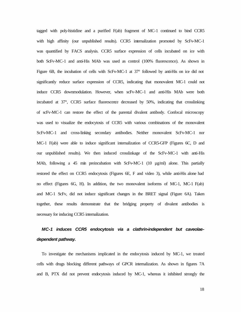
18
tagged with poly-histidine and a purified F(ab) fragment of MC-1 continued to bind CCR5
with high affinity (our unpublished results). CCR5 internalization promoted by ScFv-MC-1
was quantified by FACS analysis. CCR5 surface expression of cells incubated on ice with
both ScFv-MC-1 and anti-His MAb was used as control (100% fluorescence). As shown in
Figure 6B, the incubation of cells with ScFv-MC-1 at 37° followed by anti-His on ice did not
significantly reduce surface expression of CCR5, indicating that monovalent MC-1 could not
induce CCR5 downmodulation. However, when scFv-MC-1 and anti-His MAb were both
incubated at 37°, CCR5 surface fluorescence decreased by 50%, indicating that crosslinking
of scFv-MC-1 can restore the effect of the parental divalent antibody. Confocal microscopy
was used to visualize the endocytosis of CCR5 with various combinations of the monovalent
ScFv-MC-1 and cross-linking secondary antibodies. Neither monovalent ScFv-MC-1 nor
MC-1 F(ab) were able to induce significant internalization of CCR5-GFP (Figures 6C, D and
our unpublished results). We then induced crosslinkage of the ScFv-MC-1 with anti-His
MAb, following a 45 min preincubation with ScFv-MC-1 (10 µg/ml) alone. This partially
restored the effect on CCR5 endocytosis (Figures 6E, F and video 3), while anti-His alone had
no effect (Figures 6G, H). In addition, the two monovalent isoforms of MC-1, MC-1 F(ab)
and MC-1 ScFv, did not induce significant changes in the BRET signal (Figure 6A). Taken
together, these results demonstrate that the bridging property of divalent antibodies is
necessary for inducing CCR5 internalization.
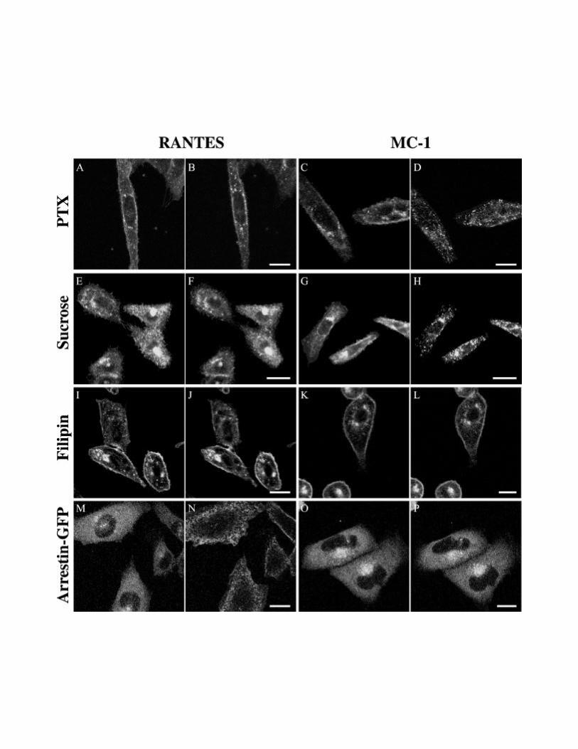
MC-1 induces CCR5 endocytosis via a clathrin-independent but caveolae-
dependent pathway.
To investigate the mechanisms implicated in the endocytosis induced by MC-1, we treated
cells with drugs blocking different pathways of GPCR internalization. As shown in figures 7A
and B, PTX did not prevent endocytosis induced by MC-1, whereas it inhibited strongly the
19
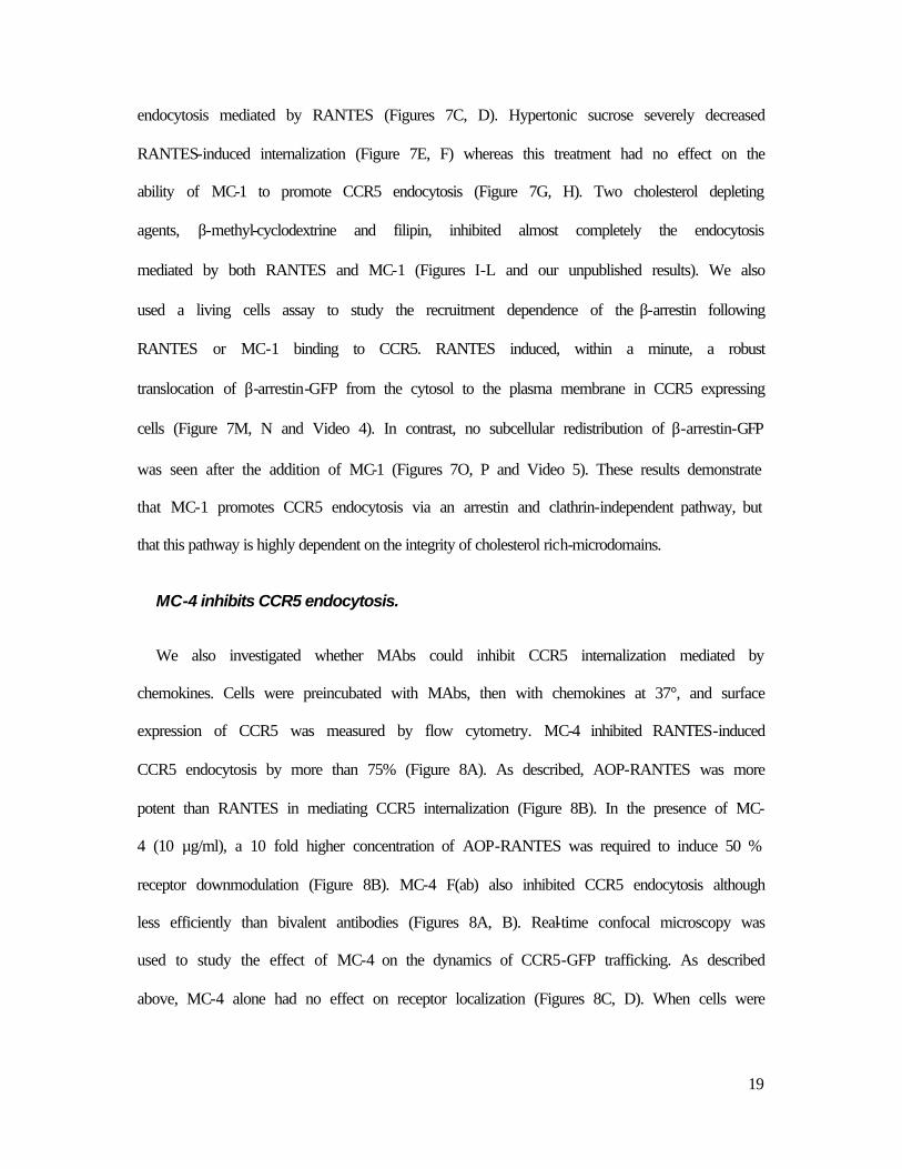
endocytosis mediated by RANTES (Figures 7C, D). Hypertonic sucrose severely decreased
RANTES-induced internalization (Figure 7E, F) whereas this treatment had no effect on the
ability of MC-1 to promote CCR5 endocytosis (Figure 7G, H). Two cholesterol depleting
agents, β-methyl-cyclodextrine and filipin, inhibited almost completely the endocytosis
mediated by both RANTES and MC-1 (Figures I-L and our unpublished results). We also
used a living cells assay to study the recruitment dependence of the β-arrestin following
RANTES or MC-1 binding to CCR5. RANTES induced, within a minute, a robust
translocation of β-arrestin-GFP from the cytosol to the plasma membrane in CCR5 expressing
cells (Figure 7M, N and Video 4). In contrast, no subcellular redistribution of β-arrestin-GFP
was seen after the addition of MC-1 (Figures 7O, P and Video 5). These results demonstrate
that MC-1 promotes CCR5 endocytosis via an arrestin and clathrin-independent pathway, but
that this pathway is highly dependent on the integrity of cholesterol rich-microdomains.
MC-4 inhibits CCR5 endocytosis.
We also investigated whether MAbs could inhibit CCR5 internalization mediated by
chemokines. Cells were preincubated with MAbs, then with chemokines at 37°, and surface
expression of CCR5 was measured by flow cytometry. MC-4 inhibited RANTES-induced
CCR5 endocytosis by more than 75% (Figure 8A). As described, AOP-RANTES was more
potent than RANTES in mediating CCR5 internalization (Figure 8B). In the presence of MC-
4 (10 µg/ml), a 10 fold higher concentration of AOP-RANTES was required to induce 50 %
receptor downmodulation (Figure 8B). MC-4 F(ab) also inhibited CCR5 endocytosis although
less efficiently than bivalent antibodies (Figures 8A, B). Real-time confocal microscopy was
used to study the effect of MC-4 on the dynamics of CCR5-GFP trafficking. As described
above, MC-4 alone had no effect on receptor localization (Figures 8C, D). When cells were

20
stimulated with 100 nM RANTES in the presence of MC-4, enhancement of membrane
fluorescence and ruffling were observed (Figures 8E,-G and Video 6), as in the absence of
MC-4. However, no subsequent internalization occurred, and membrane ruffling could be
observed all along the 45 min observation period (Figure 8G). Isotype control IgG had no
effect on CCR5-GFP localization (Figures 8H, I) or RANTES-induced internalization (Figure
8J).

21
Discussion
MAbs recognizing the second extracellular loop of CCR5 antagonize
chemokine binding
We have characterized 5 anti-CCR5 MAbs in terms of epitope mapping, inhibition of HIV
Env binding and chemokine function, and investigated their ability to activate CCR5 and
modulate its intracellular trafficking. On the basis of their recognition of various CCR5-
CCR2b chimeras, these MAbs could be classified into three groups. The epitopes of MC-4,
MC-5 and MC-7 are located in the amino-terminal domain, that of MC-1 within ECL2, while
that of MC-6 involves multiple extracellular domains. MC-5 recognizes a linear epitope
including the first 2 residues, that of MC-7 includes Y10 and D11. These are clearly two
dominant epitopes, recognized also by a number of other MAbs (Wu et al., 1997b; Hill et al.,
1998; Lee et al., 1999; Olson et al., 1999). ECL2, the longest extracellular loop of CCR5, also
contains dominant epitopes. Both MC-1 and 2D7 mapped to the first part of ECL2 but
recognize different epitopes. MC-6 binding, like most multidomain MAbs, was highly
dependent on K171 and E172.
Numerous studies have highlighted the importance of CCR5 amino-terminus and ECL2 to
chemokine binding and HIV coreceptor function (Rucker et al., 1996; Samson et al., 1997;
Farzan et al., 1999). ECL2 is particularly important for chemokine binding and selectivity,
while the N-terminus plays the dominant role for the gp120-CCR5 interaction. In line with
these observations, MAbs such as 539, 531, and 2D7, recognizing ECL2 epitopes inhibit
efficiently chemokine binding and signaling, as well as HIV entry (Lee et al., 1999). Our
present results are consistent with these previous observations. Both the ECL2 MAb MC-1,
and the multidomain MAb MC-6, that rely on ECL2 among other domains, efficiently
22
inhibited binding of RANTES, MIP-1β and Env to CCR5, as well as the functional response
to chemokines. The N-terminal MAb MC-5 inhibited binding of MIP-1β and gp120, but had
no effect on RANTES binding and signaling, while MC-4 had no effect. The differential
inhibition of MIP-1β and RANTES function by MC-5 provides further evidence that CCR5
ligands may use different extracellular residues for high affinity binding.
Partial activation of CCR5 by a conformation-sensitive multidomain MAb
Chemokine receptors are coupled to heterotrimeric G proteins belonging to both Pertussis
toxin-sensitive Gαi and Pertussis toxin-insensitive Gαq families. They regulate in turn a
number of intracellular cascades, including inhibition of cAMP production, intracellular Ca2+
release, and activation of PI3-kinase and MAP-kinases. These cascades mediate the biological
effects of chemokines, such as chemotaxis and/or increase of integrin adhesiveness (Sanchez-
Madrid and del Pozo, 1999). Activation of GPCRs upon ligand binding is believed to involve
the reorganization of the transmembrane helix bundle, unmasking intracellular domains that
interact with G proteins, and trigger their activation (Wess, 1997). Like chemokines, MAbs
might also, by binding to CCR5, modify the equilibrium between its inactive and active states,
and either trigger signaling cascades, or prevent their activation.
MAbs directed at GPCRs and displaying agonist activities are rare, and were not reported
so far for CCR5. However, an anti-CCR2b MAb was shown to activate this receptor in B cells
(Frade et al., 1997). Our MAbs were tested in three functional assays. We showed that the
multidomain MAb MC-6 induced CCR5 activation in GTPγS and cAMP accumulation
assays. MC-6 was however unable to promote intracellular calcium release, whereas
chemokines were equally efficient in all three assays. This discrepancy suggests that MC-6
and chemokines stabilize different active conformations of CCR5, coupling differently to

23
intracellular signaling cascades. Differential coupling of a receptor to G proteins according to
ligands has been proposed for CXCR2 (Hall et al., 1999). The conformational epitope of MC-
6 includes multiple CCR5 domains involved in chemokine binding, which might explain why
MC-6 promotes at least some of the conformational changes required for receptor activation.
The absence of modification of BRET signal following bivalent MC-6 addition would suggest
that the oligomerization state of the receptor is not modified in this process. As the MC-6
F(ab) fragment has no signaling properties, it remains however possible that bivalent
monoclonals modify the conformation of pre-existing dimers without changing the distance
between the BRET donor (luciferase) and the BRET acceptor (yellow GFP).
MAb-induced internalization of CCR5 oligomers involves an arrestin-
independent caveolae-dependent pathway.
A molecule that would trigger efficient CCR5 internalization without activating
intracellular signaling cascades would have obvious advantages in terms of therapeutic
usefulness as an HIV entry blocker. MC-1, although unable to activate the receptor in a
number of bioassays, induced CCR5-GFP internalization. The redistribution dynamics was
however strikingly different to that promoted by chemokines, as CCR5-GFP containing
vesicles formed more rapidly but did not fuse with each other nor with larger endosomal
structures. On the other hand, CCR5 activation by the MC-6 MAb did not induce CCR5-GFP
endocytosis, demonstrating that G protein activation and receptor internalization can be
totally dissociated. In the classical model of receptor desensitization, binding of a ligand to its
receptor leads to G-protein activation, allowing the recruitment of regulatory proteins that
mediate phosphorylation of the receptor (G protein-coupled receptor kinases), inhibition of
signaling (arrestins), and ultimately its clathrin-mediated endocytosis (AP-2, dynamin)
(Lefkowitz, 1998). However, recent experimental evidence has shown that receptor activation

24
and internalization can be dissociated. First, a number of mutant receptors are unable to
signal, but internalize normally, while other mutants do not internalize despite normal
signaling capacity (Cheung et al., 1990; Hunyady et al., 1994; Bennett et al., 2000). Secondly,
morphine, an agonist of the µ opioid receptor, does not induce endocytosis, while the receptor
is internalized well in response to other ligands (Keith et al., 1996). An antagonist of the CCK
receptor mediating endocytosis has also been described (Roettger et al., 1997). Finally,
constitutive activity of GPCR mutants does not necessarily result in enhanced basal
phosphorylation and internalization (Thomas et al., 2000; Mhaouty-Kodja et al., 1999). These
observations led to the suggestion that receptors may exist in multiple active (and inactive)
conformational states, each corresponding to a specific range of functional properties
(Thomas et al., 2000). In this context, it is conceivable that different MAbs may stabilize
preferentially one of these usually transient conformations. MC-6 would stabilize a signaling
status of CCR5, whereas MC-1 stabilizes a conformation triggering internalization.
The use of monovalent and bivalent forms of MC-1 allowed us to investigate whether this
internalization-prone conformation involves oligomeric structures. The two monovalent
versions of MC-1 continued to bind specifically CCR5 but could no longer mediate
internalization of the receptor, while cross-linking by a secondary Ab partially rescued the
ability of the single chain Fv fragment to induce receptor endocytosis. This partial restoration
might be due to the fact that crosslinking of the monovalent forms of MC-1 by anti-His
antibodies does not necessarily restore the original geometry of the bivalent monoclonal.
Moreover, BRET analysis demonstrated that bivalent MC-1, but not its monovalent forms,
modify the interaction between CCR5 polypeptides.
For many membrane receptor families, such as growth factor or cytokine receptors,
dimerization is necessary both for signal transduction and endocytosis. Although GPCRs were
25
until recently believed to operate as monomers, experimental evidence now suggests that
homo- or hetero-dimerization occurs, and is sometimes necessary for normal receptor
trafficking and function (Hebert and Bouvier, 1998). Using biophysical approaches, several
groups have observed constitutive homo or hetero-dimerization of GPCRs in living cells
(Angers et al., 2000; Overton et al., 2000; McVey et al., 2001; Kroeger et al., 2001;
Rocheville et al., 2000a; Rocheville et al., 2000b). For some, but not all receptors, a change in
the energy transfer level was observed following ligand addition. As stated in these studies,
modifications of fluorescent resonance energy transfer (FRET) or BRET signals do not allow
to distinguish clearly between the de novo association of receptor subunits, and the
conformation change of pre-existing dimers, modifying the relative distance between the two
probes. Homodimerization (α-factor receptor) and heterodimerization of receptors (dopamine
D2 and somatostatin SST5, SST1 and SST5, β2 adrenergic and opioid OP1, bradykinin B2 and
angiotensin AT1), have been shown to affect their signaling and internalization properties
(Yesilaltay and Jennes, 2000; Overton et al., 2000; Rocheville et al., 2000a; Rocheville et al.,
2000b; Jordan et al., 2001; AbdAlla et al., 2000).
CCR5 homodimerization has been described, as well as heterodimerization with CCR2b
(Mellado et al., 1999; Rodriguez-Frade et al., 1999; Benkirane et al., 1997). However,
whether this process is regulated by ligand binding or necessary for any of the CCR5
functions has not been conclusively demonstrated so far. We provide here the evidence that
bridging CCR5 polypeptides by bivalent antibodies is necessary for the induction of
internalization by MC-1, and modification of energy transfer in BRET assays. These data
demonstrate the involvement of oligomers in the MC-1 induced internalization process.
However, they do not allow to discriminate between the de novo association of monomeric
CCR5, aggregation of pre-existing CCR5 dimers or structural reorganization of these

26
constitutive dimers.
MC-1 mediated CCR5 internalization involves a pathway independent of arrestin and
clathrin, but dependent on cholesterol-rich caveolae. Two recent studies reported that CCR5
could be found in membrane raft microdomains and this subcellular localization may
contribute to the ability of CCR5 to mediate chemotaxis and to support HIV infection (Manes
et al., 2000; Manes et al., 1999). The pathway of MC-1 induced endocytosis contrasts with
the internalization process promoted by chemokine agonists, which is arrestin and clathrin-
dependent. Whether the dimerization or oligomerization state of CCR5 is also involved in the
chemokine-induced internalization pathway will require further investigation.
To the best of our knowledge, MC-1 is the first MAb able to promote endocytosis of a
native G protein-coupled receptor. Internalization of tagged receptors (human muscarinic M1
and thyrotropin-releasing hormone receptors) has however been reported following incubation
with antibodies directed against the tags (Petrou et al. 1997, Tolbert et al. 1998).
A MAb binding to CCR5 N-terminus prevents internalization without affecting
chemokine-induced signaling
MC-4 was able to potently and specifically inhibit endocytosis mediated by RANTES and
AOP-RANTES, without preventing intracellular signaling. Instead, persistent ruffling of the
plasma membrane was seen in confocal microscopy, demonstrating prolonged activation of
the receptor. Recently, a MAb (CCR5-02), mapping to the same N-terminal epitope of CCR5
as MC-4 was found to inhibit HIV infection. This effect was attributed to receptor
dimerization (Vila-Coro et al., 2000). Interestingly, antibodies directed against the amino-
terminal domain of the bradykinin B2 receptor, that is involved in dimerization, have been
shown to reduce receptor internalization induced by bradykinin (AbdAlla et al., 1999). The

27
inhibition of CCR5 endocytosis by MC-4 is probably not related to the state of CCR5
oligomerization since MC-4 did not induce changes in BRET signal, and its monovalent
version was also able to inhibit CCR5 endocytosis. Therefore, this effect likely results from
the stabilization of a particular conformation of the receptor.
Conclusion
We have characterized five MAbs that recognize CCR5 expressed on primary cells. They
map to distinct epitopes including the N-terminal segment (MC-4, MC-5, MC-7), the second
extracellular loop (MC-1), or both (and other) domains (MC-6). Many of these antibodies
exhibit functional properties (partial activation of CCR5 signaling pathways, stimulation of
internalization without signaling, inhibition of internalization without impairing signaling)
that together suggest the existence of multiple active conformation states of CCR5. The
differential properties of monovalent and divalent forms of the MC-1 antibody on both
endocytosis and bioluminescence energy transfer also indicate that the endocytic pathway
activated by this antibody involves CCR5 oligomers. Finally, some of the monoclonal
antibodies have interesting properties that might be used in different fields. The MC-1 MAb,
that competes for gp120 binding and promotes efficient internalization of the receptor without
triggering intracellular signaling, might constitute the basis for the development of anti-HIV
therapeutic agents. Other MAbs, that appear to stabilize active conformations or dimers,
might also be useful as tools to purify and crystallize active states of CCR5 for structural
studies.

28
Acknowledgements
Expert technical assistance was provided by M.J. Simons, H. Nguyen Tran, M. E. Decobecq,
and T. Rupp. This work was supported by a grant from the Deutsche Forschungsgemeinschaft
(SFB 464), the Actions de Recherche Concertées of the Communauté Fra
the French Agence Nationale de Recherche sur le SIDA, the Centre de Recherche Inter-
universitaire en Vaccinologie, the Belgian programme on Interuniversity Poles of attraction
initiated by the Belgian State, Prime Minister's Office, Science Policy Programming, the
BIOMED and BIOTECH programmes of the European Community (grants BIO4-CT98-0543
and BMH4-CT98-2343), the Fonds de la Recherche Scientifique Médicale of Belgium,
Télévie and the Fondation Médicale Reine Elisabeth to M.P. The scientific responsibility is
assumed by the authors. C.B. is Aspirant, and J.M.V. is Chercheur Qualifié, of the Belgian
Fonds National de la Recherche Scientifique. V.W. is recipient of a First fellowship of the
Région Wallonne. We thank Amanda Proudfoot for kindly providing RANTES, Robin Offord
and Brigitte Dufour for the synthesis of AOP-RANTES, Mark Scott for providing β-arrestin-
2-GFP and the AIDS Research and Reference Reagent Program for providing soluble CD4
and the 2D7 anti-CCR5 monoclonal antibody.
29
References
AbdAlla,S., Zaki,E., Lother,H., and Quitterer,U. (1999). Involvement of the amino
terminus of the B(2) receptor in agonist-induced receptor dimerization. J. Biol. Chem., 274,
26079-26084.
AbdAlla,S., Lother,H., and Quitterer,U. (2000). AT1-receptor heterodimers show
enhanced G-protein activation and altered receptor sequestration. Nature, 407, 94-98.
Alkhatib,G., Locati,M., Kennedy,P.E., Murphy,P.M., and Berger,E.A. (1997). HIV-1
coreceptor activity of CCR5 and its inhibition by chemokines: independence from G protein
signaling and importance of coreceptor downmodulation. Virology, 234, 340-348.
Angers,S., Salahpour,A., Joly,E., Hilairet,S., Chelsky,D., Dennis,M., and Bouvier,M.
(2000). Detection of beta 2-adrenergic receptor dimerization in living cells using
bioluminescence resonance energy transfer (BRET). Proc. Natl. Acad. Sci. U. S. A, 97, 3684-
3689.
Amara,A., Gall,S.L., Schwartz,O., Salamero,J., Montes,M., Loetscher,P., Baggiolini,M.,
Virelizier,J.L., and Arenzana-Seisdedos,F. (1997). HIV coreceptor downregulation as
antiviral principle: SDF-1α- dependent internalization of the chemokine receptor CXCR4
contributes to inhibition of HIV replication. J. Exp. Med., 186, 139-146.
Baba,M., et al. (1999) A small-molecule, nonpeptide CCR5 antagonist with highly potent
and selective anti-HIV-1 activity. Proc. Natl. Acad. Sci. U. S. A., 96, 5698-5703.

30
Baggiolini,M. (1998). Chemokines and leukocyte traffic. Nature, 392, 565-568.
Benkirane,M., Jin,D.Y., Chun,R.F., Koup,R.A., and Jeang,K.T. (1997). Mechanism of
transdominant inhibition of CCR5-mediated HIV-1 infection by ccr5∆32. J. Biol. Chem., 272,
30603-30606.
Bennett,T.A., Maestas,D.C., and Prossnitz,E.R. (2000). Arrestin binding to the G protein-
coupled N-formyl peptide receptor is regulated by the conserved "DRY" sequence. J. Biol.
Chem. 275, 24590-24594.
Berger,E.A., Murphy,P.M., and Farber,J.M. (1999). Chemokine receptors as HIV-1
coreceptors: roles in viral entry, tropism, and disease. Annu. Rev. Immunol., 17, 657-700.
Blanpain,C., et al. (1999a). Multiple charged and aromatic residues in CCR5 amino-
terminal domain are involved in high affinity binding of both chemokines and HIV-1 Env
protein. J. Biol. Chem., 274, 34719-34727.
Blanpain,C., et al. (1999b). Extracellular cysteines of CCR5 are required for chemokine
binding, but dispensable for HIV-1 coreceptor activity. J. Biol. Chem., 274, 18902-18908.
Blanpain,C., et al. (2000). Multiple nonfunctional alleles of CCR5 are frequent in various
human populations. Blood, 96, 1638-1645.
Blanpain,C., et al. (2001). Palmitoylation of CCR5 is critical for receptor trafficking and
efficient activation of intracellular signaling pathways. J. Biol. Chem., 276, 23795-23804.

31
Cheung,A.H., Dixon,R.A., Hill,W.S., Sigal,I.S., and Strader,C.D. (1990). Separation of the
structural requirements for agonist-promoted activation and sequestration of the beta-
adrenergic receptor. Mol. Pharmacol., 37, 775-779.
Cocchi,F., DeVico,A.L., Garzino-Demo,A., Arya,S.K., Gallo,R.C., and Lusso,P. (1995).
Identification of RANTES, MIP-1 alpha, and MIP-1 beta as the major HIV- suppressive
factors produced by CD8+ T cells. Science, 270, 1811-1815.
Farzan,M., et al. (1998). A tyrosine-rich region in the N terminus of CCR5 is important for
human immunodeficiency virus type 1 entry and mediates an association between gp120 and
CCR5. J. Virol., 72, 1160-1164.
Frade,J.M., Mellado,M., del Real,G., Gutierrez-Ramos,J.C., Lind,P., and Martinez,A.
(1997). Characterization of the CCR2 chemokine receptor: functional CCR2 receptor
expression in B cells. J. Immunol., 159, 5576-5584.
Gerard, C. and Rollins, B.J. (2001). Chemokines and disease. Nat. Immunol. 2, 108-115.
Hall,D.A., Beresford,I.J., Browning,C., and Giles,H. (1999). Signalling by CXC-
chemokine receptors 1 and 2 expressed in CHO cells: a comparison of calcium mobilization,
inhibition of adenylyl cyclase and stimulation of GTPgammaS binding induced by IL-8 and
GROalpha. Br. J. Pharmacol., 126, 810-818.
Hebert,T.E. and Bouvier,M. (1998). Structural and functional aspects of G protein-coupled
receptor oligomerization. Biochem. Cell Biol., 76, 1-11.

32
Hill,C.M., et al. (1998). The amino terminus of human CCR5 is required for its function as
a receptor for diverse human and simian immunodeficiency virus envelope glycoproteins.
Virology, 248, 357-371.
Hunyady,L., Baukal,A.J., Balla,T., and Catt,K.J. (1994). Independence of type I
angiotensin II receptor endocytosis from G protein coupling and signal transduction. J. Biol.
Chem., 269, 24798-24804.
Keith,D.E., Murray,S.R., Zaki,P.A., Chu,P.C., Lissin,D.V., Kang,L., Evans,C.J., and von
Zastrow,M. (1996). Morphine activates opioid receptors without causing their rapid
internalization. J. Biol. Chem., 271, 19021-19024.
Kroeger,K.M., Hanyaloglu,A.C., Seeber,R.M., Miles,L.E., and Eidne,K.A. (2001).
Constitutive and agonist-dependent homo-oligomerization of the thyrotropin-releasing
hormone receptor. Detection in living cells using bioluminescence resonance energy transfer.
J. Biol. Chem., 276, 12736-12743.
Lee,B., et al. (1999). Epitope mapping of CCR5 reveals multiple conformational states and
distinct but overlapping structures involved in chemokine and coreceptor function. J. Biol.
Chem., 274, 9617-9626.
Lefkowitz,R.J. (1998). G protein-coupled receptors. III. New roles for receptor kinases and
beta-arrestins in receptor signaling and desensitization. J. Biol. Chem., 273, 18677-18680.
Liu,R., Paxton,W.A., Choe,S., Ceradini,D., Martin,S.R., Horuk,R., MacDonald,M.E.,

33
Stuhlmann,H., Koup,R.A., and Landau,N.R. (1996). Homozygous defect in HIV-1 coreceptor
accounts for resistance of some multiply-exposed individuals to HIV-1 infection. Cell, 86,
367-377.
Lopalco,L., Barassi,C., Pastori,C., Longhi,R., Burastero,S.E., Tambussi,G., Mazzotta,F.,
Lazzarin,A., Clerici,M., and Siccardi,A.G. (2000). CCR5-reactive antibodies in seronegative
partners of HIV-seropositive individuals down-modulate surface CCR5 in vivo and neutralize
the infectivity of R5 strains of HIV-1 In vitro. J. Immunol., 164, 3426-3433.
Mack,M., Riethmuller,G., and Kufer,P. (1995). A small bispecific antibody construct
expressed as a functional single -chain molecule with high tumor cell cytotoxicity. Proc. Natl.
Acad. Sci. U. S. A., 92, 7021-7025.
Mack,M., et al. (1998). Aminooxypentane-RANTES induces CCR5 internalization but
inhibits recycling: a novel inhibitory mechanism of HIV infectivity. J. Exp. Med., 187, 1215-
1224.
Mack,M., Kleinschmidt,A., Bruhl,H., Klier,C., Nelson,P.J., Cihak,J., Plachy,J.,
Stangassinger,M., Erfle,V., Schlondorff,D. (2000). Transfer of the chemokine receptor CCR5
between cells by membrane-derived microparticles: a mechanism for cellular human
immunodeficiency virus 1 infection. Nat. Med. 6, 769-775.
Manes,S., Mira,E., Gomez-Mouton,C., Lacalle,R.A., Keller,P., Labrador,J.P., and
Martinez,A. (1999). Membrane raft microdomains mediate front-rear polarity in migrating
cells. EMBO J., 18, 6211-6220.

34
Manes,S., del Real,G., Lacalle,R.A., Gomez-Mouton,C., Sanchez-Palomino,S.,
Delgado,R., Alcami,J., Mira,E., and Martinez-A,C. (2000). Membranes raft microdomains
mediate lateral assemblies required for HIV-1 infection. EMBO Reports, 1, 190-196.
McVey,M., Ramsay,D., Kellett,E., Rees,S., Wilson,S., Pope,A.J., and Milligan,G. (2001).
Monitoring receptor oligomerization using time-resolved fluorescence resonance energy
transfer and bioluminescence resonance energy transfer. The human delta -opioid receptor
displays constitutive oligomerization at the cell surface, which is not regulated by receptor
occupancy. J. Biol. Chem., 276, 14092-14099.
Mellado,M., Rodriguez-Frade,J.M., Vila-Coro,A.J., de Ana,A.M., and Martinez,A. (1999).
Chemokine control of HIV-1 infection. Nature, 400, 723-724.
Mhaouty-Kodja,S., Barak,L.S., Scheer,A., Abuin,L., Diviani,D., Caron,M.G., and
Cotecchia,S. (1999). Constitutively active alpha-1b adrenergic receptor mutants display
different phosphorylation and internalization features. Mol. Pharmacol., 55, 339-347.
Moore,J.P. and Stevenson,M. (2000). New Targets for inhibitior of HIV-1 replication. Nat.
Rev. Mol. Cell Biol., 1, 40-49
Murphy,P.M., Baggiolini,M., Charo,I.F., Hebert,C.A., Horuk,R., Matsushima,K.,
Miller,L.H., Oppenheim,J.J., and Power,C.A. (2000). International union of pharmacology.
XXII. Nomenclature for chemokine receptors. Pharmacol. Rev., 52, 145-176.
Olson,W.C., et al. (1999). Differential inhibition of human immunodeficiency virus type 1

35
fusion, gp120 binding, and CC-chemokine activity by monoclonal antibodies to CCR5. J.
Virol., 73, 4145-4155.
Oppermann,M., Mack,M., Proudfoot,A.E., and Olbrich,H. (1999). Differential effects of
CC chemokines on CC chemokine receptor 5 (CCR5) phosphorylation and identification of
phosphorylation sites on the CCR5 carboxyl terminus. J. Biol. Chem., 274, 8875-8885.
Orlandi,R., Gussow,D.H., Jones,P.T., and Winter,G. (1989). Cloning immunoglobulin
variable domains for expression by the polymerase chain reaction. Proc. Natl. Acad. Sci. U. S.
A., 86, 3833-3837.
Overton,M.C. and Blumer,K.J. (2000). G-protein-coupled receptors function as oligomers
in vivo. Curr. Biol., 10, 341-344.
Petrou,C., Chen,L., and Tashjian,A.H.J. (1997). A receptor-G protein coupling-
independent step in the internalization of the thyrotropin-releasing hormone receptor. J. Biol.
Chem., 272, 2326-2333.
Rocheville,M., Lange,D.C., Kumar,U., Patel,S.C., Patel,R.C., and Patel,Y.C. (2000a).
Receptors for dopamine and somatostatin: formation of hetero-oligomers with enhanced
functional activity. Science, 288, 154-157.
Rocheville,M., Lange,D.C., Kumar,U., Sasi,R., Patel,R.C., and Patel,Y.C. (2000b).
Subtypes of the somatostatin receptor assemble as functional homo- and heterodimers. J. Biol.
Chem., 275, 7862-7869.

36
Rodriguez-Frade,J.M., Vila-Coro,A.J., Martin,A., Nieto,M., Sanchez-Madrid,F.,
Proudfoot,A.E., Wells,T.N., Martinez,A., and Mellado,M. (1999). Similarities and differences
in RANTES- and (AOP)-RANTES-triggered signals: implications for chemotaxis. J. Cell
Biol., 144, 755-765.
Roettger,B.F., Ghanekar,D., Rao,R., Toledo,C., Yingling,J., Pinon,D., and Miller,L.J.
(1997). Antagonist-stimulated internalization of the G protein-coupled cholecystokinin
receptor. Mol. Pharmacol., 51, 357-362.
Rucker,J., et al. (1996). Regions in beta-chemokine receptors CCR5 and CCR2b that
determine HIV-1 cofactor specificity. Cell, 87, 437-446.
Samson,M., Labbe,O., Mollereau,C., Vassart,G., and Parmentier,M. (1996a). Molecular
cloning and functional expression of a new human CC-chemokine receptor gene.
Biochemistry, 35, 3362-3367.
Samson,M., et al. (1996b). Resistance to HIV-1 infection in caucasian individuals bearing
mutant alleles of the CCR-5 chemokine receptor gene. Nature, 382, 722-725.
Samson,M., LaRosa,G., Libert,F., Paindavoine,P., Detheux,M., Vassart,G., and
Parmentier,M. (1997). The second extracellular loop of CCR5 is the major determinant of
ligand specificity. J. Biol. Chem., 272, 24934-24941.
Sanchez-Madrid,F. and del Pozo,M.A. (1999). Leukocyte polarization in cell migration
and immune interactions. EMBO J., 18, 501-511.

37
Signoret,N., Pelchen-Matthews,A., Mack,M., Proudfoot,A.E., and Marsh,M. (2000).
Endocytosis and recycling of the HIV coreceptor CCR5. J. Cell Biol., 151, 1281-1294.
Simmons,G., Clapham,P.R., Picard,L., Offord,R.E., Rosenkilde,M.M., Schwartz,T.W.,
Buser,R., Wells,T.N.C., and Proudfoot,A.E. (1997). Potent inhibition of HIV-1 infectivity in
macrophages and lymphocytes by a novel CCR5 antagonist. Science, 276, 276-279.
Thomas,W.G., Qian,H., Chang,C.S., and Karnik,S. (2000). Agonist-induced
phosphorylation of the angiotensin II (AT(1A)) receptor requires generation of a
conformation that is distinct from the inositol phosphate-signaling state. J. Biol. Chem., 275,
2893-2900.
Tolbert,L.M. and Lameh,J. (1998). Antibody to epitope tag induces internalization of
human muscarinic subtype 1 receptor. J. Neurochem., 70, 113-119.
Trkola,A., Dragic,T., Arthos,J., Binley,J.M., Olson,W.C., Allaway,G.P., Cheng-Mayer,C.,
Robinson,J., Maddon,P.J., and Moore,J.P. (1996). CD4-dependent, antibody-sensitive
interactions between HIV-1 and its co- receptor CCR-5. Nature, 384, 184-187.
Vila-Coro,A.J., Mellado,M., Martin,d.A., Lucas,P., del Real,G., Martinez,A., and
Rodriguez-Frade,J.M. (2000). HIV-1 infection through the CCR5 receptor is blocked by
receptor dimerization. Proc. Natl. Acad. Sci. U. S. A, 97, 3388-3393.
Wess,J. (1997). G-protein-coupled receptors: molecular mechanisms involved in receptor
activation and selectivity of G-protein recognition. FASEB J., 11, 346-354.

38
Wu,L., et al. (1997a). CCR5 levels and expr ession pattern correlate with infectability by
macrophage-tropic HIV-1, in vitro. J. Exp. Med., 185, 1681-1691.
Wu,L., et al. (1997b). Interaction of chemokine receptor CCR5 with its ligands: multiple
domains for HIV-1 gp120 binding and a single domain for chemokine binding. J. Exp. Med.,
186, 1373-1381.
Yesilaltay,A. and Jenness,D.D. (2000). Homo-oligomeric complexes of the yeast alpha-
factor pheromone receptor are functional units of endocytosis. Mol. Biol. Cell, 11, 2873-2884.

39
Legends to tables and figures
Table 1. Epitope mapping of anti-CCR5 MAbs.
The MAbs used in this study were tested for their immunoreactivity against a set of CCR5-
CCR2b chimeras and CCR5 point mutants. Chimeras are coded as described in the legend of
Figure 1. All constructs were stably transfected in CHO-K1 cells and expressed at levels
similar to wtCCR5 (50-150%). Results were normalized relative to the mean-channel
fluorescence obtained for each MAb with wtCCR5. +, (+) and – indicate fluorescence levels
that correspond to 50-150 % , 10-50 % and <10 % respectively of the MCF obtained for
wtCCR5. Background fluorescence was always lower than 1% of wtCCR5 MCF. All
experiments have been repeated at least twice. Representative data are shown in Figure 1.
ND: not done.
Figure 1. Immunostaining of CCR5-CCR2b chimeras and CCR5 point
mutants with anti-CCR5 MAbs.
CCR5-CCR2b chimeras are coded according to the origin of the N-terminal domain (first
digit), and of the three extracellular loops (last three digits): 5555 and 2222 represent CCR5
and CCR2b respectively, 2555 represents a chimeric receptor containing the amino-terminal
domain of CCR2b and the three extracellular loops of CCR5. CCR5 point mutants are
designated by the nature of their amino-acid substitution.
CHO-K1 cells stably expressing these constructs were stained with saturating
concentrations of the different anti-CCR5 MAbs and phycoerythrin-conjugated anti-mouse
IgG, and analyzed by FACS. Fluorescence histograms are shown together with the
background fluorescence obtained for cells expressing CCR2b.

40
Figure 2. CCR5 activation by the multidomain MAb MC-6.
Effect of different concentrations of RANTES (A), and anti-CCR5 MAbs at 10 µg/ml (B)
on the binding of [35S]-GTPγS to membranes of CHO-K1 cells expressing human CCR5. Data
are presented as raw cpm values (A) or normalized (B) to basal (0%) and maximal
[35S]GTPγS binding in response to RANTES (100%). These experiments were repeated four
times with similar results. All data points were analyzed in triplicates (error bars: s.e.m.). C.
Inhibition of forskolin-stimulated cAMP accumulation. CHO-K1 cells expressing CCR5 were
incubated for 20 min with 5 µM forskolin and RANTES or the various MAbs, and cAMP was
measured by ELISA. The results were normalized for basal (0%, in the presence of forskolin
only) and maximal response (100%, cAMP levels obtained with a saturating concentration of
RANTES). The experiments were performed in triplicate (error bars: s.e.m.), and the figure
represents a typical experiment out of two performed independently.
Figure 3. Inhibition of MIP-1ββ, RANTES and HIV Env binding and receptor
activation.
A) Different concentrations of anti-CCR5 MAbs ( : 0 µg/ml, : 0.1 µg/ml, : 1
µg/ml, : 10 µg/ml) were tested for their ability to compete with binding of [125I]-MIP-1β or
[125I]-RANTES to CHO cells expressing CCR5. Results were normalized for non-specific
binding (0%) and specific binding in the absence of competitor (100%). B) Mab mediated
inhibition of functional responses to MIP-1β and RANTES in cell lines coexpressing CCR5
and apoaequorin. Cells were pre-incubated for 30 minutes with MAbs, thenMIP-1β or

41
RANTES (1 nM) was added, and luminescence was recorded for 30 s. Results were
normalized for the chemokine response in the absence of antibodies (100%) and the basal
luminescence of the cells (0%). C) Influence of MAbs on the binding of [125I]-gp120 derived
from the macrophage-tropic HIV-1 strain JRFL to CCR5 expressing CHO-K1 cells, in the
presence of sCD4 (100 nM). Results were normalized for non-specific binding (0%) and
specific binding in the absence of competitor (100%). All experiments were repeated at least
twice with similar results. All points were performed in triplicates (error bars: s.e.m.).
Figure 4. MC-1 MAb induces CCR5 endocytosis in CHO cells.
CHO cells expressing CCR5 were incubated with MC-1( ), MC-4 ( ) (A) or RANTES
( ) (B) for 30 minutes at 37°. Cell surface CCR5 was detected by flow-cytometry using a
saturating concentration of the same MAbs or MC-4 for RANTES induced CCR5
downmodulation. Results were normalized for the fluorescence of unstimulated cells (100%)
and for the background fluorescence (0%). All experiments were repeated at least twice with
similar results.
Figure 5. Downmodulation of cell surface CCR5-GFP by RANTES and MC-1.
A) CHO-K1 cells expressing CCR5-GFP (green) were incubated with alexafluor
transferrin (red) for 30 minutes, washed twice with media, and analyzed by confocal
microscopy for colocalization (yellow). Dynamics of CCR5-GFP was measured after
stimulation of the cells by RANTES or MC-1 MAb. The cells were exposed to 100 nM
RANTES or 10 µg/ml MC-1 in DMEM/F12 medium at 37°, under confocal microscopy.
Images were acquired every 3 minutes for 45 minutes. CCR5-GFP distribution is shown 0,
15, 30 and 45 minutes following addition of RANTES (B, C, D and E) or MC-1 (F, G, H and
I). This session is representative of at least 5 similar experiments. Bar: 5 µm. Videos of the

42
experiments described in this figure are available on the internet version of this paper.
Figure 6. Receptor oligomerization are involved in MC-1 induced CCR5
endocytosis.
A) 293T cells expressing CCR5-luc and CCR5-YFP were incubated with coelenterazine,
and the luminescence and the fluorescence signals were quantified before and after the
addition of the indicated MAb. The BRET ratio was defined as [(emission at 485 nm ±
20)/(emission at 530 nm ± 25)]-[(emission at 485 nm ± 20)/(emission at 530 nm ± 25)] for
CCR5-luc expressed alone in the same experiments. Variation in BRET signal as compared to
baseline (addition of buffer alone) after 10 minutes of incubation with antibodies is displayed.
All experiments were repeated at least twice with similar results. All points were performed in
triplicates (error bars: s.e.m.). B) CHO cells expressing CCR5 were incubated with ScFv-MC-
1 (3 µg/ml) for 45 minutes at 4° or 37°, washed twice with cold PBS and incubated with anti-
His at 4° or 37° as indicated. Surface expression of CCR5 was measured by flow cytometry
with PE- labeled anti-mouse Ab. Fluorescence was normalized for cells incubated with ScFv
MC-1 and anti-His both at 4° (100%), and for background fluorescence (0%). C-H) Confocal
microscopy. Cells expressing CCR5-GFP were incubated with ScFv-MC-1 (10 µg/ml) at 37°
and are shown before (C) and 45 minutes after the incubation (D). Cells were then washed 3
times with buffer (E) and incubated with an anti-polyhistidine Ab (anti-His, 4 µg/ml) for 45
minutes (F). Videos of the experiment described in this figure are available on the internet
version of this paper. Anti-His Ab alone did not result in CCR5-GFP endocytosis after 45
minutes of observation (G, H). All experiments were at least repeated twice with similar
results.
Figure 7. MC-1 induces CCR5 endocytosis by an arrestin and clathrin-

43
independent, caveolae-dependent pathway.
(A-L). CHO-K1 cells expressing CCR5-GFP were incubated for 18 hours with 100 ng/ml
Pertussis toxin 100 (A-D) or for 1 hour with 0.45M sucrose (E-H), or or 2.5 µg/ml filipin (I-
L) for 1 hour at 37°. Cells were then stimulated with 100 nM RANTES (A-B, E-F, I-J) or 10
µg/ml MC-1 (C-D, G-H, K-L), and the dynamics of CCR5-GFP was recorded by confocal
microscopy. Responses are shown before (A, C, E, G, I, K) and 30 minutes after (B, D, F, H,
J, L) ligand addition. (M-P). CHO-K1 cells stably expressing CCR5 were transfected with β-
arrestin-GFP. 24 hours after transfection, subcellular redistribution of β-arrestin-GFP was
analyzed by confocal microscopy. The cells were exposed to 100 nM RANTES (M-N) or 10
µg/ml MC-1 (O-P). Images were acquired every 15 seconds for 10 minutes. Responses are
shown before (M, O), 2 minutes (N) or 10 minutes (P) following ligands addition. Videos of
the dynamics of β-arrestin-GFP trafficking in response to RANTES stimulation, as described
in this figure, are available on the internet version of this paper. All experiments were
repeated at least twice with similar results. Bar: 20 µm.
Figure 8. MC-4 inhibits CCR5 endocytosis mediated by chemokines.
(A, B) CHO-K1 cells expressing CCR5 were pre-incubated with medium (¢) , 10 µg/ml
MC-4 (�) or MC-4 F(ab) (r) for 30 minutes on ice and then incubated with RANTES (A) or
AOP-RANTES (B) for 30 minutes at 37°. Surface expression of CCR5 was measured by flow
cytometry using the antibody MC-4 or MC-4 F(ab) (15 µg/ml, on ice) and FITC-conjugated
secondary Abs. Fluorescence was normalized as 100% in the absence of chemokines and 0%
for background fluorescence. All experiments were repeated at least three times with similar
results. Cells expressing CCR5-GFP are shown before (C), and 30 minutes (D) after MC-4
addition (10 µg/ml) at 37°. Cells were then stimulated with 100 nM RANTES, and the
44
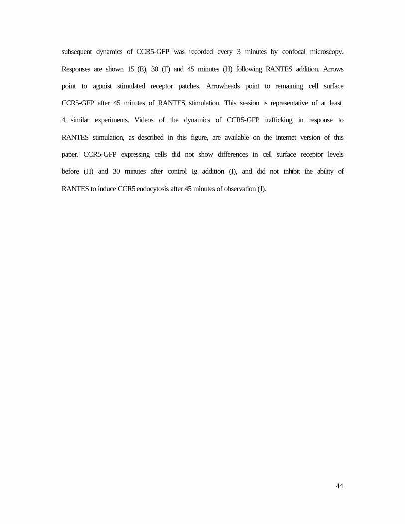
subsequent dynamics of CCR5-GFP was recorded every 3 minutes by confocal microscopy.
Responses are shown 15 (E), 30 (F) and 45 minutes (H) following RANTES addition. Arrows
point to agonist stimulated receptor patches. Arrowheads point to remaining cell surface
CCR5-GFP after 45 minutes of RANTES stimulation. This session is representative of at least
4 similar experiments. Videos of the dynamics of CCR5-GFP trafficking in response to
RANTES stimulation, as described in this figure, are available on the internet version of this
paper. CCR5-GFP expressing cells did not show differences in cell surface receptor levels
before (H) and 30 minutes after control Ig addition (I), and did not inhibit the ability of
RANTES to induce CCR5 endocytosis after 45 minutes of observation (J).
45
Online supplemental material.
Video 1: CHO-K1 cells expressing CCR5-GFP were observed before and after the
addition of 100 nM RANTES. Frames were captured every 3 min. A montage of images taken
from this movie is presented in Figure 5.
Video 2: CHO-K1 cells expressing CCR5-GFP were observed before and after the
addition of 10 µg/ml MC-1. Frames were captured every 3 min. A montage of images taken
from this movie is presented in Figure 5.
Video 3: CHO-K1 cells expressing CCR5-GFP were incubated with MC-1 ScFv 10 µg/ml
for 45 min, rince 3 times with 1 ml DMEM/F12, and observed before and after the addition of
anti-His (4 µg/ml) for 30 min, and images were collected every 3 min. A montage of images
taken from this movie is presented in Figure 6.
Video 4: CHO-K1 cells expressing CCR5 were transfected with β-arrestin 2-EGFP, 24
hours after transfection transfected cells were observed before and after the addition of 100
nM RANTES for 10 min, and images were collected every 15 seconds. A montage of images
taken from this movie is presented in Figure 7.
Video 5: CHO-K1 cells expressing CCR5 were transfected with β-arrestin 2-EGFP, 24
hours after transfection transfected cells were observed before and after the addition of 10
µg/ml MC-1 for 10 min, and images were collected every 20 seconds. A montage of images
taken from this movie is presented in Figure 7.
Video 6: CHOK1 cells expressing CCR5-GFP were incubated for 45 min with 10 µg/ml
MC-4, and observed before and after the addition of 100 nM RANTES. Frames were captured

46
every 3 min. A montage of images taken from this movie is presented in Figure 8.

47
CCR5-CCR2b chimera 2D7 3A9 MC-1 MC-4 MC-5 MC-6 MC-7
5555 + + + + + + + 2222 - - - - - - - 5222 - + - + + - + 2555 + - + - - (+) - 5255 + + + + + - + 5525 - + - + + - + 2252 + - + - - - - 2225 - - - - - - -
ECL2 N-ter ECL2 N-ter N-ter MD N-ter
CCR5 amino-terminal mutants CCR5 + + + + + + +
∆∆ 2 ND + + ND (+) + ND ∆∆ 2-3 ND + + ND - + ND ∆∆ 2-4 ND + + ND - + ND ∆∆ 2-5 - + + + - + (+) ∆∆ 2-9 ND + + + - + - ∆∆ 2-13 ND + + (+) - + - ∆∆ 2-17 ND + + - - + - D2A ND + + + + + + Y3A ND + + + + + + Q4A ND + + + + + + V5A ND + + + + + + S6A ND + + + + + + S7A ND + + + + + - P8A ND + + + + + - I9A ND + + + + + +
Y10A ND + + + + + - D11A ND + + + + + - I12A ND + + + + + + N13A ND + + + + + + E18A ND + + + + + +
CCR5 second extracellular loop mutants 5555 + + + + + + +
55(25)5 - + - + + - + 55(52)5 + + + + + - + 22(25)5 - - - - - - - 22(52)5 + - + - - - - K171A - ND + + + - ND E172A - ND + + + - ND

48