Oligomerization of the chromatin-structuring protein H-NS
11
Molecular Microbiology (2000) 36(4), 962–972 Oligomerization of the chromatin-structuring protein H-NS Clare P. Smyth, 1 Thomas Lundba ¨ck, 1 Debora Renzoni, 1 Giuliano Siligardi, 2 Rebecca Beavil, 3 Meredith Layton, 4 Julie M. Sidebotham, 5 Jay C. D. Hinton, 5 Paul C. Driscoll, 1,4 Christopher F. Higgins 5,6 and John E. Ladbury 1 * 1 Department of Biochemistry and Molecular Biology, University College London, Gower Street, London, WC1E 6BT, UK. 2 EPSRC National Chiroptical Spectroscopy Centre, Kings College London, Department of Pharmacy, Manresa Road, London, SW3 6LX, UK. 3 The Randall Institute, Kings College London, 26–29 Drury Lane, London, WC2B 5RL, UK. 4 The Ludwig Institute for Cancer Research, 91 Riding House Street, London, W1P 8BT, UK. 5 Nuffield Department of Clinical Biochemistry, Institute for Molecular Medicine, University of Oxford, John Radcliffe Hospital, Oxford, OX3 9DS, UK. 6 MRC Clinical Sciences Centre, Imperial College School of Medicine, Hammersmith Hospital, DuCane Road, London, W12 0NN, UK. Summary H-NS is a major component of the bacterial nucleoid, involved in condensing and packaging DNA and modulating gene expression. The mechanism by which this is achieved remains unclear. Genetic data show that the biological properties of H-NS are influenced by its oligomerization properties. We have applied a variety of biophysical techniques to study the structural basis of oligomerization of the H-NS protein from Salmonella typhimurium. The N-terminal 89 amino acids are responsible for oligomerization. The first 64 residues form a trimer dominated by an a- helix, likely to be in coiled–coil conformation. Extending this polypeptide to 89 amino acids gener- ated higher order, heterodisperse oligomers. Simi- larly, in the full-length protein no single, defined oligomeric state is adopted. The C-terminal 48 residues do not participate in oligomerization and form a monomeric, DNA-binding domain. These N- and C-terminal domains are joined via a flexible linker which enables them to function independently within the context of the full-length protein. This novel mode of oligomerization may account for the unusual binding properties of H-NS. Introduction The bacterial chromosome is efficiently packaged into a compact structure, the nucleoid (Drlica and Rouviere- Yaniv, 1987; Pettijohn, 1988). However, the expression of a significant proportion of the genome at any one time, and rapid cell doubling times, require a dynamic structure and organization able to respond rapidly to changes in environmental conditions as gene expression is modu- lated. A number of DNA-binding proteins have been impli- cated in the organization of bacterial chromatin (Spassky et al., 1984; Drlica and Rouviere-Yaniv, 1987). The two most abundant of these are HU and H-NS. H-NS is an approximately 15 kDa protein that is present at 10 5 copies cell 21 (Higgins et al., 1990; Ussery et al., 1994; Williams and Rimsky, 1997). H-NS binds DNA in a relatively sequence-independent manner, and although a preference for intrinsically curved sequences has been reported (Bracco et al., 1989; Yamada et al., 1990; Owen- Hughes et al., 1992), this cannot fully explain its mode of action in vivo (Jordi et al., 1997). H-NS has also been implicated as a global regulator of gene expression (Higgins et al., 1988; Hulton et al., 1990; Laurent-Winter et al., 1997). Based on this lack of sequence specificity for binding DNA, and the fact that H-NS influences DNA topology in vivo and in vitro (Higgins et al., 1988; Hulton et al., 1990; Tupper et al., 1994), the mechanism by which H-NS influences transcription is likely to differ from that of more typical sequence-specific transcriptional regulators. H-NS is an unusual protein because within as few as 136 residues genetic data have indicated that there are at least two distinct functional domains. Genetic data suggest that oligomerization of H-NS is likely to be important for its function (Ueguchi et al., 1996; 1997; Williams et al., 1996). The oligomeric state of H-NS has variously been reported as a dimer, trimer, tetramer or hexamer, depending on experimental conditions and method of analysis (Falconi et al., 1988; Spurio et al., 1997; Ueguchi et al., 1997). It has been established that the N-terminal two-thirds of H-NS (residues 1–91) is sufficient for self-association to occur, whereas the C-terminus is required for DNA binding (Ueguchi et al., Q 2000 Blackwell Science Ltd Received 11 October, 1999; revised 7 March, 2000; accepted 11 March, 2000. *For correspondence. E-mail [email protected]. ac.uk; Tel. (144) 02076 797012; Fax (144) 020276 797193.
-
Upload
independent -
Category
Documents
-
view
3 -
download
0
Transcript of Oligomerization of the chromatin-structuring protein H-NS

Molecular Microbiology (2000) 36(4), 962±972
Oligomerization of the chromatin-structuringprotein H-NS
Clare P. Smyth,1 Thomas LundbaÈck,1 Debora
Renzoni,1 Giuliano Siligardi,2 Rebecca Beavil,3
Meredith Layton,4 Julie M. Sidebotham,5 Jay C. D.
Hinton,5 Paul C. Driscoll,1,4 Christopher F. Higgins5,6
and John E. Ladbury1*1Department of Biochemistry and Molecular Biology,
University College London, Gower Street, London,
WC1E 6BT, UK.2EPSRC National Chiroptical Spectroscopy Centre, Kings
College London, Department of Pharmacy, Manresa
Road, London, SW3 6LX, UK.3The Randall Institute, Kings College London, 26±29
Drury Lane, London, WC2B 5RL, UK.4The Ludwig Institute for Cancer Research, 91 Riding
House Street, London, W1P 8BT, UK.5Nuffield Department of Clinical Biochemistry, Institute for
Molecular Medicine, University of Oxford, John Radcliffe
Hospital, Oxford, OX3 9DS, UK.6MRC Clinical Sciences Centre, Imperial College School
of Medicine, Hammersmith Hospital, DuCane Road,
London, W12 0NN, UK.
Summary
H-NS is a major component of the bacterial nucleoid,
involved in condensing and packaging DNA and
modulating gene expression. The mechanism by
which this is achieved remains unclear. Genetic
data show that the biological properties of H-NS are
influenced by its oligomerization properties. We have
applied a variety of biophysical techniques to study
the structural basis of oligomerization of the H-NS
protein from Salmonella typhimurium. The N-terminal
89 amino acids are responsible for oligomerization.
The first 64 residues form a trimer dominated by an a-
helix, likely to be in coiled±coil conformation.
Extending this polypeptide to 89 amino acids gener-
ated higher order, heterodisperse oligomers. Simi-
larly, in the full-length protein no single, defined
oligomeric state is adopted. The C-terminal 48
residues do not participate in oligomerization and
form a monomeric, DNA-binding domain. These N-
and C-terminal domains are joined via a flexible linker
which enables them to function independently within
the context of the full-length protein. This novel mode
of oligomerization may account for the unusual
binding properties of H-NS.
Introduction
The bacterial chromosome is efficiently packaged into a
compact structure, the nucleoid (Drlica and Rouviere-
Yaniv, 1987; Pettijohn, 1988). However, the expression of
a significant proportion of the genome at any one time,
and rapid cell doubling times, require a dynamic structure
and organization able to respond rapidly to changes in
environmental conditions as gene expression is modu-
lated.
A number of DNA-binding proteins have been impli-
cated in the organization of bacterial chromatin (Spassky
et al., 1984; Drlica and Rouviere-Yaniv, 1987). The two
most abundant of these are HU and H-NS. H-NS is an
approximately 15 kDa protein that is present at 105
copies cell21 (Higgins et al., 1990; Ussery et al., 1994;
Williams and Rimsky, 1997). H-NS binds DNA in a
relatively sequence-independent manner, and although
a preference for intrinsically curved sequences has been
reported (Bracco et al., 1989; Yamada et al., 1990; Owen-
Hughes et al., 1992), this cannot fully explain its mode of
action in vivo (Jordi et al., 1997). H-NS has also been
implicated as a global regulator of gene expression
(Higgins et al., 1988; Hulton et al., 1990; Laurent-Winter
et al., 1997). Based on this lack of sequence specificity for
binding DNA, and the fact that H-NS influences DNA
topology in vivo and in vitro (Higgins et al., 1988; Hulton
et al., 1990; Tupper et al., 1994), the mechanism by which
H-NS influences transcription is likely to differ from that of
more typical sequence-specific transcriptional regulators.
H-NS is an unusual protein because within as few as
136 residues genetic data have indicated that there are at
least two distinct functional domains. Genetic data
suggest that oligomerization of H-NS is likely to be
important for its function (Ueguchi et al., 1996; 1997;
Williams et al., 1996). The oligomeric state of H-NS has
variously been reported as a dimer, trimer, tetramer or
hexamer, depending on experimental conditions and
method of analysis (Falconi et al., 1988; Spurio et al.,
1997; Ueguchi et al., 1997). It has been established that
the N-terminal two-thirds of H-NS (residues 1±91) is
sufficient for self-association to occur, whereas the
C-terminus is required for DNA binding (Ueguchi et al.,
Q 2000 Blackwell Science Ltd
Received 11 October, 1999; revised 7 March, 2000; accepted 11March, 2000. *For correspondence. E-mail [email protected]; Tel. (144) 02076 797012; Fax (144) 020276 797193.

1996; Williams et al., 1996). However, it has also recently
been suggested that mutations in the C-terminus of the
protein may also influence its self-associated state
(Spurio et al., 1997). Genetic data have also led to the
suggestion that H-NS consists of at least three functional
domains, responsible for self-association, transcriptional
repression and DNA-binding respectively (Ueguchi et al.,
1997).
In order to gain a more complete understanding of the
mode of action of H-NS, how it compacts bacterial DNA
and influences transcription at specific promoters, it is
essential to determine in detail its oligomeric state and the
residues which contribute to this. We therefore applied a
number of complementary biophysical techniques to
examine the self-associated state of H-NS from Salmo-
nella typhimurium under equilibrium conditions, over a
concentration range from the low mM to mM. These data
are consistent with a model in which H-NS is composed of
two structurally independent domains. These N- and
C-terminal domains are joined by a flexible linker. The
C-terminal DNA-binding domain plays no role in oligomer-
ization. The N-terminal 64 amino acids of H-NS form
stable trimers. Extending the polypeptide to incorporate
residues 65±89 results in the formation of an unusual
higher order heterodisperse oligomeric form of the
protein. This heterodisperse oligomeric state is main-
tained in the full-length protein. These unique properties
suggest a mechanism by which H-NS is able to condense
DNA in the bacterial nucleoid.
Results
We have investigated the domain structure of H-NS
directly using biochemical and biophysical methods.
Taking a reductionist approach, we investigated selected
polypeptide sequences from H-NS of S. typhimurium to
enable assignment of structural properties to distinct
regions within the primary sequence. The polypeptides
were chosen based on structural prediction data (see
below) and previously reported soluble truncated forms of
H-NS (Shindo et al., 1995; Ueguchi et al., 1997). The
following three polypeptides were investigated: N-terminal
residues 1±64 and 1±89 and C-terminal residues 89±136
(Fig. 1A and B). The properties of these polypeptides
were used to obtain an understanding of the oligomeriza-
tion properties of full-length H-NS. H-NSC20S (H-NS with
Fig. 1. A. Primary sequence of H-NS from S.typhimurium using the one letter codes for theamino acids (Hulton et al., 1990). The proteinused in this study had the naturally occurringcysteine at position 20 mutated to a serineresidue (grey box). Highlighted in bold are thefluorescent tyrosine and tryptophan residues.A putative DNA-binding region, which ispossibly unstructured in the uncomplexedprotein (see Results) and is abundant in basicresidues is boxed.B. Schematic illustration of full-length H-NSand the three C-terminally and N-terminallytruncated forms of H-NS used in this study.All the N-terminal forms had the cysteine toserine mutation at position 20.C. Prediction of coiled±coil structure in the N-terminal parts of H-NS using the COILS
program (version 2.1; 31). The probability forcoiled±coil formation is reported as a functionof residue number in H-NS, using the scoringmatrixes MTK (¼) and MTIDK (Ð). A windowof 28 residues was used as recommended forpredictions of new coiled±coils. To reduce thepossibility of generating false positives, theprogram's weighting option was employed (i.e.the two hydrophobic positions a and d of theheptad repeat are assigned the same weightas the five hydrophilic positions b, c, e, f andg, see Lupas, 1996).
Oligomerization of H-NS 963
Q 2000 Blackwell Science Ltd, Molecular Microbiology, 36, 962±972
cysteine at position 20 replaced by serine) was used for
all these experiments because it has the same CD
spectrum, melting characteristics and similar DNA binding
properties as the wild-type protein, but obviates the
potential complexities of disulphide bond formation in
biophysical experiments. The C20 residue is not highly
conserved between bacterial H-NS proteins (Dorman
et al., 1999).
NMR spectroscopic analysis of H-NS
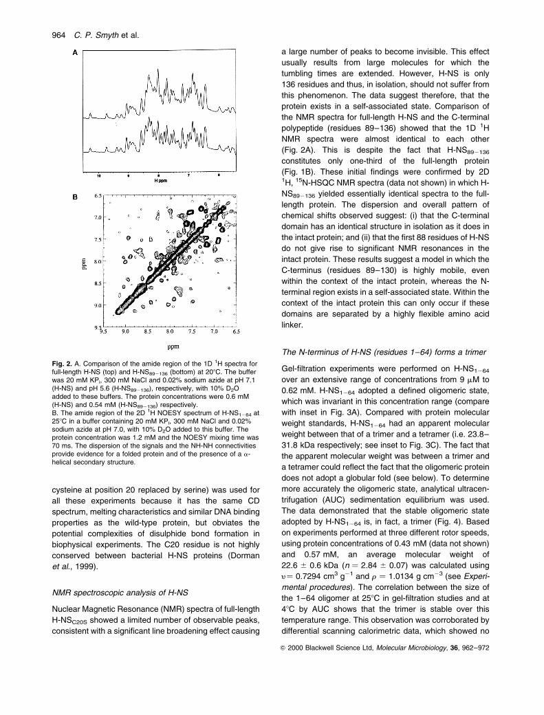
Nuclear Magnetic Resonance (NMR) spectra of full-length
H-NSC20S showed a limited number of observable peaks,
consistent with a significant line broadening effect causing
a large number of peaks to become invisible. This effect
usually results from large molecules for which the
tumbling times are extended. However, H-NS is only
136 residues and thus, in isolation, should not suffer from
this phenomenon. The data suggest therefore, that the
protein exists in a self-associated state. Comparison of
the NMR spectra for full-length H-NS and the C-terminal
polypeptide (residues 89±136) showed that the 1D 1H
NMR spectra were almost identical to each other
(Fig. 2A). This is despite the fact that H-NS892136
constitutes only one-third of the full-length protein
(Fig. 1B). These initial findings were confirmed by 2D1H, 15N-HSQC NMR spectra (data not shown) in which H-
NS892136 yielded essentially identical spectra to the full-
length protein. The dispersion and overall pattern of
chemical shifts observed suggest: (i) that the C-terminal
domain has an identical structure in isolation as it does in
the intact protein; and (ii) that the first 88 residues of H-NS
do not give rise to significant NMR resonances in the
intact protein. These results suggest a model in which the
C-terminus (residues 89±130) is highly mobile, even
within the context of the intact protein, whereas the N-
terminal region exists in a self-associated state. Within the
context of the intact protein this can only occur if these
domains are separated by a highly flexible amino acid
linker.
The N-terminus of H-NS (residues 1±64) forms a trimer
Gel-filtration experiments were performed on H-NS1264
over an extensive range of concentrations from 9 mM to
0.62 mM. H-NS1264 adopted a defined oligomeric state,
which was invariant in this concentration range (compare
with inset in Fig. 3A). Compared with protein molecular
weight standards, H-NS1264 had an apparent molecular
weight between that of a trimer and a tetramer (i.e. 23.8±
31.8 kDa respectively; see inset to Fig. 3C). The fact that
the apparent molecular weight was between a trimer and
a tetramer could reflect the fact that the oligomeric protein
does not adopt a globular fold (see below). To determine
more accurately the oligomeric state, analytical ultracen-
trifugation (AUC) sedimentation equilibrium was used.
The data demonstrated that the stable oligomeric state
adopted by H-NS1264 is, in fact, a trimer (Fig. 4). Based
on experiments performed at three different rotor speeds,
using protein concentrations of 0.43 mM (data not shown)
and 0.57 mM, an average molecular weight of
22.6 ^ 0.6 kDa (n � 2.84 ^ 0.07) was calculated using
y� 0.7294 cm3 g21 and r � 1.0134 g cm23 (see Experi-
mental procedures). The correlation between the size of
the 1±64 oligomer at 258C in gel-filtration studies and at
48C by AUC shows that the trimer is stable over this
temperature range. This observation was corroborated by
differential scanning calorimetric data, which showed no
Fig. 2. A. Comparison of the amide region of the 1D 1H spectra forfull-length H-NS (top) and H-NS892136 (bottom) at 208C. The bufferwas 20 mM KPi, 300 mM NaCl and 0.02% sodium azide at pH 7.1(H-NS) and pH 5.6 (H-NS892136), respectively, with 10% D2Oadded to these buffers. The protein concentrations were 0.6 mM(H-NS) and 0.54 mM (H-NS892136) respectively.B. The amide region of the 2D 1H NOESY spectrum of H-NS1264 at258C in a buffer containing 20 mM KPi, 300 mM NaCl and 0.02%sodium azide at pH 7.0, with 10% D2O added to this buffer. Theprotein concentration was 1.2 mM and the NOESY mixing time was70 ms. The dispersion of the signals and the NH-NH connectivitiesprovide evidence for a folded protein and of the presence of a a-helical secondary structure.
964 C. P. Smyth et al.
Q 2000 Blackwell Science Ltd, Molecular Microbiology, 36, 962±972

change in excess heat capacity in this temperature range
(data not shown), consistent with no change in oligomeric
state over the physiological temperature range.
The trimeric region of H-NS is primarily a -helical
Computer-based structural prediction programs (Lupas,
1996) showed that the N-terminal 64 residues possess a
tendency to adopt a coiled±coil motif (Fig. 1C). The
presence of a characteristic heptad repeat in these
N-terminal amino acids, and sequence similarity to well-
known coiled±coil proteins, such as myosin, keratin, and
dystrophin led to the suggestion that the self-association
of H-NS could be mediated by interactions between
a-helices (Ussery et al., 1994). This coiled±coil con-
formation presumably provides the structural basis for
self-association into the trimer.
To test these predictions, circular dichroism (CD)
spectrometry was used. The magnitude of the CD in the
peptide backbone region between 185 and 260 nm of
H-NS1264 was strongly concentration dependent in the
range 0.3±30 mM (in 10 mM KPi, 10 mM NaCl at pH 7.0).
Changes in the CD spectra were pronounced in the two
negative maxima at close to 208 and 222 nm and the
positive maximum at close to 192 nm (Fig. 5A), demon-
strating that the self-association of H-NS1264 is coupled to
the formation of a a-helical structure (Table 1). This is
consistent with a coiled±coil structure. At concentrations
above 30 mM, no change in CD spectra was observed,
indicating that a defined structural state had been attained
(Fig. 5A).
Residues 1±64 of H-NS gave measurable NMR spectra
(Fig. 2B), in contrast to their behaviour in the context of
the intact protein. The limited line broadening and
relaxation data were consistent with H-NS1264 forming a
defined oligomeric state with a molecular weight signifi-
cantly less than the large disperse oligomers formed by
full-length H-NS (see above). The spectra obtained were
also consistent with a significant proportion of a a-helical
structure (Fig. 2B).
H-NS1289 adopts higher self-associated states
Compared with H-NS1264, significant NMR spectral line
width broadening was observed for H-NS1289. This
suggested that residues from 64 to 89 might contribute
to the formation of higher order structures. We therefore
investigated the oligomerization properties of H-NS1289.
Gel-filtration traces obtained for H-NS1289 at a range of
concentrations are shown in Fig. 3B. The asymmetric
nature of the broad peaks (in marked contrast to
H-NS1264) are consistent with H-NS1289 adopting a wide
range of different self-associated states with an increased
average molecular weight at increased concentrations.
Fig. 3. Gel-filtration assay on full length and truncated forms ofH-NSC20S. The figures give the optical density at 220 nM as afunction of elution time (minutes). The samples were applied to aSuperose 12 column in 20 mM KPi and 300 mM NaCl at pH 7.0and 258C at the indicated concentrations unless otherwise stated.A. H-NS1264 at 9.6, 19.3, 38.5, 77.1, 155, 308 and 617 mM. Theinset gives the traces obtained for a monomeric control protein(ovalbumin) at concentrations ranging from 3 to 49 mM.B. H-NS1289 at 8.9, 17.9, 35.7, 71.5, 143, 286 and 572 mM. Theinset shows the results on full-length H-NS at 8.9, 17.9, 35.7, 71.4,143, 162, 260 and 344 mM.C. H-NS892136 at 13.4, 26.6, 52.6, 105, 210, 289 and 578 mM in1.8 mM KPi, 10 mM NaPi, 140 mM NaCl and 2.7 mM KCl at pH 7.4and 258C. The optical density was recorded at 280 nm. Given inthe inset is the result observed for a molecular weight standard set,with molecular weights of 1.25, 17, 44 and 158 kDa from right toleft. There is also an undefined peak corresponding to the voidvolume and a 660 kDa protein standard (not used in the analysis).
Oligomerization of H-NS 965
Q 2000 Blackwell Science Ltd, Molecular Microbiology, 36, 962±972

Distinct oligomeric states could not be resolved. Compar-
ison of peak maxima (Fig. 3B) with globular protein
molecular weight standards gave apparent molecular
weights ranging from 24 kDa at 8.9 mM to 198 kDa at
0.57 mM. These numbers correspond to a change in the
most abundant oligomeric state of H-NS1289 from some-
what larger than a dimer to something in the order of 20
mers within this concentration range. The gel-filtration
experiments, despite the potential inaccuracies in deter-
mining the molecular weight of the 1±89 polypeptide
oligomer, do serve to demonstrate very clearly a
difference in behaviour when compared with the 1±64
polypeptide.
We next used AUC to determine the oligomeric state of
the 1±89 polypeptide. Again, and in contrast to H-NS1264,
AUC data for H-NS1289 could not be accurately modelled
for a single species. The average buoyant molecular
mass of this polypeptide was found to vary significantly
with centrifugation speed, ranging from approximately
68 kDa at 8000 r.p.m. to 32 kDa at 12000 r.p.m. for a
protein concentration of 0.58 mM. The increase of M (1-y
r) with decreasing rotor speed demonstrated that
H-NS1289 did not reach an equilibrium distribution of
oligomeric states at this concentration. As the speed was
reduced, the extent to which the larger components could
contribute to the average molecular weight of the sample
increased, indicating that at the faster speeds the larger
complexes travelled to the bottom of the cell and were not
detected. It is important to note that the sample did not
exhibit time-dependent aggregation, because the
observed masses did not depend on the order of data
collection at the different speeds. The AUC experiments
show that H-NS1289 forms self-associated complexes of
considerable size, in agreement with the above gel-
filtration data. These data clearly demonstrate dramati-
cally different oligomerization behaviour to trimeric
H-NS1264.
The C-terminal domain of H-NS (residues 89±136) is a
monomer
Gel-filtration traces for H-NS892136 at a range of con-
centrations suggested a molecular weight that is between
that predicted for a monomer and a dimer (Fig. 3C). The
symmetrical nature of the gel filtration peaks at all
concentrations, and the constant position of the peak,
confirmed that H-NS892136 is present in a single, defined
state that is concentration independent. AUC sedimenta-
tion equilibrium data for H-NS892136 gave an apparent
stoichiometry of 1.3 (data not shown). We interpret these
data as showing that the polypeptide is monomeric and
attribute the intermediate stoichiometries to possible
partial unfolding of this polypeptide, in agreement with
the solution structural data for H-NS912136 from Escher-
ichia coli in which some structural disorder was observed
(Shindo et al., 1995).
CD spectra for the H-NS892136 polypeptide were
significantly different from those of the N-terminal domain
(inset Fig. 3A). The spectra were not dominated by
a-helix, in agreement with the previously reported NMR
structure for C-terminal H-NS from E. coli (Shindo et al.,
1995). We can not explain the very small concentration
dependence seen in the CD spectra for H-NS892136 (see
inset to Fig. 5B), but note that the magnitude is close to
the experimental uncertainty.
The C-terminal (residues 89±136) forms an independent
domain within the context of the full-length protein
The observation that only residues 89±136 contributed to
the NMR spectra of H-NSC20S, and the location of trypsin-
sensitive cleavage sites between residues 82±90 (Cusick
and Belfort, 1998), suggest that H-NS892136 exists as an
independent domain within the context of the intact
protein. We set out to test this experimentally. Melting
Fig. 4. Analytical ultracentrifugationsedimentation equilibrium data on H-NS1264
at 0.57 mM at centrifugation speeds of A8000 and B 15500 r.p.m. The experimentswere carried out in 20 mM KPi and 300 mMNaCl at pH 7.0 and 48C. The solid linesrepresent best-fits with M (1 2 y r) values of37176 and 5876 for these protein solutions.The residuals of the fits are shown in eachcase, demonstrating that these are randomlydistributed around zero.
966 C. P. Smyth et al.
Q 2000 Blackwell Science Ltd, Molecular Microbiology, 36, 962±972

studies using NMR involve measuring the change of a
given proton chemical shift from a particular residue as
the protein is heated. The chemical shifts at high field
resonances of individual residues in the C-terminal
domain can be followed as a function of temperature for
H-NS892136 using NMR spectroscopy. The temperature
dependence of the chemical shift at 0.12 p.p.m. (208C) is
shown in Fig. 6A. The absence of a clear high tempera-
ture end-point prevents a precise determination of the
melting temperature (Tm), but an approximate measure
can be obtained by fitting for the Tm while floating the high
temperature chemical shift. This results in an apparent Tm
of 618C for H-NS892136 (pH 5.6) using a two-state
transition model. Chemical shift data for the full-length
protein (as described for H-NS892136 above) shows a
transition with a Tm of 588C (pH 7.0). This is derived from
chemical shifts identified as being associated with protons
in the C-terminal domains (because the N-terminal suffers
from severe line broadening in the full-length NMR
spectra). Because the majority of aromatic residues are
found in the C-terminus of H-NS, the thermal melting of
full-length H-NS (at 31 mM) was also measured using CD
spectroscopy. Analysis of the temperature-induced
changes in the aromatic region at 288 nm (Fig. 6B)
gave a Tm of 598C. The Tm values for H-NSC20S were in
good agreement with those of the isolated domain,
suggesting that the presence of the N-terminal region of
the protein has little effect on the stability of the
C-terminus. This is consistent with H-NS892136 being a
structurally independent domain existing as a monomer
(see above) and rules out the possibility of inter-domain
interactions. The inability of residues 89±136 to associate
within the context of the full-length protein is a require-
ment for the highly mobile domain required to produce the
NMR spectra described above.
The melting temperature derived for the C-terminal
domain correlates well with a previously reported value of
around 578C derived from fluorescence spectroscopic
studies on E. coli H-NS (Tippner and Wagner, 1995). This
value is also likely to correspond to the melting of the
independent region of residues 89±136 because Trp 108
is the major fluorophore in the protein.
Full-length H-NSC20S adopts higher self-associated states
The gel-filtration peaks for H-NSC20S were broad and
clearly asymmetric at all concentrations (inset Fig. 3B), as
seen for H-NS1289. These results substantiate the
Fig. 5. Circular dichroism studies on full length and truncated formsof H-NS in 10 mM KPi and 10 mM NaCl at pH 7.0 and 258C unlessotherwise stated. The figures show the delta epsilon D1 as afunction of wavelength (l) in the peptide backbone region (185±260 nM).A. H-NS1264 at 0.33 (¼.), 3.3 (± ±), 33 (Ð) and 166 (.-.-) mMshowing that the full-length spectrum is dominated by contributionsfrom the N-terminal 64 residues. The inset gives the spectrum ofH-NS892136 at 0.32 (¼.), 3.2 (± ± ±), 32 (____) and 75.5 (.-.-) mMin 5 mM KPi and 10 mM NaCl at pH 7.0 and 258C. The C-terminalpart of the protein gives a comparatively weak CD spectrum. Thesmall changes in the CD spectrum of H-NS892136 withconcentration are negligible in comparison with those observed infull-length H-NS and H-NS1264.B. Full-length H-NS at 0.31 (¼.), 3.1 (± ± ±) and 31 (____) mM.The inset shows the spectrum of full-length H-NS at 3.1 (¼.), 31(± ±) and 310 (____) mM in 10 mM KPi and 300 mM NaCl atpH 7.0 and 258C (lower protein concentrations could not beexamined due to the strong absorption of buffer components inlonger path-length cells).
Table 1. Deconvolution of the circular dichroism data.
Protein/Polypeptide Concentration (mM) a-Helix (%)
H-NSC20S 310 45.3a
31 47b (44.3a)3.1 30.4b (34.4a)0.31 14.8b
H-NS1264b 166 55
33 53.23.3 34.90.33 19.4
H-NS892136c 75.5 10.7
32 10.53.2 9.60.32 7.8
a. In 10 mM KPi and 300 mM NaCl at pH 7.0 and 258C.b. In 10 mM KPi and 10 mM NaCl at pH 7.0 and 258C.c. In 5 mM KPi and 10 mM NaCl at pH 7.0 and 258C.
Oligomerization of H-NS 967
Q 2000 Blackwell Science Ltd, Molecular Microbiology, 36, 962±972

hypothesis that H-NSC20S self-associates to form large
complexes as the concentration is increased, and that no
single defined state is reached within the concentration
range tested. In this case, a comparison with molecular
weight standards gave apparent molecular weights of
51 kDa at 8.9 mM and 281 kDa at 0.34 mM, for these
peak maxima. These numbers correspond to a change in
the oligomeric state from somewhat larger than a trimer to
approximately 20-mers within this concentration range,
similar to the data for H-NS1289. The peaks were smooth
and distinct oligomeric states could not be resolved even
when other columns were used (inset to Fig. 3B).
The behaviour of full-length protein, as observed by
AUC, resembled that of H-NS1289 (data not shown). The
buoyant molecular mass of H-NSC20S ranged from 37 kDa
at 8000 r.p.m. to 26 kDa at 12000 r.p.m. for a protein
concentration of 52 mM. These are lower molecular
masses than previously observed for the 1±89 polypep-
tide, but this can be explained by the significant difference
in concentration and protein size (more of the larger
H-NSC20S will travel to the bottom of the cell and not
contribute to the analysis). Additional AUC sedimentation
velocity experiments were carried out on H-NSC20S (data
not shown) and a large set of interacting and non-
interacting models were used in an attempt to fit the data
(including monomer±dimer, monomer±tetramer±octamer,
monomer±trimer±hexamer±nonamer). None of these
models led to an improved fit of the data and, furthermore,
the derivatives of the velocity data curves suggest a
multicomponent system. The presence of a range of
oligomeric states agrees with the above gel-filtration data,
in which the width of the asymmetric peak for any given
concentration spans approximately a 10-fold difference in
molecular weight.
The behaviour observed on gel filtration could, poten-
tially, be explained if H-NSC20S dissociates from a defined
oligomeric state while travelling through the column. This
has previously been invoked to explain the behaviour of
mutants of the coiled±coil leucine zipper protein GCN4
(Zeng et al., 1997). However, in the light of the above
AUC data, we favour the explanation that the protein
exists in a range of different oligomeric states.
CD spectra for the full-length H-NS protein were very
similar to those obtained for residues 1±64 (Fig. 3B).
Again, the CD signal was strongly concentration depen-
dent and accompanied by the formation of an a-helical
secondary structure at lower concentrations (Table 1). It
was not possible to assess whether the a-helix content
reached a maximum at high concentration (as seen in the
case of H-NS1264) owing to insolubility of H-NSC20S at
10 mM NaCl. The protein appears to aggregate/precipi-
tate in low saline conditions. Under higher salt conditions
(300 mM NaCl), H-NSC20S is soluble and CD spectra at
high concentration could be obtained. The CD spectra
resembled those observed in low salt conditions. At high
protein concentrations (up to 310 mM) the a-helical
content of the sample did not increase significantly
(Table 1 and inset Fig. 5B), demonstrating that the
oligomeric state which is accompanied by structure
formation has become saturated. This does not necessa-
rily mean that maximum oligomeric state of H-NSC20S has
been achieved, because another mechanism for this self-
association, which is not accompanied by additional
structure formation, could prevail. Indeed, this appears
to be the case because, although the formation of a
a-helix has a limit at concentrations in the region above
30 mM, gel filtration as well as AUC experiments show
that the formation of higher oligomeric states continues at
concentrations above this. These findings are in contrast
Fig. 6. A. NMR melting experiments of H-NS892136 and full-lengthH-NS. The chemical shift at 0.12 p.p.m. (208C) was followed as afunction of temperature (15±688C). The buffer was 20 mM KPi,300 mM NaCl and 0.02% sodium azide at pH 7.0 (H-NS) or pH 5.6(H-NS892136), respectively, with 10% D2O and 0.00125% TSPadded to these buffers. The protein concentrations were 0.22 mM(H-NS) and 0.54 mM (H-NS892136) respectively. The change inchemical shift with temperature was fitted to a sigmoidal model, asdemonstrated by the solid lines, to obtain an apparent melting pointof 588C for full-length H-NS (B) and 618C for H-NS892136 (O)respectively.B. Aromatic region of the full-length H-NS (31 mM) spectrum at33.8, 41.3, 48.9, 55.4, 62.5, 74.3 and 88.28C. The inset gives thedelta epsilon (D1) at 288 nm as a function of temperature and thesolid line represents the non-linear least-square fit to the data witha melting temperature of 598C as described in the text.
968 C. P. Smyth et al.
Q 2000 Blackwell Science Ltd, Molecular Microbiology, 36, 962±972

to the situation for H-NS1264 (Fig. 3A), which self-
associates with the concomitant formation of a a-helix,
but once the trimeric form has been reached then no
further self-association is observed.
Discussion
Genetic data have demonstrated that H-NS consists of at
least two domains and can exist in oligomeric forms that
contribute to its biological activity. Biochemical studies of
the oligomeric state have, however, led to conflicting
conclusions. We have therefore attempted to characterize
this behaviour directly using biochemical and biophysical
approaches. The data reported here demonstrate that
H-NS does not exist in a single, defined, oligomeric state
and highlight the complex and unusual nature of H-NS
oligomerization. The protein consists of two distinct
domains separated by a highly flexible linker. The
C-terminal DNA-binding domain (amino acids 89±136)
does not contribute to oligomerization and, in the context
of the intact oligomeric protein, is freely mobile. Oligomer-
ization is due to the N-terminal domain (residues 1±89).
Amino acids 1±64 form a discrete trimer, probably a
coiled±coil. The addition of 25 amino acids (residues 1±
89) results in the formation of larger, heterodisperse
oligomers (up to at least 20 mers) in a concentration-
dependent fashion. Full-length H-NS also adopts a
concentration-dependent heterodisperse oligomeric state
of up to 20 mers.
Trimer formation is biologically relevant because the
same residues of H-NS expressed in vivo produce a
dominant-negative effect when expressed in hns1
bacteria (Ueguchi et al., 1997). Trimerization probably
involves a coiled±coil configuration and we have demon-
strated the formation of a increased a-helical structure
upon oligomerization. Trimer formation increases with
concentration up to about 30 mM. Trimer formation is also
temperature independent, at least within the physiological
range. Trimer formation is likely to form a building block
for higher order oligomerization.
Increasing the polypeptide length to include residues
65±89 resulted in further self-association, giving rise to
heterodisperse, higher order oligomeric states. The size
of the oligomers increased with concentration. This
oligomerization was also seen in the full-length protein:
it appears that residues 65±89 form the platform for this
oligomerization.
Remarkably, like residues 1±89, full length also H-NS
forms heterodisperse, higher order oligomers in a
concentration-dependent fashion. At concentrations
below 30 mM, there is concentration-dependent formation
of a-helix, consistent with the building block being the
colied±coil trimers seen for residues 1±64. At higher
concentrations, further self-association continued in a
manner similar to that observed for amino acids 1±89.
This two-step self-association, the formation of trimers
and then association into larger heterodisperse oligomers
whose size increases with concentration is a unique
property of H-NS and unusual for such a comparatively
small protein. The finding that H-NS does not adopt a
discrete oligomeric state accounts for published data,
which appear, at first sight, inconsistent in reporting
oligomeric forms of H-NS from dimer to hexamer (Falconi
et al., 1988; Spurio et al., 1997; Ueguchi et al., 1997).
The C-terminal domain of H-NS, in both the free form
and covalently associated within the context of the intact
protein, was shown by NMR spectroscopy to be structu-
rally independent and isolated by a flexible linker, allowing
it a high degree of mobility such that it can rotate freely.
The reported structure of the C-terminal of H-NS from E.
coli (Shindo et al., 1995) shows that the globular fold
begins at about residue 95, so the flexible linker is
probably included in the H-NS892136 polypeptide. The role
of this flexible linker is yet to be resolved; however, it is
presumably required for the C-terminal domain to adopt
the necessary orientations within a large oligomeric
structure in order to interact with DNA.
The high concentration of H-NS in bacterial cells means
that the prevalence of high order oligomers will provide a
scaffold around which DNA can interact and facilitate its
compaction. As the isolated C-terminal domain shows low
affinity for DNA (Shindo et al., 1995), it is likely that the co-
operative effect of binding of the oligomeric protein leads
to enhanced affinity within the context of the full-length
protein. The availability of a C-terminal DNA binding
domain on a flexible linker avoids steric constraint on the
binding of DNA and its subsequent packaging.
Experimental procedures
Bacterial strains and plasmids
E. coli strain DH5a was used for all gene cloning experi-ments. Plasmid pJSH6, which encodes the C20S mutant ofthe full-length H-NS, was constructed as described below.Chromosomal DNA from S. typhimurium was purified asdescribed elsewhere (Maniatis et al., 1982). The primers 5 0-CAAACTGGAGACT-CATATGAGCGAAGCACTTAAAATTC-3 0 and 5 0-CACGGATCCGGTT-CCTGGGTATTAAGAAG-TAAC-3 0 were used to amplify, by PCR, the H-NS structuralgene (Hulton et al., 1990), and to introduce BamHI and NdeIrestriction sites at the 5 0 and 3 0 ends respectively. The PCRproduct was cloned into the SmaI site of pT7-3 (Tabor andRichardson, 1985) to generate pJSH1. The hns gene wasexcised from pJSH1 on an EcoRI±HindIII fragment andsubsequently cloned into pALTER for site-directed mutagen-esis (Altered Sites 2 system from Promega). The C20Smutation was introduced into the hns gene, using the primer5 0-GCGCAGGCAAGAGAAAGCACTCTGGAAACGC-3 0. TheC20S hns gene was completely sequenced to ensure no
Oligomerization of H-NS 969
Q 2000 Blackwell Science Ltd, Molecular Microbiology, 36, 962±972

other mutations had been introduced. The gene was excisedfrom pALTER with BamHI and NdeI and ligated into BamHI±NdeI-digested pET14b and named pJSH6 (Studier andMoffat, 1986; Rosenberg et al., 1987). This also introduced anN-terminal 6 � His-tag followed by a thrombin cleavage site.
Plasmids pCPS1 and pCPS2, which overexpress H-NS1264 and H-NS1289 respectively, were constructed.Truncated hns genes, coding for residues 1±64 and 1±89,were amplified with pJSH6 as the template, using primers5 0-CAAACTGGAGACTCATATGAGCGAAGCACTTAAAATT-C-3 0 and 5 0-CACGGATCCCTATTATAACATTTCACGATAC-TGTTGCAGTTT-3 0 or 5 0-CACGGATCCCTATTAGCGTTTAGCTTTGGTACCGGATTTAGC-3 0 respectively. Restrictionsites NdeI and BamHI were introduced at the 5 0 and the 3 0
ends respectively. The PCR products were digested withNdeI and BamHI and ligated into pET14(b), introducing an N-terminal 6 � His tag and thrombin cleavage site as describedabove. DNA sequencing was used to verify the identity of thetruncated hns genes.
Protein expression, purification and biological activity
E. coli BL21 (lDE3 plysS) cells were transformed, separatelywith plasmids pJSH6, pCPS1 and pCPS2 to express full-length H-NSC20S, H-NS1264 or H-NS1289 respectively. Next,500 ml flasks containing 150 ml of Luria±Bertani (LB) media,chloramphenicol (34 mg ml21) and carbenicillin (200mg ml21) were inoculated with cells from a freshly trans-formed colony. Then 10 ml of the overnight cultures wereused to inoculate 2 l flasks containing fresh media andantibiotics (500 ml) and grown in a shaking incubator at200 r.p.m. and 378C until an OD600 of 0.5 was reached. TheT7 promoter was then induced by the addition of isopropyl-b-D-thiogalactopyranoside (IPTG; Melford Laboratories) to afinal concentration of 0.5 mM and incubated at 200 r.p.m.and 308C for 3 h. The cells were centrifuged at 5000 r.p.m. ina SORVALL GS3 rotor and the resulting cell pellet was storedat 2708C.
Cell lysis was by sonication in 20 mM Tris, 500 mM NaCl,5% glycerol, 1% Triton X-100 and 0.5 mM 4-(2-aminoethyl)benzene sulphonyl fluoride hydrochloride (AEBSF; MelfordLaboratories) at pH 8.0. The lysate was centrifuged toremove insoluble cell debris. The supernatant was heatedto 608C for 15 min, recentrifuged to remove any denaturedprotein and applied to a TALONe metal affinity columnequilibrated with 20 mM Tris, 500 mM NaCl and 5% glycerolat pH 8.0. His-tagged H-NS protein was eluted in 20 mM Tris,500 mM NaCl, 0.5 M imidazole and 5% glycerol at pH 8.0.The protein was precipitated with 70% ammonium sulphateand subsequently resuspended in 20 mM Tris, 150 mMNaCl, 2.5 mM CaCl2 and 5% glycerol at pH 8.0. Thrombin(Bovine Thrombin, Calbiochem) was added to remove theHis-tag, and the protein diluted four times with distilled waterand then applied to a Pharmacia MonoS column equilibratedin 10 mM KPi 1 mM Na-EDTA at pH 7.5. H-NS was theneluted in 10 mM KPi, 600 mM NaCl and 1 mM Na-EDTA atpH 7.5. Thrombin cleavage of the His-tag resulted in aprotein product with four additional amino acids, GSHM, atthe N-terminus of each H-NS-derived protein.
For the purification of H-NS1264 and H-NS1289 polypep-
tides, the heating step after the initial cell lysis andcentrifugation steps was not performed. Furthermore, sub-sequent to thrombin cleavage, the polypeptides werereapplied to the TALONe metal affinity column and collectedas flow through, thus removing the His-tag, which remainsbound to the column.
The N-terminally truncated H-NS892136 protein was purifiedas follows. The supernatant containing full-length His-taggedH-NSC20S protein was applied to the TALONe metal affinitycolumn equilibrated in 20 mM Tris, 150 mM NaCl and2.5 mM CaCl2 at pH 8.0. Trypsin was added to the column,resulting in cleavage of the full-length protein (Cusick et al.,1998). The H-NS892136 polypeptide was eluted with theabove buffer. The protein was purified further using theMonoS column as described above.
Full-length H-NSC20S, H-NS1264, H-NS1289 and H-NS892136 protein samples were concentrated according tothe requirements of a given experiment (see below), usingeither an Amicon stirred cell or centriprep (Amicon) concen-trators, and dialysed into the required buffer, depending uponthe experiment to be performed. The polypeptides werestored under the experimental buffer at 48C prior to use.
The size and homogeneity (. 95% in all cases) of thepurified proteins was confirmed by denaturing SDS±PAGEand staining with Coomassie brilliant blue R-250. The massof each of the proteins was confirmed using mass spectro-metry. The following results were obtained with the expectedmolecular weight given within brackets. H-NSC20S,15806 g mol21 (15805); H-NS1264, 7959 g mol21 (7958);H-NS1289, 10516 g mol21 (10512); and H-NS892136, 5311/5467 g mol21 (5314/5470). The difference in molecularweight between the two species in the H-NS892136 sampleis equivalent to that of an arginine residue, indicating that thetrypsin cleavage yields two different products (Cusick et al.,1998). All the experimental results are identical to theexpected masses within experimental uncertainty.
The biological activity of the C20S full-length protein wascompared with that of wild-type H-NS from S. typhimurium(Hulton et al., 1990), using in vitro DNA mobility shift assays.Plasmid pAF3 (Jordi et al., 1997) was purified and digestedwith the restriction enzyme HaeIII. The DNA restrictionfragments were incubated with wild-type H-NS or H-NSC20S
(in an overall amount ranging from 0.1 to 6.0 mg), in 10 mlreaction mixtures containing 10 mM Tris, 0.2 mM Na-EDTA,15 mM KCl, 2 mM spermidine and 15% glycerol at pH 7.5.After 10 min at room temperature, protein±DNA complexeswere resolved on 3% agarose gels, using TBE (89 mM Tris-Borate, 89 mM Boric acid and 10 mM Na-EDTA at pH 8.0) asthe running buffer. The DNA fragments were stained withethidium bromide and visualized using a UV lamp. H-NSC20S
caused a shift in the mobility of DNA, which was comparableto that of wild-type H-NS (data not shown).
Circular dichroism spectroscopy
Proteins were extensively dialysed against a buffer contain-ing 10 mM potassium phosphate (KPi) and 10 mM NaCl atpH 7.0 at 48C unless otherwise stated. H-NS892136 and thefull-length H-NSC20S that were to be used for the lowtemperature study were, instead, dialysed against a buffer
970 C. P. Smyth et al.
Q 2000 Blackwell Science Ltd, Molecular Microbiology, 36, 962±972

containing 15 mM KPi and 30 mM NaCl at pH 7.0 and thendiluted three times using water and ethanediol respectively.CD spectra were recorded on Jasco 600 or 700 spectro-polarimeters with JOVAN attachments for low temperaturemeasurements. Spectra for the peptide backbone region(185±320 nm) were recorded using different path-lengthcells, ranging from 0.01 to 2 cm, depending on the proteinconcentration. Spectra for the aromatic region (240±340 nm)were recorded in a 5 cm path-length cell. All spectra wereaveraged over two or four scans, using a resolution of 0.2 nmand a scan rate of 10 nm per minute. All the samples wereallowed to reach thermal equilibrium in the cell, thetemperature of which was controlled using a Hewlett Packardpeltier system. The estimation of secondary structure contentwas performed using a principle component repressionmethod (PLSIQPRD of GRAMS/3.2 suite program) with acalibration data set of 16 proteins obtained elsewhere(Hennessey and Johnson, 1981).
Nuclear magnetic resonance spectroscopy
The 1H 1D and 2D Nuclear Overhauser Effect Sprectroscopy(NOESY) spectra (Macura et al., 1981) were recorded on aVarian UNITYplus 500 MHz spectrometer. The NMRPIPE
(Delaglio et al., 1995) and AZARA 2.0 (Boucher, 1996) softwarewas used to process and analyse the data. Next, 3-(trimethylsilyl)-propionate (TSP) was added to the samplesused for the NMR melting studies as an internal standard forthe referencing of the proton chemical shifts.
Gel filtration
A range of concentrations of H-NSC20S, H-NS1264, H-NS1289,H-NS892136 or a control protein (ovalbumin) were applied to aBIOCAD/INTEGRAL system equipped with a PharmaciaSuperose 12 column. All proteins were dialysed against10 mM KPi and 300 mM NaCl at pH 7.0 prior to loading thecolumn. The column was equilibrated with either the dialysisbuffer or phosphate-buffered saline (PBS) (1.8 mM KPi,10 mM NaPi, 140 mM NaCl and 2.7 mM KCl at pH 7.4) asindicated in the text. A calibration of the expected flow rateswas obtained by running molecular weight standards (Bio-Rad Gel Filtration Standard) in 10 mM KPi and 300 mM NaClat pH 7.0. These covered the range of molecular weightsfrom 1350 to 670 kDa.
Analytical ultracentrifugation
Analytical ultracentrifugation experiments were performedusing a Beckman XL-A analytical ultracentrifuge. Data wereacquired as an average of 25 absorbance measurements at awavelength of 280 nm and a radial spacing of 0.001 cm. H-NS protein was prepared in 20 mM KPi, 300 mM NaCl and0.05% sodium azide at pH 7.0. All sedimentation equilibriumexperiments were run using double sector charcoal-filledcentrepieces and column lengths of approximately 4 mm.Solvent densities were calculated from standard tables(Handbook of Chemistry and Physics, 1968, CRC Press,Oxford). Sedimentation equilibrium experiments were con-ducted at various rotor speeds, ranging from 8000 to
17000 r.p.m., and temperatures of 4.0, 15.0 and 208C. Thetime required for the attainment of equilibrium was estab-lished by running at a given rotor speed until successivescans, taken 12 h apart, were invariant. Data were thenanalysed to obtain the buoyant molecular mass, M (1 2 y r),using the Optima XL-A data analysis software (version 2.0,Beckman) running under Microcal Origin 2.8, by fitting datafrom each scan to:
Ar � exp�ln�Ao� 1 HM�1 2 yr��r2 2 r2o�� 1 E �1�
where Ao is the absorbance at a reference point ro, Ar is theabsorbance at a given radial position r, H represents v2/2RT,with v being the angular speed in rad s21, R, the gasconstant and T the absolute temperature, and E is a smallbaseline correction term. Residuals were calculated bysubtracting the best fit eq. 1 from the experimental data. Amodified version of the equation, including terms for self-association or multicomponent systems, was also used(BECKMAN ANALYSIS software version 2.0).
Acknowledgements
We would like to thank D. Ussery for his contribution to initiating
this study. J.E.L. is a Wellcome Trust Senior Research Fellow.T.L. was supported by the Swedish Foundation for International
Cooperation in Research and Higher Education and the Swedish
National Sciences Council. This work was supported by a
Wellcome Trust Programme Grant (Ref. 045490) to C.F.H. andJ.C.D.H. and by the ICRF.
References
Boucher, W. (1996) AZARA v2.0. Cambridge, UK: Department ofBiochemistry, University of Cambridge.
Bracco, L., Kotlarz, D., Kolb, A., Diekmann, S., and Buc, H.
(1989) Synthetic curved DNA-sequences can act as transcrip-
tional activators in Escherichia coli. EMBO J 8: 4289±4296.Cusick, M.E., and Belfort, M. (1998) Domain structure and RNA
annealing activity of the Escherichia coli regulatory protein
StpA. Mol Microbiol 28: 847±857.Delaglio, F., Grzesisek, S., Vuister, G.W., Zhu, G., Pfeifer, J., and
Bax, A. (1995) NMR-Pipe 2 A multidimensional spectral
processing system based on UNIX pipes. J Biomol NMR 6:
277±293.Dorman, C.J., Hinton, J.C.D., and Free, A. (1999) Domain
organization and oligomerization among H-NS-like nucleoid-
associated proteins in bacteria. Trends Microbiol 7: 124±128.
Drlica, K., and Rouviere-Yaniv, J. (1987) Histone-like proteins ofbacteria. Microb Rev 51: 301±319.
Falconi, M., Gualtieri, M.T., LaTeana, A., Losso, M.A., and Pon,
C.L. (1988) Proteins from the prokaryotic nucleoid 2 Primaryand quaternary structure of the 15 kDa Escherichia coli DNA-
binding protein, H-NS. Mol Microbiol 2: 323±329.
Hennessey, J., and Johnson, W.C. (1981) Information content in
the circular dichroism of proteins. Biochemistry 20: 1085±1094.
Higgins, C.F., Dorman, C.J., Stirling, D.A., Waddell, L., Booth,
I.R., May, G., et al. (1988) A physiological role for DNA
supercoiling in the osmotic regulation of gene expression in S.typhimurium and E. coli. Cell 52: 569±584.
Higgins, C.F., Hinton, J.C.D., Hulton, C.S.J., Owen-Hughes, T.,
Pavitt, G.D., and Seirafi, A. (1990) Protein H1: a role for
Oligomerization of H-NS 971
Q 2000 Blackwell Science Ltd, Molecular Microbiology, 36, 962±972

chromatin structure in the regulation of bacterial gene expres-sion and virulence? Mol Microbiol 4: 2007±2012.
Hulton, C.S.J., Seirafi, A., Hinton, J.C.D., Sidebotham, J.M.,
Waddell, L., Pavitt, G.D., et al. (1990) Histone-like protein H1
(H-NS), DNA supercoiling and gene expression in bacteria.Cell 63: 631±642.
Jordi, B.J.A.M., Fielder, A.E., Burns, C.M., Hinton, J.C.D., Dover,
N., Ussery, D.W., et al. (1997) DNA binding is not sufficient forH-NS mediated repression of proU expression. J Biol Chem
272: 12083±12090.
Laurent-Winter, C., Ngo, S., Danchin, A., and Bertin, P. (1997)
Role of Escherichia coli histone-like nucleoid structuring proteinin bacterial metabolsim and stress response. Identification of
targets by two-dimensional electrophoresis. Eur J Biochem
244: 767±773.
Lupas, A. (1996) Coiled-coils: New structures and new functions.Trends Biochem Sci 21: 375±382.
Macura, S., Huang, Y., Suter, D., and Ernst, R.R. (1981) Two-
dimensional chemical exchange and cross-relaxation spectro-
scopy of coupled nuclear spins. J Magn Reson 43: 259±281.Maniatis, T., Fritsch, E.F., and Sambrook, J. (1982) Molecular
Cloning: a Laboratory Manual. Cold Spring Harbor, NY: Cold
Spring Harbor Laboratory Press.Owen-Hughes, T.A., Pavitt, G.D., Santos, D.S., Sidebotham,
J.M., Hulton, C.S.J., Hinton, J.C.D., et al. (1992) The
chromatin-associated protein H-NS interacts with curved
DNA to influence DNA topology and gene expression. Cell71: 255±265.
Pettijohn, D.E. (1988) Histone-like proteins and bacterial chromo-
some structure. J Biol Chem 263: 12793±12796.
Rosenberg, H.A., Lade, B.N., Chui, D.S., Lin, S.W., Dunn, J.J.,and Studier, F.W. (1987) Vectors for selective expression of
cloned DNAs by T7 RNA polymerase. Gene 56: 125±135.
Shindo, H., Iwaki, T., Ieda, R., Kurumizaka, H., Ueguchi, C.,Mizuno, T., et al. (1995) Solution structure of the DNA-binding
domain of a nucleoid-associated protein, H-NS, from Escher-
ichia coli. FEBS Microbiol Lett 360: 125±131.
Spurio, R., Falconi, M., Brandi, A., Pon, C.L., and Gualerzi, C.O.(1997) The oligomeric structure of nucleoid protein H-NS is
necessary for recognition of intrinsically curved DNA and for
DNA bending. EMBO J 16: 1795±1805.
Studier, F.W., and Moffat, B.A. (1986) Use of bacteriophage T7RNA polymerase to direct selective high level expression of
cloned genes. J Mol Biol 189: 113±130.
Tabor, S., and Richardson, C.C. (1985) A Bacteriophage T7 RNA
polymerase promoter system for controlled exclusive expres-sion of specific genes. Proc Natl Acad Sci USA 82: 1074±
1078.
Tippner, D., and Wagner, R. (1995) Fluorescence analysis of the
Escherichia coli transcription regulator, H-NS reveals two
distinguishable complexes dependent on binding to specificor nonspecific DNA sites. J Biol Chem 38: 22243±22247.
Tupper, A.E., Owen-Hughes, T.A., Ussery, D.W., Santos, D.S.,
Ferguson, D.J.P., Sidebotham, J.M., et al. (1994) The
chromatin-associated protein H-NS alters DNA topology invitro. EMBO J 13: 258±268.
Ueguchi, C., Suzuki, T., Yoshida, T., Tanaka, K., and Mizuno, T.
(1996) Systematic mutational analysis revealing the functional
domain organization of Escherichia coli nucleoid protein H-NS.J Mol Biol 263: 149±162.
Ueguchi, C., Seto, C., Suzuki, T., and Mizuno, T. (1997)
Clarification of the dimerization domain and its functional
significance for the Escherichia coli nucleoid protein H-NS. JMol Biol 274: 145±151.
Ussery, D.W., Hinton, J.C.D., Jordi, B.J.A.M., Granum, P.E.,
Seirafi, A., Stephen, R.J., et al. (1994) The chromatin-
associated protein H-NS. Biochimie 76: 968±980.
Williams, R.M., and Rimsky, S. (1997) Molecular aspects of theE. coli nucleoid protein H-NS: a central controller of gene
regulatory networks. FEMS Microbiol Lett 156: 175±185.
Williams, R.M., Rimsky, S., and Buc, H. (1996) Probing the
structure, function, and interactions of the Escherichia coli H-NS and StpA proteins by using dominant negative derivatives.
J Bacteriol 178: 4335±4343.
Yamada, H., Muramatsu, S., and Mizuno, T. (1990) An
Escherichia coli protein that preferentially binds to sharply
curved DNA. J Biochem (Tokyo) 108: 420±425.
Zeng, X., Herdon, A.M., and Hu, J.C. (1997) Buried asparagines
determine the dimerization specificities of leucine zipper
mutants. Proc Natl Acad Sci USA 94: 3673±3678.
972 C. P. Smyth et al.
Q 2000 Blackwell Science Ltd, Molecular Microbiology, 36, 962±972