"Goat-Stags, Philosopher-Kings, and Eudaimonism in the Republic"
Upload
unispital-baselCategory
view
4download
0
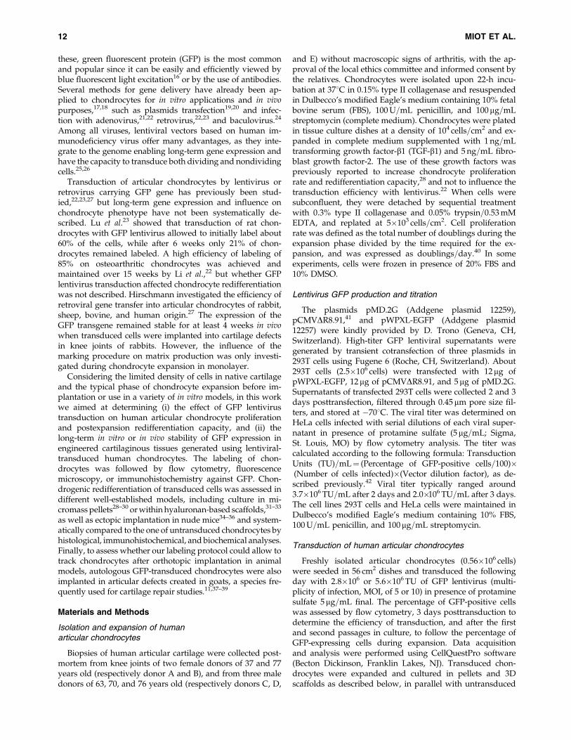
In Vitro and In Vivo Validation of Human and GoatChondrocyte Labeling by Green Fluorescent
Protein Lentivirus Transduction
Sylvie Miot, Ph.D.,1 Roberto Gianni-Barrera, Ph.D.,1 Karoliina Pelttari, Ph.D.,1 Chitrangada Acharya, Ph.D.,1
Pierre Mainil-Varlet, M.D.,2 Henriette Juelke, M.D.,2 Claude Jaquiery, M.D.,1 Christian Candrian, M.D.,1,3
Andrea Barbero, Ph.D.,1 and Ivan Martin, Ph.D.1
We investigated whether human articular chondrocytes can be labeled efficiently and for long-term with a greenfluorescent protein (GFP) lentivirus and whether the viral transduction would influence cell proliferation andtissue-forming capacity. The method was then applied to track goat articular chondrocytes after autologousimplantation in cartilage defects. Expression of GFP in transduced chondrocytes was detected cytofluorimetricallyand immunohistochemically. Chondrogenic capacity of chondrocytes was assessed by Safranin-O staining, im-munostaining for type II collagen, and glycosaminoglycan content. Human articular chondrocytes were efficientlytransduced with GFP lentivirus (73.4� 0.5% at passage 1) and maintained the expression of GFP up to 22 weeks ofin vitro culture after transduction. Upon implantation in nude mice, 12 weeks after transduction, the percentage oflabeled cells (73.6� 3.3%) was similar to the initial one. Importantly, viral transduction of chondrocytes did notaffect the cell proliferation rate, chondrogenic differentiation, or tissue-forming capacity, either in vitro or in vivo.Goat articular chondrocytes were also efficiently transduced with GFP lentivirus (78.3� 3.2%) and maintained theexpression of GFP in the reparative tissue after orthotopic implantation. This study demonstrates the feasibility ofefficient and relatively long-term labeling of human chondrocytes for co-culture on integration studies, and indi-cates the potential of this stable labeling technique for tracking animal chondrocytes for in cartilage repair studies.
Introduction
Cartilage tissue engineering can provide functionalcartilaginous constructs that can be used for controlled
in vitro studies of chondrogenesis and potentially for in vivoarticular cartilage repair.1 For example, tissue-engineeredcartilage has been introduced as a model to investigate in-teractions between different zonal populations of articularchondrocytes,2 between primary and passaged chon-drocytes,3 or between chondrocytes and other cell types,including osteoblasts,2,4 human embryonic stem cells,5 andsynoviocytes.6 Engineered cartilaginous tissues have alsobeen used as in vitro model systems to study the integrationprocess of a cartilage graft with native cartilage7–9 or bonetissue.10 In most of these coculture or integration studies,however, some of the key mechanistic questions related tothe functional contribution of specific cell populations couldnot be addressed, since chondrocytes were either unlabeledor labeled with a membrane fluorescent dye,2 which is sub-jected to natural fading with time.
Due to limited scientific evidence of the role of implantedchondrocytes in a cartilage defect and of their extent ofparticipation in the repair process, chondrocyte labelingwould also be extremely important before cell grafting inanimal models. So far, only Dell’Accio et al.11 used a fluo-rescent dye, PKH26, to label goat articular chondrocytes andshowed that implanted cells persisted for at least 14 weeks inthe defects and contributed to structural cartilage repair in agoat model of autologous chondrocyte implantation. Thisstudy represents an important milestone in the field, but themethod used for labeling was not ideal for long-term studiesbecause of the natural fading of PKH26 and the decrease inintensity of labeling with cell divisions.11,12
With the perspective of performing controlled in vitro orin vivo studies on the interaction between defined chon-drocyte populations and other cell systems, a reliable tech-nique for sustained chondrocyte labeling should be used.Besides membrane fluorochromes,11,13 other methods for celltracking include the use of radioactive isotopes,14 Y chromo-some–specific probes,15 or delivery of reporter genes. Among
1Departments of Surgery and of Biomedicine, University Hospital Basel, Basel, Switzerland.2Institute of Pathology, University of Bern, Bern, Switzerland.3Ospedale Regionale di Lugano, FMH Chirurgie, Orthopadie und Traumatologie des Bewegungsapparates, Lugano, Switzerland.
TISSUE ENGINEERING: Part CVolume 16, Number 1, 2010ª Mary Ann Liebert, Inc.DOI: 10.1089=ten.tec.2008.0698
11
these, green fluorescent protein (GFP) is the most commonand popular since it can be easily and efficiently viewed byblue fluorescent light excitation16 or by the use of antibodies.Several methods for gene delivery have already been ap-plied to chondrocytes for in vitro applications and in vivopurposes,17,18 such as plasmids transfection19,20 and infec-tion with adenovirus,21,22 retrovirus,22,23 and baculovirus.24
Among all viruses, lentiviral vectors based on human im-munodeficiency virus offer many advantages, as they inte-grate to the genome enabling long-term gene expression andhave the capacity to transduce both dividing and nondividingcells.25,26
Transduction of articular chondrocytes by lentivirus orretrovirus carrying GFP gene has previously been stud-ied,22,23,27 but long-term gene expression and influence onchondrocyte phenotype have not been systematically de-scribed. Lu et al.23 showed that transduction of rat chon-drocytes with GFP lentivirus allowed to initially label about60% of the cells, while after 6 weeks only 21% of chon-drocytes remained labeled. A high efficiency of labeling of85% on osteoarthritic chondrocytes was achieved andmaintained over 15 weeks by Li et al.,22 but whether GFPlentivirus transduction affected chondrocyte redifferentiationwas not described. Hirschmann investigated the efficiency ofretroviral gene transfer into articular chondrocytes of rabbit,sheep, bovine, and human origin.27 The expression of theGFP transgene remained stable for at least 4 weeks in vivowhen transduced cells were implanted into cartilage defectsin knee joints of rabbits. However, the influence of themarking procedure on matrix production was only investi-gated during chondrocyte expansion in monolayer.
Considering the limited density of cells in native cartilageand the typical phase of chondrocyte expansion before im-plantation or use in a variety of in vitro models, in this workwe aimed at determining (i) the effect of GFP lentivirustransduction on human articular chondrocyte proliferationand postexpansion redifferentiation capacity, and (ii) thelong-term in vitro or in vivo stability of GFP expression inengineered cartilaginous tissues generated using lentiviral-transduced human chondrocytes. The labeling of chon-drocytes was followed by flow cytometry, fluorescencemicroscopy, or immunohistochemistry against GFP. Chon-drogenic redifferentiation of transduced cells was assessed indifferent well-established models, including culture in mi-cromass pellets28–30 or within hyaluronan-based scaffolds,31–33
as well as ectopic implantation in nude mice34–36 and system-atically compared to the one of untransduced chondrocytes byhistological, immunohistochemical, and biochemical analyses.Finally, to assess whether our labeling protocol could allow totrack chondrocytes after orthotopic implantation in animalmodels, autologous GFP-transduced chondrocytes were alsoimplanted in articular defects created in goats, a species fre-quently used for cartilage repair studies.11,37–39
Materials and Methods
Isolation and expansion of humanarticular chondrocytes
Biopsies of human articular cartilage were collected post-mortem from knee joints of two female donors of 37 and 77years old (respectively donor A and B), and from three maledonors of 63, 70, and 76 years old (respectively donors C, D,
and E) without macroscopic signs of arthritis, with the ap-proval of the local ethics committee and informed consent bythe relatives. Chondrocytes were isolated upon 22-h incu-bation at 378C in 0.15% type II collagenase and resuspendedin Dulbecco’s modified Eagle’s medium containing 10% fetalbovine serum (FBS), 100 U=mL penicillin, and 100mg=mLstreptomycin (complete medium). Chondrocytes were platedin tissue culture dishes at a density of 104 cells=cm2 and ex-panded in complete medium supplemented with 1 ng=mLtransforming growth factor-b1 (TGF-b1) and 5 ng=mL fibro-blast growth factor-2. The use of these growth factors waspreviously reported to increase chondrocyte proliferationrate and redifferentiation capacity,28 and not to influence thetransduction efficiency with lentivirus.22 When cells weresubconfluent, they were detached by sequential treatmentwith 0.3% type II collagenase and 0.05% trypsin=0.53 mMEDTA, and replated at 5�103 cells=cm2. Cell proliferationrate was defined as the total number of doublings during theexpansion phase divided by the time required for the ex-pansion, and was expressed as doublings=day.40 In someexperiments, cells were frozen in presence of 20% FBS and10% DMSO.
Lentivirus GFP production and titration
The plasmids pMD.2G (Addgene plasmid 12259),pCMVDR8.91,41 and pWPXL-EGFP (Addgene plasmid12257) were kindly provided by D. Trono (Geneva, CH,Switzerland). High-titer GFP lentiviral supernatants weregenerated by transient cotransfection of three plasmids in293T cells using Fugene 6 (Roche, CH, Switzerland). About293T cells (2.5�106 cells) were transfected with 12 mg ofpWPXL-EGFP, 12 mg of pCMVDR8.91, and 5mg of pMD.2G.Supernatants of transfected 293T cells were collected 2 and 3days posttransfection, filtered through 0.45 mm pore size fil-ters, and stored at �708C. The viral titer was determined onHeLa cells infected with serial dilutions of each viral super-natant in presence of protamine sulfate (5 mg=mL; Sigma,St. Louis, MO) by flow cytometry analysis. The titer wascalculated according to the following formula: TransductionUnits (TU)=mL¼ (Percentage of GFP-positive cells=100)�(Number of cells infected)�(Vector dilution factor), as de-scribed previously.42 Viral titer typically ranged around3.7�106 TU=mL after 2 days and 2.0�106 TU=mL after 3 days.The cell lines 293T cells and HeLa cells were maintained inDulbecco’s modified Eagle’s medium containing 10% FBS,100 U=mL penicillin, and 100 mg=mL streptomycin.
Transduction of human articular chondrocytes
Freshly isolated articular chondrocytes (0.56�106 cells)were seeded in 56 cm2 dishes and transduced the followingday with 2.8�106 or 5.6�106 TU of GFP lentivirus (multi-plicity of infection, MOI, of 5 or 10) in presence of protaminesulfate 5mg=mL final. The percentage of GFP-positive cellswas assessed by flow cytometry, 3 days posttransduction todetermine the efficiency of transduction, and after the firstand second passages in culture, to follow the percentage ofGFP-expressing cells during expansion. Data acquisitionand analysis were performed using CellQuestPro software(Becton Dickinson, Franklin Lakes, NJ). Transduced chon-drocytes were expanded and cultured in pellets and 3Dscaffolds as described below, in parallel with untransduced
12 MIOT ET AL.
cells, used as controls. For cytofluorimetry, cells were re-trieved from pellets by sequential treatment with 0.3% type IIcollagenase and 0.05% trypsin=0.53 mM EDTA.
Culture of human articular chondrocytesin micromass pellets
Second-passage cells, in aliquots of 5�105, were centri-fuged in polypropylene conical tubes (Sarstedt, Numbrecht,Germany) to form spherical pellets, which were cultured for2 weeks in 0.5 mL of a chemically defined serum-free me-dium containing 10 ng=mL TGF-b1 and 10�7 M dexametha-sone, as previously described.28
Culture of human articular chondrocyteson porous 3D scaffolds
HYAFF�11 nonwoven meshes made of esterified hya-luronic acid (Fidia Advanced Biopolymers, Abano Terme,Italy) were cored as discs (6 mm diameter, 1.5 mm thick), andplaced on dishes coated with a thin film of 1% agarose.Second-passage chondrocytes (5�106=scaffold) were distrib-uted on the top surface. Constructs were statically culturedfrom 2 to 20 weeks in complete medium supplemented with10 mg=mL insulin, 0.1 mM ascorbic acid 2-phosphate, and10 ng=mL TGF-b3, with culture medium replaced twice aweek. These supplements were previously shown to enhancechondrogenesis of dedifferentiated human chondrocytesduring culture into HYAFF11 nonwoven meshes.35 Con-structs (n¼ 4 per condition and time point) were either pre-cultured for 2 weeks and further implanted in nude miceas described below or were harvested at the time points 2,4, 8, 12, 16, and 20 weeks, bisected, and analyzed by histol-ogy, immunohistochemistry, biochemistry, and fluorescencemicroscopy.
Ectopic implantation of human articularchondrocyte constructs
Human articular chondrocyte–HYAFF11� constructs cul-tured for 2 weeks (with control or transduced cells) wereimplanted in the back of two nude mice (CD-1 nu=nu,athymic, 6–8-week-old females) in a pocket between excisedmuscle fascia and subcutaneous tissue. All animals in thisstudy were cared for and treated according to institutionalguidelines. Each mouse received four grafts, two generatedby control cells and two generated by transduced cells.Constructs were harvested after 8 weeks and processed forhistological and immunohistochemical analyses and fluo-rescence microscopy.
Generation and orthotopic implantation of autologousgoat articular chondrocytes constructs
Surgical procedures were performed following protocolapproval by the cantonal veterinary office (Amt fur Land-wirtschaft des Kantons Bern) using sterile techniques andunder general anesthesia. Anaesthetic induction was obtainedby intramuscular injection of xylazin (Vetoquinol, Ittigen,Switzerland), followed by intravenous application of mid-azolam (Roche Pharma, Basel, Switzerland), and the in-traoperative anesthesia was maintained by isofluoraninhalation. Postoperative antibiotic (Amoxycillin; Glaxo-SmithKline AG, Munchenbuchsee, Switzerland) and anal-
getic (Flunixin; Biochema, Uzwil, Switzerland) therapy wasapplied for another 3 days. In a first surgery, both posteriorlegs of two skeletally adult female Saanen goats (older than 18months) were laterally operated. To harvest the biopsies, fourcircular defects of 6 mm in diameter and 0.4–0.5 mm in depthwere created in the cartilage of the trochlea groove usinga disposable biopsy punch (Stiefel, Offenbach am Main,Germany). Chondrocytes isolated from the excised cartilagewere transduced at MOI of five using the protocols previouslydescribed for human chondrocytes. Transduced chondrocyteswere then expanded for two passages in complete mediumsupplemented with 5 ng=mL fibroblast growth factor-2.43 Thepercentage of GFP-positive cells was quantified cytofluori-metrically at the end of the second expansion as previouslydescribed. Collagen type I=III membranes (Chondrogide�;Geistlich, Wolhusen, Switzerland) were cored as discs (6 mmdiameter), and cells were statically seeded at a density of2�106 cells=scaffold on the porous side of the dry membrane,the smooth side being rather impermeable to cells. Constructswere then cultured for 2 weeks in a complete medium sup-plemented with 10mg=mL insulin, 0.1 mM ascorbic acid 2-phosphate, and 10 ng=mL TGF-b3, with the culture mediumreplaced twice a week. Four weeks after biopsy withdrawal,in a second surgical intervention the biopsy sites in thetrochlea femoris were re-cored to a final size of 6 mm diam-eter�2 mm thickness and filled with the in vitro preculturedconstructs, which were sutured to the surrounding cartilagewith four stitches. Goats were sacrificed 4 weeks postopera-tively by injection of an overdose of phenobarbital, andsamples were harvested for the immunohistochemical detec-tion of GFP-labeled chondrocytes.
Histological and immunohistochemical analyses
Cell pellets, engineered tissues, ectopic implants, or re-parative tissues were fixed in 4% formalin, embedded inparaffin, cross sectioned (5 mm thick for pellets and 7 mmthick for engineered tissues and implants), and stained withSafranin-O for sulfated glycosaminoglycans (GAG). In someexperiments, pellets or constructs were cryo-embedded; 7-mm-thick sections were assessed for the expression of GFP bydirect observation using a fluorescent microscope (Axiophot;Zeiss, Jena, Germany), using transmitted light images of thesame microscopic field to view all cells.
For immunohistochemical assessment of GFP expression,sections were deparaffinized and incubated with 0.1% tryp-sin for 20 min at 378C and afterward with rabbit serum (di-lution 1:20) for 10 min. For detection of GFP in tissuesgenerated using human chondrocytes, sections were incu-bated for 1 h with a goat polyclonal antibody against GFP(dilution 1:200) (ab6673; Abcam, Cambridge, United King-dom), followed by a secondary antibody, rabbit anti-goatbiotinylated (dilution 1:400) (E0466; Dako, Carpenteria, CA).For detection of GFP in tissues generated using goat chon-drocytes, sections were incubated for 1 h with a rabbitpolyclonal antibody against GFP (dilution 1:200) (ab290;Abcam), followed by a secondary antibody, horse anti-rabbitbiotinylated (dilution 1:100) (BA-1400; Vector, Burlingame,CA). The avidin–biotin complex coupled with alkalinephosphatase (ABC=AP complex; Dako) was used for thestaining according to manufacturer’s instructions followedby incubation with Fast Red substrate and Levamisole
CHONDROCYTE LABELING BY GFP LENTIVIRUS TRANSDUCTION 13

(Dako). The percentage of GFP-positive chondrocytes wasderived by manual counting of the number of cells positivelystained by Fast Red and of the number of total cells, whichwere viewed by counterstaining with hematoxylin. The im-plants were surrounded by a thin layer of fibroblastic cells,which was not considered for analysis, since most likelyformed by mice cells. Four microscopic fields for each crosssection, corresponding to an average of 323� 35 cells=field,were assessed.
For collagen type II immunohistochemistry, sections wereprocessed using an antibody against type II collagen(II-II6B3, Hybridoma Bank, University of Iowa) as previouslydescribed.43
Biochemical analysis
Cell pellets and engineered tissues were digested withproteinase K solution as previously described.44 GAG con-tent was measured spectrophotometrically after reaction
with dimethylmethylene blue dye,45 with chondroitin sulfateas a standard. The GAG content was normalized to the DNAamount, which was measured using a CyQUANT cell pro-liferation assay kit (Molecular Probes, Eugene, OR), with calfthymus DNA as a standard.
Results and Discussion
Lentiviral transduction of chondrocytes
Human articular chondrocytes (donor B) were transducedwith the recombinant GFP lentivirus at MOI of 5 and 10. In apreliminary experiment, the percentage of chondrocytespositive for GFP as analysed by flow cytometry 6 daysposttransduction was not significantly higher when thetransduction was performed at MOI 10 as compared to MOI5 (75.6% vs. 75.0%). For this reason, further experiments wereall conducted at MOI 5.
Three days posttransduction with GFP lentivirus, labeledchondrocytes could be detected by fluorescence microscopy
FIG. 1. Representative pic-tures of expanded chon-drocytes untransduced(control) (A, C) or after 3 daysof transduction with GFPlentivirus (transduced) (B, D),acquired under fluorescencelight (A, B) or under phasecontrast (C, D). (E) Flowcytometry analysis of chon-drocytes at passage 2. Blackline, control chondrocytes;gray line, transduced chon-drocytes. Scale bars: 100 mm.GFP, green fluorescentprotein.
14 MIOT ET AL.

(Fig. 1A, B). The percentage of GFP-positive cells 3 daysposttransduction was 71.1� 2.6% as determined by cyto-fluorimetry with cells from donors B, C, D, and E. At passage2, the fraction of labeled cells could be quantified by a clearpositive peak by cytofluorimetry (Fig. 1E). The percentage of
GFP-positive cells after the first passage (corresponding to,respectively, 2.8 and 3.9 doublings for cells from donors Aand B) was 73.6� 1.4%, and remained essentially stable(73.4� 0.5%) after the second passage (corresponding to,respectively, 7.2 and 7.5 doublings for cells from donors Aand B).
Cell proliferation rate and redifferentiationcapacity in pellets
The proliferation rate of chondrocytes, assessed until thesecond passage of expansion, was only slightly variable us-ing cells from the two donors, and was not significantlydifferent when cells were untreated (control) or transducedwith GFP lentivirus (Fig. 2). These results again confirm thelimited intradonor variability in the response of chon-drocytes to GFP transduction.
The redifferentiation capacity of expanded chondrocytes(donor A) was assessed in 3D micromass pellet cultures. Theintensity of staining for sulfated GAG (Safranin-O staining,
FIG. 2. Cell proliferation rate (as doublings=day) of chon-drocytes isolated from cartilage biopsies of the two donors,transduced with GFP lentivirus or untransduced (control)and expanded for two passages.
FIG. 3. Chondrocyte redifferentiation in 3D pellets. Pellets generated from untransduced (control) (I), transduced (II), ortwice transduced (III) chondrocytes from donor A were stained by Safranin-O (A) or observed unstained under fluorescencelight (B) or transmitted light (C). The GAG=DNA ratio (mg=mg) of the pellets is indicated below each picture in (A). Scale bars:100 mm. Color images available online at www.liebertonline.com=ten.
CHONDROCYTE LABELING BY GFP LENTIVIRUS TRANSDUCTION 15
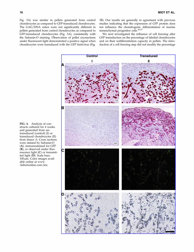
Fig. 3A) was similar in pellets generated from controlchondrocytes as compared to GFP-transduced chondrocytes.The GAG=DNA ratios were not significantly different inpellets generated from control chondrocytes as compared toGFP-transduced chondrocytes (Fig. 3A), consistently withthe Safranin-O staining. Observation of pellet cryosectionsunder fluorescent light demonstrated a positive signal whenchondrocytes were transduced with the GFP lentivirus (Fig.
3B). Our results are generally in agreement with previousstudies indicating that the expression of GFP protein doesnot influence the chondrogenic differentiation of murinemesenchymal progenitor cells.46,47
We next investigated the influence of cell freezing afterGFP transduction on the percentage of labeled chondrocytesand on their redifferentiation capacity in pellets. The intro-duction of a cell freezing step did not modify the percentage
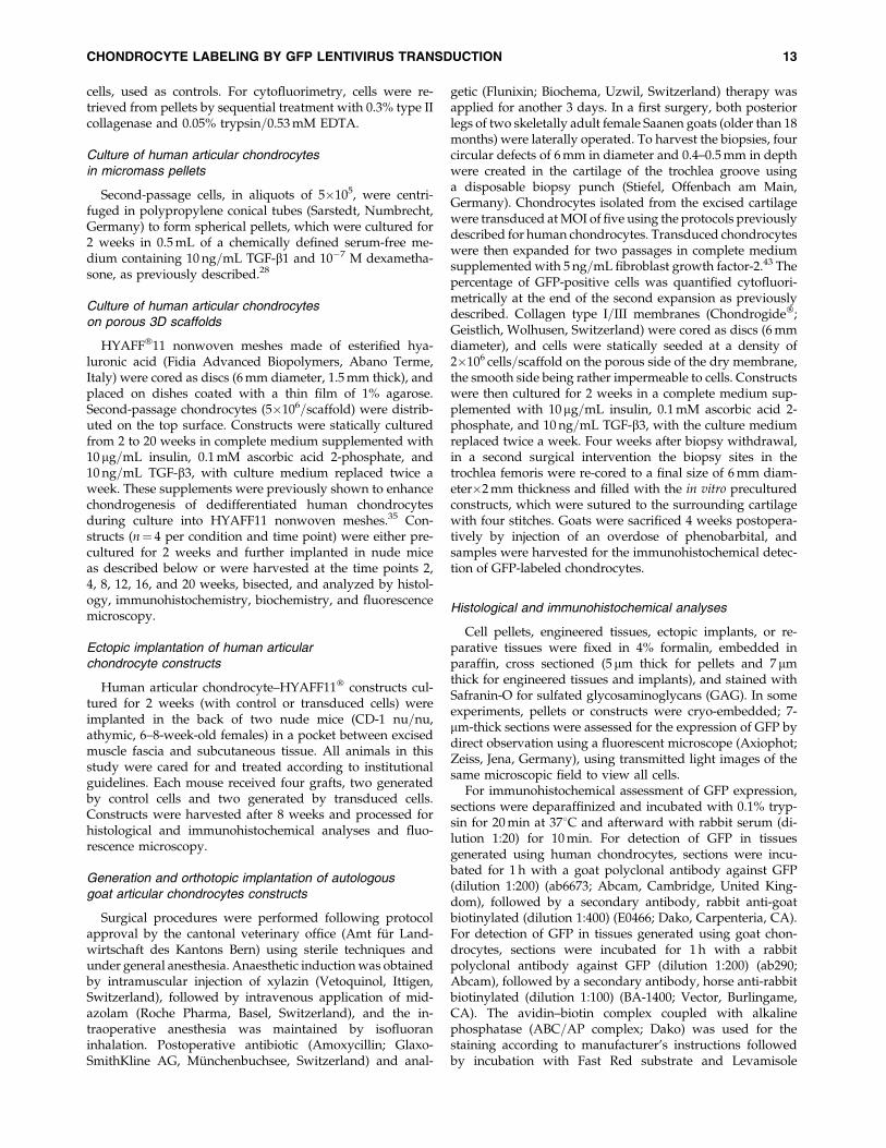
FIG. 4. Analysis of con-structs cultured for 4 weeksand generated from un-transduced (control) (I) ortransduced chondrocytes (II)from donor A. Cross sectionswere stained by Safranin-O(A), immunostained for GFP(B), or observed under fluo-rescence light (C) or transmit-ted light (D). Scale bars:100 mm. Color images avail-able online at www.liebertonline.com=ten.
16 MIOT ET AL.
of isolated fluorescent chondrocytes, as quantified by cyto-fluorimetry (73.1% positive cells after the freezing step vs.73.6% before cell freezing). Our findings are in line withthose by Li et al.,22 who reported that the expression of GFPin transduced osteoarthritic chondrocytes was maintainedafter freezing and re-culture.
Pellets generated from transduced chondrocytes with orwithout freezing exhibited similar intensity of staining forsulfated GAG (data not shown) and GAG=DNA ratios(10.5� 0.1 with freezing step vs. 11.2� 0.6 without freezingstep). After 2 weeks in pellets, the fluorescence intensity oftransduced chondrocytes was comparable if a freezing stepwas performed or not during previous cell expansion (datanot shown).
To possibly increase the percentage of GFP-labeled cells, asecond cycle of transduction was performed on alreadytransduced chondrocytes. This second cycle of transductionincreased the percentage of fluorescent chondrocytes from73.6% to 85.6� 1.5% at passage 1 as detected by cyto-fluorimetry. This is consistent with Li et al.,22 reporting that
after two cycles of retroviral transduction the percentage oflabeled human chondrocytes increased from 85% to 96%.Pellets generated from double-transduced chondrocytes ex-hibited a similar intensity of staining for sulfated GAG aswell as similar GAG=DNA ratios as compared to thosegenerated from cells transduced only once (Fig. 3A). Ob-servation of pellet cryosections under fluorescent lightqualitatively indicated a more homogenous green staining inpellets generated from double-transduced chondrocytes ascompared to cells transduced only once (Fig. 3B). The per-centages of single- or double-transduced chondrocytes en-zymatically retrieved from pellets after 2 weeks culture andstill positive for GFP averaged, respectively, 62.5� 2.3% or75.9� 4.6%, as assessed by cytofluorimetry.
Cell redifferentiation capacity in 3D scaffolds
As an independent model of chondrogenic differentiationand tissue formation, untransduced (control) or transducedchondrocytes (donor A), after 2 weeks of expansion, were
FIG. 5. Analysis of con-structs implanted in nudemice for 8 weeks and gener-ated from untransduced (con-trol) (I) or transducedchondrocytes (II) from donorA. Cross sections were stainedby Safranin-O (A), im-munostained for type II colla-gen (B), or immunostained forGFP with hematoxylin coun-terstain (C). Scale bar 100 mm.Insets in (C) display higher-magnification images of thecells immunostained for GFPand counterstained byhematoxylin. Color imagesavailable online at www.liebertonline.com=ten.
CHONDROCYTE LABELING BY GFP LENTIVIRUS TRANSDUCTION 17
cultured in HYAFF�11 scaffolds for up to additional 20weeks. Engineered cartilaginous tissues continuously ma-tured in vitro until 8 weeks, as indicated by Safranin-Ostaining, but longer culture time led to progressive degra-dation of the tissue, as typically occurs under static cultureconditions.48 After 4 weeks of culture, generated constructsdisplayed faint staining for Safranin-O, which was of similarintensity for control and transduced chondrocytes (Fig. 4A).Immunohistochemistry for GFP protein indicated a strongpositive staining in constructs generated from transducedcells (Fig. 4B), in correspondence both of elongated cells atthe construct periphery and of more rounded cells in thecentral part of the tissue. After 8 weeks or longer culturetime, cells localized at the tissue periphery were still pre-dominantly positively stained for GFP, whereas the constructcore was essentially necrotic, not allowing reliable assess-ment of positivity for GFP staining (data not shown).
By fluorescence analysis of cryosections, HYAFF�11 fibersappeared unspecifically fluorescent in constructs generatedfrom control or transduced chondrocytes (Fig. 4C). The samemicroscopic fields were thus observed also in light micros-copy to distinguish fluorescent HYAFF�11 fibers from fluo-rescent cells (Fig. 4D). This analysis allowed to confirm thatonly constructs generated using transduced chondrocytescontained GFP-positive cells.
Cell redifferentiation capacity and trackingafter ectopic implantation in nude mice
Expanded, transduced, or untransduced chondrocytes(donor A) were cultured in HYAFF�11 scaffolds for 2 weeksand further implanted subcutaneously in nude mice for 8
weeks. The chondrogenic redifferentiation capacity was as-sessed via staining for sulfated GAG, which appeared to beof strong and similar intensity in implants generated usinguntransduced or transduced chondrocytes (Fig. 5A). A sim-ilar intensity of immunostaining for collagen type II wasobserved in implants generated from control or transducedchondrocytes (Fig. 5B). Collagen type II positive staining wasdetected in the same regions stained for sulfated GAG. GFPexpression was demonstrated by immunohistochemistry inimplants generated using transduced chondrocytes (Fig. 5C),indicating that in vivo cells were still labeled and detectable.Manual counting of cells positively stained for GFP and of allcells with hematoxylin-stained nuclei was performed on his-tological sections of implants generated using transducedchondrocytes. Such counting demonstrated that 73.6� 3.3%of the cells in the implants were positive for GFP after12 weeks of transduction, corresponding to a complete main-tenance of labeling by the transduced cells. Fluorescenceanalysis of cryosections of the implants confirmed the pres-ence of GFP-expressing chondrocytes (data not shown).
Transduced chondrocytes implanted in nude mice did notshow an apparent reduction in the number of positive cellsafter 12 weeks, while transduced chondrocytes cultured for2 weeks in 3D pellets exhibited around 10% reduction ofGFP positivity. In the two experimental designs, we useddifferent culture systems (scaffolds vs. 3D pellets) as well asdistinct methodologies to determine the number of labelledcells (direct counting after immunohistochemistry vs. cyto-fluorimetry after enzymatic digestion of pellets). The meth-odologies employed in this study could explain relativevariations observed in the number of GFP-positive chon-drocytes.
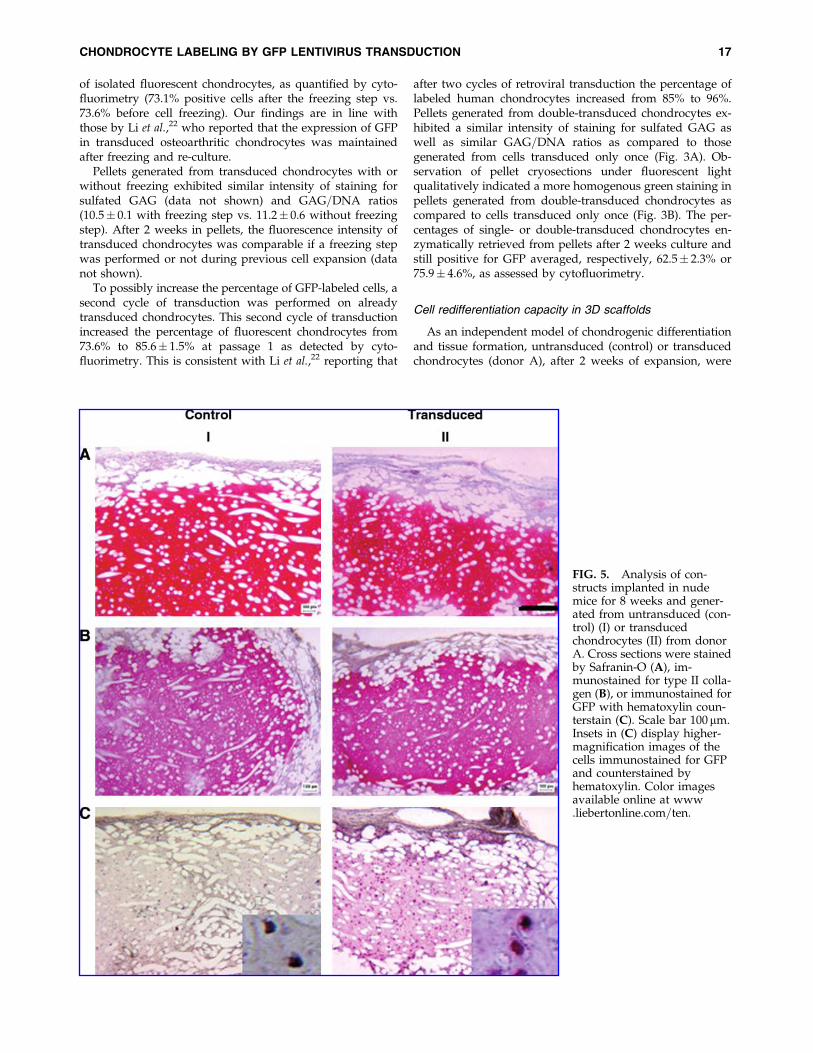
FIG. 6. Analysis of the con-structs implanted in goat car-tilage defects for 4 weeks. (A)GFP-labeled autologouschondrocytes, cultured inChondrogide membranes for2 weeks, were implanted intogoat trochlea defects. (B) After4 weeks the defects were filledwith a smooth repair tissue.(C, D) Immunohistochemicalstaining demonstrated a largeamount of GFP-positive cellsin the reparative tissue. Colorimages available online atwww.liebertonline.com=ten.
18 MIOT ET AL.
Cell tracking after orthotopic implantationin a goat model
The percentage of GFP-positive goat articular chon-drocytes after the second passage was slightly higher after asingle transduction than that measured in human articularchondrocyte cultures, averaging 78.3� 3.2% as measured bycytofluorimetry. This percentage is in the same range of thatreported by Ueblacker et al.49 and indicates that the protocolused for labeling human chondrocytes is efficient also for thetransduction of chondrocytes from other species.
GFP-labeled chondrocytes were cultured in Chondrogidemembranes for 2 weeks and then implanted into defectsgenerated in the trochlea of goats (Fig. 6A). After 4 weeks,visual observations showed an apparent good filling of thedefect with a relatively smooth surface (Fig. 6B). No in-flammatory reactions, possibly related to scaffold degrada-tion products, or blood vessel ingrowth were observed in therepair tissue. Immunohistochemical analysis demonstrated alarge amount of GFP-positive cells (Fig. 6C, D) in the re-parative tissue, indicating that the grafted chondrocytes re-mained viable in the defect. Quantification of GFP-positivecells from these sections (as percentage of total) appeared notreliable, due to the possible infiltration of cells from othercompartments (e.g., the subchondral bone or synovialmembrane) in the grafted material, which could not be dis-tinguished from nonlabeled implanted cells. Although in ourstudy only two goats have been used and a relative shortfollow-up time was considered, the results represent a proofof principle that the proposed GFP transduction methodwould enable to track implanted chondrocytes in orthotopiccartilage repair studies and possibly to evaluate the contri-bution of grafted cells in the repair process.
Conclusions
We report that primary human articular chondrocytescan be transduced by a GFP lentiviral vector at high effi-ciency, with labeled cells detected up to 22 weeks aftertransduction using in vitro 3D cultures, and up to 8 or 12weeks in in vivo models, namely, orthotopic implantation ingoats and subcutaneous implantation in nude mice. Im-portantly, chondrocyte transduction did not affect the cellproliferation rate or the postexpansion redifferentiation ca-pacity. The developed and validated technique could beused as a tool to engineer labeled cartilage tissues, and in-vestigate the interaction between chondrocytes and othercell populations during crucial processes (e.g., migration,differentiation, and integration) in in vitro systems or im-munodeficient animal models. We additionally proved thatthis method could be extended to the transduction ofchondrocytes from other species, thus allowing evaluatingthe fate of implanted cells in orthotopic cartilage repairstudies in large animal models.
Acknowledgments
We would like to thank Fidia Advanced Biopolymers forsupplying HYAFF�11 nonwoven meshes, Geistlich for sup-plying Chondrogide membranes, Prof. D. Trono (Geneva) forkindly providing plasmids for the generation of GFP lenti-virus, and Ms. Francine Wolf for her technical support withthe immunohistochemical analyses.
Disclosure Statement
No competing financial interests exist.
References
1. Freed, L.E., Martin, I., and Vunjak-Novakovic, G. Frontiersin tissue engineering. In vitro modulation of chondrogenesis.Clin Orthop Relat Res 367S, S46, 1999.
2. Jiang, J., Nicoll, S.B., and Lu, H.H. Co-culture of osteoblastsand chondrocytes modulates cellular differentiation in vitro.Biochem Biophys Res Commun 338, 762, 2005.
3. Gan, L., and Kandel, R.A. In vitro cartilage tissue formationby co-culture of primary and passaged chondrocytes. TissueEng 13, 831, 2007.
4. Nakaoka, R., Hsiong, S.X., and Mooney, D.J. Regulation ofchondrocyte differentiation level via co-culture with osteo-blasts. Tissue Eng 12, 2425, 2006.
5. Vats, A., Bielby, R.C., Tolley, N., Dickinson, S.C., Boccaccini,A.R., Hollander, A.P., Bishop, A.E., and Polak, J.M. Chon-drogenic differentiation of human embryonic stem cells: theeffect of the micro-environment. Tissue Eng 12, 1687, 2006.
6. Kurz, B., Steinhagen, J., and Schunke, M. Articular chon-drocytes and synoviocytes in a co-culture system: influenceon reactive oxygen species-induced cytotoxicity and lipidperoxidation. Cell Tissue Res 296, 555, 1999.
7. Hunter, C.J., and Levenston, M.E. Maturation and integra-tion of tissue-engineered cartilages within an in vitro defectrepair model. Tissue Eng 10, 736, 2004.
8. Obradovic, B., Martin, I., Padera, R.F., Treppo, S., Freed,L.E., and Vunjak-Novakovic, G. Integration of engineeredcartilage. J Orthop Res 19, 1089, 2001.
9. Zhang, Z., McCaffery, J.M., Spencer, R.G., and Francomano,C.A. Growth and integration of neocartilage with nativecartilage in vitro. J Orthop Res 23, 433, 2005.
10. Tognana, E., Chen, F., Padera, R.F., Leddy, H.A., Chris-tensen, S.E., Guilak, F., Vunjak-Novakovic, G., and Freed,L.E. Adjacent tissues (cartilage, bone) affect the functionalintegration of engineered calf cartilage in vitro. OsteoarthritisCartilage 13, 129, 2005.
11. Dell’Accio, F., Vanlauwe, J., Bellemans, J., Neys, J., De, B.C.,and Luyten, F.P. Expanded phenotypically stable chon-drocytes persist in the repair tissue and contribute to carti-lage matrix formation and structural integration in a goatmodel of autologous chondrocyte implantation. J OrthopRes 21, 123, 2003.
12. Krause, D.S., Theise, N.D., Collector, M.I., Henegariu, O.,Hwang, S., Gardner, R., Neutzel, S., and Sharkis, S.J. Multi-organ, multi-lineage engraftment by a single bone marrow-derived stem cell. Cell 105, 369, 2001.
13. Chawla, K., Klein, T.J., Schumacher, B.L., Jadin, K.D., Shah,B.H., Nakagawa, K., Wong, V.W., Chen, A.C., Masuda, K.,and Sah, R.L. Short-term retention of labeled chondrocytesubpopulations in stratified tissue-engineered cartilaginousconstructs implanted in vivo in mini-pigs. Tissue Eng 13,
1525, 2007.14. Shapiro, F., Koide, S., and Glimcher, M.J. Cell origin and
differentiation in the repair of full-thickness defects of ar-ticular cartilage. J Bone Joint Surg Am 75, 532, 1993.
15. Ball, S.T., Goomer, R.S., Ostrander, R.V., Tontz, W.L., Jr.,Williams, S.K., and Amiel, D. Preincubation of tissue en-gineered constructs enhances donor cell retention. Clin Or-thop Relat Res 420, 276, 2004.
16. Tsien, R.Y. The green fluorescent protein. Annu Rev Bio-chem 67, 509, 1998.
CHONDROCYTE LABELING BY GFP LENTIVIRUS TRANSDUCTION 19

17. Cucchiarini, M., Madry, H., Ma, C., Thurn, T., Zurakowski,D., Menger, M.D., Kohn, D., Trippel, S.B., and Terwilliger,E.F. Improved tissue repair in articular cartilage defectsin vivo by rAAV-mediated overexpression of human fibro-blast growth factor 2. Mol Ther 12, 229, 2005.
18. Yokoo, N., Saito, T., Uesugi, M., Kobayashi, N., Xin, K.Q.,Okuda, K., Mizukami, H., Ozawa, K., and Koshino, T. Re-pair of articular cartilage defect by autologous transplanta-tion of basic fibroblast growth factor gene-transducedchondrocytes with adeno-associated virus vector. ArthritisRheum 52, 164, 2005.
19. Madry, H., and Trippel, S.B. Efficient lipid-mediatedgene transfer to articular chondrocytes. Gene Ther 7, 286,2000.
20. Zhang, S.K., Liu, Y., Song, Z.M., Fu, C.F., and Xu, X.X. Greenfluorescent protein as marker in chondrocytes over-expressing human insulin-like growth factor-1 for repair ofarticular cartilage defects in rabbits. Chin J Traumatol 10, 10,2007.
21. Dinser, R., Kreppel, F., Zaucke, F., Blank, C., Paulsson, M.,Kochanek, S., and Maurer, P. Comparison of long-termtransgene expression after non-viral and adenoviral genetransfer into primary articular chondrocytes. Histochem CellBiol 116, 69, 2001.
22. Li, Y., Tew, S.R., Russell, A.M., Gonzalez, K.R., Hard-ingham, T.E., and Hawkins, R.E. Transduction of passagedhuman articular chondrocytes with adenoviral, retroviral,and lentiviral vectors and the effects of enhanced expressionof SOX9. Tissue Eng 10, 575, 2004.
23. Lu, F.Z., Kitazawa, Y., Hara, Y., Jiang, J.Y., and Li, X.K.Long-term gene expression using the lentiviral vector in ratchondrocytes. Clin Orthop Relat Res 439, 243, 2005.
24. Chen, H.C., Lee, H.P., Ho, Y.C., Sung, M.L., and Hu, Y.C.Combination of baculovirus-mediated gene transfer androtating-shaft bioreactor for cartilage tissue engineering.Biomaterials 27, 3154, 2006.
25. Lever, A.M. Lentiviral vectors: progress and potential. CurrOpin Mol Ther 2, 488, 2000.
26. Naldini, L., Blomer, U., Gallay, P., Ory, D., Mulligan, R.,Gage, F.H., Verma, I.M., and Trono, D. In vivo gene deliveryand stable transduction of nondividing cells by a lentiviralvector. Science 272, 263, 1996.
27. Hirschmann, F., Verhoeyen, E., Wirth, D., Bauwens, S.,Hauser, H., and Rudert, M. Vital marking of articularchondrocytes by retroviral infection using green fluores-cence protein. Osteoarthritis Cartilage 10, 109, 2002.
28. Jakob, M., Demarteau, O., Schafer, D., Hintermann, B.,Dick, W., Heberer, M., and Martin, I. Specific growth factorsduring the expansion and redifferentiation of adult humanarticular chondrocytes enhance chondrogenesis and carti-laginous tissue formation in vitro. J Cell Biochem 81, 368,2001.
29. Yaeger, P.C., Masi, T.L., de Ortiz, J.L., Binette, F., Tubo,R., and McPherson, J.M. Synergistic action of transforminggrowth factor-beta and insulin-like growth factor-I inducesexpression of type II collagen and aggrecan genes inadult human articular chondrocytes. Exp Cell Res 237,
318, 1997.30. Yoo, J.U., Barthel, T.S., Nishimura, K., Solchaga, L., Caplan,
A.I., Goldberg, V.M., and Johnstone, B. The chondrogenicpotential of human bone-marrow-derived mesenchymalprogenitor cells. J Bone Joint Surg Am 80, 1745, 1998.
31. Farhadi, J., Fulco, I., Miot, S., Wirz, D., Haug, M., Dick-inson, S.C., Hollander, A.P., Daniels, A.U., Pierer, G.,
Heberer, M., and Martin, I. Precultivation of engineeredhuman nasal cartilage enhances the mechanical propertiesrelevant for use in facial reconstructive surgery. Ann Surg244, 978, 2006.
32. Grigolo, B., Lisignoli, G., Piacentini, A., Fiorini, M., Gobbi,P., Mazzotti, G., Duca, M., Pavesio, A., and Facchini, A.Evidence for redifferentiation of human chondrocytes grownon a hyaluronan-based biomaterial (HYAff 11): molecular,immunohistochemical and ultrastructural analysis. Bioma-terials 23, 1187, 2002.
33. Solchaga, L.A., Dennis, J.E., Goldberg, V.M., and Caplan,A.I. Hyaluronic acid-based polymers as cell carriers for tis-sue-engineered repair of bone and cartilage. J Orthop Res 17,
205, 1999.34. Dell’Accio, F., De, B.C., and Luyten, F.P. Molecular markers
predictive of the capacity of expanded human articularchondrocytes to form stable cartilage in vivo. ArthritisRheum 44, 1608, 2001.
35. Moretti, M., Wendt, D., Dickinson, S.C., Sims, T.J.,Hollander, A.P., Kelly, D.J., Prendergast, P.J., Heberer, M.,and Martin, I. Effects of in vitro preculture on in vivo de-velopment of human engineered cartilage in an ectopicmodel. Tissue Eng 11, 1421, 2005.
36. Mueller-Rath, R., Gavenis, K., Gravius, S., Andereya, S.,Mumme, T., and Schneider, U. In vivo cultivation of humanarticular chondrocytes in a nude mouse-based containeddefect organ culture model. Biomed Mater Eng 17, 357, 2007.
37. Barry, F.P. Mesenchymal stem cell therapy in joint disease.Novartis Found Symp 249, 86, 2003.
38. Jackson, D.W., Lalor, P.A., Aberman, H.M., and Simon, T.M.Spontaneous repair of full-thickness defects of articularcartilage in a goat model. A preliminary study. J Bone JointSurg Am 83A, 53, 2001.
39. Murphy, J.M., Fink, D.J., Hunziker, E.B., and Barry, F.P.Stem cell therapy in a caprine model of osteoarthritis.Arthritis Rheum 48, 3464, 2003.
40. Barbero, A., Grogan, S., Schafer, D., Heberer, M., Mainil-Varlet, P., and Martin, I. Age related changes in human ar-ticular chondrocyte yield, proliferation and post-expansionchondrogenic capacity. Osteoarthritis Cartilage 12, 476,2004.
41. Zufferey, R., Nagy, D., Mandel, R.J., Naldini, L., and Trono,D. Multiply attenuated lentiviral vector achieves efficientgene delivery in vivo. Nat Biotechnol 15, 871, 1997.
42. Follenzi, A., and Naldini, L. HIV-based vectors. Preparationand use. Methods Mol Med 69, 259, 2002.
43. Miot, S., Scandiucci de, F.P., Wirz, D., Daniels, A.U., Sims,T.J., Hollander, A.P., Mainil-Varlet, P., Heberer, M., andMartin, I. Cartilage tissue engineering by expanded goatarticular chondrocytes. J Orthop Res 24, 1078, 2006.
44. Hollander, A.P., Heathfield, T.F., Webber, C., Iwata, Y.,Bourne, R., Rorabeck, C., and Poole, A.R. Increaseddamage to type II collagen in osteoarthritic articular car-tilage detected by a new immunoassay. J Clin Investig 93,
1722, 1994.45. Farndale, R.W., Buttle, D.J., and Barrett, A.J. Improved
quantitation and discrimination of sulphated glycosamino-glycans by use of dimethylmethylene blue. Biochim BiophysActa 883, 173, 1986.
46. Lin, Y., Tian, W., Chen, X., Yan, Z., Li, Z., Qiao, J., Liu, L.,Tang, W., and Zheng, X. Expression of exogenous or endo-genous green fluorescent protein in adipose tissue-derivedstromal cells during chondrogenic differentiation. Mol CellBiochem 277, 181, 2005.
20 MIOT ET AL.
47. Ogawa, R., Mizuno, H., Watanabe, A., Migita, M., Shimada,T., and Hyakusoku, H. Osteogenic and chondrogenic dif-ferentiation by adipose-derived stem cells harvested fromGFP transgenic mice. Biochem Biophys Res Commun 313,
871, 2004.48. Wendt, D., Stroebel, S., Jakob, M., John, G.T., and Martin, I.
Uniform tissues engineered by seeding and culturing cells in3D scaffolds under perfusion at defined oxygen tensions.Biorheology 43, 481, 2006.
49. Ueblacker, P., Wagner, B., Vogt, S., Salzmann, G., Wexel, G.,Kruger, A., Plank, C., Brill, T., Specht, K., Hennig, T.,Schillinger, U., Imhoff, A.B., Martinek, V., and Gansbacher,B. In vivo analysis of retroviral gene transfer to chondrocyteswithin collagen scaffolds for the treatment of osteochondraldefects. Biomaterials 28, 4480, 2007.
Address correspondence to:Ivan Martin, Ph.D.
Departments of Surgery and of BiomedicineUniversity Hospital Basel
Hebelstrasse 20ZLF, Room 405
Basel 4031Switzerland
E-mail: [email protected]
Received: December 23, 2008Accepted: March 27, 2009
Online Publication Date: May 18, 2009
CHONDROCYTE LABELING BY GFP LENTIVIRUS TRANSDUCTION 21

This article has been cited by:
1. Jiarong Chen, Fuyou Wang, Yi Zhang, Xuhong Jin, Lin Zhang, Yong Feng, Xiao Lin, Liu Yang. 2012. In Vivo Tracking ofSuperparamagnetic Iron Oxide Nanoparticle Labeled Chondrocytes in Large Animal Model. Annals of Biomedical Engineering40:12, 2568-2578. [CrossRef]
2. Li-You An, Yu-Guo Yuan, Bao-Li Yu, Ting-Jia Yang, Yong Cheng. 2012. Generation of human lactoferrin transgenic clonedgoats using donor cells with dual markers and a modified selection procedure. Theriogenology 78:6, 1303-1311. [CrossRef]
3. Sinan Güven, Marianna Karagianni, Mandy Schwalbe, Simone Schreiner, Jian Farhadi, Sylvain Bula, Karen Bieback, Ivan Martin,Arnaud Scherberich. 2012. Validation of an Automated Procedure to Isolate Human Adipose Tissue–Derived Cells by Using theSepax® Technology. Tissue Engineering Part C: Methods 18:8, 575-582. [Abstract] [Full Text HTML] [Full Text PDF] [FullText PDF with Links]
4. Chitrangada Acharya, Adetola Adesida, Paul Zajac, Marcus Mumme, Jens Riesle, Ivan Martin, Andrea Barbero. 2012. Enhancedchondrocyte proliferation and mesenchymal stromal cells chondrogenesis in coculture pellets mediate improved cartilageformation. Journal of Cellular Physiology 227:1, 88-97. [CrossRef]
Copyright © 2022 FDOKUMEN