Delineating structural characteristics of viral capsid proteins ...
207
Delineating structural characteristics of viral capsid proteins critical for their functional assembly by Shanshan Cheng A dissertation submitted in partial fulfillment of the requirements for the degree of Doctor of Philosophy (Bioinformatics) in the University of Michigan 2014 Doctoral Committee: Professor Charles L. Brooks III, Chair Assistant Professor Barry J. Grant Professor Kerby A. Shedden Professor Janet L. Smith Professor Yang Zhang
-
Upload
khangminh22 -
Category
Documents
-
view
3 -
download
0
Transcript of Delineating structural characteristics of viral capsid proteins ...

Delineating structural characteristics of viral capsid proteins critical for their functional assembly
by
Shanshan Cheng
A dissertation submitted in partial fulfillment of the requirements for the degree of
Doctor of Philosophy (Bioinformatics)
in the University of Michigan 2014
Doctoral Committee:
Professor Charles L. Brooks III, Chair Assistant Professor Barry J. Grant Professor Kerby A. Shedden Professor Janet L. Smith Professor Yang Zhang

© Shanshan Cheng 2014 All Rights Reserved

ii
To my parents,
my husband and my daughter

iii
ACKNOWLEDGEMENTS
I am very fortunate to have been advised by Dr. Charles Brooks, who has been a
truly wonderful mentor for my graduate career. His genuine interest in advancing
understanding of the basic sciences, as well as his scientific vision, has always been a
source of inspiration for me. The instrumental guidance he provided me with has shaped
the way I approach science, and I will be grateful for all that he taught me in many years
to come.
I would also like to acknowledge my committee members, Drs. Barry Grant, Kerby
Shedden, Janet Smith and Yang Zhang, for all the insightful suggestions and feedback
they gave that helped improve the quality of my work. In addition, I would like to thank
Dr. Georgios Skiniotis, who is not on my thesis committee, but from whom I have
learned a lot about cryo-electron microscopy.
I am also thankful to all current and past members of the Brooks lab. Working in such
a diverse and vibrant environment has given me new perspectives and has complemented
my Ph.D. training in many ways. In addition, I would like to particularly thank David
Braun, who manages the computer resources in the lab, and Kate Dyki, our admin
assistant, for providing full support throughout my graduate studies.
My gratitude also extends to all the friendly faculty and staff in the Bioinformatics
program. Dr. Margit Burmeister was the one that recruited me to this wonderful program.

iv
Dr. Daniel Burns was always approachable for academic advice. Julia Eussen, our
graduate coordinator, was very patient with my endless questions.
I want to express my heart-felt gratitude to my parents, Jingxuan Cheng and Liqun
Zhang. I would simply not be where I am now without their unconditional love for me
and their self-sacrifices to support me to pursue my dreams. Special thanks also goes to
my dear husband, Chaolong Wang, whose continuous support and encouragement gave
me the strength to overcome every hurdle. Thank you for being in my life. Finally, to my
beloved daughter, Zixuan Ann Wang, for coming to this world 10 days after my thesis
defense and for bringing so much joy in my life.

v
TABLE OF CONTENTS DEDICATION ii
ACKNOWLEDGEMENTS iii
LIST OF TABLES viii
LIST OF FIGURES ix
ABSTRACT xi
CHAPTER
I. Introduction 1
1.1 General background 1 1.2 Aim of the study and motivation 4 1.3 Overview of thesis chapters 10
II. Viral capsid proteins are segregated in structural fold space 14
2.1 Introduction 14 2.2 Materials and Methods 18
2.2.1 Comparison metric 18 2.2.2 Data collection 20 2.2.3 Shared folds as the test statistic 23 2.2.4 Statistical significance of the test statistic 24 2.2.5 Cross-checking with other functional classes of proteins 24
2.3 Results 25 2.3.1 Representative folds adopted by viral capsid proteins 25 2.3.2 Viral capsid proteins are segregated in structural fold space from
generic proteins 28
2.3.3 Estimation of statistical significance 28 2.3.4 Other functional classes 33
2.4 Discussion 33 2.4.1 Possible differences in domain definition 33 2.4.2 What are the capsid-like proteins and why they do not form shells 35

vi
2.4.3 Scarcity and possible bias in the data 38 2.4.4 Implications on protein-protein interaction and other applications 38 2.4.5 Are viral capsid interfaces also unique to viruses? 40
III. PCalign: a method to quantify physicochemical similarity of protein-
protein interfaces
44
3.1 Introduction 44 3.2 Methods 47
3.2.1 Extract interfacial residues 47 3.2.2 Identify initial alignments 49 3.2.3 Iterative refinement 51
3.3 Results 54 3.3.1 Validation of the scoring function 54 3.3.2 Comparison of performance with existing methods 56 3.3.3 Application of the method in detecting convergently evolved
similar interfaces 62
3.4 Discussion and conclusions 69
IV. Template-based protein inhibitor design for influenza hemagglutinin 111
4.1 Introduction 111 4.2 Methods 117
4.2.1 Interface library construction 117 4.2.2 Initial candidate selection 118 4.2.3 Criteria for evaluating likelihood of binding activity 119 4.2.4 Relaxation of initial structure 121 4.2.5 Selection of residues for redesign 123
4.3 Results 124 4.3.1 Correlation between PC-score with the native complex and the
predicted binding energy 124
4.3.2 Top ranked predictions 125 4.4 Discussion 131
V. Protein-protein interfaces in viral capsids are structurally unique 139
5.1 Introduction 139 5.2 Materials and Methods 141
5.2.1 Data collection 143 5.2.2 Comparison metric 144 5.2.3 Quantify overlap in structural space 145 5.2.4 Statistical significance of the test statistic 146
5.3 Results 146 5.3.1 Similar sizes but different oligomerization states 146 5.3.2 Protein-protein interfaces formed by capsid proteins are distinct
from those formed by cellular proteins 149

vii
5.3.3 Overlap in structural space of protein-protein interfaces 151 5.4 Discussion 157
5.4.1 Result of comparative study is not sensitive to quality of structural data
157
5.4.2 Domain-swapped interfaces are not treated differently 159 5.4.3 Implications of protein-protein interfaces in capsids being
unique 159
5.4.4 Concluding remarks 162
VI. Conclusion 168
BIBLIOGRAPHY 179

viii
LIST OF TABLES
Table 2.1 The 21 folds covered by structural relatives of capsid proteins 29
Table 2.2 Seven additional functional classes of proteins studied 34
Table A.1 Capsid proteins added from SCOP that are not deposited in VIPERdb 42
Table 3.1 Three examples of viral mimicry achieved via convergent evolution 64
Table B.1 Mean Cα-Cα distances 75
Table B.2 Standard deviation of Cα-Cα distances 76
Table B.3 Percentage of correctly mapped interfacial residues by PCalign in quasi-equivalent protein-protein interfaces within the same capsid
79
Table B.4 Statistical significance of PC-scores 85
Table B.5 The interface equivalence measured by Q-score and PC-score for different T=3 viruses
86
Table B.6 Protein dimers grouped by their familial annotation by SCOPPI 88
Table 4.1 Basic properties of candidate proteins 127
Table 4.2 Structural relaxation with simulated annealing 129
Table 4.3 Improvement with computational redesign 130
Table C.1 List of additional capsid protein dimers 164
Table C.2 Functional annotation of capsid-like generic interfaces 166

ix
LIST OF FIGURES
Figure 2.1 Capsid shells and the folded topology of a typical capsid protein 16
Figure 2.2 Comparison in structural fold space of capsid proteins and non-capsid ones
19
Figure 2.3 Domain size distribution 22
Figure 2.4 Clustering to find representative capsid folds 26
Figure 2.5 The 56 representative capsid folds 27
Figure 2.6 Capsid proteins are structurally distant from generic proteins 31
Figure 2.7 Statistical significance of test statistic 32
Figure A.1 Statistical significance of test statistic for small domains 43
Figure 3.1 The ROC curves for predicting highly related interfaces using three methods, PCalign, Ialign, and I2I-SiteEngine
59
Figure 3.2 Recognition of interface similarity across unrelated interfaces by PCalign and Ialign
61
Figure 3.3 Three examples of viral mimicry resulting from convergent evolution
65
Figure B.1 Distribution of structural models in the PDB with resolution lower than 3.5 Å
71
Figure B.2 Matthews Correlation Coefficient for the amino acid type-specific distance criterion and a general Cα-Cα distance cutoff criterion
74
Figure B.3 The mean of PC-scoreraw across random, unrelated pairs whose interface sizes are between 95% and 105% of a given interface size
82
Figure B.4 The distribution of PC-scores 84

x
Figure B.5 An example of similar interfaces across unrelated dimers missed by existing methods but captured by PCalign
87
Figure B.6 Running time reported by the three programs
110
Figure 4.1 Template-based protein inhibitor design protocol
113
Figure 4.2 Structure of a HA trimer
115
Figure 4.3 Antibodies targeting HA
116
Figure 4.4 PC-scores of computational designs
126
Figure 4.5 Top four designed inhibitors
132
Figure 4.6 Mutations introduced in top four designs
133
Figure 5.1 Comparison between inter-subunit interfaces in viral capsids and protein-protein interfaces in generic protein complexes
142
Figure 5.2 Permutation test for estimating the statistical significance
147
Figure 5.3 Density distribution of interface sizes for the generic set and the capsid set
148
Figure 5.4 Different oligomerization states in the two sets
150
Figure 5.5 Statistical significance of the test statistic
152
Figure 5.6 Equal connectivity in the two sets
153
Figure 5.7 Capsid protein-protein interfaces are different from generic ones
154
Figure 5.8 Sizes of the 418 capsid protein-portein interfaces in the overlap region
156
Figure 5.9 Examples of similar interfaces from the capsid set and the generic set
158
Figure 6.1 Comparison of inter-subunit interfaces in small plant viruses 174

xi
ABSTRACT
Viral capsids exhibit elaborate and symmetrical architectures of defined sizes and
remarkable mechanical properties not seen with cellular macromolecular complexes. The
limited coding capacity of viral genome necessitates economization upon one or a few
identical gene products known as capsid proteins for shell assembly. The functional
uniqueness of this class of proteins prompts questions on key structural features critically
important for their higher order organization. In this thesis, I develop the statistical
framework and computational tools to pinpoint the structural characteristics of viral
capsid proteins exclusive to the virosphere by testing a series of hypotheses, providing
understanding of the physical principles governing molecular self-association that can
inform rational design of nanomaterials and therapeutics. In the first chapter, I compare
the folded topology of capsid proteins with those of generic proteins, and establish that
capsid proteins are segregated in structural fold space, highlighting the geometric
constraints of these building blocks for tiling into a closed shell. Second, I develop a
software program, PCalign, for quantifying the physicochemical similarity between
protein-protein interfaces. This tool overcomes the major limitation of current methods
by using a reduced representation of structural information, greatly expanding the
structural interface space that can be investigated through inclusion of large
macromolecular assemblies that are often not amenable to high resolution experimental
techniques. As an application of this method, I propose a computational framework for

xii
template-based protein inhibitor design, leading to the prediction of putative binders for a
therapeutic target, the influenza hemagglutinin. In silico evaluations of these candidate
drugs parallel those of known protein binders, offering great promise in expanding
therapeutic options in the clinic. Lastly, I examine protein-protein interfaces using
PCalign, and find strong statistical evidence for the disconnectivity between capsid
proteins and cellular proteins in structural interface space. I thus conclude that the basic
shape and the sticky edges of these Lego pieces act concertedly to create the sophisticated
shell architecture. In summary, the novel tools contributed by this dissertation work lead
to delineation of structural features of viral capsid proteins that make them functionally
unique, providing an understanding that will serve as the basis for prediction and design.

1
CHAPTER I
Introduction
1.1. General background:
Viruses, a frequently overlooked domain of pseudo-living organisms, are gaining
attention in the scientific community due to the unparalleled reach and diversity of these
systems, with an abundance that is estimated to outnumber living cells by an order of
magnitude [1]. Since the 1980s there has been a paradigm shift from the traditional view
of viruses as a long-standing foe of human health [2], to their being a rich source of
information on biodiversity [3], on their role in shaping evolution through genetic
exchanges with hosts [4], and on the insights they provide into the design principle of
nanoscale biological containers [2]. Our current understanding of the virosphere is still
relatively limited, and growing efforts are now channeled into studying this important
section that is an indispensable and integral part of the biosphere.
Traditionally, viruses have been classified based on a variety of phenotypical features,
such as their host organisms (animal viruses, plant viruses, etc.), their overall morphology
(helical, icosahedral or complex), and the types of genetic materials they carry (dsDNA
viruses, ssDNA viruses, dsRNA viruses, +ssRNA viruses, -ssRNA viruses, ssRNA-RT
viruses, and dsDNA-RT viruses). A major obstacle in deriving a systematic taxonomy for
viruses, like with the domains of cellular life, is the lack of a unifying genetic marker,

2
compounded with the fact that viruses frequently undergo lateral gene transfer as opposed
to vertical transmission. With recent advances in experimental techniques that have
determined the structures of viral capsids, there is accumulated evidence that viruses
previously thought to be unrelated, including those infecting different domains of hosts
[5-7], can share remarkable structural and architectural similarity in their viral capsid
proteins, providing support for a newly formulated hypothesis that attempts to group
together viruses by the structural similarity of the viral coat proteins to define viral
lineage [8]. This proposal appears reasonable, given that viral capsid proteins are
arguably the single hallmark feature present in all viruses, in the same way that
ribosomes are signature of the cellular empire.
This thesis work is centered on viral capsid proteins, which are a functionally unique
class of proteins in the virosphere. They play a critical role in the life cycle of viruses,
providing a protective coat that encapsulates the viral genome to be delivered to host cells
they infect. In carrying out this central function, capsid proteins first have to self-
assemble into a closed shell of a defined size inside the highly crowded cellular
environment at the stage of replication, undergo a series of maturation steps to stabilize
the shell in the case of some viruses, and later dissociate from one another in the newly
infected host cells to release the genetic materials, manifesting remarkable dynamic and
mechanical properties [9]. Other than the central function of protecting the genetic
material, there are other more refined roles of capsid proteins that are differentiated
among different viruses, depending on their detailed mechanism of infection. For
instance, enveloped viruses, which are membrane-covered viruses, typically have
glycoproteins (here broadly considered as coat proteins or capsid proteins) that specialize

3
in inducing membrane fusion with the endosome upon endocytosis, in addition to their
structural role.
Most of the currently solved structures of viral capsids are icosahedral, and we have
focused our studies on these available structural data, which are collectively available in
the database VIPERdb [10]. An icosahedron contains 20 triangular faces, with 2-, 3- and
5-fold symmetry, and consists of 60 copies of an icosahedral asymmetric unit (IAU).
Given the limited coding capacity of viral genomes, viruses economize upon a single type
or a few types of capsid proteins that tile on an icosahedron lattice. Within each IAU,
there are T-number of capsid proteins, where the T-number is the triangulation number
that takes discrete values given by T=h2+hk+k2, h and k being non-negative integers. An
icosahedron has twelve vertices, and thus an icosahedral virus consists of 12 pentamers
placed at the vertices and 10(T-1) hexamers tiled on the flat surfaces. For T=1 viruses, all
capsid proteins are placed in an identical environment. For T>1 viruses, the sequence-
wise identical capsid proteins are placed in different chemical environments, and interact
with one another in slightly different fashions. This construction principle, termed ‘quasi-
equivalence’ by Caspar and Klug [11], allows large viruses with more than 60 protein
subunits to form, while still obeying icosahedral symmetry. In this case, quasi-symmetry
axes are introduced, which do not necessarily coincide with the icosahedral symmetry
axes, depending on the classification of different T-numbers as discussed in detail in [12].
While most icosahedral viruses have symmetries that are explained by the quasi-
equivalence theory, a few exceptions exist. The best known example of deviation from
the quasi-equivalence theory is the polyoma viruses. Instead of having 12 pentamers and
10(T-1) hexamers, polyoma viruses consist of only pentamers, which occupy positions on

4
an icosahedral lattice that correspond to T=7 symmetry [13] and classified as such. The
total number of capsid protein subunits is thus 360, which implies a forbidden T-number
based on the formula, contrary to the expected 420 for a T=7 virus. Another case that
disobeys the quasi-equivalence rule is L-A viruses, which are comprised of 120 protein
subunits that again points to a forbidden T-number of 2. It is therefore commonly viewed
as having a symmetry of T=1, except with dimers making up the IAU [14]. Finally,
different capsid protein subunits in picornaviruses have different peptide sequences, yet
all adopt the same structural fold, which can also be regarded as forming pseudo-quasi-
equivalent interfaces [15]. Despite these aberrations, the quasi-equivalence theory
remains an important cornerstone that has wide applicability in describing the
architecture of most icosahedral viruses, and the T-number is generally regarded as a
useful metric in quantifying the size of viral capsids.
1.2. Aim of the study and motivation:
One observation about viral capsids is of interest to us; although symmetric
macromolecular assemblies are not lacking in the cellular domains of life, exemplified by
ferritin cages and the chaperone GroEL/ES complexes, they are not found to be on such a
large scale as viral capsids. In other words, viral capsid proteins represent a functionally
unique class of proteins dedicated to make large cages for the protection and
transportation of other macromolecules. Given that function follows form, we ask the
question of what structural features of viral capsid proteins are key to determining their
higher order organization, with the goal of understanding the fundamental physical
principles governing assembly of biological containers of defined sizes.
5
Prior to our work, an important finding about the structure-function relationship of
viral capsid proteins has established the role of topological constraints- specifically the
trapezoidal shape adopted by most viral capsid proteins- in tiling into a closed spherical
shell with no holes, no overlaps and no gross structural variability [16], which are
essential properties for capsid shells designed to offer protection to the viral genome with
maximal genetic economy. With the aim to gain a deeper understanding to fully address
the question raised above, I set out to investigate in this thesis work how various
structural features of viral capsid proteins compare with generic, non-capsid proteins. The
answer to our question has three-fold important implications; first is to provide guidelines
for the design of nano-packaging tools in various biomedical applications and materials
science, second is to inform the rational design of antiviral drugs that minimize toxic
effects, and lastly to enrich our knowledge of protein-protein interactions in general by
mining through the myriad of structural data for viral capsids. These three aspects are
presented as follows.
The ability of viral capsid proteins to self-assemble into cages of defined sizes offers
great promise in a range of biomedical applications, including vaccine development, gene
therapy, bio-imaging and drug delivery. In fulfilling these functionalities, viral capsid
proteins are typically expressed in the absence of the infectious genomic material they
encapsulate, with the exception of the use of viruses for transfection in gene therapy, and
these empty viral shells are often called virus-like particles (VLPs) without virulent
activity. Since as early as in the 1990s, immunogenic peptides have been genetically
fused with viral capsid proteins to be presented on the surface of the assembled capsids to
enhance recognition by antibodies to elicit immune response [17-44], with some of the

6
VLP-based vaccines making it to the clinical trial stage. The use of viruses as vectors of
gene therapy also has a long history, and has been known for their efficiency for the
reason that viruses are naturally designed to transfect living cells with their own genetic
material, via sophisticated mechanisms selected by evolution. These vectors are
generated as recombinant viral genomes packaged by viral capsid proteins, incorporating
the therapeutic gene of interest, allowing self-replication in some cases (especially in the
treatment of cancer) while prohibiting replication in others for a ‘one-hit’ transient
expression of the therapeutic gene using replication-defective viruses [45]. Different
types of viral vectors including retroviruses, adenoviruses, adeno-associated viruses and
herpes simplex viruses have been developed [46], with notable successes in the treatment
for inherited monogenic diseases [47-51]. In the area of bio-imaging, VLPs can either be
convalently conjugated to dye molecules on the viral surface to take advantage of the
large display area for maximized signal, as well as the precise positioning to prevent the
dye molecules from fluorescent quenching, or they can encapsulate fluorescent cores as
cargos, leaving the outer surface for modifications to achieve specific targeting [52, 53].
These techniques have been widely applied in various biomedical studies, including but
not limited to imaging of tumor angiogenesis in vivo with fluorescent cowpea mosaic
virus sensors [54-56], packaging quantum dots and infrared chromophores into simian
viruses and brome mosaic viruses for in vitro imaging [57, 58], and using cowpea mosaic
viruses, cowpea chlorotic viruses and MS2 viruses as carriers of MRI contrast reagents
for diagnostic and therapeutic decisions [59-62]. Lastly, formulations of small drug
molecules delivered by viral containers have also been developed for treatment of cancer

7
[63-68] and bacterial infections [69], greatly prolonging the activity of the drug
molecules which otherwise undergo fast clearance.
In materials science applications, viral capsids are equally popular as bio-templates
given their remarkable mechanical properties and diversity in sizes and shapes. Phage
display libraries, for one, have been used for identifying peptides with specific binding to
materials, which are in turn engineered onto viral capsid proteins for directing nucleation
of inorganic nanocrystals of different sizes, composition and morphology [70-74]. The
interior of VLPs has been explored to confine nanomaterials synthesis within, with
examples ranging from mineralization of anionic polyoxometalate salts [75], to formation
of nanowires in helical viruses [76, 77], to creation of light harvesting systems by
chemically attaching chromophores to viral coat proteins [78, 79]. Selective engineering
of exposed residues on the outer surface of viral capsid proteins also allows spatially
controlled crosslinking patterns in various nanodevices such as sensors and electronic
circuits [51, 80-82]. Lastly, altering assembled architectures by tuning inter-subunit
interactions of viral capsid proteins [83] enables some degree of control over magnetic
properties of the magnetic materials synthesized within [84].
The need for precise control in all of the above design applications is highly
important; on the exterior surface, functional moieties are arranged with specific
geometries (such as defined spacing) for optimally targeting specific sites of interest. At
the interface between protein subunits of the capsid shell, the slightest modifications can
imply altered stability and overall architecture. For materials encapsulated in the viral
interior, the shell has to assume the right size to tailor to different cargos or has to be
decorated with modules internally to direct the assembly. Understanding the physical

8
principles that govern viral shell assembly is essential to achieve predictable results when
such new functionalities are imparted onto the viral templates. What specific structure is
required for the building block to satisfy geometric constraints in forming a closed shell?
In what ways do these Lego pieces associate with one another? We can glean some
preliminary insights into these unanswered questions by comparing the structural features
of capsid proteins that form large protein cages and the non-capsid proteins that do not
form large protein cages, and delineate characteristics that differentiate the function of
capsid proteins.
A second reason for us to study viral capsid proteins is the increasing recognition of
their being attractive therapeutic targets in viral infections. Traditionally, efforts on
developing antiviral drugs have largely focused on viral enzymes [85], which are
involved in important steps in the life cycle of viruses. Examples of these targets include
viral DNA polymerases and reverse transcriptases, which are essential for replication of
the viral genome, with known drugs such as acyclovir, brivudin, zidovudine, nevirapine
and other nucleoside analogues [86, 87]; viral proteases, which process newly
synthesized viral proteins for maturation, targeted by saquinavir, ritonavir and other
peptidomimetic substrate analogues [88]; neuraminidases, the glycoproteins found on
influenza viruses that allow newly formed virions to escape the infected host cell,
inhibited by drugs such as zanamivir and oseltamivir [89-93]. While efficacious, these
drugs may raise selectivity concerns due to the potential functional overlap between viral
enzymes and some cellular enzymes. The emergence of techniques that allow monitoring
of the viral capsid assembly [94, 95] has opened up avenues to investigate drugs targeting
viral capsid proteins, which are functionally unique to viruses and thus may serve as a

9
more specific drug target. A few pioneering studies have led to the discovery of
molecules/compounds that interfere with capsid assembly or capsid uncoating. Pleconaril,
for instance, is known to bind to a hydrophobic pocket on the capsid protein VP1 of
enteroviruses, which rigidifies the capsid shell and prevents the capsid proteins from
undergoing conformational changes necessary for disassembly [96-98]. A peptide, CA-I,
inserts in between the dimer interface of HIV capsid proteins, while another allosteric
small molecule inhibitor of HIV, CAP-1, alters the interface geometry to disrupt the viral
capsid assembly [99-103]. Given the different structural aspects one can target on viral
capsid proteins, be it the structural core or the protein-protein interface, we should be
cautious about drug selectivity. It is thus helpful to evaluate which structural features of
viral capsid proteins are exclusive in the virosphere and therefore pathogen-specific, so as
to rationalize antiviral drug design that minimally interferes with normal cellular
activities.
Lastly, viral capsid proteins provide a great example of protein plasticity in
accommodating varied modes of protein-protein interactions. As introduced earlier,
quasi-equivalence of inter-subunit interfaces in viral capsids provides a general
framework for large icosahedral viruses to maximize their genetic economy. Different
modes of interaction between genetically identical capsid proteins can be achieved via an
array of mechanisms; in small plant viruses this is frequently in the form of the switch
between order/disorder of a terminal arm of the capsid proteins, or RNA segments, or a
combination of both [104-106], and in larger viruses often helper proteins are required to
control the size of the assembled complex, such as the tape protein P30 that defines the
vertex-to-vertex distance between two pentamers in PRD1 viruses [107]. Structural data

10
of viral capsids thus provide a wealth of information on fine-tuning protein-protein
interactions with subtle conformational switching among the constituent monomers,
which can lend insights into fundamental principles of molecular recognition. Before we
apply such knowledge gained to for instance improve scoring functions for protein-
protein docking studies, we need to establish its generalizability, by assessing whether the
inter-subunit interfaces in viral capsids span the structural interface space of all proteins
including cellular ones.
1.3. Overview of thesis chapters:
Motivated by these open questions, I performed large scale comparative analysis
between available structural models of all capsid proteins and non-capsid proteins,
focusing on two primary structural features of viral capsid proteins, namely the folded
topology and the protein-protein interfaces. Chapter II discusses my findings on the
structural overlap in protein fold space between viral capsid proteins and cellular proteins,
which informs the geometric constraints of individual building blocks for tiling into a
closed shell. Chapter III describes a computational tool I developed, PCalign, to quantify
physical and chemical similarities between a given pair of protein-protein interfaces.
Because no current method prior to this work deals sufficiently with low-resolution
structural data that is typical with viral capsids, this tool (PCalign) is specifically
designed for assessing the degree to which capsid inter-subunit interfaces are
representative of all interfacial patterns present in cellular macromolecular complexes, as
explored in detail in Chapter V. In Chapter IV, I apply the same tool to designing protein
inhibitors of the influenza virus hemagglutinin protein, and predict a number of new
candidate binders that can further undergo experimental validation, illustrating the

11
usefulness of the tool in setting up a general framework for search of novel protein-based
drug leads.
In Chapter II, I mainly explore the question of whether the prevalent jellyroll fold of
capsid proteins is exclusively found in the virosphere, to which end I examine
representative capsid protein chains in the VIrus Particle ExploreR database (VIPERdb)
[10] as well as a non-redundant subset of protein domains in the Structural Classification
Of Proteins (SCOP) database [108], not including the viral capsid entries. In this work, I
design a novel statistical framework for analyzing the degree of connectivity between two
mutually exclusive sets of data, where the partition is made based on functional
annotation while the connectivity is drawn based on structural similarity. This same
framework is also applied in Chapter V when studying protein-protein interfaces.
Unlike the case of comparing individual protein structures, for which a state-of-the-
art tool (TMalign) is available [109], a metric for quantifying similarity between protein-
protein interfaces that accounts for patterns of interest to us is lacking. One major
limitation with currently available implementations for methods that compare a given
pair of protein-protein interfaces is that they all require atomic details of structural
models as input to define what constitutes an interface. However, large macromolecular
complexes such as viral capsids are often times not amenable to X-ray crystallography
due to the challenge in crystal formation, and lower-resolution techniques such as cryo-
Electron Microscopy (cryo-EM) serve as a complement that provides structural models
typically with fewer details [110, 111]. In the VIPERdb database [10], for instance, there
are 29 cryo-EM structural models (10 of which have homologues determined by X-ray
crystallography) for icosahedral viral capsids in addition to 392 X-ray crystal structures,

12
which is a small but non-negligible proportion. Chapter III of my thesis covers
specifically a method that I developed, PCalign, to address this limitation, taking into
account the extent to which the spatial and chemical arrangement of residues lining two
protein-protein interfaces overlap with each other. This method facilitates comparison
across structural models with different levels of detail by relying on a hierarchical
definition of interfacial residues, using a distance criteria optimized by a correlation study
that I carried out through mining the Protein Data Bank (PDB) [112].
Chapter IV of my thesis illustrates an application of the method PCalign other than
the intended large scale comparative studies detailed in Chapter V, i.e. template-based
protein inhibitor design for a given therapeutic target, which is the hemagglutinin of
influenza viruses in this study. Motivated by recent works of David Baker and coworkers
[113, 114], which used an ab initio approach to successfully design protein inhibitors for
the hemagglutinin protein, I attempted to address the same problem by searching the
existing library of protein-protein interfaces to identify putative binders for the viral
protein, which interact with their respective native partners in similar fashions as the viral
protein complexes with a known antibody, based on interface similarity recognized by
the PCalign program. These putative binders, bearing a binding site that resembles the
paratope, can then serve as a starting point for redesign for affinity for the target protein,
using an array of computational modelling tools. Candidate protein inhibitors identified
as such can then undergo further experimental validation to verify the feasibility of our
design protocol.
Knowing that the geometric shape of capsid proteins plays a role in determining the
assembled architecture, I was next interested in whether the glue that holds these building

13
blocks together is also subject to evolutionary constraints. Chapter V addresses this
question by testing the hypothesis that the protein-protein interfaces in viral capsids are
representative of generic protein-protein interfaces using a similar statistical framework
as with the folded topology. The comparative analysis of a non-redundant set of inter-
subunit interfaces in viral capsids from VIPERdb and protein-protein interfaces in non-
capsid protein complexes from the PDB allows us to assess if the overlap in structural
space of protein-protein interfaces of the two sets is significantly small. Together with my
earlier study of the folded topology of viral capsid proteins, we can delineate important
structural features in viral capsid proteins in directing the final complex formation of
nanoscale containers.
In summary, I have developed a general statistical framework for testing the
uniqueness of structural features for a given functional class of proteins, as well as a
computational tool, available as a software program, to quantify physicochemical
similarity between protein-protein interfaces. These methodology developments led to
novel biological insights into the design principles of large biological assemblies, with
broad applications in biomedical and materials science.

14
CHAPTER II
Viral capsid proteins are segregated in structural fold space
2.1 Introduction:
Viral capsid proteins protect the viral genome by forming a closed protein shell
around it. Most of currently found viral shells with known structure are spherical in shape
and observe icosahedral symmetry [115]. Comprised of a large number of proteins, such
large, symmetrical complexes assume a geometrically sophisticated architecture not seen
in other biological assemblies. Here we make a distinction between protein cages in viral
capsid shells that have sizes ranging from about 10 nm to about 90 nm in radius (Figure
2.1(A)), and other oligomeric containers of a much smaller scale, such as ferritins and
chaperones. In the simplest form, 60 identical copies of an icosahedral asymmetric unit
(IAU) are assembled with 5:3:2 symmetry, by positioning three IAUSs on each of the 20
triangular faces of the icosahedron [116]. The triangulation-number, or T-number, can be
used to describe the number of proteins in each icosahedral asymmetric unit and therefore
the size of the virus. Thus the number of capsid proteins in each shell is a multiple of 60,
such as 180 proteins for a T=3 virus and 240 proteins for a T=4 virus. While T=1 viruses
can place each protein in an identical environment, other viruses having multiple proteins
per IAU achieve the symmetry by following the ‘quasi-equivalence’ principle proposed
by Caspar and Klug [11]. Also worth noting is that large viruses, such as double-stranded

15
RNA (dsRNA) viruses, deviate from this principle, while preserving a rigid icosahedral
symmetry nonetheless [116].
Geometry of the complex architecture aside, another striking feature of viral capsid
proteins lies in the folded topology of the monomers, with the canonical jelly-roll β barrel
appearing most prevalent (but not unique) as a core structural motif among capsid
proteins that make up these viral shells of varying sizes [117]. Traditionally, this fold has
also been termed as a wedge shape [118], an RNA virus capsid domain [119], a β-barrel
[120], a β-sandwich [121], and an eight-stranded antiparallel β-barrel fold with a β-roll
topology [122], all of which are consistent with the overall morphological characteristic
of the fold (Figure 2.1(B)). Remarkable diversity in the loop regions connecting the β
strands has been observed across different viruses, with variations in length and in
inserted segments ranging from secondary structural elements to complete domains [123].
This signature fold of capsid proteins has been extensively studied [124, 125], and has
also been compared with non-viral proteins in many separate works, most of which aimed
to investigate the evolutionary relationship between viruses and their hosts. Other than
the jelly-roll β barrel, there are also the Greek key β barrel with six strands [126], the
helix bundle [127] and the immunoglobulin-like fold [128].
Given the unique geometry of the complex formed by viral capsid proteins, one
interesting question arises as to whether the structural folds of capsid proteins that
assemble into this distinct architecture are also unique to viruses. By comparing the
structural topology of capsid proteins that form the icosahedral shells and generic
proteins that interact to form other types of complexes, we can potentially establish a link
between capsid fold and capsid architecture, or the lack thereof. The answer to this

16
Figure 2.1. Capsid shells and the folded topology of a typical capsid protein. (A) Representative icosahedral viral capsid structures with varying sizes. The Satellite Tobacco Mosaic Virus which is a T=1 virus has a radius of 8.8 nm, and the Paramecium bursaria Chlorella virus 1 (PBCV-1) which is a pT=169 virus has a radius of 92.9 nm. Here pT stands for ‘pseudo T number’, which simply means the subunits are not chemically identical (the primary sequences are different). These protein shells are large in that they are assembled from tens of up to hundreds of protein monomers, and they are highly symmetrical. (B) The signature jelly-roll of viral capsid proteins, with 8 β-strands forming two antiparallel sheets, exemplified by a satellite tobacco mosaic virus protein subunit here (PDB code: 1a34). The wedge or trapezoidal shape of this particular fold immediately reveals six flat surfaces for monomer-monomer interaction; the sides, the two loop ends and the top and the bottom. The prevalence of the jelly-roll fold among capsid proteins might be related to their relative ease for tiling.

17
question can lend novel insights to protein-protein interactions, in terms of how folds of
protein monomers, as opposed to their surface chemistry, might be related to the
assembled multimer complex architecture. Furthermore, the ability of many viral capsid
proteins to self-assemble spontaneously makes them an attractive platform for synthetic
manipulation across the fields of biomedical applications and nanosciences [2].
Understanding how much influence viral capsid folds place on the assembled architecture
is likely to provide guiding principles in the design of drug delivery systems and
nanomaterials.
In this work, we present, to the best of our knowledge, the first attempt to examine
whether the structural folds of viral capsid proteins set them apart from generic proteins,
and with how much statistical significance. We recognize that a general assumption is
that any class of proteins with a unique function is expected to be found in exclusive
folds, which may or may not hold, given that folded topology is a coarse description of
structural characteristics. Thus in addition to testing our hypothesis in the specific case of
viral capsid proteins, we perform similar analysis for a few representative classes of
proteins with diverse functions. At a finer level of granularity, i.e., the superfamily level,
Abroi and Gough also surveyed the classification of all viral proteins and the other
superkingdoms to study their genetic interactions in evolutionary history [3]. We
distinguish our work by restricting our analysis to viral capsid proteins, which are
functionally unique in viruses, in order to establish the link between topology of the
building block and the assembled complex architecture. In another related work, Janin
and coworkers provided an extensive analysis of physicochemical characteristics of
protein-protein interfaces in icosahedral viruses, and compared them with generic

18
protein-protein interfaces [129]. Rather than adopting the same approach of enumerating
what’s similar and what’s different between the two classes, we will employ a direct
comparison metric to evaluate whether there is significant statistical evidence supporting
our conjecture that viral capsid proteins are structurally unique.
2.2 Materials and methods:
To test our hypothesis that viral capsid folds are not commonly found in generic
proteins, we proceed to evaluate if the proportion of non-viral capsid proteins that share
similar structural folds with viral capsid proteins is significantly small (Figure 2.2),
based on a well-defined quantitative measure.
2.2.1 Comparison metric
We chose the Template Modeling-score (TM-score) [109] as our structural
comparison metric, for the following reasons. This structure-alignment-based scoring
function using the fr-TM-align algorithm [130] is very fast to compute and suits our
large-scale comparison; it is normalized, or protein size independent, making the
comparison between pairs of domains with complex topology and pairs with simpler ones
fair; it has been established in large scale benchmark studies that most of the pairs of
proteins with a TM-score of more than 0.5 have the same fold classification, and most of
those with a TM-score of less than 0.5 are in different fold classes [131]. In addition, a
TM-score of 0.4 has also been extensively used as a criterion to decide if a pair of
structures are similar or not [132]. Given that many proteins within the same SCOP fold
can have a TM-score of 0.4 and higher, we chose the TM-score of 0.4 as the threshold to
validate our hypothesis.

19
Figure 2.2. Comparison in structural fold space of capsid proteins and non-capsid ones. Capsid proteins form large, highly symmetric protein shells (left, PDB code: 3kic), while generic proteins form other types of complexes (right), exemplified here by an RNA polymerase elongation complex (PDB code: 2o5i). Overlap between the structural space of viral capsid proteins and that of generic proteins signifies the set of non-capsid ‘relatives’ of capsid proteins. Figure is for illustration purposes and not drawn to scale.

20
Briefly, the structural alignment score is defined as
TM-score = 𝑴𝑴𝑴 � 𝟏𝑳𝑵∑ 𝟏
𝟏+�𝒅𝒊𝒅𝟎�𝟐
𝑳𝑻𝒊=𝟏 �, (2.1)
where LN and LT are the lengths of the two peptides being compared, di is the distance
between the Cα atoms of the structurally equivalent residues, and d0 is a normalization
score to make the alignment length-independent. The term Max stands for an optimal
superimposition between the two structures to minimize distances between structurally-
equivalent residues. We define structural distance between a pair of proteins by (1−TM-
score), which ranges from zero to one.
2.2.2 Data collection
In our work, we included all of capsid, nucleocapsid and envelope proteins for
analysis, which we collectively call capsid proteins, because of their common structural
role in forming the viral shell despite differentiated functions in a few cases. We
collected the viral capsid protein set from the VIrus Particle ExploreR (VIPERdb) [10],
which is a database of icosahedral virus capsid structures, with 319 entries in total.
Altogether 1174 protein chains having at least 80 residues were extracted from these
entries, as short peptides are known to assume very simple topologies. These 1174 were
further cut into domains; while 452 proteins have domain annotations in SCOP, 637
proteins have homologues (sharing a sequence identity of at least 40%) that are well-
annotated by SCOP. The remaining 85 were examined visually and dissected into

21
individual domains. Lastly, the non-compact domains (extended structure with little
secondary structure content) are removed, leaving 1447 domains in total.
We used the non-redundant set of 10569 proteins covering 1195 folds from the
database Structural Classification Of Proteins (SCOP) 1.75 [133] filtered at 40%
sequence identity, available from the ASTRAL compendium [134], to constitute our total
protein set. This set was further reduced to 8921 proteins covering 1047 folds after
removal of short peptides with fewer than 80 residues. The viral capsid protein set was
then subtracted from the total protein set to yield the non-capsid protein set. In addition,
24 capsid proteins in the total protein set that were originally not deposited in VIPERdb
were added to the capsid set and removed from the non-capsid set (Appendix A, Table
A.1). A sequence filter of 40% identity was then applied to the domains of the capsid set,
which resulted in 151 domains that are sequence-wise non-redundant.
As viruses across the same family are known to share limited sequence identity
despite remarkable structural resemblance, a further structural filter was applied to the
capsid set of 151 domains by clustering analysis. We performed hierarchical clustering
via the average linkage method, and selected the cluster medoids of the resulting N
clusters as our structurally non-redundant capsid set. Optimal partitioning of the data
from hierarchical clustering was obtained by choosing the minimal number of clusters
such that all intra-cluster distances are less than 0.6, using our structural distance measure.
This criterion is based on the rationale that we would like to sort out the most
representative capsid structures, without their repeating one another resulting in unfair
comparison with the permutation test that we will describe shortly.

22
Figure 2.3. Domain size distribution. Shown in pink is the density distribution of the lengths of non-capsid proteins, and that of capsid proteins is shown in blue. Viral capsid proteins appear to have overall larger domains compared to their cellular counterparts, with a few exceptionally complex domains having more than 600 residues. 600 was later used as a size cutoff in order to examine the two sets that are of comparable sizes.

23
A preliminary survey of the two sets revealed differences in the sizes of domains. As
shown in Figure 2.3, a typical capsid domain (blue) has approximately 180 residues,
compared to about 150 residues for a typical non-capsid domain (pink). This size
comparison is purely based on existing structural data of viral capsid proteins, but we do
see a larger proportion of complex topologies in certain capsid domains, as opposed to
the under-representation of longer folds in generic proteins. In order to preclude the
possibility of concluding that capsid and non-capsid proteins have different folds that are
in fact largely a result of the difference in length, we performed an additional separate
analysis by removing domains having longer than 600 residues in both datasets.
2.2.3 Shared folds as the test statistic
After obtaining the non-redundant viral capsid set and the non-capsid set, we quantify
the extent to which the structural space of the non-capsid set overlaps with that of the
capsid set in the following manner. We performed an all-against-all structural comparison
between the non-capsid set and the capsid set. For each member in the non-capsid set, we
select its nearest neighbor in the capsid set, and use the distance between the two to
represent how far structurally this particular non-capsid protein is to viral capsid proteins.
With the structural distances between all non-capsid proteins and their nearest neighbors
in capsids in hand, we then filter the non-capsid set by retaining only proteins that are
less than 0.6 away from capsid proteins. We thus obtain the final distribution of distances
between viral capsid proteins and those non-capsid proteins that structurally resemble
capsid proteins. Among these ‘relatives’ of viral capsid proteins, we count the number of
folds covered by them, following the fold classification in SCOP. This defines our test
statistic, which we term as ‘shared folds’ in the rest of the chapter.

24
2.2.4 Statistical significance of the test statistic
To estimate the statistical significance of the number of shared folds between capsids
and non-capsid proteins, we calculated the probability of observing at most the same
number of shared folds by random chances by running a permutation test on the total
protein set. The total set of proteins was randomly partitioned into set A and set B, with
set A consisting of an equal number of proteins as that in the capsid set, and set B being
their complement in the total set. The same procedure as described above was carried out
to obtain the number of shared folds between this particular set A and their non-self
counterparts. To avoid finding ‘relatives’ in set B that are evolutionarily closely related to
(i.e. belonging to the same family) the proteins in set A, we further excluded ‘self folds’
from the shared folds found, as an approximation to, or a lower bound of, folds shared
with non-self proteins. Here ‘self fold’ is defined as the fold annotation by SCOP of a
particular structural analogue found in the large protein set that is already covered by any
protein in the small set of proteins. Altogether 10,000 independent permutations were
done to give rise to the estimated distribution of shared folds, based on which the p-value
of our test statistic can be evaluated.
2.2.5 Cross-checking with other functional classes of proteins
To examine if unique function generally implies unique folds, we chose a few
functional classes of proteins to perform the same analysis described above for capsid
proteins. Seven classes were chosen, namely kinases, globins, dehydrogenases,
DNA/RNA polymerases, chaperones, antigens and muscle proteins, with functions
ranging from catalysis, to transport to signal transduction. The total protein set which is

25
filtered at 40% sequence identity level was partitioned into two sets based on SCOP
annotations at the domain level; one being the functional class and the other being the
complementary set, and the statistical significance of shared folds is again estimated by
permutation tests.
2.3 Results:
2.3.1 Representative folds adopted by viral capsid proteins
We found 56 clusters for the viral capsid set, using the criterion described in the
Materials and Methods section. These clusters are fairly compact, with all members
within each cluster being less than 0.6 apart from one another. Furthermore, the clusters
are maximally separated, with only 26 pairs of proteins (0.24%) from two different
clusters being closer than 0.4. In Figure 2.4, we show the statistics demonstrating a good
separation between clusters that are reasonably homogeneous. The resulting 56 cluster
medoids thus represent the distinct domain architecture adopted by capsid proteins.
Figure 2.5 illustrates these 56 clusters with all members in each cluster superimposed
on one another. The alignment shows high structural similarity across the same cluster,
while different clusters display mostly different folding topologies, in agreement with our
quantitative assessment. There are a fairly large number of singlet clusters that are unlike
one another, mostly because the structural data for these few viral families are lacking.
The few most populated clusters correspond to the canonical jelly-roll fold, with
variations in the terminal ends.

26
Figure 2.4. Clustering to find representative capsid folds. Shown here are all pairwise distances between members from the same cluster (grey) and between members from different clusters (blue). The numbers on top of each bar indicate the number of pairs that fall into that bin. Partitioning was chosen such that each cluster is maximally homogeneous, with no members within the same cluster being farther than 0.6 apart.

27
Figure 2.5. The 56 representative capsid folds. Domains within one cluster are superimposed on one another to show good structural alignment, with number of members in each cluster indicated. The prevalence of singlet clusters reflects the scarcity of structural data for many viral families.

28
2.3.2 Viral capsid proteins are segregated in structural fold space from generic
proteins
By comparing the viral capsid set and the non-capsid set, we found altogether 2078
generic proteins sharing similar topology with viral capsid proteins, based on a distance
cutoff of 0.6. These 2078 proteins cover 210 folds in total. If we disregard marginally
similar capsid-like proteins by looking at those within a distance 0.5 of capsid proteins
only, we find altogether 600 proteins covering 21 folds (Table 2.1). A further inspection
of the distribution of shared folds for randomly sampled sets of 56 proteins and their non-
self counterparts immediately reveals that viral capsid proteins are structurally separated
from generic proteins. Referring to Figure 2.6, the cumulative fraction of non-self
proteins across the entire structural distance spectrum from viral capsid proteins is clearly
shifted to the right compared to those of the 10,000 permutation tests. Through this plot,
we expect to arrive at the answer that capsid proteins are different from generic proteins
regardless of the distance cutoff used in defining similar folds.
2.3.3 Estimation of statistical significance
The distribution of shared folds, estimated from the 10,000 permutation tests, is
plotted in Figure 2.7. The number of capsid-like folds shared by non-capsid proteins
hence lies on the extreme left tail of the distribution, demonstrating that viral capsid folds
are far less populated in structural fold space compared to generic proteins (Figure 2.7).
The one-tailed p-value of our test statistic is less than 0.0001, and we thus conclude that
there is significant statistical evidence against the null hypothesis that viral capsid folds

29
Table 2.1. The 21 folds covered by structural relatives of capsid proteins. 14 out of these 21 folds are either greek-key or jelly-roll (the latter fold being a specific variation of the former). Remarkably, 17 folds are specific to non-capsid proteins, and are only marginally similar to capsid proteins in structure.
fold (as in SCOP)
name of fold description of fold whether contains capsid proteins
example of non-capsid relatives
SCOP ID of example
b.1 Immunoglobulin-like beta-sandwich
sandwich; 7 strands in 2 sheets; greek-key. some members of the fold have additional strands
Yes Titin, I27 d1tiua_
b.2 Common fold of diphtheria toxin/transcription factors/cytochrome f
sandwich; 9 strands in 2 sheet; greek-key; subclass of immunoglobin-like fold
No Runt-related transcription factor 1
d1eaqa_
b.6 Cupredoxin-like sandwich; 7 strands in 2 sheets, greek-key variations: some members have additional 1-2 strands
No Auracyanin d1qhqa_
b.7 C2 domain-like sandwich; 8 strands in 2 sheets; greek-key
No Chaperone protein Caf1M
d1p5va2
b.14 Calpain large subunit, middle domain (domain III)
sandwich; 8 strands in 2 sheets; jelly-roll No M-Calpain d1df0a2
b.18 Galactose-binding domain-like
sandwich; 9 strands in 2 sheets; jelly-roll No Xyn10B carbohydrate-binding module
d1h6ya_
b.22 TNF-like sandwich, 10 strands in 2 sheets; jelly-roll
No Tumor necrosis factor superfamily member 4
d2hewf1
b.23 CUB-like sandwich, 10 strands in 2 sheets; jelly-roll
No Acidic seminal fluid protein (spermadhesin)
d1sfpa_
b.29 Concanavalin A-like lectins/glucanases
sandwich; 12-14 strands in 2 sheets; complex topology
Yes Sugar binding protein
d1is3a_
b.47 Trypsin-like serine proteases
barrel, closed; n=6, S=8; greek-key duplication: consists of two domains of the same fold
Yes human alpha-thrombin
d1h8d.1
b.71 Glycosyl hydrolase domain
folded sheet; greek-key No alpha-galactosidase d1uasa1
b.82 Double-stranded beta-helix
one turn of helix is made by two pairs of antiparallel strands linked with short turns has appearance of a sandwich of distinct architecture and jelly-roll topology
No transcriptional regulator, HTH_3 family
d1y9qa2
b.121 Nucleoplasmin-like/VP (viral coat and capsid proteins)
sandwich; 8 strands in 2 sheets; jelly-roll; some members can have additional 1-2 strands characteristic interaction between the domains of this fold allows the formation of five-fold and pseudo six-fold assemblies
Yes Nucleoplasmin-like protein (histone chaperone)
d1nlqa_
b.132 Supernatant protein factor (SPF), C-terminal domain
sandwich; 8 strands in 2 sheets; jelly-roll; similarity to the Nucleoplasmin-like/VP fold
No Lipid Binding Protein
d1olma2
b.135 Superantigen (mitogen) Ypm
sandwich; 9 strands in 2 sheets; jelly-roll No superantigen from Yersinia pseudotuberculosis
d1pm4a_
c.2 NAD(P)-binding Rossmann-fold domains
core: 3 layers, a/b/a; parallel beta-sheet of 6 strands, order 321456
No Shikimate dehydrogenase
d1nyta1

30
c.16 Lumazine synthase 3 layers, a/b/a; core: parallel beta-sheet of 4 strands, order 2134
No lumazine synthase d1ejba_
c.23 Flavodoxin-like 3 layers, a/b/a; parallel beta-sheet of 5 strand, order 21345
No Lysine aminomutase d1xrsb1
c.37 P-loop containing nucleoside triphosphate hydrolases
3 layers: a/b/a, parallel or mixed beta-sheets of variable sizes
No elongation factor SelB
d1wb1a4
c.44 Phosphotyrosine protein phosphatases I-like
3 layers: a/b/a; parallel beta-sheet of 4 strands, order 2134
No IIBcellobiose d1iiba_
c.66 S-adenosyl-L-methionine-dependent methyltransferases
core: 3 layers, a/b/a; mixed beta-sheet of 7 strands, order 3214576; strand 7 is antiparallel to the rest
No salicylic acid carboxyl methyltransferase
d1m6ex_

31
Figure 2.6. Capsid proteins are structurally distant from generic proteins. Each curve plots the empirical cumulative fraction distribution of distances between one set of 56 proteins and their nearest neighbor in the complementary set. The comparison between the capsid set and the non-capsid proteins is colored in blue, while those from the 10,000 permutation tests are colored in grey. The average empirical cumulative fraction distribution of the 10,000 permutation tests is colored in red. The capsid set is clearly further away from its non-self set compared to what happens with random chances.

32
Figure 2.7. Statistical significance of test statistic. No single case in the 10,000 permutations has resulted in 210 or fewer shared folds between the set of 56 protein domains and their complement set, which makes the p-value of our test statistic less than 0.0001, as an upper bound for the statistical significance.

33
span the protein fold space. We also show in Figure A.1 (Appendix A) that the p-value of
our test statistic, based on the datasets containing domains of comparable sizes only, is
0.0002, therefore excluding size as a compounding factor contributing to the difference in
fold. In conclusion, viral capsid folds are unique to viruses.
2.3.4 Other functional classes
The seven other functional classes of proteins we examined range in size from 18 to
297 in the total set of 8921 proteins. When compared with their complementary set, the
number of shared folds with non-self proteins is found to be statistically insignificant,
with a one-tailed p-value greater than 0.05 in all cases (Table 2.2). This is not surprising,
given that cellular proteins have evolved over a relatively shorter period of time
compared to viral proteins, and therefore their folds are more similar to one another as
compared to viral ones, similar being defined by having a TM-score of greater than 0.4.
We thus showed that it is not always true that unique function implies unique structural
folds. Without making this assumption, we further proved that viral capsid proteins are
segregated in structural fold space, which is remarkable.
2.4 Discussion:
2.4.1 Possible differences in domain definition
In this work, our major interest is to compare the independently folded domains of
capsid proteins with generic protein domains, so as to reveal their relationship with the
higher order of structural organization. Domains defined in this work therefore refer to
integral structural units that are connected by single peptide to neighboring domains,

34
Table 2.2. Seven additional functional classes of proteins studied. These are found to be not significantly distinguished in their folded topology. The shared folds between each functional class of proteins and their complement are not significantly small compared to what happens with random chances, with a one-tailed p-value greater than 0.05 in every case, suggesting that these cellular proteins are highly connected in structural fold space.
Functional class Size of class Subgroups, if any, included One-tail p-value
Kinase 213 - 0.1449 Globin 32 Myoglobin and hemoglobin 0.4154 Dehydrogenase 297 - 0.3461 Polymerase 67 DNA/RNA polymerase 0.0572 Chaperone 33 - 0.2925 Antigen 49 - 0.4411 Muscle 18 Actin, myosin, titin, nebulin 0.1972

35
although in a few cases these criteria are not fully met. We followed strictly the definition
of domains in SCOP to make fair comparison with generic proteins collected from the
same database. Our work does not focus on a finer granularity of structure such as
subdomains, or motifs, which might have been called ‘domains’ in certain literature for
the interpretation of their evolutionary origin. While our choice of domain definition
addresses our question of interest adequately, we also note that the question of whether
viral folds and generic proteins are evolutionarily segregated can be answered by
comparing subdomains or structural motifs, which is outside the scope of discussion here.
2.4.2 What are the capsid-like proteins and why they do not form shells
Prior to our work, several studies have reported that certain classes of cellular
proteins also share similar topologies or structural cores with certain capsid proteins.
These include the tumor necrosis factor superfamily [121], the serine proteases [126], the
superantigen class [135], the concavalin A class [124], and the CUB-like domains [136].
All of the above classes of proteins were among the generic proteins that we found to
share similar folds as capsid proteins, as expected. In addition, analysis of our set of 600
non-viral relatives of capsid proteins revealed that many virus proteases, certain
hydrolases, transcription regulators and histone chaperones also shared close topological
characteristics with viral capsid proteins (Table 2.1).
We first examined the structural relatives that are highly similar to capsid proteins
(within a distance 0.4 or less). Many of these structural relatives possess the typical jelly-
roll topology, with some variations in each case. The tumor necrosis factor superfamily is
characterized by 10 strands in two sheets, with the core eight strands having identical

36
connectivity as that of a standard capsid jelly-roll. Truncation in one strand and addition
of two extra strands make them slightly different in shape compared to capsid proteins.
The CUB-like domains in spermadhesins display a particular variation of the jelly-roll
topology in terms of connectivity, including reversed β-strands, two disulphide bridges
and two additional β-strands. They thus share a minimal structural core with capsid
proteins (specifically the bean pod mottle virus capsid protein), but have shorter β-strands
and overall smaller shape as a distinction. Superantigen Ypm is yet another class that
overlaps significantly in structure with capsid proteins, especially satellite tobacco
necrosis virus capsid proteins. Other than an additional disulphide bond connecting the C
terminus with one β-strand that differentiates itself, superantigen Ypm also has a much
more compact structure compared to capsid proteins, owing to its shorter loops
connecting the β-strands. The supernatant protein factor protein consists of two domains,
and the C-terminal domain also follows the jelly-roll topology that resembles satellite
tobacco necrosis virus most, with minute differences in the concavity of the two β-sheets.
The histone chaperone proteins are characterized by the same topology as capsid proteins,
with some of them having one or two additional strands. Remarkably, all of these
proteins discussed occur naturally (as opposed to crystal packing) as heterodimers (the
monomers having identical topology), trimers, pentamers or hexamers, although their
modes of interaction differ from that of capsid proteins in many cases. This suggests that
the β-sandwich formed by proteins with varying connectivity generally facilitates
aggregation, presumably because of the greasy, flat surfaces presented by their wedge-
like shapes to promote monomer association.

37
In addition to these structural analogues found naturally in oligomeric states, we also
identified quite a few proteins in the immunoglobulin fold and the methyltransferase fold
that are highly similar to capsid proteins; however, they typically occur as part of some
multi-domain proteins, such as the N-terminal binding fragment of the human polymeric
immunoglobulin receptor. It thus might not be feasible to simultaneously arrange all
domains on a shell in such cases, which may explain why we are not observing
multimeric complexes for these proteins. We omit here discussion on the remaining types
of protein domains, mainly for the reason of their limited structural similarity to capsid
proteins (distance-wise more than 0.4 apart). These proteins typically either appear
smaller in size or are tightly coupled with other domains, and consequently significantly
different in shape, and have not been observed to form symmetric complexes in general.
Given the above interesting observations, we need to highlight that the structural
relatives of capsid proteins only marginally resemble capsid proteins to the extent of their
common structural core, as evident from the large structural distances (majority are
greater than 0.4) between the two classes. Decorations on top of this level of similarity
directly differentiate the exposed edges of the proteins, such that geometrical
complementarities along multiple symmetry axes are easily satisfied by repeating units of
the same monomers in the case of capsid proteins but not in the other. In other words, the
positions in which monomers interact with one another are also fine-tuned by geometric
and physicochemical factors of protein-protein interfaces. We thus do not observe any
protein cages assembled from these cellular proteins despite their sharing similar
structural topologies with capsid proteins. Lastly, we speculate that the structural but not
functional close relationship between these few classes of proteins and capsid proteins

38
resulted from ancient genetic interactions between viruses and their hosts, although
further investigation is needed to support this view.
2.4.3 Scarcity and possible bias in the data
An important aspect that cannot be overlooked is that we have drawn our conclusion
in this work based solely on existing structural data of capsid proteins taken from
icosahedral viruses. We cannot exclude possibilities of identifying novel viral capsid
folds that span a larger subspace of protein folds in future, as predicted in several recent
publications [1-3] given the diversity of the virosphere. This is especially so when we
take into account the current challenges in determining the structure of viral proteins
embedded in lipid membranes for enveloped viruses. In addition, experimental
limitations in determining the structure of large assemblies place a heavy bias in highly
symmetrical viral particles, and thus statistics for irregularly shaped viruses such as HIV
are missing in our analysis. Given all structural data available up to this date, we have
derived our conclusion with rigor and confidence, but we remain open to potential
changes should abundant novel discoveries be made.
2.4.4 Implications on protein-protein interaction and other applications
Our study provided support for the hypothesis that viral capsid proteins, which are
functionally unique in viruses in constructing protein shells, are also structurally unique
in terms of their folding topology. This implies that protein-protein interactions, in the
case of viral capsids at least, confer evolutionary constraints on capsid proteins,
specifically on their folds. Bhadur and Janin [137] found that residues making up capsid

39
cores are more conserved than interface residues and surface residues, which highlights a
greater selective pressure on capsid structural core. Interpreted together, the characteristic
folds (and therefore fundamental shapes) of capsid proteins are most likely a consequence
of geometric requirements of the building block so as to form the cage-like
macromolecular assembly, which corroborates the theory proposed by Mannige and
Brooks that demonstrates a trapezoid as the only shape available to capsid proteins for
monohedral tiling into an icosahedron [16]. From a more general point of view, core
residues of cellular proteins have also been shown to evolve at a slower rate compared to
interface and surface residues [138], with a 25%-35% higher conservation score
compared to surface residues. Most studies that investigated the degree to which proteins
are subject to constraints due to their interactions with other proteins mainly focused on
interface residues [138-140], and it remains to be established whether the greater
conservation of structural cores of generic proteins is similarly affected by the interaction
with their partners during evolution. Our work sheds light on this missing link by
studying the particular case of viral capsid proteins, and it will be interesting to verify
whether this evolutionary constraint is true in general.
Additionally, virus-like particles (VLPs), which are self-assembling capsid shells
without the infectious viral genetic materials encapsulated, are already a popular choice
among a variety of nanoparticle platforms for a wide range of applications both in the
biomedical arena and in materials science [141-147]. For a comprehensive review,
readers may refer to this paper [148]. Compared to other nanoparticle materials, VLPs
offer several advantages, including the full range of protein templates they provide that
adapt to diverse environmental conditions including extreme thermal environments [149],

40
their proteinaceous nature which makes them biodegradable [52], and their plasticity to a
wide range of synthetic manipulations [150-152]. For biomedical applications, VLP
design has been formulated for targeted delivery of drug molecules [53], tissue-specific
imaging reagents [151], as well as novel vaccine development [153]. VLPs have also
been extensively explored as nanocontainers [75] and nanotubes [154] in materials
science. In order to fulfill their desired purposes, VLPs are introduced into new
functional modules to facilitate specific interactions with the intended biological sites or
nonbiological surfaces, to alter the overall architecture and stability [155], and to package
various cargos as well as directing the cage assembly [156]. Our work laid out the
fundamental principle in such tailored design of VLP platforms; in order to preserve the
assembled architecture of viral capsid shells, it is important for the newly formulated
protein subunits to adhere to the library of viral capsid folds. In other words, significant
adaptations that result in unfolding or misfolding of capsid proteins are undesirable.
Where human creativity has no bound in exploring all synthetic possibilities, feasibility
has its bound; decorations on VLPs should minimally disrupt the folded topology and
geometry of the building block to make it work.
2.4.5 Are viral capsid interfaces also unique to viruses?
Having established that viral capsid proteins possess distinct folds, we would like to
take one step further by examining whether the protein-protein interfaces in viral capsid
assemblies are also unique to viruses. Because differences in monomer structure do not
imply differences in protein-protein interfaces [132], our conclusion of the uniqueness of
capsid fold cannot be directly extended to capsid interfaces. The results of this second
41
comparison will again have interesting implications. Should capsid interfaces resemble
those of generic ones, the mode of capsid-capsid interation is then governed purely by
physicochemical laws, and evolution merely plays a part in dictating the building block
structure for their proper tiling. If, on the other hand, we learn that viral capsid interfaces
are quantitatively different from interfaces formed by their cellular counterparts, we can
then tap on this difference and design pathogen-specific antiviral drugs targeted at
disintegrating the protection shells, without disrupting normal cellular activities. This
question is fully addressed in Chapter V.
In summary, our comprehensive analysis of the viral capsid proteins and their cellular
counterparts revealed the segregation of capsid proteins in structural fold space. This
provides important clues to requirements of the building blocks for the distinctive viral
shell architecture; the unique folds of viral capsid proteins present favorable geometry to
allow effective packing and assembly into the right complex architecture. With this in
mind, the design of gene therapy delivery agents as well as nanoparticles, both targeted at
making packing tools, can be tailored to satisfy geometric constraints by following
closely the viral capsid templates nature has created for us.

42
Appendix A
Additional information for CHAPTER II
Table A.1. Capsid proteins added from SCOP that are not deposited in VIPERdb.
SCOP id Description of protein
d1wyka Sindbis virus capsid protein
d2df7a1 structural polyprotein VP2
d1vpsa Polyomavirus vp1 pentamer
d1hx6a2 major capsid protein
d1p2za1 Hexon protein
d1p2za2 Hexon protein
d1ahsa African horse sickness virus (serotype 4) vp7
d1bvp11 bluetongue virus coat protein vp7
d1bvp12 bluetongue virus coat protein vp7
d1qhda1 viral capsid vp6
d1qhda2 viral capsid vp6
d1jmu.1 protein mu-1
d1u7ka Gag polyprotein
d2eiaa2 Eiav capsid protein p26
d1em9a Gag polyprotein capsid protein p27
d1tx9a1 Scaffolding protein D
d1kqra VP4
d1svba2 Tick-borne encephalitis virus glycoprotein
d2alaa2 Structural polyprotein (P130)
d1slqa VP4
d1m3ya1 The Major capsid protein of PBCV-1, Vp54
d1m3ya2 The Major capsid protein of PBCV-1, Vp54
d2pxrc1 Gag-Pol polyprotein (Pr160Gag-Pol)
d2v33a1 E1 envelope glycoprotein

43
Figure A.1. Statistical significance of test statistic for small domains. For permutation analysis performed on protein domains of 600 residues or fewer, only 2 out of the 10,000 permutations showed 210 or fewer shared folds between the set of 53 proteins and their complement set, which makes the p-value of our test statistic 0.0002 and thus still significant.

44
CHAPTER III
PCalign: a method to quantify physicochemical similarity of protein-protein interfaces
3.1. Introduction:
Protein-protein interactions play important functional roles in almost all biological
activities, including, but not restricted to, signal transduction, gene regulation, catalytic
enzymatic activities and maintenance of structure [157]. Characterization and
classification of protein-protein interactions would allow us to organize information in
protein-protein interaction networks, to make predictions on their function, as well as to
facilitate drug design targeted at interfering with those disease-associated protein-protein
interactions. While many high-throughput studies are directed toward mapping out the
entire protein interactome, providing information on whether two given proteins interact
or not, the question of how two proteins interact still requires three-dimensional structural
models of protein complexes. Advances in experimental techniques in recent years have
led to exponential growth in structural data available for protein complexes [112], and the
rise of low-resolution alternative techniques such as cryo-Electron Microscopy (cryo-EM)
have made it possible to visualize even large macromolecular complexes that were
previously not amenable to crystallization [110]. With increasing efforts from structural
genomics initiatives to populate the structural space of protein complexes [158], we

45
expect the gap between protein-protein interaction networks and the matching structural
details to close quickly.
At the center of protein-protein interactions are the binding surfaces, or interfacial
residues which form contacts between binding partners and stabilize protein complexes.
Characteristics of residues lining interfaces have been extensively studied, some focusing
on their collective statistics such as hydrophobicity, buried surface area, depth index and
planarity [159-161], others focusing on hot spot residues which contribute significantly to
the free energy of binding [162, 163]. While these approaches provide insights into the
mechanism of protein-protein recognition, they are not suitable for measuring similarities
between a given pair of protein-protein interfaces. The latter is useful for revealing
potential biological relationships between different complexes, for instance, to discover
evolutionarily closely-related protein complexes, or to recognize competitive binding
partners for the same protein, or to search potential off-targets for drug molecules that
target specific protein-protein interfaces, or to identify structural templates for protein-
protein docking studies. Therefore a suitable method to directly compare protein-protein
interfaces across randomly selected protein complexes and to quantitatively assess their
pairwise similarities is highly desirable.
Depending on the specific biological question being asked, methods for protein-
protein interface comparison with different focuses have been developed. Most of the
methods are based on structural alignment of protein-protein interfaces, as this provides
the most straightforward way to both visualize and quantitatively measure interface
similarities. Gao and Skolnick developed the dynamic programming-based algorithm
Ialign [164] to detect protein-protein interfaces with shared geometric patterns. The
46
currently available implementation for this method initially defines interfacial residues
using atomic details, and then uses positions of the Cα atoms for structural alignment and
scoring, and includes a sequence-order dependent version [164] and a sequence-order
independent version [165]. Shulman-Peleg et al. developed I2I-SiteEngine [166] to
compare the physicochemical properties of the functional groups forming protein-protein
interfaces, which uses an algorithm similar to pharmacophore mapping. Both of these
methods work well for accurately predicting functional relationships between protein
complexes determined at atomic resolution. Despite different levels of details involved in
the scoring stage (one with Cα atoms and one with side chain atoms and molecular
surface), both of these methods require high resolution of the structural models in order
to determine what constitutes an interface. As a result, they are not applicable in cases
where the data quality of structural models is relatively poor and only backbone atoms
are traceable.
The current statistics in the Protein Data Bank (PDB) [112] shows that a very small
proportion of the deposited entries were determined at low-resolution, with only 1379
entries having a resolution of 3.5 Å or lower compared to 78511 atomic resolution
models (statistics for proteins only). Many of these structures are macromolecular
complexes, such as viral capsid shells, that are typically solved by cryo-EM, and may
capture protein-protein interaction patterns that occur exclusively in large oligomeric
complexes. The current under-representation of these macromolecular complexes in the
database merely reflects the limitation of experiments, and does not justify complete
neglect of their contribution to our knowledge of protein-protein interactions. In addition,
with increasing efforts from the Structural Genomics project to target high-value, novel

47
protein structures, we expect homology modeling techniques to be valuable tools to fill
up the structural space. The modeled structures generated as such are unlikely to be high
in quality, but nonetheless should contain useful information. In other words, there is
significant added value in exploring these coarse-grained structural models when
studying protein-protein interfaces, calling for an interface comparison method that can
traverse through different resolutions of structural models.
In this work, we develop a method that combines the advantages of existing methods
to quantify the similarity of any given pair of protein-protein interfaces in terms of their
physicochemical properties. This method not only disregards the sequence-order of
interface fragments in performing the structural alignment, but also takes into account the
mapping of different chemical types of amino acid residues. More importantly, our
method facilitates comparison of structural models determined at different resolutions,
greatly expanding the structural space of protein-protein interactions that can be studied
systematically.
3.2. Methods:
3.2.1. Extract interfacial residues
While many structural models for protein monomers and homodimers can be
determined at atomic resolution by X-ray crystallography or NMR techniques, the
structures of large macromolecular complexes are typically solved by cryo-EM and are
hence low in resolution. In spite of their lower resolution, these coarser structural models
are nonetheless informative and can aid our understanding of protein-protein interactions.
Statistics from PDB show over 600 structural models only contain information on the Cα

48
atoms, with many of them populating the lower end of the resolution spectrum (Figure
B.1 in Appendix B). To facilitate comparison of interfaces across models with different
levels of details, we apply a hierarchical approach in defining interfacial residues with a
distance criterion, as described below.
Given the structural model of a protein dimer determined at atomic resolution, we
define two residues to be in contact if at least two heavy atoms, one from each residue,
are within 4.5Å. The collection of all residues that are in contact with at least one other
residue in the binding partner is considered the set of interfacial residues. When side
chain information is not available, we use a Cα-Cα distance cutoff criterion to determine
if two residues are in contact. Traditionally, a common distance cutoff is used for all
types of amino acids. Considering the fact that different amino acids have side chains that
vary in size, the Cα-Cα distance between different types of pairs of amino acids that
make a contact via their side chains may differ by a non-negligible amount. To account
for the side chain size factor, we examined the statistics of Cα-Cα distances for different
pairs of amino acids (e.g., a Ser-Lys pair) that are in contact from the PDB. Similar to
what Kolinski and Skolnick did in parameterizing pairwise interactions between side
chain groups of different amino acids [167], our distance cutoff for Cα-Cα distances is
determined by the following,
cutoff𝑖,𝑗 = mean𝑖,𝑗 + ξ × sd𝑖,𝑗 (3.1)
where i,j represent a given pair of residues of amino acid type i and of amino acid type j
respectively (i, j can be the same type). Meani,j and sdi,j represent the average values of
the Cα-Cα distances in the set of high-resolution (37474) structures in the PDB and their

49
standard deviation respectively. The statistics are listed in Appendix B, Table B.1 and
B.2. ξ represents a multiplication factor that is of a fixed value across different residue
types, and its optimal value was determined to be 0.5 based on our correlation study (see
Appendix B, Figure B.2).
After extraction of the interfacial residues, only the coordinates of the Cα atoms are
retained for use in computing the pairwise interface similarity score, so as to allow
comparison of interfaces with different levels of structural details. This bare bones
criterion sufficiently captures the skeleton architecture that hosts amino acid residues of
various chemical types at an interface, without adding noise to the data representation
that arises from fluctuations of the side chain orientations.
3.2.2. Identify initial alignments
In order to quantitatively assess the degree to which two sets of interfacial residues
resemble each other spatially and chemically, we first superimpose the two sets of
interfacial residues. As interfacial residues are fragments that are clustered at the binding
site without these fragments necessarily following the peptide sequence order, we chose
the sequence order-independent comparison technique, geometric hashing [168-171], to
find the transformation needed to superimpose one interface onto the other. This
algorithm treats each interface as a set of color-labeled points (of the Cα atoms) scattered
in the three-dimensional space, where the color corresponds to the chemical type of each
residue. The goal is to find a transformation (i.e. translation and rotation) applied to one
point cloud to be overlaid with the other so as to maximize the number of points that
match spatially and chemically. Details of the algorithm dealing with simply the
geometric properties of the interfacial residues are explained in Appendix B (B.2). In

50
short, the algorithm uses a voting procedure to count the number of points that can be
matched between two sets of points for a given superposition, where a point i is described
by a feature defined by the Cartesian coordinates of the point, (xi,yi,zi). The superposition
that receives a high vote corresponds to one having many points that can be matched
between the two sets of interfacial residues.
There are two additional factors to be taken into account in this problem; first is that
an interface is not a single entity (a set of points) but rather consists of two binding
fragments, A and B. Thus in aligning one interface to another, one needs to
simultaneously align fragment A in interface 1 to its counterpart, fragment A’ in
interface2, and B to B’, and not allow crossing over. To achieve this, we add to the
feature of each residue (xyz-coordinates originally) an additional attribute of fragment
label (either binding site A or binding site B), and count the votes only when both the
coordinates and the fragment labels match. Without knowing the correspondence of
binding fragments prior to alignment, we attempt both ways by swapping the fragment
labels.
In addition to the fragment label, another factor to be considered is the chemical label
that is associated with each residue. Given the coarse-grained nature of our method (using
only Cα atoms), we applied a reductive method to classify the 20 amino acids based on
the prominent functional group in each side chain. The assignment of the individual
functional groups in the side chain is based on the definition in earlier work by Schmitt et
al [172]. Depending on what functional groups are present in a specific amino acid, we
classified them to one of the following six categories: donor (K, R), acceptor (E, D),
mixed donor/acceptor (N, Q, S, T), aromatic (F, W), Aliphatic (C, A, I, L, M, P, V, G)

51
and mixed donor/acceptor or aromatic (H,Y). This classification scheme largely agrees
with previous assignment of the residue type [173]. Since the six classes are not mutually
exclusive, we allow matches across two different classes as long as they share at least one
common functional group. For instance, asparagine has both a donor group, ND2-HD21,
and an acceptor group, OD1, and thus classified as “mixed donor/acceptor”. Therefore
asparagine can be matched with either arginine due to the shared functional group of a
donor, or aspartate due to the shared functional group of an acceptor. In summary, we
consider features representing residues as equal if their binned xyz-coordinates and
fragment labels are the same and their chemical labels match (not necessarily identical).
Lastly, different pairs of orthogonal bases typically yield degenerate transformation
matrices, and thus those receiving sufficiently high votes are further clustered to retain
representative transformations as a last step, with the 100 top-ranking transformations
processed for further refinement.
3.2.3. Iterative refinement
The previous step proposed candidate transformations to superimpose the two
interfaces initially, and in the iterative refinement step we aim to further improve the
structural alignment in order to maximize the final similarity score. Based on each
proposed initial alignment, a list of structurally equivalent pairs of residues between the
two interfaces can be identified. Here we used maximum weight matching in bipartite
graphs [174] to identify structural equivalence, which is the problem of optimizing one-
to-one mapping between two sets of nodes based on the weight of the edge that connects
two nodes (one from each set). We implemented the Hungarian algorithm [175], which is
explained in detail in Appendix B (B.3).

52
In our particular problem, we’d like the weight to reflect spatially how close two
residues are and also how well their chemical types match, and hence we’ve chosen the
following scheme to quantify equivalence between ith residue in interface 1 and jth residue
in interface 2,
equivalence-score𝑖𝑗 = 1
1+0.25×�1−𝐼𝑖𝑖(same chem type)�+𝑑𝑖𝑖
2
16
(3.2)
where Iij(same chem type) is the indicator function that takes the value of 1 when the pair
of residues (i, j) share the same chemical type and 0 when they don’t. dij is the Euclidean
distance between the Cα atoms of the two residues in Å after structural superposition.
After obtaining the list of equivalent residues, we then apply the Kabsch algorithm [176]
to translate and rotate the second interface so as to minimize the sum of squared errors
between all the equivalent pairs of residues. Based on this new structural superposition,
we obtain a new updated list of structurally equivalent residues using maximum weight
matching in bipartite graphs, which will be submitted to the same procedure for
refinement. This process is iterated until no further improvement is possible.
The overall scoring function, PC-scoreraw, based on the converged alignment is given
by
PC-scoreraw = 𝒇𝒄𝑳𝑴𝒂𝒂
∑ 𝟏
𝟏+𝟎.𝟐𝟐×�𝟏−𝑰𝒊𝒊(same chem type)�+𝒅𝒊𝒊𝟐
𝟏𝟏
𝑳𝑴𝒂𝒊𝒊=𝟏 (3.3)
where Lave is the average number of interfacial residues for the pair of interfaces
compared, and Lali is the number of all aligned residues identified by the aforementioned
53
algorithm that have an equivalence-score of 0.20 or higher. fc is the ratio of common
contacts between the two sets of aligned interfacial residues, and is calculated as
𝒇𝒄 = �⃖��⃗ 𝟏∙�⃖��⃗ 𝟐
(�⃖��⃗ 𝟏∙�⃖��⃗ 𝟏+�⃖��⃗ 𝟐∙�⃖��⃗ 𝟐)/𝟐 (3.4)
where 𝑁1 and 𝑁2 are Lali× Lali matrices representing the contact maps of the aligned
interfacial residues in interface 1 and those in interface 2 respectively. The dot operation
represents the inner product. This scoring function is largely adapted from the scoring
function of IS-score for the program Ialign [164], given its demonstrated excellent
performance in the original study, with the modifications here to specifically address our
question of interest.
Finally, the raw PC-score is further scaled by the following equation to remove the
dependency of the score on the interface size to derive our final scoring function:
PC-score = PC-score𝐫𝐫𝐫𝒂𝒍𝟎.𝟑/𝒂𝒍 (𝟎.𝟏𝟏+𝟎.𝟐𝟐×𝟎.𝟐𝟗𝑳𝑴𝒂𝒂)
(3.5)
This scaling function was derived by fitting the curve of the raw PC-score as a function
of the size of randomly selected pairs of interfaces being compared (see Appendix B,
Figure B.3).
The candidate alignment that receives the highest PC-score represents the optimal
alignment solution, and its associated PC-score gives the measurement of
physicochemical similarity between the two interfaces being compared. This scoring
function is normalized between 0 and 1, and takes the value of 1 when comparing two
identical interfaces. The associated statistical significance with a PC-score is derived
54
empirically from the distribution of PC-scores for random interface alignments
(Appendix B, Figure B.4).
3.3. Results:
3.3.1. Validation of the scoring function
Although the quantification of protein-protein interface similarity has no
corresponding experimental observables to benchmark against, we can nonetheless
evaluate whether the scoring function is reasonable by comparing it with other physically
sound metrics. This is only a proof of concept, but still provides useful information in
terms of judging the performance of the method. Specifically, we tested our scoring
function against the Q-score in quasi-equivalent viral capsid protein-protein interfaces.
The Q-score is a normalized score based on equivalent residue contacts in interfaces
formed by viral capsid proteins [177]. Viral capsid proteins are special structural proteins;
repeating units of the same capsid protein assemble into large, symmetric shells that
embed the viral genetic materials inside. An inter-subunit interface within a capsid is thus
formed by two monomers with the same peptide sequence. In the smallest icosahedral
viruses, 60 copies of the same protein tile the icosahedral shell, where each protein is
placed in the same environment. Correspondingly, all the interfaces with the same
dimerization states are chemically identical. For larger viruses, however, multiples (with
the multiplicity denoted by the Triangulation number or T-number) of 60 copies of the
same protein assemble into macromolecular complexes that also obey icosahedral
symmetry. Based on the theory proposed by Caspar and Klug [11], this can be achieved
by allowing slightly varied modes of interaction in the proteins such that those protein-

55
protein interfaces following strict 2-fold, 3-fold or 5-fold symmetry (as in an icosahedron)
and those which do not are quasi-equivalent to each other, but not identical.
The Q-score was developed to specifically quantify the level to which two quasi-
equivalent interfaces resemble each other. First the contact map represented by an N×N
matrix of 1’s and 0’s between the two binding partners of each interface is calculated,
where N is the number of amino acids in the capsid protein. The Q-score is computed by
taking the normalized inner product of the two contact maps of the interfaces:
Q-score = 2𝑁𝑎∙𝑁𝑏
𝑁𝑎∙𝑁𝑎+𝑁𝑏∙𝑁𝑏 (3.6)
=𝟐∑ ∑ 𝑵𝒊,𝒋
𝑴 𝑵𝒊,𝒋𝒃
𝒋𝒊
∑ ∑ �(𝑵𝒊,𝒋𝑴 )𝟐+(𝑵𝒊,𝒋
𝒃 )𝟐�𝒋𝒊 (3.7)
where 𝑁𝑎 and 𝑁𝑏 are matrices representing the contact maps of interface a and interface
b respectively.
Hence the Q-score reflects the ratio of common contacts between two interfaces, and
is equal to 1 for identical interfaces and 0 for two interfaces with no common contacts.
This quantification metric is thus a reasonable one with straightforward physical
interpretation in the case of capsid protein-protein interactions. We therefore attempted
to compare our interface similarity score with the published Q-scores for 18 T=3 viruses
in [177].
Our results show that our interface similarity score largely agrees with the Q-score in
viral capsid protein-protein interfaces, with a high overall correlation coefficient of 0.93
(Appendix B, Table B.5). Although the Q-score only measures geometric properties, we

56
showed that with the chemical type taken into consideration in our method, the two agree
well, suggesting that our scoring metric is reasonably accurate in capturing the physical
properties of interfaces.
3.3.2. Comparison of performance with existing methods
The performance of PCalign is compared with two existing methods, Ialign with its
sequence-order independent version and I2I-SiteEngine [166], by testing how well these
methods can distinguish highly similar interfaces from less similar ones. We manually
collected from the database, Structural Classification Of Protein-Protein Interfaces
(SCOPPI) [178], a set of interfaces which are grouped into several different families
based on their evolutionary relationship, and evaluate if our scoring function, as well as
the two existing methods, can provide a reasonable cutoff value to separate interfaces that
are highly related from those that are not, where the “relatedness” label is given by pairs
of interfaces annotated to be in the same group. We should highlight here that the goal of
our method is to quantify the physical and chemical properties of protein-protein
interfaces, regardless of the familial relationship between the monomers forming the
interfaces. Thus benchmarking similarity of interfaces against evolutionary relatedness of
the monomers forming the interfaces does not accurately reflect the performance of the
three methods. Nonetheless, it gives us a crude measurement of how confident we can be
in applying our method to study interfaces in general.
Together we collected 609 dimers from 124 pairs of protein families in SCOPPI,
where dimers within the same group are obtained after applying a 50% sequence
redundancy filter in the database, and are also selected to have similar interaction modes
(i.e., they present the same “faces” at the binding sites, based on the database

57
classification) [178]. The chosen families cover a wide range of structural folds, as
annotated by the first letter and the succeeding number in their SCOP ID (Appendix B,
Table B.6). We then performed an all-against-all comparison for these 609 interfaces
using the three methods, and obtained their respective scores for each pair compared. For
I2I-SiteEngine, we included all three scores reported by the program, including the match
score, the total score, and the t-score [166]. In addition to applying our method to the
original data, in order to demonstrate the robustness of our method in application to noisy
low-resolution models, we applied the same analysis described above to the “backbone”
set, which is the same data set reported here except that all structural models were first
“corrupted” to retain their Cα atoms only, and then had the positions of the Cα atom
perturbed in a random direction by a magnitude that follows a Gaussian distribution
centered at 0 with a standard deviation of 1 Å. This creates an artificial low-resolution
dataset for testing the robustness of our method. The two existing methods do not deal
with low-resolution data and are thus not applied to this backbone dataset. For any given
cutoff value of each score, we tabulated the counts of true positives, false negatives, false
positives and true negatives for all pairs of interfaces, where real positives correspond to
pairs of interfaces belonging to the same pair of protein families, and predicted positives
correspond to pairs of interfaces that have a similarity score higher than the given cutoff
value. We thus derived the receiver operating characteristic (ROC) curve of the three
methods so as to evaluate if the method serves as a good classifier of related/unrelated
interfaces.
Our results show that our method performs comparably to the two exsiting methods
in capturing most of the interface similarity reflected in the evolutionary relationship. As

58
shown in Figure 3.1, the area under the curve (AUC) value of predictions derived from
IS-score (in orange), which corresponds to the alignment program Ialign, ranks the
highest at 0.980. Following Ialign, our PCalign applied to the original data (in red) and to
the backbone data (in magenta) resulted in AUC values of 0.970 and 0.955 respectively.
The three scores reported by I2I-SiteEngine, the match score (in blue), the total score (in
green), and the t-score (in cyan), gave an AUC value of 0.831, 0.884 and 0.909
respectively. From this result we see that Ialign is a very useful tool in predicting highly
related interfaces. Although our method may appear inferior when applied to this dataset,
it should be noted that our method is developed to detect similar interfaces, which may
result not only from evolutionary relatedness but also arise from nature’s recycling her
limited choices of interface design. What were reported by PC-score to be FPs could well
be putative positives based on interface and not monomer structural similarity, which can
be captured by our program and may be dismissed by Ialign. To verify if indeed this is
the case, we carried out further analysis as follows.
Because we are interested in knowing if PCalign does better than Ialign in
recognizing interface similarity across unrelated protein dimers, we select one
representative structure from each of the 124 clusters of protein dimers, and compare all-
against-all. For each pair compared, we tabulate the fraction of aligned interfacial
residues (i.e. coverage) as well as the RMSD between the aligned interfacial residues.
Ideally, a good structural alignment program should find high coverage and low RMSD
values. Note that this criterion is purely geometric. As shown in Figure 3.2(A), PCalign
aligns slightly more interfacial residues on average compared to Ialign, with lower

59
Figure 3.1. The ROC curves for predicting highly related interfaces using three methods, PCalign, Ialign, and I2I-SiteEngine. As shown by the red and magenta curves, our method PCalign gives an AUC value of 0.970, and for the backbone set 0.955. In comparison, Ialign gives an AUC of 0.980. I2I-SiteEngine performs slightly worse, with those predicted by match score, total score and t-score having AUC values of 0.831, 0.884 and 0.909 respectively.

60
RMSD as well, even though PCalign considers both geometric and chemical aspects of
protein-protein interfaces. When the chemical term in our scoring function (Equations 3.2
and 3.3) is turned off to only compare structural alignment from a geometric point of
view, the advantage of PCalign over Ialign becomes more pronounced (Figure 3.2(B)). In
addition, we find for each of the 124 interfaces its nearest neighbor in the non-redundant
set based on PC-score and IS-score respectively, and perform the same analysis. We
again observe the same trend for the closest, unrelated match identified by the two
methods, with PCalign marginally outperforming Ialign with and without the chemical
term considered (Figure 3.2(C), (D)).
As such, our method may detect more often than other methods highly similar but
non-related interfaces that are counted as FPs in this benchmark test, which is the
question of interest that our method aims to address and which cannot be accurately
assessed by the classification analysis in Figure 3.1. To further check the odds of our
method outperforming Ialign versus the other way round in terms of getting a better
structural alignment, we tabulate the statistics of each scenario among all 185136 pairs
compared, again using the same geometric criterion as in Figure 3.2. With the chemical
term switched on, PCalign outperforms Ialign in 50838 pairs, with an average coverage
of 0.623 and an average RMSD of 3.64 Å, as compared to 0.483 and 4.10 Å for Ialign
respectively. Ialign is found to outperform PCalign in 34087 cases, with an average
coverage of 0.619 and an average RMSD of 3.60 Å, as compared to 0.486 and 4.04 Å for
PCalign. If we remove the compounding factor of chemical types for fairer comparison,
PCalign outperforms Ialign in 57639 cases, with an average coverage of 0.629 and an
average RMSD of 3.63 Å, as compared to 0.486 and 4.09 Å for Ialign respectively. In

61
Figure 3.2. Recognition of interface similarity across unrelated interfaces by PCalign and Ialign. The comparison is based on two geometric criteria; fraction of aligned residues (coverage) and RMSD of aligned residues. (A) All-against-all pairwise comparison, with PCalign (Ialign) aligning on average 53.1±13.3% (51.4±13.8%) of residues with RMSD of 3.725±0.371Å (3.810±0.473Å). (B) All-against-all pairwise comparison, with PCalign (Ialign) aligning on average 54.8±13.2% (51.4±13.8%) of residues with RMSD of 3.686±0.378Å (3.810±0.473Å), where the chemical term in PCalign is turned off to capture geometric similarity only. (C) Closest unrelated interface in the set of 124 dimers, with PCalign (Ialign) aligning on average 68.4±14.5% (68.3±15.6%) of residues with RMSD of 3.483±0.366Å (3.563±0.502Å). (D) Closest unrelated interface in the set of 124 dimers, with PCalign (Ialign) aligning on average 70.1±15.4% (68.3±15.6%) of residues with RMSD of 3.466±0.371Å (3.563±0.502Å), considering the geometric part of the scoring function in PCalign only. In all scenarios, PCalign does slightly better than Ialign in recognizing geometric similarities across unrelated interfaces, and using a scoring function that considers both chemical and geometric properties in PC-score performs less well compared to using one that considers purely geometric properties in PC-score, due to the fact that this analysis uses purely geometric criteria.

62
contrast, Ialign does better than PCalign in only 27790 cases, with an average coverage of
0.630 and an average RMSD of 3.56Å, as compared to 0.504 and 3.99 Å for PCalign. In
summary, we have an odds ratio of 1.5 for PCalign doing better than Ialign with the
original scoring function of PCalign, and an odds ratio of 2.1 for PCalign performing
better when we only consider the physical environment of protein-protein interfaces.
Figure B.5 in Appendix B gives an anecdotal illustration of a scenario where PCalign
recognizes significant structural similarity between two unrelated interfaces that is missed
by Ialign.
In terms of computational time, our method is slower than Ialign and faster than I2I-
SiteEngine (Appendix B, Figure B.6). This higher computational cost arises from the
algorithm complexity, which samples a larger initial alignment space by disregarding the
peptide topology. Nonetheless, such cost is sufficiently low for our method to be applied
to large-scale comparison studies. We would therefore argue that the performance of our
method parallels those of existing methods. Additionally, our method tackles structural
models spanning the resolution spectra, which existing methods fail to do, and is able to
detect spatial and chemical patterns shared by interfaces regardless of their sequence
similarity in the constituent monomers. We thus expect our method to be a handy tool in
exploring the repertoire of protein-protein interfaces and understanding their structural
relationships.
3.3.3. Application of the method in detecting convergently evolved similar
interfaces

63
Our method quantifies interface similarity based on the spatial and chemical
organization of discontinuous interface fragments. This method therefore accounts for
sequence order-independent patterns shared between protein-protein interfaces that arise
not necessarily from divergent evolution, but potentially from convergent evolution,
which leads to identification of functional relationships masked by apparent lack of
structural resemblance.
One such interesting example is viral mimicry. Over the long-standing history of
pathogen-host interaction, viruses have evolved various strategies to evade detection by
the host immune system [179], to manipulate the cellular signaling network to their
advantage [180], and to hijack the cellular transcription and translation machinery for
self-replication [181]. Among these strategies is molecular mimicry, which can arise
sometimes from viruses capturing host genes followed by deriving their homologues via
divergent evolution, and more frequently from viruses independently evolving similar
binding sites without any sequence or structural similarity to the endogenous protein they
compete with [182]. The latter is especially of interest, as being able to identify which
endogenous proteins are displaced by these viral proteins when such mimicry is masked
by the lack of sequence and structural similarity can significantly enhance our
understanding of how viruses interfere with the cellular pathways for their purposes.
To illustrate the usefulness of our tool in detecting interface mimicry in virus-host
interaction, we show here three examples of viral mimicry that are well understood
(Table 3.1). The first example concerning immune evasion is that of the Murid
herpesvirus 4 M3 protein, which binds strongly to the CC chemokine ligand 2/monocyte
chemoattractant protein 1 (CCL2/MCP-1) [183]. It is known that chemokines play a

64
Table 3.1. Three examples of viral mimicry achieved via convergent evolution. The first seven columns describe the identity of the proteins involved as well as their representative PDB IDs and corresponding chains used for our analysis. The eighth column gives the sequence identity between each viral protein and the endogenous binding partner it displaces, using the program ClustalW [201]. The ninth column gives the template modeling score (TM-score) between each viral protein and the endogenous binding partner it mimics, where TM-score measures the structural similarity between two proteins [109], and a TM-score of 0.4 or higher typically implies high structural similarity. The last column corresponds to the PC-score of the two interfaces measured by our program, and a PC-score of 0.4 or higher implies highly similar interfaces. In all three examples, the cognate binding partner of the target protein shares no sequence or structural similarity with the viral protein, as evident from the low sequence identity and TM-scores. In all three cases, PCalign computes a significantly high score for the two interfaces, recognizing the interfacial patterns that the viral proteins evolved to mimic over time. We also compute the interface similarity scores returned by the two existing measures. For Ialign, the non-sequential version and not the sequential version recognizes the convergently evolved interfaces as significantly similar with IS-scores of 0.393, 0.436 and 0.299 for the three cases respectively. For I2I-SiteEngine, we had no success generating the input files for the first case of M3 protein for which the program reported an error. For the remaining two cases, the match scores are 27 and 22, the total scores 1339 and 648, and the t-scores 47 and 44 respectively. While it is not clear whether these scores are statistically significant, a comparison with the large scale study performed on the SCOPPI benchmark dataset suggests that these scores are not sufficient (the latter suggest a match score of at least 31 to be classified as similar interfaces).
Viral protein PDB ID Human target protein
PDB ID (complexed with viral protein)
PDB ID (complexed with cognate partner)
Displaced human binding partner
PDB ID
Seq identity
TM-score
PC-score
Murid herpesvirus 4, M3
2nz1A C-C motif chemokine 2
2nz1D 1dokA C-C motif chemokine 2
1dokB 7% 0.17940 0.445
Simian virus 5, nonstructural protein V
2b5lC DNA damage-binding protein 1
2b5lA 3ei4A DNA damage-binding protein 2
3ei4B 4% 0.21976 0.546
Nipah virus, glycoprotein G
2vskA Ephrin-B2
2vskB 2hleB Ephrin type-B receptor 4
2hleA 12% 0.24913 0.430

65
Figure 3.3. Three examples of viral mimicry resulting from convergent evolution. The first example is that of the M3 protein mimicking CCL2 in complexing with another CCL2 monomer (A,B,C), the second being the V protein competing with DDB2 in binding with DDB1 (D,E,F), and the third case being the G protein targeting the ephrin B2 ligand in similar ways with its native ephrin type-B receptor 4 (G,H,I). They are shown with the two complexes superimposed (A,D,G), with a focused view of the matched interfacial residues (B,E,H), and with just one binding site on the viral protein and that on the host protein it mimics (C,F,I). In all illustrations the viral protein is colored in blue, and the host protein it displaces is colored in cyan.

66
The human target protein is colored red when bound with the viral protein, and orange when complexed with its cognate binding partner. The small spheres represent the Cα positions of all the interfacial residues present in the original complex, while the large spheres represent those which are structurally equivalent in the virus-host protein complex and in the endogenous complex. Figures are generated by the VMD software [184].

67
crucial role in inducing directed chemotaxis for trafficking of nearby leukocytes [185],
which is part of the host immune response. Studies have shown that oligomerization of
the CC chemokines, among other types of chemokines, is critical for recruiting cells in
vivo [186]. Herpesvirus thus evolved the M3 protein as a decoy receptor for CCL2 as
shown in the experimental structure [183], which binds strongly to the chemokine at the
same site where it forms a homodimer with another chemokine [187], therefore inhibiting
oligomerization of chemokines that is necessary for its recruitment of leukocytes. Using
our method PCalign, we found that despite the complete lack of sequence and structural
similarity between the viral protein and the one it displaces, the M3-CCL2 interface
indeed overlaps extensively with that of the CCL2 homodimer interface, with a high PC-
score of 0.445 (Figure 3.3(A), (B), (C)).
In a second case pertaining to viral pathogenesis, the Simian virus 5 V proteins target
the DNA damage-binding protein 1 (DDB1), a protein involved in the ubiquitin-
proteasome pathway, leading to degradation of the STAT1 protein [188]. The latter
results in the type I interferon signaling pathway being blocked, effectively preventing
the establishment of a cellular antiviral environment [189]. In achieving this function, the
viral protein has adopted a similar binding site as that of the DNA damage-binding
protein 2 (DDB2), which is known to form a complex with DDB1 to participate in UV-
induced nucleotide excision repair [190], as well as in stimulating E2F1-activated
transcription [191]. Experimental evidence exists that the V protein and DDB2 bind to
DDB1 in a mutually exclusive manner [192]. Thus through sequestering DDB1 and
inhibiting its association with DDB2, V proteins are expected to disrupt the normal
function of the UV-DDB complex in DNA repair and cell cycle regulation, which are

68
associated with the viral pathogenesis [193]. Through analyzing the structural models of
the V protein-DDB1 complex [194] and the UV-DDB complex [190], we again found
significant interface similarity with a PC-score of 0.546, and it is clear from Figure
3.3(D) that such mimicry is established from the viruses’ rapid mutation leading to the
converged interface, rather than from divergent evolution.
The last example of viral mimicry involves the mechanism of viral entry into host
cells. Nipah viruses employ their attachment glycoprotein G (NiV-G) for anchoring to the
cell surface before initiating membrane fusion, specifically via binding of the
glycoprotein G to ephrin-B2 [195, 196], which is a transmembrane ligand for the ephrin
B class of receptor tyrosine kinases. Comparison of the NiV-G-ephrin-B2 complex and
the cognate ephrin-B2-ephrin-B4 receptor complex reveals striking similarity in their
structures [197, 198], preserving key interactions at the G-H binding loop in both cases
[199]. Although experimental evidence for the viral protein’s competitive binding to
ephrin-B2 with the target protein’s cognate receptor remains to be established, the
observed interface structural similarity has already spurred propositions of therapeutic
schemes that target the anchor site of the viral protein while avoiding disrupting the
endogenous ephrin receptor interactions [199]. Unsurprisingly, this interface mimicry is
also captured by our method with a high PC-score of 0.430, demonstrating the power of
our method in identifying shared patterns correlated with biological significance.
We also performed the same analysis using the two existing methods. While the non-
sequential version of Ialign also detected significant similarity in all three cases, I2I-
SiteEngine assigns scores that do not quantify these mimicked interfaces as sufficiently
similar (Table 3.1). Based on these anecdotal analyses of protein-protein interfaces

69
bearing biological significance with limited overall structural similarity in the constituent
proteins, Ialign and PCalign appear to be the recommended methods for detecting the
interface similarity when all structural details are available.
3.4. Discussion and conclusions:
Characterizing, classifying and annotating protein-protein interactions are
fundamental to understanding the structural or functional relationship between proteins,
and to provide additional insights into what can be revealed by studying individual
proteins alone. Central to protein-protein interactions from a structural point of view is
protein-protein interfaces, which may be dissimilar for similar monomers, and similar for
dissimilar monomers. Structural comparison of protein-protein interfaces is thus expected
to aid in organizing information hidden in the protein-protein interaction network, and
enable predictions for novel biological functions undisclosed by protein monomer
structures.
This work presents PCalign, a method to quantitatively measure interface similarity
for a given pair of protein-protein interfaces, taking into account the chemical and spatial
patterns of residues lining the interfaces. It primarily uses a geometric hashing algorithm
to identify the optimal superimposition of two sets of discontinuous fragments of
interfacial residues while disregarding their connectivity. Based on the optimal
superimposition, a normalized scoring function, PC-score, is calculated to reflect the
extent to which the two sets of interfacial residues overlap with each other in terms of
their physicochemical properties. A major contribution of this new method is that
PCalign adopts a coarse-grained approach in representing interfaces, aligning interfaces

70
and scoring the alignment, therefore it is able to accommodate input data across different
resolutions. This is expected to gain advantage over existing methods in the next era of
structural bioinformatics, given the rate at which large macromolecular complexes solved
at nano-resolutions continue to populate the pool of structural data. Performance-wise,
we demonstrated that our method is comparable to existing methods in terms of accuracy
and computational complexity, and is able to detect significant structural similarities at
protein-protein interfaces, which are sometimes missed by existing methods.
As PCalign is aimed at capturing the overall degree of equivalence between protein-
protein interfaces, a necessary limitation with such a design is the lack of sensitivity
towards local structural motifs shared among interfaces that are globally dissimilar
(‘globally’ here refers to the entire interface, rather than the entire monomer structure). In
such situations the local signal becomes diluted out upon normalization against the
average interface size. In this regard, caution needs to be taken when screening for small
signature motifs embedded in large interfaces using our method that was designed for
measuring global similarity among interfaces.
With this new tool for protein-protein interface comparison, we would now like to
expand our investigation of structural properties of protein-protein interfaces by
analyzing large macromolecular assemblies, such as viral capsids. This class of proteins
presents unique structural and functional characteristics unseen in cellular protein
complexes [200], and the wealth of information contained in their structural data may
enrich our knowledge of protein-protein interfaces in general. We expect PCalign to be a
useful tool in exploring some interesting questions pertaining to the higher-order
organization of these assemblies.

71
Appendix B
Additional information for CHAPTER III
Figure B.1. Distribution of structural models in the PDB with resolution lower than 3.5 Å. While models with all atomic details (shown in blue) are mostly clustered on the higher end of the resolution spectrum, structural models with coordinates of Cα atoms only, in comparison, are more likely to be populated on a wide range of resolutions, especially towards the lower end.

72
B.1 Hierarchical definition of interfacial residues:
To identify the optimal ξ value, we took a non-redundant set of 4248 protein dimers
determined at atomic resolution, and selected all residues that had their Cα atom within
15Å of at least another Cα atom of a residue belonging to the binding partner. All pairs of
residues, one from each chain, in each dimer then comprise our total set. We can compute
the Matthews correlation coefficient (MCC) for each given ξ value,
MCC = TP×TN−FP×FN�(TP+FP)(TP+FN)(TN+FP)(TN+FN)
(B.1)
where true positives (TP) represent the number of residues that are in contact based on
the heavy atoms within 4.5Å definition and “predicted” to be in contact based on the
residue-specific Cα-Cα distance cutoff criterion with a given ξ value. False positives (FP)
represent the number of residues that are not in contact based on the side chain definition
but “predicted” to be in contact based on the backbone criterion. True negatives (TN)
represent the number of residues that are not in contact based on the side chain criterion
and also “predicted” to be not in contact based on the backbone criterion. False negatives
(FN) represent the number of residues that are not in contact based on the side chain
criterion but are “predicted” to be in contact based on the backbone criterion. Thus MCC
gives a quantitative measure of how well the two definitions of contacting residues match
for a given ξ value. We varied the ξ value, which stands for the fraction of the standard
deviation to be added to the mean value, from -1.0 to 2.0, and chose the ξ value that
maximizes the MCC with the side chain distance criterion. In addition, we also tested a
range of generic Cα-Cα distance cutoffs (from 6Å to 12Å) that are invariant for the

73
residue type and computed their respective MCC with the side chain distance criterion, in
order to compare with the performance of our amino acid type-specific criterion.
In the range of ξ values we calculated in determining a good type-specific Cα-Cα
distance cutoff, we found a ξ value of 0.5 gave the highest MCC value of about 0.48
(Figure B.2 in blue). In comparison, the optimal general Cα-Cα distance cutoff that is
invariant to residue type, shown to be 8 Å, only yielded a MCC value of about 0.42
(Figure B.2 in red). A potential explanation for the fairly low MCC value in our residue
type-specific criterion could be that, the rich repertoire of side-chain rotamers at the
interface region results in large fluctuations in their Cα-Cα distances that almost cancel
out any significant differences resulting from different residue types. This can be inferred
from the comparable magnitudes in the standard deviation values of Cα-Cα distances
across various types of residue pairs and in the difference between their mean values
(Table B.1 and Table B.2). Despite the small improvement, given that the residue-type-
specific Cα-Cα distance cutoff criterion matches better with the heavy-atom based
criterion, we have incorporated this criterion with a ξ value of 0.5 into our hierarchical
definition of contacting residues.

74
Figure B.2. Matthews Correlation Coefficient for the amino acid type-specific distance criterion and a general Cα-Cα distance cutoff criterion. The horizontal axis on top (red) gives the range of the distance cutoffs in Å we tested for the general Cα-Cα distance cutoff criterion, which resulted in a peak MCC value of 0.42 when the cutoff is chosen to be 8 Å (shown in red circles). The horizontal axis at the bottom (blue) shows the range of ξ values, which are the multiplicity factor of the standard deviation to be added to the mean value for each residue-residue type, and we obtained the highest MCC value of 0.48 when ξ = 0.5.

75
Table B.1. Mean Cα-Cα distances. Statistics are for a given pair of contacting residues of specific types based on statistics in Protein Data Bank, where contacting residues are defined by having at least two heavy atoms, one from each residue, that are less than 4.5 Å apart.
A R N D C E Q H I L K M F P S T W Y V
A 5.53 7.99 6.42 6.24 6.03 6.76 6.97 6.91 6.74 6.97 6.96 7.14 7.59 6.10 5.70 6.27 7.89 7.68 6.36
R 7.99 10.31 8.80 9.32 8.02 9.87 9.21 9.29 8.84 8.73 9.88 8.95 9.32 8.40 8.41 8.57 9.30 9.75 8.40
N 6.42 8.80 7.31 7.46 6.65 7.95 7.79 8.12 7.33 7.61 8.37 8.02 8.23 6.83 6.82 7.01 8.78 8.71 7.09
D 6.24 9.32 7.46 7.53 6.64 8.06 8.06 8.34 7.32 7.27 8.77 7.66 8.00 6.73 6.68 6.81 8.72 8.98 6.87
C 6.03 8.02 6.65 6.64 5.93 7.09 7.69 7.97 7.27 7.43 7.58 7.59 8.08 6.64 6.37 6.54 8.48 7.99 6.74
E 6.76 9.87 7.95 8.06 7.09 8.63 8.49 8.90 7.61 7.86 9.40 8.07 8.39 7.40 7.36 7.47 9.11 9.31 7.35
Q 6.97 9.21 7.79 8.06 7.69 8.49 8.74 8.66 7.88 8.13 8.75 8.28 8.65 7.49 7.35 7.60 8.92 9.25 7.60
H 6.91 9.29 8.12 8.34 7.97 8.90 8.66 8.56 8.00 8.26 8.74 8.38 8.70 7.31 7.50 7.70 9.32 9.15 7.67
I 6.74 8.84 7.33 7.32 7.27 7.61 7.88 8.00 7.93 8.14 7.91 8.31 8.61 7.27 6.82 7.41 9.23 8.58 7.53
L 6.97 8.73 7.61 7.27 7.43 7.86 8.13 8.26 8.14 8.23 7.99 8.38 8.77 7.49 7.16 7.61 9.23 8.76 7.77
K 6.96 9.88 8.37 8.77 7.58 9.40 8.75 8.74 7.91 7.99 9.54 8.44 8.47 7.77 7.97 8.02 8.84 9.30 7.54
M 7.14 8.95 8.02 7.66 7.59 8.07 8.28 8.38 8.31 8.38 8.44 8.49 8.89 7.71 7.13 7.70 9.39 8.84 7.94
F 7.59 9.32 8.23 8.00 8.08 8.39 8.65 8.70 8.61 8.77 8.47 8.89 9.21 7.72 7.58 8.19 9.97 9.29 8.45
P 6.10 8.40 6.83 6.73 6.64 7.40 7.49 7.31 7.27 7.49 7.77 7.71 7.72 6.71 6.42 6.85 8.10 8.30 6.99
S 5.70 8.41 6.82 6.68 6.37 7.36 7.35 7.50 6.82 7.16 7.97 7.13 7.58 6.42 6.15 6.41 8.13 8.14 6.60
T 6.27 8.57 7.01 6.81 6.54 7.47 7.60 7.70 7.41 7.61 8.02 7.70 8.19 6.85 6.41 6.71 8.43 8.47 6.93
W 7.89 9.30 8.78 8.72 8.48 9.11 8.92 9.32 9.23 9.23 8.84 9.39 9.97 8.10 8.13 8.43 9.96 9.90 8.80
Y 7.68 9.75 8.71 8.98 7.99 9.31 9.25 9.15 8.58 8.76 9.30 8.84 9.29 8.30 8.14 8.47 9.90 9.37 8.40
V 6.36 8.40 7.09 6.87 6.74 7.35 7.60 7.67 7.53 7.77 7.54 7.94 8.45 6.99 6.60 6.93 8.80 8.40 7.11

76
Table B.2. Standard deviation of Cα-Cα distances. Statistics are for a given pair of contacting residues of specific types based on statistics in Protein Data Bank, where contacting residues are defined by having at least two heavy atoms, one from each residue, that are less than 4.5 Å apart.
A R N D C E Q H I L K M F P S T W Y V
A 0.84 1.94 1.04 1.06 0.92 1.28 1.32 1.37 1.07 1.13 1.65 1.43 1.48 0.81 0.87 0.88 1.78 1.73 0.84
R 1.94 2.70 2.14 2.02 1.94 2.23 2.21 2.19 2.03 1.95 2.52 2.08 2.14 1.95 2.02 2.02 2.24 2.47 1.92
N 1.04 2.14 1.45 1.44 1.31 1.58 1.70 1.49 1.37 1.43 1.96 1.62 1.79 1.18 1.25 1.32 1.85 2.00 1.18
D 1.06 2.02 1.44 1.67 1.24 1.80 1.65 1.61 1.27 1.42 1.89 1.52 1.73 1.19 1.08 1.22 1.95 1.90 1.12
C 0.92 1.94 1.31 1.24 1.19 1.44 1.42 1.35 1.30 1.18 1.81 1.48 2.05 1.04 1.04 1.11 2.10 1.89 1.19
E 1.28 2.23 1.58 1.80 1.44 2.10 1.79 1.85 1.48 1.41 2.09 1.59 1.77 1.32 1.38 1.40 1.97 2.13 1.28
Q 1.32 2.21 1.70 1.65 1.42 1.79 1.95 1.87 1.51 1.50 2.10 1.73 1.87 1.41 1.44 1.41 1.99 2.24 1.40
H 1.37 2.19 1.49 1.61 1.35 1.85 1.87 2.09 1.63 1.49 2.04 1.81 1.75 1.40 1.47 1.50 2.14 2.19 1.42
I 1.07 2.03 1.37 1.27 1.30 1.48 1.51 1.63 1.47 1.40 1.67 1.59 1.70 1.14 1.19 1.29 1.90 1.88 1.28
L 1.13 1.95 1.43 1.42 1.18 1.41 1.50 1.49 1.40 1.51 1.63 1.62 1.83 1.24 1.28 1.25 1.96 1.81 1.23
K 1.65 2.52 1.96 1.89 1.81 2.09 2.10 2.04 1.67 1.63 2.95 1.92 1.89 1.77 1.87 1.94 2.10 2.32 1.57
M 1.43 2.08 1.62 1.52 1.48 1.59 1.73 1.81 1.59 1.62 1.92 1.92 1.93 1.45 1.59 1.56 2.15 2.06 1.57
F 1.48 2.14 1.79 1.73 2.05 1.77 1.87 1.75 1.70 1.83 1.89 1.93 2.16 1.56 1.55 1.74 2.45 2.16 1.66
P 0.81 1.95 1.18 1.19 1.04 1.32 1.41 1.40 1.14 1.24 1.77 1.45 1.56 1.17 0.98 1.01 1.61 1.77 0.99
S 0.87 2.02 1.25 1.08 1.04 1.38 1.44 1.47 1.19 1.28 1.87 1.59 1.55 0.98 1.10 1.10 1.81 1.91 1.04
T 0.88 2.02 1.32 1.22 1.11 1.40 1.41 1.50 1.29 1.25 1.94 1.56 1.74 1.01 1.10 1.25 1.92 1.93 1.10
W 1.78 2.24 1.85 1.95 2.10 1.97 1.99 2.14 1.90 1.96 2.10 2.15 2.45 1.61 1.81 1.92 2.69 2.34 1.87
Y 1.73 2.47 2.00 1.90 1.89 2.13 2.24 2.19 1.88 1.81 2.32 2.06 2.16 1.77 1.91 1.93 2.34 2.67 1.84
V 0.84 1.92 1.18 1.12 1.19 1.28 1.40 1.42 1.28 1.23 1.57 1.57 1.66 0.99 1.04 1.10 1.87 1.84 1.15

77
B.2 Geometric hashing:
Geometric hashing is divided into two phases; the construction phase and the voting
phase. The construction phase constructs a lookup table for each structure, and is
computed only once. The voting phase retrieves the two tables corresponding to the two
structures being compared and finds the best transformations to apply to one of the
interfaces to be overlaid with the other.
In the construction phase, a lookup table is built for an interface, which describes the
projected coordinates of each point (residue) based on different reference bases. Each
orthogonal basis (�⃑�, �⃑�, 𝑧) is defined by an ordered triplet of points (residues i, j, k, which
are interfacial residues that occur consecutively in their sequence order) in the following
manner,
�⃑� = 𝑣𝚤���⃑ − 𝑣𝚥���⃑ , (B.2)
𝑦′���⃑ = 𝑣𝑘����⃑ − 𝑣𝚥���⃑ , (B.3)
𝑧 = �⃑� × 𝑦′���⃑ , (B.4)
�⃑� = 𝑧 × �⃑�. (B.5)
where 𝑣𝚤���⃑ , 𝑣𝚥���⃑ and 𝑣𝑘����⃑ are the coordinates of residues i, j, k respectively. The × operation
refers to the cross product. Each point is then projected onto this particular reference
basis to obtain its new coordinates, which are then discretized based on a given grid size
(we used 4 Å in our case). The new, discretized coordinates of the point (a feature) is
then recorded as a hash key pointing to the basis that leads to this specific transformation.

78
All possible ordered triplets are computed and registered into the lookup table based on
their transformed coordinates (the features).
The voting procedure browses the two lookup tables that have been pre-computed and
finds pairs of orthogonal bases that generate a sufficiently large number of features that
match. As we look up each hash key (feature) in the first table, we find the matching key
in the second table, and give one vote to all the pairs of orthogonal bases pointed by this
feature in the two tables. Consequently pairs of orthogonal bases that receive a high vote
count correspond to bases to re-orient the two structures so that they have many points
that overlap. These bases are then used to compute candidate initial alignments. Our
extensive tests show that this approach yields the final optimal score with sufficient
sampling (Table B.3).

79
Table B.3. Percentage of correctly mapped interfacial residues by PCalign in quasi-equivalent protein-protein interfaces within the same capsid. In quasi-equivalent inter-subunit interfaces within a capsid, structurally equivalent residues have an exact one-to-one correspondence due to the monomers being sequence-wise identical. It is evident from these numbers that our sampling is sufficient in correctly assigning equivalence to pairs of residues for which the correspondence is known. There are, however, a few cases where not all matched pairs have the same residue ID, and they mostly occur in the comparison between A1B5 and C1C6, and between A1A2 and B1C6. These comparisons are between a “bent” interface on a five-fold symmetry axis and a “flat” interface on a six-fold symmetry axis, which are therefore less equivalent, as reflected by the lower PC-scores in the brackets. In these cases, the “mismatches” identified by the program are the consequence of maximizing the overlap of physicochemical patterns between less equivalent interfaces, rather than having resulted from inefficient sampling.
Virus name
PDB ID
A1B1: B1C1
A1C1: B1C1
A1B1: A1C1
A1B5: C1-C6
A1A2: B1C2
A1A2: B1C6
B1C2: B1C6
TNV 1c8n 100% 100% 100% 100% 100% 100% 97% (0.483)
SMV 1smv 100% 100% 100% 100% 100% 100% 100%
SBMV 4sbv 100% 100% 100% 97% (0.722) 100% 100% 100%
RYMV 1f2n 100% 100% 100% 100% 100% 100% 100%
BBV 2bbv 100% 100% 100% 91% (0.595) 100% 95% (0.597) 100%
NOV 1nov 100% 100% 100% 96% (0.558) 100% 100% 85% (0.495)
PAV 1f8v 100% 100% 100% 88% (0.433) 100% 93% (0.400) 100%
TBSV 2tbv 100% 100% 100% 100% 100% 100% 100%
NV 1ihm 100% 100% 100% 100% 100% 98% (0.737) 100%
CCMV 1cwp 100% 100% 100% 100% 100% 96% (0.545) 100%
CMV 1f15 100% 100% 100% 100% - - 100%
TYMV 1auy 97% (0.722) 100% 100% 100% 100% 92% (0.715) 100%
DYMV 1ddl 100% 94% (0.620) 100% 100% 100% 100% 100%
PhMV 1qjz 100% 100% 100% 100% 94% (0.695) 97% (0.569) 97% (0.698)
B1B2: A1C6
B1B2: C1A2
A1C6: C1A2
GA 1gav 100% 94% (0.748) 100% 100% 100% 100% 100%
FR 1frs 100% 100% 100% 100% 100% - 91% (0.700)
MS2 2ms2 100% 100% 100% 100% 100% - 95% (0.693)
QB 1qbe 94% (0.772) 92% (0.680) 100% 100% 100% 100% 87% (0.755)

80
B.3 Hungarian algorithm:
The Hungarian algorithm is used for finding maximal weight matching in bipartite
graphs [175]. Briefly, the algorithm starts with some initial state, and iteratively improves
the objective function until it is optimized.
Key definitions included in this algorithm are as follows:
A bipartite graph, G(V,E), consists of two sets of notes, X and Y, that satisfy V= X ∪ Y
and X ∩ Y = ∅, and the set of edges E ⊆ X × Y. The weights of the edges are given by
w(x,y) for x ∈ X, y ∈ Y. A matching is a subset of edges, M ⊆ E, such that ∀v ∈ V, at most
one edge in M is incident upon v. The neighborhood of v, N(v), is all vertices that share
an edge with v, and the neighborhood of a set of vertices S, N(S), is all vertices that share
an edge with a vertix in S. The vertex labelling function is defined by l: V→ R, which is
feasible if l(x) + l(y) ≥ w(x,y) ∀ x ∈ X, y ∈ Y. For Gl(V,El), if El satisfies El = {(x,y):
l(x)+l(y) = w(x,y)}, then Gl(V,El) is an equality subgraph.
Based on Kuhn-Munkres Theorem, if l is feasible and M is a perfect matching in El,
then M is a maximum weight matching, which is what the algorithm aims to find.
The Hungarian algorithm works as such:
Initialization: ∀y∈Y, l(y)=0; ∀x∈X, l(x) = max𝑦∈𝑌{𝑤(𝑥,𝑦)}. Set M=∅.
Iteration: while M is not perfect,
1.Choose an unmatched vertex u∈X, and set S={u},T=∅.

81
2.If the Nl(S) = T, define a slack variable, 𝜹 =𝐦𝐦𝐦𝒖∈𝑺,𝒚∈𝒀\𝑻(𝒂(𝑴) + 𝒂(𝒚) −
𝒘(𝑴,𝒚)), and update labelling as such:
l′(v) = �𝑙(𝑣) − 𝛿, 𝑣𝑣𝑣𝑙(𝑣) + 𝛿, 𝑣𝑣𝑣𝑙(𝑣), otherwise
(B.6)
3.If the Nl(S) ≠ T, pick y ∈ Nl(S) − T,
If y is free, augment M and go back to while loop.
If y is matched to some vertex z, extend the alternating tree by setting S = S ∪ {z},
T = T ∪ {y}. Go back to 2.
When the iteration stops, M is perfect, and it is the maximum weight matching we
look for. In the search for structurally equivalent residues across different interfaces, we
define the weight function, w(x,y), by the equivalence-score (Equation 3.2) in the
Methods section (3.2).
B.4 Correcting for length-dependency of raw PC-score:
Earlier studies have reported the dependency of raw alignment scores on the sizes of
interfaces being compared [164]. We therefore applied a similar approach to make the
scoring function independent of the interface size, by considering the mean PC-scoreraw
of all random pairs whose interface sizes are ±5% of a given size. When performed on a
non-redundant set of 1797 interfaces, we observed that the raw score showed exponential
dependence on the interface size, as shown in Figure B.3.

82
Figure B.3. The mean of PC-scoreraw across random, unrelated pairs whose interface sizes are between 95% and 105% of a given interface size. The length-corrected PC-score based on Equation 3.5 of the main text, derived from the curve fitting here, becomes 0.3 for randomly chosen interface pairs regardless of their size.

83
B.5 Statistical significance of PC-scores:
To derive the p-value of any PC-score, we generated the distribution of PC-scores for
comparing 1,613,706 pairs of interfaces, using the same dataset as in Figure B.3. Based
on the distribution of PC-scores for random alignments, shown in Figure B.4, we can
estimate the p-value of a given PC-score s empirically by obtaining the area under the
curve to the right of s. A few representative p-values and their corresponding PC-scores
are provided in Table B.4.
B.6 Example of PCalign outperforming Ialign:
Figure B.5 gives an example where PCalign outperforms Ialign in finding better
structural alignment between two unrelated interfaces. For this pair of unrelated dimers,
one being a chorismate mutase and the other being a hypothetical protein with unknown
function, the monomers share an overall low structural similarity, measured by a low
Template Modeling-score (TM-score) [109] of 0.29132. However, PCalign recognizes
significant interface similarity that is missed by Ialign, by finding structural alignment
that has higher coverage and lower RMSD.
B.7 Computational costs of different interface comparison methods:
For comparison of computational time among the different methods, we collected the
statistics of the running time reported by each program for the 185136 pairs we compared,
excluding the one-time cost for preparing the dataset in the cases of PCalign and I2I-
SiteEngine. Our performance lies between that of Ialign and I2I-SiteEngine, as shown in
the boxplot of Figure B.6.

84
Figure B.4. The distribution of PC-scores. This is obtained by tabulating the all-against-all pairwise interface comparison for a non-redundant set of 1797 interfaces.

85
Table B.4. Statistical significance of PC-scores.
p-value 0.05 0.01 0.005 0.001 0.0005 0.0001 0.00001
PC-score 0.370 0.407 0.424 0.483 0.541 0.699 0.849

86
Table B.5. The interface equivalence measured by Q-score and PC-score for different T=3 viruses. The “/” separates the Q-score reported in [177] and the PC-score computed using our method. The two scoring functions match well with an overall correlation coefficient of 0.93, and show better agreement in some viruses than in others (last column).
Virus name*
PDB ID
A1B1:B1C1 A1C1:B1C1 A1B1:A1C1 A1B5:C1C6 A1A2:B1C2 A1A2:B1C6 B1C2:B1C6 c.c. within virus
TNV 1c8n 0.93/0.97 0.95/0.96 0.98/0.97 0.47/0.53 0.81/0.84 0.30/0.58 0.29/0.52 0.97
SMV 1smv 0.97/0.93 0.97/0.94 0.97/0.97 0.38/0.57 0.76/0.78 0.37/0.58 0.38/0.55 0.99
SBMV 4sbv 0.92/0.93 0.90/0.94 0.93/0.95 0.36/0.52 0.74/0.80 0.28/0.52 0.26/0.53 0.99
RYMV 1f2n 0.70/0.75 0.73/0.76 0.94/0.95 0.15/0.33 0.93/0.92 0.53/0.47 0.52/0.49 0.96
BBV 2bbv 0.90/0.88 0.93/0.94 0.90/0.90 0.33/0.42 0.88/0.90 0.54/0.45 0.53/0.47 0.97
NOV 1nov 0.82/0.93 0.91/0.93 0.86/0.96 0.40/0.41 0.87/0.86 0.41/0.47 0.44/0.47 0.98
PAV 1f8v 0.70/0.80 0.78/0.86 0.72/0.79 0.29/0.37 0.74/0.81 0.33/0.41 0.42/0.48 1.00
TBSV 2tbv 0.87/0.84 0.90/0.83 0.85/0.91 0.77/0.77 0.86/0.83 0.44/0.55 0.42/0.53 0.97
NV 1ihm 0.77/0.79 0.74/0.82 0.84/0.87 0.96/0.93 0.63/0.70 0.69/0.70 0.58/0.66 0.97
CCMV 1cwp 0.77/0.66 0.80/0.67 0.74/0.68 0.90/0.83 0.46/0.58 0.47/0.56 0.96/0.96 0.88
CMV 1f15 0.92/0.74 0.71/0.7 0.77/0.87 0.78/0.83 - - 0.92/0.92 0.36
TYMV 1auy 0.59/0.73 0.80/0.83 0.68/0.78 0.88/0.90 0.54/0.66 0.57/0.70 0.85/0.89 0.99
DYMV 1ddl 0.79/0.81 0.56/0.65 0.71/0.76 0.93/0.92 0.72/0.82 0.78/0.79 0.90/0.89 0.97
PhMV 1qjz 0.51/0.62 0.66/0.67 0.51/0.59 0.49/0.63 0.42/0.59 0.44/0.52 0.42/0.62 0.66
B1B2:A1C6 B1B2:C1A2 A1C6:C1A2
GA 1gav 0.55/0.62 0.62/0.60 0.74/0.84 0.88/0.92 0.55/0.53 0.34/0.51 0.79/0.79 0.92
FR 1frs 0.52/0.62 0.57/0.62 0.74/0.73 0.86/0.92 0.52/0.61 - 0.45/0.55 0.97
MS2 2ms2 0.39/0.56 0.52/0.56 0.71/0.78 0.92/0.94 0.50/0.57 - 0.45/0.64 0.95
QB 1qbe 0.44/0.70 0.69/0.66 0.64/0.68 0.92/0.94 0.58/0.57 0.36/0.47 0.60/0.65 0.84
*Tobacco necrosis virus (TNV), Sesbania mosaic virus (SMV), Southern bean mosaic virus (SBMV), Rice yellow mottle virus (RYMV), Black beetle virus (BBV), Nodamura virus (NOV), Pariacoto virus (PAV), Tomato bushy stunt virus (TBSV), Norwalk virus (NV), Cowpea chlorotic mottle virus (CCMV), Cucumber mosaic virus (CMV), Turnip yellow mosaic virus (TYMV), Desmodium yellow mottle virus (DYMV), Physalis mottle virus (PhMV), Bacteriophage GA (GA), Bacteriophage FR (FR), Bacteriophage MS2 (MS2), Bacteriophage Q beta (QB).

87
Figure B.5. An example of similar interfaces across unrelated dimers missed by existing methods but captured by PCalign. (A) The homodimer [PDB code: 2d8d] in blue and red and the homodimer [PDB: 2d7v] in cyan and orange show no global structural similarity in their monomers, but both dimers formed intertwined interfaces in a fashion that resembles two hairpins clipped to each other. (B) Ialign aligns 74 residues out of 103 interfacial residues of the smaller interface with an RMSD of 3.87Å, and the IS-score of 0.220 has a p-value > 0.05 which makes the interface similarity insignificant. (C) PCalign aligns 93 out of 103 interfacial residues of the smaller interface with an RMSD of 3.47 Å, and the PC-score of 0.418 has a p-value of 0.007, which regards the interface similarity as significantly high. This is illustrated by the interface alignment showing just one binding site, given the interface is symmetric. Figures are generated by the VMD software.

88
Table B.6. Protein dimers grouped by their familial annotation by SCOPPI. All dimers within the same group are less than 50% identical sequence-wise, and have the same dimerization states. The first alphabet and the first number of the SCOP classification describe the folded topology of the monomer structure, the third number representing its superfamily and the last its family. As shown in the table, the selected groups of protein dimers cover a diverse range of folds, and there are no “shared” monomers (in terms of their familial annotation) across different groups.
Grp PDB ID Family of chain 1 SCOP classification
Family of chain 2 SCOP classification
1 1all_AB Phycocyanin-like phycobilisome proteins
a.1.1.3 Phycocyanin-like phycobilisome proteins
a.1.1.3
1eyx_AB
1ha7_AB
2 3bro_AB MarR-like transcriptional regulators
a.4.5.28 MarR-like transcriptional regulators
a.4.5.28
1lj9_AB
2a61_AB
2hr3_AB
2fxa_AB
1s3j_AB
1hsj_AB
1z9c_AB
1lnw_AB
2frh_AB
2eth_AB
2fbk_AB
3 1nek_AB Succinate dehydrogenase/ fumarate reductase flavoprotein C-terminal domain
a.7.3.1 2Fe-2S ferredoxin domains from multidomain proteins
d.15.4.2
1e7p_AB
1kfy_AB
4 2f8n_DK Nucleosome core histones a.22.1.1 Nucleosome core histones a.22.1.1
2aro_AB
2fj7_AB
5 2yw7_AE Ferritin a.25.1.1 Ferritin a.25.1.1

89
1ji4_AD
2bkc_AC
2ux1_AC
1eum_AF
1sq3_AB
1vlg_AF
1n1q_AB
1nf4_AB
2fjc_AD
2htn_AB
1mfr_AG
1jre_AB
1zuj_AB
1z6o_AM
1tk6_AB
6 1fg9_AC Interferons/interleukin-10 (IL-10) a.26.1.3 Fibronectin type III b.1.2.1
1j7v_LR
1lqs_LR
7 2cx9_AB Medium chain acyl-CoA dehydrogenase-like, C-terminal domain
a.29.3.1 Medium chain acyl-CoA dehydrogenase, NM (N-terminal and middle) domains)
e.6.1.1
2reh_AB
1t9g_AB
1ivh_AB
1jqi_AB
1rx0_AC
8 2gzu_AB SinR domain-like a.35.1.3 SinR domain-like a.35.1.3
1zzc_AB
1y7y_AB
2b5a_AB
2ofy_AB

90
9 2mys_AC Calmodulin-like a.39.1.5 Motor proteins c.37.1.9
1w7i_AB
1br1_AB
1l2o_AC
10 1ea4_AB CopG-like a.43.1.3 CopG-like a.43.1.3
1x93_AB
1p94_AB
11 1v2a_AB Glutathione S-transferase (GST), C-terminal domain
a.45.1.1 Glutathione S-transferase (GST), N-terminal domain
c.47.1.5
1tu7_AB
1pn9_AB
1tw9_AB
2aaw_AC
1y6e_AB
2c8u_AB
1pl2_AB
1zgn_AB
1m0u_AB
2ab6_AB
1gul_AB
1gwc_BC
1oyj_AB
1k0c_AB
1v40_AD
1axd_AB
2pmt_AB
1n2a_AB
1ljr_AB
12 2elc_AD Nucleoside phosphorylase/phosphoribosyltra
a.46.2.1 (Nucleoside phosphorylase/phosphoribosyl
a.46.2.1
1kgz_AB

91
2gvq_AD nsferase N-terminal domain transferase N-terminal domain
1brw_AB
1azy_AB
13 1p51_AB Prokaryotic DNA-bending protein)
a.55.1.1 Prokaryotic DNA-bending protein
a.55.1.1
1b8z_AB
1exe_AB
2o97_AB
1ihf_AB
14 2a1j_AB Hef domain-like a.60.2.5 Hef domain-like a.60.2.5
1x2i_AB
2aq0_AB
1z00_AB
15 1f5q_AB Cyclin a.74.1.1 Protein kinases, catalytic subunit
d.144.1.7
1g3n_AC
1xo2_AB
2f2c_AB
1fvv_AB
1w98_AB
16 2nz8_AB DBL homology domain (DH-domain)
a.87.1.1 G proteins c.37.1.8
1foe_AB
1ki1_AB
1lb1_AB
2dfk_AB
1x86_AB
17 2pv7_AB TyrA dimerization domain-like a.100.1.12 TyrA dimerization domain-like
a.100.1.12
2g5c_AC
2f1k_AB
18 1tnb_AB Protein prenyltransferases a.102.4.3 Protein prenylyltransferase a.118.6.1
1ltx_AB

92
1sa5_AB
19 1aj8_AB Citrate synthase a.103.1.1 Citrate synthase a.103.1.1
1ixe_AB
1o7x_AB
4cts_AB
1nxg_AB
20 1dd4_AB Ribosomal protein L7/12, oligomerisation (N-terminal) domain
a.108.1.1 Ribosomal protein L7/12, oligomerisation (N-terminal) domain
a.108.1.1
1dd3_AB
21 1s9d_AE Sec7 domain a.118.3.1 G proteins c.37.1.8
1re0_AB
1r8q_AE
22 1zk8_AB Tetracyclin repressor-like, C-terminal domain
a.121.1.1 Tetracyclin repressor-like, C-terminal domain
a.121.1.1
2gfn_AB
2hku_AB
1z0x_AB
1vi0_AB
2g3b_AB
1rpw_AB
2zoz_AB
1rkt_AB
1t33_AB
3loc_AB
1bjy_AB
23 2gpp_AB Nuclear receptor ligand-binding domain
a.123.1.1 Nuclear receptor ligand-binding domain
a.123.1.1
1yy4_AB
1h9u_AB
1uhl_AB
1m7w_AB
1xls_AE

93
1fm6_AD
1r1k_AD
1xdk_AB
1zeo_AB
1ot7_AB
1p8d_AB
24 1gk2_AD HAL/PAL-like a.127.1.2 HAL/PAL-like a.127.1.2
1w27_AB
1t6p_AC
25 2q80_AB Isoprenyldiphosphate synthases a.128.1.1 Isoprenyldiphosphate synthases)
a.128.1.1
1v4e_AB
1rtr_AB
1rqi_AB
26 2d8d_AB Dimericchorismatemutase a.130.1.1 Dimericchorismatemutase a.130.1.1
1ecm_AB
2h9c_AB
27 1wwm_AB
TENA/THI-4 a.132.1.3 TENA/THI-4 a.132.1.3
1rtw_AB
2gm8_AB
1yaf_AC
1udd_AB
1z72_AB
28 1cl5_AB Vertebrate phospholipase A2 a.133.1.2 Vertebrate phospholipase A2 a.133.1.2
1y38_AB
1oyf_AB
29 2ijc_AF Atu0492-like a.152.1.3 Atu0492-like a.152.1.3
2gmy_AB
2oyo_AB
2prr_AB

94
2pfx_AB
30 1fqv_AB Skp1 dimerisation domain-like a.157.1.1 F-box domain a.158.1.1
1nex_AB
1p22_AB
2ovr_AB
31 1y74_AD L27 domain a.194.1.1 L27 domain a.194.1.1
1vf6_AC
1y76_AD
1rso_AB
1zl8_AB
32 2qgs_AB HD domain a.211.1.1 (HD domain a.211.1.1
2pjq_BC
3dto_AB
3djb_AB
33 2jr2_AB YejL-like a.284.1.1 YejL-like a.284.1.1
2juz_AB
2jrx_AB
34 1n0l_AB Pilus chaperone b.1.11.1 Pilus subunits b.2.3.2
2uy7_AB
1pdk_AB
2j2z_AB
1ze3_CH
1p5u_AB
2co6_AB
35 1fft_AB Periplasmic domain of cytochrome c oxidase subunit II
b.6.1.2 Cytochrome c oxidase subunit I-like
f.24.1.1
1ehk_AB
3dtu_AB
2dys_AB
36 1fi8_AC Ecotin, trypsin inhibitor b.16.1.1 Eukaryotic proteases b.47.1.2

95
1id5_HI
1azz_AC
1n8o_BE
1xx9_AC
1p0s_HE
1ezu_BC
37 1uxe_AB Adenovirus fiber protein "knob" domain
b.21.1.1 Adenovirus fiber protein "knob" domain
b.21.1.1
1qiu_AB
1nob_AB
38 1xu2_AR TNF-like b.22.1.1 BAFF receptor-like g.24.1.2
1oqd_AK
1oqe_AK
1xu1_AR
39 1ukg_AB Legume lectins b.29.1.1 Legume lectins b.29.1.1
2dvg_AB
1qmo_AE
1qot_AB
1avb_AB
1fat_AB
2ltn_AC
40 1n9e_AB Amine oxidase catalytic domain b.30.2.1 Amine oxidase catalytic domain
b.30.2.1
2oov_AB
1ivu_AB
1qal_AB
1ksi_AB
41 2b24_AB Ring hydroxylating alpha subunit ISP domain
b.33.1.2 Ring hydroxylating alpha subunit catalytic domain
d.129.3.3
1uli_AB
1ndo_AB
42 2cz0_AB Nitrile hydratase beta chain b.34.4.4 Nitrile hydratase alpha chain d.149.1.1

96
1v29_AB
1ugr_AB
43 1hx5_AB GroES b.35.1.1 GroES b.35.1.1
1wnr_AB
1pf9_OP
44 1n9s_AB Sm motif of small nuclear ribonucleoproteins, SNRNP
b.38.1.1 Sm motif of small nuclear ribonucleoproteins, SNRNP
b.38.1.1
1i8f_AB
1d3b_AB
1b34_AB
1m8v_AB
1i5l_AB
1i4k_AB
45 1lt5_DE Bacterial AB5 toxins, B-subunits b.40.2.1 Bacterial AB5 toxins, B-subunits
b.40.2.1
1qb5_DE
46 1ggp_AB Ricin B-like b.42.2.1 Plant cytotoxins d.165.1.1
2q3n_AB
2mll_AB
1hwn_AB
2aai_AB
47 1usc_AB NADH:FMN oxidoreductase-like b.45.1.2 NADH:FMN oxidoreductase-like
b.45.1.2
1rz1_AB
1i0r_AB
48 1hkw_AB Eukaryotic ODC-like b.49.2.3 Alanine racemase-like, N-terminal domain
c.1.6.1
1qu4_AB
1twi_AB
49 2rsp_AB Retroviral protease (retropepsin) b.50.1.1 Retroviral protease (retropepsin)
b.50.1.1
1ivp_AB
2p3b_AB
1ec2_AB

97
3fiv_AB
50 1ytf_CD Transcription factor IIA (TFIIA), beta-barrel domain
b.56.1.1 Transcription factor IIA (TFIIA), beta-barrel domain
b.56.1.1
1nvp_CD
1nh2_CD
51 2pbk_AB Herpes virus serine proteinase, assemblin
b.57.1.1 Herpes virus serine proteinase, assemblin
b.57.1.1
1o6e_AB
1at3_AB
1id4_AB
1nju_AB
52 2pa7_AB dTDP-sugar isomerase b.82.1.1 dTDP-sugar isomerase b.82.1.1
1pm7_AB
1ofn_AB
1dzr_AB
1wlt_AB
2ixl_AB
53 1f7p_AB dUTPase-like b.85.4.1 dUTPase-like b.85.4.1
1q5h_AB
1smc_AB
54 1gkq_AD Hydantoinase (dihydropyrimidinase)
b.92.1.3 Hydantoinase (dihydropyrimidinase)
b.92.1.3
1k1d_AB
1gkr_AB
2fvm_AC
55 2vq0_AB Tombusviridae-like VP b.121.4.7 Tombusviridae-like VP b.121.4.7
1c8n_AB
4sbv_AB
1f2n_AB
56 2nad_AB (Formate/glycerate dehydrogenases, NAD-domain
c.2.1.4 Formate/glycerate dehydrogenases, NAD-domain
c.2.1.4
1j49_AB
1qp8_AB

98
1ygy_AB
1psd_AB
1gdh_AB
57 1z7x_WX 28-residue LRR c.10.1.1 Ribonuclease A-like d.5.1.1
1a4y_AB
2bex_AC
58 1yg8_AG Clp protease, ClpP subunit c.14.1.1 Clp protease, ClpP subunit c.14.1.1
2ce3_AG
1y7o_AG
1tg6_AG
2f6i_AG
59 1lqm_AB Uracil-DNA glycosylase c.18.1.1 (Uracil-DNA glycosylase inhibitor protein
d.17.5.1
2j8x_AB
1udi_EI
60 2c57_AB Type II 3-dehydroquinate dehydratase
c.23.13.1 Type II 3-dehydroquinate dehydratase
c.23.13.1
1gqo_AB
1v1j_AB
61 1ufv_AB Pantothenatesynthetase (Pantoate-beta-alanine ligase, PanC))
c.26.1.4 Pantothenatesynthetase (Pantoate-beta-alanine ligase, PanC)
c.26.1.4
1iho_AB
3coy_AB
62 1zpd_AB Pyruvate oxidase and decarboxylase Pyr module
c.36.1.5 Pyruvate oxidase and decarboxylase PP module
c.36.1.9
1pyd_AB
1ozh_AB
1ovm_AB
2c31_AB
1upb_AB
1n0h_AB
1mcz_AB
63 1hoo_AB Nitrogenase iron protein-like c.37.1.10 Nitrogenase iron protein-like c.37.1.10

99
1lny_AB
1dj3_AB
64 2tec_EI Subtilases c.41.1.1 CI-2 family of serine protease inhibitors
d.40.1.1
1mee_AI
1y3f_EI
65 1nw2_AD Thioltransferase c.47.1.1 Thioltransferase c.47.1.1
1xwb_CD
1ep8_AB
1f9m_AB
66 1a49_AB Pyruvate kinase, C-terminal domain
c.49.1.1 Pyruvate kinase, C-terminal domain
c.49.1.1
1pky_AB
1pkl_AB
67 1g5c_AB beta-carbonic anhydrase, cab c.53.2.1 beta-carbonic anhydrase, cab c.53.2.1
2esf_AB
1ekj_AB
68 2nrh_AB CoaX-like c.55.1.13 CoaX-like c.55.1.13
2f9w_AB
3bf1_AB
69 1je1_AD Purine and uridinephosphorylases
c.56.2.1 Purine and uridinephosphorylases
c.56.2.1
1k9s_AD
1odi_AB
1nw4_AB
1tgv_AB
1ybf_AB
1t8w_AB
70 1bxg_AB Aminoacid dehydrogenases c.58.1.1 Aminoacid dehydrogenases c.58.1.1
1leh_AB
1bvu_AF
1v9l_AF

100
1nr7_AE
71 1bq3_AB Cofactor-dependent phosphoglycerate mutase
c.60.1.1 Cofactor-dependent phosphoglycerate mutase
c.60.1.1
2a9j_AB
1rii_AB
72 1a96_AB Phosphoribosyltransferases (PRTases)
c.61.1.1 Phosphoribosyltransferases (PRTases)
c.61.1.1
1vdm_AB
1pzm_AB
1hgx_AB
1grv_AB
1tc1_AB
1hmp_AB
1cjb_AB
1qk4_AB
73 1iy9_AD Spermidine synthase c.66.1.17 Spermidine synthase c.66.1.17
2o0l_AB
1mjf_AB
1uir_AB
2b2c_AB
2q41_AD
1jq3_AD
74 1cs1_AD Cystathionine synthase-like c.67.1.3 Cystathionine synthase-like c.67.1.3
1gc0_AB
1n8p_AD
1i43_AD
75 1h3m_AB Cytidylytransferase c.68.1.13 Cytidylytransferase c.68.1.13
1vpa_AB
1vgw_AB
76 2abq_AB Ribokinase-like c.72.1.1 Ribokinase-like c.72.1.1
2awd_AB

101
2ajr_AB
2dcn_AB
1tz3_AB
1gqt_AB
1v19_AB
2afb_AB
1vm7_AB
77 2bri_AB PyrH-like c.73.1.3 PyrH-like c.73.1.3
2brx_AB
2v4y_AF
78 1gc8_AB Dimericisocitrate&isopropylmalate dehydrogenases
c.77.1.1 Dimericisocitrate&isopropylmalate dehydrogenases
c.77.1.1
1wpw_AB
1a05_AB
1t0l_AB
2g4o_AB
1hqs_AB
79 2bht_AB (Tryptophan synthase beta subunit-like PLP-dependent enzymes
c.79.1.1 Tryptophan synthase beta subunit-like PLP-dependent enzymes
c.79.1.1
1o58_AB
1oas_AB
1pwh_AB
1tzm_AB
1ve5_AD
1j0b_AB
1v7c_AB
1m54_AB
1wdw_BD
1wkv_AB
1e5x_AB
80 1viv_AB mono-SIS domain c.80.1.3 mono-SIS domain c.80.1.3

102
1vim_AB
3bjz_AB
1x94_AB
1tk9_AD
81 1vle_MN Formate dehydrogenase/DMSO reductase, domains 1-3
c.81.1.1 Ferredoxin domains from multidomain proteins
d.58.1.5
1kqg_AB
1h0h_AB
82 1ad3_AB ALDH-like c.82.1.1 ALDH-like c.82.1.1
1wnd_AD
1euh_AB
1ez0_AD
1o02_AB
1uzb_AB
1bpw_AB
83 1vgv_AB UDP-N-acetylglucosamine 2-epimerase
c.87.1.3 UDP-N-acetylglucosamine 2-epimerase
c.87.1.3
1v4v_AB
1o6c_AB
84 1jja_AC Glutaminase/Asparaginase c.88.1.1 Glutaminase/Asparaginase c.88.1.1
1hfw_AC
2ocd_AB
1djo_AB
2d6f_AB
85 1jhz_AB L-arabinose binding protein-like c.93.1.1 L-arabinose binding protein-like
c.93.1.1
1lbi_AB
1sxi_AD
1byk_AB
86 1mm6_AB Phosphate binding protein-like) c.94.1.1 Phosphate binding protein-like c.94.1.1
2a5t_AB
1y1m_AB

103
87 1u0m_AB Chalcone synthase-like c.95.1.2 Chalcone synthase-like c.95.1.2
1ub7_AB
1xpk_AB
2eft_AB
1tee_AB
1mzj_AB
1u0u_AB
2qnz_AB
88 1ux1_AD Cytidine deaminase c.97.1.1 Cytidine deaminase c.97.1.1
2z3h_AD
2fr6_AD
1r5t_AC
89 1vhy_AB YggJ C-terminal domain-like c.116.1.5 YggJ C-terminal domain-like c.116.1.5
1v6z_AB
1vhk_AB
90 2h1o_AB PIN domain c.120.1.1 PIN domain c.120.1.1
1v8o_AB
1v96_AB
91 1vgq_AB CoA-transferase family III (CaiB/BaiF)
c.123.1.1 CoA-transferase family III (CaiB/BaiF)
c.123.1.1
1x74_AB
1xa3_AB
92 1poi_AC CoA transferase alpha subunit-like
c.124.1.2 CoA transferase alpha subunit-like
c.124.1.2
1ope_AB
1k6d_AB
2ahu_AD
93 2g4d_AB Adenain-like d.3.1.7 Ubiquitin-related d.15.1.1
2bkr_AB
2ckh_AB
1euv_AB

104
94 2z7a_AB Ketosteroidisomerase-like d.17.4.3 Ketosteroidisomerase-like d.17.4.3
1ohs_AB
1cqs_AB
95 1qy9_AB PhzC/PhzF-like d.21.1.2 PhzC/PhzF-like d.21.1.2
1u1w_AB
1u0k_AB
96 2i7r_AB Antibiotic resistance proteins d.32.1.2 Antibiotic resistance proteins d.32.1.2
2a4x_AB
1ecs_AB
2pjs_AB
1xrk_AB
1r9c_AB
1nki_AB
97 2h4u_AB PaaI/YdiI-like d.38.1.5 PaaI/YdiI-like d.38.1.5
1wm6_AC
2f3x_AB
1t82_AB
1yoc_AB
2fs2_AB
1sh8_AB
1sbk_AC
1q4s_AB
2ov9_AB
98 1ues_AB Fe,Mn superoxide dismutase (SOD), C-terminal domain
d.44.1.1 Fe,Mn superoxide dismutase (SOD), C-terminal domain
d.44.1.1
2nyb_AB
1bsm_AB
1kkc_AB
1wb8_AB
99 1tkk_AF Enolase N-terminal domain-like d.54.1.1 Enolase N-terminal domain- d.54.1.1

105
2dw6_AB like
3muc_AB
2gl5_AB
1sjd_AB
1wue_AB
1wuf_AB
1yey_AB
1r0m_AC
100 1r3n_AB Bacterial exopeptidase dimerisation domain
d.58.19.1 Bacterial exopeptidase dimerisation domain
d.58.19.1
1vgy_AB
1ysj_AB
1vix_AB
1cg2_AD
1z2l_AB
101 2p92_AB TM1457-like d.64.2.1 TM1457-like d.64.2.1
1s12_AD
2g0j_AB
2idl_AB
102 1nfh_AB DNA-binding protein AlbA d.68.6.1 DNA-binding protein AlbA d.68.6.1
2bky_AX
1udv_AB
103 1xho_AB Chorismate mutase d.79.1.2 Chorismate mutase d.79.1.2
1com_AB
1ode_AB
104 1u1i_AB Dihydrodipicolinatereductase-like
d.81.1.3 Dihydrodipicolinatereductase-like
d.81.1.3
1arz_AD
1jkf_AB
1p1k_AB
1yl7_AB

106
1vm6_AD
1r0l_AB
3dap_AB
105 1rm6_AB CO dehydrogenase flavoprotein C-terminal domain-like
d.87.2.1 Molybdenum cofactor-binding domain
d.133.1.1
1t3q_BC
1jrp_AB
1ffv_BC
106 2b67_AB NADH oxidase/flavinreductase d.90.1.1 NADH oxidase/flavinreductase
d.90.1.1
1oon_AB
2ifa_AB
1f5v_AB
1v5y_AB
2fre_AB
107 1j2g_AD Urate oxidase (uricase) d.96.1.4 Urate oxidase (uricase) d.96.1.4
1xxj_AC
2yzd_AB
1vax_AB
1ws3_AC
108 1y4o_AB Roadblock/LC7 domain d.110.7.1 Roadblock/LC7 domain d.110.7.1
1veu_AB
1j3w_AB
109 1vhg_AB MutT-like d.113.1.1 MutT-like d.113.1.1
1viu_AB
1viq_BC
110 1syn_AB Thymidylate synthase/dCMPhydroxymethylase
d.117.1.1 Thymidylate synthase/dCMPhydroxymethylase
d.117.1.1
1b49_AC
1f28_AB
1bsp_AB
1qzf_AB

107
111 1kij_AB DNA gyrase/MutL, N-terminal domain
d.122.1.2 DNA gyrase/MutL, N-terminal domain
d.122.1.2
1mx0_AB
1qzr_AB
1s16_AB
1ei1_AB
112 1m35_AD Creatinase/aminopeptidase d.127.1.1 Creatinase/aminopeptidase d.127.1.1
1pv9_AB
1chm_AB
113 2qlv_AB Ssp2 C-terminal domain-like d.129.6.2 AMPKBI-like d.353.1.1
2v92_AB
2ooy_AB
114 2gac_AB (Glycosyl)asparaginase d.153.1.5 (Glycosyl)asparaginase d.153.1.5
1k2x_AB
1apz_AB
115 1sb2_AB C-type lectin domain d.169.1.1 C-type lectin domain d.169.1.1
1ukm_AB
1x2w_AB
1fvu_AB
1uex_AB
1oz7_AB
1v4l_AB
116 1inn_AB Autoinducer-2 production protein LuxS
d.185.1.2 Autoinducer-2 production protein LuxS
d.185.1.2
1j6w_AB
1j6x_AB
117 2pkh_AB UTRA domain d.190.1.2 UTRA domain d.190.1.2
3bwg_AB
2ikk_AB
2ooi_AB
3ddv_AB

108
2fa1_AB
2p19_AC
3cnv_AB
118 2onf_AB Ohr/OsmC resistance proteins d.227.1.1 Ohr/OsmC resistance proteins d.227.1.1
1ukk_AB
1qwi_AB
1lql_AB
1n2f_AB
2d7v_AB
2opl_AB
119 1twj_AB PurS subunit of FGAM synthetase
d.284.1.1 PurS subunit of FGAM synthetase
d.284.1.1
1gtd_AB
1vq3_AB
120 1si8_AC Heme-dependent catalases e.5.1.1 Heme-dependent catalases e.5.1.1
1m7s_AC
1a4e_AB
1th2_AC
1ggj_AC
121 1dk4_AB Inositol monophosphatase/fructose-1,6-bisphosphatase-like
e.7.1.1 Inositol monophosphatase/fructose-1,6-bisphosphatase-like
e.7.1.1
1vdw_AB
1lbv_AB
1imd_AB
122 2e75_AD ISP transmembrane anchor f.23.12.1 Cytochrome b of cytochrome bc1 complex (Ubiquinol-cytochrome c reductase)
f.21.1.2
1q90_BR
2ibz_CE
2bcc_CE
123 2axt_AD Bacterial photosystem II reaction centre, L and M subunits
f.26.1.1 Bacterial photosystem II reaction centre, L and M subunits
f.26.1.1
2gmr_LM
1qov_LM

109
6prc_LM
124 1pdg_AB Platelet-derived growth factor-like
g.17.1.1 Platelet-derived growth factor-like
g.17.1.1
1bj1_VW
1rv6_VW
1wq9_AB
1vpp_VW
1kat_VW

110
Figure B.6. Running time reported by the three programs. The time in seconds is plotted in log scale. PCalign finishes a comparison within a second on average, whereas Ialign completes within a fraction of a second. I2I-SiteEngine is roughly an order of magnitude slower. The longest computational time for these methods is spent on comparing (single-domained) dimers with over 200 interfacial residues, shown in circles on the very top of each box.
111
CHAPTER IV
Template-based protein inhibitor design for influenza hemagglutinin
4.1. Introduction:
The importance of protein-protein interactions in a wide range of cellular functions
and diseases has instilled great interest in designing protein-protein interfaces for binding
affinity, with the goal of modifying, creating or inhibiting protein-protein interactions.
Part of the interest is intellectual; being able to predict proteins that bind with each other
and with the correct interface configuration provides the ultimate test to our
understanding of how proteins recognize one another to form macromolecular complexes,
and the other part is the great therapeutic potential offered by designing proteins to target
and interfere with disease-related interactions [202]. Recent advances in the field of
affinity design have seen successes in redesigning antibodies for enhanced binding
affinity [203], creation of a chimeric protein by fusing domains of different
functionalities through a designed interface [204], modifying a protein that is
physiologically monomeric to self-associate into stable oligomers [205, 206], and the de
novo design of two protein inhibitors that bind with influenza hemagglutinin protein [113,
114]. The last work is especially of interest to us, as it demonstrates the feasibility of
creating diagnostic and/or therapeutic agents to target an arbitrarily chosen protein
surface.

112
Given the structural model of the target protein, David Baker and coworkers
employed a computational design approach that entails two fundamental steps; first is the
identification of hot spot residues which make highly optimized van der Waals, hydrogen
bonding and electrostatic interactions with the target protein that essentially anchor the
association, followed by stitching these hot spot residues onto a scaffold protein that has
a patch which is geometrically complementary to the target protein surface [113, 114].
Out of the 88 computational designs created, two that survived experimental validation
were further “evolved” experimentally to select favorable mutations to enhance binding
affinity. This computational protocol is de novo in the sense that the scaffold proteins
from which the designs were created are physiologically unrelated to the target protein of
interest, and have their surfaces re-engineered to bring about binding activity that was
absent with the wild type proteins.
In view of the success of the de novo approach in designing protein binders for a
given target protein surface, we would like to test an alternative route to propose
candidate proteins that may bind with a target protein, under the premise that a structural
model exists for the target protein in complex with another protein at the desired binding
site. This approach, which we term as template-based, is rationalized based on the
observation that nature has a limited set of choices for interface designs [165], and
dissimilar proteins can associate in similar fashions [207]. We illustrate our idea in Figure
4.1, where we screen the library of currently available structural models for protein-
protein interfaces that resemble the one formed between our target protein and its native
partner, and use those protein dimers with similar interfaces as a starting point for the
design of potential proteins that can bind with the target. In this approach, we do not

113
Figure 4.1. Template-based protein inhibitor design protocol. Using the structural model of the complex formed by hemagglutinin (yellow) and CR6261 (blue) as the template of a protein-protein interface, we search the Protein Data Bank for similar interfacial patterns between other proteins (green and red). Upon the identification of such dimers, we then replace the antibody (blue) by the protein that has an equivalent binding site as the paratope (red), based on the structural alignment of the interface region, and propose this protein as a potential binder for the target viral protein.

114
require the proposed protein binder to share structural similarity with the native partner of
the target protein, as long as there is significant local similarity at the interface region.
The system we chose to test our template-based inhibitor design is the same as that in
[113, 114], specifically the hemagglutinin (HA) protein from the 1918 H1N1 influenza
pandemic [208]. Influenza viruses pose a serious threat to public health, leading to
250,000-500,000 deaths annually around the globe [209]. Despite intensive efforts on
therapeutic and vaccine development, limited success has been achieved so far in
combating this highly adaptable virus. The HA protein is a membrane glycoprotein found
on the surface of this enveloped virus. Together with another glycoprotein,
neuraminidase, they define the subtypes of different influenza viruses, such as the swine
flu H1N1 and the avian flu H5N1. HA has been well characterized as a primary drug
target [210], given its essential role in the earlier stage of viral infection- from binding of
influenza viruses to their sialylated cell-surface receptors, to mediating the transfer of
viral generic material into the cytoplasm via membrane fusion with the endosome. Being
the major surface antigen recognized by neutralizing antibodies, the HA rapidly
undergoes antigenic variation under immune pressure to escape detection, posing a great
challenge in the design of vaccines and therapeutics that can effectively target different
strains of influenza viruses [211]. This bottleneck underscores the need for novel
inhibitor designs that adopt alternative mechanisms to provide a broader range of
protection.
The HA complex consists of a trimer of two protein subunits, HA1 and HA2, which
are disulfide-linked and are derived from the proteolysis of one polypeptide during
maturation [212]. HA1 forms the head region (shown in Figure 4.2) which directly binds

115
Figure 4.2. Structure of a HA trimer. Only one asymmetric unit of the three (PDB code: 3gbn) is shown in cartoon representation, and the other two are illustrated in grey surface representation. HA1 (blue) forms the globular head that recognizes and binds with cell surface receptors, while HA2 (red) forms the stem region and contains the fusion peptide that is required for membrane fusion with the endosome. Figure is generated by the software Visual Molecular Dynamics (VMD) [184].

116
Figure 4.3. Antibodies targeting HA. Different antibodies target different regions of HA (yellow), with most of them neutralizing the hypervariable globular head region, shown here overlaid with one another by aligning the HA structure. The head-bound antibodies include BH151 (PDB code: 1eo8, shown in green), HC63 (PDB code: 1ken, shown in pink), HC45 (PDB code: 1qfu, shown in purple) and HC19 (PDB code: 2vir, shown in orange). The broadly neutralizing antibody, CR6261 (PDB code: 3gbn), shown in blue, binds instead to the stem region that is highly conserved across different strains of influenza.

117
to cellular receptors for endocytosis, while HA2 comprises the stem region, with the
terminal arm forming the fusion peptide that is responsible for initiating membrane fusion
upon entry. Inside the lowered pH environment of the endosome, HA undergoes a large
conformational change that allows the insertion of the fusion peptide into the endosome
membrane, and subsequently the viral genetic material gets released into the cytoplasm
[213]. A majority of the neutralizing antibodies that bind with HA recognize the highly
variable head region [214-217], thereby physically blocking the interaction between the
head region and the cell surface receptors. These antibodies typically bind to different
patches on the head domain (shown in Figure 4.3) and are strain-specific, offering
suboptimal protection with limited breadth. In contrast, the HA stem region is highly
conserved across different strains and subtypes of influenza, and can be recognized by
several broadly neutralizing antibodies [218-220], one of which has been recently co-
crystallized with both the HA from the 1918 H1N1 and that from the recent H5N1 avian
influenza [221]. Given the promising heterosubtypic protection offered by inhibitors
targeting this epitope that is constrained from mutation, we chose the structural model of
the broadly neutralizing antibody, CR6261, in complex with the HA from H1N1 (PDB
ID: 3gbn) as our template to identify naturally existing protein dimers that interact in a
similar fashion, with the ultimate objective of proposing novel protein inhibitors that can
offer expanded therapeutic options in the clinic.
4.2. Methods:
4.2.1. Interface library construction
To construct the library of protein dimer interfaces, we downloaded 75,694 structural
models from Protein Data Bank (statistics as for Sep 2011) [112], which were screened

118
against PISA [222] to extract 165257 protein dimer interfaces assigned to be biologically
significant (as opposed to due to crystal packing). 123395 dimers remain after pruning
those with too small an interface (<10 residues) or too large an interface (>200 residues).
These dimers are then grouped based on sequence identity of 50% (we require both
chains of the protein dimers, which can be homodimers or heterodimers, within the same
group to have at least 50% sequence identity), in order to select representative dimer
interfaces to reduce the size of the library for screening. Since two protein dimers with
similar sequences can interact in different orientations, we perform an all-against-all
pairwise structural comparison of the protein-protein interfaces within each group using
PCalign [223], and further cluster using complete-linkage clustering of the dimers that
have interface similarities of PC-scores greater than 0.5. We then select one
representative dimer from each cluster to reduce the search space, and this collection of
sequence-wise non-redundant protein dimers constitute our final interface library.
4.2.2. Initial candidate selection
The complex comprising of the 1918 H1N1 influenza virus HA and Fab CR6261
(PDB code 3gbn) is used as our template structure to screen the interface library for
similarly arranged geometric and chemical patterns. Using the program PCalign, we first
retain dimers that have interface similarities with the target structure of PC-scores greater
than 0.36. This value was shown to discriminate similar interfaces against different ones
based on our previous study [223]. We then apply a size cutoff between 50 and 100
amino acids to the protein chains that have equivalent binding sites as the antibody, as we
believe peptides that are too small are prone to degradation while large bulky proteins
may diffuse too slowly to associate effectively with the HA. Because our interface

119
similarity measure takes into account both binding fragments, we extract from each dimer
the chain that has an equivalent binding site as the antibody (we call it the candidate
protein), and make a chimera complex of the HA and the candidate protein based on
structural alignment of the interfaces of the two native complexes. This approach enables
us to delineate the contribution to interface similarity by the candidate protein, and we
can perform a re-ranking of all the candidate proteins using PC-scores between the
chimera complexes and the HA-antibody complex.
4.2.3. Criteria for evaluating likelihood of binding activity
For any predicted candidate protein, we use three independent criteria to assess its
binding potential to the target protein. First we calculate the approximate binding energy
using classical molecular mechanics force field via the GBSA method [224], as in
Equation 4.1:
∆𝐺 = ∆𝐸𝑀𝑀 + ∆𝐺𝑠𝑠𝑠𝑠,𝑝 + ∆𝐺𝑠𝑠𝑠𝑠,𝑛𝑝 − 𝑣∆𝑣, (4.1)
where ∆𝐺 is the binding free energy, ∆𝐸𝑀𝑀 is the interaction energy between the target
protein and the candidate protein, calculated as the enthalpy difference between the total
system and the individual components, using the CHARMM force field (Equation 4.2)
[225].
𝐸𝑀𝑀 = � 𝑘𝑏(𝑏 − 𝑏0)2𝑏𝑠𝑛𝑏𝑠
+ � 𝑘𝜃(𝜃 − 𝜃0)2𝑎𝑛𝑎𝑠𝑎𝑠
+ � 𝑘𝜑(1 + cos(𝑛𝑛 − 𝛿))𝑏𝑖ℎ𝑎𝑏𝑒𝑎𝑠𝑠
+ � 𝑘𝜔(𝜔 − 𝜔0)2𝑖𝑖𝑝𝑒𝑠𝑝𝑎𝑒
+ � 𝑘𝑢(𝑢 − 𝑢0)2𝑈𝑒𝑎𝑦−𝐵𝑒𝑎𝑏𝑠𝑎𝑦

120
+∑ 𝜀𝑖𝑗 ��𝑒𝑖𝑖𝑚𝑖𝑚
𝑒𝑖𝑖�12
− 2 �𝑒𝑖𝑖𝑚𝑖𝑚
𝑒𝑖𝑖�6
�𝑛𝑠𝑛−𝑏𝑠𝑛𝑏𝑎𝑏 𝑝𝑎𝑖𝑒𝑠 + ∑ 𝑞𝑖𝑞𝑖𝜖𝑒𝑖𝑖𝑛𝑠𝑛−𝑏𝑠𝑛𝑏𝑎𝑏 𝑝𝑎𝑖𝑒𝑠 , (4.2)
where the first five terms account for the bond stretching, the angle bending, the torsion
angles, the out of plane bending and the Urey-Bradley term which total up the bonded
energy. The sixth term which is the van der Waals energy and the seventh term which is
the electrostatic interactions contribute to the non-bonded energy. ∆𝐺𝑠𝑠𝑠𝑠,𝑝 is the
electrostatic contribution to the solvation energy calculated using the Generalized Born
equation, and ∆𝐺𝑠𝑠𝑠𝑠,𝑛𝑝 is an empirical term calculated as 0.015 kcal/Å2 × SASA (solvent
accessible surface area) to approximate the hydrophobic component of the solvation
energy. The last term in Equation 4.1 accounts for the entropic cost of fixing the two
proteins in the bound states with each other, and is neglected as an approximation in our
calculation for reasons of the high computational cost involved. We believe this does not
significantly affect our evaluation of the binding free energy comparisons across different
candidate proteins with the same target protein. When only a single snapshot of a
structure is available, such as the minimized configuration, the binding free energy is
calculated based on that snapshot. When a simulation trajectory is available for a system,
the last 10 ps of the trajectory is used to obtain an average binding free energy from all
frames recorded.
The second criterion used is to gauge if blind docking analysis reveals our predicted
binding configuration as favorable for any given protein candidate docked onto the target
protein. For this purpose we adopt the software developed by Zhiping Weng’s group,
Zdock [226], which is an initial-stage docking algorithm that explores the six degrees of
freedom of individual proteins that are treated as rigid bodies. Their scoring function

121
incorporates a pairwise statistical potential, shape complementarity, electrostatics, and
desolvation in the fast fourier transform-based algorithm [227], based on which the top-
scoring 2000 configurations are reported with the default parameters. Given this docking
algorithm is relatively coarse-grained without the next refinement step, we also use a
loose criterion to count “near-native” configurations, where we consider a configuration
returned by Zdock as near-native if at least three contacts (residue-based) are captured
out of all contacts from our proposed binding configuration. The fraction of near-native
configurations contained in the top 2000 poses reported by Zdock then reflects how likely
our candidate protein indeed associates with the target protein at the intended site.
Lastly, we also use the software FoldX to predict the binding energy of our proposed
candidate proteins and the target [228]. The energy function differs from the classical
molecular mechanics one described in our first criterion mainly in two aspects; one is that
the different energy terms such as hydrogen bonding have been weighted empirically,
and the other being the crude entropy estimation based on statistical analysis of the
dihedral angles of the peptide backbone, whereas entropy is ignored in the first criterion.
4.2.4. Relaxation of initial structure
The candidate protein identified by PCalign is initially placed against the target
protein based on the structural alignment of the interfaces of the template complex and of
the query complex. This may unavoidably introduce steric clashes as the two proteins that
are not natural interacting partners become forced together. A brief minimization of the
initial configuration in implicit solvent is performed for all candidate proteins. For further

122
relaxation to allow side chains to be repacked, a simulated annealing step described as
follows is carried out.
Without any restraints applied to the system during the annealing procedure, it was
observed that a candidate protein tends to quickly diffuse away from the target surface
due to the sub-optimal interactions present initially to sufficiently stabilize the association.
Thus the following restraints were added to keep a candidate protein within the proximity
of HA. Based on the minimized configuration of the complex, a list of contacting
residues is generated using the contact definition in [223]. These contacts are used for
setting up distance restraints during the heating procedure, where a flat-bottom restraint
defined in Equation 4.3 is applied to each pair of contacting residues to keep the two
proteins from diffusing away from each other:
𝐸(𝑅) =
⎩⎨
⎧0.5 × 𝑘𝑖𝑖𝑛 × (𝑅 − 𝑅𝑖𝑖𝑛)2 𝑅 < 𝑅𝑖𝑖𝑛0.0 𝑅𝑖𝑖𝑛 < 𝑅 < 𝑅𝑖𝑎𝑚0.5 × 𝑘𝑖𝑎𝑚 × (𝑅 − 𝑅𝑖𝑎𝑚)2 𝑅𝑖𝑎𝑚 < 𝑅 < 𝑅𝑠𝑖𝑖𝑓𝑖𝑎𝑚 × (𝑅 − (𝑅𝑠𝑖𝑖 + 𝑅𝑖𝑎𝑚) 2⁄ ) 𝑅 > 𝑅𝑠𝑖𝑖
, (4.3)
where E(R) is the biasing potential, and R is the distance in Å between the Cα atoms of
two particular residues from the contact list at a given time point during simulation. The
two spring constants, kmin and kmax, are both set to 1 kcal/mol/Å2. Given the distance d
between a particular pair of contacting residues in the starting configuration, Rmin and
Rmax are set to be (d-2) Å and (d+2) Å respectively to ensure the two residues stay within
close proximity within each other. fmax which is set to 2 kcal/mol/Å defines the final
asymptote slope as the two residues drift further apart. Finally, Rlim is defined as Rmax +
fmax/kmax to ensure continuity of the derivative.

123
In addition to the distance restraints between the HA and the candidate protein, a
harmonic restraint with respect to their starting positions is applied to the Cα atoms of
the HA protein chains with a force constant of 5.0 kcal/mol/Å2 to keep the target protein
in place, and a harmonic restraint with respect to the starting configuration is applied to
the Cα atoms of the candidate protein with a force constant of 5.0 kcal/mol/Å2 to allow
the candidate protein to explore around the binding site of the HA without permitting
structural distortions. These restraints are applied to the system during the first 10 ps of
the simulation when the system is heated up from 300 K to 400 K, after which the
restraints are removed to allow the system to cool back down to 300 K over another 10 ps
of simulation.
4.2.5. Selection of residues for redesign
Following the simulated annealing procedure, two rounds of stepwise mutation
analysis are carried out to identify residues for redesign. Rather than attempting
mutations for all interfacial residues, we focus on only those residues that have
mismatched chemical types with their structural equivalent ones from the paratope based
on the initial structural alignment generated by PCalign, and mutate them to any of the
residues with matched chemical types, in order to maximize the binding free energy with
minimal mutations introduced to the original candidate proteins. A few exceptions are
left untouched; Cys, Pro and Gly residues are kept due to their special structural roles in
proteins in general. Residues with their side chains facing away from the binding site are
also left unchanged as they do not directly participate in stabilizing the complex
association. Lastly, residues forming salt bridges with other residues are also not included
for redesign, as introducing mutations at these positions may destabilize the candidate

124
protein structure. The position of the Cα atom and the center of mass of the side chain are
used for building the model of each mutated residue, which is further relaxed by the same
annealing procedure. Following one mutation of a single residue, each mutation is
evaluated by the estimated binding free energy described in Section 4.2.3, and the
mutation that results in maximal improvement of binding free energy is accepted for the
next round of mutation analysis. After two point mutations are introduced in each
candidate protein, the final proposed binding configuration is assessed using all three
criteria for evaluation.
4.3. Results:
4.3.1. Correlation between PC-score with the native complex and the predicted
binding energy
To assess if interface similarity with the native complex may serve as a good
predictor for binding activity, we take the 88 initial computational designs from [113],
and compare the interface similarities between these structural models and the HA-
CR6261 complex. As only two out of the 88 computational designs showed reproducible
binding activity during experimental validation, we do not have the actual binding data
for these 88 structural models except for the computed binding energy reported in the
paper, which is what we use in this analysis. As shown in Figure 4.4, there is very limited
correlation between the computed binding energy and the interface similarity with the
native complex (reported by PC-score), with a correlation coefficient of -0.27. However,
it is worth noting that the two designs that survived experimental validation are found to
have a high PC-score, one being 0.565 and the other 0.664, implying the binding sites

125
having high resemblance to the paratope on CR6261. This gives us confidence in
choosing a good template as the starting point for computational design.
4.3.2. Top ranked predictions
Upon screening the interface library, we identified altogether 10 protein chains that
have an interaction environment which highly resembles our template complex, based on
a PC-score cutoff of 0.5. To judge how likely it is that these 10 protein will bind to the
target site of the HA protein, we include three reference states to compare the candidates
with; one being the crystal structure of HA-CR6261 (PDB code: 3gbn), and the other two
being the crystal structures of the two de novo designs from [113, 114] (PDB codes: 4eef
and 3r2x). Because the two designed inhibitors have undergone affinity maturation
through experiments, we are also interested in how the initial computational designs
perform using our evaluation criteria for comparison purposes, which are thus included in
our analysis as well.
Table 4.1 summarizes a preliminary survey of characteristics of the predicted protein
complexes, with the reference states highlighted in red. These statistics are collected
based on the initial complex configurations generated using structural alignment of the
interfaces only. With the exception of the antibody heavy chain that has over 200
residues, being a two-domain protein itself, the de novo designs and the predicted
candidate proteins have about the same length, with comparable interface sizes as well.
These initial configurations of the candidate proteins placed next to the target proteins
are then minimized and further relaxed by simulated annealing, which is shown to
generally improve the proposed binding poses (Table 4.2). Especially when the estimated

126
Figure 4.4. PC-scores of computational designs. Comparison between the 88 de novo computational designs with the native HA-CR6261 complex results in PC-scores that are poorly correlated with their computed energy in Rosetta energy units (R.e.u.). The two designs (HB36 and HB80) that showed actual binding activity are illustrated in red, bound to the target protein in yellow. Both scored high in terms of interface similarity with the native complex.

127
Table 4.1. Basic properties of candidate proteins. Compared with the three reference structures highlighted in red, our predicted protein inhibitors have about equal attributes. These include interface similarity measure with the native HA-CR6261 complex for the predicted or solved bound configuration of the candidate protein and HA, the size of the known binder or candidate protein, the interface size of the predicted or solved complex structure, and the buried surface area of the predicted or solved complex. Each protein is annotated by its PDB code and the chain identifier. The two entries in brackets, HB36 and HB80, are the original computational designs that have yet undergone experimental affinity maturation.
Binding protein PC-score with native complex
Protein size (a.a.)
NO. of interfacial
residues (a.a.) ΔSASA (Å2) CR6261 1 226 36 1660
3r2xC (HB36) 0.558 (0.565) 93 37 1800 4eefG (HB80) 0.580 (0.664) 45 33 1660
1yrnA 0.535 61 28 1900 1ohzB 0.500 60 40 1800 2x9aC 0.519 65 40 1160 3ihqB 0.512 75 32 1500 1pk1B 0.561 89 22 1670 2b87A 0.525 58 38 1990 2y0sL 0.543 92 38 1860 3ossC 0.515 68 33 1840 1s1qB 0.584 76 32 2290 1qfnA 0.561 85 32 2590

128
binding free energy is considered, side-chain repacking led to much more favored
complex configurations, rendering the results judged by the first criterion much closer to
our three reference states. In comparison, in terms of blind docking analysis by Zdock
and prediction by FoldX, there is still a significant gap between our predictions and the
reference structures, suggesting room for further improvement.
The 10 candidate proteins are then subjected to stepwise mutation analysis. For the
majority of these proteins, two point mutations appear sufficient to significantly improve
the estimated binding free energies (Table 4.3), with the exception of one protein (PDB
code 2y0s, chain L) for which no second mutation that brings about noticeable
improvement can be identified. While not all three criteria reach a consensus regarding
the effect of the mutations introduced for each candidate protein, the majority of the
proteins showed improvement based on at least two of the three criteria. Remarkably,
four proteins performed comparably with the reference states when evaluated by the three
types of assessment, namely chain B of PDB code 1ohz, chain C of PDB code 2x9a,
chain B of PDB code 1pk1, and chain A of PDB code 1qfn. It should be highlighted that
although these results are only sub-optimal compared to the two de novo designs, which
have gone through “evolution” with experiments, our predictions almost parallel those of
the original computational designs from [113], with minimal mutations needed to bring
about putative binding activity.
The four designs mentioned above in complex with the HA protein are illustrated in
Figure 4.5. With the exception of chain C of 2x9a which is largely beta-sheet, the other
proteins are mostly helical in terms of secondary structure content. Upon close
examination of the candidate proteins, we see that substitutions recommended by our

129
Table 4.2. Structural relaxation with simulated annealing. For each of the three criteria used for evaluating the likelihood of binding, a comparison is given before simulated annealing (after minimization) and after. For majority of the candidate proteins, simulated annealing improves the proposed binding pose, demonstrated by better scoring with the evaluation criteria. Nonetheless, for blind docking analysis and energy estimated by FoldX, most of these candidate proteins scored more poorly compared to the reference states shown in red.
Binding protein
<ΔGbinding> (kcal/mol)
Near-native poses identified by Zdock
After minimization (%) ΔG by FoldX (kcal/mol)
After
minimization
After simulated annealing
After minimization
After simulated annealing
After minimization
After simulated annealing
CR6261 -85.1 -73.1 3.5 1.05 -14.15 -10.95 3r2xC
(HB36) -60.6 (-45.6) -60.1 (-53.4) 6.95 (3.25) 2.15 (1.95) -12.35 (-10.14) -11.05 (-5.03) 4eefG
(HB80) -76.9 (-76.4) -76.0 (-56.5) 6.9 (4.3) 8.35 (1.6) -7.38 (-7.87) -12.14 (-9.13) 1yrnA -6.5 -43.4 0.35 0.55 9.62 1.38 1ohzB -27.7 -47.3 2.05 3.85 -7.68 -6.36 2x9aC -32.5 -34.7 0.4 0.95 -4.96 -0.43 3ihqB -51.0 -47.2 0.8 1.4 -2.79 0.74 1pk1B -13.8 -47.3 1.15 1.35 1.84 0.22 2b87A 4.9 -27.6 0.45 0.05 3.14 0.57 2y0sL -42.1 -59.2 1.2 0.5 -5.68 -3.57 3ossC -19.2 -37.8 0.25 0.95 1.97 1.54 1s1qB -62.4 -63.5 1.2 1.05 3.10 -2.44 1qfnA -23.2 -41.9 1 1.8 8.18 2.24

130
Table 4.3. Improvement with computational redesign. Each candidate protein is subjected to stepwise mutations, with the most favorable mutation retained for the next round. Each round improves over the previous one (<ΔGbinding>1 over <ΔGbinding>0, and <ΔGbinding>2 over <ΔGbinding>1). The final designs scored comparably with our reference states when evaluated by the three criteria. The last column lists the root mean squared distance (RMSD) of the final design based on Cα atoms with respect to its starting structure, and the low RMSD value suggests minimal structural distortion introduced to the candidate protein.
Binding protein
<ΔGbinding>0 (kcal/mol)
<ΔGbinding>1 (kcal/mol)
<ΔGbinding>2 (kcal/mol) Zdock
ΔG by FoldX (kcal/mol)
RMSD (Å)
CR6261 -73.1 - - 1.05 -10.95 2.3 3r2xC
(HB36) -60.1 (-53.4) - - 2.15 (1.95) -11.05 (-5.03) 1.0 (1.0) 4eefG
(HB80) -76.0 (-56.5) - - 8.35 (1.6) -12.14 (-9.13) 1.9 (1.2) 1yrnA -43.4 -61.1 (E102R) -68.1 (Q87E) 0.1 3.23 0.8 1ohzB -47.3 -59.8 (S45W) -65.7 (R53W) 2.1 -11.55 1.1 2x9aC -34.7 -41.0 (D14N) -48.4 (D46V) 1.8 -6.35 1.1 3ihqB -47.2 -72.0 (E255W) -81.7 (V260Y) 1.1 -3.63 1.1 1pk1B -47.3 -55.9 (E56V) -65.8 (S32M) 1.7 -6.21 0.9 2b87A -27.6 -45.5 (D32N) -59.3 (R35F) 0.5 -3.01 1.7 2y0sL -59.2 -73.1 (H47Y) - 1.45 -4.24 1.4 3ossC -37.8 -49.1 (R137Q) -57.5 (V150Q) 0.8 0.63 1.3 1s1qB -63.5 -69.3 (Q62K) -78.9 (I44W) 0.3 -1.7 0.9 1qfnA -41.9 -56.4 (E56K) -72.2 (D74R) 1.1 -5.84 1.7
131
computational analysis are generally intuitive (Figure 4.6). For the xylanase taken from
the cellulosome complex in Ruminiclostridium thermocellum (chain B of PDB code
1ohz), the two substitutions (S45W and R53W) mainly serve to reduce the desolvation
costs to favor binding, while modifying the side chain volume at the same time to better
fill the void between the inhibitor and the target protein (Figure 4.6, A1 and A2). For the
viral attachment protein G3P (2x9a chain C) from Enterobacteria phage If1, both of the
mutations (D14N and D46V) remove the repulsion with like charges from the nearby
residues (D46 and D19 of HA2 respectively) on the target site, thereby improving the
binding energy (Figure 4.6, B1 and B2). The sex-comb-on-midleg protein found in
Drosophila melanogaster (chain B of 1pk1) is optimized to undergo the mutation of
E56V to avoid repulsion with like charges from the nearby (D19 of HA2) in HA (Figure
4.6, C1), and the mutation of S32M to reduce the desolvation cost and to favor
hydrophobic interaction with the surrounding environment on the target site (Figure 4.6,
C2). Finally, for glutaredoxin 1 from Escherichia coli (chain A of PDB code 1qfn), both
substitutions (E56K and D74R) improve the electrostatic interaction by forming salt
bridges with E57 of HA2 and D46 of HA2 from HA respectively (Figure 4.6, D1 and D2),
which significantly improves the estimated binding free energy (Table 4.3).
4.4. Discussion:
This work presents a computational approach to design protein inhibitors for a target
protein, when a structural model for the target protein in complex with a known binder
exists. The key idea is to use the protein-protein interface for the known complex as a
template to search for naturally present protein dimers that interact in similar fashions,
from which a substitution of the known binder of the target protein can be made by its

132
Figure 4.5. Top four designed inhibitors. In all panels, HA is colored in yellow while the predicted binding protein is colored in red; (A) chain B of PDB code 1ohz, (B) chain C of PDB code 2x9a, (C) chain B of PDB code 1pk1 and (D) chain A of PDB code 1pk1.

133

134
Figure 4.6. Mutations introduced in top four designs. In all panels, HA is colored in yellow while the designed binding protein is colored in red, both in cartoon representation. The mutated residues on the designed proteins are shown in bonds representation, colored by the atom types. The binding site on the target protein that is near the redesigned residues is drawn in surface representation, with non-polar residues colored in while, polar residues in green, acidic residues in red and basic residues in blue. (A1,2) illustrate the modified redidues for chain B of PDB code 1ohz. (B1,2) show the modified residues for chain C of PDB code 2x9a. (C1,2) are the mutations for chain B of PDB code 1pk1. (D1,2) show the mutations for chain A of PDB code 1qfn.

135
structural equivalent based on interface alignment. The concept of using templates in
predicting protein-protein interactions is not new; based on sequence and/or structural
similarity of the individual proteins, numerous studies have been carried out to model
protein-protein interactions between some target proteins using the complex structure of
their homologue pairs [229-241]. In addition, methods also exist that predict protein-
protein interactions by focusing on local similarities at the interface region, either based
on sequence-structure compatibility [242] or purely from a structural point of view [243-
245], the latter of which is commonly applied in docking studies. The novel contribution
of our work is that it is the first application, to the best of our knowledge, of the template-
based method in therapeutic design.
The key to success of the template-based methods in modelling protein-protein
interactions is the target-query similarity measure. Our metric used in this study, we
believe, captures the minimally sufficient information to quantify interface similarity.
First of all, the topology of interfacial residues is not considered, in recognition of the
fact that interfaces consist of discontiguous fragments in space. Second, chemical types
of the interfacial residues are accounted for when scoring the similarity. Lastly, only the
coordinates of the Cα atoms and not the side chains are used for computing the scoring
function, based on the rationale that the Cα atoms alone provide the skeleton of the
protein scaffold that accommodates interfacial residues with certain chemical and spatial
arrangement, while still allowing the flexibility of introducing mutations for re-design
purposes. Therefore our method, PCalign, is well-suited for our intended inhibitor design
for a given target protein.

136
While it is not clear at this stage whether our computational predictions are indeed
valid without being confirmed by experiments, we have shown in our work a plausible
alternative route to design protein binders for a target of interest when a structural
template is available. Based on the evaluation by the different assessment criteria we used,
this method appears to offer great promise in generating testable hypotheses. Beyond
these computational predictions, we are in the process of setting up experimental
collaborations to have these designs tested for binding activity. Once validated, we hope
to improve our design through experiment-guided affinity maturation, using techniques
such as site-directed mutagenesis or error-prone polymerase chain reaction to identify
favorable mutations that enhance binding activity, which in turn should better inform us
the physical principles that underlie molecular recognition. Within the scope of this work,
we believe we have demonstrated a computational framework that puts us in a good
starting point in exploring therapeutic options for a given drug target.
As an extension of the current work on HA inhibitor design, it is worth examining
whether our proposed candidate proteins have cross-reactivity with other HA strains and
subtypes, similar to the broadly neutralizing antibody CR6261, given that the intended
binding site of our designed proteins should largely overlap with that of the antibody.
Such tests can be carried out for different HAs using techniques such as gel filtration,
yeast display or biolayer interferometry experiments. If inhibitory activity can indeed be
detected, we will be more confident about the feasibility of our template-based drug
design approach in providing additional diagnostic or therapeutic tools to battle against
influenza viruses.

137
Once binding activity can be established for the predicted protein inhibitors, we are
also interested in whether any toxic effects will be associated with these designs. In other
words, identifying potential off-targets of the protein inhibitors is equally important in
carefully evaluating the feasibility of any drug candidates, as interference with normal
cellular activity involving these off-targets can be highly undesirable. For the 10
candidates we identified, five of them are bacterial proteins, one archaeal, one viral, and
the remaining three eukaryotic proteins. With the exception of the ubiquitin from humans
(chain B of PDB code 1s1q), the other two eukaryotic proteins are from yeasts and fruit
flies respectively, which are considered to be on the lower end of the eukaryotic
hierarchy. Therefore it is unlikely that these candidate proteins bind directly with human
proteins to result in toxicity. However, precaution needs to be taken in examining
whether the proposed target site binding region of the candidate proteins share significant
sequence similarity with their human homologues, if any. Conservation of the interface
region of the candidate proteins across different kingdoms could lead to unwanted off-
target effects if they cross-react with the human homologues of their binding partners,
disrupting the associated cellular function in the host system. Other than interference that
results from shared binding sites for the HA equivalent between the candidate proteins
and their human homologues, another source of toxicity can also come from remote
binding sites with other proteins located far away from the designed ones on the
candidate proteins. This is especially the case if the candidate protein is found in an
oligomer as opposed to a dimer in its physiological state, so that the candidate protein is
simultaneously interacting with other macromolecules. Three out of the 10 predictions we
have belong to this category, for which redesign for interfacial residues located on the

138
secondary binding sites may be necessary to prevent undesirable association with the
human counterparts of those additional partners, should a homologous functional
complex exists in our system.

139
CHAPTER V
Protein-protein interfaces in viral capsids are structurally unique
5.1. Introduction:
Viral capsids display an elaborate and symmetrical architecture that is not seen in
other macromolecular complexes formed by cellular proteins. Most available structural
models of viral capsids observe icosahedral symmetry, where 60 copies of the
icosahedral asymmetric unit tile the 20 triangular faces. Each icosahedral asymmetric unit
contains one or more protein subunits, with the number denoted by the Triangulation
number or T-number. In T=1 viruses, all 60 capsid proteins are placed in an identical
environment. The majority of the T>1 viruses with more than 60 capsid proteins in the
capsid shell obey the quasi-equivalence principle proposed by Caspar and Klug [11],
permitting slightly varied modes of interaction between capsid proteins in different
structural environments, with a few exceptions that either still adhere to the overall
icosahedral symmetry [246, 247] or slightly distort such symmetry [248].
The unique functional role of viral capsid proteins prompts questions regarding their
structural characteristics, given that function follows form. Previously, our study on
comparing the folded topology of viral capsid proteins and generic cellular proteins
revealed the lack of connectivity in the structural space between the two [200],
highlighting the geometric constraints to which the building blocks must conform to form

140
a closed shell. In the current chapter, we concern ourselves with whether the protein-
protein interfaces in viral capsids are similarly subjected to such selective pressure, that is,
whether evolution plays a role in shaping how capsid proteins associate, or if physical
laws of complementarity alone are sufficient in guiding the self-assembly of proteins in
general.
Other than their distinct higher order organization, capsid proteins display an
interesting feature of interface plasticity, allowing different intermolecular contacts to be
formed by sequence-wise identical proteins in one static quaternary structure [104], as
opposed to dynamically controlled variation in intermolecular interactions in some other
biological systems [249]. Understanding how such fine-tuning of protein-protein
interactions is achieved in viral capsids will enhance our knowledge of the fundamental
principles of protein association, which we can apply to areas such as making better
predictions in protein-protein docking studies by improving the scoring functions
accordingly. This is only true, however, if the knowledge we gain from the wealth of
interaction data in viral capsids is generalizable to all proteins, which necessitates the
assessment of whether the inter-subunit interfaces in viral capsids span the structural
interface space of all proteins including cellular ones.
A third aspect that motivates the comparison of interfaces in viral capsids versus
those in cellular protein complexes is whether the design of antiviral drugs targeting viral
capsid assembly can be rationalized to minimize toxicity. While focus of current
treatment for viral diseases has largely been on viral enzymes [250], viral capsid proteins
are emerging as a highly promising yet underexplored therapeutic target, with some
recent advances in small molecule and peptide inhibitors that can bind HIV capsid
141
proteins [99, 100] at the inter-subunit interfaces to disrupt the initial viral capsid
assembly. As computational efforts towards structure-based drug design continue to
propose new candidates for experimental validation [251], precaution needs to be taken
to evaluate the selectivity of these drug candidates. If we find that the inter-subunit
interfaces in capsids are indeed uniquely found in viruses, we can focus on these
pathogen-specific sites for therapeutic development without worrying about off-target
effects that may disrupt normal cellular activities.
To address the question of whether the modes of protein-protein recognition seen in
viral capsids are representative of those found in small oligomeric protein-protein
complexes that are typically formed by cellular proteins, we perform a structural
comparative analysis across all pairwise-interacting dimers in viral capsids versus those
in generic protein complexes for all structural models available to date. The same
question was raised by Joel Janin and coworkers [129], but with a different approach that
comprehensively surveyed various structural features of capsid protein-protein interfaces,
crystal contacts and protein-protein complexes, such as buried surface area, chemical
composition and atomic packing. In our work, we make the first attempt to directly test
the hypothesis that the inter-subunit protein-protein interfaces found in viral capsids are
structurally unique. Figure 5.1 outlines the design of our analysis. Specifically, we would
like to examine if the structural overlap in the protein-protein interface space between the
set of all capsid-forming proteins and the set of all non-capsid forming proteins is
significantly small. If this is true, we then have statistical evidence supporting the
uniqueness of inter-subunit interfaces in viral capsids.
5.2. Materials and Methods:

142
Figure 5.1. Comparison between inter-subunit interfaces in viral capsids and protein-protein interfaces in generic protein complexes. The protein-protein interfaces formed by capsid proteins, illustrated on the left (PDB code: 3kic), constitutes the structural space drawn in blue, while the generic protein-protein interfaces, exemplified by those found in an RNA polymerase elongation complex shown on the right (PDB code: 2o5i), constitutes the structural space drawn in grey. Overlap in the Venn diagram refers to the subset of generic protein-protein interfaces that sufficiently resemble some capsid inter-subunit interfaces, based on certain quantitative criterion for structural similarity, and hence signifies the extent to which interfacial patterns found in cellular protein complexes can be represented by those in viral capsids.

143
5.2.1. Data collection
Each viral capsid shell contains 60 copies of the icosahedral asymmetric unit (IAU),
which consists of T-number of protein subunits. Therefore all pairwise dimer interfaces
involving at least one protein subunit from the first IAU sufficiently represent all unique
protein-protein interfacial patterns found in that particular virus. For the 421 entries in the
database VIrus Particle ExploreR (VIPERdb) [10], which include procapsids (capsids in
the premature form) as well as subviral particles that form as a result of modified
conditions, yet all obeying icosahedral symmetry, we collect 1930 dimers involving
subunits from the first asymmetric unit, with at least 10 interfacial residues and not
exceeding 200 interfacial residues in those dimers. These cutoffs were chosen based on
the rationale that pairwise interacting protein dimers with too small an interface are
insufficiently stabilized in the complexed state, and too large a protein-protein interface
typically involves multiple domains. Moreover, 50 additional entries of capsid protein
dimers that we identified in the generic interface set (described in the following section)
were removed from that set and added to the capsid interface set (the complete list of
these 50 entries is found in Table C.1). These 1980 capsid dimers were then clustered
using complete-linkage hierarchical agglomerative clustering cut at a PC-score
(explained in Section 5.2.2) of 0.5 into 551 groups, from each of which a medoid
structure is chosen to be included in the final representative set of capsid protein-protein
interfaces. It is worth noting that the final clustering procedure has precluded many quasi-
equivalent interfaces to be collectively included in the capsid set and only one or few
representatives of those are retained. However, we believe that the representative set
144
captures the coarse overall geometric and chemical moieties found in capsid shells and
thus suffices for our comparative study.
For the generic interface set, 75694 structural models were collected from the Protein
Data Bank [112], which were screened against PISA [222] to extract 165257 protein
dimer interfaces assigned to be biologically significant (as opposed to due to crystal
packing). 123395 remained after pruning those with fewer than 10 interfacial residues or
more than 200 interfacial residues. This set was then grouped into dimers that have
pairwise sequence identity of at least 50%. Within each group, representative dimers are
chosen so that their interfaces have PC-scores lower than 0.5 with one another, given
similar monomers can interact in dissimilar ways. The entries corresponding to inter-
subunit interfaces in viral capsids are removed from this reduced set and appended to the
viral capsid set as described earlier. Finally, the same structural clustering procedure is
performed to retain representative dimers only to constitute our generic dimer set
consisting of 20014 pairwise generic interfaces.
5.2.2. Comparison metric
For quantifying the similarity between two interfaces of a given pair of protein dimers,
we use the program PCalign [223], which returns a normalized score, PC-score, based on
structural alignment of two interfaces. PC-score takes the following form:
PC-score = PC-scorerawln 0.3 ln(0.14+0.29×0.97𝐿𝑎𝑎𝑎)⁄
, (5.1)
where Lave is the average of the number of interfacial residues for the pair of protein
dimers compared, and PC-scoreraw is computed by

145
PC-scoreraw = 𝑓𝑐𝐿𝑎𝑎𝑎
∑ 1
1+0.25×�1−𝐼𝑖𝑖(same chem type)�+𝑑𝑖𝑖2
16
𝐿𝑎𝑎𝑖𝑖=1 , (5.2)
based on the structural alignment of the two interfaces identified by PCalign. Here fc is
the ratio of common contacts between the two interfaces aligned given by
𝑓𝑐 = 𝑁1�⃖���⃗ ∙𝑁2�⃖���⃗
(𝑁1�⃖���⃗ ∙𝑁1�⃖���⃗ +𝑁2�⃖���⃗ ∙𝑁2�⃖���⃗ ) 2⁄ , (5.3)
where 𝑁1�⃖��⃗ and 𝑁2�⃖���⃗ are matrices representing the contact maps of the aligned interfacial
residues in the pair of interfaces being compared. The dot operation represents the inner
product of two matrices. Iii(same chem type) is the indicator function that takes the value
of 1 if the ith pair of aligned residues share the same chemical functional group and 0 if
they do not (see [223]). dii is the spatial distance in Å between the Cα atoms of the ith pair
of aligned residues. This scoring function ranges from 0 to 1, with 1 resulting from
comparison of identical interfaces (self-comparison).
5.2.3. Quantify overlap in structural space
To have a quantitative measure of the extent to which generic protein-protein
interfaces represent patterns found at capsid inter-subunit interfaces, we perform the
following analysis. We carry out M×N comparisons of protein-protein interfaces using
PCalign between all M members in the capsid set and all N members in the generic set.
For each of the N generic protein-protein interfaces, we find its nearest neighbor in the
capsid set, where the structural distance is defined by (1 – PC-score). For a given generic
protein-protein interface, the distance between itself and its nearest neighbor in the capsid
set reflects how connected it is to the capsid set in the interface space. We then select the

146
generic protein-protein interfaces whose distances with their respective nearest neighbor
are less than 0.5, and this set of generic interfaces is considered to share significant
structural similarity with some inter-subunit interface in viral capsids, representing the
overlapping region in the Venn diagram shown in Figure 5.1. The count of these capsid-
like generic interfaces is used as the test statistic for our hypothesis testing.
5.2.4. Statistical significance of the test statistic
To assess whether the shared structural space between generic protein-protein
interfaces and inter-subunit interfaces found in viral capsids is significantly small, we
estimate the p-value of our test statistic by a permutation test, as summarized in Figure
5.2. Similar to what was done previously [200], the total set of protein-protein interfaces
is first randomly partitioned into two sets, A and B, that have the same number of
interfaces as the capsid set and the generic set respectively. We then count the number of
interfaces in the larger set B that highly resemble at least one interface in the smaller set
A, which is our variable of interest. By repeating this experiment 10,000 times, we obtain
the distribution of structural overlap between any two mutually exclusive sets of the
given sizes, and can therefore estimate the probability of obtaining a value smaller than
or equal to our test statistic by random chance.
5.3. Results:
5.3.1. Similar sizes but different oligomerization states
For the representative 551 capsid protein-protein interfaces and 20014 generic
protein-protein interfaces, we first examine the size distribution of each set, specifically
the number of interfacial residues present in a protein dimer. From Figure 5.3, we see that

147
Figure 5.2. Permutation test for estimating the statistical significance. Under the assumption of our null hypothesis that inter-subunit interfaces in viral capsids are no different from generic protein-protein interfaces, we can exchange labels between the capsid set and the non-capsid set (i.e. partition the total set) in a random fashion to obtain a set A that mirrors the capsid set, and their complement set B that is the equivalent of the generic set. All possible values of the structural overlap between set A and set B under the rearrangement of the labels give us the distribution of structural overlap, from which we can obtain the statistical significance of our test statistic.
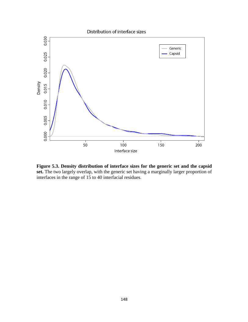
148
Figure 5.3. Density distribution of interface sizes for the generic set and the capsid set. The two largely overlap, with the generic set having a marginally larger proportion of interfaces in the range of 15 to 40 interfacial residues.

149
there is no pronounced difference in the distribution of interface sizes in the two sets, at
least not in the range of interface sizes (between 10 and 200) we have considered in our
analysis. The capsid set compared to the generic set has marginally a larger number of
residues making contacts at an interface. The most frequently seen interface size for a
generic protein dimer is 22 interfacial residues, while an inter-subunit interface in viral
capsids typically has 24 interfacial residues. Nonetheless, this difference is well within
the standard deviation of 30 residues in the distribution and thus negligible. Having
comparable interface sizes in the two sets implies that differences identified, if any,
should be mainly attributed to the geometry and the chemical properties of the interfacial
residues.
The oligomerization states, however, differ significantly between the two sets, as
expected. Similar to what was done in [129], we examined, for each protein of all dimers
included in our analysis, the number of pairwise protein-protein interfaces that the protein
is simultaneously involved in. This number of interacting partners gives a crude
description of the higher order organization of proteins in their quaternary states. As
shown in Figure 5.4, a typical generic protein forms a dimer, while capsid proteins are
frequently interacting with 5 other proteins within the same shell, highlighting the overall
complexity of capsid shell formation. The different oligomerization states may underline
potential differences we find in the two sets of proteins.
5.3.2. Protein-protein interfaces formed by capsid proteins are distinct from those
formed by cellular proteins
The all-against-all comparison between pairwise protein-protein interfaces in viral

150
Figure 5.4. Different oligomerization states in the two sets. This figure plots the density distribution of the number of simultaneous interacting partners in the same protein complex for a given protein that forms an interface in the generic set and the capsid set. A capsid protein, on average, has 4.2 neighboring capsid proteins within the same shell, while the average number of interacting partners for a generic protein is only 2.2.

151
capsids and cellular protein complexes identified altogether 418 generic interfaces that
resemble capsid ones, where the similarity is defined based on a distance cutoff of 0.5.
Compared with the distribution of any two sets of the same sizes, estimated from the
10,000 permutation tests, this overlap in structural space falls on the extreme left as
shown in Figure 5.5, which is significantly small with a one-tailed p-value < 0.0001. We
further show in Figure 5.6 that such a disconnectivity between the capsid set and the
generic set in the protein-protein interface space is not the result of differences in the
sampling density of the two sets; in fact, the intra-set connectivity is about equal for the
two sets. We thus have statistical evidence that in terms of interfacial patterns formed
between protein dimers, inter-subunit interfaces in viral capsids are not representative of
generic protein-protein interfaces found in cellular protein complexes.
The same conclusion can be arrived at even when a different distance cutoff is chosen.
Figure 5.7 plots the cumulative fraction of 20014 interfaces that are within a certain
structural distance of their nearest neighbor in the complementary set of 551 interfaces.
The blue curve corresponding to the comparison between the capsid set and the generic
set is slightly shifted to the right of the grey curves representing the 10,000 permutations,
which suggests that inter-subunit interfaces in viral capsids are more different from
generic protein-protein interfaces compared to what happens as a result of random chance.
This holds true across the entire spectrum of structural distances, including the cutoff of
0.5 chosen previously.
5.3.3. Overlap in structural space of protein-protein interfaces
As we examine the 418 generic interfaces that resemble at least one inter-subunit

152
Figure 5.5. Statistical significance of the test statistic. Out of 10,000 permutations, no single case results in 418 or fewer structurally similar interfaces identified between a randomly selected set of 551 interfaces and their complement set of 20014 interfaces, which makes the one-tailed p-value of our test statistic less than 0.0001. Hence there is strong statistical evidence supporting the hypothesis that capsid interfaces are unlike generic ones.

153
Figure 5.6. Equal connectivity in the two sets. This plot shows that the within-set structural distance distribution for the capsid interface set is well overlaid on top of that for the generic interface set, which suggests that the structural space of both sets have been sampled equally densely to give rise to the data sets, demonstrating the uniformness of the total interface set that justifies the soundness of the permutation test.

154
Figure 5.7. Capsid protein-protein interfaces are different from generic ones. Shown here in the empirical cumulative fraction distribution of distances between one set of 551 protein-protein interfaces and their nearest neighbor in the complementary set, we see that capsid protein-protein interfaces are structurally more distant from non-capsid ones, represented by the blue curve, than the random background, represented by 10,000 grey curves. The average empirical cumulative fraction distribution is colored in red. The range of structural distances plotted is from 0.35 to 0.75 to show a better resolved picture, while in theory this can range from 0 to 1.

155
interface in some capsid, we see their structural analogues in the capsid set are generally
small in size, with more than 70% of them having fewer than 20 interfacial residues
(Figure 5.8). For these protein dimers that form a small area of contact, there are most
likely only limited ways of arranging a few points (i.e. interfacial residues) spatially.
Thus for the smaller interfaces in viral capsids, it is intuitively easier to find similar
patterns among generic interfaces. Furthermore, interactions rendered by a few interfacial
residues are unlikely to contribute to a great amount of binding energies compared with
larger interfaces (which should not be confused with the idea of a few hot spot residues
anchoring protein-protein association in general). In fact, most of these protein-protein
interfaces in viral capsids that overlap with generic ones are found in between
capsomeres, which are pentamers or hexamers and generally considered to be stable
assembly intermediates for viral capsids, rather than within a capsomere, indicating that
they are less active players in the shell formation process. This overlap in structural space
of protein-protein interfaces in the two sets of data thus may not have significant
biological implications.
Unlike the case with comparing the folded topology between capsid proteins and
generic proteins, where we found a few representative classes of cellular proteins that
resemble capsid ones which also forms symmetric oligomers, no similar trend can be
easily identified with the capsid-like generic interfaces in terms of the functional
annotation of the constituent protein monomers. A complete list of the diverse range of
GO terms associated with these protein interfaces is summarized in Table C.2. Among
these, the most abundant hits are associated with proteins in the immune system. This is
not surprising, given the frequent encounters and close interaction between the host

156
Figure 5.8. Sizes of the 418 capsid protein-protein interfaces in the overlap region. Majority of these capsid interfaces that structurally resemble some generic interfaces are small in size, with the exception of one interface that has a size of 85 interfacial residues, being a long coiled coil structure.

157
immune system and the viral coat proteins before the virus gains access to the cell
interior. What appears most frequent in these small, capsid-like generic interfaces are
clusters of discontiguous fragments from loop regions, rather than well-defined
secondary structural elements, and the constituent proteins forming these small contacts
are typically part of larger oligomeric complexes as opposed to homodimers. Figure 5.9
illustrates a few examples of these cases, with the constituent proteins covering
functional classes such as transferases (Figure 5.9 (A)), cell adhesion (Figure 5.9 (B)) and
ligases (Figure 5.9(C)).
5.4. Discussion:
5.4.1. Result of comparative study is not sensitive to quality of structural data
Structural comparison between biological molecules relies on having reasonable
structural models solved by experimental techniques. The large, elaborate nature of
macromolecular complexes such as viral capsids often creates limits on the sample
availability and regularity, resulting in lower resolution of the structures determined.
Consequently, the atomic coordinates of these structural models are less accurate and
sometimes with details missing, compared to those of small protein complexes involving
cellular proteins. Furthermore, in the case of viral capsids, the symmetry of the assembled
architecture is utilized for averaging over the experimental density of all asymmetric
units for coordinate derivation, leading to additional sources of imprecision. Nonetheless,
the structural comparison tool that we used in this study scores the similarity between a
pair of protein-protein interfaces with reduced representation of their structural
information, both geometrically and chemically, and we have demonstrated previously
[200] that our comparison metric can recognize significant similarity between related

158
Figure 5.9. Examples of similar interfaces from the capsid set and the generic set. In all panels, the viral capsid proteins are colored in cyan and orange, while the cellular protein dimers are colored in blue and red. The monomer proteins are drawn in either cartoon representation or tube representation, the latter for structural models lacking side-chain information. The Cα atoms of the aligned interfacial residues are shown in van der Waals representation. (A) The interface formed by the generic protein dimer (PDB code 1xiq, chain IDs B and F) is aligned to the capsid interface (PDB code 1x9p, chain A in the first IAU and chain A in the 16th IAU). (B) The generic interface between chain A and chain B of PDB code 3m45 is aligned to the capsid interface formed by chain B and chain C within the same IAU in PDB code 1n6g. (C) The generic interface between chain G and chain H of PDB code 3d54 is aligned to the capsid interface between chain F of the first IAU and chain F of the second IAU in PDB entry 3muw.

159
protein- protein interfaces in artificially created data that are highly corrupted. Given the
robustness of our method against noisy data, it is unlikely that the conclusion drawn
based on our comparative study will be affected by the inaccuracy of the atomic positions
in these structural models.
5.4.2. Domain-swapped interfaces are not treated differently
Domain swapping is commonly found in homomeric complexes, and it refers to two
identical proteins exchanging the same structural elements to form dimer interfaces that
replace the original intramolecular contacts in the monomeric states. Since its first
recovery in diphtheria toxin [252], many domain-swapped structures of diverse origin
have been identified [253]. These interfaces are best characterized by their highly
intertwined nature, blurring the boundary between intramolecular contacts and
intermolecular ones. In our work, we have not delineated this class of protein-protein
interfaces from the rest of generic interfaces for special consideration, for the main reason
that despite the possible advantage of the domain swapping mechanism in formation of
large, stable protein complexes, domain swapping is still rare (on the order of 10) among
currently solved structures [254] and remains less well understood for unambiguous
annotation. This being the case, we do not expect the negligible fraction contributed by
this class of protein-protein interfaces to have significant bearing on the overall
conclusion of our study.
5.4.3. Implications of protein-protein interfaces in capsids being unique
Viral capsid shells represent large macromolecular assemblies that exist on a scale not
seen in most of cellular macromolecular complexes. Given the unique function of capsid

160
proteins in making shells, we are interested in whether the sticky patches on these
building blocks that piece them together are also structurally unique, in order to pinpoint
features critical for the design principle of large biological containers. Our hypothesis
testing has provided strong statistical evidence of the distinctiveness of the interfacial
patterns found between capsid proteins from those between cellular proteins, which has
the important implication that how capsid proteins recognize and associate with one
another is governed not simply by physical principles of shape complementarity alone,
but is retained specifically by evolution to favor the higher order organization. This
conclusion that capsid interfaces are subjected to evolutionary constraints is in agreement
with the previous findings that changes in inter-subunit geometry have direct impact on
the oligomerization states of protein subunits [255], which in turn should confer selective
pressure on protein-protein interactions. Combined with our earlier observation that the
folded topology of viral capsid proteins is also segregated from those of cellular proteins,
we arrive at the conclusion that the basic shape of these Lego pieces and the molecular
recognition sites on their edges act concertedly to create the sophisticated shell
architecture as designed.
In terms of rationalizing therapeutic design of antiviral drugs, the finding of this work
favors the view that pathogen specificity can be achieved, given that protein-protein
interfaces in viral capsids are significantly different from those involved in cellular
activities. While this statement holds true, we would like to caution against the danger of
introducing inhibitors targeting the smaller interfaces in viral capsids, especially those not
found within the same capsomere, which can possibly fall into the structural overlap
between capsid protein-protein interfaces and generic ones. As shown in Table C.2, these
161
potential off-targets in the cellular domain cover a wide range of biological functions,
including various enzymatic activities and gene regulation that are crucial for life. A
more conservative approach would be to focus on those larger interfaces that provide
greater stabilization energy for the capsid shell, with the hope of not only having greater
inhibitive power for the assembly process but also with fewer side effects.
Lastly, our study has confirmed that interfacial patterns in viral capsids are not
representative of those in cellular protein complexes. While most cellular proteins
function by forming binary interactions, either transiently or permanently, viral capsid
proteins are characterized by interacting with multiple partners simultaneously in the
multiple-component assemblies. What we are most interested in with regards to
properties of inter-subunit interfaces in viral capsids is the control of quasi-equivalence,
which provides a great example of exquisite fine-tuning of modes of interaction via
conformational switching of identical gene products. However, as we established in this
study, the larger interfaces within capsomeres do not share much structural similarity
with generic protein-protein interfaces, and it is precisely the alternation between the
concave pentamers and the flat hexamers that best manifests the quasi-equivalent
property. Therefore, what we learn about physical principles governing protein-protein
recognition from capsid proteins may not easily extend to protein-protein interactions at
large.
The above aspects cover the implications of our findings in this work. What our
conclusion does not imply, however, is the general uniqueness of structural
characteristics of all viral proteins. The exclusive sets of folded topology and distinct
patterns of association with one another found in viral capsid proteins are correlated with

162
their specific function that is missing for cellular life forms. Unlike capsid proteins, other
viral proteins may assume roles that partially overlap with cellular proteins. Examples
include viral proteases that process polypeptides for maturation, which contain motifs for
hydrolysis of peptide bonds similar to cellular proteases that lyse misfolded peptides for
recycling, and reverse transcriptases in retroviruses necessarily share similar nucleotide
binding sites with DNA polymerases in cellular machinery. Related studies on the
structural relationship between general classes of viral proteins and cellular proteins have
been carried out, with focuses on virus-host interaction in humans [182] and
thermodynamic stabilities [256]. The first study established extensive overlap between
virus-host interactions and endogenous interactions within the host for competitive
binding, suggesting that the uniqueness of capsid protein-protein interfaces is a result of
functional requirement rather than their viral origin. The second study concluded on the
high adaptability of viral proteins for effective interaction with proteins in the host, as
opposed to thermostable proteins, which are less tolerant of the deleterious effects of
mutations. This second study presents an additional aspect of viral proteins, specifically
their chemical composition and disorder propensity. One needs to be precautious in
extrapolating this unique biophysical property of viral proteins to all protein-protein
interfaces that are viral derived without further investigation.
5.4.4. Concluding remarks
To conclude, we have shown in this study via rigorous hypothesis testing that inter-
subunit interfaces in the large, elaborate viral capsid assemblies are significantly different
from protein-protein interfaces found in the smaller and simpler cellular protein
complexes. This difference in capsid interfaces is most likely the consequence of the

163
functional requirement for making the specific architecture of shells for encapsulation of
the viral genome, which on average involves more neighbors per protein subunit as well
as assumes a more expanded organization compared with cellular complexes that are
often times binary and more compact and collapsed in nature. Our results should provide
deeper understanding of the nature of self-association in large biological containers for
creating predictable designs in a variety of therapeutic and materials science applications.

164
Appendix C
Additional information for CHAPTER V
Table C.1. List of additional capsid protein dimers. These entries are not deposited in the VIPERdb, and were subsequently moved from the generic interface set to the capsid set before a structural redundancy filter was applied to both sets.
PDB code Chain identifiers Virus name 1ahs AB African horse sickness virus 1ak4 CD Human immunodeficiency virus 1 1bvp 56 Bluetongue virus 1c5e AC Enterobacteria phage lambda 1ebo DF Ebola virus 1ei7 AB Tobacco mosaic virus 1em9 AB Rous sarcoma virus 1fn9 AB Reovirus 1hiw AR Human immunodeficiency virus 1 1hiw QR Human immunodeficiency virus 1 1jmu BH Reovirus 1jmu DE Reovirus 1jmu DF Reovirus 1jmu EF Reovirus 1jmu FI Reovirus 1no7 AB Human herpesvirus 1
1p65 AB Porcine reproductive and respiratory syndrome virus
1r6r AB Dengue virus 2 1u7k EF AKR (endogenous) murine leukemia virus 1vps AE Murine polyomavirus 1wnc BF SARS coronavirus 1zv8 GK SARS coronavirus 2beq DF SARS coronavirus 2cse 1V Mammalian orthoreovirus 1 Lang 2cse 1W Mammalian orthoreovirus 1 Lang 2cse AK Mammalian orthoreovirus 1 Lang 2cse AR Mammalian orthoreovirus 1 Lang 2cse AY Mammalian orthoreovirus 1 Lang

165
2cse JU Mammalian orthoreovirus 1 Lang 2cse LU Mammalian orthoreovirus 1 Lang 2cse QX Mammalian orthoreovirus 1 Lang 2cse VW Mammalian orthoreovirus 1 Lang 2cse VZ Mammalian orthoreovirus 1 Lang 2cse WZ Mammalian orthoreovirus 1 Lang 2eia AB Equine infectious anemia virus 2ezo BC Simian immunodeficiency virus 2gic AC Vesicular stomatitis Indiana virus 2gic BC Vesicular stomatitis Indiana virus 2gsy BD Infectious bursal disease virus 2gsy GN Infectious bursal disease virus 2i2s AB Porcine rotavirus 2vvf AB Pseudoalteromonas phage PM2 2zl5 AB Norwalk virus 3bwq AE Simian virus 40 3h00 BD Human immunodeficiency virus type 1 3izo BC Human adenovirus 5 3rkc AB Hepatitis E virus 3sln AC Norovirus Hu 3sln BJ Norovirus Hu 3sln DH Norovirus Hu

166
Table C.2. Functional annotation of capsid-like generic interfaces. The 418 generic interfaces that structurally resemble some inter-subunit interfaces in viral capsids are highly diverse in terms of the biological functions they are involved in. This table summarizes the counts of entries in each functional category.
Functional annotation NO. of capsid-like generic interfaces Alpha-helical bundle 1 Antimicrobial protein 1 Apoptosis 2 Bacterial cell division 1 Beta-clamp 1 Biosynthetic protein 1 Blood clotting 3 CAMP-binding protein 1 Catalytic antibody 1 Cell adhesion 8 Cell cycle 3 Cell invasion 1 Cell motility protein 1 Chaperone 5 Chemokine 1 Chemotaxis 1 Chromatin regulator 1 Chromosome segregation 1 Complex (ligand/receptor) 4 Contractile protein 4 Cytokine 2 Cytotoxin 1 Decarboxylase 1 De novo protein 6 DNA binding protein 10 DNA integration 1 Electron transport 8 Endocytosis/Exocytosis 3 Enterotoxin 1 Gene regulation 8 Growth factor 1 Heme-binding 1 Hormone 6 Hydrolase 56 Immune system 28 Immunoglobulin 3 Isomerase 1 Lectin 2

167
Leucine zipper 1 Ligase 5 Light-harvesting protein 1 Lipid binding protein 1 Lipoprotein 1 Lyase 6 Membrane protein 11 Metal binding protein 3 Murine class I MHC 1 Neuropeptide 2 Neurotoxin 1 Nucleic acid binding protein 2 Oxidoreductase 22 Oxygen storage/transport 5 Peptide binding protein 1 Peroxidase 1 Phosphoribosyl transferase 1 Photosynthesis 10 Plant protein 1 Proteinase inhibitor 1 Protein binding 9 Protein transport 4 Replication 1 Retinoic-acid transport 1 Ribosomal protein 24 RNA binding protein 3 Signaling protein 13 Structural genomics, unknown
20 Structural protein 6 Sugar binding protein 3 Synthetic protein model 1 T cell receptor 1 Toxin 6 Transcription 23 Transferase 34 Tryptophan biosynthesis 1 Unknown bacterial hydrolase 1 Unknown function 6 Viral protein 12
168
CHAPTER VI
Conclusion
Viral capsids exhibit elaborate and symmetrical architectures not seen in cellular
multimeric protein complexes. The limited genome coding capacity of viruses
necessitates the economic use of a single or a few types of capsid proteins, which can
efficiently and faithfully self-assemble into a homogeneous particle of a defined size.
This thesis work is aimed at pinpointing structural characteristics of viral capsid proteins
that govern the functional assembly of capsid shells, with the ultimate goal of gaining a
deeper understanding of the design principles that control such events for various
biomedical and materials science applications.
In Chapter II [200], I explored the folded topology of all viral capsid proteins for
which structural data are available, and compared them with a representative set of
generic proteins, in order to test the hypothesis that these shell-forming building blocks
assume unique geometry. This large scale comparative analysis, using Template
Modeling (TM)-score, identified only 2078 non-capsid proteins that have marginal
structural resemblance to capsid proteins, covering altogether 210 folds following the
definition in SCOP, which is far less than the number of shared folds between randomly
partitioned sets of the same sizes. The statistical significance of this test statistic,
estimated from our permutation test, is very high with a p-value less than 0.0001. This

169
leads to our conclusion that viral capsid proteins are segregated in structural fold space.
During this work, I developed a novel statistical framework that is suitable for assessing
whether a structural feature is uniquely found in any functional class of proteins.
Incidentally, the same analysis performed on several other functional classes of proteins,
such as muscle proteins and kinases, did not establish exclusive sets of folds for these
classes, which makes the major conclusion of the work even more significant. The results
of this study provide novel insights into how structural folds of capsid proteins, which
directly determine the shape of these building blocks, might be constrained during
evolution by requirement of the assembled cage-like architecture. Additionally, my work
highlights the guideline for synthetic maneuvers of virus-based nanoplatforms when
imparting new functionalities, which is to adhere to the library of folds adopted by capsid
proteins that nature has created specifically to make shells.
Chapter III [223] describes a computational tool I developed to compare the
physicochemical properties of interfaces between two given protein dimers, focusing on
local similarities at the interface region that participate in molecular recognition while
disregarding the evolutionary relatedness between the constituent protein monomers.
Unlike existing methods which require atomic details of the input structural models, my
method can deal with relatively poor quality data determined at lower resolutions,
allowing structural comparison across models with different levels of detail. This major
contribution greatly expands the structural space that can be studied by including protein-
protein interaction patterns found in large macromolecular complexes, exemplified by
viral capsids, which are typically solved by lower-resolution experimental techniques.
Such improvement was made possible by defining intermolecular contacts using minimal

170
structural information available, based on statistical knowledge from mining the Protein
Data Bank, and our amino acid type-specific definition of contacts is shown to
outperform the generic distance cutoff criterion used traditionally. Additionally, the
geometric hashing algorithm used in my method effectively addresses the fact that
interfacial residues are discontiguous fragments clustered spatially regardless of their
connectivity, which optimally captures the central properties of the protein-protein
interfaces that interest us. I demonstrated the power of the PCalign method in accurately
recognizing highly similar interfaces across related protein dimers both on a real dataset
and on a simulated “corrupted” dataset, showing its robustness against noise.
Furthermore, through comparison with existing methods, I showed that my method has
better sampling on average to yield optimal structural alignment, mapping a larger
fraction of structurally equivalent interfacial residues with lower root mean squared error.
Finally, I illustrate the usefulness of the PCalign method in recognizing interesting
biological relationships masked by apparent lack of structural similarity by a few
examples of viral mimicry, where viruses convergently evolved similar binding sites on
viral proteins to target cellular proteins in the host system, without these viral proteins
bearing any evolutionary relationship with the native partners of their target proteins.
Chapter IV illustrates an application of the method developed in Chapter III to
computer-aided drug design. This work is rationalized on the hypothesis that nature has a
limited set of choices in arranging interfacial residues spatially and chemically. Using the
recently solved crystal structure of the 1918 influenza virus hemagglutinin protein in
complex with a neutralizing antibody CR6261 as a template, I screened a non-redundant
subset of all protein dimers in the Protein Data bank to look for similar protein-protein

171
interaction patterns to those between the epitope and the paratope. Encouraged by our
finding that two recently designed protein inhibitors [113, 114] for the same target via a
de novo approach scored discriminatively higher by our program PCalign than the
computational models that failed experimental validation, I selected 10 top-ranking
proteins which act as the antibody equivalent based on the structural alignment of the
interfaces as a starting point for designing binders for our target protein. These candidates
were further optimized by rationally selecting mutations at the binding site to favor the
interaction. Out of the 10 initial candidates, four of them gave comparable measures of
likelihood of binding to the viral protein as that of the original neutralizing antibody,
based on several independent evaluation criteria such as estimated binding free energies
and percent of near-native poses correctly docked. Experimental collaboration is now
under way to have our predictions tested for binding activity. This work presents a
generalizable pipeline of computationally designing novel protein-based inhibitors for
any given therapeutic target, using structural templates identified by the PCalign tool.
Having established that viral capsid proteins are represented by unique folded
topologies in Chapter II, I next investigated whether protein-protein interfaces in viral
capsids are representative of generic ones found in cellular protein complexes in Chapter
V. The former attribute essentially concerns the shape of the Lego pieces for shell
formation, while the latter addresses the sticky patches that hold the pieces together.
From the comparative analysis of a non-redundant set of 551 inter-subunit interfaces in
viral capsids and a non-redundant set of 20014 protein-protein interfaces in non-capsid
protein complexes, we found that 418 generic protein-protein interfaces (about 2%) share
similar physicochemical patterns with some protein-protein interface in the capsid set.

172
This overlap in the structural space of protein-protein interfaces is again significantly
small with a p-value < 0.0001, based on a permutation test on the total set of protein-
protein interfaces. We thus conclude that the exclusive protein-protein interaction
patterns found in viral capsid proteins are necessary, but not the only, determinants of
their higher order organization. This observation is in agreement with Chothia and
coworkers’ finding of interfaces having direct influence on the oligomeric states of
homomeric tetramers and hexamers from eleven protein families [255]. Together with my
earlier study of the folded topology of viral capsid proteins, we can establish the
importance of both having the geometry that conforms with the mathematical constraints
and having special physicochemical complementarity among individual building blocks
in directing the final complex formation of viral capsid shells.
Looking beyond, a related question that remains to be fully understood is the control
of quasi-equivalence in viruses. Viruses display a highly diverse range of mechanisms in
fine-tuning interactions between capsid proteins to acquire a defined shell size. For small
viruses, conformational switching of capsid protein subunits allows the formation of
different intermolecular contacts, which typically involves a molecular switch in the
terminal arm that does not appear highly conserved in sequence. For the majority of the
canonical T=3 and T=4 viruses, this switch is ordered in subunits in a certain
environment while disordered in others. Leviviruses provide an exception to this rule,
without any apparent conformational switching mechanism among their protein subunits,
yet still assuming a T=3 icosahedral symmetry. Larger viruses use more complex ways to
dictate the size of their shells, often with the help of additional scaffolding proteins,
exemplified by the tape-measure protein in PRD1 viruses. Given that capsid proteins of

173
most icosahedral viruses of varying sizes have a common structural core, represented by
the ubiquitous jellyroll fold, it is particularly intriguing what the unifying molecular
signature at the protein-protein interfaces is, if there is any, that plays a role in
determining the T-number of the assembled architecture, in addition to the interplay with
RNA/DNA molecules.
A preliminary survey was carried out for T=1, T=3 and T=4 viruses by analyzing the
set of unique capsid inter-subunit interfaces in their pentamers and hexamers. While a
pentamer is convex, with pairwise interacting dimers forming a bent interface, a hexamer
is planar, and interfaces in a hexamer can be either bent along the 5-fold symmetry axis,
or relatively flat along the 3-fold symmetry axis. The capsid proteins in these small plant
viruses all have the signature jellyroll topology, with small variations in the connecting
loops of the beta-strands. Comparing the protein-protein interfaces across the board
therefore can reveal potential molecular fingerprints related to T-numbers.
Figure 6.1 shows the comparison result for inter-subunit interfaces in a few
representative T=1, T=3 and T=4 viruses, taking one capsid from each family from the
VIPERdb database. T=1 viruses include a satellite virus (PDB code: 1stm), a
sobemovirus (PDB code: 1vak), a bromovirus (PDB code: 1yc6), and a circovirus (PDB
code: 3r0r), of which the sobemovirus and the bromovirus are both T=1 subviral particles
resulting from mutations of what are otherwise T=3 viruses. T=3 viruses include a
tymovirus (PDB code: 1ddl), a bromovirus (PDB code: 1js9), a calicivirus (PDB code:
2gh8), a sobemovirus (PDB code: 2izw), a tombusvirus (PDB code: 2tbv) and a
nodavirus (PDB code: 2z2q). Finally, one tetravirus (PDB code: 1ohf) represents a T=4
virus. All dimer interfaces in unique environments on the icosahedral shell are compared

174
Figure 6.1. Comparison of inter-subunit interfaces in small plant viruses. The separation between different interfaces approximately represents the structural distance between them. While interfaces found within the same capsid tend to cluster together, there is no apparent similarity among interfaces found within capsids of the same T-number, or interfaces found within equivalent structural environment (pentamers or hexamers).

175
against one another using PCalign, and their mutual distance, defined as (1 - PC-score), is
visualized via a multi-dimensional scaling plot shown in Figure 6.1. This plot captures
the structural distance between different interfaces based on projection onto the first two
principle components (PC1 and PC2). The color grouping refers to interfaces found at a
pentamer, the bent interfaces in a hexamer (hexamer1), and the flat interfaces in a
hexamer (hexamer2).
The distribution of different interfaces in Figure 6.1 shows that generally inter-
subunit interfaces within the same family are more similar to one another than across
different families, and this is also true for the two cases of the T=1 mutant and the T=3
wild type in the same family, which are clustered together respectively. The plot does not
show a clear pattern that groups interfaces by the T-number of the capsid they are found
in. Also worth noting is that the pentamer interfaces resemble the bent hexamer interfaces
more than the flat hexamer interfaces, as expected. Overall, there is lack of correlation
between interface similarity and the higher-order organization of capsid proteins. An
interesting observation is that for the T=3 nodavirus and the T=4 tetravirus, there have
been reported evolutionary relatedness in terms of capsid maturation strategies [105, 257].
Despite the different conformational switching mechanisms and particle architectures for
these two viruses, the interfacial patterns show remarkable similarity, shown here as
clustered together, suggesting alternative molecular motifs that converge to the same
regulatory elements for tuning the hinge angles between interacting dimers.
The diversity of conformational switching mechanisms prompts the question of
whether a general underlying rule exists to dictate the size of the assembled capsid.
Previously, capsomeres (i.e. pentamers and hexamers) mixed in pre-defined ratios are
176
modeled in simulations to study the dynamic assembly of viral capsids of different T-
numbers [258], which has largely reproduced assembled icosahedral capsids of the
expected size in silico in addition to other aberrant morphologies. This leads to our
hypothesis that the T-number of icosahedral viral shells is determined by the pre-existing
ratio of the different capsomeres, which in turn is influenced by the pre-disposition of the
capsid protein subunits to form pairwise bent or flat interfaces. Indeed, when examining
the capsid proteins in different environments on an icosahedral shell, we found tractable
trends about the proportion of different conformations, specifically the order/disorder
distribution, corresponding to the T-number. In T=1 viruses where all protein subunits
are in identical environment, there is only one conformation. In T=4 viruses, there are
four different positions for the protein subunits, which are by convention labeled as A, B,
C and D. For providence virus and Nudaurelia capensis ω virus found in the tetraviridae
family, two out of the four subunits assume an ordered configuration in the terminal arms
while the other two have the disordered configuration, although one virus uses an N-
terminal switch and the other a C-terminal switch. For T=3 viruses, there are different
classes depending on the overall morphology of the shell, known as the truncated
icosahedron and the rhombic triacontahedron [104]. Out of the three subunits A, B and C,
only one assumes an ordered configuration in the terminal arm in the rhombic
triacontahedron subtype, whereas there are two subunits that are ordered in the terminal
arms in the truncated icosahedron subtype. In other words, the order/disorder ratios seem
correlated with the T-numbers and the subtypes within the same T-number. If indeed this
is the key regulating factor of the higher order organization, one can in theory make

177
reliable predictions by estimating the relative frequencies of the ordered/disordered states
in the conformational ensemble of a given capsid protein.
In practice, however, the above hypothesis is difficult to be tested computationally.
This is mainly due to the fact that a ratio of 2:2 for the two states in T=4 viruses
corresponds to the two states being in equilibrium with each other, and the ratio of 2:1 or
1:2 for the two states in T=3 viruses corresponds to a small free energy difference of
about 0.4 kcal/mol, which is below the sensitivity of any molecular dynamics simulation
techniques. An alternative approach may be to estimate the disorder propensity of a
molecular switch using only information from its peptide sequence via Bioinformatics
tools, such as DynaMine [259], and validate the predictions through experimental means,
the latter of which can be laborious and expensive. Deciphering the Da Vinci code hidden
in individual capsid proteins that ultimately defines the shell size remains a challenging
task to explore in future.
The structural determination of the large and complex assembly of viral capsids in
sub-nano resolution marks a milestone in structural biology, bringing exciting
opportunities to learn about the natural design principles behind these finely-controlled
biological containers. This dissertation has contributed a novel statistical framework as
well as a computational tool that enabled the systematic survey of different structural
features of viral capsid proteins crucial for their function. Through rigorous hypothesis
testing, I have delineated the folded topology as well as interfacial patterns of viral capsid
proteins as important attributes retained specifically by evolution for shell assembly. My
findings should provide guidelines for designing and modulating the assembly of
biological building blocks into defined higher order structures in various nanotechnology

178
applications, as well as rationalize the design of antiviral drugs towards targeting
pathogen-specific sites for improved therapeutic efficacy and reduced toxicity.

179
BIBLIOGRAPHY
1. Suttle CA: Viruses in the sea. Nature 2005, 437(7057):356-361. 2. Douglas T, Young M: Viruses: making friends with old foes. Science 2006,
312(5775):873-875. 3. Abroi A, Gough J: Are viruses a source of new protein folds for organisms? - Virosphere
structure space and evolution. BioEssays : news and reviews in molecular, cellular and developmental biology 2011, 33(8):626-635.
4. Nasir A, Forterre P, Kim KM, Caetano-Anolles G: The distribution and impact of viral lineages in domains of life. Frontiers in microbiology 2014, 5:194.
5. Khayat R, Tang L, Larson ET, Lawrence CM, Young M, Johnson JE: Structure of an archaeal virus capsid protein reveals a common ancestry to eukaryotic and bacterial viruses. Proceedings of the National Academy of Sciences of the United States of America 2005, 102(52):18944-18949.
6. Fokine A, Leiman PG, Shneider MM, Ahvazi B, Boeshans KM, Steven AC, Black LW, Mesyanzhinov VV, Rossmann MG: Structural and functional similarities between the capsid proteins of bacteriophages T4 and HK97 point to a common ancestry. Proceedings of the National Academy of Sciences of the United States of America 2005, 102(20):7163-7168.
7. Krupovic M, Bamford DH: Virus evolution: how far does the double beta-barrel viral lineage extend? Nature reviews Microbiology 2008, 6(12):941-948.
8. Bamford DH, Grimes JM, Stuart DI: What does structure tell us about virus evolution? Current opinion in structural biology 2005, 15(6):655-663.
9. Roos WH, Ivanovska IL, Evilevitch A, Wuite GJ: Viral capsids: mechanical characteristics, genome packaging and delivery mechanisms. Cellular and molecular life sciences : CMLS 2007, 64(12):1484-1497.
10. Carrillo-Tripp M, Shepherd CM, Borelli IA, Venkataraman S, Lander G, Natarajan P, Johnson JE, Brooks CL, 3rd, Reddy VS: VIPERdb2: an enhanced and web API enabled relational database for structural virology. Nucleic acids research 2009, 37(Database issue):D436-442.
11. Caspar DL, Klug A: Physical principles in the construction of regular viruses. Cold Spring Harbor symposia on quantitative biology 1962, 27:1-24.
12. Mannige RV, Brooks CL, III: Periodic table of virus capsids: implications for natural selection and design. PloS one 2010, 5(3):e9423.
13. Keef T, Twarock R, Elsawy KM: Blueprints for viral capsids in the family of polyomaviridae. Journal of theoretical biology 2008, 253(4):808-816.
14. Naitow H, Tang J, Canady M, Wickner RB, Johnson JE: L-A virus at 3.4 A resolution reveals particle architecture and mRNA decapping mechanism. Nature structural biology 2002, 9(10):725-728.
15. Cheng RH, Reddy VS, Olson NH, Fisher AJ, Baker TS, Johnson JE: Functional implications of quasi-equivalence in a T = 3 icosahedral animal virus established by cryo-electron microscopy and X-ray crystallography. Structure 1994, 2(4):271-282.

180
16. Mannige RV, Brooks CL, III: Tilable nature of virus capsids and the role of topological constraints in natural capsid design. Physical review E, Statistical, nonlinear, and soft matter physics 2008, 77(5 Pt 1):051902.
17. Lin T, Porta C, Lomonossoff G, Johnson JE: Structure-based design of peptide presentation on a viral surface: the crystal structure of a plant/animal virus chimera at 2.8 A resolution. Folding & design 1996, 1(3):179-187.
18. McLain L, Durrani Z, Wisniewski LA, Porta C, Lomonossoff GP, Dimmock NJ: Stimulation of neutralizing antibodies to human immunodeficiency virus type 1 in three strains of mice immunized with a 22 amino acid peptide of gp41 expressed on the surface of a plant virus. Vaccine 1996, 14(8):799-810.
19. Langeveld JP, Kamstrup S, Uttenthal A, Strandbygaard B, Vela C, Dalsgaard K, Beekman NJ, Meloen RH, Casal JI: Full protection in mink against mink enteritis virus with new generation canine parvovirus vaccines based on synthetic peptide or recombinant protein. Vaccine 1995, 13(11):1033-1037.
20. Obeid OE, Partidos CD, Howard CR, Steward MW: Protection against morbillivirus-induced encephalitis by immunization with a rationally designed synthetic peptide vaccine containing B- and T-cell epitopes from the fusion protein of measles virus. Journal of virology 1995, 69(3):1420-1428.
21. Porta C, Spall VE, Loveland J, Johnson JE, Barker PJ, Lomonossoff GP: Development of cowpea mosaic virus as a high-yielding system for the presentation of foreign peptides. Virology 1994, 202(2):949-955.
22. Usha R, Rohll JB, Spall VE, Shanks M, Maule AJ, Johnson JE, Lomonossoff GP: Expression of an animal virus antigenic site on the surface of a plant virus particle. Virology 1993, 197(1):366-374.
23. Pumpens P, Grens E: HBV core particles as a carrier for B cell/T cell epitopes. Intervirology 2001, 44(2-3):98-114.
24. Stahl SJ, Murray K: Immunogenicity of peptide fusions to hepatitis B virus core antigen. Proceedings of the National Academy of Sciences of the United States of America 1989, 86(16):6283-6287.
25. Kratz PA, Bottcher B, Nassal M: Native display of complete foreign protein domains on the surface of hepatitis B virus capsids. Proceedings of the National Academy of Sciences of the United States of America 1999, 96(5):1915-1920.
26. Birkett A, Lyons K, Schmidt A, Boyd D, Oliveira GA, Siddique A, Nussenzweig R, Calvo-Calle JM, Nardin E: A modified hepatitis B virus core particle containing multiple epitopes of the Plasmodium falciparum circumsporozoite protein provides a highly immunogenic malaria vaccine in preclinical analyses in rodent and primate hosts. Infection and immunity 2002, 70(12):6860-6870.
27. Oliveira GA, Wetzel K, Calvo-Calle JM, Nussenzweig R, Schmidt A, Birkett A, Dubovsky F, Tierney E, Gleiter CH, Boehmer G et al: Safety and enhanced immunogenicity of a hepatitis B core particle Plasmodium falciparum malaria vaccine formulated in adjuvant Montanide ISA 720 in a phase I trial. Infection and immunity 2005, 73(6):3587-3597.
28. Jegerlehner A, Tissot A, Lechner F, Sebbel P, Erdmann I, Kundig T, Bachi T, Storni T, Jennings G, Pumpens P et al: A molecular assembly system that renders antigens of choice highly repetitive for induction of protective B cell responses. Vaccine 2002, 20(25-26):3104-3112.

181
29. Billaud JN, Peterson D, Barr M, Chen A, Sallberg M, Garduno F, Goldstein P, McDowell W, Hughes J, Jones J et al: Combinatorial approach to hepadnavirus-like particle vaccine design. Journal of virology 2005, 79(21):13656-13666.
30. Netter HJ, Macnaughton TB, Woo WP, Tindle R, Gowans EJ: Antigenicity and immunogenicity of novel chimeric hepatitis B surface antigen particles with exposed hepatitis C virus epitopes. Journal of virology 2001, 75(5):2130-2141.
31. Sojikul P, Buehner N, Mason HS: A plant signal peptide-hepatitis B surface antigen fusion protein with enhanced stability and immunogenicity expressed in plant cells. Proceedings of the National Academy of Sciences of the United States of America 2003, 100(5):2209-2214.
32. Bisht H, Chugh DA, Swaminathan S, Khanna N: Expression and purification of Dengue virus type 2 envelope protein as a fusion with hepatitis B surface antigen in Pichia pastoris. Protein expression and purification 2001, 23(1):84-96.
33. Bisht H, Chugh DA, Raje M, Swaminathan SS, Khanna N: Recombinant dengue virus type 2 envelope/hepatitis B surface antigen hybrid protein expressed in Pichia pastoris can function as a bivalent immunogen. Journal of biotechnology 2002, 99(2):97-110.
34. Schlienger K, Mancini M, Riviere Y, Dormont D, Tiollais P, Michel ML: Human immunodeficiency virus type 1 major neutralizing determinant exposed on hepatitis B surface antigen particles is highly immunogenic in primates. Journal of virology 1992, 66(4):2570-2576.
35. Eckhart L, Raffelsberger W, Ferko B, Klima A, Purtscher M, Katinger H, Ruker F: Immunogenic presentation of a conserved gp41 epitope of human immunodeficiency virus type 1 on recombinant surface antigen of hepatitis B virus. The Journal of general virology 1996, 77 ( Pt 9):2001-2008.
36. Frazer IH, Quinn M, Nicklin JL, Tan J, Perrin LC, Ng P, O'Connor VM, White O, Wendt N, Martin J et al: Phase 1 study of HPV16-specific immunotherapy with E6E7 fusion protein and ISCOMATRIX adjuvant in women with cervical intraepithelial neoplasia. Vaccine 2004, 23(2):172-181.
37. Dale CJ, Liu XS, De Rose R, Purcell DF, Anderson J, Xu Y, Leggatt GR, Frazer IH, Kent SJ: Chimeric human papilloma virus-simian/human immunodeficiency virus virus-like-particle vaccines: immunogenicity and protective efficacy in macaques. Virology 2002, 301(1):176-187.
38. Liu WJ, Liu XS, Zhao KN, Leggatt GR, Frazer IH: Papillomavirus virus-like particles for the delivery of multiple cytotoxic T cell epitopes. Virology 2000, 273(2):374-382.
39. Griffiths JC, Berrie EL, Holdsworth LN, Moore JP, Harris SJ, Senior JM, Kingsman SM, Kingsman AJ, Adams SE: Induction of high-titer neutralizing antibodies, using hybrid human immunodeficiency virus V3-Ty viruslike particles in a clinically relevant adjuvant. Journal of virology 1991, 65(1):450-456.
40. Weber J, Cheinsong-Popov R, Callow D, Adams S, Patou G, Hodgkin K, Martin S, Gotch F, Kingsman A: Immunogenicity of the yeast recombinant p17/p24:Ty virus-like particles (p24-VLP) in healthy volunteers. Vaccine 1995, 13(9):831-834.
41. Gilbert SC, Plebanski M, Harris SJ, Allsopp CE, Thomas R, Layton GT, Hill AV: A protein particle vaccine containing multiple malaria epitopes. Nature biotechnology 1997, 15(12):1280-1284.
42. Deml L, Speth C, Dierich MP, Wolf H, Wagner R: Recombinant HIV-1 Pr55gag virus-like particles: potent stimulators of innate and acquired immune responses. Molecular immunology 2005, 42(2):259-277.

182
43. Niikura M, Takamura S, Kim G, Kawai S, Saijo M, Morikawa S, Kurane I, Li TC, Takeda N, Yasutomi Y: Chimeric recombinant hepatitis E virus-like particles as an oral vaccine vehicle presenting foreign epitopes. Virology 2002, 293(2):273-280.
44. Maurer P, Jennings GT, Willers J, Rohner F, Lindman Y, Roubicek K, Renner WA, Muller P, Bachmann MF: A therapeutic vaccine for nicotine dependence: preclinical efficacy, and Phase I safety and immunogenicity. European journal of immunology 2005, 35(7):2031-2040.
45. Young LS, Searle PF, Onion D, Mautner V: Viral gene therapy strategies: from basic science to clinical application. The Journal of pathology 2006, 208(2):299-318.
46. Jolly D: Viral vector systems for gene therapy. Cancer gene therapy 1994, 1(1):51-64. 47. Cavazzana-Calvo M, Hacein-Bey S, de Saint Basile G, Gross F, Yvon E, Nusbaum P, Selz F,
Hue C, Certain S, Casanova JL et al: Gene therapy of human severe combined immunodeficiency (SCID)-X1 disease. Science 2000, 288(5466):669-672.
48. Hacein-Bey-Abina S, Le Deist F, Carlier F, Bouneaud C, Hue C, De Villartay JP, Thrasher AJ, Wulffraat N, Sorensen R, Dupuis-Girod S et al: Sustained correction of X-linked severe combined immunodeficiency by ex vivo gene therapy. The New England journal of medicine 2002, 346(16):1185-1193.
49. Imren S, Fabry ME, Westerman KA, Pawliuk R, Tang P, Rosten PM, Nagel RL, Leboulch P, Eaves CJ, Humphries RK: High-level beta-globin expression and preferred intragenic integration after lentiviral transduction of human cord blood stem cells. The Journal of clinical investigation 2004, 114(7):953-962.
50. Ostedgaard LS, Rokhlina T, Karp PH, Lashmit P, Afione S, Schmidt M, Zabner J, Stinski MF, Chiorini JA, Welsh MJ: A shortened adeno-associated virus expression cassette for CFTR gene transfer to cystic fibrosis airway epithelia. Proceedings of the National Academy of Sciences of the United States of America 2005, 102(8):2952-2957.
51. Chatterji A, Ochoa WF, Ueno T, Lin T, Johnson JE: A virus-based nanoblock with tunable electrostatic properties. Nano letters 2005, 5(4):597-602.
52. Yildiz I, Shukla S, Steinmetz NF: Applications of viral nanoparticles in medicine. Current opinion in biotechnology 2011, 22(6):901-908.
53. Lee LA, Wang Q: Adaptations of nanoscale viruses and other protein cages for medical applications. Nanomedicine : nanotechnology, biology, and medicine 2006, 2(3):137-149.
54. Leong HS, Steinmetz NF, Ablack A, Destito G, Zijlstra A, Stuhlmann H, Manchester M, Lewis JD: Intravital imaging of embryonic and tumor neovasculature using viral nanoparticles. Nature protocols 2010, 5(8):1406-1417.
55. Steinmetz NF, Cho CF, Ablack A, Lewis JD, Manchester M: Cowpea mosaic virus nanoparticles target surface vimentin on cancer cells. Nanomedicine (Lond) 2011, 6(2):351-364.
56. Lewis JD, Destito G, Zijlstra A, Gonzalez MJ, Quigley JP, Manchester M, Stuhlmann H: Viral nanoparticles as tools for intravital vascular imaging. Nature medicine 2006, 12(3):354-360.
57. Jung B, Rao AL, Anvari B: Optical nano-constructs composed of genome-depleted brome mosaic virus doped with a near infrared chromophore for potential biomedical applications. ACS nano 2011, 5(2):1243-1252.
58. Li F, Zhang ZP, Peng J, Cui ZQ, Pang DW, Li K, Wei HP, Zhou YF, Wen JK, Zhang XE: Imaging viral behavior in Mammalian cells with self-assembled capsid-quantum-dot hybrid particles. Small 2009, 5(6):718-726.

183
59. Datta A, Hooker JM, Botta M, Francis MB, Aime S, Raymond KN: High relaxivity gadolinium hydroxypyridonate-viral capsid conjugates: nanosized MRI contrast agents. Journal of the American Chemical Society 2008, 130(8):2546-2552.
60. Allen M, Bulte JW, Liepold L, Basu G, Zywicke HA, Frank JA, Young M, Douglas T: Paramagnetic viral nanoparticles as potential high-relaxivity magnetic resonance contrast agents. Magnetic resonance in medicine : official journal of the Society of Magnetic Resonance in Medicine / Society of Magnetic Resonance in Medicine 2005, 54(4):807-812.
61. Anderson EA, Isaacman S, Peabody DS, Wang EY, Canary JW, Kirshenbaum K: Viral nanoparticles donning a paramagnetic coat: conjugation of MRI contrast agents to the MS2 capsid. Nano letters 2006, 6(6):1160-1164.
62. Prasuhn DE, Jr., Yeh RM, Obenaus A, Manchester M, Finn MG: Viral MRI contrast agents: coordination of Gd by native virions and attachment of Gd complexes by azide-alkyne cycloaddition. Chem Commun (Camb) 2007(12):1269-1271.
63. Steinmetz NF, Hong V, Spoerke ED, Lu P, Breitenkamp K, Finn MG, Manchester M: Buckyballs meet viral nanoparticles: candidates for biomedicine. Journal of the American Chemical Society 2009, 131(47):17093-17095.
64. Stephanopoulos N, Tong GJ, Hsiao SC, Francis MB: Dual-surface modified virus capsids for targeted delivery of photodynamic agents to cancer cells. ACS nano 2010, 4(10):6014-6020.
65. Ren Y, Wong SM, Lim LY: Folic acid-conjugated protein cages of a plant virus: a novel delivery platform for doxorubicin. Bioconjugate chemistry 2007, 18(3):836-843.
66. Lu Y, Low PS: Folate-mediated delivery of macromolecular anticancer therapeutic agents. Advanced drug delivery reviews 2002, 54(5):675-693.
67. Brown WL, Mastico RA, Wu M, Heal KG, Adams CJ, Murray JB, Simpson JC, Lord JM, Taylor-Robinson AW, Stockley PG: RNA bacteriophage capsid-mediated drug delivery and epitope presentation. Intervirology 2002, 45(4-6):371-380.
68. Abbing A, Blaschke UK, Grein S, Kretschmar M, Stark CM, Thies MJ, Walter J, Weigand M, Woith DC, Hess J et al: Efficient intracellular delivery of a protein and a low molecular weight substance via recombinant polyomavirus-like particles. The Journal of biological chemistry 2004, 279(26):27410-27421.
69. Suci PA, Varpness Z, Gillitzer E, Douglas T, Young M: Targeting and photodynamic killing of a microbial pathogen using protein cage architectures functionalized with a photosensitizer. Langmuir : the ACS journal of surfaces and colloids 2007, 23(24):12280-12286.
70. Kramer RM, Li C, Carter DC, Stone MO, Naik RR: Engineered protein cages for nanomaterial synthesis. Journal of the American Chemical Society 2004, 126(41):13282-13286.
71. Flynn CE, Mao CB, Hayhurst A, Williams JL, Georgiou G, Iverson B, Belcher AM: Synthesis and organization of nanoscale II-VI semiconductor materials using evolved peptide specificity and viral capsid assembly. Journal of Materials Chemistry 2003, 13(10):2414-2421.
72. Mao C, Solis DJ, Reiss BD, Kottmann ST, Sweeney RY, Hayhurst A, Georgiou G, Iverson B, Belcher AM: Virus-based toolkit for the directed synthesis of magnetic and semiconducting nanowires. Science 2004, 303(5655):213-217.
73. Sarikaya M, Tamerler C, Jen AK, Schulten K, Baneyx F: Molecular biomimetics: nanotechnology through biology. Nature materials 2003, 2(9):577-585.

184
74. Whaley SR, English DS, Hu EL, Barbara PF, Belcher AM: Selection of peptides with semiconductor binding specificity for directed nanocrystal assembly. Nature 2000, 405(6787):665-668.
75. Douglas T, Young M: Host-guest encapsulation of materials by assembled virus protein cages. Nature 1998, 393(6681):152-155.
76. Knez M, Bittner AM, Boes F, Wege C, Jeske H, Maiss E, Kern K: Biotemplate synthesis of 3-nm nickel and cobalt nanowires. Nano letters 2003, 3(8):1079-1082.
77. Dujardin E, Peet C, Stubbs G, Culver JN, Mann S: Organization of metallic nanoparticles using tobacco mosaic virus templates. Nano letters 2003, 3(3):413-417.
78. Endo M, Fujitsuka M, Majima T: Porphyrin light-harvesting arrays constructed in the recombinant tobacco mosaic virus scaffold. Chemistry 2007, 13(31):8660-8666.
79. Miller RA, Presley AD, Francis MB: Self-assembling light-harvesting systems from synthetically modified tobacco mosaic virus coat proteins. Journal of the American Chemical Society 2007, 129(11):3104-3109.
80. Sen Gupta S, Kuzelka J, Singh P, Lewis WG, Manchester M, Finn MG: Accelerated bioorthogonal conjugation: a practical method for the ligation of diverse functional molecules to a polyvalent virus scaffold. Bioconjugate chemistry 2005, 16(6):1572-1579.
81. Blum AS, Soto CM, Wilson CD, Cole JD, Kim M, Gnade B, Chatterji A, Ochoa WF, Lin TW, Johnson JE et al: Cowpea mosaic virus as a scaffold for 3-D patterning of gold nanoparticles. Nano letters 2004, 4(5):867-870.
82. Wang Q, Lin T, Tang L, Johnson JE, Finn MG: Icosahedral virus particles as addressable nanoscale building blocks. Angew Chem Int Ed Engl 2002, 41(3):459-462.
83. Tang J, Johnson JM, Dryden KA, Young MJ, Zlotnick A, Johnson JE: The role of subunit hinges and molecular "switches" in the control of viral capsid polymorphism. Journal of structural biology 2006, 154(1):59-67.
84. Klem MT, Resnick DA, Gilmore K, Young M, Idzerda YU, Douglas T: Synthetic control over magnetic moment and exchange bias in all-oxide materials encapsulated within a spherical protein cage. Journal of the American Chemical Society 2007, 129(1):197-201.
85. De Clercq E: Strategies in the design of antiviral drugs. Nature reviews Drug discovery 2002, 1(1):13-25.
86. McGuigan C, Yarnold CJ, Jones G, Velazquez S, Barucki H, Brancale A, Andrei G, Snoeck R, De Clercq E, Balzarini J: Potent and selective inhibition of varicella-zoster virus (VZV) by nucleoside analogues with an unusual bicyclic base. Journal of medicinal chemistry 1999, 42(22):4479-4484.
87. McGuigan C, Barucki H, Carangio A, Blewett S, Andrei G, Snoeck R, De Clercq E, Balzarini J, Erichsen JT: Highly potent and selective inhibition of varicella-zoster virus by bicyclic furopyrimidine nucleosides bearing an aryl side chain. Journal of medicinal chemistry 2000, 43(26):4993-4997.
88. Patick AK, Potts KE: Protease inhibitors as antiviral agents. Clinical microbiology reviews 1998, 11(4):614-627.
89. von Itzstein M, Wu WY, Kok GB, Pegg MS, Dyason JC, Jin B, Van Phan T, Smythe ML, White HF, Oliver SW et al: Rational design of potent sialidase-based inhibitors of influenza virus replication. Nature 1993, 363(6428):418-423.
90. Barnett JM, Cadman A, Gor D, Dempsey M, Walters M, Candlin A, Tisdale M, Morley PJ, Owens IJ, Fenton RJ et al: Zanamivir susceptibility monitoring and characterization of influenza virus clinical isolates obtained during phase II clinical efficacy studies. Antimicrobial agents and chemotherapy 2000, 44(1):78-87.

185
91. Hayden FG, Osterhaus AD, Treanor JJ, Fleming DM, Aoki FY, Nicholson KG, Bohnen AM, Hirst HM, Keene O, Wightman K: Efficacy and safety of the neuraminidase inhibitor zanamivir in the treatment of influenzavirus infections. GG167 Influenza Study Group. The New England journal of medicine 1997, 337(13):874-880.
92. Hayden FG, Treanor JJ, Fritz RS, Lobo M, Betts RF, Miller M, Kinnersley N, Mills RG, Ward P, Straus SE: Use of the oral neuraminidase inhibitor oseltamivir in experimental human influenza: randomized controlled trials for prevention and treatment. JAMA : the journal of the American Medical Association 1999, 282(13):1240-1246.
93. Nicholson KG, Aoki FY, Osterhaus AD, Trottier S, Carewicz O, Mercier CH, Rode A, Kinnersley N, Ward P: Efficacy and safety of oseltamivir in treatment of acute influenza: a randomised controlled trial. Neuraminidase Inhibitor Flu Treatment Investigator Group. Lancet 2000, 355(9218):1845-1850.
94. Whalley K: Probing viral assembly. Nature Reviews Drug Discovery 2006, 5(4):282-283. 95. Stray SJ, Johnson JM, Kopek BG, Zlotnick A: An in vitro fluorescence screen to identify
antivirals that disrupt hepatitis B virus capsid assembly. Nature biotechnology 2006, 24(3):358-362.
96. Hadfield AT, Oliveira MA, Kim KH, Minor I, Kremer MJ, Heinz BA, Shepard D, Pevear DC, Rueckert RR, Rossmann MG: Structural Studies on Human Rhinovirus-14 Drug-Resistant Compensation Mutants. J Mol Biol 1995, 253(1):61-73.
97. Desmond RA, Accortt NA, Talley L, Villano SA, Soong SJ, Whitley RJ: Enteroviral meningitis: Natural history and outcome of pleconaril therapy. Antimicrobial agents and chemotherapy 2006, 50(7):2409-2414.
98. Logan D, Abughazaleh R, Blakemore W, Curry S, Jackson T, King A, Lea S, Lewis R, Newman J, Parry N et al: Structure of a Major Immunogenic Site on Foot-and-Mouth-Disease Virus. Nature 1993, 362(6420):566-568.
99. Ternois F, Sticht J, Duquerroy S, Krausslich HG, Rey FA: The HIV-1 capsid protein C-terminal domain in complex with a virus assembly inhibitor. Nature structural & molecular biology 2005, 12(8):678-682.
100. Tang C, Loeliger E, Kinde I, Kyere S, Mayo K, Barklis E, Sun Y, Huang M, Summers MF: Antiviral inhibition of the HIV-1 capsid protein. J Mol Biol 2003, 327(5):1013-1020.
101. Ganser-Pornillos BK, Cheng A, Yeager M: Structure of full-length HIV-1 CA: a model for the mature capsid lattice. Cell 2007, 131(1):70-79.
102. Sticht J, Humbert M, Findlow S, Bodem J, Muller B, Dietrich U, Werner J, Krausslich HG: A peptide inhibitor of HIV-1 assembly in vitro. Nature structural & molecular biology 2005, 12(8):671-677.
103. Kelly BN, Kyere S, Kinde I, Tang C, Howard BR, Robinson H, Sundquist WI, Summers MF, Hill CP: Structure of the antiviral assembly inhibitor CAP-1 complex with the HIV-1 CA protein. J Mol Biol 2007, 373(2):355-366.
104. Johnson JE: Functional implications of protein-protein interactions in icosahedral viruses. Proceedings of the National Academy of Sciences of the United States of America 1996, 93(1):27-33.
105. Speir JA, Taylor DJ, Natarajan P, Pringle FM, Ball LA, Johnson JE: Evolution in action: N and C termini of subunits in related T = 4 viruses exchange roles as molecular switches. Structure 2010, 18(6):700-709.
106. Barcena J, Verdaguer N, Roca R, Morales M, Angulo I, Risco C, Carrascosa JL, Torres JM, Caston JR: The coat protein of Rabbit hemorrhagic disease virus contains a molecular switch at the N-terminal region facing the inner surface of the capsid. Virology 2004, 322(1):118-134.

186
107. Abrescia NG, Cockburn JJ, Grimes JM, Sutton GC, Diprose JM, Butcher SJ, Fuller SD, San Martin C, Burnett RM, Stuart DI et al: Insights into assembly from structural analysis of bacteriophage PRD1. Nature 2004, 432(7013):68-74.
108. Murzin AG, Brenner SE, Hubbard T, Chothia C: SCOP: a structural classification of proteins database for the investigation of sequences and structures. J Mol Biol 1995, 247(4):536-540.
109. Zhang Y, Skolnick J: Scoring function for automated assessment of protein structure template quality. Proteins 2004, 57(4):702-710.
110. Rossmann MG, Morais MC, Leiman PG, Zhang W: Combining X-ray crystallography and electron microscopy. Structure 2005, 13(3):355-362.
111. Frank J: Single-particle imaging of macromolecules by cryo-electron microscopy. Annual review of biophysics and biomolecular structure 2002, 31:303-319.
112. Berman HM, Westbrook J, Feng Z, Gilliland G, Bhat TN, Weissig H, Shindyalov IN, Bourne PE: The Protein Data Bank. Nucleic acids research 2000, 28(1):235-242.
113. Fleishman SJ, Whitehead TA, Ekiert DC, Dreyfus C, Corn JE, Strauch EM, Wilson IA, Baker D: Computational design of proteins targeting the conserved stem region of influenza hemagglutinin. Science 2011, 332(6031):816-821.
114. Whitehead TA, Chevalier A, Song Y, Dreyfus C, Fleishman SJ, De Mattos C, Myers CA, Kamisetty H, Blair P, Wilson IA et al: Optimization of affinity, specificity and function of designed influenza inhibitors using deep sequencing. Nature biotechnology 2012, 30(6):543-548.
115. Rux JJ, Burnett RM: Spherical viruses. Current opinion in structural biology 1998, 8(2):142-149.
116. Harrison SC: The familiar and the unexpected in structures of icosahedral viruses. Current opinion in structural biology 2001, 11(2):195-199.
117. Johnson JE, Chiu W: Structures of virus and virus-like particles. Current opinion in structural biology 2000, 10(2):229-235.
118. Filman DJ, Hogle JM: Virology. Architecture with a difference. Nature 1991, 351(6322):100-101.
119. Harrison SC: What do viruses look like? Harvey lectures 1989, 85:127-152. 120. Abad-Zapatero C, Abdel-Meguid SS, Johnson JE, Leslie AG, Rayment I, Rossmann MG,
Suck D, Tsukihara T: Structure of southern bean mosaic virus at 2.8 A resolution. Nature 1980, 286(5768):33-39.
121. Jones EY, Stuart DI, Walker NP: Structure of tumour necrosis factor. Nature 1989, 338(6212):225-228.
122. Hosur MV, Schmidt T, Tucker RC, Johnson JE, Gallagher TM, Selling BH, Rueckert RR: Structure of an insect virus at 3.0 A resolution. Proteins 1987, 2(3):167-176.
123. Chapman MS, Liljas L: Structural folds of viral proteins. Advances in protein chemistry 2003, 64:125-196.
124. Chelvanayagam G, Heringa J, Argos P: Anatomy and evolution of proteins displaying the viral capsid jellyroll topology. J Mol Biol 1992, 228(1):220-242.
125. Krupovic M, Bamford DH: OPINION Virus evolution: how far does the double beta-barrel viral lineage extend? Nature Reviews Microbiology 2008, 6(12):941-948.
126. Choi HK, Tong L, Minor W, Dumas P, Boege U, Rossmann MG, Wengler G: Structure of Sindbis virus core protein reveals a chymotrypsin-like serine proteinase and the organization of the virion. Nature 1991, 354(6348):37-43.

187
127. Conway JF, Cheng N, Zlotnick A, Wingfield PT, Stahl SJ, Steven AC: Visualization of a 4-helix bundle in the hepatitis B virus capsid by cryo-electron microscopy. Nature 1997, 386(6620):91-94.
128. Rey FA, Heinz FX, Mandl C, Kunz C, Harrison SC: The Envelope Glycoprotein from Tick-Borne Encephalitis-Virus at 2 Angstrom Resolution. Nature 1995, 375(6529):291-298.
129. Bahadur RP, Rodier F, Janin J: A dissection of the protein-protein interfaces in icosahedral virus capsids. J Mol Biol 2007, 367(2):574-590.
130. Pandit SB, Skolnick J: Fr-TM-align: a new protein structural alignment method based on fragment alignments and the TM-score. BMC bioinformatics 2008, 9:531.
131. Xu JR, Zhang Y: How significant is a protein structure similarity with TM-score=0.5? Bioinformatics 2010, 26(7):889-895.
132. Gao M, Skolnick J: Structural space of protein-protein interfaces is degenerate, close to complete, and highly connected. Proceedings of the National Academy of Sciences of the United States of America 2010, 107(52):22517-22522.
133. Murzin AG, Brenner SE, Hubbard T, Chothia C: Scop - a Structural Classification of Proteins Database for the Investigation of Sequences and Structures. J Mol Biol 1995, 247(4):536-540.
134. Chandonia JM, Hon G, Walker NS, Lo Conte L, Koehl P, Levitt M, Brenner SE: The ASTRAL Compendium in 2004. Nucleic acids research 2004, 32(Database issue):D189-192.
135. Donadini R, Liew CW, Kwan AH, Mackay JP, Fields BA: Crystal and solution structures of a superantigen from Yersinia pseudotuberculosis reveal a jelly-roll fold. Structure 2004, 12(1):145-156.
136. Varela PF, Romero A, Sanz L, Romao MJ, Topfer-Petersen E, Calvete JJ: The 2.4 angstrom resolution crystal structure of boar seminal plasma PSP-I/PSP-II: a zona pellucida-binding glycoprotein heterodimer of the spermadhesin family built by a CUB domain architecture. J Mol Biol 1997, 274(4):635-649.
137. Bahadur RP, Janin J: Residue conservation in viral capsid assembly. Proteins 2008, 71(1):407-414.
138. Mintseris J, Weng ZP: Structure, function, and evolution of transient and obligate protein-protein interactions. Proceedings of the National Academy of Sciences of the United States of America 2005, 102(31):10930-10935.
139. Valdar WSJ, Thornton JM: Protein-protein interfaces: Analysis of amino acid conservation in homodimers. Proteins-Structure Function and Bioinformatics 2001, 42(1):108-124.
140. Guharoy M, Chakrabarti P: Conservation and relative importance of residues across protein-protein interfaces. Proceedings of the National Academy of Sciences of the United States of America 2005, 102(43):15447-15452.
141. Resch-Genger U, Grabolle M, Cavaliere-Jaricot S, Nitschke R, Nann T: Quantum dots versus organic dyes as fluorescent labels. Nat Methods 2008, 5(9):763-775.
142. McNerny DQ, Leroueil PR, Baker JR: Understanding specific and nonspecific toxicities: a requirement for the development of dendrimer-based pharmaceuticals. Wires Nanomed Nanobi 2010, 2(3):249-259.
143. Jesorka A, Orwar O: Liposomes: Technologies and Analytical Applications. Annu Rev Anal Chem 2008, 1:801-832.
144. Giljohann DA, Seferos DS, Daniel WL, Massich MD, Patel PC, Mirkin CA: Gold Nanoparticles for Biology and Medicine. Angew Chem Int Edit 2010, 49(19):3280-3294.

188
145. Fortin JP, Wilhelm C, Servais J, Menager C, Bacri JC, Gazeau F: Size-sorted anionic iron oxide nanomagnets as colloidal mediators for magnetic hyperthermia. Journal of the American Chemical Society 2007, 129(9):2628-2635.
146. Qiao RR, Yang CH, Gao MY: Superparamagnetic iron oxide nanoparticles: from preparations to in vivo MRI applications. Journal of Materials Chemistry 2009, 19(35):6274-6293.
147. Khemtong C, Kessinger CW, Gao J: Polymeric nanomedicine for cancer MR imaging and drug delivery. Chem Commun (Camb) 2009(24):3497-3510.
148. Uchida M, Klem MT, Allen M, Suci P, Flenniken M, Gillitzer E, Varpness Z, Liepold LO, Young M, Douglas T: Biological containers: Protein cages as multifunctional nanoplatforms. Adv Mater 2007, 19(8):1025-1042.
149. Rice G, Tang L, Stedman K, Roberto F, Spuhler J, Gillitzer E, Johnson JE, Douglas T, Young M: The structure of a thermophilic archaeal virus shows a double-stranded DNA viral capsid type that spans all domains of life. Proceedings of the National Academy of Sciences of the United States of America 2004, 101(20):7716-7720.
150. Douglas T, Strable E, Willits D, Aitouchen A, Libera M, Young M: Protein engineering of a viral cage for constrained nanomaterials synthesis. Adv Mater 2002, 14(6):415-+.
151. Lewis JD, Destito G, Zijlstra A, Gonzalez MJ, Quigley JP, Manchester M, Stuhlmann H: Viral nanoparticles as tools for intravital vascular imaging. Nat Med 2006, 12(3):354-360.
152. Arora PS, Kirshenbaum K: Nano-tailoring; stitching alterations on viral coats. Chemistry & biology 2004, 11(4):418-420.
153. Plummer EM, Manchester M: Viral nanoparticles and virus-like particles: platforms for contemporary vaccine design. Wires Nanomed Nanobi 2011, 3(2):174-196.
154. Knez M, Bittner AM, Boes F, Wege C, Jeske H, Maiss E, Kern K: Biotemplate synthesis of 3-nm nickel and cobalt nanowires. Nano Lett 2003, 3(8):1079-1082.
155. Speir JA, Munshi S, Wang GJ, Baker TS, Johnson JE: Structures of the Native and Swollen Forms of Cowpea Chlorotic Mottle Virus Determined by X-Ray Crystallography and Cryoelectron Microscopy. Structure 1995, 3(1):63-78.
156. Pokorski JK, Steinmetz NF: The Art of Engineering Viral Nanoparticles. Mol Pharmaceut 2011, 8(1):29-43.
157. Nooren IM, Thornton JM: Diversity of protein-protein interactions. The EMBO journal 2003, 22(14):3486-3492.
158. Bravo J, Aloy P: Target selection for complex structural genomics. Current opinion in structural biology 2006, 16(3):385-392.
159. Bahadur RP, Chakrabarti P, Rodier F, Janin J: A dissection of specific and non-specific protein-protein interfaces. J Mol Biol 2004, 336(4):943-955.
160. Jones S, Thornton JM: Principles of protein-protein interactions. Proceedings of the National Academy of Sciences of the United States of America 1996, 93(1):13-20.
161. Sheinerman FB, Norel R, Honig B: Electrostatic aspects of protein-protein interactions. Current opinion in structural biology 2000, 10(2):153-159.
162. Bogan AA, Thorn KS: Anatomy of hot spots in protein interfaces. J Mol Biol 1998, 280(1):1-9.
163. Morrow JK, Zhang S: Computational prediction of protein hot spot residues. Current pharmaceutical design 2012, 18(9):1255-1265.
164. Gao M, Skolnick J: iAlign: a method for the structural comparison of protein-protein interfaces. Bioinformatics 2010, 26(18):2259-2265.

189
165. Gao M, Skolnick J: Structural space of protein-protein interfaces is degenerate, close to complete, and highly connected. Proceedings of the National Academy of Sciences of the United States of America 2010, 107(52):22517-22522.
166. Shulman-Peleg A, Mintz S, Nussinov R, Wolfson HJ: Protein-protein interfaces: recognition of similar spatial and chemical organizations. In: Algorithms in Bioinformatics. Berline: Springer; 2004: 194-205.
167. Kolinski A, Skolnick J: Discretized Model of Proteins .1. Monte-Carlo Study of Cooperativity in Homopolypeptides. J Chem Phys 1992, 97(12):9412-9426.
168. Tsai CJ, Lin SL, Wolfson HJ, Nussinov R: A dataset of protein-protein interfaces generated with a sequence-order-independent comparison technique. J Mol Biol 1996, 260(4):604-620.
169. Nussinov R, Wolfson HJ: Efficient detection of three-dimensional structural motifs in biological macromolecules by computer vision techniques. Proceedings of the National Academy of Sciences of the United States of America 1991, 88(23):10495-10499.
170. Bachar O, Fischer D, Nussinov R, Wolfson H: A computer vision based technique for 3-D sequence-independent structural comparison of proteins. Protein engineering 1993, 6(3):279-288.
171. Fischer D, Wolfson H, Lin SL, Nussinov R: Three-dimensional, sequence order-independent structural comparison of a serine protease against the crystallographic database reveals active site similarities: potential implications to evolution and to protein folding. Protein science : a publication of the Protein Society 1994, 3(5):769-778.
172. Schmitt S, Kuhn D, Klebe G: A new method to detect related function among proteins independent of sequence and fold homology. J Mol Biol 2002, 323(2):387-406.
173. Sander O, Sing T, Sommer I, Low AJ, Cheung PK, Harrigan PR, Lengauer T, Domingues FS: Structural descriptors of gp120 V3 loop for the prediction of HIV-1 coreceptor usage. PLoS computational biology 2007, 3(3):e58.
174. Dewerra D: Algorithmic Graph-Theory - Gibbons,A. Eur J Oper Res 1986, 26(1):179-179. 175. Kuhn HW: The Hungarian Method for the assignment problem. Nav Res Log 2005,
52(1):7-21. 176. Kabsch W: Solution for Best Rotation to Relate 2 Sets of Vectors. Acta Crystallogr A
1976, 32(Sep1):922-923. 177. Damodaran KV, Reddy VS, Johnson JE, Brooks CL, 3rd: A general method to quantify
quasi-equivalence in icosahedral viruses. Journal of molecular biology 2002, 324(4):723-737.
178. Winter C, Henschel A, Kim WK, Schroeder M: SCOPPI: a structural classification of protein-protein interfaces. Nucleic acids research 2006, 34(Database issue):D310-314.
179. Alcami A: Viral mimicry of cytokines, chemokines and their receptors. Nature reviews Immunology 2003, 3(1):36-50.
180. Hajishengallis G, Lambris JD: Microbial manipulation of receptor crosstalk in innate immunity. Nature reviews Immunology 2011, 11(3):187-200.
181. Lopez S, Arias CF: Rotavirus-host cell interactions: an arms race. Current opinion in virology 2012, 2(4):389-398.
182. Franzosa EA, Xia Y: Structural principles within the human-virus protein-protein interaction network. Proceedings of the National Academy of Sciences of the United States of America 2011, 108(26):10538-10543.
183. Alexander-Brett JM, Fremont DH: Dual GPCR and GAG mimicry by the M3 chemokine decoy receptor. The Journal of experimental medicine 2007, 204(13):3157-3172.

190
184. Humphrey W, Dalke A, Schulten K: VMD: visual molecular dynamics. Journal of molecular graphics 1996, 14(1):33-38, 27-38.
185. Devreotes PN, Zigmond SH: Chemotaxis in eukaryotic cells: a focus on leukocytes and Dictyostelium. Annual review of cell biology 1988, 4:649-686.
186. Proudfoot AE, Handel TM, Johnson Z, Lau EK, LiWang P, Clark-Lewis I, Borlat F, Wells TN, Kosco-Vilbois MH: Glycosaminoglycan binding and oligomerization are essential for the in vivo activity of certain chemokines. Proceedings of the National Academy of Sciences of the United States of America 2003, 100(4):1885-1890.
187. Lubkowski J, Bujacz G, Boque L, Domaille PJ, Handel TM, Wlodawer A: The structure of MCP-1 in two crystal forms provides a rare example of variable quaternary interactions. Nature structural biology 1997, 4(1):64-69.
188. Precious B, Childs K, Fitzpatrick-Swallow V, Goodbourn S, Randall RE: Simian virus 5 V protein acts as an adaptor, linking DDB1 to STAT2, to facilitate the ubiquitination of STAT1. Journal of virology 2005, 79(21):13434-13441.
189. Didcock L, Young DF, Goodbourn S, Randall RE: The V protein of simian virus 5 inhibits interferon signalling by targeting STAT1 for proteasome-mediated degradation. Journal of virology 1999, 73(12):9928-9933.
190. Scrima A, Konickova R, Czyzewski BK, Kawasaki Y, Jeffrey PD, Groisman R, Nakatani Y, Iwai S, Pavletich NP, Thoma NH: Structural basis of UV DNA-damage recognition by the DDB1-DDB2 complex. Cell 2008, 135(7):1213-1223.
191. Shiyanov P, Hayes SA, Donepudi M, Nichols AF, Linn S, Slagle BL, Raychaudhuri P: The naturally occurring mutants of DDB are impaired in stimulating nuclear import of the p125 subunit and E2F1-activated transcription. Molecular and cellular biology 1999, 19(7):4935-4943.
192. Leupin O, Bontron S, Strubin M: Hepatitis B virus X protein and simian virus 5 V protein exhibit similar UV-DDB1 binding properties to mediate distinct activities. Journal of virology 2003, 77(11):6274-6283.
193. Lin GY, Lamb RA: The paramyxovirus simian virus 5 V protein slows progression of the cell cycle. Journal of virology 2000, 74(19):9152-9166.
194. Li T, Chen X, Garbutt KC, Zhou P, Zheng N: Structure of DDB1 in complex with a paramyxovirus V protein: viral hijack of a propeller cluster in ubiquitin ligase. Cell 2006, 124(1):105-117.
195. Negrete OA, Levroney EL, Aguilar HC, Bertolotti-Ciarlet A, Nazarian R, Tajyar S, Lee B: EphrinB2 is the entry receptor for Nipah virus, an emergent deadly paramyxovirus. Nature 2005, 436(7049):401-405.
196. Bonaparte MI, Dimitrov AS, Bossart KN, Crameri G, Mungall BA, Bishop KA, Choudhry V, Dimitrov DS, Wang LF, Eaton BT et al: Ephrin-B2 ligand is a functional receptor for Hendra virus and Nipah virus. Proceedings of the National Academy of Sciences of the United States of America 2005, 102(30):10652-10657.
197. Bowden TA, Aricescu AR, Gilbert RJ, Grimes JM, Jones EY, Stuart DI: Structural basis of Nipah and Hendra virus attachment to their cell-surface receptor ephrin-B2. Nature structural & molecular biology 2008, 15(6):567-572.
198. Chrencik JE, Brooun A, Kraus ML, Recht MI, Kolatkar AR, Han GW, Seifert JM, Widmer H, Auer M, Kuhn P: Structural and biophysical characterization of the EphB4*ephrinB2 protein-protein interaction and receptor specificity. The Journal of biological chemistry 2006, 281(38):28185-28192.
199. Lee B, Ataman ZA, Jin L: Evil versus 'eph-ective' use of ephrin-B2. Nature structural & molecular biology 2008, 15(6):540-542.

191
200. Cheng S, Brooks CL, III: Viral capsid proteins are segregated in structural fold space. PLoS computational biology 2013, 9(2):e1002905.
201. Li KB: ClustalW-MPI: ClustalW analysis using distributed and parallel computing. Bioinformatics 2003, 19(12):1585-1586.
202. Kortemme T, Baker D: Computational design of protein-protein interactions. Current opinion in chemical biology 2004, 8(1):91-97.
203. Lippow SM, Wittrup KD, Tidor B: Computational design of antibody-affinity improvement beyond in vivo maturation. Nature biotechnology 2007, 25(10):1171-1176.
204. Chevalier BS, Kortemme T, Chadsey MS, Baker D, Monnat RJ, Stoddard BL: Design, activity, and structure of a highly specific artificial endonuclease. Molecular cell 2002, 10(4):895-905.
205. Salgado EN, Ambroggio XI, Brodin JD, Lewis RA, Kuhlman B, Tezcan FA: Metal templated design of protein interfaces. Proceedings of the National Academy of Sciences of the United States of America 2010, 107(5):1827-1832.
206. Stranges PB, Machius M, Miley MJ, Tripathy A, Kuhlman B: Computational design of a symmetric homodimer using beta-strand assembly. Proceedings of the National Academy of Sciences of the United States of America 2011, 108(51):20562-20567.
207. Keskin O, Tsai CJ, Wolfson H, Nussinov R: A new, structurally nonredundant, diverse data set of protein-protein interfaces and its implications. Protein science : a publication of the Protein Society 2004, 13(4):1043-1055.
208. Gamblin SJ, Haire LF, Russell RJ, Stevens DJ, Xiao B, Ha Y, Vasisht N, Steinhauer DA, Daniels RS, Elliot A et al: The structure and receptor binding properties of the 1918 influenza hemagglutinin. Science 2004, 303(5665):1838-1842.
209. World Health Organization [http://www.who.int/mediacentre/factsheets/fs211/en/] 210. Wiley DC, Skehel JJ: The structure and function of the hemagglutinin membrane
glycoprotein of influenza virus. Annual review of biochemistry 1987, 56:365-394. 211. Pica N, Palese P: Toward a universal influenza virus vaccine: prospects and challenges.
Annual review of medicine 2013, 64:189-202. 212. Wilson IA, Skehel JJ, Wiley DC: Structure of the haemagglutinin membrane glycoprotein
of influenza virus at 3 A resolution. Nature 1981, 289(5796):366-373. 213. Bullough PA, Hughson FM, Skehel JJ, Wiley DC: Structure of influenza haemagglutinin at
the pH of membrane fusion. Nature 1994, 371(6492):37-43. 214. Fleury D, Daniels RS, Skehel JJ, Knossow M, Bizebard T: Structural evidence for
recognition of a single epitope by two distinct antibodies. Proteins 2000, 40(4):572-578. 215. Barbey-Martin C, Gigant B, Bizebard T, Calder LJ, Wharton SA, Skehel JJ, Knossow M: An
antibody that prevents the hemagglutinin low pH fusogenic transition. Virology 2002, 294(1):70-74.
216. Fleury D, Barrere B, Bizebard T, Daniels RS, Skehel JJ, Knossow M: A complex of influenza hemagglutinin with a neutralizing antibody that binds outside the virus receptor binding site. Nature structural biology 1999, 6(6):530-534.
217. Fleury D, Wharton SA, Skehel JJ, Knossow M, Bizebard T: Antigen distortion allows influenza virus to escape neutralization. Nature structural biology 1998, 5(2):119-123.
218. Julien JP, Lee PS, Wilson IA: Structural insights into key sites of vulnerability on HIV-1 Env and influenza HA. Immunological reviews 2012, 250(1):180-198.
219. Throsby M, van den Brink E, Jongeneelen M, Poon LL, Alard P, Cornelissen L, Bakker A, Cox F, van Deventer E, Guan Y et al: Heterosubtypic neutralizing monoclonal antibodies

192
cross-protective against H5N1 and H1N1 recovered from human IgM+ memory B cells. PloS one 2008, 3(12):e3942.
220. Kashyap AK, Steel J, Oner AF, Dillon MA, Swale RE, Wall KM, Perry KJ, Faynboym A, Ilhan M, Horowitz M et al: Combinatorial antibody libraries from survivors of the Turkish H5N1 avian influenza outbreak reveal virus neutralization strategies. Proceedings of the National Academy of Sciences of the United States of America 2008, 105(16):5986-5991.
221. Ekiert DC, Bhabha G, Elsliger MA, Friesen RH, Jongeneelen M, Throsby M, Goudsmit J, Wilson IA: Antibody recognition of a highly conserved influenza virus epitope. Science 2009, 324(5924):246-251.
222. Krissinel E, Henrick K: Inference of macromolecular assemblies from crystalline state. J Mol Biol 2007, 372(3):774-797.
223. Cheng S, Zhang Y, Brooks CL, III: PCalign: a method to quantify physicochemical similarity of protein-protein interfaces. (submitted) 2014.
224. Lee MS, Feig M, Salsbury FR, Jr., Brooks CL, III: New analytic approximation to the standard molecular volume definition and its application to generalized Born calculations. Journal of computational chemistry 2003, 24(11):1348-1356.
225. Brooks BR, III CLB, Jr ADM, Nilsson L, Petrella RJ, Roux B, Won Y, Archontis G, Bartels C, Boresch S et al: CHARMM: The biomolecular simulation program. Journal of computational chemistry 2009, 30(10):1545-1614.
226. Pierce BG, Hourai Y, Weng Z: Accelerating protein docking in ZDOCK using an advanced 3D convolution library. PloS one 2011, 6(9):e24657.
227. Mintseris J, Pierce B, Wiehe K, Anderson R, Chen R, Weng Z: Integrating statistical pair potentials into protein complex prediction. Proteins 2007, 69(3):511-520.
228. Schymkowitz J, Borg J, Stricher F, Nys R, Rousseau F, Serrano L: The FoldX web server: an online force field. Nucleic acids research 2005, 33(Web Server issue):W382-388.
229. Mosca R, Ceol A, Aloy P: Interactome3D: adding structural details to protein networks. Nat Methods 2013, 10(1):47-53.
230. Aloy P, Russell RB: Interrogating protein interaction networks through structural biology. Proceedings of the National Academy of Sciences of the United States of America 2002, 99(9):5896-5901.
231. Aloy P, Bottcher B, Ceulemans H, Leutwein C, Mellwig C, Fischer S, Gavin AC, Bork P, Superti-Furga G, Serrano L et al: Structure-based assembly of protein complexes in yeast. Science 2004, 303(5666):2026-2029.
232. Guerler A, Govindarajoo B, Zhang Y: Mapping monomeric threading to protein-protein structure prediction. Journal of chemical information and modeling 2013, 53(3):717-725.
233. Mukherjee S, Zhang Y: Protein-protein complex structure predictions by multimeric threading and template recombination. Structure 2011, 19(7):955-966.
234. Lu L, Lu H, Skolnick J: MULTIPROSPECTOR: an algorithm for the prediction of protein-protein interactions by multimeric threading. Proteins 2002, 49(3):350-364.
235. Chen H, Skolnick J: M-TASSER: an algorithm for protein quaternary structure prediction. Biophysical journal 2008, 94(3):918-928.
236. Hosur R, Peng J, Vinayagam A, Stelzl U, Xu J, Perrimon N, Bienkowska J, Berger B: A computational framework for boosting confidence in high-throughput protein-protein interaction datasets. Genome biology 2012, 13(8):R76.
237. Singh R, Park D, Xu J, Hosur R, Berger B: Struct2Net: a web service to predict protein-protein interactions using a structure-based approach. Nucleic acids research 2010, 38(Web Server issue):W508-515.

193
238. Kundrotas PJ, Lensink MF, Alexov E: Homology-based modeling of 3D structures of protein-protein complexes using alignments of modified sequence profiles. International journal of biological macromolecules 2008, 43(2):198-208.
239. Kundrotas PJ, Alexov E: Predicting 3D structures of transient protein-protein complexes by homology. Biochimica et biophysica acta 2006, 1764(9):1498-1511.
240. Fukuhara N, Kawabata T: HOMCOS: a server to predict interacting protein pairs and interacting sites by homology modeling of complex structures. Nucleic acids research 2008, 36(Web Server issue):W185-189.
241. Tyagi M, Hashimoto K, Shoemaker BA, Wuchty S, Panchenko AR: Large-scale mapping of human protein interactome using structural complexes. EMBO reports 2012, 13(3):266-271.
242. Hosur R, Xu J, Bienkowska J, Berger B: iWRAP: An interface threading approach with application to prediction of cancer-related protein-protein interactions. J Mol Biol 2011, 405(5):1295-1310.
243. Zhang QC, Petrey D, Garzon JI, Deng L, Honig B: PrePPI: a structure-informed database of protein-protein interactions. Nucleic acids research 2013, 41(Database issue):D828-833.
244. Zhang QC, Petrey D, Deng L, Qiang L, Shi Y, Thu CA, Bisikirska B, Lefebvre C, Accili D, Hunter T et al: Structure-based prediction of protein-protein interactions on a genome-wide scale. Nature 2012, 490(7421):556-560.
245. Sinha R, Kundrotas PJ, Vakser IA: Protein docking by the interface structure similarity: how much structure is needed? PloS one 2012, 7(2):e31349.
246. Liddington RC, Yan Y, Moulai J, Sahli R, Benjamin TL, Harrison SC: Structure of simian virus 40 at 3.8-A resolution. Nature 1991, 354(6351):278-284.
247. Stehle T, Yan Y, Benjamin TL, Harrison SC: Structure of murine polyomavirus complexed with an oligosaccharide receptor fragment. Nature 1994, 369(6476):160-163.
248. Gipson P, Baker ML, Raytcheva D, Haase-Pettingell C, Piret J, King JA, Chiu W: Protruding knob-like proteins violate local symmetries in an icosahedral marine virus. Nature communications 2014, 5:4278.
249. Milligan RA: Protein-protein interactions in the rigor actomyosin complex. Proceedings of the National Academy of Sciences of the United States of America 1996, 93(1):21-26.
250. Bocanegra R, Rodriguez-Huete A, Fuertes MA, Del Alamo M, Mateu MG: Molecular recognition in the human immunodeficiency virus capsid and antiviral design. Virus research 2012, 169(2):388-410.
251. ElSawy KM, Twarock R, Verma CS, Caves LS: Peptide inhibitors of viral assembly: a novel route to broad-spectrum antivirals. Journal of chemical information and modeling 2012, 52(3):770-776.
252. Bennett MJ, Choe S, Eisenberg D: Domain swapping: entangling alliances between proteins. Proceedings of the National Academy of Sciences of the United States of America 1994, 91(8):3127-3131.
253. Liu Y, Eisenberg D: 3D domain swapping: as domains continue to swap. Protein science : a publication of the Protein Society 2002, 11(6):1285-1299.
254. Rousseau F, Schymkowitz J, Itzhaki LS: Implications of 3D domain swapping for protein folding, misfolding and function. Advances in experimental medicine and biology 2012, 747:137-152.
255. Perica T, Chothia C, Teichmann SA: Evolution of oligomeric state through geometric coupling of protein interfaces. Proceedings of the National Academy of Sciences of the United States of America 2012, 109(21):8127-8132.

194
256. Tokuriki N, Oldfield CJ, Uversky VN, Berezovsky IN, Tawfik DS: Do viral proteins possess unique biophysical features? Trends in biochemical sciences 2009, 34(2):53-59.
257. Johnson JE, Munshi S, Liljas L, Agrawal D, Olson NH, Reddy V, Fisher A, McKinney B, Schmidt T, Baker TS: Comparative studies of T = 3 and T = 4 icosahedral RNA insect viruses. Archives of virology Supplementum 1994, 9:497-512.
258. Nguyen HD, Brooks CL, III: Generalized structural polymorphism in self-assembled viral particles. Nano letters 2008, 8(12):4574-4581.
259. Cilia E, Pancsa R, Tompa P, Lenaerts T, Vranken WF: From protein sequence to dynamics and disorder with DynaMine. Nature communications 2013, 4:2741.