Increased apoptosis of CD4 and CD8 T lymphocytes in the airways of horses with recurrent airway...
10
SHORT COMMUNICATION Increased apoptosis of CD4 and CD8 T lymphocytes in the airways of horses with recurrent airway obstruction Gabriel Moran & Virginia A. Buechner-Maxwell & Hugo Folch & Claudio Henriquez & Juan S. Galecio & Barbara Perez & Cristian Carrasco & Miguel Barria Accepted: 4 May 2011 /Published online: 19 May 2011 # Springer Science+Business Media B.V. 2011 Abstract Recurrent airway obstruction (RAO, also known as equine heaves) is an inflammatory condition similar to human asthma caused by exposure of susceptible horses to poorly ventilated stable environments. The disease is characterized by neutrophilic airway inflammation, mucus hypersecretion and reversible bronchoconstriction. This inflammatory process is mediated by several factors, including antibodies, cytokines, resident cells of the airway and inflammatory cellular components that arrive in the respiratory tract. An increasing body of evidence has lent support to the concept that a dysregulation of T cell apoptosis may play a central role in the development of airway inflammation and the associated asthma. Therefore, the aim of this study was to investigate early and late apoptosis of CD4 and CD8 T cell subpopulations obtained from the airways of acute RAO-positive animals after exposure to hay/ straw. The percentages of CD4 and CD8 T cells and their associated frequencies of apoptosis were quantified using flow cytometry. Hay/straw exposure induced clinical airway obstruction, airway neutrophilia and increased airway mucus production in RAO-positive horses. In addition, Vet Res Commun (2011) 35:447–456 DOI 10.1007/s11259-011-9482-x G. Moran (*) Department of Pharmacology, Faculty of Veterinary Science, Universidad Austral de Chile, 567 Valdivia, Chile e-mail: [email protected] V. A. Buechner-Maxwell Department of Large Animal Clinical Sciences, Virginia-Maryland Regional College of Veterinary Medicine, Blackburg, VA 24061–0442, USA H. Folch : M. Barria Department of Immunology, Faculty of Medicine, Universidad Austral de Chile, 567 Valdivia, Chile C. Henriquez : B. Perez Graduate School, Faculty of Veterinary Science, Universidad Austral de Chile, 567 Valdivia, Chile J. S. Galecio Department of Veterinary Clinical Sciences, Faculty of Veterinary Science, Universidad Austral de Chile, 567 Valdivia, Chile C. Carrasco Department of Histology and Pathology, Faculty of Medicine, Universidad Austral de Chile, 567 Valdivia, Chile
-
Upload
independent -
Category
Documents
-
view
1 -
download
0
Transcript of Increased apoptosis of CD4 and CD8 T lymphocytes in the airways of horses with recurrent airway...
SHORT COMMUNICATION
Increased apoptosis of CD4 and CD8 T lymphocytesin the airways of horses with recurrentairway obstruction
Gabriel Moran & Virginia A. Buechner-Maxwell &Hugo Folch & Claudio Henriquez & Juan S. Galecio &
Barbara Perez & Cristian Carrasco & Miguel Barria
Accepted: 4 May 2011 /Published online: 19 May 2011# Springer Science+Business Media B.V. 2011
Abstract Recurrent airway obstruction (RAO, also known as equine heaves) is aninflammatory condition similar to human asthma caused by exposure of susceptible horses topoorly ventilated stable environments. The disease is characterized by neutrophilic airwayinflammation, mucus hypersecretion and reversible bronchoconstriction. This inflammatoryprocess is mediated by several factors, including antibodies, cytokines, resident cells of theairway and inflammatory cellular components that arrive in the respiratory tract. An increasingbody of evidence has lent support to the concept that a dysregulation of Tcell apoptosis may playa central role in the development of airway inflammation and the associated asthma. Therefore,the aim of this study was to investigate early and late apoptosis of CD4 and CD8 T cellsubpopulations obtained from the airways of acute RAO-positive animals after exposure to hay/straw. The percentages of CD4 and CD8 T cells and their associated frequencies of apoptosiswere quantified using flow cytometry. Hay/straw exposure induced clinical airway obstruction,airway neutrophilia and increased airwaymucus production in RAO-positive horses. In addition,
Vet Res Commun (2011) 35:447–456DOI 10.1007/s11259-011-9482-x
G. Moran (*)Department of Pharmacology, Faculty of Veterinary Science, Universidad Austral de Chile, 567Valdivia, Chilee-mail: [email protected]
V. A. Buechner-MaxwellDepartment of Large Animal Clinical Sciences, Virginia-Maryland Regional College of VeterinaryMedicine, Blackburg, VA 24061–0442, USA
H. Folch :M. BarriaDepartment of Immunology, Faculty of Medicine, Universidad Austral de Chile, 567 Valdivia, Chile
C. Henriquez : B. PerezGraduate School, Faculty of Veterinary Science, Universidad Austral de Chile, 567 Valdivia, Chile
J. S. GalecioDepartment of Veterinary Clinical Sciences, Faculty of Veterinary Science, Universidad Austral deChile, 567 Valdivia, Chile
C. CarrascoDepartment of Histology and Pathology, Faculty of Medicine, Universidad Austral de Chile, 567Valdivia, Chile
allergen exposure increased the percentage of CD4 Tcells in RAO-positive horses as well as thefrequency of early and late apoptosis in both CD4 and CD8 lymphocyte subpopulations. Theseresults suggest that the higher frequency of lymphocyte apoptosis may play a role in diseaseprogression of horses afflicted with RAO and may partially explain the characteristic remissionof this pathological condition once the allergen source is removed. However, further studies areneeded to clarify the role of T cell apoptosis in RAO-affected horses.
Keywords Apoptosis . Tcells . Flow cytometry . Recurrent Airway Obstruction (RAO) .
Horses
AbbreviationsBALF bronchoalveolar lavage fluidBALL bronchoalveolar lavage lymphocytesPBS phosphate buffer solutionRAO recurrent airway obstruction7-AAD 7-aminoactinomycin D
Introduction
Recurrent airway obstruction (RAO, “heaves”) is an asthma-like condition that developsin mature horses following stabling and exposure to dusty hay and straw (Robinson2001). Affected horses respond to this exposure by developing airway bronchoconstric-tion, neutrophilic inflammation and airway hyper-responsiveness. RAO is characterizedby pulmonary neutrophilia and excessive mucous production, resulting in reduceddynamic lung compliance, increased pulmonary resistance and pleural pressureexcursions (Derksen et al. 1985; Jackson et al. 2000).
It is commonly accepted that RAO disease is a bronchial hypersensitivity reaction induced byinhaled molds and organic dust (Derksen et al. 1988; McGorum et al. 1993a, b). However,although the immunological aspects of RAO have been extensively studied, the precisesequence of events is still not well understood (Leguillette 2003; Moran and Folch 2011). Ingeneral, airway inflammation involves the activation of pathogenic inflammatory cells,modulation of gene transcription factors and release of inflammatory mediators (Bureau et al.2000a, b). Type I hypersensitivity, which is IgE-mediated (Halliwell et al. 1993;Schmallenbach et al. 1998; Eder et al. 2000; Eder et al. 2001; Curik et al. 2003; Künzle etal. 2007; Tahon et al. 2009; Moran et al. 2010a,b), and type III hypersensitivity reactions havebeen suggested to play roles in airway inflammation (Franchini et al. 2000; Lavoie et al. 2001).
T cells are important modulators of the immune response that are responsible for thepathogenesis of RAO (Robinson 2001; Moran and Folch 2011). The mechanism used by Tcells to cause pathogenicity is likely through secretion of pro-inflammatory Th1 and Th2cytokines induced by the humoral immune system (Ainsworth et al. 2003; Cordeau et al.2004; Giguere et al. 2002; Lavoie et al. 2001; Riihimäki et al. 2008). Previous data has shownthat RAO-affected horses produce these two types of apparently antagonistic cytokinesdepending on the stage of their disease and the timing of sample collection (Horohov et al.2005). Through the use of quantitative PCR, researchers have demonstrated thatbronchoalveolar lavage fluid (BALF) cells recovered from horses with heaves have increasedexpression of IFN-γ mRNA but no change in IL-4 or IL-13 mRNA levels (Giguere et al.2002; Ainsworth et al. 2003; Horohov et al. 2005). Studies using in situ hybridization found asignificant increase in the number of lung lymphocytes expressing IL-4 and IL-5 mRNA inRAO-affected horses 24 h after challenge in a dusty environment. These changes were
448 Vet Res Commun (2011) 35:447–456
intensified after 9 days, at which time the number of cells positive for IFN-γ mRNA haddecreased (Cordeau et al. 2004). In addition, it has also previously been shown that cytokineexpression in airway lymphocytes is also influenced by the length of time that the RAO-affected horse has experienced clinical disease (Pietra et al. 2007).
Apoptosis is generally defined as a genetic program that eliminates unneeded, senescent,or damaged cells (Thompson 1995). Moreover, apoptosis is an important regulatorymechanism in the selection and containment of an immunocompetent T cell population, inT cell development and during immune responses. Dysregulation of apoptosis has beenimplicated in a range of diseases including tumors, neurodegenerative disorders,autoimmunity (Cohen 1999) and, perhaps, allergic asthma (Vignola et al. 1999; Woolleyet al. 1996). Studies in human patients have demonstrated that reduced T cell apoptosisplays an important role in the pathogenesis of allergic bronchial asthma (Cormican et al.2001; Vignola et al. 1999; De Rose et al. 2004). These findings are also consistent inmurine models of asthma (Jayaraman et al. 1999; Tong et al. 2006; Finotto et al. 2007). Inaddition, increasing lines of evidence suggest that changes in programmed cell deathmechanisms in both mobile and resident cells of the airway may directly contribute to thedevelopment and clinical severity of asthma (Vignola et al. 2000).
Given the link between apoptosis and asthma as well as the important role thatlymphocytes play in the initial recruitment and activation of inflammatory cells andmaintenance of disease in RAO-affected horses, the present work aimed to study theapoptotic status of CD4 and CD8 lymphocytes in normal and acute RAO-affected horses.
Materials and methods
Horses
Ten adult female horses were used in this study. Five horses with heaves (ages ranging from 4 to20 years; body weights ranging from 420 to 450 kg) had a history of chronic respiratory diseasefollowing exposure to moldy hay (RAO herd). Five healthy control adult horses (ages rangingfrom 19 to 28 years; body weights ranging from 420 to 450 kg) with no detectable respiratorydisease and without history and diagnosis of RAO were also studied.
All experimental procedures were approved by Institutional Animal Care and useCommittee at Virginia Tech and by the Bioethics Committee for the Use of Animals inBiomedical Research of the Universidad Austral de Chile.
Hay/straw challenge
For the hay/straw challenge, horses were housed for 4 days in a poorly ventilated stable andfed with a mixture of good quality hay and hay with visible mold growth. This environmenthas previously been shown to induce airway inflammation and dysfunction in RAO-affected horses (McGorum et al. 1993a). The responses to the challenges was determinedusing clinical scoring, endoscopic examination and BALF cytology. Prior to the study, allhorses were kept in a remission environment.
Clinical evaluation
Complete physical examinations and clinical scores were performed twice a day for eachhorse and included evaluations of rectal temperature, heart rate, respiratory rate, hydration,
Vet Res Commun (2011) 35:447–456 449
gut motility, and auscultations of heart and lung. Clinical RAO scores were assigned aspreviously described by Robinson et al. (2000).
Endoscopic examination
Horses were restrained in stocks and sedated by intravenous administration of 0.01 mg/kgbutorphanol (Fort Dodge, IA) and 0.01 mg/kg detomidine (Pfizer Exton, PA). A 3-m longendoscope was passed through the nasal passages and into the trachea until it wedged in afourth-generation airway. The amount of mucus visible in the trachea was graded on a scale of 0to 5, as previously described by Gerber et al. (2004).
Bronchoalveolar lavage fluid (BALF) collection and cytology
The endoscope was advanced until wedged, air was extracted by aspiration with a syringe, andthree 100-ml aliquots of 37°C sterile saline solution were infused and re-aspirated. Theaspirated fluid samples were mixed and pooled in a sterile specimen cup placed on ice. Aftercollection, BALF samples were centrifuged at 200 x g for 15 min at 4°C. Total nucleated cellcounts were determined from the cellular pellet using a hemacytometer. Differential cell countswere evaluated in a cytospin slide stained with a modified Wright’s stain.
Preparation of BALF cells
Recovered BALF cells were placed in 30 ml of phosphate buffered saline (PBS) andcentrifuged at 600 x g for 15 min. The supernatant was discarded after each spin cycle.After the second cell wash, the cells were resuspended in 2 ml of PBS. An aliquot of thissuspension was taken, and living cells were counted using trypan blue staining andsubsequently adjusted to a concentration of 1x10⁶ cells/ml.
Identification of CD4 and CD8 T lymphocytes in BALF by flow cytometry
Fluorescent staining of BALF lymphocytes (BALL)was performed as follows. Prior to stainingBALF cells, anti-horse CD4 and CD8mouse IgG1 antibodies (VMRD, Inc, USA) were labeledusing the Zenon® mouse IgG kit. Anti-CD4 was labeled with an anti-mouse IgG Fabconjugated to Alexa Fluor 647, and anti-CD8 was labeled with an anti-mouse IgG Fabconjugated to Alexa 488 using labeling kits (Invitrogen, USA) according to the manufacturer’sinstructions. Staining antibodies were diluted in buffer, added to cells, incubated for 30 min inthe dark at 4°C and washed. Data were collected using a FACS Aria with three lasers (405 nm,violet; 488 nm, blue; and 633 nm, red) and analyzed using FlowJo (Treestar, Inc.).
Apoptosis determination in BALF lymphocytes
Early and late apoptotic lymphocytes were quantified by flow cytometry using acommercial Annexin-V Pacific Blue kit (BD Biosciences) according to the manufacturer’sinstructions. Cells were briefly suspended in Annexin-binding buffer at a concentration of1×106 cells/ml. From this suspension, 100-μl aliquots were dispensed into tubes, and 5 μlof Annexin V-Pacific Blue (0.5 μg/ml) and 10 μl of 7-aminoactinomycin D (7-AAD) wereadded. The tubes were shaken and incubated in the dark for 15 min at room temperature.After incubation, 400 μl of Annexin-binding buffer was added, and the samples wereanalyzed immediately by flow cytometry.
450 Vet Res Commun (2011) 35:447–456
Statistical analysis
Differences between groups were determined using an unpaired Student`s two-tailed t-test.Graph generation and statistical analyses were performed using Prism software (version 5.0;GraphPad).
Results
Clinical score, mucus score and BALF neutrophils in RAO-positive horses and normalcontrols
Hay/straw exposure significantly increased the clinical score of RAO-affected horsescompared with normal controls (P=0.029). After 4 days of hay/straw exposure, RAO-affected horses exhibited a mean clinical score of 4 (range 4–6), while control horses hada mean clinical score of 2 (range 0–2). Mucus scores were also significantly increased inRAO-affected horses compared with normal animals (P=0.002); the mean score forRAO-positive animals was 2 with a range of 1.5–2.5, while the mean score for normalanimals was 0. In addition, hay/straw challenge significantly increased the absolutenumbers of BALF neutrophils in RAO-affected horses compared with the control group(P=0.001).
Quantification of BALL subpopulations in RAO-positive horses and normal controls
Hay/straw exposure had no statistically significant effect on the percentage of BALL withintotal BALF cells when control and RAO-affected horses were compared. In control horses,the mean percentage of BALL was 37.9% (range 35%–41.6%), and in RAO-positivehorses, the mean was 38.63% (range 25.18%–54.98%). CD4 and CD8 lymphocytes wereeasily distinguished from total BALL using flow cytometry. Hay/straw exposuresignificantly increased (P=0.001) the percentage of CD4 cells in RAO-affected horsesafter 4 days of antigen exposure (mean 41.87%, range 38.17–44.25%) when compared withthe control group (mean 24.76%, range 23.34–30.58%). However, there were no detectablechanges in the CD8 population in either group after hay/straw exposure.
Quantification of early and late apoptosis in CD4 and CD8 BALL obtainedfrom RAO-positive horses and normal controls
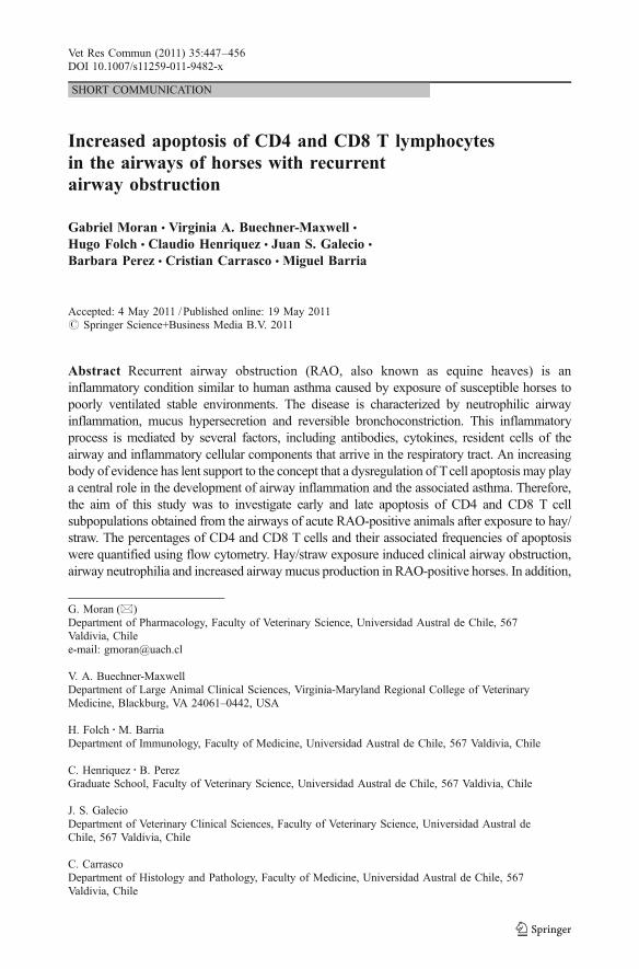
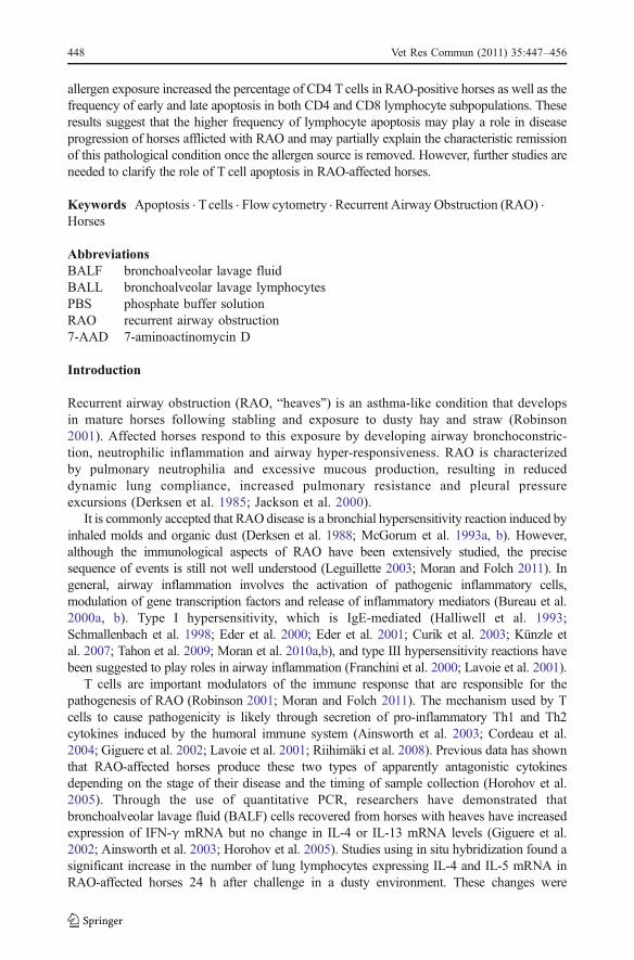
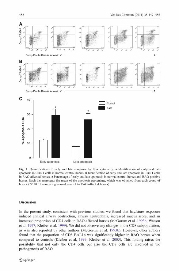
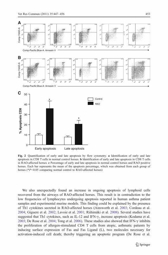
The frequencies of early and late apoptosis in CD4 cells of the RAO-positive animalsafter hay/straw exposure were significantly higher compared with those of the healthycontrols (Fig. 1; P=0.005 and 0.002, respectively). In the RAO-affected horses, themean frequencies of early and late apoptosis were 29.36% and 26.39%, respectively,whereas in the healthy horses, the mean frequencies of early and late apoptosis were12.21% and 7.6%, respectively. Similar results were obtained for CD8 cells. Thefrequencies of both early and late apoptosis in the CD8 cells of the RAO-affected horseswere significantly higher compared with that of normal horses (Fig. 2; P=0.009 and0.01, respectively). In the RAO-positive horses, the mean frequencies of early and lateapoptosis in CD8 cells were 35.75% and 20.46%, respectively, while the meanfrequencies of apoptosis in the control horses were 9.79% of CD8 cells in earlyapoptosis, and 7.76% in late apoptosis.
Vet Res Commun (2011) 35:447–456 451
Discussion
In the present study, consistent with previous studies, we found that hay/straw exposureinduced clinical airway obstruction, airway neutrophilia, increased mucus score, and anincreased proportion of CD4 cells in RAO-affected horses (McGorum et al. 1993b; Watsonet al. 1997; Kleiber et al. 1999). We did not observe any changes in the CD8 subpopulation,as was also reported by other authors (McGorum et al. 1993b). However, other authorsfound that the proportion of CD8 BALLs was significantly higher in RAO horses whencompared to controls (Kleiber et al. 1999; Kleiber et al. 2005). This finding raises thepossibility that not only the CD4 cells but also the CD8 cells are involved in thepathogenesis of RAO.
Comp-Pacific Blue-A: Annexin V
A
B
Com
p-7A
AD
-AC
omp-
7AA
D-A
Comp-Pacific Blue-A: Annexin V
C
Early apoptosis Late apoptosis0
10
20
30
40Control
RAO**
% A
po
pto
sis
CD
4
Fig. 1 Quantification of early and late apoptosis by flow cytometry. a Identification of early and lateapoptosis in CD4 T cells in normal control horses. b Identification of early and late apoptosis in CD4 T cellsin RAO-affected horses. c Percentage of early and late apoptosis in normal control horses and RAO positivehorses. Each bar represents the mean of the apoptosis percentage, which was obtained from each group ofhorses (*P<0.01 comparing normal control to RAO-affected horses)
452 Vet Res Commun (2011) 35:447–456
We also unexpectedly found an increase in ongoing apoptosis of lymphoid cellsrecovered from the airways of RAO-affected horses. This result is in contradiction to thelow frequencies of lymphocytes undergoing apoptosis reported in human asthma patientsamples and experimental murine models. This finding could be explained by the presenceof Th1 cytokines secreted in RAO-affected horses (Ainsworth et al. 2003; Cordeau et al.2004; Giguere et al. 2002; Lavoie et al. 2001; Riihimäki et al. 2008). Several studies havesuggested that Th1 cytokines, such as IL-12 and IFN-γ, increase apoptosis (Kodama et al.2003; De Rose et al. 2004; Tong et al. 2006). These studies also showed that IFN-γ inhibitsthe proliferation of allergen-stimulated CD4 T cells from atopic, asthmatic patients byinducing surface expression of Fas and Fas Ligand (L), two molecules necessary foractivation-induced cell death; thereby triggering an apoptotic program (De Rose et al.
Comp-Pacific Blue-A: Annexin V
A
B
Com
p-7A
AD
-AC
omp-
7AA
D-A
Comp-Pacific Blue-A: Annexin V
C
Early apoptosis Late apoptosis0
10
20
30
40
50
Control
RAO*
*
% A
po
pto
sis
CD
8
Fig. 2 Quantification of early and late apoptosis by flow cytometry. a Identification of early and lateapoptosis in CD8 T cells in normal control horses. b Identification of early and late apoptosis in CD8 T cellsin RAO-affected horses. c Percentage of early and late apoptosis in normal control horses and RAO positivehorses. Each bar represents the mean of the apoptosis percentage, which was obtained from each group ofhorses (*P<0.05 comparing normal control to RAO-affected horses)
Vet Res Commun (2011) 35:447–456 453
2004). These results are consistent with those obtained by Tong et al. (2006) in a murinemodel of asthma. Similarly, Kodama et al. (2003) suggested that administration of IL-12 isprotective in a Th2-dependent model of allergic airway inflammation, at least partly viaenhanced apoptosis of CD4 T cells. However, the apoptotic phenomenon presented in ourstudy was in the setting of acute RAO. Therefore, more studies are needed to examineapoptosis and cytokine profiles in other stages of the disease.
Apoptosis has emerged as a major mechanism in the clearance of activated T cellsduring the resolution of an inflammatory response (Akbar and Salmon 1997).Inadequate T cell apoptosis in asthma patients appears to interfere with normal T cellelimination, resulting in T cell accumulation, which contributes to chronic inflammationand may be the major underlying cause for tissue damage, remodeling and repair (Mülleret al. 2006; Vignola et al. 2000). Spinozzi et al. (1998) reported that pulmonary T cellsisolated from BALF of atopic asthma patients showed hypoexpression of Fas and FasL;this result may explain the low frequency of apoptosis in this group of patients. Inaddition, basal levels of apoptotic activity were significantly lower in BALL fromasthmatic subjects compared with peripheral blood lymphocytes from the same subjects.These data indicate that airway inflammation in asthma is associated with a reducedsusceptibility to apoptosis, which may lead to enhanced survival of lymphocytes in thebronchial mucosa and prolonged inflammation (Müller et al. 2006). Other molecules areinvolved in the programmed cell death process, including members of the Bcl-2 genefamily, which are known to inhibit apoptosis. Studies have shown that Bcl-2 expression isincreased in lymphoid cells obtained from the airways of asthmatic patients and thatneutralization of IL-10, an important inducer of Bcl-2, decreases Bcl-2 expression andapoptosis of cells from the respiratory tract of asthmatic patients (Hamzaoui et al. 1999a;Hamzaoui et al. 1999b). Because RAO in horses is a condition similar to human asthma,these apoptotic mechanisms might have an important role in the inflammatory processesdescribed above. Identifying the cellular and molecular mechanisms underlying thereduction of inflammatory cell apoptosis will be crucial for new and effective therapeuticstrategies in RAO-affected horses.
In summary, dysregulated apoptosis may play an important role in the pathogenesis ofallergic airway inflammation. Our study shows that acute RAO-affected horses haveincreased apoptosis of airway lymphocytes, which may partially explain the rapidresolution of this pathology once the allergen is removed. Interestingly, this process is incontrast to human asthma, where such cell clearance mechanisms are reduced, leading tochronic inflammation. To that end, further studies will be required to clarify the role of Tcell apoptosis in RAO-affected horses.
Acknowledgements This work was supported by Center for Sciences and Global Sustainability (VirginiaTech/UACh) and FONDECYT Nº 11100196 (Conicyt- Chilean Government).
Conflict of interest statement None of the authors have any financial or personal relationships that couldinappropriately influence or bias the content of the paper.
References
Ainsworth DM, Grunig G, Matychak MB, Young J, Wagner B, Erb HN, Antczak DF (2003) Recurrentairway obstruction (RAO) in horses is characterized by IFN-gamma and IL-8 production inbronchoalveolar lavage cells. Vet Immunol Immunopathol 96:83–91
454 Vet Res Commun (2011) 35:447–456
Akbar AN, Salmon M (1997) Cellular environments and apoptosis: tissuemicroenvironments controlactivated T cell death. Immunol Today 18:726–6
Bureau F, Bonizzi G, Kirschvink N, Delhalle S, Desmecht D, Merville MP, Bours V, Lekeux P (2000a)Correlation between nuclear factor-κB activity in bronchial brushing samples and lung dysfunction in ananimal model of asthma. Am J Respir Crit Care Med 161:1314–1321
Bureau F, Delhalle S, Bonizzi G, Fievez L, Dogne S, Kirschvink N, Vanderplasschen A, Merville MP, BoursV, Lekeux P (2000b) Mechanisms of persistent NF- κB activity in the bronchi of an animal model ofasthma. J Immunol 165:5822–5830
Cohen JJ (1999) Apoptosis: mechanisms of life and death in the immune system. J Allergy Clin Immunol103:548–554
Cordeau ME, Joubert P, Dewachi O, Hamid Q, Lavoie JP (2004) IL-4, IL-5 and IFN-gamma mRNAexpression in pulmonary lymphocytes in equine heaves. Vet Immunol Immunopathol 97:87–96
Cormican L, O’Sullivan S, Burke CM, Poulter LW (2001) IFNgamma but not IL-4 T cells of the asthmaticbronchial wall show increased incidence of apoptosis. Clin Exp Allergy 31:731–739
Curik I, Fraser D, Eder C, Achmann R, Swinburne J, Binns M, Crameri R, Brem G, Sölkner J, Marti E(2003) Association between MHC gene region and variation of serum IgE levels against specific mouldallergens in the horse. Genet Sel Evol 35:117–190
De Rose V, Cappello P, Sorbello V, Ceccarini B, Gani F, Bosticardo M, Fassio S, Novelli F (2004) IFN-gamma inhibits the proliferation of allergen-activated T lymphocytes from atopic, asthmatic patients byinducing Fas/FasL-mediated apoptosis. J Leukoc Biol 76:423–432
Derksen FJ, Robinson NE, Armstrong PJ, Slocombe RF (1985) Airway reactivity in ponies with recurrentairway obstruction (“heaves”). J Appl Physiol 58:598–604
Derksen FJ, Robinson NE, Scott JS, Stick JA (1988) Aerosolized Micropolyspora faeni antigen as acause of pulmonary dysfunction in ponies with recurrent airway obstruction (heaves). Am J Vet Res49:933–938
Eder C, Crameri R, Mayer C, Eicher R, Straub R, Gerber H, Lazary S, Marti E (2000) Allergen-specific IgElevels against crude mould and storage mite extracts and recombinant mould allergens in sera fromhorses affected with chronic bronchitis. Vet Immunol Immunopathol 73:241–253
Eder C, Curik I, Brem G, Crameri R, Bodo I, Habe F, Lazary S, Sölkner J, Marti E (2001) Influence ofenvironmental and genetic factors on allergen-specific immunoglobulin E levels in sera from Lipizzanhorses. Equine Vet J 33:714–720
Finotto S, Eigenbrod T, Karwot R, Boross I, Doganci A, Ito H, Nishimoto N, Yoshizaki K, Kishimoto T,Rose-John S, Galle PR, Neurath MF (2007) Local blockade of IL-6R signaling induces lung CD4+ Tcell apoptosis in a murine model of asthma via regulatory T cells. Int Immunol 19:685–693
Franchini M, Gill U, Von Fellenberg R, Bracher VV (2000) Interleukin-8 concentration and neutrophilchemotactic activity in bronchoalveolar lavage fluid of horses with chronic obstructive pulmonarydisease following exposure to hay. Am J Vet Res 11:1369–1374
Gerber V, Lindberg A, Berney C, Robinson NE (2004) Airway mucus in recurrent airway obstruction-short-term response to environmental challenge. J Vet Intern Med 18:92–97
Giguere S, Viel L, Lee E, MacKay RJ, Hernandez J, Franchini M (2002) Cytokine induction in pulmonaryairways of horses with heaves and effect of therapy with inhaled fluticasone propionate. Vet ImmunolImmunopathol 85:147–158
Halliwell RE, McGorum BC, Irving P, Dixon PM (1993) Local and systemic antibody production in horsesaffected with chronic obstructive pulmonary disease. Vet Immunol Immunopathol 38:201–215
Hamzaoui A, Hamzaoui K, Salah H, Chabbou A (1999a) Lymphocytes apoptosis in patients with acuteexacerbation of asthma. Mediat Inflamm 8:237–243
Hamzaoui K, Hamzaoui A, Zakraoui L, Chabbou A (1999b) Expression of Bcl-2 in inflammatory sites frompatients with active Behçet’s disease. Mediat Inflamm 8:101–106
Horohov DW, Beadle RE, Mouch S, Pourciau SS (2005) Temporal regulation of cytokine mRNA expressionin equine recurrent airway obstruction. Vet Immunol Immunopathol 108:237–245
Jackson CA, Berney C, Jefcoat AM, Robinson NE (2000) Environmen and prednisone interactions in thetreatment of recurrent airway obstruction (heaves). Equine Vet J 32:432–438
Jayaraman S, Castro M, O’Sullivan M, Bragdon MJ, Holtzman MJ (1999) Resistance to Fas-mediated T cellapoptosis in asthma. J Immunol 162:1717–1722
Kleiber C, Grünig G, Jungi T, Schmucker N, Gerber H, Davis WC, Straub R (1999) Phenotypic analysis ofbronchoalveolar lavage fluid lymphocytes in horses with chronic pulmonary disease. ZentralblVeterinärmed A 46:177–184
Kleiber C, McGorum BC, Horohov DW, Pirie RS, Zurbriggen A, Straub R (2005) Cytokine profiles ofperipheral blood and airway CD4 and CD8 T lymphocytes in horses with recurrent airway obstruction.Vet Immunol Immunopathol 104:91–97
Vet Res Commun (2011) 35:447–456 455
Kodama T, Kuribayashi K, Nakamura H, Fujita M, Fujita T, Takeda K, Dakhama A, Gelfand EW,Matsuyama T, Kitada O (2003) Role of interleukin-12 in the regulation of CD4+ T cell apoptosis in amouse model of asthma. Clin Exp Immunol 131:199–205
Künzle F, Gerber V, van der Haegen A, Wampfler B, Straub R, Marti E (2007) IgE-bearing cells inbronchoalveolar lavage fluid and allergen-specific IgE levels in sera from RAO-affected horses. J VetMed A Physiol Pathol Clin Med 54:40–47
Lavoie JP, Maghni K, Desnoyers M, Taha R, Martin JG, Hamid QA (2001) Neutrophilic airwayinflammation in horses with heaves is characterized by a Th2-type cytokine profile. Am J Respir CritCare Med 164:1410–1413
Leguillette R (2003) Recurrent airway obstruction-heaves. Vet Clin Equine Prac 19:63–68McGorum BC, Dixon PM, Halliwell RE (1993a) Evaluation of intradermal mould antigen in the diagnosis of
equine chronic obstructive pulmonary disease. Equine Vet J 25:273–275McGorum BC, Dixon PM, Halliwell RE (1993b) Phenotipic analysis of peripheral blood and
brocheoalveolar lavage fluid lymphocytes in control and chronic obstructive pulmonary disease affectedhorses, before and after natural (hay and straw) challenges. Vet Immunol Immunophatol 36:207–222
Morán G, Folch H (2011) Recurrent airway obstruction in horses – an allergic inflammation: a review. VetMed 56:1–13
Morán G, Burgos R, Araya O, Folch H (2010a) In vitro bioassay to detect reaginic antibodies from the serumof horses affected with recurrent airway obstruction. Vet Res Commun 34:91–99
Morán G, Folch H, Burgos R, Araya O, Barria M (2010b) Detection of reaginic antibodies against Faeniarectivirgula from the serum of horses affected with recurrent airway obstruction by an in vitro bioassay.Vet Res Commun 34:719–726
Müller M, Grunewald J, Olgart Höglund C, Dahlén B, Eklund A, Stridh H (2006) Altered apoptosis inbronchoalveolar lavage lymphocytes after allergen exposure of atopic asthmatic subjects. Eur Respir J28:513–522
Pietra M, Peli A, Bonato A, Ducci A, Cinotti S (2007) Equine bronchoalveolar lavage cytokines in thedevelopment of recurrent airway obstruction. Vet Res Commun 31:313–316
Riihimäki M, Raine A, Art T, Lekeux P, Couëtil L, Pringle J (2008) Partial divergence of cytokine mRNAexpression in bronchial tissues compared to bronchoalveolar lavage cells in horses with recurrent airwayobstruction. Vet Immunol Immunopathol 122:256–264
Robinson NE (2001) International workshop on equine chronic airway disease Michigan State University.Equine Vet J 33:5–19
Robinson NE, Olszewski MA, Boehler D (2000) Relationship between clinical signs and lung function inhorses with recurrent airway obstruction (heaves) during a bronchodilator trial. Equine Vet J 32:393–400
Schmallenbach KH, Rahman I, Sasse HH, Dixon PM, Halliwell RE, McGorum BC, Miller HR (1998)Studies on pulmonary and systemic Aspergillus Fumigatus-specific IgE and IgG antibodies in horsesaffected with chronic obstructive pulmonary disease (COPD). Vet Immunol Immunopathol 66:245–256
Spinozzi F, Fizzotti M, Agea E, Piattoni S, Droetto S, Russano A, Forenza N, Bassotti G, Grignani F,Bertotto A (1998) Defective expression of Fas messenger RNA and FAS receptor on pulmonary T cellsfrom patients with asthma. Ann Intern Med 128:363–369
Tahon L, Baselgia S, Gerber V, Doherr MG, Straub R, Robinson NE, Marti E (2009) In vitro allergy testcompared to intradermal testing in horses with recurrent airway obstruction. Vet Immunol Immunopathol127:85–93
Thompson CB (1995) Apoptosis in the pathogenesis and treatment of disease. Science 267:1456–1462Tong J, Bandulwala HS, Clay BS, Anders RA, Shilling RA, Balachandran DD, Chen B, Weinstock JV,
Solway J, Hamann KJ, Sperling AI (2006) Fas-positive T cells regulate the resolution of airwayinflammation in a murine model of asthma. J Exp Med 203:1173–1184
Vignola AM, Chanez P, Chiappara G, Siena L, Merendino A, Reina C, Gagliardo R, Profita M, Bousquet J,Bonsignore G (1999) Evaluation of apoptosis of eosinophils, macrophages, and T lymphocytes inmucosal biopsy specimens of patients with asthma and chronic bronchitis. J Allergy Clin Immunol103:563–573
Vignola AM, Chiappara G, Gagliardo R, Gjomarkaj M, Merendino A, Siena L, Bousquet J, Bonsignore G(2000) Apoptosis and airway inflammation in asthma. Apoptosis 5:473–485
Watson JL, Stott JL, Blanchard MT, Lavoie JP, Wilson, Gershwin LJ, Wilson DW (1997) Phenotipiccharacterization of lymphocyte subpopulations in horses affected with chronic obstructive pulmonarydisease and in normal controls. Vet Pathol 34:108–116
Woolley KL, Gibson PG, Carty K, Wilson AJ, Twaddell SH, Woolley MJ (1996) Eosinophil apoptosis andthe resolution of airway inflammation in asthma. Am J Respir Crit Care Med 154:237–243
456 Vet Res Commun (2011) 35:447–456