Characterisation of gut, lung, and upper airways microbiota in ...
13
HAL Id: hal-02388057 https://hal.uca.fr/hal-02388057 Submitted on 30 Nov 2019 HAL is a multi-disciplinary open access archive for the deposit and dissemination of sci- entific research documents, whether they are pub- lished or not. The documents may come from teaching and research institutions in France or abroad, or from public or private research centers. L’archive ouverte pluridisciplinaire HAL, est destinée au dépôt et à la diffusion de documents scientifiques de niveau recherche, publiés ou non, émanant des établissements d’enseignement et de recherche français ou étrangers, des laboratoires publics ou privés. Characterisation of gut, lung, and upper airways microbiota in patients with non-small cell lung carcinoma Study protocol for case-control observational trial Study Protocol Clinical Trial Rea Bingula, Marc Filaire, Nina Radosevic-Robin, Jean-Yves Berthon, Annick Bernalier-Donadille, Marie-Paule Vasson, Emilie Thivat, Fabrice Kwiatkowski, Edith Filaire To cite this version: Rea Bingula, Marc Filaire, Nina Radosevic-Robin, Jean-Yves Berthon, Annick Bernalier-Donadille, et al.. Characterisation of gut, lung, and upper airways microbiota in patients with non-small cell lung carcinoma Study protocol for case-control observational trial Study Protocol Clinical Trial. Medicine, Lippincott, Williams & Wilkins, 2018, 97 (50), pp.e13676. 10.1097/MD.0000000000013676. hal- 02388057
-
Upload
khangminh22 -
Category
Documents
-
view
1 -
download
0
Transcript of Characterisation of gut, lung, and upper airways microbiota in ...

HAL Id: hal-02388057https://hal.uca.fr/hal-02388057
Submitted on 30 Nov 2019
HAL is a multi-disciplinary open accessarchive for the deposit and dissemination of sci-entific research documents, whether they are pub-lished or not. The documents may come fromteaching and research institutions in France orabroad, or from public or private research centers.
L’archive ouverte pluridisciplinaire HAL, estdestinée au dépôt et à la diffusion de documentsscientifiques de niveau recherche, publiés ou non,émanant des établissements d’enseignement et derecherche français ou étrangers, des laboratoirespublics ou privés.
Characterisation of gut, lung, and upper airwaysmicrobiota in patients with non-small cell lung
carcinoma Study protocol for case-control observationaltrial Study Protocol Clinical Trial
Rea Bingula, Marc Filaire, Nina Radosevic-Robin, Jean-Yves Berthon, AnnickBernalier-Donadille, Marie-Paule Vasson, Emilie Thivat, Fabrice Kwiatkowski,
Edith Filaire
To cite this version:Rea Bingula, Marc Filaire, Nina Radosevic-Robin, Jean-Yves Berthon, Annick Bernalier-Donadille, etal.. Characterisation of gut, lung, and upper airways microbiota in patients with non-small cell lungcarcinoma Study protocol for case-control observational trial Study Protocol Clinical Trial. Medicine,Lippincott, Williams & Wilkins, 2018, 97 (50), pp.e13676. �10.1097/MD.0000000000013676�. �hal-02388057�

Dow
nloadedfrom
https://journals.lww.com
/md-journalby
BhDMf5ePH
Kav1zEoum1tQ
fN4a+kJLhEZgbsIH
o4XMi0hC
ywCX1AW
nYQp/IlQ
rHD3YeLEAQ
tdJ82csOMQL965C
Mulzzm
3SajIxNyVltrf68w
Z8cYBr8nUBQ
==on
12/26/2018
Downloadedfromhttps://journals.lww.com/md-journalbyBhDMf5ePHKav1zEoum1tQfN4a+kJLhEZgbsIHo4XMi0hCywCX1AWnYQp/IlQrHD3YeLEAQtdJ82csOMQL965CMulzzm3SajIxNyVltrf68wZ8cYBr8nUBQ==on12/26/2018
Characterisation of gut, lung, and upper airwaysmicrobiota in patients with non-small cell lungcarcinomaStudy protocol for case-control observational trialRea Bingula, MSa,∗, Marc Filaire, MDa,b, Nina Radosevic-Robin, MDc, Jean-Yves Berthon, PhDd,Annick Bernalier-Donadille, PhDe, Marie-Paule Vasson, PhDa,f, Emilie Thivat, PhDg,h,Fabrice Kwiatkowski, MSg,h, Edith Filaire, PhDa,d
AbstractBackground:Several studies have confirmed the important role of the gut microbiota in the regulation of immune functions and itscorrelation with different diseases, including cancer. While brain-gut and liver-gut axes have already been demonstrated, theexistence of a lung-gut axis has been suggested more recently, with the idea that changes in the gut microbiota could affect the lungmicrobiota, and vice versa. Likewise, the close connection between gut microbiota and cancer of proximal sites (intestines, kidneys,liver, etc.) is already well established. However, little is known whether there is a similar relation when looking at world’s number onecause of death from cancer—lung cancer.
Objective: Firstly, this study aims to characterise the gut, lung, and upper airways (UAs) microbiota in patients with non-small celllung cancer (NSCLC) treated with surgery or neoadjuvant chemotherapy plus surgery. Secondly, it aims to evaluate a chemotherapyeffect on site-specificmicrobiota and its influence on immune profile. To our knowledge, this is the 1st study that will analysemulti-sitemicrobiota in NSCLC patients along with site-specific immune response.
Methods: The study is a case-controlled observational trial. Forty NSCLC patients will be divided into 2 groups depending on theiranamnesis: Pchir, patients eligible for surgery, or Pct-chir, patients eligible for neoadjuvant chemotherapy plus surgery. Compositionof the UAs (saliva), gut (faeces), and lung microbiota (from broncho-alveolar lavage fluid (BALF) and 3 lung pieces: “healthy” tissuedistal to tumour, peritumoural tissue and tumour itself) will be analysed in both groups. Immune properties will be evaluated on thelocal (evaluation of the tumour immune cell infiltrate, tumour classification and properties, immune cell phenotyping in BALF; humanneutrophil protein (HNP) 1–3, b-defensin 2, and calprotectin in faeces) and systemic level (blood cytokine and immune cell profile).Short-chain fatty acids (SCFAs) (major products of bacterial fermentation with an effect on immune system) will be dosed in faecalsamples. Other factors such as nutrition and smoking status will be recorded for each patient. We hypothesise that smoking statusand tumour type/grade will be major factors influencing both microbiota and immune/inflammatory profile of all sampling sites.Furthermore, due to non-selectivity, the same effect is expected from chemotherapy.
Abbreviations: ANSM = The French National Agency for Medicines and Health Products Safety (Agence nationale de sécurité dumédicament et des produits de santé), BAL = broncho-alveolar lavage, BALF = broncho-alveolar lavage fluid, BMI = body massindex, CIFRE = Industrial Research Training Agreements grant (Convention industrielle de formation par la recherché), CRP = C-reactive protein, CT = chemotherapy, ELISA= enzyme-linked immunosorbent assay, FDR= false discovery rate, GF = germ-free, GI= gastrointestinal, HBSS = Hank’s balanced salt solution, HNP = human neutrophil peptide, ICI = immune checkpoint inhibitor, IL =interleukin, MCLB = mammalian cell lysis buffer, NSCLC = non-small cell lung cancer, OTU = outer taxonomic unit, Pchir = patientsurgery, fr. «patient chirurgie», Pct-chir = patients chemotherapy plus surgery, fr. «patient chimiothérapie —chirurgie», qPCR =quantitative polymerase chain reaction, SCFAs = short-chain fatty acids, UAs = upper airways.
Keywords: chemotherapy, gut-lung axis, immune response, microbiota, non-small cell lung cancer, tumour microenvironment
Funding: This work is financially supported by the Auvergne Region, European Regional Development Fund Industrial Research Training Agreements (CIFRE) grant. Thestudy sponsor is Jean Perrin Center, Clermont-Ferrand, France. Collection, management or analysis and data interpretation, decision to publish, or preparation of themanuscript is independent of funding institutions.
The authors have no conflicts of interest to disclose.a University of Clermont-Auvergne, UMR 1019 INRA-UCA, Human Nutrition Unit (UNH), Clermont-Ferrand, bCentre Jean Perrin, Thoracic Surgery Department,Clermont-Ferrand, c INSERM U1240, University Clermont Auvergne, Centre Jean Perrin, Department of Pathology, Clermont-Ferrand, dGreentech SA, BiopoleClermont-Limagne, Saint-Beauzire, e UMR0454 MEDIS, INRA/UCA, Saint-Genes-Champanelle, f Centre Jean Perrin, CHU Gabriel-Montpied, Clinical Nutrition Unit,Clermont-Ferrand, gUniversity of Clermont-Auvergne, INSERM U1240 Imagerie Moléculaire et Stratégies Théranostiques, Clermont-Ferrand, hCentre Jean Perrin,Clinical Research Department, Clermont-Ferrand, France.∗Correspondence: Rea Bingula, University of Clermont-Auvergne, UMR 1019 INRA-UCA, Human Nutrition Unit (UNH), Clermont-Ferrand, France (e-mail: [email protected]).
Copyright © 2018 the Author(s). Published by Wolters Kluwer Health, Inc.This is an open access article distributed under the Creative Commons Attribution License 4.0 (CCBY), which permits unrestricted use, distribution, and reproduction inany medium, provided the original work is properly cited.
Medicine (2018) 97:50(e13676)
Received: 21 November 2018 / Accepted: 22 November 2018
http://dx.doi.org/10.1097/MD.0000000000013676
Study Protocol Clinical Trial Medicine®
OPEN
1
1. Introduction
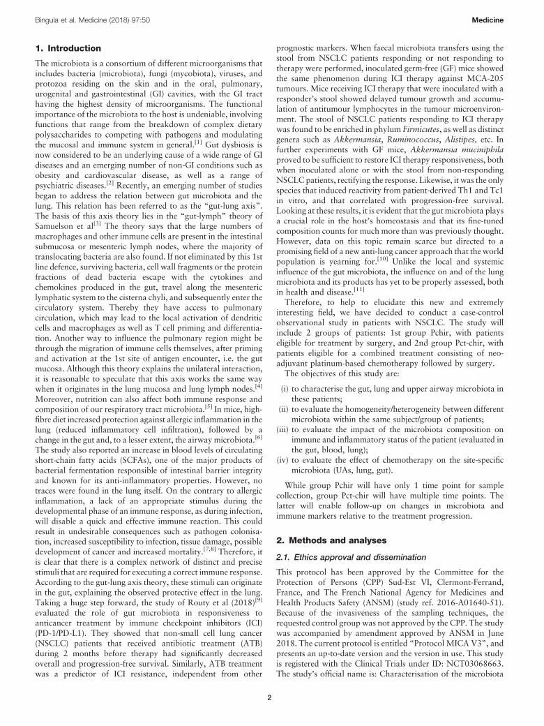
The microbiota is a consortium of different microorganisms thatincludes bacteria (microbiota), fungi (mycobiota), viruses, andprotozoa residing on the skin and in the oral, pulmonary,urogenital and gastrointestinal (GI) cavities, with the GI tracthaving the highest density of microorganisms. The functionalimportance of the microbiota to the host is undeniable, involvingfunctions that range from the breakdown of complex dietarypolysaccharides to competing with pathogens and modulatingthe mucosal and immune system in general.[1] Gut dysbiosis isnow considered to be an underlying cause of a wide range of GIdiseases and an emerging number of non-GI conditions such asobesity and cardiovascular disease, as well as a range ofpsychiatric diseases.[2] Recently, an emerging number of studiesbegan to address the relation between gut microbiota and thelung. This relation has been referred to as the “gut-lung axis”.The basis of this axis theory lies in the “gut-lymph” theory ofSamuelson et al[3] The theory says that the large numbers ofmacrophages and other immune cells are present in the intestinalsubmucosa or mesenteric lymph nodes, where the majority oftranslocating bacteria are also found. If not eliminated by this 1stline defence, surviving bacteria, cell wall fragments or the proteinfractions of dead bacteria escape with the cytokines andchemokines produced in the gut, travel along the mesentericlymphatic system to the cisterna chyli, and subsequently enter thecirculatory system. Thereby they have access to pulmonarycirculation, which may lead to the local activation of dendriticcells and macrophages as well as T cell priming and differentia-tion. Another way to influence the pulmonary region might bethrough the migration of immune cells themselves, after primingand activation at the 1st site of antigen encounter, i.e. the gutmucosa. Although this theory explains the unilateral interaction,it is reasonable to speculate that this axis works the same waywhen it originates in the lung mucosa and lung lymph nodes.[4]
Moreover, nutrition can also affect both immune response andcomposition of our respiratory tract microbiota.[5] In mice, high-fibre diet increased protection against allergic inflammation in thelung (reduced inflammatory cell infiltration), followed by achange in the gut and, to a lesser extent, the airway microbiota.[6]
The study also reported an increase in blood levels of circulatingshort-chain fatty acids (SCFAs), one of the major products ofbacterial fermentation responsible of intestinal barrier integrityand known for its anti-inflammatory properties. However, notraces were found in the lung itself. On the contrary to allergicinflammation, a lack of an appropriate stimulus during thedevelopmental phase of an immune response, as during infection,will disable a quick and effective immune reaction. This couldresult in undesirable consequences such as pathogen colonisa-tion, increased susceptibility to infection, tissue damage, possibledevelopment of cancer and increased mortality.[7,8] Therefore, itis clear that there is a complex network of distinct and precisestimuli that are required for executing a correct immune response.According to the gut-lung axis theory, these stimuli can originatein the gut, explaining the observed protective effect in the lung.Taking a huge step forward, the study of Routy et al (2018)[9]
evaluated the role of gut microbiota in responsiveness toanticancer treatment by immune checkpoint inhibitors (ICI)(PD-1/PD-L1). They showed that non-small cell lung cancer(NSCLC) patients that received antibiotic treatment (ATB)during 2 months before therapy had significantly decreasedoverall and progression-free survival. Similarly, ATB treatmentwas a predictor of ICI resistance, independent from other
prognostic markers. When faecal microbiota transfers using thestool from NSCLC patients responding or not responding totherapy were performed, inoculated germ-free (GF) mice showedthe same phenomenon during ICI therapy against MCA-205tumours. Mice receiving ICI therapy that were inoculated with aresponder’s stool showed delayed tumour growth and accumu-lation of antitumour lymphocytes in the tumour microenviron-ment. The stool of NSCLC patients responding to ICI therapywas found to be enriched in phylum Firmicutes, as well as distinctgenera such as Akkermansia, Ruminococcus, Alistipes, etc. Infurther experiments with GF mice, Akkermansia muciniphilaproved to be sufficient to restore ICI therapy responsiveness, bothwhen inoculated alone or with the stool from non-respondingNSCLC patients, rectifying the response. Likewise, it was the onlyspecies that induced reactivity from patient-derived Th1 and Tc1in vitro, and that correlated with progression-free survival.Looking at these results, it is evident that the gut microbiota playsa crucial role in the host’s homeostasis and that its fine-tunedcomposition counts for much more than was previously thought.However, data on this topic remain scarce but directed to apromising field of a new anti-lung cancer approach that the worldpopulation is yearning for.[10] Unlike the local and systemicinfluence of the gut microbiota, the influence on and of the lungmicrobiota and its products has yet to be properly assessed, bothin health and disease.[11]
Therefore, to help to elucidate this new and extremelyinteresting field, we have decided to conduct a case-controlobservational study in patients with NSCLC. The study willinclude 2 groups of patients: 1st group Pchir, with patientseligible for treatment by surgery, and 2nd group Pct-chir, withpatients eligible for a combined treatment consisting of neo-adjuvant platinum-based chemotherapy followed by surgery.The objectives of this study are:
(i) to characterise the gut, lung and upper airway microbiota inthese patients;
(ii) to evaluate the homogeneity/heterogeneity between differentmicrobiota within the same subject/group of patients;
(iii) to evaluate the impact of the microbiota composition onimmune and inflammatory status of the patient (evaluated inthe gut, blood, lung);
(iv) to evaluate the effect of chemotherapy on the site-specificmicrobiota (UAs, lung, gut).
While group Pchir will have only 1 time point for samplecollection, group Pct-chir will have multiple time points. Thelatter will enable follow-up on changes in microbiota andimmune markers relative to the treatment progression.
2. Methods and analyses
2.1. Ethics approval and dissemination
This protocol has been approved by the Committee for theProtection of Persons (CPP) Sud-Est VI, Clermont-Ferrand,France, and The French National Agency for Medicines andHealth Products Safety (ANSM) (study ref. 2016-A01640-51).Because of the invasiveness of the sampling techniques, therequested control group was not approved by the CPP. The studywas accompanied by amendment approved by ANSM in June2018. The current protocol is entitled “Protocol MICA V3”, andpresents an up-to-date version and the version in use. This studyis registered with the Clinical Trials under ID: NCT03068663.The study’s official name is: Characterisation of the microbiota
Bingula et al. Medicine (2018) 97:50 Medicine
2

(gut, lung, and upper airways) in patients with non-small cell lungcarcinoma: exploratory study (acronym: MICA). Writteninformed consent is obtained from all patients before enrolmentin the study. The results are planned for presentation atconferences and publication in peer-reviewed journals in early2019. All samples will be preserved for 15 years according to thepractice of the sponsoring institution (Centre Jean Perrin).Samples will be available to other investigators if they want toperform complementary studies that consider NSCLC afteradditional consent obtained from patient. However, because ofFrench regulations regarding patient information files, patients’data will not be available.
2.2. Study outcomes
As its primary outcome, this study will characterise the lung andUAs microbiota in 2 groups of 20 patients with NSCLC. GroupPchir will include patients eligible for surgery without chemo-therapy. Group Pct-chir will include patients eligible for surgeryafter platinum-based chemotherapy. The UAs microbiota will beevaluated from saliva, while lung microbiota will be evaluatedfrom 3 lung explants: “healthy” lung tissue, tumour andperitumoural tissue; and broncho-alveolar lavage fluid (BALF)from the tumour’s proximity (same lobe). Following bacterialDNA extraction, microbiota will be analysed by qPCR and 16Sribosomal rRNA gene sequencing using the Illumina MiSeqplatform.Secondary outcomes of this study are set as follows:
i) to study the effect of neoadjuvant chemotherapy on micro-biota by evaluating:a. the variation of the proportion of the phylum Firmicutes
(as the phylum is highly represented in all of the differenttypes of samples considered in this study),[4]
b. the variation of the proportion of the bacterial genera perphylum in different types of samples (faeces, saliva, BALF,lung tissue/peritumoural tissue/tumour),
c. the concordance of genera between sample locations (e.g.saliva vs. BALF, healthy lung tissue vs. peritumoural tissuevs. tumour);by qPCR and 16S rRNA gene sequencing;
ii) to study the homogeneity/heterogeneity between lung, upperairways and gut microbiota for each and between both groups(evaluated by 16S rRNA gene sequencing and qPCR) andbetween different time points (group Pct-chir),
iii) to evaluate immune/inflammatory status:a. in the gut: by dosing b-defensin 2, human neutrophil
peptides HNP1-3, calprotectin (ELISA) and SCFAs (gasliquid chromatography)
b. in plasma: by dosing plasmatic cytokines (Luminex), C-reactive protein (CRP) (ELISA) and immune cell pheno-typing (flow cytometry)
c. in the lung: by immune cell phenotyping (flow cytometry)and characterisation of immune infiltrate in lung tumourbiopsies obtained during the operation (by immunohis-tochemistry).
2.3. Patient recruitment
The study pre-considers all patients diagnosed with NSCLC andpresented before the Thoracic Oncologic Committee of the JeanPerrin Centre, Clermont-Ferrand, France. Inclusion criteria arepresented in Table 1. Inclusion in the study is consecutive andparallel for both groups. A written informed consent is obtainedfrom each patient participating in the study before inclusion.Depending on their diagnosis, patients are included in one of the2 groups: Pchir (patients eligible for surgery only), or Pct-chir(patients eligible for surgery after neoadjuvant platinum-basedchemotherapy).
2.4. Patient and public involvement
Neither patients nor the Patient Committee were involved in thedesign of this study. If desired, patients can be informed of thestudy’s results by the investigator or physician.
2.5. Trial design and timeline
This study is a case-control observational trial. Recruitment intothe study started in May 2017 and will end in May 2019. Theestimated complete duration of the study is 29 months, with anaverage follow-up period per patient of 1.5 and 3.5–4.5 monthsfor the Pchir and Pct-chir groups, respectively. Intermediaryanalyses will take place after obtaining all samples from half ofthe patient quota (n = 20) regardless of the group, withoutinterruption of the further recruitment to the study.The trial design is presented in Figure 1. Eligible patients meet
official study personnel at an outpatient appointment (Visit 1)where all the details of the protocol are thoroughly explained. Atthis visit, patients give their written consent to participate in the
Table 1
Inclusion and exclusion criteria for patients.
Inclusion criteria Exclusion criteria
• NSCLC patient with an indication of surgery or neoadjuvant chemotherapy plus surgery • Cognitive difficulties• >18 and <80 years of age • Refusal or inability to give clear consent to participate• BMI <29.9 • Acute digestive or pulmonary infections in the past
2 months (requiring antibiotic treatment)• Not-treated with antibiotics, corticoids or immunosuppressive drugsfor at least the past 2 months • Inflammatory intestinal pathologies
• Signed written consent before enrolment in the study • Colostomy• Affiliated with the Social Security System • Partial or complete gastrectomy
• Previous oesophageal surgery• Previous otorhinolaryngeal cancer treated by radiotherapy or surgery• Inability to conform to the study’s requirements• Deprivation of a right to decide by an administrative or juridical entity• Ongoing participation or participation in another study <1 month ago• Ground-glass opacity
Bingula et al. Medicine (2018) 97:50 www.md-journal.com
3
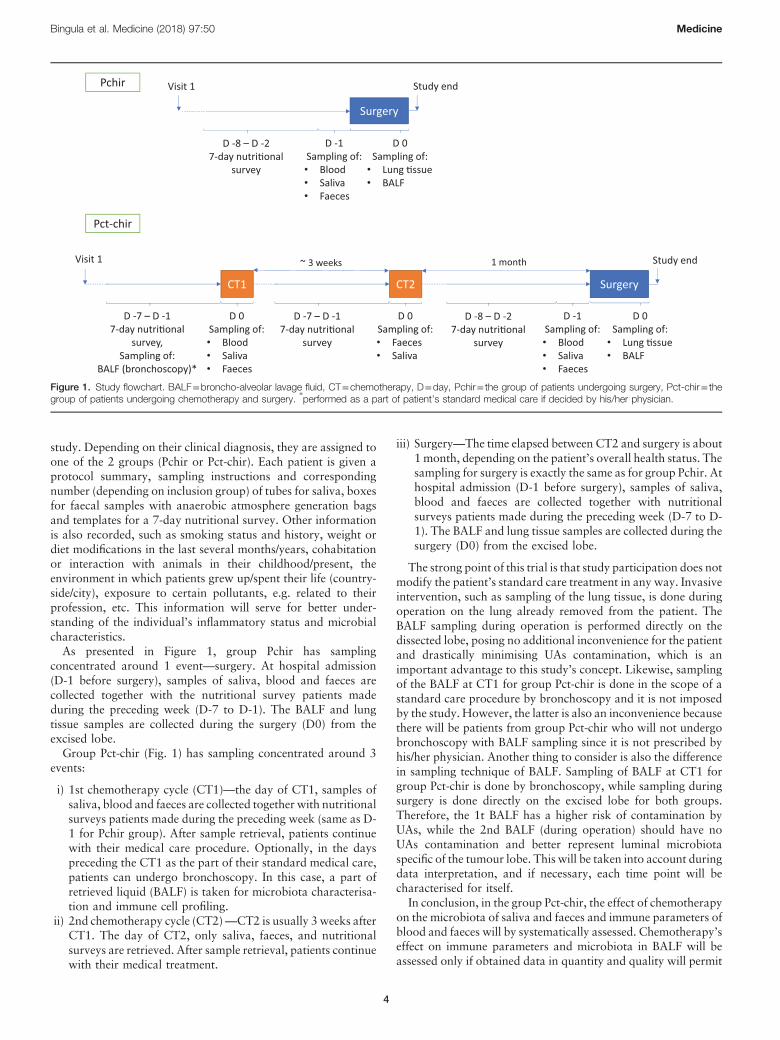
study. Depending on their clinical diagnosis, they are assigned toone of the 2 groups (Pchir or Pct-chir). Each patient is given aprotocol summary, sampling instructions and correspondingnumber (depending on inclusion group) of tubes for saliva, boxesfor faecal samples with anaerobic atmosphere generation bagsand templates for a 7-day nutritional survey. Other informationis also recorded, such as smoking status and history, weight ordiet modifications in the last several months/years, cohabitationor interaction with animals in their childhood/present, theenvironment in which patients grew up/spent their life (country-side/city), exposure to certain pollutants, e.g. related to theirprofession, etc. This information will serve for better under-standing of the individual’s inflammatory status and microbialcharacteristics.As presented in Figure 1, group Pchir has sampling
concentrated around 1 event—surgery. At hospital admission(D-1 before surgery), samples of saliva, blood and faeces arecollected together with the nutritional survey patients madeduring the preceding week (D-7 to D-1). The BALF and lungtissue samples are collected during the surgery (D0) from theexcised lobe.Group Pct-chir (Fig. 1) has sampling concentrated around 3
events:
i) 1st chemotherapy cycle (CT1)—the day of CT1, samples ofsaliva, blood and faeces are collected together with nutritionalsurveys patients made during the preceding week (same as D-1 for Pchir group). After sample retrieval, patients continuewith their medical care procedure. Optionally, in the dayspreceding the CT1 as the part of their standard medical care,patients can undergo bronchoscopy. In this case, a part ofretrieved liquid (BALF) is taken for microbiota characterisa-tion and immune cell profiling.
ii) 2nd chemotherapy cycle (CT2)—CT2 is usually 3 weeks afterCT1. The day of CT2, only saliva, faeces, and nutritionalsurveys are retrieved. After sample retrieval, patients continuewith their medical treatment.
iii) Surgery—The time elapsed between CT2 and surgery is about1 month, depending on the patient’s overall health status. Thesampling for surgery is exactly the same as for group Pchir. Athospital admission (D-1 before surgery), samples of saliva,blood and faeces are collected together with nutritionalsurveys patients made during the preceding week (D-7 to D-1). The BALF and lung tissue samples are collected during thesurgery (D0) from the excised lobe.
The strong point of this trial is that study participation does notmodify the patient’s standard care treatment in any way. Invasiveintervention, such as sampling of the lung tissue, is done duringoperation on the lung already removed from the patient. TheBALF sampling during operation is performed directly on thedissected lobe, posing no additional inconvenience for the patientand drastically minimising UAs contamination, which is animportant advantage to this study’s concept. Likewise, samplingof the BALF at CT1 for group Pct-chir is done in the scope of astandard care procedure by bronchoscopy and it is not imposedby the study. However, the latter is also an inconvenience becausethere will be patients from group Pct-chir who will not undergobronchoscopy with BALF sampling since it is not prescribed byhis/her physician. Another thing to consider is also the differencein sampling technique of BALF. Sampling of BALF at CT1 forgroup Pct-chir is done by bronchoscopy, while sampling duringsurgery is done directly on the excised lobe for both groups.Therefore, the 1t BALF has a higher risk of contamination byUAs, while the 2nd BALF (during operation) should have noUAs contamination and better represent luminal microbiotaspecific of the tumour lobe. This will be taken into account duringdata interpretation, and if necessary, each time point will becharacterised for itself.In conclusion, in the group Pct-chir, the effect of chemotherapy
on the microbiota of saliva and faeces and immune parameters ofblood and faeces will by systematically assessed. Chemotherapy’seffect on immune parameters and microbiota in BALF will beassessed only if obtained data in quantity and quality will permit
Surgery
D 0Sampling of:• Lung �ssue• BALF
D -8 – D -27-day nutri�onal
survey
D -1Sampling of:• Blood• Saliva• Faeces
Study endVisit 1Pchir
Surgery
D 0Sampling of:• Lung �ssue• BALF
Visit 1
D -7 – D -17-day nutri�onal
survey,Sampling of:
BALF (bronchoscopy)*
D 0Sampling of:• Blood• Saliva• Faeces
CT1
~ 3 weeks
D -7 – D -17-day nutri�onal
survey
D 0Sampling of:• Faeces • Saliva
CT2
D -8 – D -27-day nutri�onal
survey
D -1Sampling of:• Blood• Saliva• Faeces
1 month Study end
Pct-chir
Figure 1. Study flowchart. BALF=broncho-alveolar lavage fluid, CT=chemotherapy, D=day, Pchir= the group of patients undergoing surgery, Pct-chir= thegroup of patients undergoing chemotherapy and surgery.
∗performed as a part of patient’s standard medical care if decided by his/her physician.
Bingula et al. Medicine (2018) 97:50 Medicine
4

it (as explained above). When talking of lung tissue, “healthy”lung tissue, taken at distance from the tumour, will be consideredas a control[12] tissue for comparison with peritumoural andtumoural tissue, as well as with BALF. Also, characterisation willbe done respective to the tumour type where possible (sufficientpatient number with the same tumour type).A demanded control group for the study was not authorised by
the Ethics Committee, due to the invasiveness or evident inabilityto realise certain sampling steps (bronchoscopy, lung tissuesampling). Therefore, the focus will be on characterisation of thesite-related microbiota and its connection to local and systemicimmunity in NSCLC respective of the treatment group (Pchir orPct-chir). Likewise, the question is whether there is an initialdifference between group Pchir and group Pct-chir (before CT1),and what its nature is. Equally, how does chemotherapy modifygroup Pct-chir, and whether it becomes more alike or differentfrom group Pchir regarding its different properties (microbialtaxa ratios, inflammatory properties) when followed in time(fromCT1 to surgery). Furthermore, obtained results consideringmicrobiota composition and abundance, where possible, willaddress similar studies[13–16] only in a descriptive matter and thesame will be done with immune parameters.[17–21]
3. Sampling and data recording
3.1. Nutritional survey
The dietary habits are evaluated for each patient for the 7 dayspreceding chemotherapy (Pct-chir) and/or surgery (Pchir and Pct-chir). At Visit 1 (see Fig. 1), all participants receive a detailedverbal explanation, written instructions and the survey with anexample. They are asked to maintain their usual dietary habitsduring the survey period and to record as accurately as possiblethe amount, type and preparation of food and fluid consumed. Ifthey consume commercial and ready meals, they are also asked tonote brand names. The quantity of food or drink can be expressedin either precise measures (weight) or in common householdmeasures, such as cups, tablespoons, etc. In the case of anyquestions or ambiguities, patients are encouraged to contact thestudy personnel. These data will help to estimate each patient’soverall nutritional status and help to explain the microbiologicalanalysis of faecal samples following the survey, as well as thepatient’s immune and inflammatory status. We anticipated thatthe patients might change their dietary habits during thechemotherapy duration, which is why recording was requestedbefore each chemotherapy treatment and surgery, and during 1week. The primary objective is to use what was recorded ascomplementary data to the faecal microbiota analysis (sampledthe day after the end of each survey), to better explain thelongitudinal modification of microbiota, if any. This is due to thefact that faecal microbiota can overcome significant changes inonly a few days relative to a diet change.[22]
3.2. Saliva
At Visit 1, after recording any evidence of oral health problems orinjuries, each patient receives a tube for saliva collection(Sarstedt). It is necessary to fill the tube with a minimum of 1mL of saliva (designated on the tube) on an empty stomach by thepassive drooling method on the morning of hospital admissionfor the chemotherapy session (Pct-chir) and/or surgery (Pchir,Pct-chir) (Fig. 1). The sample is stored at�80 °C for later bacterialDNA extraction and metagenomic sequencing.
3.3. Blood
Blood sampling is done following the patient’s admission tohospital, in the day/hours preceding the prescribed clinicaltreatment, depending on the group (Fig. 1). A 10mL of freshblood are collected in EDTA treated tubes, where 500mL ofwhole blood is immediately used for immune cell phenotyping byflow cytometry. The remainder is centrifuged for 10min 2000�gat 4°C to obtain plasma, which is then aliquoted and stored at�80°C for further analyses (cytokine/interleukin analysis byLuminex, CRP dosage by ELISA).
3.4. Faeces
At Visit 1, each patient receives a sampling box, an anaerobicatmosphere generation bag (GENbag anaer, Biomérieux) anddetailed printed instructions on how to handle the samples at his/her home. Faecal samples are collected following the 1-weeknutritional survey (i.e. on the day of chemotherapy before the druginfusion or the day preceding surgery). In brief, sampling is donedirectly into the sampling box, and after removing the protectivefoil and placing the anaerobic atmosphere generation bag in thebox, the box is closed firmly and placed in the cold (+4°C). Patientsare asked if they have the ability to transport the sample in aninsulated bag to preserve cold conditions. If there is no suchpossibility, the study personnel supplies the patient with therequested bag. The sampling should be done within 12hourspreceding the hospital admission and sample retrieval. Therefore,patients are asked to do the sampling the morning of hospitaladmission if possible. If there are any problems, the patient is askedto contact the protocol personnel to ensure that the sample isprocessed in time. On reception, the sample is aliquoted 3�1g forbacterial DNA extraction, and 2�5–10g (depending on avail-ability) for dosage of faecal calprotectin, b-defensin 2, HNP1-3(ELISA) and SCFAs (gas liquid chromatography). Aliquots arestored immediately at �80°C until analysis. Approximately 1g offresh sample is used for bacterial culture of the main functionarygroups of microorganisms (total anaerobic bacteria, mucin-degrading bacteria, lactic acid producing bacteria, sulphate-reducing bacteria and Enterobacteriaceae).
3.5. Lung tissue and BALF
Only for group Pct-chir, BALF is sampled at inclusion to thestudy. This sample is taken as a part of patient’s standard careprotocol if decided by his/her physician, and is not taken as anadditional sample for this study. The BAL is performed by routinebronchoscopy procedure.Sampling of lung tissue and BALF during surgery is performed
for both groups, after partial or complete pneumonectomy. Theremoved lung tissue is placed in a sterile vessel and the tumourposition is determined by palpation. A piece of healthy lung distalto the tumour, with a minimum size of 1cm�1cm�1cm, is thenclamped. The clamp is left in place during the followingprocedure. The stich on the bronchus is cut away and using asterile syringe the lung is inflated through the bronchus. Lavage isperformed by instilling 2�40mL of sterile physiological saline.After each instillation, the maximum amount of liquid inside thebronchus is retrieved (8–10mL in total), poured into a sterile 50mL tube and placed immediately on ice, designated as “BALF”.At the end, the clamped wedge is cut off and designated as“healthy lung”. A slice of the tumour, with a minimal weight of400mg, containing the tumour cross-section is excised alongwith
Bingula et al. Medicine (2018) 97:50 www.md-journal.com
5

peritumoural tissue, after which the 2 are separated based onhistological difference. All tissues are frozen 1st in liquid nitrogenand then placed at �80°C for long-term storage until DNAextraction. The mirror piece of the excised tumour slice is storedin paraffin and later analysed by immunohistochemistry forcharacterisation of tumour infiltrate. A 3mL of BALF areimmediately used for immune cell analysis by flow cytometry andthe remainder is stored at �80°C for later DNA extraction.
4. Methods and analyses
Saliva, “healthy” lung, peritumoural, and tumour tissue, BALFand faecal samples will be used for bacterial DNA extraction,followed by qPCR and 16S rRNA gene sequence analysis toestablish microbial profiles. Fresh faeces samples will be used forbacterial culture, and frozen aliquots for dosage of faecalcalprotectin, b-defensin 2, HNP1-3 (ELISA), and SCFAs (gasliquid chromatography). The BALF and plasma samples will bothbe analysed by flow cytometry (immune cell phenotyping), whileplasma will also be used for cytokines (Luminex) and CRP(ELISA) dosage. Tumour tissue stored in paraffin will be used forthe analysis of immune infiltrate by immunohistochemistry. Eachprocedure is explained in detail in the following sections.
4.1. Nutritional status
Nutritional data for each patient will be analysed using theNutrilog 2.3 software package, a computerised database (Pro-form) that calculates food composition from the French standardreference.[23]
4.2. Cytokines
Stored plasma will be analysed for cytokines corresponding (butnot restricted) to the following profiles: Th1, Th2, Th17, Treg.Samples will be analysed using Luminex kits: HSTCMAG-28SK,HTH17MAG-14K and TGFBMAG-64K-01 (Merck Millipore).Analyses will be conducted by the phenotyping service ofCREFRE, Toulouse, France.
4.3. Evaluation of tumour immune infiltrate
Immunohistochemistry will be performed on tumour tissue usingthe specific antibodies to detect subpopulations of immune cells asfollows: cytotoxicT-lymphocytes (anti-CD8, clone SP16, Thermo-Fisher Scientific), regulatory T-lymphocytes (anti-FoxP3, cloneSP97, ThermoFisher Scientific), B-lymphocytes (anti-CD20, cloneSP32, Cell Marque). The immune response checkpoint axis PD-1–PD-L1will be assessed byanti-PD-1 (cloneNAT105,CellMarque)and anti-PD-L1 (clone 28-8, Abcam). All staining will beperformed by a fully automated, standardised procedure (Bench-mark XT, Ventana/Roche). The number of lymphoid cellsexpressing each antigen, except PD-L1, will be determined within5 consecutive x40 microscopic fields, starting from the invasivefront toward the tumour centre, used as a parameter reflecting thetumour’s quantity of a given immune cell subpopulation. PD-L1will be assessed for both immune and tumour cells and reported asthe percentage of each population expressing the antigen.
4.4. Immune cell phenotyping
Immune cell phenotyping will be performed on fresh samples ofblood (0.5mL) and BALF (3mL) by flow cytometry. Leukocyteswill be obtained after haemolysis (solution of 155mMNH4Cl, 12
mM NaHCO3, 0.1mM EDTA) for 15min at room temperature,followed by 10min centrifugation at 600�g. Before centrifuga-tion, BALF will be filtered through a porous gauze to eliminatemucus and reduce the viscosity. Lymphocyte subpopulations willbe phenotyped using the following antibodies: anti-CD3-VioBlue,anti-CD4-APC-Vio770, anti-CD25-APC, anti-CD127-VioBrightFITC, anti-CD183 (CXCR3)-PE-Vio770, anti-CD294 (CRTH2)-PE, anti-CD196 (CCR6)-PE-Vio615, anti-CD15-FITC, anti-CD62L-PE, anti-CD11b-PE-Vio770andViobility 405/520fixabledye, all purchased from Miltenyi Biotec. A T lymphocytes CD4+
will be characterised as CD3+CD4+ cells. Subpopulations of Tlymphocytes CD4+ will be characterised as follows: Th1 asCD3+CD4+CD183+, Th2 as CD3+CD4+CD294+, Th17 asCD3+CD4+CD196+,TregasCD3+CD25+CD127- andneutrophilsas CD15+CD11b+CD62L+/ CD15+CD11b-CD62L+ for “tether-ing” form, and CD15+CD11b+CD62L- for active form. Due tohigh debris background in BALF samples, utilisation of theViability dye is essential and utilisation of intracellular dyes isexcluded. The data will be acquired using LSRII, BD Biosciences.
4.5. Inflammatory/antimicrobial markers and short chainfatty acids (SCFAs) analysis
Faecal samples will be analysed for calprotectin (kit Calprest NG,Eurospital,with anadaptationonBEP2000 (Siemens)),b-defensin2(bDefensin 2 ELISAKit, Immundiagnostik, Bensheim), andHNP1-3 (humanHNP1-3ELISAKit,Hycult biotech). All 3markerswill bemeasured in the Laboratory of Functional Coprologie, GH Pitié-Salpêtrière, Paris. TheC-reactive proteinwill bemeasured in plasmaby CRP human ELISA kit (Enzo Life Sciences). The SCFAconcentration will be dosed after water extraction of acidifiedfaecal samples using gas liquid chromatography (Nelson 1020,Perkin-Elmer) in the Commensals and Probiotics-Host InteractionsLaboratory, Micalis Institute, INRA UMR 1319, France.
4.6. DNA extraction
A DNA extraction on all samples will be performed in batches toreduce the possibility of manipulation errors between extractions.
4.6.1. Sample pre-treatment. Lung tissue. Lung tissue will betakendirectly from liquidnitrogen, broken into smaller pieceswith amortar and pestle, and homogenised in Hank’s balanced saltsolution (HBSS) (Sigma-Aldrich) in gentleMACSM tubes (MiltenyiBiotec). The ratioof buffer volume:sampleweightwill bedeterminedfor each sample, and adapted volume will be used for each of thefollowing steps. The programs used will be those adapted for lungand tumour tissue (Miltenyi Biotec). The homogenate obtained willbe treated with collagenase D (Sigma-Aldrich) (2mg/mL finalconcentration) at 37°Cfor15min, followedby10minat 2000�gatroom temperature (RT). The pellet will be resuspended in 2–5mL(depending on the initial sample weight) of mammalian cell lysisbuffer (MCLB),[24] and repeatedly vortexed for 5min at RT. Thereaction will be stopped with adding an equal volume ofneutralisation buffer.[24] After 2 washes with PBS (Sigma)(2000�g for 10min), the pellet will be used for DNA extractionusing the adapted protocol of Godon et al.[25]
Saliva and BALF. Saliva and BALF will first be brought to RTand vortexed. 1mL of saliva and 5mL of BALF will be used forDNA extraction. Whole BALF will be used for extraction, tominimise the loss of bacterial communities.[26] BALF will becentrifuged (7000�g, 10min) and 3mL of MCLB will be addedto the pellet, while saliva will be treated directly with 1mL ofMCLB. Both will be vortexed for 5min at RT, followed by
Bingula et al. Medicine (2018) 97:50 Medicine
6

addition of neutralisation buffer. DNA extraction will beperformed directly on the pellet after centrifugation at 7000�g for 10min at RT and a washing step (PBS).Faeces. Faecal samples will have no pre-treatment and
extraction will begin directly on frozen samples.
4.6.2. DNA extraction. DNA extraction will be performed byusing the adapted protocol of Godon et al.,[25] i.e. InternationalHuman Microbiome Standards Standard Operating Protocol forFecal Samples (IHMS SOP) 07 V1. Briefly, 4M guanidinethiocyanate and 10% N-lauroyl sarcosine will be added directlyon frozen samples or pellets for 10min at RT. After the additionof 5%N-lauroyl sarcosine and homogenisation by vortexing, thesamples will be incubated for 1h at 70°C. All of the samples willbe transferred to Lysing Matrix B tubes (MPBio) andhomogenised using FastPrep-24 Instrument (MPBio), 4�45 sat 6.5ms�1. Between each cycle, the samples will be cooled on icefor 2min. One micro-spoon of polyvinylpolypyrrolidone will beadded to each tube, followed by vortexing and centrifugation for3min at 18,000�g. The supernatant will be removed and placedin a new 2mL tube and the pellet will be washed with TENP andcentrifuged for 3min at 18,000�g, and the new supernatant willbe added to that which was harvested previously. The pooledtube will be centrifuged for 1min at 18,000�g and thesupernatant will be transferred to a new 2mL tube. One volumeof isopropanol will be added to the supernatant, gently mixed byturning the tube and incubated for 10min at RT. Aftercentrifugation for 5min at 18,000�g, the pellet will beresuspended in 0.1Mphosphate buffer, pH 8, and 5Mpotassiumacetate and incubated overnight at 4°C. The samples will then becentrifuged for 30min at 18,000�g and 4°C. The supernatantwill be transferred to a new 2mL tube, and after the addition ofRNase (final concentration 40mg/mL), incubated at 37°C for 30min. Nucleic acids will be precipitated with absolute ethanol and3M sodium acetate, followed by centrifugation at maximumspeed for 3min. The pellet will be washed with 70% ethanol,dried and resuspended in 100mL of TE buffer. DNA quality andconcentration will be estimated by agarose gel electrophoresisand nanodrop (NanoDropND-1000) measurement, respectively.Considering the low biomass samples, background controls
have been made throughout the sampling and extraction process.The physiological serum used to perform BAL is 1st sampled withthe same syringe that is afterward used for lavage from the samevessel containing physiological serum. This sample is used as a“negative sampling control”. During the DNA extraction, miliQwater is used as a “negative background control” sample, treatedwill all the reagents and passing all the procedures along with thereal samples. These “negative” samples will be analysed alongwith the real samples.All the reagents used in DNA extraction and sample pre-
treatments were either autoclaved, filtered through 20mm filtersor purchased sterile. All the tools and pipettes were thoroughlywashed and disinfected between extractions of different sampletypes, to minimise the transfer from high biomass samples. Also,DNA extraction from lung tissue samples (3 samples per patient)was randomised (each extraction “batch” never contained onlyone sample type from different patients or all the samples fromthe same patient), to minimise the “batch” effect.
4.7. Molecular analyses of microbiota4.7.1. The 16S rRNA gene sequencing. The genomic DNAfrom saliva, faeces, BALF, “healthy” lung, peritumoural andtumoural tissue, and negative controls will be analysed by
sequencing of the bacterial 16S rRNA gene by DNAVision,Belgium, using Illumina MiSeq technology. After PCR amplifi-cation of the targeted region V3–V4, libraries will be indexedusing the NEXTERA XT Index kit V2. The sequencing is carriedout in paired-end sequencing (2�250bp) by targeting an averageof 10,000 reads per sample. Software used for bioinformaticanalysis will be QIIME (Quantitative Insights Into MicrobialEcology), with a cut-off value of 5000 reads per sample foranalysis. For each sample the following will be determined:
a) alpha and beta diversity,b) comparison of alpha diversities based on a 2-sample t test
using non-parametric (Monte Carlo) method,c) statistical significance of sample groupings using distance
matrices (Adonis method),d) comparison of OTU frequencies across sample groups
(comparison is performed at OTU, Phylum, Class, Order,Family and Genus level)
Multiple comparisons will be realised, both between differentgrouping criteria and between samples.Considering the low taxonomic levels (Species), sequencing is
best used as an indicative tool for further analyses by qPCR.
4.7.2. qPCR. In our study, qPCR will be done in 2 phases:a) Pre-16S sequencing analysisIn this phase, qPCR will have 2 purposes: 1st, to confirm and
further characterise the bacterial functionary groups in faecesevaluated by bacterial culture (providing the information ofviable bacteria inside specific functionary group); and 2nd, toquantify pathogens/commensals in respiratory and intestinalsystem, known to be implicated in tumourigenesis/pro or anti-inflammatory reactions[27–32] using specific primers.The qPCR will provide information of absolute quantity of
taxa/species of interest in each sample, which is information thatcannot be obtained by sequencing (only relative abundance).b) Post-16S analysisInour study, the sequencinghas thepurposeof sample“screening”.
Itwill give us an ideaof the compositionof themicrobial communitiesfrom different sites and originating from different conditions (patientwith tumours eligible for chemotherapy/surgery) based on minimumof 5000 reads. However, sequencing stays a technique to determinerelative abundance. Therefore, once the overall composition of eachsample is determined, we will proceed with:
1. quantification of the specific outer taxonomic units (OTUs)/taxa we determine as relevant in either relative or normalisedabundance not analysed during pre-16S analyses
2. enlarging the primer list specific for the new discovered OTUsof interest
The current list of primers optimised for our study is shown inTable 2. Each primer couple is tested for specificity on 60 referentspecies. The QPCRwill be done using the Rotor-Gene Qmachine(Qiagen). Additional primer couples will be tested and optimisedif found necessary, as explained.
4.8. Statistical analysis plan4.8.1. General information. This study is exploratory and mainoutcomes address the description of microbiota characteristics inlung cancer patients. Three microbiota are concerned (gut, lung,and saliva) and data are collected at different times in patientstreated by neoadjuvant chemotherapy (group Pct-chir). For otherpatients (group Pchir), different microbiota are sampled at only 1time point, at surgery.
Bingula et al. Medicine (2018) 97:50 www.md-journal.com
7

In patients treated by chemotherapy, analysis of variations ofmicrobiota induced by chemotherapy will be possible byevaluation of changes in proportions over time. On the otherhand, the analysis of microbiota with/without previous chemo-therapy will only be descriptive, as the design is not compatiblewith a non-biased comparison of both groups.
4.8.2. Sample size. The sample size calculation is based on thestudy of Montassier et al, 2015,[53] where the abundance of theprincipal phylum Firmicutes in faecal microbiota decreased by
approximately 30%, with an FDR-corrected P-value of .0002.Considering these results, 20 patients in the chemotherapy groupshould allow us to detect a similar variation in our samples, atleast in the faecal microbiota. For group balance, the same samplesize of 20 was retained for the surgery-only group.
4.8.3. Description of patients’ characteristics. Patients’ char-acteristics will be described using standard distribution parame-ters: counts, range, mean/median, confidence intervals, standarddeviation/interquartile range for quantitative parameters and, for
Table 2
The current list of qPCR primer couples.
Target group Primer sequence (5’ – 3’) Reference
Total bacteria F CGGTGAATACGTTCCCGG Furet et al, 2009 [33]
R TACGGCTACCTTGTTACGACTTBacteroidetes F AACGCTAGCTACAGGCTTAACA Dick & Field, 2004 [34]
R ACGCTACTTGGCTGGTTCAActinobacteria F TACGGCCGCAAGGCTA Trompette et al, 2014 [6]
R TCRTCCCCACCTTCCTCCGFirmicutes F GGAGYATGTGGTTTAATTCGAAGCA Guo et al, 2008 [35]
R AGCTGACGACAACCATGCACGammaproteobacteria F CMATGCCGCGTGTGTGAA Mühling et al, 2008 [36]
R ACTCCCCAGGCGGTCDACTTABacteroides/Prevotella (Bacteroidales) F CCTWCGATGGATAGGGGTT Layton et al, 2006 [37]
R CACGCTACTTGGCTGGTTCAGLactobacillus/Leuconostoc/Pediococcus F CGCCACTGGTGTTCYTCCATATA Furet et al, 2009 [33]
R AGCAGTAGGGAATCTTCCABlautia genus F GTGAAGGAAGAAGTATCTCGG Kurakawa et al, 2015 [38]
R TTGGTAAGGTTCTTCGCGTTVeillonella genus F GRAGAGCGATGGAAGCTT Tana et al, 2010 [39]
R CCGTGGCTTTCTATTCCNeisseria genus F CTGTTGGGCARCWTGAYTGC Yan et al, 2015 [40]
R GATCGGTTTTRTGAGATTGGFusobacterium genus F AAGCGCGTCTAGGTGGTTATGT Dalwai et al, 2007 [41]
R TGTAGTTCCGCTTACCTCTCCAGBacteroides thetaiotaomicron F GACCGCATGGTCTTGTTATT Haugland et al, 2010 [42]
R CGTAGGAGTTTGGACCGTGTBilophila wadsworthia F CGTGTGAATAATGCGAGGG McOrist et al, 2001 [43]
R TCTCCGGTACTCAAGCGTGAkkermansia muciniphila F CAGCACGTGAAGGTGGGGAC Collado et al, 2007 [44]
R CCTTGCGGTTGGCTTCAGATEscherichia coli F CATGCCGCGTGTATGAAGAA Huijsdens et al, 2002 [45]
R CGGGTAACGTCAATGAGCAAAFaecalibacterium prausnitzii F GGAGGAAGAAGGTCTTCGG Ramirez-Farias et al, 2008 [46]
R AATTCCGCCTACCTCTGCACTBlautia (Ruminococcus) gnavus F GGACTGCATTTGGAACTGTCAG Le Leu et al, 2015 [47]
R AACGTCAGTCATCGTCCAGAAAGRuminococcus torques F GCTTAGATTCTTCGGATGAAGAGGA Le Leu et al, 2015 [47]
R AGTTTTTACCCCCGCACCABifidobacterium bifidum F CCACATGATCGCATGTGATTG Malinen et al, 2005 [48]
R CCGAAGGCTTGCTCCCAAAEnterococcus hirae F GGCATATTTATCCAGCACTAG Daillère et al, 2016 [49]
R TAGCGTACGAAAAGGCATCCPseudomonas aeruginosa F CCAGCCATGCCGCGTGTGTGA Silva-Junior et al, 2016 [50]
R GTTGGTAACGTCAAAACAGCAAGGStreptococcus pneumonia F ACGCAATCTAGCAGATGAAGCA Chien et al, 2013 [51]
R TCGTGCG TTTTAATTCCAGCTHaemophilus influenza F AGCGGCTTGTAGTTCCTCTAACA Fukumoto et al, 2015[52]
R CAACAGAGTATCCGCCAAAAGTTFusobacterium nucleatum F CAAGCGGTGGAGCATGTG Fukumoto et al, 2015[52]
R CTAAGATGTCAAACGCTGGTAAGGMoraxella catarrhalis F GGTGAGTGCCGCTTTTACAAC Fukumoto et al, 2015[52]
R TGTATCGCCTGCCAAGACAAKlebsiella pneumoniae F CGGGCGTAGCGCGTAA Fukumoto et al, 2015[52]
R GATACCCGCATTCACATTAAACAG
Bingula et al. Medicine (2018) 97:50 Medicine
8
categorical ones, counts and frequencies. This description will alsobe made by treatment group (Pchir/Pct-chir).
4.8.4. Description of microbiota. Microbiota characteristicsconsist in several hierarchical steps including:
� Phylum: 4 main phyla are found in both lung and intestinalmicrobiota (Firmicutes, Bacteroidetes, Actinobacteria, andProteobacteria). Firmicutes are supposed to represent ∼80% ofthe intestinal microbiota biomass[54] and ∼40% of the lungmicrobiota.[55] The proportion of each component will bedescribed by its proportion of the biomass in %.
� Main classes of bacteria per phylum: these classes gatherbacteria that share important characteristics and functions (e.g.Bacilli, Clostridia, Gammaproteobacteria, etc.). Compositionof the microbial communities from different sites andoriginating from different conditions will be quantified (alphaand beta diversity, relative proportions).
� Inside classes, description by order, family and genus level willbe performed when contributory on a biological plan.
Heterogeneity between microbiota will be studied. Thecomparison of proportions of phyla, or by other taxonomic levelwill be performed to evaluate if specific adaptation characterisesthe 3 microbiota and their components. The ANOVAwill be usedto perform inter-patient comparisons. The FDR correction will beapplied when analyses are conducted within phyla.
4.8.5. Comparison of microbiota before/after chemothera-py. This comparison will be performed for each site-specificmicrobiota. Proportions of main phyla will be compared usingANOVA (mixed model) to check if an independent chemo-effectcan be objectivised, adjusting on patients and phyla (withoutFDR correction).Comparisons of taxonomic levels below phylum before/after
chemotherapy will be performed on relevant components with anFDR correction. These comparisons will be performed on bothalpha and beta diversity. Univariate paired parametric or non-parametric tests (Student t test, Mann–Whitney U test, etc.) willbe used here.
4.8.6. Comparison of microbiota between the 2 treatmentgroups. These comparisons will use the same tests as in theprevious paragraph, except tests will not be paired.
4.8.7. Relationship between inflammatory status and micro-biota. Several cytokines will be measured in blood samples. Eachresult will consist in a concentration of cytokine. The relationshipbetweenmicrobiota and inflammationwill be tested using Pearson(or Spearman rank) correlation coefficient, using FDR correction.
4.8.8. Tumoural immune infiltration. Immune reaction will bedescribed by percentage by lymphocyte type. These proportionswill be compared to correspondingmicrobiota characteristics: forexample lung microbiota and lung tumour. Statistical associationbetween these parameters will be tested as in the previousparagraph.
4.8.9. Complementary analyses.Complementary analyses willbe performed if particular biological issues can be betterdescribed.All statistical analyses will be performed using R-software
version 3.5.0 or later (R-Project, GNUGPL). Tests will be 2-sidedand the significance threshold is set at 0.05, after FDR correctionwhere needed (as for the analyses concerning taxonomic ranksbelow phylum). Data may be missing due to possible loss of
follow-up between inclusion and end of study. A description ofthe missing data and associated reasons will be given.
4.9. Data monitoring committee
A data monitoring committee is not needed in this study since thisis an observational trial and there are no intervention or securityrisks for patients.
5. Discussion
5.1. What is known
Lung cancer is a leading cause of death by cancer worldwide,responsible for 1,761,007 or 23.1% deaths in 2018 according tothe WHO.[56] It is also the most frequent cancer in men and the3rd most frequent in women.[56] While well characterisedregarding its aetiology, morphological, and molecular proper-ties,[57–59] much less is known regarding its relationship with lungmicrobiota, and almost nothing regarding its connection todistant sites such as the gut and gut microbiota. This lack ofstudies is self-explanatory when one knows that not so long agolungs were considered sterile except in case of infection.[55,60]
Recently, however, there is an emerging idea of more “systemic”influence of the gut microbiota, and its connection to the immunesystem beyond the local effect.[3,60–62] A few teams made a hugeleap in elucidating the role of the gut microbiota in chemotherapyand anticancer treatment, including lung cancer.[63–67]
5.2. What is new
Based on these studies and the questions unanswered, wedesigned a case-control observational trial underlining a multi-aspect approach to the patient. In each of our subjects, wedecided to characterise the microbiota of different sites (UAs,gut, and lung microbiota), in parallel with immune profilecharacterisation (local and systemic) while taking into accountthe patient’s life style (nutrition, smoking status, profession,etc.). Examination of these factors in patients undergoingchemotherapy before surgery enables a direct follow up ofthese parameters correlated with the treatment phase (to ourknowledge, this has never been reported for lung cancer before).Moreover, lung microbiota at surgery is sampled in 2 ways: byperforming broncho-alveolar lavage (BAL) directly on theexcised lung lobe (to eliminate possible UAs contamination andobtain the maximal microbial concentration for analysis), andby sampling lung tissue at 3 sites: “healthy” tissue distal totumour (used as a control tissue),[12] peritumoural tissue, andtumour itself. This enables sampling of both luminal and tissue/cell-bound bacteria which, according to known studies, do notshare the samemicrobial composition.[26] To our knowledge, atpresent there is no study of lung cancer that examines themicrobiota of peritumoural tissue, and even less in 4 differentlung sample types. Also, no study performedBALdirectly on thetumour lobe without passing through the UAs.
5.3. Choice of analyses
As previously reported, certain bacterial species can modify ourimmune responses differently, such as Faecalibacterium praus-nitzii or on the other hand Fusobacterium nucleatum, as well asthe whole cluster (Clostridia cluster XIV).[68,69] Therefore,tumour lymphocyte infiltration and lymphocyte composition inthe broncho-alveolar lavage fluid (BALF) will be examined and
Bingula et al. Medicine (2018) 97:50 www.md-journal.com
9
closely looked at for its relationship with sampled microbiota.Likewise, each tumour is characterised according to theTNM stage classification[70] and histological properties.Tumour architecture, localisation in the lung, and diseaseseverity are expected to dynamically interact with microbialcomposition in situ and immune profile, as seen in similarpathologic states of the lung (obstruction of the normal lungarchitecture, creation of anaerobic thermal pockets in the case ofbronchial obstructiveness, immunogenicity of the tumour,promotion of neutrophil recruitment, inflammation).[12,15,71–73]
Difference of the microbiota composition between tumoursamples of adenocarcinoma and squamous cell carcinoma hasalready been evidenced by Yu et al.[15] Interestingly, salivarymicrobiota is also proven to correlate with NSCLC type.[40]
Therefore, similar analysis direction will be taken with oursalivary and lung samples.As mentioned, intestinal microbiota has both local and
systemic influence on its host. According to the gut-lung axistheory,[4] bacteria or their products might have systemic effects,and therefore, have an effect on the lung microbial compositionand immune response. For this reason, faecal microbiota will becharacterised, as well as faecal SCFAs concentrations (knownproducts of bacterial fermentation and immune modulators/protectors of the intestinal barrier).[74] The SCFAs might bepotential mediators of the gut’s “long-distance” influence bydirect effect on the target site or indirectly via gut/circulatingimmune system stimulation. As intestinal microbiota is shown toadapt very quickly to the changes in nutrition, as well as itsinfluence on SCFAs concentrations,[22,75] nutritional recordsbefore each faecal sampling will be taken into account. We willnot only determine the composition of faecal microbiota, but alsoits “quality” and influence on intestinal health by dosage ofbacteriocins (HNP1-3, b-defensin 2), and calprotectin asinflammatory marker.[76] Broad-spectrum cytokine profilingand immune cell phenotyping in the blood will be used toevaluate systemic immune status. This holds particular impor-tance as connection to circulating IL-6 and IL-8 was previouslyreported in lung cancer,[76,77] but also to intestinal SCFAconcentrations.[78]
As explained, group Pct-chir will enable follow-up on multi-site microbiota and immune status during different treatmentphases (Fig. 1). Since the biggest problem of chemotherapy,despite its efficacy against tumour cells, is its non-selectivity(effecting epithelial layers and mucosae),[79–81] we expect to seechanges in all 3 types ofmicrobiota—salivary, faecal, and lung, asall are closely related to epithelial and mucosal layers. Similarly,immune characteristics should be altered following the chemo-therapy and above-mentioned changes in microbiota (but alsovice versa—the affected immune system will change its interac-tion with microbiota, thus modifying it). Finally, we couldhypothesise that different initial properties of the tumour (whythe patient is prescribed chemotherapy or not in the 1st place)might divide 2 patient profiles (Pchir vs. Pct-chir 1st time point)regarding both multi-site microbial and immune/inflammatorycharacteristics.
5.4. Final word
To conclude, our results will be one of the 1st to give a betterunderstanding of the close and intense interaction between themicrobiota of different, yet communicating sites and theirinteraction with the immune system in patients suffering fromlung cancer (the world’s number 1 cause of death by cancer).[82]
The strength of this study design is data collection throughmultiple non-invasive techniques that can be incorporated intothe standard medical care and treatment of the patients. Anotherstrong point is a multi-site approach towards each patient:lifestyle, nutrition, immune status, andmicrobial composition areassessed using different and complementary techniques (e.g.faecal microbiota will be assessed by techniques of molecularbiology via qPCR and sequencing, but also by bacterial culture,and in the aspect of individual’s nutrition). The main limitation islack of the “healthy” control group, not authorised by EthicsCommittee because of the invasiveness of the samplingtechniques for healthy subjects. Therefore, previously publisheddata on healthy subjects and similar cohorts will be addressedonly in a descriptive matter, while we will focus more onrelational aspects (e.g. interaction between site-specific immunityand its microbiota).We hope that our results will help in setting the basis for
developing more personalised or “alternative” approaches inlung cancer treatment, better characterisation of patient’s statusand diagnosis, as well as in finding ways of improvingchemotherapy tolerance and effectiveness (complementary pre-biotics, probiotics or symbiotics).
Author contributions
The RB and EF wrote the manuscript. MF and EF proposed thestudy design and protocol preparation. JYB and ET wereinvolved in the preparation of protocol amendments. MPVwas involved in the study design. ABD and NRR led thestudy design and protocol preparation. FK wrote the statisticalanalysis plan. MF was responsible for patient recruitment,surgery procedure and lung sampling during surgery. Preparingthe study design, sample and data collection, management,analyses and decision to submit the report for publication is theresponsibility of RB, EF and MF. All authors have reviewed thismanuscript.Conceptualization: Marc Filaire, Nina Radosevic-Robin, Jean-
Yves Berthon, Annick Bernalier-Donadille, Marie-PauleVasson, Fabrice Kwiatkowski, Edith Filaire.
Data curation: Rea Bingula, Marc Filaire.Formal analysis: Rea Bingula, Nina Radosevic-Robin.Funding acquisition: Marc Filaire, Jean-Yves Berthon, Annick
Bernalier-Donadille, Marie-Paule Vasson, Edith Filaire.Investigation: Rea Bingula, Marc Filaire, Nina Radosevic-Robin.Methodology: Rea Bingula, Marc Filaire, Nina Radosevic-
Robin, Annick Bernalier-Donadille, Marie-Paule Vasson,Fabrice Kwiatkowski, Edith Filaire.
Project administration: Marc Filaire, Jean-Yves Berthon, AnnickBernalier-Donadille, Marie-Paule Vasson, Emilie Thivat,Edith Filaire.
Software: Fabrice Kwiatkowski.Supervision: Marc Filaire, Nina Radosevic-Robin, Annick
Bernalier-Donadille, Marie-Paule Vasson, Edith Filaire.Validation: Marc Filaire, Nina Radosevic-Robin, Jean-Yves
Berthon, Annick Bernalier-Donadille, Marie-Paule Vasson,Emilie Thivat, Fabrice Kwiatkowski, Edith Filaire.
Visualization: Rea Bingula.Writing – original draft: Rea Bingula, Edith Filaire.Writing – review & editing: Marc Filaire, Nina Radosevic-
Robin, Jean-Yves Berthon, Annick Bernalier-Donadille,Marie-Paule Vasson, Emilie Thivat, Fabrice Kwiatkowski,Edith Filaire.
Rea Bingula orcid: 0000-0002-7782-7538.
Bingula et al. Medicine (2018) 97:50 Medicine
10

References
[1] Brownawell AM, Caers W, Gibson GR, et al. Prebiotics and the healthbenefits of fiber: current regulatory status, future research, and goals. JNutr 2012;124:962–74.
[2] Carding S, Verbeke K, Vipond DT, et al. Dysbiosis of the gut microbiotain disease. Microb Ecol Heal Dis 2015;26:26191doi:10.3402/mehd.v26.26191.
[3] Samuelson DR, Welsh DA, Shellito JE. Regulation of lung immunity andhost defense by the intestinal microbiota. Front Microbiol 2015;6:1–4.
[4] Bingula R, FilaireM, Radosevic-Robin N, et al. Desired turbulence? Gut-Lung axis, immunity, and lung cancer. J Oncol 2017;2017doi:10.1155/2017/5035371.
[5] Madan JC, Koestle DC, Stanton BA, et al. Serial analysis of the gut andrespiratory microbiome in cystic fibrosis in infancy: interaction betweenintestinal and respiratory tracts and impact of nutritional exposures.MBio 2012;3: doi:10.1128/mBio.00251-12.
[6] Trompette A, Gollwitzer ES, Yadava K, et al. Gut microbiota metabolismof dietary fiber influences allergic airway disease and hematopoiesis. NatMed 2014;20:159–66.
[7] Mazmanian SK, Cui HL, Tzianabos AO, et al. An immunomodulatorymolecule of symbiotic bacteria directs maturation of the host immunesystem. Cell 2005;122:107–18.
[8] Hooper LV, Littman DR, Macpherson AJ. Interactions between themicrobiota and the immune system. Science 2015;336:1268–73.
[9] Routy B, Le Chatelier E, Derosa L, et al. Gut microbiome influencesefficacy of PD-1-based immunotherapy against epithelial tumors. Science2018;359:91–7.
[10] Wasserman H. Cancer Facts and Statistics. Am Cancer Soc 2015;19–21.[11] Sze MA, Tsuruta M, Yang S-WJ, et al. Changes in the bacterial
microbiota in gut, blood, and lungs following acute LPS instillationinto mice lungs. PLoS One 2014;9:e111228doi:10.1371/journal.pone.0111228.
[12] Sze MA, Dimitriu PA, Suzuki M, et al. Host response to the lungmicrobiome in chronic obstructive pulmonary disease. Am J Respir CritCare Med 2015;192:438–45.
[13] Segal LN, Clemente JC, Tsay J-CJ, et al. Enrichment of lung microbiomewith supraglottic taxa is associated with increased pulmonaryinflammation. Microbiome 2013;1:19doi:10.1186/2049-2618-1-19.
[14] Lee SH, Sung JY, Yong D, et al. Characterization of microbiome inbronchoalveolar lavage fluid of patients with lung cancer comparingwithbenign mass like lesions. Lung Cancer 2016;102:89–95.
[15] Yu G, Gail MH, Consonni D, et al. Characterizing human lung tissuemicrobiota and its relationship to epidemiological and clinical features.Genome Biol 2016;17:163doi:10.1186/s13059-016-1021-1.
[16] Farup PG, Rudi K, Hestad K. Faecal short-chain fatty acids - a diagnosticbiomarker for irritable bowel syndrome? BMC Gastroenterol2016;16:51doi:10.1186/s12876-016-0446-z.
[17] Tufman A, Huber RM, Völk S, et al. Interleukin-22 is elevated in lavagefrom patients with lung cancer and other pulmonary diseases. BMCCancer 2016;16:409doi:10.1186/s12885-016-2471-2.
[18] Matanic D, Beg-Zec Z, Stojanovic D, et al. Cytokines in patients withlung cancer. Scand J Immunol 2003;57:173–8.
[19] Li J, Wang Z, Mao K, et al. Clinical significance of serum T helper 1/Thelper 2 cytokine shift in patients with non-small cell lung cancer. OncolLett 2014;8:1682–6.
[20] Kim HO, Kim H-S, Youn J-C, et al. Serum cytokine profiles in healthyyoung and elderly population assessed using multiplexed bead-based immunoassays. J Transl Med 2011;9:113doi:10.1186/1479-5876-9-113.
[21] Langhorst J, Junge A, Rueffer A, et al. Elevated human b-defensin-2levels indicate an activation of the innate immune system in patients withirritable bowel syndrome. Am J Gastroenterol 2009;104:404–10.
[22] David LA, Maurice CF, Carmody RN, et al. Diet rapidly andreproducibly alters the human gut microbiome. Nature 2014;505:559–63.
[23] Ferry M, Mischlich D, Alix E, et al. Nutrition de La Personne âgée -Aspects Fondamentaux, Cliniques et Psycho-Sociaux. 2018;Elsevier-Masson, https://books.google.fr/books?id=aMwDWYORaJIC&dq=nutrition+de+la+personne+agee+ferry+4e+edition&hl=fr&source=gbs_navlinks_s. Accessed January 18,.
[24] Trung NT, Hien TTT, Huyen TTT, et al. Enrichment of bacterial DNAfor the diagnosis of blood stream infections. BMC Infect Dis2016;16:235doi:10.1186/s12879-016-1568-1.
[25] Godon J, Zumstein E, Dabert P, et al. Molecular microbial diversity of ananaerobic digestor as determined by small-subunit rDNA sequenceanalysis. Apllied Environ Microbiol 1997;63:2802–13.
[26] Dickson RP, Erb-downward JR, Prescott HC, et al. Cell-associatedbacteria in the human lung microbiome. Microbiome 2014;2:1–0.
[27] Fink J, Mathaba LT, Stewart GA, et al. Moraxella catarrhalis stimulatesthe release of proinflammatory cytokines and prostaglandin E 2 fromhuman respiratory epithelial cells and monocyte-derived macrophages.FEMS Immunol Med Microbiol 2006;46:198–208.
[28] Levin TR. The best laid plans: adaptation is an essential part of goingfrom efficacy research to program implementation. Gastroenterology2017;152:693–4. doi:10.1053/j.gastro.2017.01.025.
[29] Mortaz E, Adcock IM, Ricciardolo FLM, et al. Anti-inflammatory effectsof Lactobacillus Rahmnosus and Bifidobacterium breve on cigarettesmoke activated human Macrophages. PLoS One 2015;10:e0136455doi:10.1371/journal.pone.0136455.
[30] Sivan A, Corrales L, Hubert N, et al. Commensal Bifidobacteriumpromotes antitumor immunity and facilitates anti-PD-L1 efficacy.Science 2015;350:1084–9.
[31] Wu D, Hou C, Li Y, et al. Analysis of the bacterial community inchronic obstructive pulmonary disease sputum samples by denaturinggradient gel electrophoresis and real-time PCR. BMC Pulm Med2014;14:1–7.
[32] Daillère R, Vétizou M, Waldschmitt N, et al. Enterococcus hirae andBarnesiella intestinihominis Facilitate Cyclophosphamide-Induced Ther-apeutic Immunomodulatory Effects. Immunity 2016;45:931–43.
[33] Furet J-P, Firmesse O, Gourmelon M, et al. Comparative assessment ofhuman and farm animal faecal microbiota using real-time quantitativePCR. FEMS Microbiol Ecol 2009;68:351–62.
[34] Dick LK, Field KG, Dick K, et al. Rapid estimation of numbers of fecalbacteroidetes by use of a quantitative PCR assay for 16S rRNA genes.Appl Environ Microbiol 2004;70:5695–7.
[35] Guo X, Xia X, Tang R, et al. Development of a real-time PCRmethod forFirmicutes and Bacteroidetes in faeces and its application to quantifyintestinal population of obese and lean pigs. Lett Appl Microbiol2008;47:367–73.
[36] Mühling M,Woolven-Allen J, Murrell JC, et al. Improved group-specificPCR primers for denaturing gradient gel electrophoresis analysis of thegenetic diversity of complex microbial communities. ISME J 2008;2:379–92.
[37] Layton A, McKay L, Williams D, et al. Development of Bacteroides 16SrRNA gene TaqMan-based real-time PCR assays for estimation of total,human, and bovine fecal pollution in water. Appl Environ Microbiol2006;72:4214–24.
[38] Kurakawa T, Ogata K, Matsuda K, et al. Diversity of intestinalClostridium coccoides group in the Japanese population, as demonstrat-ed by reverse transcription-quantitative PCR. PLoS One 2015;10:e0152753.
[39] Tana C, Umesaki Y, Imaoka A, et al. Altered profiles of intestinalmicrobiota and organic acids may be the origin of symptoms inirritable bowel syndrome. Neurogastroenterol Motil 2010;22:512–9.e114-5.
[40] Yan X, Yang M, Liu J, et al. Discovery and validation of potentialbacterial biomarkers for lung cancer. Am J Cancer Res 2015;5:3111–22.
[41] Dalwai F, Spratt DA, Pratten J. Use of quantitative PCR and culturemethods to characterize ecological flux in bacterial biofilms. J ClinMicrobiol 2007;45:3072–6.
[42] Haugland RA, Varma M, Sivaganesan M, et al. Evaluation of geneticmarkers from the 16S rRNA gene V2 region for use in quantitativedetection of selected Bacteroidales species and human fecal waste byqPCR. Syst Appl Microbiol 2010;33:348–57.
[43] McOrist AL, Warhurst M, McOrist S, et al. Colonic infection byBilophila wadsworthia in pigs. J Clin Microbiol 2001;39:1577–9.
[44] Collado MC, Derrien M, Isolauri E, et al. Intestinal integrity andAkkermansia muciniphila, a mucin-degrading member of the intestinalmicrobiota present in infants, adults, and the elderly. Appl EnvironMicrobiol 2007;73:7767–70.
[45] Huijsdens XW, Linskens RK, Mak M, et al. Quantification of bacteriaadherent to gastrointestinal mucosa by real-time PCR. J Clin Microbiol2002;40:4423–7.
[46] Ramirez-Farias C, Slezak K, Fuller Z, et al. Effect of inulin on the humangut microbiota: stimulation of Bifidobacterium adolescentis andFaecalibacterium prausnitzii. Br J Nutr 2009;101:541–50.
[47] Le Leu RK, Winter JM, Christophersen CT, et al. Butyrylated starchintake can prevent red meat-induced O6-methyl-2-deoxyguanosineadducts in human rectal tissue: a randomised clinical trial. Br J Nutr2015;114:220–30.
[48] Malinen E, Rinttila T, Kajander K, et al. Analysis of the fecal microbiotaof irritable bowel syndrome patients and healthy controls with real-timePCR. Am J Gastroenterol 2005;100:373–82.
Bingula et al. Medicine (2018) 97:50 www.md-journal.com
11

[49] Daillère R, Vétizou M, Waldschmitt N, et al. Enterococcus hirae andBarnesiella intestinihominis facilitate Cyclophosphamide-Induced thera-peutic immunomodulatory effects. Immunity 2016;45:931–43.
[50] Silva-Junior WP, Martins AS, Xavier PCN, et al. Etiological profile ofearly neonatal bacterial sepsis by multiplex qPCR. J Infect Dev Ctries2016;10: doi:10.3855/jidc.7474.
[51] Chien Y-W, Vidal JE, Grijalva CG, et al. Density interactions amongStreptococcus pneumoniae, Haemophilus influenzae and Staphylococcusaureus in the nasopharynx of young Peruvian children. Pediatr Infect DisJ 2013;32:72–7.
[52] FukumotoH, Sato Y,HasegawaH, et al. Development of a new real-timePCR system for simultaneous detection of bacteria and fungi inpathological samples. Int J Clin Exp Pathol 2015;8:15479–88.
[53] Montassier E, Gastinne T, Vangay P, et al. Chemotherapy-drivendysbiosis in the intestinal microbiome. Aliment Pharmacol Ther 2015;42:515–28.
[54] Tap J, Mondot S, Levenez F, et al. Towards the human intestinalmicrobiota phylogenetic core. Environ Microbiol 2009;11:2574–84.
[55] Hilty M, Burke C, Pedro H, et al. Disordered microbial communities inasthmatic airways. PLoS One 2010;5:e8578doi:10.1371/journal.pone.0008578.
[56] Wild BWS and CP.World Cancer Report 2014.; 2014. doi:9283204298.[57] Aisner DL, Marshall CB. Molecular pathology of non-small cell lung
cancer: a practical guide. Am J Clin Pathol 2012;138:332–46.[58] TravisW, Brambilla E,Muller-Mermelink H, et al. Patology and genetics
of tumours of the lung, pleura, thyumus and heart. IARC Pres 2004;10:1–344.
[59] Miller YE. Pathogenesis of lung cancer. Am J Respir Cell Mol Biol2005;33:216–23.
[60] Dickson RP, Erb-Downward JR, Huffnagle GB. The role of the bacterialmicrobiome in lung disease. Expert Rev Respir Med 2013;7:245–57.
[61] He Y, Wen Q, Yao F, et al. Gut–lung axis: the microbial contributionsand clinical implications. Crit Rev Microbiol 2016;0:1–5.
[62] Tsay T-B, Yang M-C, Chen P-H, et al. Gut flora enhance bacterialclearance in lung through toll-like receptors 4. J Biomed Sci 2011;18:68.
[63] Gui Q-F, Lu H-F, Zhang C-X, et al. Well-balanced commensalmicrobiota contributes to anti-cancer response in a lung cancer mousemodel. Genet Mol Res 2015;14:5642–51.
[64] Gopalakrishnan V, Spencer CN, Nezi L, et al. Gut microbiomemodulates response to anti-PD-1 immunotherapy in melanoma patients.Science 2018;359:97–103.
[65] Viaud S, Saccheri F,Mignot G, et al. The intestinal microbiota modulatesthe anticancer immune effects of cyclophosphamide. Science 2013;342:971–6.
[66] Dzutsev A, Goldszmid RS, Viaud S, et al. The role of the microbiota ininflammation, carcinogenesis, and cancer therapy. Eur J Immunol 2015;45:17–31.
[67] Goubet A-GL, Daillè R, Routy B, et al. The impact of the intestinalmicrobiota in therapeutic responses against cancer. C R Biol 2018;341:284–9.
[68] Kostic AD, Chun E, Robertson L, et al. Fusobacterium nucleatumpotentiates intestinal tumorigenesis and modulates the tumor-immunemicroenvironment. Cell Host Microbe 2013;14:207–15.
[69] Blander JM, Longman RS, Iliev ID, et al. Regulation of inflammation bymicrobiota interactions with the host. Nat Immunol 2017;18:851–60.
[70] Eberhardt WE, Mitchell A, Crowley J, et al. The IASLC lung cancerstaging project: proposals for the revision of the M descriptors in theforthcoming eighth edition of the TNM classification of lung cancer. JThorac Oncol 2015;10:1515–22.
[71] Eftimiadi C, Tonetti M, Cavallero A, et al. Short-chain fatty acidsproduced by anaerobic bacteria inhibit phagocytosis by human lungphagocytes. J Infect Dis 1990;161:138–42.
[72] Erb-Downward JR, Thompson DL, Han MK, et al. Analysis of the lungmicrobiome in the “Healthy” smoker and in COPD. PLoS One 2011;6:e16384doi:10.1371/journal.pone.0016384.
[73] Sze MA, Dimitriu PA, Hayashi S, et al. The lung tissue microbiome inchronic obstructive pulmonary disease. Am J Respir Crit Care Med2012;185:1073–80.
[74] Pang T, Leach ST, Katz T, et al. Fecal biomarkers of intestinal healthand disease in children. Front Pediatr 2014;2:6doi:10.3389/fped.2014.00006.
[75] Cuervo A, Salazar N, Ruas-Madiedo P, et al. Fiber from a regular diet isdirectly associated with fecal short-chain fatty acid concentrations in theelderly. Nutr Res 2013;33:811–6.
[76] Sanmamed MF, Carranza-Rua O, Alfaro C, et al. Serum interleukin-8reflects tumor burden and treatment response across malignancies ofmultiple tissue origins. Clin Cancer Res 2014;20:5697–707.
[77] Lippitz BE. Cytokine patterns in patients with cancer: a systematicreview. Lancet Oncol 2013;14:218–28. doi:10.1016/S1470-2045(12)70582-X.
[78] Biagi E, Nylund L, Candela M, et al. Through ageing, and beyond: gutmicrobiota and inflammatory status in seniors and centenarians. PLoSOne 2010;5: doi:10.1371/journal.pone.0010667.
[79] Yu J. Intestinal stem cell injury and protection during cancer therapy.Transl Cancer Res 2013;2:384–96.
[80] Beck PL, Wong JF, Li Y, et al. Chemotherapy- and radiotherapy-inducedintestinal damage is regulated by intestinal trefoil factor. Gastroenterol-ogy 2004;126:796–808.
[81] Chaveli-López B, Bagán-Sebastián JV. Chemotherapy-induced oralmucositis treatment treatment of oral mucositis due to chemotherapy.J Clin Exp Dent 2016;8:201–10.
[82] Ferlay J, Soerjomataram I, Dikshit R, et al. Cancer incidence andmortality worldwide: sources, methods and major patterns in GLOBO-CAN. Int J cancer 2015;136:E359–86.
Bingula et al. Medicine (2018) 97:50 Medicine
12