Lipid Recycling between the Plasma Membrane ... - CiteSeerX
13
Lipid Recycling between the Plasma Membrane and Intracellular Compartments: Transport and Metabolism of Fluorescent Sphingomyelin Analogues in Cultured Fibroblasts Michael Koval and Richard E. Pagano Department of Embryology,Carnegie Institution of Washington, Baltimore, Maryland 21210-3301 Abstract. We examined the metabolism and intracellu- lar transport of the D-erythro and L-threo stereoisomers of a fluorescent analogue of sphingomye- lin, N-(N-[6-[(7-nitrobenz-2-oxa-l,3-d iazol-4-yl )amino] caproyl])-sphingosylphosphorylcholine (C6-NBD-SM), in Chinese hamster ovary (CHO-K1) fibroblast mono- layers. C6-NBD-SM was integrated into the plasma membrane bilayer by transfer of C6-NBD-SM monomers from liposomes to cells at 7°C. The cells were washed, and within 10-15 min of being warmed to 37°C, C6-NBD-SM was internalized from the plasma membrane to a perinuclear location that colocalized with the centriole and was distinct from the lysosomes and the Golgi apparatus. This perinuclear region was also labeled by internalized rhodamine-conjugated transferrin. C6-NBD-SM en- docytosis was not inhibited when the microtubules were disrupted with nocodazole; rather, the fluorescent lipid was distributed in vesicles throughout the cell pc- riphery instead of being internalized to the perinuclear region of the cell. The metabolism of C6-NBD-SM to other fluorescent sphingolipids at 37°C and its effect on C6-NBD-SM transport was also examined. To study plasma membrane lipid recycling, C6-NBD- SM was first inserted into the plasma membrane of CHO-K1 cells and then allowed to be internalized by the cells at 37°C. Any C6-NBD-SM remaining at the plasma membrane was then removed by incubation with nonfluorescent liposomes at 7°C, leaving cells containing only internalized fluorescent lipid. The re- turn of C6-NBD-SM to the plasma membrane from in- tracellular compartments upon further 37°C incubation was then observed. The half-time for a complete round C6-NBD-SM recycling between the plasma membrane and intracellular compartments was ~40 min. Pretreatment of cells with either monensin or nocodazole did not inhibit C6-NBD-SM recycling. ECYC LINGof plasma membrane receptors is a process involving membrane vesicle budding, fusion/fission and transport (10, 38). Since intracellular vesicles are involved in all known steps of the recycling process, there should also be considerable lipid transport in conjunction with protein recycling. However, no direct evidence for plasma membrane lipid recycling between intracellular com- partments and the cell surface is available. Previous work in our laboratory used fluorescent acyl chain-labeled glycerolipids as probes for studying endocyto- sis in cultured cells (32, 37). However, these probes are not practical for studies of lipid recycling because such studies require prolonged incubations at 37°C during which the fluorescent glycerolipids are extensively hydrolyzed by cel- lular phospholipases, releasing fluorescent fatty acid (37). Since sphingomyelin (SM)' is a major lipid constituent of the plasma membrane that is highly resistant to acyl chain hydrolysis (1), we have used a fluorescent analog of SM, N-(N-[6-[(7-nitrobenz-2-oxa-l,3-diazol-4-y/) amino]caproyl])- sphingosylphosphorylcholine (C6-NBD-SM), to circumvent this problem in the present study. Using Chinese hamster ovary fibroblast cell line (CHO-K1) cells, we examined the internalization of this fluorescent lipid from the plasma membrane to intracellular compartments, and its subsequent return to the plasma membrane ("recycling") with time. We also studied the effect of SM stereochemistry on its metab- olism and intracellular transport using stereoisomers of C6-NBD-SM corresponding to the natural D-erythro or non- natural L-threo forms of SM. Finally, the patterns of intracel- lular labeling obtained with C6-NBD-SM were compared to those of a recycling protein to learn whether the same com- partments are involved in lipid and protein recycling. This work is in partial fulfillment of the requirements for a doctoral degree by M. Koval in the Department of Biology, The Johns Hopkins University, Baltimore, MD. 1. Abbreviations used in this paper: Cer, ceramide; CHO-KI, Chinese ham- ster ovary fibroblast cell line; DOPC, dioleoylphosphatidylcholine; GlcCer, glucosylceramide; HCMF, 10 mM Hepes-buffered calcium and magne- sium-free Puck's saline, pH 7.4; HMEM, Hepes-buffered MEM; LUVET, large unilamellar vesicle by extrusion techniques; C6-NBD, N-[6-[(7-nitro- benz-2-oxa-l,3-diazol-4-yl)amino]caproyl]; C6-NBD-Cer, N-(C6-NBD)- sphingosine; C6-NBD-GIcCer, N-(C6-NBD)-glucosylsphingosine; C6-NBD- SM, N-(C6-NBD)-sphingosylphosphorylcholine; Rh, rhodamine; Rh-Tf, Rh-conjugated transferrin; SM, sphingomyelin; SRh, sulforhodamine; SUV, small unilamellar vesicle. © The Rockefeller University Press, 0021-9525/89/06/2169/13 $2.00 The Journal of Cell Biology, Volume 108, June 1989 2169-2181 2169
-
Upload
khangminh22 -
Category
Documents
-
view
0 -
download
0
Transcript of Lipid Recycling between the Plasma Membrane ... - CiteSeerX

Lipid Recycling between the Plasma Membrane and Intracellular Compartments: Transport and Metabolism of Fluorescent Sphingomyelin Analogues in Cultured Fibroblasts Michael Koval and Richard E. Pagano
Department of Embryology, Carnegie Institution of Washington, Baltimore, Maryland 21210-3301
Abstract. We examined the metabolism and intracellu- lar transport of the D-erythro and L-threo stereoisomers of a fluorescent analogue of sphingomye- lin, N-( N-[ 6-[ ( 7-nitrobenz- 2-oxa-l,3-d iazol-4-y l )amino] caproyl])-sphingosylphosphorylcholine (C6-NBD-SM), in Chinese hamster ovary (CHO-K1) fibroblast mono- layers. C6-NBD-SM was integrated into the plasma membrane bilayer by transfer of C6-NBD-SM monomers from liposomes to cells at 7°C. The cells were washed, and within 10-15 min of being warmed to 37°C, C6-NBD-SM was internalized from the plasma membrane to a perinuclear location that colocalized with the centriole and was distinct from the lysosomes and the Golgi apparatus. This perinuclear region was also labeled by internalized rhodamine-conjugated transferrin. C6-NBD-SM en- docytosis was not inhibited when the microtubules were disrupted with nocodazole; rather, the fluorescent lipid was distributed in vesicles throughout the cell pc-
riphery instead of being internalized to the perinuclear region of the cell. The metabolism of C6-NBD-SM to other fluorescent sphingolipids at 37°C and its effect on C6-NBD-SM transport was also examined.
To study plasma membrane lipid recycling, C6-NBD- SM was first inserted into the plasma membrane of CHO-K1 cells and then allowed to be internalized by the cells at 37°C. Any C6-NBD-SM remaining at the plasma membrane was then removed by incubation with nonfluorescent liposomes at 7°C, leaving cells containing only internalized fluorescent lipid. The re- turn of C6-NBD-SM to the plasma membrane from in- tracellular compartments upon further 37°C incubation was then observed. The half-time for a complete round C6-NBD-SM recycling between the plasma membrane and intracellular compartments was ~40 min. Pretreatment of cells with either monensin or nocodazole did not inhibit C6-NBD-SM recycling.
ECYC LING of plasma membrane receptors is a process involving membrane vesicle budding, fusion/fission and transport (10, 38). Since intracellular vesicles are
involved in all known steps of the recycling process, there should also be considerable lipid transport in conjunction with protein recycling. However, no direct evidence for plasma membrane lipid recycling between intracellular com- partments and the cell surface is available.
Previous work in our laboratory used fluorescent acyl chain-labeled glycerolipids as probes for studying endocyto- sis in cultured cells (32, 37). However, these probes are not practical for studies of lipid recycling because such studies require prolonged incubations at 37°C during which the fluorescent glycerolipids are extensively hydrolyzed by cel- lular phospholipases, releasing fluorescent fatty acid (37). Since sphingomyelin (SM)' is a major lipid constituent of
the plasma membrane that is highly resistant to acyl chain hydrolysis (1), we have used a fluorescent analog of SM, N-(N-[6-[(7-nitrobenz-2-oxa-l,3-diazol-4-y/) amino]caproyl])- sphingosylphosphorylcholine (C6-NBD-SM), to circumvent this problem in the present study. Using Chinese hamster ovary fibroblast cell line (CHO-K1) cells, we examined the internalization of this fluorescent lipid from the plasma membrane to intracellular compartments, and its subsequent return to the plasma membrane ("recycling") with time. We also studied the effect of SM stereochemistry on its metab- olism and intracellular transport using stereoisomers of C6-NBD-SM corresponding to the natural D-erythro or non- natural L-threo forms of SM. Finally, the patterns of intracel- lular labeling obtained with C6-NBD-SM were compared to those of a recycling protein to learn whether the same com- partments are involved in lipid and protein recycling.
This work is in partial fulfillment of the requirements for a doctoral degree by M. Koval in the Department of Biology, The Johns Hopkins University, Baltimore, MD.
1. Abbreviations used in this paper: Cer, ceramide; CHO-KI, Chinese ham- ster ovary fibroblast cell line; DOPC, dioleoylphosphatidylcholine; GlcCer, glucosylceramide; HCMF, 10 mM Hepes-buffered calcium and magne-
sium-free Puck's saline, pH 7.4; HMEM, Hepes-buffered MEM; LUVET, large unilamellar vesicle by extrusion techniques; C6-NBD, N-[6-[(7-nitro- benz-2-oxa-l,3-diazol-4-yl)amino]caproyl]; C6-NBD-Cer, N-(C6-NBD)- sphingosine; C6-NBD-GIcCer, N-(C6-NBD)-glucosylsphingosine; C6-NBD- SM, N-(C6-NBD)-sphingosylphosphorylcholine; Rh, rhodamine; Rh-Tf, Rh-conjugated transferrin; SM, sphingomyelin; SRh, sulforhodamine; SUV, small unilamellar vesicle.
© The Rockefeller University Press, 0021-9525/89/06/2169/13 $2.00 The Journal of Cell Biology, Volume 108, June 1989 2169-2181 2169
Materials and Methods
Materials Dioleoylphosphatidylcholine (DOPC) and dioleoylphosphatidylethanol- amine were obtained from Avanti Polar Lipids, Inc. (Birmingham, AL). C6- NBD-aminohexanoic acid, sulforhodamine chloride and sulforhodamine dextran, 10 kD (SRh-dextran) were purchased from Molecular Probes Inc. (Eugene, OR). Sodium cacodylate was from Electron Microscopy Sciences (Fort Washington, PA). Triphenylphosphine was from Aldrich Chemical Co. (Milwaukee, WI). All organic solvents were purchased from Burdick & Jackson Laboratories Inc. (Muskegon, MI). Rhodamine(Rh)-conjugated second antibodies were from Organon Teknika-Cappel (West Chester, PA). Rh-conjugated Ricinus communis agglutinin 120 was purchased from Vector Laboratories, Inc. (Burlingame, CA). Unless otherwise stated, all other materials were obtained from Sigma Chemical Co. (St. Louis, MO).
Cell Culture Monolayer cultures of CHO-K1 fibroblasts (CCL 61; American Type Cul- ture Collection, Rockville, MD) were grown in MEM Alpha medium (No. 410-2000; Gibco Laboratories, Grand Island, NY) supplemented with 5 % FBS in a water-saturated atmosphere of 5 % CO2 in air. Cells were grown for 48-72 h on No. 1 thickness, 25-mm acid-washed glass coverslips to 20% confluency for microscopy, or on 60-mm plastic tissue culture dishes to 80% confluency for biochemical analysis.
Lipid Synthesis and Analysis C6-NBD-SM was synthesized from C6-NBD-fatty acid and sphingosyl- phosphorylcholine by oxidation-reduction condensation with triphenyl- phosphine and 2-2'-dipyridyldisulflde (19, 24). TLC of the reaction mixture on silica gel 60 plates (E. Merck, Darmstadt, FRG) developed in CHCI3/ CH3OH/28% NH4OH/H20 (72:48:2:9, vol/vol/vol/vol) resolved two products, C6-NBD-SM1 (Rf = 0.31) and C6-NBD-SM2 (Rf = 0.36), both having the expected molecular mass of 740.9 D as determined by mass spec- trometry.
Samples of each C6-NBD-SM isomer were hydrolyzed to C6-NBD- ceramide (Cer) using sphingomyelinase (human placenta) in vitro as previ- ously described (25). Reverse-phase HPLC (33) of the resulting fluorescent Cers showed that hydrolysis of C6-NBD-SM1 produced only D-erythro-C6- NBD-Cer, while hydrolysis of C6-NBD-SM2 produced only L-threo-C6- NBD-Cer. Thus, we identified C6-NBD-SM1 as D-erythro-C6-NBD-SM and C6-NBD-SM2 as L-threo-C6-NBD-SM.
N-Sul forhodamine(SRh)-conjugated dioleoylphosphatidylethanolamine (43) and N-(C6-NBD)-o-erythro-sphingosine (C6-NBD-Cer; 33) were syn- thesized and purified as previously described. Concentrations of lipid stock solutions were determined by phosphorus measurement (35) or by reference to known concentrations of fluorescent standards.
Lipid Vesicles Small unilamellar vesicles (SUV) were formed by ethanol injection (20) as follows. C6-NBD-SM and DOPC (typically 2:3; mol/mol) were mixed in chloroform/methanol (2:1), dried first under nitrogen, then in vacuo, and dissolved in ethanol (2.8 mM total lipid concentration). This ethanol solu- tion was injected into 17.5 vol of 10 mM Hepes-buffered calcium and magnesium-free Puck's saline (HCMF) while vortex-mixing. The vesicle preparation was dialyzed at 4°C overnight against Hepes-buffered MEM, pH 7.4 (HMEM), and diluted to a final concentration of 25 ~M total lipid in HMEM (unless otherwise specified).
DOPC vesicles for the back-exchange procedure (see below) were pre- pared as large unilamellar vesicles by extrusion (LUVET) as follows (14). DOPC in chloroform was dried first under nitrogen, then in vacuo, and sus- pended into 2 ml HCMF to obtain a final DOPC concentration of 10-40 mM. This suspension was frozen in liquid nitrogen, thawed at 37°C, frozen and thawed again, and then passed 10 times through two stacked 25-mm polycarbonate filters (0.1/xm pore size; Nuclepore Corp., Pleasanton, CA) in an extruder device (Lipex Biomembranes, Inc., Vancouver, Canada) at 250 lb/in 2. The resulting LUVETs were diluted with HMEM to a final con- centration of 400 ~M DOPC in HMEM.
Incubation of Lipid Vesicles with Cells Monolayer cultures were cooled to 7°C for 5 min, washed twice with HMEM and then incubated with vesicles containing fluorescent lipid, typi- cally using 25 #M C6-NBD-SM/DOPC (2:3; mol/mol) SUV in HMEM at 7°C for 30 min (standard conditions). Incubations were stopped by washing the cells three times with cold HMEM. In most experiments, the cultures were subsequently warmed to 37°C by adding prewarmed HMEM to the cells and incubating at 37°C in a water-saturated incubator.
To remove C6-NBD-SM associated with the plasma membrane, the cells were back-exchanged (37, 40) by incubating at 7°C with back-exchange medium (DOPC LUVETs in HMEM; see above). The back-exchange medium was replaced every 5 min with fresh 7°C back-exchange medium for a total of six treatments.
After back-exchange, cultures sometimes were further incubated at 37°C with either prewarmed HMEM alone or prewarmed HMEM containing back-exchange medium (to remove any C6-NBD-lipid transported to the plasma membrane during the second 37°C incubation). After each 15-min period of reincubation at 37°C, the medium was replaced with the same type of prewarmed medium.
Analysis of Fluorescent Lipid Metabolism Monolayer cultures of cells treated with fluorescent lipid were scraped into 1 ml HMEM with a policeman (Teflon, Wilmington, DE). The culture dish was washed with an additional 1 ml HMEM that was combined with the cell suspension. The cell suspension was centrifuged at 500 g for 5 min at 4°C, and the resulting cell pellet was resuspended in 200 ~1 HMEM. A 40- /zl aliquot of this suspension was assayed for DNA content with either bis- benzimide H 33258 (21) or diphenylamine (23) using a salmon sperm DNA standard. Triton X-100 was addedto the remaining 160 #1 to a fnal concen- tration of 1% (wt/vol), and the NBD and SRh fluorescence was measured using a fluorimeter (model 8000C; SLM Instruments, Inc., Urbana, IL). The amounts of C6-NBD-SM or N-SRh-dioleoyl phosphatidylethanolamine in HMEM present were determined by reference to standard curves.
To examine C6-NBD-SM metabolism, the cell pellet was resuspended in 900/zl HMEM. A 100-#1 aliquot of this suspension was assayed for DNA content, and the lipids were extracted from the remaining 800 p.l using the procedure of Bligh and Dyer (3) using 0.9% NaCI and 10 mM HCI in the aqueous phase. Lipid extracts were chromatographed on silica gel 60 thin- layer plates using CHCI3/CH3OH/28% NH4OH/H20 (72:48:2:9, vol/vol/ vol/vol) as the developing solvent. TLC plates were analyzed quantitatively as follows. A Newvicon camera (Dage-MTI Inc., Michigan City, IN) was used to obtain a video image of a TLC plate illuminated by UV light. The video image was then digitized using an IP-512 image processing system (Imaging Technology, Inc., Woburn, MA). Regions of the digitized image corresponding to areas of the plate containing a single C6-NBD-lipid spe- cies were identified by the operator as regions to be quantified. Blank areas adjacent to selected regions were used to determine the amount of back- ground signal resulting from the TLC plate. The amount of NBD fluores- cence was then calculated as the difference between the total intensity within each region and the background intensity. Absolute amounts of the C6- NBD-lipid species were determined by reference to known amounts of fluorescent lipid chromatographed and analyzed under the same conditions.
Calculation of Results The percentage of C6-NBD-SM removed by the back-exchange process % (C6-NBD-SM)rem was calculated using the equation
%(C6-NBD-SM)rem = [1 - ((C6-NBD-SM)Bx/ (C6-NBD-SM)tot)] × 100, (1)
where the amount of C6-NBD-SM was determined as pmole fluorescent SM/#g DNA in both back-exchanged cells (C6-NBD-SM)ax and non- back-exchanged cells (C6-NBD-SM)tot. The amount of C6-NBD-lipid re- moved by back-exchange reflects the amount of fluorescent lipid located at the plasma membrane (25, 27, 37, 44).
The amount of newly synthesized C6-NBD-SM in o-erythro-C6-NBD- SM-labeled cells was calculated from the amount of cell-associaled C6- NBD-glucosylceramide (GIcCer) by:
(newly synthesized C6-NBD-SM) = 1.41 • (C6-NBD-GIcCer), (2)
where 1.41 was the ratio of C6-NBD-SM to C6-NBD-GIcCer synthesized by CHO-K1 cells following incubation with D-erythro-C6-NBD-Cer (see
The Journal of Cell Biology, Volume 108, 1989 2170
Results). The amount of Ct-NBD-SM that was not metabolized was deter- mined by:
(nonmetabolized Ct-NBD-SM) = (total C6-NBD-SM) - (newly synthesized Ct-NBD-SM). (3)
Values in Eqs. 2 and 3 were determined as pmole Ct-NBD-SM/#g DNA.
Microscopy and Rh-Transferrin Labeling Procedures A microscope (model IM-35, Carl Zeiss, Inc., Thornwood, NY) equipped with epifluorescence optics was used. Filter combinations eliminated cross- over between NBD and rhodamine fluorescence channels.
Rhodamine-conjugated transferrin (Rh-Tf) was kindly provided by Drs. T. McGraw and E Maxfield (Columbia University). For colocalization studies, ceils were first labeled with Ct-NBD-SM for 30 min at 7°C, washed, and then incubated at 37°C for 30 min in HMEM containing 20 #g/ml Rh-Tf. The cells were then treated with back-exchange medium at 7°C and photographed sequentially using optics appropriate for NBD and rhodamine fluorescence.
Lysosome Labeling Lysosomes were visualized by indirect immunofluorescence using an anti- body to a 95-kD lysosomal glycoprotein (anti-lgp95), kindly provided by Drs. S. Schmid and I. Mellman (Yale University). Cells were first labeled with C6-NBD-SM for 30 rain at 70C and then warmed for 30 rain at 37°C, followed by treatment with back-exchange medium at 7°C. All remaining steps were performed at room temperature. The cells were fixed (22) using 3% paraformaldehyde-0.02% glutaraldehyde in PBS for 15 rain, washed, and then photographed using optics appropriate for NBD fluorescence. The samples were then treated with 0.2 M glycine in H20 for 5 rain, followed by a 10-s treatment with 100% methanol at -20°C to render the cells per- meable to antibodies. The cells were then washed with PBS containing 0.2 % gelatin, and incubated with anti-lgp95 at a 1:100 dilution in PBS-gelatin for 30 min. The cells were washed, incubated with rhodamine-conjugated rab- bit anti-mouse IgG at a 1:100 dilution in PBS-gelatin for 30 rain, and then washed with PBS. Cells previously photographed were relocated and pho- tographed using optics appropriate for rhodamine fluorescence.
Alternatively, cells were labeled with C6-NBD-SM for 30 min at 70C and then warmed to 37°C for 1 h in HMEM containing 2 mg/ml SRh- dextran (10 kD), followed by a 1-h incubation at 37°C in HMEM alone (6, 39). The cells were treated with back-exchange medium at 7°C and then photographed using optics appropriate for beth NBD and rhodamine fluorescence.
gelatin for 30 min to label the Golgi apparatus (46). The cells were washed with PBS-gelatin and cells previously photographed were relocated and photographed using optics appropriate for Rh fluorescence.
Miscellaneous Procedures Nocodazole. Ceils were preincubated with nocodazole (10 #g/ml) for 90 min at 37°C by adding a 1000× nocodazole stock solution (in DMSO) to the culture medium. All incubation solutions contained 10 #g/ml noco- dazole.
Monensin. Cells were preincubated with monesin (10 #M) for 90 min at 37°C by adding a 1000× stock solution (in ethanol) into the culture medium. All incubation solutions contained 10 #M monensin.
Energy depletion was performed by incubating cells at 7°C with SUV containing fluorescent lipid in the presence of 5 mM sodium azide and 50 mM 2-deoxyglucose for 30 rain, followed by 37°C incubation in HMEM containing 5 mM sodium azide and 50 mM 2-deoxyglucose for 30 min (37).
Results
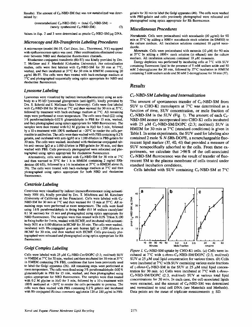
C~-NBD-SM Labeling and Internalization The amount of spontaneous transfer of Ct-NBD-SM from SUV to CHO-K1 monolayers at 7°C was determined as a function of time, SUV concentration, or mole fraction of Ct-NBD-SM in the SUV (Fig. 1). The amount of each Ct- NBD-SM isomer incorporated into CHO-K1 cells incubated with 25 #M C6-NBD-SM/DOPC (2:3; mol/mol) SUV in HMEM for 30 min at 7°C (standard conditions) is given in Table I. In some experiments, the SUV used for labeling also contained 2 mole % N-SRh-DOPE, a nonexchangeable fluo- rescent lipid marker (37, 40, 41) that provided a measure of SUV nonspecificaUy adsorbed to the cells. From these ex- periments, we calculate that >96% of the cell-associated C6-NBD-SM fluorescence was the result of transfer of fluo- rescent SM to the plasma membrane of cells treated under standard incubation conditions.
Cells labeled with SUV containing C6-NBD-SM at 7°C
Centriole Labeling Centrioles were visualized by indirect immunofluorescence using autoanti- body 5051 (4), kindly provided by Drs. T. Mitchison and M. Kirschner 40
< (University of California at San Francisco). Cells were labeled with C6- z NBD-SM for 30 min at 7°C and then wanned for 15 min at 37°C. All re- o maining steps were performed at room temperature. The cells were fixed ~ 30 using 1.6% paraformaldehyde in fixing buffer (0.1 M sodium cacodylate/ ~. 0.1 M sucrose) for 15 min and photographed using optics appropriate for ,~ NBD fluorescence. The samples were then treated with 0.1% Triton X-100 z . 20
in fixing buffer for 3 min, washed with HCMF, and incubated with autoanti- - o
body 5051 at a 1:100 dilution in HCMF for 30 min. The cells were washed, ~. incubated with Rh-conjugated goat anti-human IgG at 1:200 dilution in l o
HCMF for 30 min, and then washed with HCME Cells previously pho- tographed were relocated and photographed using optics appropriate for Rh fluorescence.
Golgi Complex Labeling Cells were labeled with 25 #M C6-NBD-Cer/DOPC (2:3; mol/mol) SUV
o in HMEM at 7°C for 30 rain, washed and then incubated for 30 min at 37 C in HMEM containing 5% FBS, conditions that have been previously used to label the Golgi apparatus (26). All remaining steps were performed at room temperature. The cells were fixed using 3 % paraformaldehyde-0.02 % glutaraldehyde in PBS for 15 min, washed, and then photographed using optics appropriate for NBD fluorescence. The samples were then treated with 0.2 M glycine in H20 for 5 min, followed by a 10-s treatment with 100% methanol at -20°C to render the cells permeable to proteins. The cells were then washed with PBS containing 0.2% gelatin and incubated with Rh-conjugated Ricinus communis agglutinin 120 at 100 #g/ml in PBS-
a b
10 20 30 40 50 0.2 0.4 0.6 60 Minutes Mole Fraction nmol/ml
C
Figure 1. C6-NBD-SM uptake by CHO-K1 cells. (a) Cells were in- cubated at 7°C with L-threo-C6-NBD-SM/DOPC (2:3, mol/mol) SUV at 25 # M total lipid concentrat ion for various t imes. (b) Cells were incubated at 7°C with SUV containing various mole fractions of L-threo-C6-NBD-SM in the S U V at 2 5 / z M total lipid concen- tration for 30 min. (c) Cells were incubated at 7°C with L-threo- C t - N B D - S M / D O P C (2:3, mol/mol) SUV at various total lipid concentrat ions for 30 min. In each case, the cell-associated lipids were extracted, and the amount of C t -NBD-SM was determined and normalized to total cell D N A (see Materials and Methods) . Data points are the mean of triplicate measurements + SD.
Koval and Pagano Plasma Membrane Lipid Recycling 2171
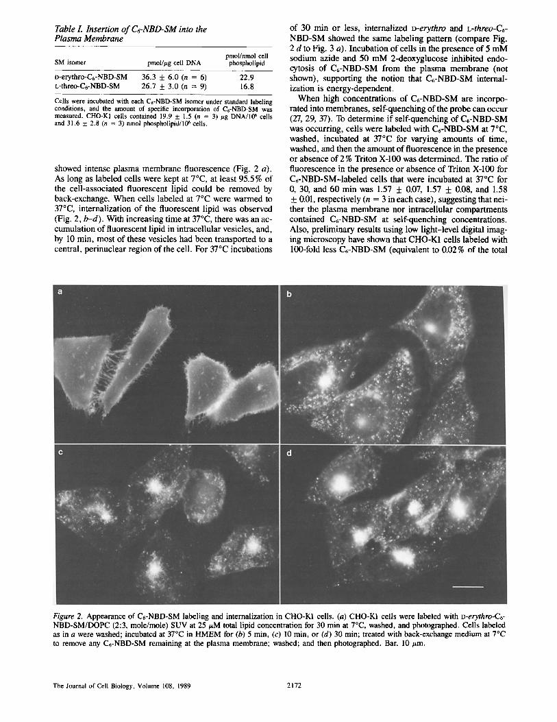
Table I. Insertion of C6-NBD-SM into the Plasma Membrane
pmol/nmol cell SM isomer pmol/#g cell DNA phospholipid
o-erythro-C6-NBD-SM 36.3 + 6.0 (n = 6) 22.9 L-threo-C6-NBD-SM 26.7 + 3.0 (n = 9) 16.8
Cells were incubated with each C6-NBD-SM isomer under standard labeling conditions, and the amount of specific incorporation of C6-NBD-SM was measured. CHO-KI cells contained 19.9 + 1.5 (n = 3) #g DNA/IlY' cells and 31.6 5:2.8 (n = 3) nmol phospholipid/lO 6 cells.
showed intense plasma membrane fluorescence (Fig. 2 a). As long as labeled cells were kept at 7°C, at least 95.5% of the cell-associated fluorescent lipid could be removed by back-exchange. When cells labeled at 7°C were warmed to 37°C, internalization of the fluorescent lipid was observed (Fig. 2, b-d). With increasing time at 37°C, there was an ac- cumulation of fluorescent lipid in intracellular vesicles, and, by 10 min, most of these vesicles had been transported to a central, perinuclear region of the cell. For 37°C incubations
of 30 min or less, internalized D-erythro and L-threo-C6- NBD-SM showed the same labeling pattern (compare Fig. 2 d to Fig. 3 a). Incubation of cells in the presence of 5 mM sodium azide and 50 mM 2-deoxyglucose inhibited endo- cytosis of C6-NBD-SM from the plasma membrane (not shown), supporting the notion that C6-NBD-SM internal- ization is energy-dependent.
When high concentrations of C6-NBD-SM are incorpo- rated into membranes, self-quenching of the probe can occur (27, 29, 37). To determine if self-quenching of C6-NBD-SM was occurring, cells were labeled with C6-NBD-SM at 7°C, washed, incubated at 37°C for varying amounts of time, washed, and then the amount of fluorescence in the presence or absence of 2 % Triton X-100 was determined. The ratio of fluorescence in the presence or absence of Triton X-100 for C6-NBD-SM-labeled cells that were incubated at 37°C for 0, 30, and 60 min was 1.57 + 0.07, 1.57 + 0.08, and 1.58 + 0.01, respectively (n = 3 in each case), suggesting that nei- ther the plasma membrane nor intracellular compartments contained C6-NBD-SM at self-quenching concentrations. Also, preliminary results using low light-level digital imag- ing microscopy have shown that CHO-K1 cells labeled with 100-fold less C6-NBD-SM (equivalent to 0.02% of the total
Figure 2. Appearance o f C6-NBD-SM label ing and internalizat ion in CHO-K1 cells. (a) CHO-K1 cells were labeled with D-erythro-C6- NBD-SM/DOPC (2:3, mole/mole) SUV at 25/zM total lipid concentration for 30 min at 7°C, washed, and photographed. Cells labeled as in a were washed; incubated at 37°C in HMEM for (b) 5 min, (c) 10 min, or (d) 30 min; treated with back-exchange medium at 7°C to remove any C6-NBD-SM remaining at the plasma membrane; washed; and then photographed. Bar, 10/~m.
The Journal of Cell Biology, Volume 108, 1989 2172
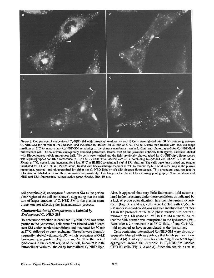
Figure 3. Comparison of endocytosed C6-NBD-SM with lysosomal markers. (a and b) Cells were labeled with SUV containing L-threo- C6-NBD-SM for 30 min at 70C, washed, and incubated in HMEM for 30 min at 37°C. The cells were then treated with back-exchange medium at 7°C to remove any C6-NBD-SM remaining at the plasma membrane, washed, fixed and photographed for C6-NBD-lipid fluorescence (a). The cells were subsequently rendered permeable, treated with an antilysosomal antibody (anti-lgp95), and then labeled with Rh-conjugated rabbit anti-mouse IgG. The cells were washed and the field previously photographed for C6-NBD-lipid fluorescence was rephotographed for Rh fluorescence (b). (c and d) Cells were labeled with SUV containing D-erythro-C6-NBD-SM in HMEM for 30 min at 7°C, washed, and incubated for 1 h at 37°C in HMEM containing 2 mg/ml SRh-dextran. The cells were then washed and further incubated for 1 h at 37°C in HMEM alone, treated with back-exchange medium at 7°C to remove C6-NBD-SM remaining at the plasma membrane, washed, and photographed for either (c) C6-NBD-lipid or (d) SRh-dextran fluorescence. This procedure does not require relocation of labeled cells and thus minimizes the possibility of a change in the plane of focus during photography. Note the absence of NBD and SRh fluorescence colocalization (arrowheads). Bar, 10 ttm.
cell phospholipid) endocytose fluorescent SM to the perinu- clear region of the cell (not shown), suggesting that the addi- tion of larger amounts of C6-NBD-SM to the plasma mem- brane was not affecting the internalization process.
Characterization of Compartments Labeled by Endocytosed C~-NBD-SM To determine whether internalized C6-NBD-SM was trans- ported to the lysosomes, cells were first labeled with fluores- cent SM under standard conditions and incubated for 30 min at 370C, followed by back-exchange. The cells were then sub- sequently labeled with anti-lgp95, which recognizes a 95-kD lysosomal glycoprotein (Fig. 3, a and b). Note the lack of lysosomes in the central region of the cell, in contrast to the intracellular vesicles labeled by internalized C6-NBD-lipid.
Also, it appeared that very little fluorescent lipid accumu- lated in the lysosomes under these conditions as indicated by a lack of probe colocalization. In a complementary experi- ment (Fig. 3, c and d), cells were labeled with C6-NBD- SM under standard conditions and then incubated at 37°C for 1 h in the presence of the fluid phase marker SRh-dextran, followed by a 1-h chase at 37°C in HMEM alone to insure that the SRh-dextran was transported to the lysosomes (39). Even after a 2-h incubation at 37°C, little, if any, C6-NBD- lipid appeared to have accumulated in the lysosomes.
Cells containing internalized C6-NBD-SM were also sub- sequently labeled with an antibody that labels pericentriolar material (4). Endocytic vesicles containing fluorescent lipid aggregated around the centriole in C6-NBD-SM-labeled CHO-K1 cells (Fig. 4, a and b). Since the centriole acts as
Koval and Pagano Plasma Membrane LipM Recycling 2173

Figure 4. Colocalization of endocytosed C6-NBD-SM with the centriole and the effect of microtubule disruption on C6-NBD-SM internal- ization. In (a and b), cells were labeled with SUV containing L-threo-C6-NBD-SM for 30 min at 7°C, washed, and incubated in HMEM for 15 min at 3"/°C. The cells were then treated with back-exchange medium at 7°C to remove any C6-NBD-SM remaining at the plasma membrane, washed, fixed and photographed for C6-NBD-lipid fluorescence (a). Note that the plane of focus selected does not enable the visualization of fluorescently labeled peripheral endosomes. The cells were subsequently rendered permeable, treated with antibody 5051, which recognizes pericentriolar material, and then labeled with Rh-conjugated goat anti-human IgG. The cells were washed and the field previously photographed for C6-NBD-lipid fluorescence was rephotographed for Rh fluorescence (b). (c) Cells were preincubated with 10 #g/ml nocodazole for 90 min at 37°C in culture medium, washed, and labeled with SUV containing L-threo-C6-NBD-SM in HMEM with 10/zg/ml nocodazole. The cells were washed, incubated with back-exchange medium containing 10/zg/ml nocodazole at 7°C to remove any C6-NBD-SM remaining at the plasma membrane, washed, and photographed. Bar, 10 #m.
I o(~
9C
8O C3 ~= :
2o
10
90
:.3 80 C3 ~ : ~ 2o
10
20 40 60 80 I O0 120
Minutes at 37°0
Figure 5. Metabolism of o-erythro and L-threo-C6-NBD-SM. Cells were labeled with SUV containing either (a) n-erythro- or (b) L-threo-C6-NBD-SM in HMEM for 30 min at 7°C, washed and incubated for the indicated times at 37°C in HMEM. The cell- associated lipids were extracted, separated by TLC, and the fluores- cent metabolites were measured and expressed as a percentage of total cell-associated C6-NBD-lipid. (m) C6-NBD-SM; (&) C6-NBD- GlcCer; (e) C6-NBD-Cer. Data points are the mean of triplicate measurements.
a microtubule organizing center, the effect of microtubule disruption was also examined. In the presence of 10/zg/ml nocodazole, C6-NBD-SM continued to be endocytosed, how- ever, microtubule disruption resulted in little internalized fluorescent lipid accumulation in the central region of the cells. Instead, C6-NBD-lipid was distributed in vesicles throughout the periphery of the cell (Fig. 4c).
Quantitation of Metabolism and Endocytosis of C~-NBD-SM
Cells labeled with either o-erythro or L-threo-C6-NBD-SM at 7°C under standard conditions and then incubated at 37°C metabolized C6-NBD-SM to other fluorescent sphingolipids (Fig. 5, a and b). Both C6-NBD-SM isomers showed partial hydrolysis to the corresponding C6-NBD-Cer isomer, but only o-erythro-C6-NBD-SM-labeled cells produced C6- NBD-GIcCer (from C6-NBD-Cer) during the 37°C incuba- tion, probably because of the stereospecificity of the conver- sion of C6-NBD-Cer to C6-NBD-GIcCer (33). No other fluorescent lipid species, including C6-NBD-fatty acid, were produced during 37°C incubations of up to 6 h in cells la- beled with either fluorescent SM isomer. In addition, a small amount (1.7 _ 0.7 %; n = 3) of o-erythro-C6-NBD-SM was hydrolyzed to C6-NBD-Cer during the 30 min incubation at 7°C.
CHO-K1 cells were labeled directly with D-erythro-C6- NBD-Cer and the conversion to C6-NBD-SM and C6-NBD-
The Journal of Cell Biology, Volume 108, 1989 2174
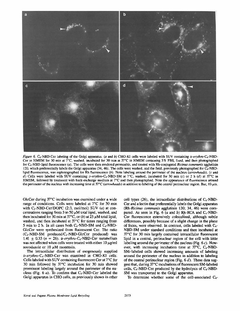
Figure 6. C6-NBD-Cer labeling of the Golgi apparatus. (a and b) CHO-K1 cells were labeled with SUV containing D-erythro-C6-NBD- Cer in HMEM for 30 min at 7°C, washed, incubated for 30 min at 37°C in HMEM containing 5 % FBS, fixed, and then photographed for C6-NBD-lipid fluorescence (a). The cells were then rendered permeable, and treated with Rh-conjugated Ricinus communis agglutinin 120, which preferentially labels the Golgi apparatus (34, 46). The cells were washed, and the field, previously photographed for C6-NBD- lipid fluorescence, was rephotographed for Rh fluorescence (b). Note labeling around the perimeter of the nucleus (arrowheads). (c and d) Cells were labeled with SUV containing D-erythro-C6-NBD-SM at 7°C, washed, incubated for 30 min (c) or 2 h (d) at 37°C in HMEM, followed by treatment with back-exchange medium at 7°C and then photographed. Note the appearance of fluorescence around the perimeter of the nucleus with increasing time at 37°C (arrowheads) in addition to labeling of the central perinuclear region. Bar, 10 t~m.
GlcCer during 37°C incubation was examined under a wide range of conditions. Cells were labeled at 7°C for 30 min with C6-NBD-Cer/DOPC (2:3, mol/mol) SUV (a) at con- centrations ranging from 3 to 50/~M total lipid, washed, and then incubated for 30 min at 37°C, or (b) at 25/~M total lipid, washed, and then incubated at 37°C for times ranging from 5 min to 2 h. In all cases both C6-NBD-SM and C6-NBD- GlcCer were synthesized from fluorescent Cer. The ratio (C6-NBD-SM produced/C6-NBD-GlcCer produced) was 1.41 + 0.33 (n = 26). o-erythro-C6-NBD-Cer metabolism was not affected when cells were treated with either 10/~g/ml nocodazole or 10/~M monensin.
The intracellular distribution of exogenously supplied D-erythro-C6-NBD-Cer was examined in CHO-K1 cells. Cells labeled with SUV containing fluorescent Cer at 7°C fox 30 min followed by 37°C incubation for 30 min showed prominent labeling largely around the perimeter of the nu- cleus (Fig. 6 a). To confirm that C6-NBD-Cer labeled the Golgi apparatus in CHO cells, as previously shown in other
cell types (26), the intracellular distributions of C6-NBD- Cer and a lectin that preferentially labels the Golgi apparatus (Rh-Ricinus communis agglutinin 120; 34, 46) were com- pared. As seen in Fig. 6 (a and b)"Rh-RCA and C6-NBD- Cer fluorescence extensively colocallzed, although subtle differences, possibly because of a slight change in the plane of focus, were observed. In contrast, ceils labeled with C6- NBD-SM under standard conditions and then incubated at 37°C for 30 min largely contained intracellular fluorescent lipid in a central, perinuclear region of the cell with little labeling around the perimeter of the nucleus (Fig. 6 c). How- ever, with increasing incubation time at 37°C, C6-NBD- SM-labeled cells showed increasing amounts of labeling around the perimeter of the nucleus in addition to labeling of the central perinuclear region (Fig. 6 d). These data sug- gest that, during 37°C incubations of fluorescent SM-labeled cells, C6-NBD-Cer produced by the hydrolysis of C6-NBD- SM was transported to the Golgi apparatus.
To determine whether some of the cell-associated C6-
Koval and Pagano Plasma Membrane Lipid Recycling 2175

'tO01
80
Z
2O
Minulms at 37 °C
Figure 7. Quantitative distribution of Ct-NBD-SM upon 37°C in- cubation. CHO-Ki cells were labeled with SUV containing o-eryth- ro-Ct-NBD-SM in HMEM for 30 rain at 7°C, washed, incubated at 37"C in HMEM for the indicated amount of time, and then either harvested immediately or, in parallel experiments, after treatment with back-exchange medium at 7"C to remove any Ct-NBD-lipid at the plasma membrane. The cell-associated lipids were extracted and analyzed by TLC as described in the text. The amount of back- exchangeable C~-NBD-lipid was calculated using Eq. 1 (see Mate- rials and Methods) and all values are expressed as percentage of total cell-associated C6-NBD-lipid. The amounts of newly synthe- sized C6-N-BD-SM and nonmetabolized C6-N-BD-SM were calcu- lated by Eq. 2 and 3, respectively (see Materials and Methods). (n) Nonmetabolized D-erythro-C6-N-BD-SM at the plasma mem- brane; (m) intracellular nonmetabolized D-erythro-C6-N-BD-SM; (zx) newly synthesized D-erythro-C6-NBD-SM at the plasma mem- brane; (A) intracellular newly synthesized D-erythro-C6-NBD- SM. Data points are the mean of triplicate measurements.
NBD-SM resulted from the hydrolysis of fluorescent SM to Ct-NBD-Cer followed by resynthesis of Ct-NBD-SM, cells were prelabeled with [32P]orthophosphate followed by incu- bation with SUV containing either o-erythro or L-threo-C6- NBD-SM at 7°C. The cells were then washed, incubated at 37°C for 2 h, and the cell-associated lipids were extracted and analyzed by two-dimensional TLC. In both cases, C6- NBD-SM isolated from these cells had incorporated 32p (not shown), suggesting that two pools of fluorescent SM were present; i.e., nonmetabolized and newly synthesized Ct-NBD-SM.
The best resolution of C6-NBD-SM from other radiolabeled lipids possible by TLC was adequate for autoradiography, but not for quantitative determination of 32p incorporation. Because CHO-K1 cells directly labeled with D-erythro-Ct- NBD-Cer produced Ct-NBD-SM and C6-NBD-GlcCer at a constant ratio (1.41:1), we used the amount of cell-associated
fluorescent GlcGer to estimate the amount of newly synthe- sized Ct-NBD-SM formed by D-erythro-Ct-NBD-SM-la- beled cells during a given experiment (Eq. 2). The amount of nonmetabolized Ct-NBD-SM was then calculated as the difference between the total cell-associated C6-NBD-SM and the amount of newly synthesized C6-NBD-SM (Eq. 3). L-threo-Ct-NBD-SM-labeled ceils did not produce any C6- NBD-GIcCer; therefore, all quantitation of Ct-NBD-SM transport was done using o-erythro-Ct-NBD-SM-labeled cells.
Fig. 7 shows the quantitative redistribution of fluorescent SM in a-erythro-C6-NBD-SM-labeled CHO-K1 cells dur- ing a 37°C incubation. Ct-NBD-SM levels at the plasma membrane declined as the result of fluorescent SM endocyto- sis and hydrolysis to C~-NBD-Cer. The amount of endocy- tosed, nonmetabolized Ct-NBD-SM reached a plateau value of ~ 27% of the total cell-associated C6-NBD-lipid after 30 min at 37°C. The amount of newly synthesized Ct- NBD-SM produced during the 2-h incubation period in- creased steadily to ,x,15% of the total Ct-NBD-lipid, but re- mained less than the amount of internalized, nonmetabolized Ct-NBD-SM. Also, only small amounts of newly synthe- sized fluorescent SM were transported to the plasma mem- brane during this period ('~3 % of the total Ct-NBD-lipid). Neither 10 #M monensin nor 10 #g/ml nocodazole inhibited either the Ct-NBD-SM internalization or Ct-NBD-GIcCer synthesis in Ct-NBD-SM-Iabeled cells (Table II).
Plasma Membrane Recycling of Ct-NBD-SM
Cells were labeled with Ct-NBD-SM under standard condi- tions, washed, incubated at 37°C for 30 min and then treated with back-exchange medium at 7°C to remove any fluores- cent SM remaining at the plasma membrane, resulting in cells containing only internalized fluorescent lipid (Fig. 8 a). Cells containing only internalized fluorescent lipid were fur- ther incubated at 37°C in HMEM and the transport of Ct- NBD-lipid from intracellular compartments to the plasma membrane was observed (Fig. 8 b). When cells that had been further incubated at 37°C were subsequently treated with back-exchange medium at 7°C, to remove any fluorescent lipid that had returned to the plasma membrane, C6-NBD- lipid labeling in the periphery of the cell was revealed (Fig. 8 c). In contrast, when cells containing only internalized C6-NBD-lipid were further incubated at 37°C in back-ex- change medium, any C6-NBD-lipid transported to the plas- ma membrane from intracellular compartments was contin-
Table II. Effect of Nocodazole and Monensin on Ct-NBD-SM Endocytosis
C6-NBD-SM internalized Ct-NBD-GIcCer synthesized
Percent of cell- Percent of cell- associated associated
Treatment pmol/#g DNA Ct-NBD-lipid pmol/#g DNA C6-NBD-lipid
Control 5.24 + 0.60 28.1 _+ 3.2 0.33 + 0,05 1,7 + 0,3
10 pM monensin 6,20 + 2.10 33.3 + 11.3 0.53 + 0.11 2.8 + 0.6 10 p.g/ml nocodazole 3.76 -t- 0.39 37.9 + 3.9 0.40 -I- 0.04 4.0 + 0.4
Cells were either untreated (control) or were preincubated with 10 ttM monensin or 10 #g/ml nocodazole in culture medium for 90 min at 37°C. All solutions used for treated cells contained either 10 #M monensin or 10 #g/ml nocodazole. Cells were labeled with SUV containing o-erythro-Ct-NBD-SM in HMEM for 30 rain at 7°C, washed, incubated in HMEM at 37°C for 30 min, and either harvested immediately or treated with back-exchange medium at 7°C. The cell- associated lipids were extracted, and both the amount of endocytosed, nonmetabolized Ct-NBD-SM and of newly synthesized Ct-NBD-GIcCer were determined as described in the legend to Fig. 7. Data are the mean of triplicate measurements + SD.
The Journal of Cell Biology, Volume 108, 1989 2176
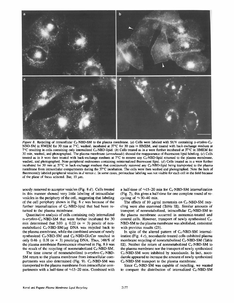
Figure 8. Recycling of intracellular C6-NBD-SM to the plasma membrane. (a) Cells were labeled with SUV containing o-erythro-C6- NBD-SM in HMEM for 30 min at 7°C, washed, incubated at 37°C for 30 min in HMEM, and treated with back-exchange medium at 7°C resulting in cells containing only internalized C6-NBD-lipid. (b) Cells treated as in a were further incubated at 37°C in HMEM for 30 min, washed, and photographed. The plasma membrane (arrowheads) showed the reappearance of fluorescent lipid labeling. (c) Ceils treated as in b were then treated with back-exchange medium at 7°C to remove any C6-NBD-lipid returned to the plasma membrane, washed, and photographed. Note peripheral endosomes containing reinternalized fluorescent lipid. (d) Cells treated as in a were further incubated for 30 min at 37°C in back-exchange medium that continuously removed any C6-NBD-lipid being transported to the plasma membrane from intracellular compartments during the 37°C incubation. The cells were then washed and photographed. Note the lack of fluorescently labeled peripheral vesicles in d versus c. In some cases, perinuclear labeling was not visible for each cell in the field because of the plane of focus selected. Bar, 10 #m.
uously removed to acceptor vesicles (Fig. 8 d). Cells treated in this manner showed very little labeling of intracellular vesicles in the periphery of the cell, suggesting that labeling of the cell periphery shown in Fig. 8 c was because of the further internalization of C6-NBD-lipid that had been re- turned to the plasma membrane.
Quantitative analysis of cells containing only internalized D-erythro-C6-NBD-SM that were further incubated for 30 min determined that 3.03 + 0.22 (n = 3) pmole of non- metabolized C6-NBD-SM/#g DNA was recycled back to the plasma membrane, while the combined amount of newly synthesized C6-NBD-SM and C6-NBD-GIcCer resulted in only 0.46 + 0.31 (n = 3) pmole/#g DNA. Thus, >86% of the plasma membrane fluorescence observed in Fig. 8 b was the result of the recycling of nonmetabolized C6-NBD-SM.
The time course of nonmetabolized D-erythro-C6-NBD- SM return to the plasma membrane from intracellular com- partments was also determined (Fig. 9). C6-NBD-SM was transported to the plasma membrane from intracellular com- partments with a half-time of ~15-20 min. Combined with
a half-time of ~15-20 min for C6-NBD-SM internalization (Fig. 7), this gives a half-time for one complete round of re- cycling of "~, 30-40 min.
The effects of 10 #g/ml monensin on C6-NBD-SM recy- cling were also examined (Table III). Similar amounts of transport of nonmetabolized, intracellular C6-NBD-SM to the plasma membrane occurred in monensin-treated and control cells. However, transport of newly synthesized C6- NBD-SM to the plasma membrane was abolished, consistent with previous results (25).
In spite of the altered pattern of C6-NBD-SM internal- ization (Fig. 4 c), nocodazole-treated cells exhibited plasma membrane recycling of nonmetabolized C6-NBD-SM (Table III). Neither the return of nonmetabolized C6-NBD-SM to the plasma membrane nor the transport of newly synthesized C6-NBD-SM were inhibited by nocodazole. In fact, noco- dazole appeared to increase the amount of newly synthesized C6-NBD-SM transport to the plasma membrane.
Since C6-NBD-SM was capable of recycling, we wanted to compare the distribution of internalized C6-NBD-SM
Koval and Pagano Plasma Membrane Lipid Recycling 2177

d 6o
~o 2
0 20 40 60
Minutes at 37 °C
Figure 9. Quantitation of internalized C6-HBD-SM returned to the plasma membrane. Cells were labeled with SUV containing D-erythro-C6-NBD-SM in HMEM at 7°C for 30 min, washed, in- cubated at 37°C for 30 min, and treated with back-exchange medium at 7°C to remove any C6-NBD-SM remaining at the plasma membrane. The cells were then further incubated for the in- dicated amount of time at 37°C in either HMEM or back-exchange medium, washed, and the cell-associated lipids were extracted and analyzed by TLC (see Materials and Methods). The amount of non- metabolized C6-NBD-SM returned to the plasma membrane was calculated using Eqs. 1-3. (B) D-erythro-C6-NBD-SM and returned to the plasma membrane; ([]) intracellular D-erythro-C6- NBD-SM. Data points are the mean of triplicate measurements _+ SD and are expressed as the percentage of total cell-associated, non- metabolized C6-NBD-SM.
with that of Rh-Tf, a recycling protein (45). Cells were la- beled with C6-NBD-SM at 7°C under standard conditions, washed, and then incubated for 30 min at 37°C in HMEM containing Rh-Tf. As shown in Fig. 10, both C6-NBD-SM and Rh-Tf were internalized to the central region of the cell; however, some intracellular vesicles in the periphery of the cell appeared to be labeled only by C6-NBD-SM. This could be, in part, because of a difference in the sensitivity of C6- NBD-lipid detection versus Rh-Tf detection.
Discussion
In this study, we have examined the transport of the o-erythro and L-threo stereoisomers of a fluorescent analogue of sphin- gomyelin, C6-NBD-SM, as probes for plasma membrane lipid transport in CHO-KI cells. Most of the intracellular C6-NBD-SM was transported along the recycling pathway illustrated in Fig. 11, pathway I. Both isomers of C~-NBD-
SM labeled only the plasma membrane when incubated with cells at 7°C. Cells labeled with C6-NBD-SM at 7°C and then incubated at 37°C endocytosed fluorescent SM to a perinuclear region of the cell that colocalized with the centri- ole. When cells containing only internalized C6-NBD-lipid were further incubated at 37°C, C6-NBD-SM was returned to the plasma membrane. Thus, C6-NBD-SM recycles be- tween the plasma membrane and intracellular compart- ments. Both D-erythro and L-threo-C6-NBD-SM were capa- ble of being recycled.
In addition to the endocytosis of C6-NBD-SM, some hy- drolyis of both C6-NBD-SM isomers to the corresponding C6-NBD-Cer isomer was observed upon 37°C incubation. C6-NBD-Cer can spontaneously undergo transbilayer move- ment (31), in contrast to C6-NBD-SM which has a highly polar head group restricting it to one leaflet of a bilayer (17). Thus, as illustrated in Fig. 11, pathway H, C~-NBD-Cer produced by the hydrolysis of C6-NBD-SM was transported to the Golgi apparatus, presumably by spontaneous diffusion (30, 33), where it was converted back to fluorescent SM and, in the case of D-erythro-C6-NBD-SM, to fluorescent GlcCer. Consistent with the synthesis of C6-NBD-SM at the Golgi apparatus (30) and the transport of newly synthesized C6- NBD-SM through the Golgi apparatus (25), the transport of newly synthesized C6-NBD-SM to the plasma membrane was inhibited by monensin, while the recycling of nonmetab- olized C6-NBD-SM was not affected by monesin treatment (Table III).
Fig. 11 depicts the partial hydrolysis of C6-NBD-SM oc- curring at the plasma membrane. This is consistent with our observation that some hydrolysis of D-erythro-C6-NBD-SM occurred at 7°C, in the absence of endocytosis. Neutral sphingomyelinases have been found in plasma membrane- enriched fractions from rat liver (16), neuroblastoma cell cultures (5), and human renal proximal tubule cell cultures (9). It is possible that C6-NBD-SM was also hydrolyzed by lysosomal (acid) sphingomyelinase (1, 7, 18); however, C6- NBD-SM did not accumulate in the lysosomes, probably because of the sorting of endocytosed C6-NBD-SM from the degradation pathway to a recycling pathway. Also, in- creasing lysosomal pH with NH4CI, which should reduce lysosomal sphingomyelinase activity, did not inhibit C6- NBD-SM hydrolysis (data not shown). Examination of C6- NBD-SM transport in Niemann-Pick fibroblasts, which are
Table IlL Effect of Nocodazole and Monensin on C6-NBD-SM Recycling
Treatment
Percent of total C6-NBD-SM
Nonmetabolized New Percent nonmetabolized Percent new C6-NBD-SM C6-NBD-SM C~-NBD-SM C6-NBD-SM
recycled transported recycled transported
Con t ro l 43 .3 + 4 .2 4 .0 ± 2 .8 68 .7 + 9 .8 10.8 + 1.0 10 /~M m o n e n s i n 56.1 _+ 19.1 O* 7 6 . 0 _+ 25 .9 O* 1 0 / ~ g / m l nocodazo le 4 6 . 8 + 5 .9 9 .9 _+ 1.8 60 .8 + 7 .7 4 3 . 0 + 7 .8
Cells were either untreated (control) or preincubated with 10 #M monensin or 10 #g/mt nocodazole in culture medium for 90 min at 37°C. All solutions used for treated cells contained either 10 p,M monensin or 10 #g/ml nocodazole. Cells were labeled with SUV containing D-erythro-C6-NBD-SM in HMEM for 30 rain at 7°C, washed, incubated at 370C in HMEM for 30 min, and then treated with back-exchange medium to remove any C6-NBD-SM remaining at the plasma membrane. The cells were then further incubated at 37°C for 30 min in either HMEM or back-exchange medium and washed. The cell-associated lipids were extracted and both the amount of nonmetabolized C6-NBD-SM recycled to the plasma membrane and of newly synthesized C6-NBD-SM transported to the plasma membrane were determined as described in the legend to Fig. 9. Data are the mean of triplicate measurements + SD, expressed as percent total cell-associated C6-NBD-SM. * Not detectable.
The Journal of Cell Biology, Volume 108, 1989 2178
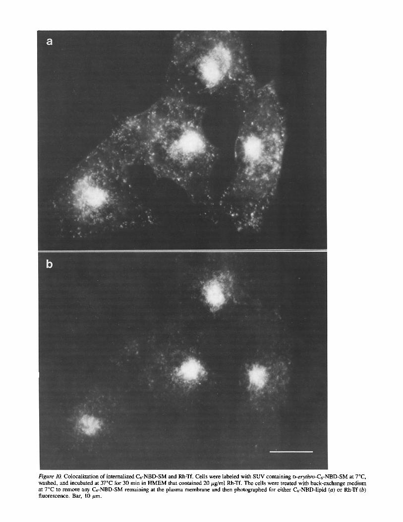
Figure 10. Colocalization of internalized C6-NBD-SM and Rh-Tf. Cells were labeled with SUV containing o-erythro-C6-NBD-SM at 7°C, washed, and incubated at 37°C for 30 min in HMEM that contained 20 #g/ml Rh-Tf. The cells were treated with back-exchange medium at 7°C to remove any C6-NBD-SM remaining at the plasma membrane and then photographed for either C6-NBD-lipid (a) or Rh-Tf (b) fluorescence. Bar, 10/xm.
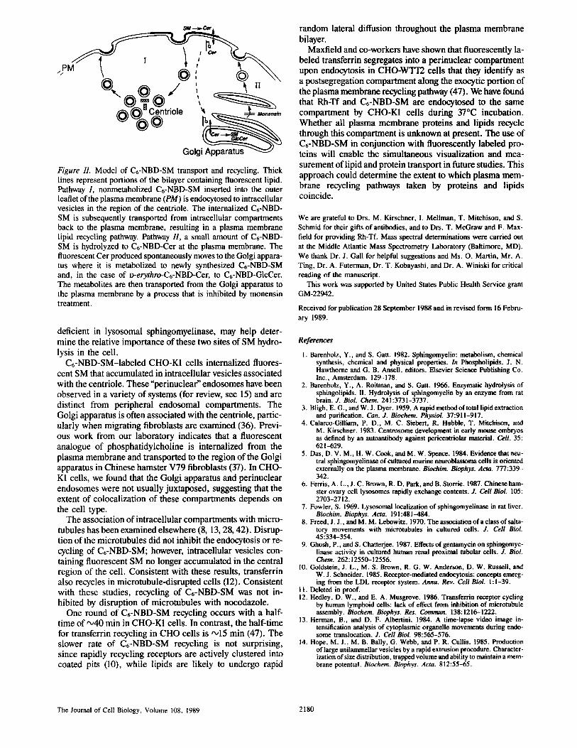
~) ~ G o l g i ~ Figure 11. Model of Cr-NBD-SM transport and recycling. Thick lines represent portions of the bilayer containing fluorescent lipid. Pathway 1, nonmetabolized Cr-NBD-SM inserted into the outer leaflet of the plasma membrane (PM) is endocytosed to intracellular vesicles in the region of the centriole. The internalized Cr-NBD- SM is subsequently transported from intracellular compartments back to the plasma membrane, resulting in a plasma membrane lipid recycling pathway. Pathway H, a small amount of Cr-NBD- SM is hydrolyzed to Cr-NBD-Cer at the plasma membrane. The fluorescent Cer produced spontaneously moves to the Golgi appara- tus where it is metabolized to newly synthesized Cr-NBD-SM and, in the case of D-erythro-Cr-NBD-Cer, to C6-NBD-GIcCer. The metabolites are then transported from the Golgi apparatus to the plasma membrane by a process that is inhibited by monensin treatment.
deficient in lysosomal sphingomyelinase, may help deter- mine the relative importance of these two sites of SM hydro- lysis in the cell.
Cr-NBD-SM-Iabeled CHO-K1 cells internalized fluores- cent SM that accumulated in intracellular vesicles associated with the centriole. These"perinuclear" endosomes have been observed in a variety of systems (for review, see 15) and are distinct from peripheral endosomal compartments. The Golgi apparatus is often associated with the centriole, partic- ularly when migrating fibroblasts are examined (36). Previ- ous work from our laboratory indicates that a fluorescent analogue of phosphatidylcholine is internalized from the plasma membrane and transported to the region of the Golgi apparatus in Chinese hamster V79 fibroblasts (37). In CHO- K1 cells, we found that the Golgi apparatus and perinuclear endosomes were not usually juxtaposed, suggesting that the extent of colocalization of these compartments depends on the cell type.
The association of intracellular compartments with micro- tubules has been examined elsewhere (8, 13, 28, 42). Disrup- tion of the microtubules did not inhibit the endocytosis or re- cycling of Cr-NBD-SM; however, intracellular vesicles con- raining fluorescent SM no longer accumulated in the central region of the cell. Consistent with these results, transferrin also recycles in microtubule-disrupted cells (12). Consistent with these studies, recycling of Cr-NBD-SM was not in- hibited by disruption of microtubules with nocodazole.
One round of C6-NBD-SM recycling occurs with a half- time of ,,o40 min in CHO-K1 cells. In contrast, the half-time for transferrin recycling in CHO cells is '~15 min (47). The slower rate of Cr-NBD-SM recycling is not surprising, since rapidly recycling receptors are actively clustered into coated pits (10), while lipids are likely to undergo rapid
random lateral diffusion throughout the plasma membrane bilayer.
Max field and co-workers have shown that fluorescently la- beled transferrin segregates into a perinuclear compartment upon endocytosis in CHO-WTT2 cells that they identify as a postsegregation compartment along the exocytic portion of the plasma membrane recycling pathway (47). We have found that Rh-Tf and Cr-NBD-SM are endocytosed to the same compartment by CHO-K1 cells during 37°C incubation. Whether all plasma membrane proteins and lipids recycle through this compartment is unknown at present. The use of Cr-NBD-SM in conjunction with fluorescently labeled pro- teins will enable the simultaneous visualization and mea- surement of lipid and protein transport in future studies. This approach could determine the extent to which plasma mem- brane recycling pathways taken by proteins and lipids coincide.
We are grateful to Drs. M. Kirschner, I. Mellman, T. Mitchison, and S. Schmid for their gifts of antibodies, and to Drs. T. McGraw and F. Max- field for providing Rh-Tf. Mass spectral determinations were carried out at the Middle Atlantic Mass Spectrometry Laboratory (Baltimore, MD). We thank Dr. J. Gall for helpful suggestions and Ms. O. Martin, Mr. A. Ting, Dr. A. Futerman, Dr. T. Kobayashi, and Dr. A. Winiski for critical reading of the manuscript.
This work was supported by United States Public Health Service grant GM-22942.
Received for publication 28 September 1988 and in revised form 16 Febru- ary 1989.
References
1. Barenholz, Y., and S. Gau. 1982. Sphingomyelin: metabolism, chemical synthesis, chemical and physical properties. In Phospholipids. J. N. Hawthorne and G. B. Ansell, editors. Elsevier Science Publishing Co. Inc., Amsterdam. 129-178.
2. Barenholz, Y., A. Roitman, and S. Gatt. 1966. Enzymatic hydrolysis of sphingolipids. U. Hydrolysis of sphingomyelin by an enzyme from rat brain. J. Biol. Chem. 241:3731-3737.
3. Bligh, E. G., and W. J. Dyer. 1959. A rapid method of total lipid extraction and purification. Can. J. Biochem. Physiol. 37:91 !-917.
4. Calarco-Gilliam, P. D., M. C. Siebert, R. Hubble, T. Mitchison, and M. Kirschner. 1983. Centrosome development in early mouse embryos as defined by an autoantibody against pericentriolar material. Cell. 35: 621-629.
5. Das, D. V. M., H. W. Cook, and M. W. Spence. 1984. Evidence that neu- tral sphingomyelinase of coltured murine neuroblastoma cells is oriented externally on the plasma membrane. Biochim. Biophys. Acta. 777:339- 342.
6. Ferris, A. L., J. C. Brown, R. D. Park, and B. Storrie. 1987. Chinese ham- ster ovary cell lysosomes rapidly exchange contents. J. Cell Biol. 105: 2703 -2712.
7. Fowler, S. 1969. Lysosomal localization of sphingomyelinase in rat liver. Biochim. Biophys. Acta. 191:481--484.
8. Freed, J. J., and M. M. Lebowitz. 1970. The association of a class of salta- tory movements with microtubules in cultured cells. J. Cell Biol. 45:334-354.
9. Ghosh, P., and S. Chaaerjee. 1987. Effects of gentamyein on sphingomye- linase activity in cultured human renal proximal tubular cells. J. Biol. Chem. 262:12550-12556.
I0. Goldstein, J. L., M. S. Brown, R. G. W. Anderson, D. W. Russell, and W. J. Schneider. 1985. Receptor-mediated endneytosis: concepts emerg- ing from the LDL receptor system. Annu. Rev. Cell Biol. 1 : i-39.
1 I. Deleted in proof. 12. Hedley, D. W., and E. A. Musgrove. 1986. Transferrin receptor cycling
by human lymphoid cells: lack of effect from inhibition of mierotubule assembly. Biochem. Biophys. Res. Commun. 138:1216-1222.
13. Herman, B., and D. F. Albertini. 1984. A time-lapse video image in- tensification analysis of cytoplasmic organelle movements during endo- some translocation. J. Cell Biol. 98:565-576.
14. Hope, M. J., M. B. Bally, G. Webb, and P. R. Cullis. 1985. Production of large unilammellar vesicles by a rapid extrusion procedure. Character- ization of size distribution, trapped volume and ability to maintain a mem- brane potential. Biochem. Biophys. Acta. 812:55-65.
The Journal of Cell Biology, Volume 108, 1989 2180

15. Hopkins, C. R. 1986. Membrane boundaries involved in the uptake and in- traceilular processing of cell surface receptors. Trends Biochem. Sci. 11 : 473--477.
16. Hostetler, K. Y., and P. J. Yazaki. 1979. The subcellular localization of neutral sphingomyelinase in rat liver. J. Lipid. Res. 20:456-463.
17. Houslay, M. D., and K. K. Stanley. 1982. Dynamics of Biological Mem- branes. John Wiley & Sons Inc., New York. 1-330.
18. Kanfer, J. N., O. M. Young, D. Shapiro, and R. O. Brady. The metabolism of sphingomyelin. I. Purification and properties of a sphingomyelin- cleaving enzyme from rat liver tissue. J. Biol. Chem. 241:1081-1084.
19. Kishimoto, Y. 1975. A facile synthesis of ceramides. Chem. Phys. Lipids. 15:33-36.
20. Kremer, J. M. H., M. W. J. v. d. Esker, C. Pathmamanoharan, and P. H. Wiersema. 1977. Vesicles of variable diameter prepared by a modified injection method. Biochemistry. 16:3932-3935.
21. Labarca, C., and K. Paigen. 1980. A simple, rapid and sensitive DNA assay procedure. Anal Biochem. 102:344-352.
22. Lewis, V., S. A. Green, M. Marsh, P. Vihko, A. Helenius, and I. Mellman. 1985. Glycoproteins of the lysosomal membrane. J. Cell Biol. 100:1839-1847.
23. Leyva, A., Jr., and W. N. Kelly. 1974. Measurement of DNA in cultured human cells. Anal. Biochem. 62:173-t79.
24. Lipsky, N. G., and R. E. Pagano. 1984. Fluorescent sphingomyelin labels the plasma membrane of cultured fibroblasts. Ann. NY Acad. Sci. 435: 306-308.
25. Lipsky, N. G., and R. E. Pagano. 1985. Intracellular translocation of fluorescent sphingolipids in cultured fibroblasts: endogenously synthe- sized sphingomyelin and glucosylcerebroside analogues pass through the Golgi apparatus en route to the plasma membrane. J. Cell Biol. 100:27-34.
26. Lipsky, N. G., and R. E. Pagano. 1985. A vital stain for the Golgi appara- tus. Science (Wash. DC). 228:745-747.
27. Martin, O. C., and R. E. Pagano. 1987. Transbilayer movement of fluores- cent analogs of phosphatidylserine and phosphatidylethanolamine at the plasma membrane of cultured cells. J. Biol. Chem. 262:5890-5898.
28. Matteoni, R., and T. E. Kreis. Translocation and clustering of endosomes and lysosomes depends on microtubules. J. Cell Biol. 105:1253-1265.
29. Nichols, J. W., and R. E. Pagano. 1981. Kinetics of soluble lipid monomer diffusion between vesicles. Biochemistry. 20:2783-2789.
30. Pagano, R. E. 1988. What is the fate of diacylglycerol produced at the Golgi apparatus? Trends. Biochem. Sci. 13:202-205.
31. Pagano, R. E. 1989. A fluorescent derivative of ceramide: physical proper- ties and use in studying the Golgi apparatus of animal cells. Methods Cell Biol. 29(Pt. A):75-85.
32. Pagano, R. E., and R. G. Sleight. 1985. Defining lipid transport pathways in animal cells. Science (Wash. DC). 229:1051-1057.
33. Pagano, R. E., and O. C. Martin. 1988. A series of fluorescent N-(acyl)-
sphingosines: synthesis, physical properties, and studies in cultured cells. Biochemistry. 27:4439--4445.
34. Pavelka, M., and A. Ellinger. 1985. Localization of binding sites for Con- canavalin A, Ricinus communis I and Helix pomata lectin in the Golgi ap- paratus of rat small intestinal absorptive cells. J. Histochem. Cytochem. 33:905-914.
35. Rouser, B., A. N. Siakotos, and S. Fleisher. 1981. Quantitative analysis of phospholipids by thin-layer chromatography and phosphorous analysis of spots. Lipids. 1:85-86.
36. Singer, S. J., and A. Kupfer. 1986. The directed migration of eukaryotic cells. Annu. Rev. Cell Biol. 2:337-365.
37. Sleight, R. G., and R. E. Pagano. 1984. Transport of a fluorescent phos- phatidylcholine analog from the plasma membrane to the Golgi appara- tus. J. Cell Biol. 99:742-751.
38. Steinman, R. M., I. S. Mellman, W. A. Muller, and Z. A. Cohn. 1983. Endocytosis and the recycling of plasma membrane. J. Cell Biol. 96:1-27.
39. Storrie, B., R. R. Pool, Jr., M. Sachdeva, K. M. Maurey, and C. Oliver. 1984. Evidence for both prelysosomal and lysosomal intermediates in en- docytic pathways. J. Cell Biol. 98:108-115.
40. Struck, D. K., and R. E. Pagano. 1980. Insertion of fluorescent phospholip- ids into the plasma membrane of a mammalian cell. J. Biol. Chem. 255:5404-5410.
41. Struck, D. K., D. Hoekstra, and R. E. Pagano. 1981. Use of resonance energy transfer to monitor membrane fusion. Biochemistry. 20:4093- 4099.
42. Swanson, J., A. Bushnell, and S. C. Silverstein. 1987. Tubular lysosome morphology and distribution within macrophages depend on the integrity of cytoplasmic microtubules. Proc. Natl. Acad. Sci. USA. 84:1921- 1925.
43. Uster, P. S., and R. E. Pagano. 1986. Resonance energy transfer micros- copy: observations of membrane-bound fluorescent probes in model membranes and in living cells. J. Cell Biol. 103:1221-1234.
44. van Meer, G., E. H. K. Stetzer, W. Wijnaendts-van-Resandt, and K. Si- mons. 1987. Sorting of sphingolipids in epithelial (Madin-Darby canine kidney) cells. J. Cell Biol. 105:1623-1635.
45. van Renswoude, J., K. R. Bridges, J. B. Harford, and R. D. Klausner. 1982. Receptor-mediated endocytosis of transferrin and the uptake of Fe in K562 cells: identification ofa nonlysosomal acidic compartment. Proc. Natl. Acad. Sci. USA. 79:6186-6190.
46. Virtanen, I., P. Ekblom, and P. Laurila. 1980. Subeellular compartmental- ization of saccharide moieties in cultured and malignant cells. J. Cell Biol. 85:429-434.
47. Yamashiro, D. J., B. Tycko, S. R. Fluss, and F. R. Maxfield. 1984. Segre- gation of transferrin to a mildly acidic (pH 6.5) para-Golgi compartment in the recycling pathway. Cell. 37:789-800.
Koval and Pagano Plasma Membrane Lipid Recycling 2181