Lipid-based nanoparticles for psoriasis treatment
22
Lipid-based nanoparticles for psoriasis treatment: a review on conventional treatments, recent works, and future prospects Ummu Umaimah Mohd Nordin, a Noraini Ahmad, * a Norazlinaliza Salim b and Nor Saadah Mohd Yusof a Psoriasis is a lingering inflammatory skin disease that attacks the immune system. The abnormal interactions between T cells, immune cells, and inflammatory cytokines causing the epidermal thickening. International guidelines have recommended topical treatments for mild to moderate psoriasis whilst systemic and phototherapy treatments for moderate to severe psoriasis. However, current therapeutic approaches have a wider extent to treat moderate to severe type of psoriasis especially since the emergence of diverse biologic agents. In the meantime, topical delivery of conventional treatments has prompted many unsatisfactory effects to penetrate through the skin (stratum corneum). By understanding the physiology of stratum corneum barrier functions, scientists have developed di fferent types of lipid-based nanoparticles like solid lipid nanoparticles, nanostructured lipid carriers, nanovesicles, and nanoemulsions. These novel drug delivery systems help the poorly solubilised active pharmaceutical ingredient reaches the targeted site seamlessly because of the bioavailability feature of the nanosized molecules. Lipid-based nanoparticles for psoriasis treatments create a paradigm for topical drug delivery due to their lipids' amphiphilic feature to efficiently encapsulate both lipophilic and hydrophilic drugs. This review highlights different types of lipid-based nanoparticles and their recent works of nano formulated psoriasis treatments. The encapsulation of psoriasis drugs through lipid nanocarriers unfold numerous research opportunities in pharmaceutical applications but also draw challenges for the future development of nano drugs. Ummu Umaimah Mohd Nordin received her BSc in Chemistry (Forensic Analysis) from Uni- versiti Teknologi MARA, Shah Alam, Malaysia in 2016. She is currently a Graduate Research Assistant (GRA) while pursuing her master's degree in chemistry under the supervision of Dr Noraini Ahmad and Dr Nor- azlinaliza Salim at the Depart- ment of Chemistry, Faculty of Science, Universiti Malaya. Her current research interests focus on colloid chemistry and nanotechnology. Noraini Ahmad received her PhD (2012) in Physical Chemistry from the Universiti Malaya. She went for a research attachment at the IQAC-CSIC, Barcelona, Spain in 2010 and 2011 funded by InForm Project, EU-FP7 and Ministry of Higher Education Malaysia, respectively. Pres- ently, Noraini is an Associate Professor at the Department of Chemistry, Faculty of Science and Fellow of UM Community and Sustainability Centre (UMCares). Her research interests focus on colloidal chemistry, liquid crystals, and formulation of nano- carrier systems for cosmeceutical, pharmaceutical and nutraceut- ical applications. She has been awarded with several research grants and won numerous awards locally and internationally. a Department of Chemistry, Faculty of Science, University of Malaya, 50603 Kuala Lumpur, Malaysia. E-mail: [email protected]; Fax: +603-79674193; Tel: +603- 79674008 b Integrated Chemical Biophysics Research, Faculty of Science, Universiti Putra Malaysia, 43400 UPM Serdang, Selangor, Malaysia Cite this: RSC Adv. , 2021, 11, 29080 Received 11th August 2021 Accepted 19th August 2021 DOI: 10.1039/d1ra06087b rsc.li/rsc-advances 29080 | RSC Adv., 2021, 11, 29080–29101 © 2021 The Author(s). Published by the Royal Society of Chemistry RSC Advances REVIEW Open Access Article. Published on 01 September 2021. Downloaded on 7/22/2022 12:08:43 AM. This article is licensed under a Creative Commons Attribution-NonCommercial 3.0 Unported Licence. View Article Online View Journal | View Issue
-
Upload
khangminh22 -
Category
Documents
-
view
0 -
download
0
Transcript of Lipid-based nanoparticles for psoriasis treatment
RSC Advances
REVIEW
Ope
n A
cces
s A
rtic
le. P
ublis
hed
on 0
1 Se
ptem
ber
2021
. Dow
nloa
ded
on 7
/22/
2022
12:
08:4
3 A
M.
Thi
s ar
ticle
is li
cens
ed u
nder
a C
reat
ive
Com
mon
s A
ttrib
utio
n-N
onC
omm
erci
al 3
.0 U
npor
ted
Lic
ence
.
View Article OnlineView Journal | View Issue
Lipid-based nano
Ur(vAcAhuNamS
current research interests focunanotechnology.
aDepartment of Chemistry, Faculty of Scien
Lumpur, Malaysia. E-mail: [email protected]
79674008
Cite this: RSC Adv., 2021, 11, 29080
Received 11th August 2021Accepted 19th August 2021
DOI: 10.1039/d1ra06087b
rsc.li/rsc-advances
29080 | RSC Adv., 2021, 11, 29080–2
particles for psoriasis treatment:a review on conventional treatments, recent works,and future prospects
Ummu Umaimah Mohd Nordin, a Noraini Ahmad, *a Norazlinaliza Salim b
and Nor Saadah Mohd Yusof a
Psoriasis is a lingering inflammatory skin disease that attacks the immune system. The abnormal interactions
between T cells, immune cells, and inflammatory cytokines causing the epidermal thickening. International
guidelines have recommended topical treatments for mild to moderate psoriasis whilst systemic and
phototherapy treatments for moderate to severe psoriasis. However, current therapeutic approaches have
a wider extent to treat moderate to severe type of psoriasis especially since the emergence of diverse biologic
agents. In the meantime, topical delivery of conventional treatments has prompted many unsatisfactory
effects to penetrate through the skin (stratum corneum). By understanding the physiology of stratum
corneum barrier functions, scientists have developed different types of lipid-based nanoparticles like solid lipid
nanoparticles, nanostructured lipid carriers, nanovesicles, and nanoemulsions. These novel drug delivery
systems help the poorly solubilised active pharmaceutical ingredient reaches the targeted site seamlessly
because of the bioavailability feature of the nanosized molecules. Lipid-based nanoparticles for psoriasis
treatments create a paradigm for topical drug delivery due to their lipids' amphiphilic feature to efficiently
encapsulate both lipophilic and hydrophilic drugs. This review highlights different types of lipid-based
nanoparticles and their recent works of nano formulated psoriasis treatments. The encapsulation of psoriasis
drugs through lipid nanocarriers unfold numerous research opportunities in pharmaceutical applications but
also draw challenges for the future development of nano drugs.
mmu Umaimah Mohd Nordineceived her BSc in ChemistryForensic Analysis) from Uni-ersiti Teknologi MARA, Shahlam, Malaysia in 2016. She isurrently a Graduate Researchssistant (GRA) while pursuinger master's degree in chemistrynder the supervision of Droraini Ahmad and Dr Nor-zlinaliza Salim at the Depart-ent of Chemistry, Faculty ofcience, Universiti Malaya. Hers on colloid chemistry and
Noraini Ahmad received her PhD(2012) in Physical Chemistryfrom the Universiti Malaya. Shewent for a research attachmentat the IQAC-CSIC, Barcelona,Spain in 2010 and 2011 fundedby InForm Project, EU-FP7 andMinistry of Higher EducationMalaysia, respectively. Pres-ently, Noraini is an AssociateProfessor at the Department ofChemistry, Faculty of Scienceand Fellow of UM Community
and Sustainability Centre (UMCares). Her research interests focuson colloidal chemistry, liquid crystals, and formulation of nano-carrier systems for cosmeceutical, pharmaceutical and nutraceut-ical applications. She has been awarded with several researchgrants and won numerous awards locally and internationally.
ce, University of Malaya, 50603 Kuala
.my; Fax: +603-79674193; Tel: +603-
bIntegrated Chemical Biophysics Research, Faculty of Science, Universiti Putra
Malaysia, 43400 UPM Serdang, Selangor, Malaysia
9101 © 2021 The Author(s). Published by the Royal Society of Chemistry
Review RSC Advances
Ope
n A
cces
s A
rtic
le. P
ublis
hed
on 0
1 Se
ptem
ber
2021
. Dow
nloa
ded
on 7
/22/
2022
12:
08:4
3 A
M.
Thi
s ar
ticle
is li
cens
ed u
nder
a C
reat
ive
Com
mon
s A
ttrib
utio
n-N
onC
omm
erci
al 3
.0 U
npor
ted
Lic
ence
.View Article Online
1 Introduction
Psoriasis is an immune-mediated inammatory dermatologicaldisease that is also widely referred to as a systemic inamma-tory disorder.1,2 Psoriasis can be indicated by a rapid build-up ofcells on the skin's surface, forming erythematous (redness)papules and plaques with silver scales.3 The red, scaly plaqueson the skin are itchy and sometimes painful.4 The psoriasislesions typically formed on joints such as elbows and knees andmay develop on any part of the skin such as hands, feet, scalp,neck, and face.5 The inammation comes from the aberrantinteractions between innate immune cells, T cells, and kerati-nocytes.6 However, psoriasis is non-contagious since it inher-ently involves innate and adaptive immune systems.7
The prevalence rate differs by region,8 which is epidemio-logically attributable to racial factors, genetic background,lifestyle, and diet.9,10 Psoriasis prevalence amongst adults andchildren is reported to be ranging from 0.91% to 8.5% and 0 to2.1%, respectively.11,12Generally, it depicts a lower occurrence inAsian and some African populations but has a higher occur-rence amongst Caucasian and Scandinavian people, notably inhigh-income countries.2,8 Nevertheless, the actual data amongstworldwide occurrence is not precise as Parisi et al. reported thatthe majority of low-income countries did not report the epide-miology of psoriasis in their countries.2
The disease may be triggered by several known factors ormay also develop over the course of time. Infections frombacteria and viruses are reportedly exacerbated psoriasisconditions.13 Besides, certain medications (such as beta-blockers, lithium antimalarials, nonsteroidal anti-inammatory medications, and tetracyclines) for adult psori-asis patients could also trigger the disease.14,15 They are alsoassociated with numerous psoriasis comorbidities, includingpsoriatic arthritis, heart disease, hypertension, dyslipidaemia,obesity, type II diabetes, metabolic syndrome, inammatory
Norazlinaliza Salim received herPhD (2013) in Oleochemistryfrom the Universiti PutraMalaysia and went fora research attachment at theIQAC-CSIC, Barcelona, Spain(2010). In 2014, she was a post-doctoral research fellow atCentre for Fundamental andFrontier Sciences in Nano-structure Self-Assembly, Uni-versiti Malaya and thereaer,was appointed as a senior
lecturer at the Centre of Foundation Studies for AgriculturalScience, Universiti Putra Malaysia. Her research interest isfocussed on nanodelivery systems such as nanoemulsion, niosome,etc. for cosmeceutical and pharmaceutical applications. She haspublished over 25 papers in peer-reviewed indexed journals, onebook chapter and ve patents.
© 2021 The Author(s). Published by the Royal Society of Chemistry
bowel disease, anxiety, and depression.14,16 From psychosocialimpacts, psoriasis has been an intimidating disease as somestudies show that the risk for anxiety and depression hasbecome dominant comorbidities in psoriasis pathophysiologythat inuences patients' physical appearance and self-esteem.17,18
In determining the type of psoriasis (mild to moderate ormoderate to severe), Psoriasis Area and Severity Index (PASI)score acts as the gold standard of severity lesion measurement,with PASI $ 10 indicating moderate to severe psoriasis.19,20 Formild to moderate psoriasis conditions, topical therapies aregenerally recommended over systemic therapies.3 The latter ispreferable in cases involving moderate to severe diseaseconditions.3,21 Conventional topical delivery of antipsoriaticdrugs typically utilises dosage methods at high concentratedtherapeutics for a certain period.22 Nonetheless, this treatmentpossesses several limitations, including inadequate andunpredictable delivery to the afflicted skin as well as localtoxicity due to skin irritation.22,23
Biologic drugs (biologics or biologic agents) are the recenttreatment strategy with superior efficacy to other treatments formoderate to severe type of psoriasis.3,24 This treatment employsrecombinant proteins to inhibit inammatory cytokines at thetarget site while functions systemically.25 The advent of biologicagents has transformed the psoriasis treatment landscape aerthe rst of its kind, etanercept, was approved by the Food andDrug Administration (FDA) in 2004.26
Currently, the breakthrough of psoriasis treatments emergesfrom the utilisation of novel drug carrier systems ornanotechnology-based approaches leading to greater drug effi-cacy and long-term effects.22,27,28 Poorly soluble active pharma-ceutical ingredients (API) can be improved through the use ofnanotechnology whilst increasing their bioavailability andphysicochemical stability.28–32 It is presumed that ineffectiveAPIs have low pharmacokinetic proles, lack of stability and
Nor Saadah Mohd Yusof ob-tained her PhD from theUniversity of Melbourne in2015. Presently, she is a seniorlecturer at the Department ofChemistry, Universiti Malaya.Her research focuses on themicelle structures in the pres-ence of various additives; andtheir transitional quanticationusing kinetics techniques. She isalso working on sonochemistryand the effect of ultrasound on
the micelle structures. Other than that, she also works on the use ofultrasound on other subjects such as to synthesize gold nano-particles, on natural rubber, sonoencapsulation, sonoextraction,sonopolymerization and sonodegradation such as on food wastesand organic pollutants.
RSC Adv., 2021, 11, 29080–29101 | 29081

RSC Advances Review
Ope
n A
cces
s A
rtic
le. P
ublis
hed
on 0
1 Se
ptem
ber
2021
. Dow
nloa
ded
on 7
/22/
2022
12:
08:4
3 A
M.
Thi
s ar
ticle
is li
cens
ed u
nder
a C
reat
ive
Com
mon
s A
ttrib
utio
n-N
onC
omm
erci
al 3
.0 U
npor
ted
Lic
ence
.View Article Online
solubility, or ones with limited dose toxicity.33 Therefore, byemploying novel drug delivery systems, these drawbacks can beovercome and thus improve the therapeutic actions.33–35 Nano-particles have been proven in preclinical and clinical studies toreduce the drug's side effects at highly controlled drug deliverywith relevant therapeutic concentrations.36 Nanoparticlesthrough the application of nanomedicine are designed toimprove the drug's half-life, thus facilitating the API delivery toits targeted action site through nanocarriers.33 Nano-based drugdelivery or known as nanocarriers are nanosized particles thatenhance the API's therapeutic effectiveness and reduce adverseeffects. This is mainly because of their large surface area to carrythe API.37 Nanocarriers are drug delivery systems employed toregulate the pharmacokinetics and pharmacodynamic proper-ties of the drug of interest.38 Widespread attention to utilisingtopical nanocarriers for skin diseases has multiplied latelybecause they provide better skin permeation and controlledrelease at a reduced dosage regimen.21
Different interests in various types of nanoparticles havebeen widely investigated for topical psoriasis treatments. Poly-meric nanoparticles, namely nanospheres39 and nanocapsules40
as well as polymer-based nanoparticles such as dendrimers,41
and polymeric micelles42 are a growing interest in these appli-cations. Generally, many types of polymers are attractivebiocompatible materials with proven favourable skin penetra-tion and deposition effects.43–45 Despite so, their constituentsare inclined to cytotoxicity while the cost of production isexpensive for a scale-up and requires high-end instruments.45,46
Hydrogels, water-insoluble cross-linked hydrophilic poly-mers, have become a popular trend in psoriasis with/withoutnanoformulations lately, as they can improve anti-psoriaticactivity aer drug encapsulation. The gels can solely act asdrug vehicles,47 or formulated into nanostructured supra-molecules,48 or integrated as excipients in various types ofnanocarriers, such as graphene oxide,49 polymeric nano-particles,43 nanosponge,50 and niosomes.51 With the growinginterest and research in hydrogels, it is expected that they wouldbecome a signicant component of nano-based drug deliverysystems in the future to further improve the therapeuticproles.52
At a more advanced approach than conventional photo-therapy treatments, photothermal therapy (PTT) is a new tech-nology combined with nano-agents to kill cancer cells bylocalised thermal damage,53 which can also be utilised forpsoriasis treatments. PTT results in the apoptosis of keratino-cytes upon photo absorption of nano-agents that is activated bylight.54 Nirmal et al. developed gold nanorods and isatin (ananti-inammatory agent) loaded into poly(lactic-co-glycolicacid) (PLGA) to form nanocomplexes for PTT, which was foundto be able to reduce hyperthermia as a result of near-infrared(NIR) irradiation and also improve psoriasiform hyper-proliferation.54 Nevertheless, the use of gold as a nano-agent isassociated with low photostability, which is liable to structuraldeformation.55 Photodynamic therapy (PDT) is also a form ofphototherapy, which is illuminated by a laser light beam ofa specic wavelength, matching the absorption band of thetopical administrated photosensitizer drug in psoriasis
29082 | RSC Adv., 2021, 11, 29080–29101
treatment. For example, a study reported by Liu et al.56 showedthat zinc phthalocyanine (ZnPc) via PDT signicantly inhibitedthe hyperproliferation of mouse vaginal epithelium induced bydiethylstilbestrol and improved propranolol- and imiquimod-induced psoriasis-like symptoms. However, the PDT mayperform damage to the plasma membrane or DNA of harmfulcells, which may lead to cell death due to the highly reactive 1O2
generated in situ. Thus, nanocarriers based on carboxymethylchitosan loaded with ZnPc has been reported.57
Lipid-based nanoparticles (also known as lipid-based nano-carriers or nano-delivery systems) have been futuristic fortopical nanocarriers in the treatments of wound-healing activityof skin diseases such as those found from psoriasis.58–60 Suchinterest in lipid-based nanoparticles is the result of their lowtoxicity potential and much easier sample preparation with low-production cost allowing mass industrial production, andbetter thermal stability when compared to other types ofnanoparticles.61,62 On top of that, their constituents are alsomore biodegradable and biocompatible relative to syntheticpolymer constituents like in other types of nanocarriers.58,63
In this review, the pathophysiology of psoriasis is brieyexplained such as the interaction of the inammatory inl-trating cells with specic cytokines and alleles in the dermallayers. Multiple diagnosis strategies and the conventionaltreatments as recommended by dermatologists depending onthe types of psoriasis are presented to provide information ondifferent existing therapies that are regulated worldwide. Aspsoriasis is a skin disease, the skin penetration barrier of thestratum corneum and the challenges in delivering currenttherapies are further explained. This includes the discussion onunsatisfactory results from existing approaches. Subsequently,the emerging roles of lipid-based nanocarriers including theirpros and cons are to be associated with the conventionalstrategies of psoriasis treatments, as well as their recent works.Lastly, current opportunities and challenges that arise from thisnovel therapeutic in treating psoriasis are also discussedcomprehensively.
2 Pathogenesis of psoriasis
Psoriasis is a T cell-mediated inammatory disease originatingfrom the autoimmune system of the body. It is also construed tostem from a combination of genetic, epigenetic, and environ-mental factors,64 and also attributable to endocrine hormonesand immunological factors.10 Human leukocyte antigen (HLA)alleles (also known as major histocompatibility complex (MHC)molecules) that reside in both innate and adaptive immunesystems and other susceptibility loci that bear genetic factorslead to the risk of developing psoriasis.65–67 It remains vaguewhether the inciting antigens are self-antigens, environmentalantigens, or some combination of the two to a certain extent.67
This autoimmune expedites the life cycle of skin cells and oenbe characterised by an excessively rare hyperproliferation ofkeratinocytes,64 caused by inammatory cell inltration thatgenerates into the epidermis and dermis skin layers.11 Inam-matory cells are from many types of leukocyte populationincluding lymphocytes, monocytes, and neutrophils that are
© 2021 The Author(s). Published by the Royal Society of Chemistry
Review RSC Advances
Ope
n A
cces
s A
rtic
le. P
ublis
hed
on 0
1 Se
ptem
ber
2021
. Dow
nloa
ded
on 7
/22/
2022
12:
08:4
3 A
M.
Thi
s ar
ticle
is li
cens
ed u
nder
a C
reat
ive
Com
mon
s A
ttrib
utio
n-N
onC
omm
erci
al 3
.0 U
npor
ted
Lic
ence
.View Article Online
accelerated by tumour necrosis factor (TNF)-a, a type ofinammatory cytokine aided by dendritic cell activation.10,68
Hence, the feedback loop between adaptive immune cells (Tcells), innate immune cells (e.g., dendritic cells, Langerhanscells, natural killer cells, macrophages, granulocytes), andresident skin cells (e.g., keratinocytes, endothelial cells) willincrease the inammatory response, thus, cause cellhyperproliferation.69
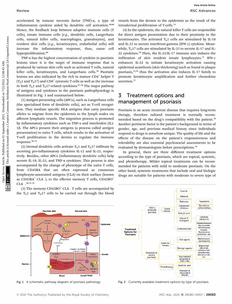
TNF-a has the highest concentration of cytokine in psoriaticlesions since it is the target of immune response that isproduced by various skin cells such as activated T cells, naturalkiller cells, keratinocytes, and Langerhans cells.70 Psoriaticlesions are also indicated by the rich in mature CD4+ helper T(Th1 and Th17) and CD8+ cytotoxic T cells as well as the increasein both Th1 and Th17-related cytokines.67,70 The major pathwayof antigens and cytokines in the psoriasis pathophysiology isillustrated in Fig. 1 and summarised below.
(1) Antigen-presenting cells (APCs), such as Langerhans cells(the specialised form of dendritic cells), act as T-cell recogni-tion. They mediate specic HLA antigens that carry psoriasisalleles to migrate from the epidermis to the lymph nodes viaafferent lymphatic vessels. The migration process is promotedby inammatory cytokines such as TNF-a and interleukin (IL)-1b. The APCs present their antigens (a process called antigenpresentation) to naıve T cells, which results in the activation ofT-cell proliferation in the dermis to regulate the immuneresponse.70–72
(2) Dermal dendritic cells activate Th1 and Th17 inltrate bysecreting pro-inammatory cytokines IL-12 and IL-23, respec-tively. Besides, other APCs (inammatory dendritic cells) helpsecrete IL-18, IL-23, and TNF-a cytokines. This process is alsoaccompanied by the change of phenotype of the naıve T cells,from CD45RA that are oen expressed as cutaneouslymphocyte-associated antigens (CLA) on their surface (knownas CD45RA+ CLA�), to the effector memory T cells, CD45RO+
CLA�.70,72–74
(3) The memory CD45RO+ CLA� T cells are accompanied bythe Th1 and Th17 cells to be carried out through the blood
Fig. 1 A schematic pathway diagram of psoriasis pathology.
© 2021 The Author(s). Published by the Royal Society of Chemistry
vessels from the dermis to the epidermis as the result of theintradermal proliferation of T-cells.74
(4) In the epidermis, the natural killer T cells are responsiblefor direct antigen presentation due to their proximity to thekeratinocytes. The activated Th1 cells are stimulated by IL-18and IL-23 to secrete interferon-gamma (IFN-g) cytokine. Mean-while, Th17 cells are stimulated by IL-23 to secrete IL-17 and IL-22 cytokines.70 Then, the IL-23/IL-17 immune axis induces theinltration of skin resident innate lymphocytes.75 IFN-genhances IL-22 to initiate keratinocyte activation causingepidermal acanthosis (skin thickening) such as found in plaquepsoriasis,72,76 thus the activation also induces IL-17 family topromote keratinocyte amplication and further chemokinesecretion.77
3 Treatment options andmanagement of psoriasis
Psoriasis is an acute recurrent disease that requires long-termtherapy, therefore tailored treatment is normally recom-mended based on the drug's compatibility with the patient.78
Another pertinent factor is the patient's background in terms ofgender, age, and previous medical history since individualsrespond to drugs is somehow unique. The quality of life and theeffects of the disease on the patient's responsiveness andtolerability are also essential psychosocial assessments to beevaluated by dermatologists before prescriptions.79
In general, there are three different treatment optionsaccording to the type of psoriasis, which are topical, systemic,and phototherapy. Milder topical treatments can be recom-mended for patients with mild to moderate psoriasis. On theother hand, systemic treatments that include oral and biologicdrugs are suitable for patients with moderate to severe type of
Fig. 2 Currently available treatment options by type of psoriasis.
RSC Adv., 2021, 11, 29080–29101 | 29083

RSC Advances Review
Ope
n A
cces
s A
rtic
le. P
ublis
hed
on 0
1 Se
ptem
ber
2021
. Dow
nloa
ded
on 7
/22/
2022
12:
08:4
3 A
M.
Thi
s ar
ticle
is li
cens
ed u
nder
a C
reat
ive
Com
mon
s A
ttrib
utio
n-N
onC
omm
erci
al 3
.0 U
npor
ted
Lic
ence
.View Article Online
psoriasis. Phototherapy treatment is commonly suggested forpatients with moderate to severe type of psoriasis in the event ofunsuccessful topical and systemic treatments. A list of treat-ment options under topical, systemic, and phototherapy aredepicted in Fig. 2.21,27
3.1 Topical treatments
The recommended rst line of defence is topical treatment, inwhich the therapeutic is applied onto the afflicted skin. Suchtopical formulations are typically rendered in cream, solution,ointment, and shampoo (for scalp treatment). A wide range oftopical treatments is available to treat psoriasis with differentmechanisms of action. Topical drugs are expedient and acces-sible but usually deliver better efficacy in combination withother topical agents. In this section, the type of topical agents issorted by older to the latest treatments relevant to themedications.
3.1.1 Coal tar. Since ancient times, coal tar has been part ofpsoriasis treatment, especially for scalp psoriasis but showsdeclining popularity in the past decades when compared toother tropicals.80,81 This is probably because of its cosmeticallyunpleasant nature such as malodorous smell and staining ofcloth, especially in patients with non-black hair.80,82 Out ofthousand compounds constituted in the tar, polycyclic arylhydrocarbon is the most analysed compound as it is believed togenerate an effect in psoriasis treatment.83 Contrariwise,carbazole of aryl hydrocarbons is suggested to exhibit a potenteffect in the coal tar.83 Even so, coal tar has raised concernsamong clinicians and patients due to its thousands of uniden-tied components.84 It is also banned in Canada and theEuropean Union for cosmetic use because of carcinogenicconcerns.85
3.1.2 Dithranol. Dithranol (also known as anthralin) is oneof the oldest treatments to alleviate plaque psoriasis86 throughinhibiting proliferation and inducing keratinocyte apoptosis, asdemonstrated through human keratinocyte (HaCaT) cells.87,88 Inessence, the direct or indirect effects of dithranol on theinammatory response of psoriasis are not entirely compre-hended, although they have been investigated by multifacetedstudies.89,90 Notwithstanding the statement, a recent studyshowed that topical dithranol leads to a rapid reduction in thePASI score, resultant in a decrease in epidermal hyper-proliferation and delayed reduction of inammatory penetra-tion in psoriatic skin.89 The study also suggested that psoriaticactivity can be suppressed by targeting the keratinocytes(residing in the epidermis) rather than the immune cells.Despite the benets, dithranol may not be powerful enough forskin permeation as it is associated with low solubility andstability, toxicity, staining, and skin irritation.91
3.1.3 Retinoids. Retinoids are the derivatives or vitamers ofVitamin A. The sole FDA-approved retinoids for psoriasistreatments are tazarotene and acitretin.92 Tazarotene is a topicaldrug, whilst acitretin is an oral agent. The retinoids work onretinoic acid receptors and retinoid-X-receptors, initiating geneexpression's modication of inammatory cytokines and thus,inhibits the keratinocyte proliferation. However, topical
29084 | RSC Adv., 2021, 11, 29080–29101
tazarotene can cause localised toxicity such as skin irritationand photosensitivity.93,94 Tazarotene's efficacy has been widelystudied through combination with topical corticosteroids asthey optimally enhance the post-treatment effects of psoriasis,in which tazarotene reduces the irritation whilst corticosteroidsprovide anti-inammatory effects.95,96
3.1.4 Calcineurin inhibitors. Tacrolimus and pimecroli-mus are two different types of topical calcineurin inhibitors(immunosuppressants) that act as the rst-line therapeuticoptions for inverse psoriasis (body fold area) for long-timeuse.97,98 They are functional in anti-inammatory and immu-nomodulatory activities to inhibit calcineurin phosphatase;hence, suppressing T-cell activation and cytokine production.99
Tacrolimus is also used to treat genital psoriasis, is well-tolerated amongst adults, and is suitable for patients who aresensitive to steroids but with certain side effects like mildpruritus and burning sensation onset the treatment.100–102
Similarly, pimecrolimus has also been used for such psoriasisconditions and facial psoriasis with equivalent potency inchildren but has less therapeutic efficacy than tacrolimus inadults.98,101,102
3.1.5 Salicylic acid. Salicylic acid is a potent keratolyticagent, oen used in combination with other topical agents suchas corticosteroids and calcineurin inhibitors. Its combinationwith calcineurin inhibitors is believed to help in the excellentdrug absorption into psoriasis plaques.103 The use of salicylicacid alone for scalp psoriasis may result in temporary sheddingof telogen hair, but the cessation of use may resolve the issue.104
Besides, it is not recommended to use salicylic acid witha concentration greater than 10% on large body surface areas.This is mainly to avoid possible side effects such as oralmucosal burn, frontal headache, and nausea. Some previousresearch has shown the potent effects of salicylic acid asa penetration enhancer through combination with a calcineurininhibitor, vitamin D analogue, and corticosteroid for better skinpermeation and sustained release prole.105–107
3.1.6 Vitamin D analogues. Calcipotriol and calcitriol aretwo types of vitamin D analogues derived from sunlight.Presumably, vitamin D deciency is associated with psoriasis asits presence at an adequate amount helps suppress theproduction of inammation's potent cytokine mediators,namely IL-2, IL-6, and IFN-g; thus, keep the body healthy.Moreover, there is a correlation between Th17 cells (that workfor a healthy immune system) and vitamin D in psoriasispatients.108 Vitamin D evidently stimulates suppressor T cellsthat regulate other cells in the immune system and inhibitscytotoxicity and, thus, is a natural killer of cell hyper-proliferation. Nevertheless, their association remains debat-able, albeit well-documented,109 and vitamin D analogues areoen recommended with other topical agents. Calcipotriol andbetamethasone dipropionate (a type of corticosteroid) has beenstudied widely to treat trunk and limb psoriasis as they bothwork synergistically to reduce inammation. The combinationtherapy of the two helps control the imbalance between regu-latory CD8+ or CD4+ T cells and pro-inammatory CCR6+
gamma delta (gd) T cells for antipsoriatic activity.110
© 2021 The Author(s). Published by the Royal Society of Chemistry

Review RSC Advances
Ope
n A
cces
s A
rtic
le. P
ublis
hed
on 0
1 Se
ptem
ber
2021
. Dow
nloa
ded
on 7
/22/
2022
12:
08:4
3 A
M.
Thi
s ar
ticle
is li
cens
ed u
nder
a C
reat
ive
Com
mon
s A
ttrib
utio
n-N
onC
omm
erci
al 3
.0 U
npor
ted
Lic
ence
.View Article Online
3.1.7 Corticosteroids. Corticosteroids are the mainstay fortopical therapies of psoriasis. They work by inhibiting therelease of phospholipase A2 and act directly on deoxy-ribonucleic acid (DNA) and inammatory cytokines.111 The useof corticosteroids alone acts as rst-line topical therapy byreducing inammation and slowing down cell hyper-proliferation, especially in genital psoriasis.112 As discussedearlier, the effects of corticosteroids are more powerful throughcombination with other topical agents, namely retinoids, sali-cylic acid, and vitamin D analogues, as evidenced by multiplestudies. However, local side effects like perioral dermatitis,striae, hypertrichosis, and infections are reportedly developedfrom the long-time use of topical corticosteroids.113 On a sidenote, studies showed that there is a moderately high perceptionof steroid phobia among our current generation, especially inwomen.114,115 In general, the studies indicate that the fear ofusing steroid-related drugs are due to skin deteriorationconditions, weight gain, asthma, and stunted growth aerobtaining sources from dermatologists and media.
Fig. 3 FDA-approved biologic agents by their classes.
3.2 Systemic treatments
The systemic treatment utilises drug that circulates through thewhole body. It can be divided into two types: (1) oral agent; and(2) biologic agent that is administered into the system byinjection or intravenous (IV) infusion. International guidelinesfor psoriasis treatments have recommended that if topical andphototherapy treatments are deemed unsuccessful, systemicnonbiologic (oral agent) and biologic therapies may be pursuedto achieve skin clearance. Since monotherapy of topical treat-ments to moderate to severe psoriasis causes many dissatis-factions, patients can also benet from systemic therapies thatprovide better efficiency and favourable tolerability to reduceclinical symptoms, accomplish treatment efficacy targets, andimprove their quality of life.116
3.2.1 Oral drugs. Systemic nonbiologic therapies work bysuppressing inammatory responses in the form of oralconsumption. Methotrexate and cyclosporine are the two famousoral, systemic therapies used for decades to treat psoriasis, oenrecommended as monotherapy or other systemic therapies.Although biologics have widespread attention in psoriasis treat-ments recently, systemic nonbiologic therapies are with manyadvantages. Oral systemic generally curb pervasive psoriasis, islower cost than older medications and biologics, and has theconvenience of use and availability.117 Besides can be adminis-tered orally, the active ingredients are also loaded in nano-formulations to treat psoriasis topically.
3.2.1.1 Methotrexate. Methotrexate is a comprehensivetreatment for all types of psoriasis conditions. It normalisespsoriasis conditions by inciting the apoptosis process of kera-tinocyte proliferation.118 It is usually administered weekly ata low single dose to reduce side effects before the dose can begradually increased to achieve its optimal efficacy.119 However,the continuous use of methotrexate would cause liver damageand pulmonary toxicity (lung damage). Besides, myelosup-pression (low blood cell production) and immunosuppression(weakened immune system) are other problems associated with
© 2021 The Author(s). Published by the Royal Society of Chemistry
methotrexate's long-term use. It is also reported thatmethotrexate-receiving patients have a higher cardiovascularevent risk than TNF inhibitor users.120
3.2.1.2 Cyclosporine. Cyclosporine (sometimes spelt ascyclosporin and known as cyclosporine A) is another type ofcalcineurin inhibitor that has been one of the most effectivesystemic psoriasis treatments. Both cyclosporine and metho-trexate's rotational use are found to help reduce long-termkidney toxicity (nephrotoxicity) whilst minimising the adverseimpacts of plaque psoriasis.121 The use of cyclosporine drug isreportedly equally safe and effective for severe paediatric andadult plaque psoriasis patients122 but with mild side effects suchas pain abdomen and an increase in serum creatinine (achemical waste product in the blood).123,124 However, livertoxicity (hepatotoxicity) has been subject to the most affectedorgan for prolonged use of cyclosporine.125
3.2.2 Biologic drugs. Aer the successful case of treatinga patient that suffered from Crohn's disease and moderate tosevere psoriasis with a TNF-a blocker in 2000,126 the last decadewitnessed the discovery of IL-17 and IL-23 pathways responsiblefor the pathogenesis of psoriasis.127,128 Biologics have pragmaticstrength of recommendations for psoriasis treatments, typicallyrecommended based on the patient's case evidence and thedrug's performance. The American Academy of Dermatology(AAD) and the National Psoriasis Foundation dene biologicagents as “engineered monoclonal antibodies and fusionproteins that exert their therapeutic actions by blocking speciccytokines or cytokine receptors critical to psoriatic inamma-tion”.26 Presently, there are 11 types of FDA-approved biologicagents for adult psoriasis treatments, classied by their cyto-kine classes (TNF, IL-12, IL-23, IL-17A) (Fig. 3). Secukinumab, asthe rst-line systemic biological treatment129 is discussed torepresent the overview of this type of advanced drug.
Secukinumab (Cosentyx®) inhibits IL-17A from binding toits cytokine receptor for inammation.130 It is highly favourablein safety prole as its efficacy demonstrates comparable withthe commonly used psoriasis treatments (e.g., methotrexate andTNF-a blockers).131 Its efficacy is widely reported in treatingmoderate to severe plaque psoriasis132 and psoriatic arthritis.133
Other than that, those with a past treatment failure, difficult-to-treat patterns of psoriasis (nail, scalp, and palmoplantarpsoriasis),134 and have clear skin desire135 can also be beneted
RSC Adv., 2021, 11, 29080–29101 | 29085

RSC Advances Review
Ope
n A
cces
s A
rtic
le. P
ublis
hed
on 0
1 Se
ptem
ber
2021
. Dow
nloa
ded
on 7
/22/
2022
12:
08:4
3 A
M.
Thi
s ar
ticle
is li
cens
ed u
nder
a C
reat
ive
Com
mon
s A
ttrib
utio
n-N
onC
omm
erci
al 3
.0 U
npor
ted
Lic
ence
.View Article Online
from this biologics. Having said that, secukinumab is alsoperceived to have the most frequent side effects, mainly infec-tions, compared with other biologic agents.136 It has been re-ported that nasopharyngitis, upper respiratory tract infections,and headaches are the expected side effects of secukinumab.133
Overall, biologic agents are considered expensive or as high-investment treatment based on the type of biologics and indi-vidual's PASI score.137 In the wake of the globally impactedpandemic era of COVID-19 (novel coronavirus disease) thatseverely affects the human respiratory system, the continuationof biologic treatments is controversial. This is due to the highrespiratory rate before the pandemic emerged.138 That raisesconcern if it could increase the consumers' susceptibility to thevirus. On the contrary, treatment discontinuation would causeloss of response when treatments are reintroduced or furtherexacerbated by antibody formation to the biologics.138
Fig. 4 The ‘brick-and-mortar’ system of the stratum corneum withinthe epidermis.
3.3 Phototherapy treatments
Photo-based therapy has been developed for psoriasis treat-ments since the 1920s, and presently serves as a mainstay forthe moderate to severe type of psoriasis.139 In general, photo-therapy is deemed an effective and comparatively affordabletreatment. For instance, it is estimated that a 3 year photo-therapy treatment would cost about $5000 as compared to the 3year duration cost of secukinumab at about $182 718.140 Pho-totherapy treatments also have relatively rare side effects,particularly immunosuppression as compared to systemictherapies.141,142 Presently, narrowband ultraviolet B (NB-UVB),excimer laser/lamp (targeted phototherapy), and psoralen plusultraviolet A (PUVA) are three different types of phototherapytreatments to treat psoriasis as recommended by internationalguidelines.
Nevertheless, phototherapy might be unpopular amongpsoriasis patients due to concerns about its carcinogenicity andis therefore not recommended for long-term treatment.141 Thedeclining usage of phototherapy is also contributed aer theadvent of biological agents.3,143 Moreover, NB-UVB is also notsuitable for psoriasis patients with photosensitive disorders(such as those suffering from xeroderma pigmentosa).141,143
Other than that, the literature reveals that PUVA undergoesa higher adverse risk than UVB and is not recommended formaintenance therapy due to cutaneous malignancy risks.144
3.3.1 Narrowband ultraviolet B (NB-UVB) and excimerlaser. NB-UVB light (311 nm) and excimer lamp/laser (308 nm)are two effective phototherapies that currently act as the rst-line treatment for stable plaque psoriasis.145 Both therapiesare equally useful for psoriasis' skin clearance.141,145 The choiceof NB-UVB is usually made aer the failure of a series of topicalagents.146 Phototherapy requires repetitive skin exposure to UVlight. The generation of phototherapy works where the UVradiation from the treatments induce apoptosis in T lympho-cytes and keratinocytes in the epidermis to control the psoriaticlesions.147 The trend of the highly cost-effective UVB treatmentis the exclusive option for a particular group of patients, such ashuman immunodeciency virus (HIV), internal malignancy,and pregnancy, where contraindication of their systemic
29086 | RSC Adv., 2021, 11, 29080–29101
immune reaction might happen (if modied while using otherpsoriasis treatments).148
The excimer laser is remarked to be more efficacious thanthe excimer lamp and is recommended by the AAD for PASI <10.141 Whilst other parts of the body can be induced by the NB-UVB phototherapy, the nose, ear, and palpebral region repre-sent difficult-to-treat areas of psoriasis. Therefore, the excimerlaser is a better phototherapy alternative to target thosementioned psoriasis areas.149 Besides being a powerful andeffective treatment, it is preferable amongst psoriasis patientsas the laser offers long-term remission of psoriasis.150
3.3.2 Psoralen plus ultraviolet A (PUVA). PUVA is a photo-chemotherapy treatment considered another effective treat-ment of chronic plaque psoriasis.151 The combination ofpsoralens and UVA has been shown to reduce PASI by 75% to80% amongst the users, making this phototherapy useful asa second-line treatment aer unresponsive topical and NB-UVBtreatments.152 Instances of specic failure treatment indicationsof NB-UVB relapse are when the patients develop pustularpsoriasis and pityriasis rubra pilaris conditions. Consequently,to introduce PUVA phototherapy to the patients aer suchpsoriasis deterioration, psoralen needs to be sensitised uponthem as a form to enhance the therapeutic procedure.146 Psor-alen is a group of light-sensitive compounds derived fromnatural plants, commonly found in Psoralea corylifolia (Babchi)that exhibit therapeutic properties.153 They are likely to instigateskin-related conditions namely burning, itching, and pigmen-tation, and also nausea aer this therapeutic action.154
4 Overcoming skin barrier challengesfor nano-based drug delivery
A thorough analysis and understanding of the various skin'sstructures and physiology is required to design drugs withimproved permeability and skin penetration. Skin is the largestorgan in the human body and is also responsible for protectingone's system against exogenous pathogens and environmentalfactors and preventing water and electrolyte loss from theinternal.155,156 As a result, the stratum corneum prevents thepenetration of larger molecules (>500 Da)157 across the outer-most layer of the skin epidermis. The barrier is referred to asa ‘brick-and-mortar system’ and comprises approximately 15layers of attened corneocytes (anucleated keratinocytes) withina multilamellar, lipid-rich extracellular matrix,158 The threeessential components of stratum corneum are: (1) naturalmoisturising factor (NMF)-laden and lipid-bound corneocytes
© 2021 The Author(s). Published by the Royal Society of Chemistry
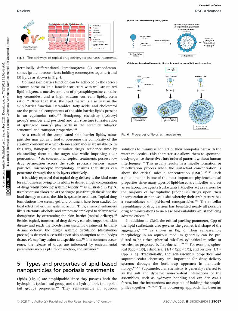
Fig. 5 The pathways of topical drug delivery for psoriasis treatments.
Fig. 6 Properties of lipids as nanocarriers.
Review RSC Advances
Ope
n A
cces
s A
rtic
le. P
ublis
hed
on 0
1 Se
ptem
ber
2021
. Dow
nloa
ded
on 7
/22/
2022
12:
08:4
3 A
M.
Thi
s ar
ticle
is li
cens
ed u
nder
a C
reat
ive
Com
mon
s A
ttrib
utio
n-N
onC
omm
erci
al 3
.0 U
npor
ted
Lic
ence
.View Article Online
(terminally differentiated keratinocytes); (2) corneodesmo-somes (proteinaceous rivets holding corneocytes together); and(3) lipids as shown in Fig. 4.
Optimal skin barrier function can be achieved by the correctstratum corneum lipid lamellar structure with well-structuredlipid bilayers, a massive amount of phytosphingosine-contain-ing ceramides, and a high stratum corneum lipid/proteinratio.159 Other than that, the lipid matrix is also vital in theskin barrier function. Ceramides, fatty acids, and cholesterolare the principal components of the skin barrier lipids presentin an equimolar ratio.160 Headgroup chemistry (hydroxylgroup's number and position) and tail structure (unsaturationof sphingoid moiety) play parts in the ceramide bilayers'structural and transport properties.161
As a result of the complicated skin barrier lipids, nano-particles may act as a tool to overcome the complexity of thestratum corneum in which chemical enhancers are unable to. Inthis way, nanoparticles stimulate drugs' residence time bychannelling them to the target size while improving theirpenetration.162 As conventional topical treatments possess lowdrug permeation across the scaly psoriasis lesions, nano-particles' microscopic morphology ensures that drugs canpenetrate through the skin layers effectively.
It is widely regarded that topical drug delivery is the ideal routefor skin diseases owing to its ability to deliver a high concentrationof drugs whilst reducing systemic toxicity,163 as illustrated in Fig. 5.Itsmechanismallows the API or drug to pass through the skin to thelocal therapy or across the skin by systemic treatment. Topical drugformulations like cream, gel, and ointment have been studied forlocal effect rather than systemic action. Thus, chemical enhancerslike surfactants, alcohols, and amines are employed to deliver activetherapeutics by overcoming the skin barrier (topical delivery).164
Besides topical, transdermal drug delivery can also target local skindisease and reach the bloodstream (systemic treatment). In trans-dermal delivery, the drug's systemic circulation (distributionprocess) is deemed successful upon skin absorption to the body'stissues via capillary action at a specic rate.165 In a common occur-rence, the release of drugs are inuenced by environmentalparameters such as pH, redox reaction, and enzymes.37
5 Types and properties of lipid-basednanoparticles for psoriasis treatments
Lipids (Fig. 6) are amphipathic since they possess both thehydrophilic (polar head group) and the hydrophobic (non-polartail group) properties.166 They self-assemble in aqueous
© 2021 The Author(s). Published by the Royal Society of Chemistry
solutions to minimise contact of their non-polar part with thewater molecules. This characteristic allows them to spontane-ously organise themselves into ordered patterns without humaninterference.167 This usually results in a micelle formation ormicellization process when the surfactant concentration isabove the critical micelle concentration (CMC).167,168 Sucha phenomenon is one of the most important physicochemicalproperties since many types of lipid-based are micelles and actas surface-active agents (surfactants). Micelles act as carriers forthe majority of hydrophobic (lipophilic) drugs upon theirincorporation at nanoscale size whereby their architecture hasa resemblance to lipid-based nanoparticles.169 The micellarresemblance of drug carriers has beneted nearly all possibledrug administrations to increase bioavailability whilst reducingadverse effects.170
In addition to CMC, the critical packing parameter, Cpp ofthe lipid surfactants also governs the geometrical shape of theaggregates,171–173 as shown in Fig. 6. Their self-assemblymorphology in an aqueous medium generally can be pre-dicted to be either spherical micelles, cylindrical micelles orvesicles, as proposed by Israelachvili.171–173 For example, spher-ical (Cpp < 1/3), cylindrical, (1/3 < Cpp < 1/2), and vesicles (1/2 <Cpp < 1). Traditionally, the self-assembly properties andsupramolecular chemistry are important for drug deliverysystems through the bottom-up approach in nanotech-nology.174,175 Supramolecular chemistry is generally referred toas the so and dynamic non-covalent interactions of theassemblies, such as hydrogen bonding and van der Waalsforces, but the interactions are capable of holding the amphi-philes together.174,176,177 This bottom-up approach has been an
RSC Adv., 2021, 11, 29080–29101 | 29087
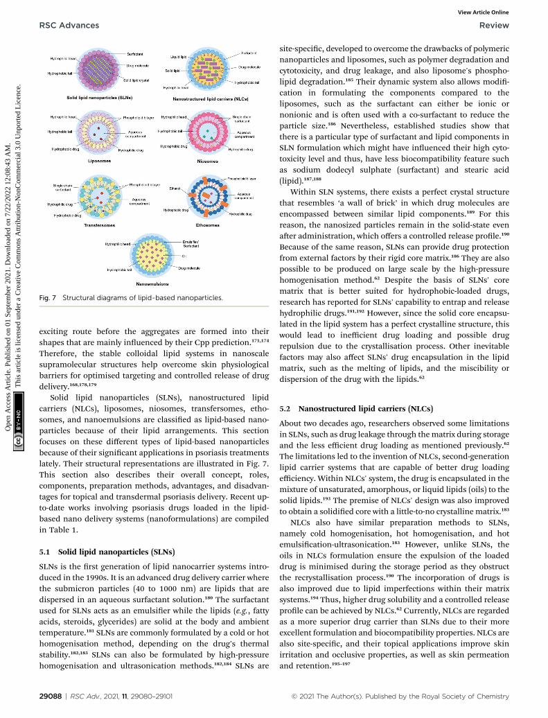
Fig. 7 Structural diagrams of lipid-based nanoparticles.
RSC Advances Review
Ope
n A
cces
s A
rtic
le. P
ublis
hed
on 0
1 Se
ptem
ber
2021
. Dow
nloa
ded
on 7
/22/
2022
12:
08:4
3 A
M.
Thi
s ar
ticle
is li
cens
ed u
nder
a C
reat
ive
Com
mon
s A
ttrib
utio
n-N
onC
omm
erci
al 3
.0 U
npor
ted
Lic
ence
.View Article Online
exciting route before the aggregates are formed into theirshapes that are mainly inuenced by their Cpp prediction.171,174
Therefore, the stable colloidal lipid systems in nanoscalesupramolecular structures help overcome skin physiologicalbarriers for optimised targeting and controlled release of drugdelivery.168,178,179
Solid lipid nanoparticles (SLNs), nanostructured lipidcarriers (NLCs), liposomes, niosomes, transfersomes, etho-somes, and nanoemulsions are classied as lipid-based nano-particles because of their lipid arrangements. This sectionfocuses on these different types of lipid-based nanoparticlesbecause of their signicant applications in psoriasis treatmentslately. Their structural representations are illustrated in Fig. 7.This section also describes their overall concept, roles,components, preparation methods, advantages, and disadvan-tages for topical and transdermal psoriasis delivery. Recent up-to-date works involving psoriasis drugs loaded in the lipid-based nano delivery systems (nanoformulations) are compiledin Table 1.
5.1 Solid lipid nanoparticles (SLNs)
SLNs is the rst generation of lipid nanocarrier systems intro-duced in the 1990s. It is an advanced drug delivery carrier wherethe submicron particles (40 to 1000 nm) are lipids that aredispersed in an aqueous surfactant solution.180 The surfactantused for SLNs acts as an emulsier while the lipids (e.g., fattyacids, steroids, glycerides) are solid at the body and ambienttemperature.181 SLNs are commonly formulated by a cold or hothomogenisation method, depending on the drug's thermalstability.182,183 SLNs can also be formulated by high-pressurehomogenisation and ultrasonication methods.182,184 SLNs are
29088 | RSC Adv., 2021, 11, 29080–29101
site-specic, developed to overcome the drawbacks of polymericnanoparticles and liposomes, such as polymer degradation andcytotoxicity, and drug leakage, and also liposome's phospho-lipid degradation.185 Their dynamic system also allows modi-cation in formulating the components compared to theliposomes, such as the surfactant can either be ionic ornonionic and is oen used with a co-surfactant to reduce theparticle size.186 Nevertheless, established studies show thatthere is a particular type of surfactant and lipid components inSLN formulation which might have inuenced their high cyto-toxicity level and thus, have less biocompatibility feature suchas sodium dodecyl sulphate (surfactant) and stearic acid(lipid).187,188
Within SLN systems, there exists a perfect crystal structurethat resembles ‘a wall of brick’ in which drug molecules areencompassed between similar lipid components.189 For thisreason, the nanosized particles remain in the solid-state evenaer administration, which offers a controlled release prole.190
Because of the same reason, SLNs can provide drug protectionfrom external factors by their rigid core matrix.186 They are alsopossible to be produced on large scale by the high-pressurehomogenisation method.62 Despite the basis of SLNs' corematrix that is better suited for hydrophobic-loaded drugs,research has reported for SLNs' capability to entrap and releasehydrophilic drugs.191,192 However, since the solid core encapsu-lated in the lipid system has a perfect crystalline structure, thiswould lead to inefficient drug loading and possible drugrepulsion due to the crystallisation process. Other inevitablefactors may also affect SLNs' drug encapsulation in the lipidmatrix, such as the melting of lipids, and the miscibility ordispersion of the drug with the lipids.62
5.2 Nanostructured lipid carriers (NLCs)
About two decades ago, researchers observed some limitationsin SLNs, such as drug leakage through thematrix during storageand the less efficient drug loading as mentioned previously.62
The limitations led to the invention of NLCs, second-generationlipid carrier systems that are capable of better drug loadingefficiency. Within NLCs' system, the drug is encapsulated in themixture of unsaturated, amorphous, or liquid lipids (oils) to thesolid lipids.193 The premise of NLCs' design was also improvedto obtain a solidied core with a little-to-no crystalline matrix.183
NLCs also have similar preparation methods to SLNs,namely cold homogenisation, hot homogenisation, and hotemulsication-ultrasonication.183 However, unlike SLNs, theoils in NLCs formulation ensure the expulsion of the loadeddrug is minimised during the storage period as they obstructthe recrystallisation process.190 The incorporation of drugs isalso improved due to lipid imperfections within their matrixsystems.194 Thus, higher drug solubility and a controlled releaseprole can be achieved by NLCs.62 Currently, NLCs are regardedas a more superior drug carrier than SLNs due to their moreexcellent formulation and biocompatibility properties. NLCs arealso site-specic, and their topical applications improve skinirritation and occlusive properties, as well as skin permeationand retention.195–197
© 2021 The Author(s). Published by the Royal Society of Chemistry
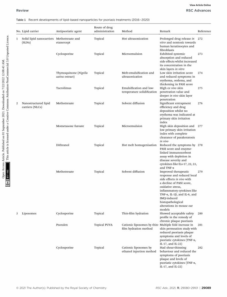
Table 1 Recent developments of lipid-based nanoparticles for psoriasis treatments (2016–2020)
No. Lipid carrier Antipsoriatic agentRoute of drugadministration Method Remark Reference
1 Solid lipid nanocarriers(SLNs)
Methotrexate andetanercept
Topical Hot ultrasonication Prolonged drug release invitro and nontoxic towardshuman keratinocytes andbroblasts
272
Cyclosporine Topical Microemulsion Exhibited systemicabsorption and reducedside effects whilst increasedits concentration in theskin layers in vitro
273
Thymoquinone (Nigellasativa extract)
Topical Melt-emulsication andultrasonication
Low skin irritation scoreand reduced symptoms inerythema, oedema, andthickening in PASI score
274
Tacrolimus Topical Emulsication and low-temperature solidication
High ex vivo skinpenetration value anddeeper in vivo skin layerpenetration
275
2 Nanostructured lipidcarriers (NLCs)
Methotrexate Topical Solvent diffusion Signicant entrapmentefficiency and drugdeposition whilst noerythema was indicated atprimary skin irritationindex
276
Mometasone furoate Topical Microemulsion High skin deposition andlow primary skin irritationindex with completeclearance of parakeratosisin vivo
277
Dithranol Topical Hot melt homogenisation Reduced the symptoms byPASI score and enzyme-linked immunosorbentassay with depletion indisease severity andcytokines like ILs-17, 22, 23,and TNF-a
278
Methotrexate Topical Solvent diffusion Improved therapeuticresponse and reduced localside effects in vivo witha decline of PASI score,oxidative stress,inammatory cytokines likeTNF-a, IL-1b, and IL-6, andIMQ-inducedhistopathologicalalterations in mouse earmodels
279
3 Liposomes Cyclosporine Topical Thin-lm hydration Showed acceptable safetyprole in the remedy ofchronic plaque psoriasis
280
Psoralen Topical PUVA Cationic liposomes by thin-lm hydration method
Multiple fold increase inskin permeation study withreduced psoriasis plaquesymptoms and levels ofpsoriatic cytokines (TNF-a,IL-17, and IL-22)
281
Cyclosporine Topical Cationic liposomes byethanol injection method
Had shear-thinningbehaviour and reduced thesymptoms of psoriasisplaque and levels ofpsoriatic cytokines (TNF-a,IL-17, and IL-22)
282
© 2021 The Author(s). Published by the Royal Society of Chemistry RSC Adv., 2021, 11, 29080–29101 | 29089
Review RSC Advances
Ope
n A
cces
s A
rtic
le. P
ublis
hed
on 0
1 Se
ptem
ber
2021
. Dow
nloa
ded
on 7
/22/
2022
12:
08:4
3 A
M.
Thi
s ar
ticle
is li
cens
ed u
nder
a C
reat
ive
Com
mon
s A
ttrib
utio
n-N
onC
omm
erci
al 3
.0 U
npor
ted
Lic
ence
.View Article Online
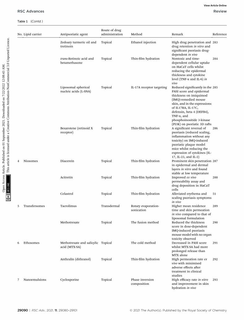
Table 1 (Contd. )
No. Lipid carrier Antipsoriatic agentRoute of drugadministration Method Remark Reference
Zedoary turmeric oil andtretinoin
Topical Ethanol injection High drug penetration anddrug retention in vitro andsignicant psoriasis drug-dependent in vivo
283
trans-Retinoic acid andbetamethasone
Topical Thin-lm hydration Nontoxic and time-dependent cellular uptakeon HaCaT cells whilstreducing the epidermalthickness and cytokinelevel (TNF-a and IL-6) invivo
284
Liposomal sphericalnucleic acids (L-SNA)
Topical IL-17A receptor targeting Reduced signicantly in thePASI score and epidermalthickness on imiquimod(IMQ)-remedied mouseskin, and in the expressionsof IL17RA, IL-17C,defensin, beta 4 (DEFB4),TNF-a, andphosphoinositide 3-kinase(PI3K) on psoriatic 3D ras
285
Bexarotene (retinoid Xreceptor)
Topical Thin-lm hydration A signicant reversal ofpsoriasis (reduced scaling,inammation without anytoxicity) on IMQ-inducedpsoriatic plaque modelmice whilst reducing theexpression of cytokines (IL-17, IL-23, and IL-2)
286
4 Niosomes Diacerein Topical Thin-lm hydration Prominent skin penetrationin epidermal and dermallayers in vitro and foundstable at low temperature
287
Acitretin Topical Thin-lm hydration Improved ex vivopermeability assay anddrug deposition in HaCaTcells
288
Celastrol Topical Thin-lm hydration Alleviated erythema andscaling psoriasis symptomsin vivo
51
5 Transfersomes Tacrolimus Transdermal Rotary evaporation-sonication
Higher mean residencetime and skin permeationin vivo compared to that ofliposomal formulation
289
Methotrexate Topical The fusion method Reduced the thicknessscore in dose-dependentIMQ-induced psoriasismousemodel with no organtoxicity observed
290
6 Ethosomes Methotrexate and salicylicacid (MTX-SA)
Topical The cold method Decreased in PASI scorewhilst MTX-SA had moreprolonged release thanMTX alone
291
Anthralin (dithranol) Topical Thin-lm hydration High permeation rate exvivo with minimisedadverse effects aertreatment in clinicalstudies
292
7 Nanoemulsions Cyclosporine Topical Phase inversioncomposition
High efficacy rate in vitroand improvement in skinhydration in vivo
293
29090 | RSC Adv., 2021, 11, 29080–29101 © 2021 The Author(s). Published by the Royal Society of Chemistry
RSC Advances Review
Ope
n A
cces
s A
rtic
le. P
ublis
hed
on 0
1 Se
ptem
ber
2021
. Dow
nloa
ded
on 7
/22/
2022
12:
08:4
3 A
M.
Thi
s ar
ticle
is li
cens
ed u
nder
a C
reat
ive
Com
mon
s A
ttrib
utio
n-N
onC
omm
erci
al 3
.0 U
npor
ted
Lic
ence
.View Article Online

Table 1 (Contd. )
No. Lipid carrier Antipsoriatic agentRoute of drugadministration Method Remark Reference
Tacrolimus Topical Spontaneousemulsication
Prolonged-release patternand dermal bioavailabilityimprovement in vitro whilstshowed depletion in serumcytokines andimprovement in psoriasissymptoms in vivo
294
Methotrexate Topical Low energy emulsication Improved skin permeationand retention in deep skinlayers ex vivo with anincrease in antipsoriaticefficacy, effective skinretention, and lesser serumand tissue accumulation invivo
295
Curcumin (naturalcompound)
Topical Low-energy emulsication Multiple fold increase inskin permeation and fasthealing in psoriatic activity
296
Imiquimod and curcumin Topical Low energy emulsication Prevented the appearanceof psoriasis-like symptomsand slowing down thepsoriatic activity
297
Review RSC Advances
Ope
n A
cces
s A
rtic
le. P
ublis
hed
on 0
1 Se
ptem
ber
2021
. Dow
nloa
ded
on 7
/22/
2022
12:
08:4
3 A
M.
Thi
s ar
ticle
is li
cens
ed u
nder
a C
reat
ive
Com
mon
s A
ttrib
utio
n-N
onC
omm
erci
al 3
.0 U
npor
ted
Lic
ence
.View Article Online
5.3 Nanovesicular systems
Nanovesicular systems (also known as nanovesicles) can typi-cally be distinguished by their unilamellar structures, whichhave become up-and-coming drug delivery and targetingsystems.198 The original vesicles are conventional liposomesbefore they are adapted into niosomes, transfersomes, andethosomes, named aer their characteristics and composi-tions.199 They can be assembled per need of their membranecomponents, and the type of the drug (hydrophobic or hydro-philic),199 which are useful for topical delivery of psoriasis.27
5.3.1 Liposomes. The liposomes are nano-, and micro-sized colloidal multilayer vesicles (50–1000 nm in diameter)consist of an aqueous compartment enclosed by natural orsynthetic lipid bilayers.37,200 Liposomes are permeable to waterand osmotically sensitive.201 Liposomes may present in multi-lamellar, small unilamellar, large unilamellar, or multivesicularshapes. Regardless of that, liposomes' size and homogeneity aremore essential for drug encapsulation compared to the lamellarnumbers.202,203 Liposomes can be prepared conventionally bythin-lm hydration, reverse-phase evaporation, detergent dial-ysis, or solvent injection method.199,204 Further formation stepsnamely sonication, high-pressure homogenisation or extrusiontechnique is required to obtain smaller unilamellarvesicles.199,204
Several drawbacks associated with phospholipid bilayers ofconventional liposomes are revealed in literature such as high-cost production, short half-life, and low solubility.199,200 Besides,the encapsulated drugmolecules within liposomesmay leak outand fuse because of their less stable surfactants (phospho-lipids).200 Phospholipids that make up the membrane compo-nents of liposomes have a negatively charged phosphate
© 2021 The Author(s). Published by the Royal Society of Chemistry
hydrophilic head group with two adjacent fatty acid chains ontheir tail. Phospholipids are disposed to undergo hydrolysis–oxidation reactions due to several factors: (1) small liposomesize and low concentration; (2) the presence of oxidant agents(chemical oxidants and enzymatic oxidants); and (3) modica-tion on fatty acyl chain. The literature also stated that liposomesize is an essential parameter for a stable liposome, whilstphospholipids' starting material could cause free radicaloxidation.205
Accordingly, some characteristics are modied to improveliposomal formulations for gene delivery application and arepertinent to antipsoriatic activity. The recent shi towardscationic liposomes by adding cationic lipids on the surface ofliposomes to form stable complexes has gained a lot of interestfor select nucleic acid delivery.206,207 In this regard, liposomesprovide some protection to the genes from degradation reac-tions. The cationic liposomal structures will get entrapped tothe large negatively charged DNA fragments, possibly aboutchromosome size, and, most importantly, can target speciccells or tissues.200
5.3.2 Niosomes. Niosomes are the best alternative to lipo-somes. They are developed to overcome the low physicalstability and expensive production of liposomes.208,209 Niosomesobtained the name because they consist of nonionic surfac-tants, thus are less toxic to the human's body since they carry nocharge.208,210 However, the earliest reported use of niosomes(known as synthetic liposomes) was in cosmetical application,coined by cosmetic giant L’Oreal in the 1970s and 1980s,211,212
before being explored in pharmaceutical applications. Sincethey are made up of nonionic surfactants, they are less hae-molytic and disrupt the cellular surfaces, thus, exhibiting
RSC Adv., 2021, 11, 29080–29101 | 29091
RSC Advances Review
Ope
n A
cces
s A
rtic
le. P
ublis
hed
on 0
1 Se
ptem
ber
2021
. Dow
nloa
ded
on 7
/22/
2022
12:
08:4
3 A
M.
Thi
s ar
ticle
is li
cens
ed u
nder
a C
reat
ive
Com
mon
s A
ttrib
utio
n-N
onC
omm
erci
al 3
.0 U
npor
ted
Lic
ence
.View Article Online
biocompatible properties.213–215 The formation of niosomes isusually with or without cholesterol, in which the latter affordsthe membrane rigidity and stability due to its chargepresence.216,217
Different preparation methods of niosomes have been re-ported including ether injection method, thin-lm hydrationmethod, emulsion method, reverse phase evaporation method,and micro uidisation method, which each could yield differentvesicles sizes.209,218,219 It is signied that a favourable niosome ofamphiphilic bilayers for high entrapment efficiency can beproduced by taking into consideration of the hydrophilic-lipophilic balance (HLB) value of nonionic surfactants between4 and 8.218,220 The choice of nonionic surfactants that falls withinthe range is typically amongst Tween, Span, and Brij. Besides,a stable niosome formation for better drug loading can be ach-ieved had long hydrocarbon chains used without a doublebond.220 It is also ubiquitous for nonionic surfactants with a highHLB value, notably Tween 20, Tween 60, and Tween 80 to inte-grate cholesterol for stabilisation due to their imbalance heyhydrophilic heads and small hydrophobic tails.220
In general, niosomes exhibit similar physicochemical prop-erties, drug delivery studies, and in vivo behaviour with lipo-somes.210,221 Even so, they have different structural compositionbilayer as niosomes exhibit higher chemical, physical, andbiological stabilities of the vesicles.221 As a result, niosomalformulations may offer several advantages over liposomes inrespect of higher drug entrapment and cheaper industrialmanufacturing.209,210,221 However, this interesting nanovesicle isrelatively new and young in pharmaceutical applicationscompared to liposomes.221 The lack of generally recognised assafe (GRAS) components in niosomes (like phospholipids inliposomes) might be one of their drawbacks,212 whilst theaforementioned commercial surfactants (Tween and Span)reportedly exhibit polydispersity feature, which is held asanother shortcoming from niosomes.210
5.3.3 Transfersomes. Transfersomes (also known as ultra-deformable liposomes or elastic liposomes),222,223 wasdesigned by Cevc and Blume in 1992.224,225 The main compo-nents of transfersomes are phospholipids (similar to lipo-somes) and edge activators (EA), a single chain surfactant that isalso a membrane-soening agent (e.g., Tween 80, Span 80, andsodium cholate).224,226 Novel approaches have been introducedto provide better deformability, but there is no general recipe informulating the vesicles since their medium suspendingcomposition must be tailored individually to each type of drugand the specic payload.222,224 Besides, the high bilayer adapt-ability and vesicle stability are the two vital factors in theprocess of forming transfersomes.224
In essence, transfersomes are more deformable and elasticto efficiently penetrate through the small channels in the skincompared to conventional liposomes.226,227 As a result of theirself-optimising deformability, the membrane changing exi-bility upon reaching and passing through the skin pores can beattained spontaneously as they open up extracellular pathwaysof the cells.227 EA acts as a permeation enhancer to enabletransdermal penetration by destabilising lipid bilayers of thevesicles while enhancing the uidity of stratum corneum
29092 | RSC Adv., 2021, 11, 29080–29101
lipid.163,225 Thus, transdermal delivery can be enhanced byavoiding the rst-pass effect and be controlled and prolongedthe duration of the drug activity. These pharmacokinetic effectsallow the efficiency of short half-life drugs and increase thephysiological and pharmacological response.228 Amongst thecommon preparation methods of transfersomes are either thin-lm hydration, reverse-phase evaporation, ethanol injection, orfreeze–thaw method.226,229 It is generally regarded that trans-fersomes are superior to liposomes in terms of skin penetra-tion226,227,230 but relatively inferior to ethosomes in terms ofdeformability.231 However, transfersomes and ethosomes haveequivalent skin deposition ability had ethosomes not incorpo-rated another type of alcohol as will be discussed in the nextsubsection.232
5.3.4 Ethosomes. Ethosomes were developed by Touitouet al. in 1996, acting as another enhanced delivery of activeagents.233–235 Ethosomes are composed of phospholipids(similar to liposomes) and ethanol, in which ethanol plays anessential role in efficient topical drug delivery.236,237 Ethosomeshave a better penetration rate through the skin, probably due totheir high ethanolic content (20–50%). The so and malleablevesicles are obtained as a result of the coexistence of bothphospholipids and ethanol as proved by Touitou et al.238,239
Similar to transfersomes, therapeutic delivery can be adminis-tered via transdermal route by ethosomal systems.240 Thus, thevesicles have a higher therapeutic efficacy over liposomes interms of the greater permeation of the drug's ability as they cansqueeze themselves through the skin pores as established byseveral studies.241–243 A study by Yang et al. showed that etho-somal formulation had a higher quantity and depth of lipo-philic drugs to be delivered across the skin (transdermaldelivery) for a longer period when compared to liposomes dueto their interdependent composition.244
The proposedmechanism of ethosome transportation acrossthe skin is well-described by the literature; however, the exactmethod of how they act as vehicles is not fully understood.237
The underlying mechanism is upon ethanol's polar head groupinteraction with the lipid regions of the skin to allow theincrease of cell membrane lipid uidity, which results inimproved skin penetrability of the ethosomes.237 These etho-somes then permeate the skin, merge with cell membranelipids, and release the system's drug.245 A recent investigation byNiu and co-workers reported that that the ethanol solutioninuenced the swelling of the stratum corneum and theincrease in the size of keratinocytes, except that the ethosomeswere not responsible for the results. The ndings alsoconcluded that the ethosomes had better transdermal perme-ation effects since some of the ethosomes fused to the upperstratum corneum whilst some others travelled and released thedrug to the intercellular space during their penetration into thedeeper skin layers.246
Amongst popular ethosomal formulations are the classicalcold method, the hot method, ethanol injection-sonication,thin-lm hydration, and reverse phase-evaporationmethods.233 Ethosomal systems can be further classied intotheir composition as (1) classical ethosomes; (2) binary etho-somes (added with another type of alcohol like isopropyl
© 2021 The Author(s). Published by the Royal Society of Chemistry
Review RSC Advances
Ope
n A
cces
s A
rtic
le. P
ublis
hed
on 0
1 Se
ptem
ber
2021
. Dow
nloa
ded
on 7
/22/
2022
12:
08:4
3 A
M.
Thi
s ar
ticle
is li
cens
ed u
nder
a C
reat
ive
Com
mon
s A
ttrib
utio
n-N
onC
omm
erci
al 3
.0 U
npor
ted
Lic
ence
.View Article Online
alcohol); and (3) transethosomes (added with a chemicalsurfactant or edge activator).233 Several studies publishedcomparative studies between these different types of ethosomesand transfersomes. For instance, upon the comparison betweenclassical ethosomes, binary ethosomes, and transfersomes, itwas reported that binary ethosomes have the highest penetra-tion depth and uorescence intensity but both types of etho-somes have lesser skin deposition than transfersomes.231
Similarly, Zhang et al. concluded that the signicant amount ofethanol had inuenced the transdermal drug delivery ratherthan the skin deposition (topical delivery) of ethosomes. Hence,the ethosomal formulations could be adjusted with propyleneglycol to prolong the movement of the vesicles and accumulatein the skin.241 On top of that, transethosomes showed thehighest drug's incorporation efficiency as opposed to etho-somes and transfersomes, and as such, were more deformablethan the two because of the presence of both ethanol andsurfactant compositions.232
5.4 Nanoemulsions
Nanoemulsions are isotropic, heterogeneous system, consistingof two immiscible liquids of a sufficient drug's dispersion innanodroplet sizes.247 The two liquids usually are either water inoil (W/O) or oil in water (O/W) or double emulsion (W/O/W andO/W/O)248 stabilised by amphiphilic surfactants, with meandroplets of <200 nm depending on the phase media.249 Thesystems have been proven to virtually encapsulate lipophilicactive compounds for enhanced skin delivery topically.250
Besides, nanoemulsions are also excellent vehicles to encapsu-late essential oils and natural bioactive compounds as stated inthe literature and proven in recent studies.251–254 The emulsiontechnique is versatile, and as such, it can be rendered in spray,gel, cream, and aerosol. Several other reasons also lead to theoverwhelming number of research that utilises nanoemulsionmethods as drug delivery, such as they are feasible, non-invasive, and cost-effective, especially for high metabolicactive drugs.255,256
The choice of surfactants is vital in reducing the surface andinterfacial tension between oil and water, which can either beionic or nonionic.249 Amongst common surfactants or emulsi-fying agents are Tween® 80, amphiphilic proteins (caseinate),phospholipids (soy lecithin), polysaccharides, and polymers(PEG).257 Generally, nonionic surfactants are regarded as saferthan anionic surfactants because they have less local irritation,which is suitable for pharmaceutical applications.258 In addi-tion, they also have a lower CMC value than their ionic coun-terparts of the same alkyl chain length for a more stable drugdelivery system.259,260 Concurrently, a co-surfactant is oenneeded whereby alcohol groups with C3–C8 chain length likeethylene glycol, propylene glycol, ethanol, and propanol areadded to enhance the entropy and stabilise the colloidalsystem.247 It is interesting to note that, nanoemulsion structurehas the closest resemblance of a micelle, out of other lipidnanocarriers, except that the innermost part of nanoemulsionsis the oil phase that solubilises hydrophobic drugs. In choosingthe oils, different types of triglycerides from long-chain
© 2021 The Author(s). Published by the Royal Society of Chemistry
triglycerides (LCT), medium-chain triglycerides (MCT), andshort-chain triglycerides (SCT) with potent therapeutic benetshave been utilised as excipients and act as determinants in thebioavailability of the encapsulated API.261 Nevertheless, it isassumed that a more stable O/W formulation could be achievedthrough a less polar oil base.262
The preparation methods of nanoemulsions can generally bedivided into high energy and low energy methods.263 The high-energy method, such as high-pressure homogenisation,produces a robust way of synthesising nanoemulsions byapplying excess shear or mechanical device, to break largedroplets into nanosized particles.263,264 However, the high-energy method merely produces 1% nanoemulsion whilst99% of its heat is dissipated off, thus, a repeated series of cyclesis required, which is more applicable in large scale produc-tion.265 On the other hand, two different types of low-energymethods, namely phase inversion composition (PIC) andphase inversion temperature (PIT) are known to be energy-saving since nanoemulsions are produced by exploiting thesystem's low interfacial property.264,266,267 The PIC and PITmethods have gained special attention lately and become morefavourable methods in which temperature and composition arekept constant, respectively.266 Given the requirements of thelow-energy methods, composition variables act as determinantsin producing nanodroplets rather than the preparation vari-ables as in the high-energy method.267
Nanoemulsions are non-equilibrium systems with thermo-dynamically unstable and kinetically stable systems that aresensitive to environmental parameters like pH and tempera-ture.268,269 Consequently, they have a propensity to undergoocculation, coalescence, and/or Ostwald ripening phenomenathat are associated with unstable emulsions.270,271 However,with appropriate optimisation in the selection of their compo-nents as well as during formulating preparation, a stablenanoemulsion system can be achieved for drug delivery.
6 Future prospects of lipid-basednanoparticles in pharmaceuticalapplications
In line with nanotechnology evolution, we witness an over-whelming number of nanotechnology uses in pharmaceuticalapplications. As a result, the utilisation of nanomaterials intonanotechnology-based drug delivery systems is futuristic, andthe market value is projected to skyrocket six-times fold forapproximately US$334 billion by 2025 compared to that of overa decade ago.298 Nanomedicine will witness rigorous researchprogress within the next 20 years as more focus on materialsdesign is driven by clinical needs.299 Therefore, a new paradigmof psoriasis treatments is moving towards this novel therapeuticplatform.300,301
Interestingly, lipid-based nanoparticles have garneredspecial attention in psoriasis treatments because of theiradaptability and feasibility approach. Each different type oflipid-based nanoparticle is distinctive since their lipid compo-nents as surfactants can be tailored per suitability. This leads to
RSC Adv., 2021, 11, 29080–29101 | 29093
RSC Advances Review
Ope
n A
cces
s A
rtic
le. P
ublis
hed
on 0
1 Se
ptem
ber
2021
. Dow
nloa
ded
on 7
/22/
2022
12:
08:4
3 A
M.
Thi
s ar
ticle
is li
cens
ed u
nder
a C
reat
ive
Com
mon
s A
ttrib
utio
n-N
onC
omm
erci
al 3
.0 U
npor
ted
Lic
ence
.View Article Online
a further novel approach as researchers can broaden theirhorizon in selecting the lipid components as the startingmaterials for more natural and greener products, such as car-danol, sugar-based surfactants, glycolipids, and bio-surfactants.302–306However, the Cpp of the lipid surfactants mustbe acknowledged since the morphology prediction woulddetermine the success of preparing any lipid-based nano-particles. For example, non-ionic lipid surfactants that have (1/2< Cpp < 1) would form vesicles (niosomes) in an aqueousmedium but other types of non-ionic lipid surfactants with (1/3< Cpp < 1/2) would form cylindrical micelles (nano-emulsions),171–173 as discussed in Section 5.0.
Moreover, the option of encapsulating the API does not limitto a certain type of lipid-based nanoparticles based on thecompilation in Table 1. In this regard, we can see the develop-ments of the same API via different nanocarriers such asmethotrexate was successfully encapsulated into SLNs,272
NLCs,276,279 transfersomes,290 ethosomes,291 and nano-emulsions,295 with signicant therapeutic proles. Further-more, these different types of lipid-based nanoparticles havesuccessfully encapsulated several different antipsoriatic agentsor API. For example, liposomes have encapsulated cyclo-sporine,280,282 psoralens,281 zedoary turmeric oil and tretinoin,283
trans-retinoic acid and betamethasone,284 L-SNA,285 and bexar-otene.286 Therefore, these discriminations demonstrate thatlipid-based nanoparticles are versatile and highly promisingdrug carriers for antipsoriatic activity. They also have a highpotential for clinical studies and thus, to be developed inpharmaceuticals.
An online platform, http://www.ClinicalTrials.gov, serves asthe clinical trials database that gathers worldwide research andany clinical studies should be reported there to avoid evidencedistortion for clinical practice.307,308 However, based on thedatabase, there is no psoriasis nano-drug yet to be approved foruse aer clinical studies. The database also shows that there isonly one psoriasis nano-drug that is undergoing clinical trial.The study (Identier: NCT03348462) successfully developed twodifferent anthralin nanoformulations encapsulated into lipo-somes and ethosomes, which has already completed phase 4clinical trial and showed higher potent effects through ethoso-mal systems.292
In the pharmaceutical industry, it was reported that com-mercialised nano drugs are currently at the rst generation ofphase II clinical trials carried out by small and medium-sizedenterprises (SMEs) before entering the market.298 The tediousprocedures of clinical trials and nancial constraints havehindered the current progress whilst big pharmaceuticalcompanies are only interested in acquiring these SMEs oncethese emerging drugs get approved by the FDA.298 Some otherchallenges to realise in pharmaceuticals include that there isa need for further nanoparticle characterisation prole forsafety and toxicity-related issues, as well as a lack ofnanotechnology-regulated guidelines for the industry.309,310 It isalso noteworthy to mention that current nanomedicine inves-tigation on nano drugs is limited to the evaluation of in vitroand in vivo studies.311 Lastly, the interactions between thetissues and lipid-based nanoparticles should also be deeply
29094 | RSC Adv., 2021, 11, 29080–29101
explored since the drug delivery systems may exhibit intricatebehaviour in the human's body.312 For example, nanoparticlesmay be deposited in the internal organs and thus interact withother immune cells, which could develop other immune reac-tions.31 However, the bottom-up approach through the self-assembled lipid nanoparticles could improve the safetymeasures.31 Besides, the application of state-of-the-art technol-ogies namely articial intelligence (AI) and machine learningcould facilitate the nanotherapeutic delivery via stimulationand modelling processes by understanding both biochemicaland physicochemical properties of the nano drugs.313 Therefore,profound knowledge on the cytotoxicity and biological activitiesof nano-drugs are vital to be further investigated inclusively toproviding coherent knowledge on their short-term and long-term effects before being pursued in clinical studies.31,311,312
7 Conclusion
Psoriasis is a skin disease that is affected by several knownfactors including environmental and genetic, causing cellhyperproliferation. Psoriasis treatments provide a wide array oftreatment options, namely topical, systemic nonbiologic,systemic biologics, and phototherapy. Despite the many anti-psoriatic drugs with different mechanisms of action, topicaltreatment serves as the most convenient way to transport thetherapeutics across the skin barrier. Lipid-based nanoparticlesare promising novel nano delivery systems to improve the APIdelivery to the target site. Recent works show that different APIscan be encapsulated into different lipid nanocarriers but lipo-somes and nanoemulsions have the highest number ofsuccessful nano-based drugs formulated for psoriasis treat-ments. Enhanced skin permeation and alleviated psoriasissymptoms are the improvements of antipsoriatic agents intandem with lipid-based nanoparticles. Besides, the therapeu-tics also show highly favourable outcomes in alleviating psori-asis lesions as they enhance skin penetration, retention, andprolonged release due to their large surface area at nanoscalesize. However, further research and opportunities with othertypes of lipid-based nanocarriers like niosomes, transfersomes,and ethosomes are needed to explore their tremendous poten-tial in the antipsoriatic application's topical delivery. In general,the production cost of nano drugs is expensive in the pharma-ceutical industry. The development of nano drugs is also asso-ciated with cytotoxicity and safety issues but could be overcomeby an in-depth understanding of their interactions in thehuman's body. Based on the compilation of the recent works ofthe API with lipid-based nanocarriers, it is also observed thatthey exhibit higher drug penetration and bioavailabilitycompared to that of the control variable (conventional treat-ments without nanocarrier). Therefore, the production of nanodrugs is guaranteed success if optimised strategies are taken innanomedicine applications.
Author contributions
Conceptualization, U. U. M. N., N. A., N. S. and N. S. M. Y.;methodology, U. U. M. N.; writing – original dra preparation,
© 2021 The Author(s). Published by the Royal Society of Chemistry
Review RSC Advances
Ope
n A
cces
s A
rtic
le. P
ublis
hed
on 0
1 Se
ptem
ber
2021
. Dow
nloa
ded
on 7
/22/
2022
12:
08:4
3 A
M.
Thi
s ar
ticle
is li
cens
ed u
nder
a C
reat
ive
Com
mon
s A
ttrib
utio
n-N
onC
omm
erci
al 3
.0 U
npor
ted
Lic
ence
.View Article Online
U. U. M. N.; writing – review & editing, N. A., N. S. and N. S. M. Y.;visualization, U. U. M. N.; supervision, N. A. and N. S.; projectadministration, N. A. and N. S.; funding acquisition, N. A.; allauthors have read and agreed to the published version of themanuscript.
Conflicts of interest
There are no conicts to declare.
Acknowledgements
The authors gratefully acknowledge the Ministry of HigherEducation Malaysia (MOHE) for the research project nancialsupport through the Fundamental Research Grant Scheme(FP037-2019A) and the University of Malaya.
References
1 J. Takeshita, S. Grewal, S. M. Langan, N. N. Mehta, A. Ogdie,A. S. Van Voorhees and J. M. Gelfand, J. Am. Acad. Dermatol.,2017, 76, 377–390.
2 R. Parisi, I. Y. Iskandar, E. Kontopantelis, M. Augustin,C. E. Griffiths and D. M. Ashcro, Br. Med. J., 2020, 369,m1590.
3 A. W. Armstrong and C. Read, J. Am. Med. Assoc., 2020, 323,1945–1960.
4 B. Elewski, A. F. Alexis, M. Lebwohl, L. S. Gold, D. Pariser,J. D. Rosso and G. Yosipovitch, J. Eur. Acad. Dermatol.Venereol., 2019, 33, 1465–1476.
5 Z. Yiu, R. Warren and A. Menter, in Handbook of Psoriasisand Psoriatic Arthritis, ed. R. Warren and A. Menter, Adis,Cham, 2016.
6 S. K. Ippagunta, R. Gangwar, D. Finkelstein, P. Vogel,S. Pelletier, S. Gingras, V. Redecke and H. Hacker, Proc.Natl. Acad. Sci. U.S.A., 2016, 113, E6162–E6171.
7 W. Masson, M. Lobo and G. Molinero, Adv. Ther., 2020, 37,2017.
8 A. Rendon and K. Schakel, Int. J. Mol. Sci., 2019, 20, 1475.9 J. M. Fernandez-Armenteros, X. Gomez-Arbones, M. Buti-Sole, A. Betriu-Bars, V. Sanmartin-Novell, M. Ortega-Bravo,M. Martınez-Alonso and J. M. Casanova-Seuma, ActasDermo-Siliogracas, 2019, 110, 385–392.
10 E. Ogawa, Y. Sato, A. Minagawa and R. Okuyama, J.Dermatol., 2018, 45, 264–272.
11 C.-H. Lin, S.-H. Chang and J.-Y. Fang, Curr. Drug Ther., 2016,11, 21–40.
12 R. Parisi, D. P. Symmons, C. E. Griffiths and D. M. Ashcro,J. Invest. Dermatol., 2013, 133, 377–385.
13 M. Rademaker, K. Agnew, N. Anagnostou, M. Andrews,K. Armour, C. Baker, P. Foley, K. Gebauer, M. Gupta andG. Marshman, Australas. J. Dermatol., 2019, 60, 91–98.
14 A. Menter, K. M. Cordoro, D. M. R. Davis, D. Kroshinsky,A. S. Paller, A. W. Armstrong, C. Connor, B. E. Elewski,J. M. Gelfand, K. B. Gordon, A. B. Gottlieb, D. H. Kaplan,A. Kavanaugh, M. Kiselica, D. Kivelevitch, N. J. Korman,M. Lebwohl, C. L. Leonardi, J. Lichten, H. W. Lim,
© 2021 The Author(s). Published by the Royal Society of Chemistry
N. N. Mehta, S. L. Parra, A. L. Pathy, E. A. Farley Prater,R. N. Rupani, M. Siegel, B. Stoff, B. E. Strober,E. B. Wong, J. J. Wu, V. Hariharan and C. A. Elmets, J.Am. Acad. Dermatol., 2020, 82, 161–201.
15 D. M. Balak and E. Hajdarbegovic, Psoriasis, 2017, 7, 87–94.16 N. J. Korman, Br. J. Dermatol., 2020, 182, 840–848.17 S. Gonzalez-Parra and E. Dauden, Actas Dermo-
Siliogracas, 2019, 110, 12–19.18 F. M. Sahi, A. Masood, N. A. Danawar, A. Mekaiel and
B. H. Malik, Cureus, 2020, 12(8), e9708.19 C. Fink, C. Alt, L. Uhlmann, C. Klose, A. Enk and
H. A. Haenssle, Br. J. Dermatol., 2019, 180, 390–396.20 D. Hagg, A. Sundstrom, M. Eriksson and M. Schmitt-
Egenolf, Am. J. Clin. Dermatol., 2017, 18, 583–590.21 V. K. Rapalli, G. Singhvi, S. K. Dubey, G. Gupta,
D. K. Chellappan and K. Dua, Biomed. Pharmacother.,2018, 106, 707–713.
22 G. Singhvi, S. Hejmady, V. K. Rapalli, S. K. Dubey andS. Dubey, in Delivery of Drugs, Elsevier, 2020, pp. 75–96.
23 M. Bhat, S. Pukale, S. Singh, A. Mittal and D. Chitkara, J.Drug Deliv. Sci. Technol., 2021, 62, 102328.
24 A. W. Armstrong, L. Puig, A. Joshi, M. Skup, D. Williams,J. Li, K. A. Betts and M. Augustin, JAMA Dermatol., 2020,156, 258–269.
25 P. Rider, Y. Carmi and I. Cohen, Int. J. Cell Biol., 2016, 2016,9259646.
26 A. Menter, B. E. Strober, D. H. Kaplan, D. Kivelevitch,E. F. Prater, B. Stoff, A. W. Armstrong, C. Connor,K. M. Cordoro, D. M. R. Davis, B. E. Elewski,J. M. Gelfand, K. B. Gordon, A. B. Gottlieb, A. Kavanaugh,M. Kiselica, N. J. Korman, D. Kroshinsky, M. Lebwohl,C. L. Leonardi, J. Lichten, H. W. Lim, N. N. Mehta,A. S. Paller, S. L. Parra, A. L. Pathy, R. N. Rupani,M. Siegel, E. B. Wong, J. J. Wu, V. Hariharan andC. A. Elmets, J. Am. Acad. Dermatol., 2019, 80, 1029–1072.
27 M. Pradhan, A. Alexander, M. R. Singh, D. Singh, S. Sarafand S. Saraf, Biomed. Pharmacother., 2018, 107, 447–463.
28 A. R. Fernandes, C. Martins-Gomes, A. Santini, A. M. Silvaand E. B. Souto, in Design of Nanostructures for VersatileTherapeutic Applications, ed. A. M. Grumezescu, WilliamAndrew Publishing, 2018, pp. 379–406.
29 M. R. Gigliobianco, C. Casadidio, R. Censi and P. DiMartino, Pharmaceutics, 2018, 10, 134.
30 W. Kaialy and M. Al Shaee, Adv. Colloid Interface Sci., 2016,228, 71–91.
31 W. Lu, J. Yao, X. Zhu and Y. Qi, Biomed. Pharmacother.,2021, 134, 111103.
32 X. Zhang, Z. Li, J. Gao, Z. Wang, X. Gao, N. Liu, M. Li,H. Zhang and A. Zheng, Molecules, 2020, 25, 1080.
33 M. Swierczewska, R. M. Crist and S. E. McNeil, inCharacterization of Nanoparticles Intended for DrugDelivery, Humana Press, New York, NY, 2018, pp. 3–16.
34 S. L. Jyothi, K. L. Krishna, V. K. Ameena Shirin, R. Sankar,K. Pramod and H. V. Gangadharappa, J. Drug Deliv. Sci.Technol., 2021, 62, 102364.
35 N. Vincent, D. D. Ramya and H. B. Vedha, Dermatol. Rep.,2014, 6, 5451.
RSC Adv., 2021, 11, 29080–29101 | 29095
RSC Advances Review
Ope
n A
cces
s A
rtic
le. P
ublis
hed
on 0
1 Se
ptem
ber
2021
. Dow
nloa
ded
on 7
/22/
2022
12:
08:4
3 A
M.
Thi
s ar
ticle
is li
cens
ed u
nder
a C
reat
ive
Com
mon
s A
ttrib
utio
n-N
onC
omm
erci
al 3
.0 U
npor
ted
Lic
ence
.View Article Online
36 T. W. Prow, J. E. Grice, L. L. Lin, R. Faye, M. Butler,W. Becker, E. M. T. Wurm, C. Yoong, T. A. Robertson,H. P. Soyer and M. S. Roberts, Adv. Drug Deliv. Rev., 2011,63, 470–491.
37 D. Medina-Cruz, B. Saleh, A. Vernet-Crua, A. Ajo, A. K. Royand T. J. Webster, in Wound Healing, Tissue Repair, andRegeneration in Diabetes, ed. D. Bagchi, A. Das and S. Roy,Academic Press, 2020, pp. 439–488.
38 C. Su, Y. Liu, R. Li, W. Wu, J. P. Fawcett and J. Gu, Adv. DrugDeliv. Rev., 2019, 143, 97–114.
39 T. Ramezanli, B. E. Kilfoyle, Z. Zhang and B. B. Michniak-Kohn, Int. J. Pharm., 2017, 516, 196–203.
40 A. L. Savian, D. Rodrigues, J. Weber, R. F. Ribeiro,M. H. Motta, S. R. Schaffazick, A. I. H. Adams, D. F. deAndrade, R. C. R. Beck and C. B. da Silva, Mater. Sci. Eng.C, 2015, 46, 69–76.
41 P. K. Tripathi, B. Gorain, H. Choudhury, A. Srivastava andP. Kesharwani, Heliyon, 2019, 5, e01343.
42 M. Lapteva, V. Santer, K. Mondon, I. Patmanidis,G. Chiriano, L. Scapozza, R. Gurny, M. Moller andY. N. Kalia, J. Control. Release, 2014, 196, 9–18.
43 K.-L. Mao, Z.-L. Fan, J.-D. Yuan, P.-P. Chen, J.-J. Yang, J. Xu,D.-L. ZhuGe, B.-H. Jin, Q.-Y. Zhu, B.-X. Shen, Y. Sohawon,Y.-Z. Zhao and H.-L. Xu, Colloids Surf. B Biointerfaces,2017, 160, 704–714.
44 S. A. Fereig, G. M. El-Zaafarany, M. G. Arafa andM. M. A. Abdel-Mottaleb, Carbohydr. Polym., 2021, 268,118238.
45 K. Yadav, A. Soni, D. Singh and M. R. Singh, Prog. Biomater.,2021, 10, 1–17.
46 S. Sur, A. Rathore, V. Dave, K. R. Reddy, R. S. Chouhan andV. Sadhu, Nano-Struct. Nano-Objects, 2019, 20, 100397.
47 M. T. C. P. Bernardes, S. B. N. Agostini, G. R. Pereira, L. P. daSilva, J. B. da Silva, M. L. Bruschi, R. D. Novaes andF. C. Carvalho, Eur. J. Pharm. Sci., 2021, 165, 105956.
48 D. Limon, K. Tallo Domınguez, M. L. Garduno-Ramırez,B. Andrade, A. C. Calpena and L. Perez-Garcıa, ColloidsSurf. B Biointerfaces, 2019, 181, 657–670.
49 Q. Li, F. Li, X. Qi, F. Wei, H. Chen and T. Wang, ColloidsSurf. B Biointerfaces, 2020, 195, 111246.
50 S. Kumar, M. Prasad and R. Rao, Mater. Sci. Eng. C, 2021,119, 111605.
51 S. Meng, L. Sun, L. Wang, Z. Lin, Z. Liu, L. Xi, Z. Wang andY. Zheng, Colloids Surf. B Biointerfaces, 2019, 182, 110352.
52 S. Jacob, A. B. Nair, J. Shah, N. Sreeharsha, S. Gupta andP. Shinu, Pharmaceutics, 2021, 13, 357.
53 X. Li, J. F. Lovell, J. Yoon and X. Chen, Nat. Rev. Clin. Oncol.,2020, 17, 657–674.
54 G. R. Nirmal, Z.-C. Lin, M.-J. Tsai, S.-C. Yang, A. Alalaiweand J.-Y. Fang, J. Controlled Release, 2021, 333, 487–499.
55 J. Chen, Z. Zeng, L. Huang, S. Luo, J. Dong, F. H. Zhou,K. Zhou, L. Wang and L. Kang, Am. J. Transl. Res., 2020,12, 3089–3115.
56 H.-Q. Liu, Y.-M. Wang, W.-F. Li, C. Li, Z.-H. Jiang, J. Bao,J.-F. Wei, H.-T. Jin and A.-P. Wang, Cell. Physiol. Biochem.,2017, 44, 200–214.
29096 | RSC Adv., 2021, 11, 29080–29101
57 P. R. Ebani, L. Stefanello, B. Luiza Kuhn, C. Piccinin Frizzo,T. Augusto Lima Burgo, C. Luisa Kloster and M. AntonioVilletti, J. Mol. Liq., 2021, 336, 116874.
58 R. Kumar, in Nanocarriers for Drug Delivery, ed. S. S.Mohapatra, S. Ranjan, N. Dasgupta, R. K. Mishra and S.Thomas, Elsevier, 2019, pp. 249–284.
59 M. Pradhan, D. Singh and M. R. Singh, J. Controlled Release,2013, 170, 380–395.
60 M. Gupta, U. Agrawal and S. P. Vyas, Expet Opin. Drug Deliv.,2012, 9, 783–804.
61 B. Garcıa-Pinel, C. Porras-Alcala, A. Ortega-Rodrıguez,F. Sarabia, J. Prados, C. Melguizo and J. M. Lopez-Romero, Nanomaterials, 2019, 9, 638.
62 P. Ghasemiyeh and S. Mohammadi-Samani, Res. Pharm.Sci, 2018, 13, 288–303.
63 S.-Y. Chuang, C.-H. Lin, T.-H. Huang and J.-Y. Fang,Nanomaterials, 2018, 8, 42.
64 C. Albanesi, in Clinical Immunology, Elsevier, 2019, pp. 871–878. e1.
65 B. Lukasch, H. Westerdahl, M. Strandh, H. Winkler,Y. Moodley, F. Knauer and H. Hoi, PeerJ, 2017, 5, e3679.
66 M. Wieczorek, E. T. Abualrous, J. Sticht, M. Alvaro-Benito,S. Stolzenberg, F. Noe and C. Freund, Front. Immunol.,2017, 8, 292.
67 V. Kumar, A. K. Abbas and J. C. Aster, Robbins BasicPathology, Elsevier, Philadelphia, PA, 10th edn, 2018.
68 J. B. Zabriskie, Essential clinical immunology, CambridgeUniversity Press, 2009.
69 M. P. Schon, Front. Immunol., 2019, 10, 1764.70 J. M. Hugh and J. M. Weinberg, Cutis, 2018, 102, 6–12.71 H. R. Hampton and T. Chtanova, Front. Immunol., 2019, 10,
1168.72 A. Wang and Y. Bai, J. Dermatol., 2020, 47, 104–113.73 C. de Jesus-Gil, L. Sans-de SanNicolas, I. Garcıa-Jimenez,
M. Ferran, A. Celada, A. Chiriac, R. M. Pujol andL. F. Santamaria-Babı, Front. Immunol., 2021, 12, 524.
74 V. R. Khairutdinov, A. F. Mikhailichenko, I. E. Belousova,E. Sh. Kuligina, A. V. Samtsov and E. N. Imyanitov, An.Bras. Dermatol., 2017, 92, 41–44.
75 B. Polese, H. Zhang, B. Thurairajah and I. L. King, Front.Immunol., 2020, 11, 242.
76 B. Wawrzycki, A. Pietrzak, E. Grywalska, D. Krasowska,G. Chodorowska and J. Rolinski, Arch. Immunol. Ther.Exp., 2019, 67, 103–108.
77 N. C. Brembilla, L. Senra and W.-H. Boehncke, Front.Immunol., 2018, 9, 1682.
78 M. Tampa, M.-I. Sarbu, M.-I. Mitran, C.-I. Mitran, C. Mateiand S.-R. Georgescu, Dis. Markers, 2018, 2018, 5823684.
79 A. M. Martins, A. Ascenso, H. M. Ribeiro and J. Marto, Mol.Neurobiol., 2020, 57, 2391–2419.
80 G. P. Thami and R. Sarkar, Clin. Exp. Dermatol., 2002, 27,99–103.
81 J. A. Zeichner, J. Clin. Aesthetic Dermatol., 2010, 3, 37–40.82 A. Bhatia, B. Singh, B. Amarji, P. Negi, A. Shukla and
O. P. Katare, Int. J. Dermatol., 2011, 50, 1246–1248.
© 2021 The Author(s). Published by the Royal Society of Chemistry
Review RSC Advances
Ope
n A
cces
s A
rtic
le. P
ublis
hed
on 0
1 Se
ptem
ber
2021
. Dow
nloa
ded
on 7
/22/
2022
12:
08:4
3 A
M.
Thi
s ar
ticle
is li
cens
ed u
nder
a C
reat
ive
Com
mon
s A
ttrib
utio
n-N
onC
omm
erci
al 3
.0 U
npor
ted
Lic
ence
.View Article Online
83 S. Sekhon, C. Jeon, M. Nakamura, L. A, D. Yan, J. J. Wu,W. Liao and T. Bhutani, J. Dermatol. Treat., 2018, 29, 230–232.
84 W. R. Heymann, J. Am. Acad. Dermatol., 2019, 80, 56–57.85 K. Blakely and M. Gooderham, Psoriasis Auckl. NZ, 2016, 6,
33–40.86 H.Mysliwiec, P. Mysliwiec, A. Baran and I. Flisiak, Adv. Med.
Sci., 2016, 61, 207–211.87 K. A. Hollywood, C. L. Winder, W. B. Dunn, Y. Xu,
D. Broadhurst, C. E. M. Griffiths and R. Goodacre, Mol.Biosyst., 2015, 11, 2198–2209.
88 C. Mo, D. Shetti and K. Wei, Molecules, 2019, 24, 2727.89 T. Benezeder, C. Painsi, V. Patra, S. Dey, M. Holcmann,
B. Lange-Asschenfeldt, M. Sibilia and P. Wolf, eLife, 2020,9, e56991.
90 J. Holstein, B. Fehrenbacher, J. Bruck, E. Muller-Hermelink,I. Schafer, M. Carevic, B. Schittek, M. Schaller, K. Ghoreschiand F. C. Eberle, J. Dermatol. Sci., 2017, 87, 236–245.
91 V. Kadian, S. Kumar, K. Saini, V. Kakkar and R. Rao, Curr.Drug Res. Rev., 2020, 12, 82–96.
92 M. S. Heath, D. R. Sahni, Z. A. Curry and S. R. Feldman,Expert Opin. Drug Metab. Toxicol., 2018, 14, 919–927.
93 I. Erol, N. U. Okur, D. Orak, H. Sipahi, A. Aydın and O. Ozer,Pharm. Dev. Technol., 2020, 25, 909–918.
94 E. L. Roux and H. Frow, Prescriber, 2020, 31, 9–17.95 E. Tanghetti, M. Lebwohl and L. Stein Gold, J. Drugs
Dermatol. JDD, 2018, 17, 1280–1287.96 L. S. Gold, M. G. Lebwohl, J. L. Sugarman, D. M. Pariser,
T. Lin, G. Martin, R. Pillai, R. Israel and T. Ramakrishna,J. Am. Acad. Dermatol., 2018, 79, 287–293.
97 G. Micali, A. E. Verzı, G. Giuffrida, E. Panebianco,M. L. Musumeci and F. Lacarrubba, Clin. Cosmet. Investig.Dermatol., 2019, 12, 953–959.
98 J. F. Merola, A. Qureshi and M. E. Husni, Dermatol. Ther.,2018, 31, e12589.
99 L. Guenther, C. Lynde and Y. Poulin, J. Cutan. Med. Surg.,2019, 23, 27S–34S.
100 S. Jindal, R. Awasthi, D. Singhare and G. T. Kulkarni, Med.Hypotheses, 2020, 142, 109838.
101 Z. Yin, J. Xu and D. Luo, J. Biomed. Res., 2011, 25, 385–391.102 X. Huang and B. Xu, Dermatology, 2015, 231, 41–49.103 R. Torsekar and M. M. Gautam, Indian Dermatol. Online J.,
2017, 8, 235.104 C. Stull, S. Grossman and G. Yosipovitch, Am. J. Clin.
Dermatol., 2016, 17, 617–624.105 S. Baboota, M. S. Alam, S. Sharma, J. K. Sahni, A. Kumar
and J. Ali, Int. J. Pharm. Investig., 2011, 1, 139–147.106 C. L. Carroll, J. Clarke, F. Camacho, R. Balkrishnan and
S. R. Feldman, Arch. Dermatol., 2005, 141, 43–46.107 A. Tosti, B. M. Piraccini, N. Cameli, F. Kokely, C. Plozzer,
G. E. Cannata and C. Benelli, Br. J. Dermatol., 1998, 139,655–659.
108 M. S. Al-Dhubaibi, Int. J. Health Sci., 2018, 12, 33.109 A. Filoni, M. Vestita, M. Congedo, G. Giudice, S. Tafuri and
D. Bonamonte, Medicine, 2018, 97, e11185.110 K. Satake, T. Amano and T. Okamoto, Sci. Rep., 2019, 9,
16322.
© 2021 The Author(s). Published by the Royal Society of Chemistry
111 S. Gabros, T. A. Nessel and P. M. Zito, in StatPearls,StatPearls Publishing, Treasure Island (FL), 2021.
112 E. C. Kleyn, E. Morsman, L. Griffin, J. J. Wu, P. Cm van deKerkhof, W. Gulliver, J. M. van der Walt and L. Iversen, J.Dermatol. Treat., 2019, 30, 311–319.
113 T. Frantz, E. G. Wright, E. A. Balogh, A. Cline, A. L. Adler-Neal and S. R. Feldman, Children, 2019, 6, 125.
114 E. Choi, N. S. Chandran and C. Tan, Singapore Med. J., 2020,61(3), 149.
115 S. Y. Song, S.-Y. Jung and E. Kim, J. Dermatol. Treat., 2019,30, 245–250.
116 G. Martin, M. Young and L. Aldredge, J. Clin. AestheticDermatol., 2019, 12, 13–26.
117 A. Menter, J. M. Gelfand, C. Connor, A. W. Armstrong,K. M. Cordoro, D. M. R. Davis, B. E. Elewski,K. B. Gordon, A. B. Gottlieb, D. H. Kaplan, A. Kavanaugh,M. Kiselica, D. Kivelevitch, N. J. Korman, D. Kroshinsky,M. Lebwohl, C. L. Leonardi, J. Lichten, H. W. Lim,N. N. Mehta, A. S. Paller, S. L. Parra, A. L. Pathy,E. F. Prater, R. S. Rahimi, R. N. Rupani, M. Siegel,B. Stoff, B. E. Strober, E. B. Tapper, E. B. Wong, J. J. Wu,V. Hariharan and C. A. Elmets, J. Am. Acad. Dermatol.,2020, 82, 1445–1486.
118 T. Elango, A. Thirupathi, S. Subramanian, P. Ethiraj,H. Dayalan and P. Gnanaraj, Clin. Exp. Med., 2017, 17,371–381.
119 C. A. da Silva, K. Von Kossel, M. Leszczynski, T. Melnik andR. Riera, Cochrane Database Syst. Rev., 2019, 2019,CD010498.
120 J. J. Wu, A. Guerin, M. Sundaram, K. Dea, M. Cloutier andP. Mulani, J. Am. Acad. Dermatol., 2017, 76, 81–90.
121 C. W. Choi, B. R. Kim, J. Ohn and S. W. Youn, Ann.Dermatol., 2017, 29, 55–60.
122 E. Baskan, S. Yazici, S. Tunali and H. Saricaoglu, J.Dermatol. Treat., 2016, 27, 328–331.
123 V. Di Lernia, L. Stingeni, V. Boccaletti, P. G. CalzavaraPinton, C. Guarneri, A. Belloni Fortina, M. Panzone,M. Corazza, I. Neri, S. Cambiaghi, C. Lasagni, E. Ficarelliand P. Gisondi, J. Dermatol. Treat., 2016, 27, 395–398.
124 S. Dogra, R. Mahajan, T. Narang and S. Handa, J. Dermatol.Treat., 2017, 28, 18–20.
125 B. R. Kim, S. Yang, E. J. Doh, C. W. Choi and S. W. Youn,Ann. Dermatol., 2018, 30, 143–149.
126 C. J. Oh, K. M. Das and A. B. Gottlieb, J. Am. Acad. Dermatol.,2000, 42, 829–830.
127 M. Frleta, S. Siebert and I. B. McInnes, Curr. Rheumatol.Rep., 2014, 16, 414.
128 J. E. Hawkes, B. Y. Yan, T. C. Chan and J. G. Krueger, J.Immunol., 2018, 201, 1605–1613.
129 K. Godse, Indian J. Dermatol., 2017, 62, 195–199.130 M. Shirley and L. J. Scott, Drugs, 2016, 76, 1135–1145.131 A. Blauvelt, Expert Opin. Drug Saf., 2016, 15, 1413–1420.132 R. Reszke and J. C. Szepietowski, Immunotherapy, 2017, 9,
229–238.133 P. Mease and I. B. McInnes, Rheumatol. Ther., 2016, 3, 5–29.
RSC Adv., 2021, 11, 29080–29101 | 29097
RSC Advances Review
Ope
n A
cces
s A
rtic
le. P
ublis
hed
on 0
1 Se
ptem
ber
2021
. Dow
nloa
ded
on 7
/22/
2022
12:
08:4
3 A
M.
Thi
s ar
ticle
is li
cens
ed u
nder
a C
reat
ive
Com
mon
s A
ttrib
utio
n-N
onC
omm
erci
al 3
.0 U
npor
ted
Lic
ence
.View Article Online
134 A. Gottlieb, J. Sullivan, M. van Doorn, A. Kubanov, R. You,A. Parneix, S. Hugot and M. Milutinovic, J. Am. Acad.Dermatol., 2017, 76, 70–80.
135 A. Johar, S. Thevarajah, A. Heng, L. C. Chan, C. C. Ch’ng,N. A. Mohd Safdar, P. Muniandy, T. Taib, W. C. Tan andK. E. Tey, Dermatol. Res. Pract., 2019, 2019, 8923168.
136 A. Egeberg, J. P. Thyssen, J. J. Wu and L. Skov, Br. J.Dermatol., 2019, 180, 116–121.
137 J. J. Wu, S. R. Feldman, S. Rastogi, B. Menges, M. Lingohr-Smith and J. Lin, J. Dermatol. Treat., 2018, 29, 769–774.
138 M. Lebwohl, R. Rivera-Oyola and D. F. Murrell, J. Am. Acad.Dermatol., 2020, 82, 1217–1218.
139 P. C. M. van de Kerkhof and F. R. de Gruijl, J. Eur. Acad.Dermatol. Venereol., 2020, 34, 926–931.
140 K. Hyde, L. A. Cardwell, R. Stotts and S. R. Feldman, Am. J.Pharm. Benets, 2018, 10, 18–21.
141 C. A. Elmets, H. W. Lim, B. Stoff, C. Connor, K. M. Cordoro,M. Lebwohl, A. W. Armstrong, D. M. R. Davis, B. E. Elewski,J. M. Gelfand, K. B. Gordon, A. B. Gottlieb, D. H. Kaplan,A. Kavanaugh, M. Kiselica, D. Kivelevitch, N. J. Korman,D. Kroshinsky, C. L. Leonardi, J. Lichten, N. N. Mehta,A. S. Paller, S. L. Parra, A. L. Pathy, E. A. Farley Prater,R. N. Rupani, M. Siegel, B. E. Strober, E. B. Wong,J. J. Wu, V. Hariharan and A. Menter, J. Am. Acad.Dermatol., 2019, 81, 775–804.
142 J. M. Vassantachart, T. Soleymani and J. J. Wu, J. DrugsDermatol. JDD, 2016, 15, 995–1000.
143 V. T. Ma, C. S. Katzman, P. L. Palmbos, R. M. Patel,J. E. Gudjonsson and A. S. Alva, Curr. Probl. Cancer CaseRep., 2020, 1, 100004.
144 A. Ighani, A. C. R. Partridge, N. H. Shear, C. Lynde,W. P. Gulliver, C. Sibbald and P. Fleming, J. Cutan. Med.Surg., 2019, 23, 204–221.
145 P. Zhang and M. X. Wu, Lasers Med. Sci., 2018, 33, 173–180.146 S. H. Ibbotson, Front. Med., 2018, 5, 184.147 A. Morita, J. Dermatol., 2018, 45, 287–292.148 E. Racz and E. P. Prens, in Ultraviolet Light in Human Health,
Diseases and Environment. Advances in ExperimentalMedicine and Biology, ed. S. Ahmad, Springer, Cham,2017, pp. 287–294.
149 V. Ardeleanu, D. S. Radaschin and A. L. Tatu, Exp. Ther.Med., 2020, 20, 52–55.
150 M. Nakamura, B. Farahnik and T. Bhutani, F1000Res., 2016,5, 1684.
151 S. Das, Int. J. Res. Rev., 2019, 6, 258–263.152 B. Farahnik, M. Nakamura, R. K. Singh, M. Abrouk,
T. H. Zhu, K. M. Lee, M. V. Jose, R. DaLovisio, J. Koo,T. Bhutani and W. Liao, Dermatol. Ther., 2016, 6, 315–324.
153 Y. Ren, X. Song, L. Tan, C. Guo, M. Wang, H. Liu, Z. Cao,Y. Li and C. Peng, Front. Pharmacol., 2020, 11, 571535.
154 J. Koo and M. Nakamura, in Clinical Cases in Phototherapy.Clinical Cases in Dermatology, Springer, Cham, 2017, pp.13–31.
155 E. S. Chambers and M. Vukmanovic-Stejic, Immunology,2020, 160, 116–125.
156 S. J. van Rensburg, A. Franken and J. L. D. Plessis, Skin Res.Technol., 2019, 25, 595–605.
29098 | RSC Adv., 2021, 11, 29080–29101
157 J. D. Bos and M. M. H. M. Meinardi, Exp. Dermatol., 2000, 9,165–169.
158 M. B. Murphrey, J. H. Miao and P. M. Zito, in StatPearls,StatPearls Publishing, Treasure Island (FL), 2020.
159 D. J. Moore and A. V. Rawlings, Int. J. Cosmet. Sci., 2017, 39,366–372.
160 K. Vavrova, A. Kovacik and L. Opalka, Eur. Pharm. J., 2017,64, 28–35.
161 Y. Badhe, R. Gupta and B. Rai, J. Mol. Model., 2019, 25, 140.162 F. Arriagada and J. Morales, Curr. Pharm. Des., 2019, 25,
455–466.163 T. Jiang, T. Wang, T. Li, Y. Ma, S. Shen, B. He and R. Mo,
ACS Nano, 2018, 12, 9693–9701.164 N. Tiwari, A. S. Sonzogni and M. Calderon, Nanomed, 2019,
14, 2891–2895.165 X. Zhou, Y. Hao, L. Yuan, S. Pradhan, K. Shrestha,
O. Pradhan, H. Liu and W. Li, Chin. Chem. Lett., 2018, 29,1713–1724.
166 T. Sych, Y. Mely andW. Romer, Philos. Trans. R. Soc. Lond. BBiol. Sci., 2018, 373, 20170117.
167 W. Lu, in Encyclopedia of Nanotechnology, ed. B. Bhushan,Springer Netherlands, Dordrecht, 2012, pp. 2371–2382.
168 H. Su, F. Wang, W. Ran, W. Zhang, W. Dai, H. Wang,C. F. Anderson, Z. Wang, C. Zheng, P. Zhang, Y. Li andH. Cui, Proc. Natl. Acad. Sci. U.S.A., 2020, 117, 4518–4526.
169 S. H. Crayton, A. K. Chen, J. F. Liu, E. M. Higbee-Dempsey,C.-H. Huang, A. Tsourkas and Z. Cheng, in ComprehensiveBiomaterials II, ed. P. Ducheyne, Elsevier, Oxford, 2017,pp. 424–466.
170 S. Rana, J. Bhattacharjee, K. C. Barick, G. Verma,P. A. Hassan and J. V. Yakhmi, in Nanostructures forCancer Therapy, ed. A. Ficai and A. M. Grumezescu,Elsevier, 2017, pp. 177–209.
171 R. A. Khalil and A. A. Zarari, Appl. Surf. Sci., 2014, 318, 85–89.
172 D. Lombardo, M. A. Kiselev, S. Magazu and P. Calandra,Adv. Condens. Matter Phys., 2015, 2015, e151683.
173 J. N. Israelachvili, D. J. Mitchell and B. W. Ninham, J. Chem.Soc., Faraday Trans. 2, 1976, 72, 1525–1568.
174 D. Lombardo, P. Calandra, L. Pasqua and S. Magazu,Materials, 2020, 13, 1048.
175 P. Iqbal, J. A. Preece and P. M. Mendes, in SupramolecularChemistry, American Cancer Society, 2012.
176 Z. Huang, W. Song and X. Chen, Front. Chem., 2020, 8, 380.177 A. Sikder and S. Ghosh, Mater. Chem. Front., 2019, 3, 2602–
2616.178 L. T. Varma, N. Singh, B. Gorain, H. Choudhury,
M. M. Tambuwala, P. Kesharwani and R. Shukla, Curr.Drug Deliv., 2020, 17, 279–291.
179 A. Walduck, P. Sangwan, Q. Anh Vo, J. Ratcliffe, J. White,B. W. Muir and N. Tran, RSC Adv., 2020, 10, 33608–33619.
180 R. H. Muller, K. Mader and S. Gohla, Eur. J. Pharm.Biopharm., 2000, 50, 161–177.
181 N. Naseri, H. Valizadeh and P. Zakeri-Milani, Adv. Pharm.Bull., 2015, 5, 305–313.
182 W. Mehnert and K. Mader, Adv. Drug Deliv. Rev., 2012, 64,83–101.
© 2021 The Author(s). Published by the Royal Society of Chemistry

Review RSC Advances
Ope
n A
cces
s A
rtic
le. P
ublis
hed
on 0
1 Se
ptem
ber
2021
. Dow
nloa
ded
on 7
/22/
2022
12:
08:4
3 A
M.
Thi
s ar
ticle
is li
cens
ed u
nder
a C
reat
ive
Com
mon
s A
ttrib
utio
n-N
onC
omm
erci
al 3
.0 U
npor
ted
Lic
ence
.View Article Online
183 V. R. Salvi and P. Pawar, J. Drug Deliv. Sci. Technol., 2019, 51,255–267.
184 A. C. Silva, E. Gonzalez-Mira, M. L. Garcıa, M. A. Egea,J. Fonseca, R. Silva, D. Santos, E. B. Souto and D. Ferreira,Colloids Surf. B Biointerfaces, 2011, 86, 158–165.
185 V. Mishra, K. K. Bansal, A. Verma, N. Yadav, S. Thakur,K. Sudhakar and J. M. Rosenholm, Pharmaceutics, 2018,10, 191.
186 Y. Duan, A. Dhar, C. Patel, M. Khimani, S. Neogi,P. Sharma, N. S. Kumar and R. L. Vekariya, RSC Adv.,2020, 10, 26777–26791.
187 C. D. Pizzol, F. B. Filippin-Monteiro, J. A. S. Restrepo,F. Pittella, A. H. Silva, P. Alves de Souza, A. Machado deCampos and T. B. Creczynski-Pasa, Int. J. Environ. Res.Public. Health, 2014, 11, 8581–8596.
188 A. H. Silva, F. B. Filippin-Monteiro, B. Mattei, B. G. Zanetti-Ramos and T. B. Creczynski-Pasa, Sci. Total Environ., 2012,432, 382–388.
189 V.-A. Duong, T.-T.-L. Nguyen and H.-J. Maeng, Molecules,2020, 25, 4781.
190 E. B. Souto, I. Baldim, W. P. Oliveira, R. Rao, N. Yadav,F. M. Gama and S. Mahant, Expet Opin. Drug Deliv., 2020,17, 357–377.
191 G. Amasya, G. Sandri, A. Onay-Besikci, U. Badilli,C. Caramella, M. C. Bonferoni and N. Tarimci, Curr. DrugDeliv., 2016, 13, 1100–1110.
192 R. A. Khallaf, H. F. Salem and A. Abdelbary, Drug Deliv.,2016, 23, 3452–3460.
193 G. M. Gelfuso, M. S. Cunha-Filho and T. Gratieri, Ther.Deliv., 2016, 7, 735–737.
194 B. Gaba, M. Fazil, S. Khan, A. Ali, S. Baboota and J. Ali, Bull.Fac. Pharm. Cairo Univ., 2015, 53, 147–159.
195 A. Khosa, S. Reddi and R. N. Saha, Biomed. Pharmacother.,2018, 103, 598–613.
196 V. K. Rapalli, V. Kaul, T. Waghule, S. Gorantla, S. Sharma,A. Roy, S. K. Dubey and G. Singhvi, Eur. J. Pharm. Sci.,2020, 152, 105438.
197 S. Vaz, R. Silva, M. H. Amaral, E. Martins, J. M. Sousa Loboand A. C. Silva, Colloids Surf. B Biointerfaces, 2019, 179, 242–249.
198 S. Wadhwa, V. Garg, M. Gulati, B. Kapoor, S. K. Singh andN. Mittal, in Pharmaceutical Nanotechnology: BasicProtocols, ed. V. Weissig and T. Elbayoumi, Springer, NewYork, NY, 2019, pp. 1–17.
199 N. Grimaldi, F. Andrade, N. Segovia, L. Ferrer-Tasies,S. Sala, J. Veciana and N. Ventosa, Chem. Soc. Rev., 2016,45, 6520–6545.
200 H. Daraee, A. Etemadi, M. Kouhi, S. Alimirzalu andA. Akbarzadeh, Artif. Cells Nanomed. Biotechnol., 2016, 44,381–391.
201 N. Filipczak, J. Pan, S. S. K. Yalamarty and V. P. Torchilin,Adv. Drug Deliv. Rev., 2020, 156, 4–22.
202 E. Elizondo, E. Moreno, I. Cabrera, A. Cordoba, S. Sala,J. Veciana and N. Ventosa, in Progress in Molecular Biologyand Translational Science, ed. A. Villaverde, AcademicPress, 2011, vol. 104, pp. 1–52.
© 2021 The Author(s). Published by the Royal Society of Chemistry
203 L. Maja, K. Zeljko and P. Mateja, J. Supercrit. Fluids, 2020,165, 104984.
204 K. S. Ahmed, S. A. Hussein, A. H. Ali, S. A. Korma, Q. Lipengand C. Jinghua, J. Drug Target., 2019, 27, 742–761.
205 A. Reis and C. M. Spickett, Biochim. Biophys. ActaBiomembr., 2012, 1818, 2374–2387.
206 M. A. Obeid, R. J. Tate, A. B. Mullen and V. A. Ferro, in LipidNanocarriers for Drug Targeting, ed. A. M. Grumezescu,William Andrew Publishing, 2018, pp. 313–359.
207 K. Yadav, D. Singh, M. R. Singh and M. Pradhan, Med.Hypotheses, 2020, 145, 110322.
208 P. Bhardwaj, P. Tripathi, R. Gupta and S. Pandey, J. DrugDeliv. Sci. Technol., 2020, 56, 101581.
209 S. Chen, S. Hanning, J. Falconer, M. Locke and J. Wen, Eur.J. Pharm. Biopharm., 2019, 144, 18–39.
210 R. Bartelds, M. H. Nematollahi, T. Pols, M. C. A. Stuart,A. Pardakhty, G. Asadikaram and B. Poolman, PloS One,2018, 13, e0194179.
211 G. M. El Maghraby, M. F. Arafa and E. A. Essa, inApplications of Nanocomposite Materials in Drug Delivery,ed. E. M. Inamuddin, A. M. Asiri and A. Mohammad,Woodhead Publishing, 2018, pp. 469–490.
212 T. G. Singh and N. Sharma, in Nanobiomaterials in GalenicFormulations and Cosmetics, ed. A. M. Grumezescu,William Andrew Publishing, 2016, pp. 149–174.
213 I. Ali, M. R. Shah, S. Yousuf, S. Ahmed, K. Shah and I. Javed,Toxicol. Res., 2018, 7, 771–778.
214 G. P. Kumar and P. Rajeshwarrao, Acta Pharm. Sin. B, 2011,1, 208–219.
215 Y. Zhang, K. Zhang, Z. Wu, T. Guo, B. Ye, M. Lu, J. Zhao,C. Zhu and N. Feng, Int. J. Pharm., 2015, 478, 138–146.
216 R. Liang, L. Chen, W. Yokoyama, P. A. Williams andF. Zhong, J. Agric. Food Chem., 2016, 64, 9180–9188.
217 M. H. Nematollahi, A. Pardakhty, M. Torkzadeh-Mahanai,M. Mehrabani and G. Asadikaram, RSC Adv., 2017, 7,49463–49472.
218 R. Rajera, K. Nagpal, S. K. Singh and D. N. Mishra, Biol.Pharm. Bull., 2011, 34, 945–953.
219 P. L. Yeo, C. L. Lim, S. M. Chye, A. P. K. Ling and R. Y. Koh,Asian Biomed., 2018, 11, 301–314.
220 B. Lu, Y. Huang, Z. Chen, J. Ye, H. Xu, W. Chen and X. Long,Molecules, 2019, 24, 2322.
221 R. Muzzalupo and E. Mazzotta, Expet Opin. Drug Deliv.,2019, 16, 1145–1147.
222 J. Chen, W.-L. Lu, W. Gu, S.-S. Lu, Z.-P. Chen and B.-C. Cai,Expet Opin. Drug Deliv., 2013, 10, 845–856.
223 B. Piumitali, U. Neeraj and J. Jyotivardhan, Int. J. Nanosci.,2020, 19, 1950033.
224 G. Cevc and A. Chopra, in Percutaneous penetrationenhancers chemical methods in penetration enhancement,Springer, 2016, pp. 39–59.
225 P.-S. Wu, Y.-S. Li, Y.-C. Kuo, S.-J. J. Tsai and C.-C. Lin,Molecules, 2019, 24, 600.
226 S. A. T. Opatha, V. Titapiwatanakun and R. Chutoprapat,Pharmaceutics, 2020, 12, 855.
227 S. Rai, V. Pandey and G. Rai, Nano Rev. Exp., 2017, 8,1325708.
RSC Adv., 2021, 11, 29080–29101 | 29099

RSC Advances Review
Ope
n A
cces
s A
rtic
le. P
ublis
hed
on 0
1 Se
ptem
ber
2021
. Dow
nloa
ded
on 7
/22/
2022
12:
08:4
3 A
M.
Thi
s ar
ticle
is li
cens
ed u
nder
a C
reat
ive
Com
mon
s A
ttrib
utio
n-N
onC
omm
erci
al 3
.0 U
npor
ted
Lic
ence
.View Article Online
228 P. Chaurasiya, E. Ganju, N. Upmanyu, S. K. Ray and P. Jain,J. Drug Deliv. Ther., 2019, 9, 279–285.
229 P. Chauhan and B. K. Tyagi, J. Drug Deliv. Ther., 2018, 8,162–168.
230 S. Jain, P. Jain, R. B. Umamaheshwari and N. K. Jain, DrugDev. Ind. Pharm., 2003, 29, 1013–1026.
231 J.-P. Zhang, Y.-H. Wei, Y. Zhou, Y.-Q. Li and X.-A. Wu, Arch.Pharm. Res., 2012, 35, 109–117.
232 A. Ascenso, S. Raposo, C. Batista, P. Cardoso, T. Mendes,F. G. Praça, M. V. L. B. Bentley and S. Simoes, Int. J.Nanomedicine, 2015, 10, 5837–5851.
233 I. M. Abdulbaqi, Y. Darwis, N. A. K. Khan, R. A. Assi andA. A. Khan, Int. J. Nanomedicine, 2016, 11, 2279–2304.
234 M. M. A. Elsayed, O. Y. Abdallah, V. F. Naggar andN. M. Khalafallah, Int. J. Pharm., 2006, 322, 60–66.
235 E. Touitou, B. Godin and C. Weiss, Drug Dev. Res., 2000, 50,406–415.
236 D. Aggarwal and U. Nautiyal, Int. J. Pharm. Med. Res., 2016,4, 354–363.
237 P. Verma and K. Pathak, J. Adv. Pharm. Technol. Res., 2010,1, 274–282.
238 D. Mishra, N. Balekar, V. Dhote and P. Mishra, 2017.239 E. Touitou, N. Dayan, L. Bergelson, B. Godin andM. Eliaz, J.
Control. Release, 2000, 65, 403–418.240 B. Godin and E. Touitou, Crit. Rev. Ther. Drug Carr. Syst.,
2003, 20, 63–102.241 L.-N. Shen, Y.-T. Zhang, Q. Wang, L. Xu and N.-P. Feng, Int.
J. Pharm., 2014, 460, 280–288.242 Y.-T. Zhang, L.-N. Shen, Z.-H. Wu, J.-H. Zhao and
N.-P. Feng, Int. J. Pharm., 2014, 471, 449–452.243 C. Fan, X. Li, Y. Zhou, Y. Zhao, S. Ma, W. Li, Y. Liu and G. Li,
BioMed Res. Int., 2013, 161943.244 L. Yang, L. Wu, D. Wu, D. Shi, T. Wang and X. Zhu, Int. J.
Nanomedicine, 2017, 12, 3357–3364.245 P. K. Jaiswal, S. Kesharwani, R. Kesharwani and D. K. Patel,
J. Drug Deliv. Ther., 2016, 6, 7–17.246 X.-Q. Niu, D.-P. Zhang, Q. Bian, X.-F. Feng, H. Li, Y.-F. Rao,
Y.-M. Shen, F.-N. Geng, A.-R. Yuan, X.-Y. Ying and J.-Q. Gao,Int. J. Pharm. X, 2019, 1, 100027.
247 V. K. Rai, N. Mishra, K. S. Yadav and N. P. Yadav, J.Controlled Release, 2018, 270, 203–225.
248 H. H. Tayeb and F. Sainsbury, Nanomed, 2018, 13, 2507–2525.
249 N. Salim, N. Ahmad, S. H. Musa, R. Hashim, T. F. Tadrosand M. Basri, RSC Adv., 2016, 6, 6234–6250.
250 S. A. Yousef, Y. H. Mohammed, S. Namjoshi, J. E. Grice,H. A. E. Benson, W. Sakran and M. S. Roberts,Pharmaceutics, 2019, 11, 639.
251 O. Bayraktar, I. Erdogan, M. D. Kose and G. Kalmaz, inNanostructures for Antimicrobial Therapy, Elsevier, 2017,pp. 395–412.
252 R. K. Harwansh, R. Deshmukh and M. A. Rahman, J. DrugDeliv. Sci. Technol., 2019, 51, 224–233.
253 A. de J. Cenobio-Galindo, J. Ocampo-Lopez, A. Reyes-Munguıa, M. L. Carrillo-Inungaray, M. Cawood,G. Medina-Perez, F. Fernandez-Luqueno andR. G. Campos-Montiel, Antioxidants, 2019, 8, 500.
29100 | RSC Adv., 2021, 11, 29080–29101
254 Q. Liu, H. Huang, H. Chen, J. Lin and Q. Wang, Molecules,2019, 24, 4242.
255 L. Pavoni, D. R. Perinelli, G. Bonacucina, M. Cespi andG. F. Palmieri, Nanomaterials, 2020, 10, 135.
256 D. S. Shaker, R. A. H. Ishak, A. Ghoneim and M. A. Elhuoni,Sci. Pharm., 2019, 87, 17.
257 E. Sanchez-Lopez, M. Guerra, J. Dias-Ferreira, A. Lopez-Machado, M. Ettcheto, A. Cano, M. Espina, A. Camins,M. L. Garcia and E. B. Souto, Nanomaterials, 2019, 9, 821.
258 C. Pulce and J. Descotes, in Human Toxicology, ed. J.Descotes, Elsevier Science B.V., Amsterdam, 1996, pp.683–702.
259 Y. Lu, E. Zhang, J. Yang and Z. Cao, Nano Res., 2018, 11,4985–4998.
260 T. Tadros, in Encyclopedia of Colloid and Interface Science,ed. T. Tadros, Springer, Berlin, Heidelberg, 2013, pp. 209–210.
261 Y. Singh, J. G. Meher, K. Raval, F. A. Khan, M. Chaurasia,N. K. Jain and M. K. Chourasia, J. Controlled Release,2017, 252, 28–49.
262 M. Miastkowska, E. Sikora and J. Ogonowski, Period.Polytech. Chem. Eng., 2014, 58, 43–48.
263 M. Kumar, R. S. Bishnoi, A. K. Shukla and C. P. Jain, Prev.Nutr. Food Sci., 2019, 24, 225–234.
264 A. Gupta, A. Z. M. Badruddoza and P. S. Doyle, Langmuir,2017, 33, 7118–7123.
265 O. Alliod, J.-P. Valour, S. Urbaniak, H. Fessi, D. Dupin andC. Charcosset, Colloids Surf., A, 2018, 557, 76–84.
266 L. Wang, X. Guan, C. Zheng, N. Wang, H. Lu and Z. Huang,Langmuir, 2020, 36, 10082–10090.
267 M. N. Yukuyama, D. D. M. Ghisleni, T. J. A. Pinto andN. A. Bou-Chacra, Int. J. Cosmet. Sci., 2016, 38, 13–24.
268 S. Manickam, K. Sivakumar and C. H. Pang, Ultrason.Sonochem., 2020, 69, 105258.
269 L. A. Pascual-Pineda, E. Flores-Andrade,M. Jimenez-Fernandez and C. I. Beristain, Int. J. Food Sci.Technol., 2015, 50, 1174–1181.
270 A. Simonazzi, A. G. Cid, M. Villegas, A. I. Romero,S. D. Palma and J. M. Bermudez, in Drug Targeting andStimuli Sensitive Drug Delivery Systems, ed. A. M.Grumezescu, William Andrew Publishing, 2018, pp. 81–116.
271 C. Solans and I. Sole, Curr. Opin. Colloid Interface Sci., 2012,17, 246–254.
272 M. Ferreira, L. Barreiros, M. A. Segundo, T. Torres,M. Selores, S. A. Costa Lima and S. Reis, Colloids Surf. BBiointerfaces, 2017, 159, 23–29.
273 S. Trombino, R. Russo, S. Mellace, G. P. Varano,A. S. Lagana, F. Marcucci and R. Cassano, J. Drug Deliv.Sci. Technol., 2019, 49, 563–569.
274 A. Ali, S. Ali, M. Aqil, S. S. Imam, A. Ahad and A. Qadir, J.Drug Deliv. Sci. Technol., 2019, 52, 713–721.
275 J.-H. Kang, J. Chon, Y.-I. Kim, H.-J. Lee, D.-W. Oh, H.-G. Lee,C.-S. Han, D.-W. Kim and C.-W. Park, Int. J. Nanomedicine,2019, 14, 5381–5396.
276 P. Tripathi, A. Kumar, P. K. Jain and J. R. Patel, Int. J. Biol.Macromol., 2018, 120, 1322–1334.
© 2021 The Author(s). Published by the Royal Society of Chemistry
Review RSC Advances
Ope
n A
cces
s A
rtic
le. P
ublis
hed
on 0
1 Se
ptem
ber
2021
. Dow
nloa
ded
on 7
/22/
2022
12:
08:4
3 A
M.
Thi
s ar
ticle
is li
cens
ed u
nder
a C
reat
ive
Com
mon
s A
ttrib
utio
n-N
onC
omm
erci
al 3
.0 U
npor
ted
Lic
ence
.View Article Online
277 N. Kaur, K. Sharma and N. Bedi, Pharm. Nanotechnol., 2018,6, 133–143.
278 P. Sathe, R. Saka, N. Kommineni, K. Raza and W. Khan,Drug Dev. Ind. Pharm., 2019, 45, 826–838.
279 Y. O. Agrawal, U. B. Mahajan, H. S. Mahajan and S. Ojha,Int. J. Nanomedicine, 2020, 15, 4763–4778.
280 R. Kumar, S. Dogra, B. Amarji, B. Singh, S. Kumar, K. Vinay,R. Mahajan and O. Katare, JAMA Dermatol., 2016, 152, 807–815.
281 S. Doppalapudi, A. Jain, D. K. Chopra and W. Khan, Eur. J.Pharm. Sci., 2017, 96, 515–529.
282 M. Walunj, S. Doppalapudi, U. Bulbake and W. Khan, J.Liposome Res., 2020, 30, 68–79.
283 J. Chen, Y. Ma, Y. Tao, X. Zhao, Y. Xiong, Z. Chen andY. Tian, J. Liposome Res., 2020, 1–15.
284 W. Wang, G. Shu, K. Lu, X. Xu, M. Sun, J. Qi, Q. Huang,W. Tan and Y. Du, J. Nanobiotechnol., 2020, 18, 80.
285 H. Liu, R. S. Kang, K. Bagnowski, J. M. Yu, S. Radecki,W. L. Daniel, B. R. Anderson, S. Nallagatla, A. Schook,R. Agarwal, D. A. Giljohann and A. S. Paller, J. Invest.Dermatol., 2020, 140, 435–444.
286 R. Saka, H. Jain, N. Kommineni, N. Chella and W. Khan, J.Drug Deliv. Sci. Technol., 2020, 58, 101691.
287 S. R. M. Moghddam, A. Ahad, M. Aqil, S. S. Imam andY. Sultana, Mater. Sci. Eng. C, 2016, 69, 789–797.
288 I. I. Abu Hashim, N. F. Abo El-Magd, A. R. El-Sheakh,M. F. Hamed and A. E.-G. H. Abd El-Gawad, Int. J.Nanomedicine, 2018, 13, 1059–1079.
289 V. Parkash, S. Maan, V. Chaudhary, V. Jogpal, G. Mittal andV. Jain, J. Bioequivalence Bioavailab., 2018, 10, 99–106.
290 M. Bahramizadeh, M. Bahramizadeh, B. Kiafar,A. H. Jafarian, A. R. Nikpoor, M. Hatamipour, H. Esmaily,Z. Rezaeemehr, S. Golmohammadzadeh, S. A. Moosavianand M. R. Jafari, Int. J. Pharm., 2019, 569, 118623.
291 A. Chandra, G. Aggarwal, S. Manchanda and A. Narula,Pharm. Nanotechnol., 2019, 7, 362–374.
292 D. Fathalla, E. M. K. Youssef and G. M. Soliman,Pharmaceutics, 2020, 12, 446.
293 S. H. Musa, M. Basri, H. R. Fard Masoumi, N. Shamsudinand N. Salim, Int. J. Nanomedicine, 2017, 12, 2427–2441.
294 S. Sahu, S. S. Katiyar, V. Kushwah and S. Jain, Nanomed,2018, 13, 1985–1998.
© 2021 The Author(s). Published by the Royal Society of Chemistry
295 P. Rajitha, P. Shammika, S. Aiswarya, A. Gopikrishnan,R. Jayakumar and M. Sabitha, J. Drug Deliv. Sci. Technol.,2019, 49, 463–476.
296 M. S. Algahtani, M. Z. Ahmad and J. Ahmad, J. Drug Deliv.Sci. Technol., 2020, 59, 101847.
297 M. S. Algahtani, M. Z. Ahmad, I. H. Nourein and J. Ahmad,Biomolecules, 2020, 10, 968.
298 R. Bosetti and S. L. Jones, Nanomed, 2019, 14, 1367–1370.299 J. J. Richardson and F. Caruso, Nano Lett., 2020, 20, 1481–
1482.300 E. C. Murphy, S. W. Schaer and A. J. Friedman, Curr.
Dermatol. Rep., 2019, 8, 14–25.301 S. Saleem, M. K. Iqubal, S. Garg, J. Ali and S. Baboota, Expet
Opin. Drug Deliv., 2020, 17, 817–838.302 N. Ahmad, R. Ramsch, M. Llinas, C. Solans, R. Hashim and
H. A. Tajuddin, Colloids Surf. B Biointerfaces, 2014, 115,267–274.
303 L. Bai and D. J. McClements, J. Colloid Interface Sci., 2016,479, 71–79.
304 M. S. Behalo, E. Bloise, L. Carbone, R. D. Sole,D. Lomonaco, S. E. Mazzetto, G. Mele and L. Mergola, J.Exp. Nanosci., 2016, 11, 1274–1284.
305 O. W. Mak, N. Seman, N. H. Harun, R. S. D. Hussen,N. M. Rodzi and T. Heidelberg, J. Surfactants Deterg.,2015, 18, 973–980.
306 A. Sedaghat Doost, J. Van Camp, K. Dewettinck and P. Vander Meeren, Food Chem., 2019, 293, 134–143.
307 N. J. DeVito, S. Bacon and B. Goldacre, Lancet, 2020, 395,361–369.
308 M. Zwierzyna, M. Davies, A. D. Hingorani and J. Hunter, Br.Med. J., 2018, 361, k2130.
309 J. K. Patra, G. Das, L. F. Fraceto, E. V. R. Campos, M. delPilar Rodriguez-Torres, L. S. Acosta-Torres, L. A. Diaz-Torres, R. Grillo, M. K. Swamy and S. Sharma, J.Nanobiotechnol., 2018, 16, 71.
310 C. L. Ventola, Pharm. Ther., 2017, 42, 742–755.311 R. Kumar, S. V. Dalvi and P. F. Siril, ACS Appl. Nano Mater.,
2020, 3, 4944–4961.312 D. Lombardo, M. A. Kiselev and M. T. Caccamo, J.
Nanomater., 2019, 2019, 3702518.313 A. V. Singh, M. H. D. Ansari, D. Rosenkranz, R. S. Maharjan,
F. L. Kriegel, K. Gandhi, A. Kanase, R. Singh, P. Laux andA. Luch, Adv. Healthc. Mater., 2020, 9, 1901862.
RSC Adv., 2021, 11, 29080–29101 | 29101