Cefuroxime axetil loaded Solid Lipid Nanoparticles for enhanced activity against S. aureus biofilm
29
Accepted Manuscript Title: Cefuroxime axetil loaded Solid Lipid Nanoparticles for enhanced activity against S. aureus biofilm Author: Bhupender Singh Parameswara Rao Vuddanda M.R. Vijayakumar Vinod Kumar Preeti S. Saxena Sanjay Singh PII: S0927-7765(14)00178-7 DOI: http://dx.doi.org/doi:10.1016/j.colsurfb.2014.03.046 Reference: COLSUB 6367 To appear in: Colloids and Surfaces B: Biointerfaces Received date: 13-8-2013 Revised date: 25-3-2014 Accepted date: 26-3-2014 Please cite this article as: B. Singh, P.R. Vuddanda, V. Kumar, P.S. Saxena, S. Singh, Cefuroxime axetil loaded Solid Lipid Nanoparticles for enhanced activity against S. aureus biofilm, Colloids and Surfaces B: Biointerfaces (2014), http://dx.doi.org/10.1016/j.colsurfb.2014.03.046 This is a PDF file of an unedited manuscript that has been accepted for publication. As a service to our customers we are providing this early version of the manuscript. The manuscript will undergo copyediting, typesetting, and review of the resulting proof before it is published in its final form. Please note that during the production process errors may be discovered which could affect the content, and all legal disclaimers that apply to the journal pertain.
Transcript of Cefuroxime axetil loaded Solid Lipid Nanoparticles for enhanced activity against S. aureus biofilm
Accepted Manuscript
Title: Cefuroxime axetil loaded Solid Lipid Nanoparticles forenhanced activity against S. aureus biofilm
Author: Bhupender Singh Parameswara Rao Vuddanda M.R.Vijayakumar Vinod Kumar Preeti S. Saxena Sanjay Singh
PII: S0927-7765(14)00178-7DOI: http://dx.doi.org/doi:10.1016/j.colsurfb.2014.03.046Reference: COLSUB 6367
To appear in: Colloids and Surfaces B: Biointerfaces
Received date: 13-8-2013Revised date: 25-3-2014Accepted date: 26-3-2014
Please cite this article as: B. Singh, P.R. Vuddanda, V. Kumar, P.S. Saxena,S. Singh, Cefuroxime axetil loaded Solid Lipid Nanoparticles for enhancedactivity against S. aureus biofilm, Colloids and Surfaces B: Biointerfaces (2014),http://dx.doi.org/10.1016/j.colsurfb.2014.03.046
This is a PDF file of an unedited manuscript that has been accepted for publication.As a service to our customers we are providing this early version of the manuscript.The manuscript will undergo copyediting, typesetting, and review of the resulting proofbefore it is published in its final form. Please note that during the production processerrors may be discovered which could affect the content, and all legal disclaimers thatapply to the journal pertain.
Page 1 of 28
Accep
ted
Man
uscr
ipt
1
Cefuroxime axetil loaded Solid Lipid Nanoparticles for enhanced 1
activity against S. aureus biofilm 2
3
Bhupender Singh1, Parameswara Rao Vuddanda1, Vijayakumar M.R1, 4
Vinod Kumar2, Preeti S. Saxena2, Sanjay Singh*1 5
6
1Department of Pharmaceutics, Indian Institute of Technology (Banaras Hindu University), 7
Varanasi-221 005, India. 8
2Department of Zoology, Faculty of Science, Banaras Hindu University, Varanasi-221 005, 9
India. 10
11
12
13
14
15
16
*correspondence author 17 Prof. Sanjay Singh 18 Department of Pharmaceutics 19 Indian Institute of Technology (BHU) 20 Varanasi – 221005 21 India 22 Email: [email protected], [email protected] 23 Telephone: +91-542-6702712 24 25
Page 2 of 28
Accep
ted
Man
uscr
ipt
2
Abstract: 1
The present research work is focused on the development of solid lipid nanoparticles 2
of cefuroxime axetil (CA-SLN) for its enhanced inhibitory activity against Staphylococcus 3
aureus produced biofilm. CA-SLN was prepared by solvent emulsification/evaporation 4
method using single lipid (stearic acid (SA)) and binary lipids (SA and tristearin (TS)). 5
Process variables such as volume of dispersion medium, concentration of surfactant, 6
homogenization speed and time were optimized. The prepared SLN were characterized for 7
encapsulation efficiency, DSC, FT-IR, SEM, AFM, in vitro drug release, stability studies and 8
in vitro anti biofilm activity against Staphylococcus aureus biofilm. Among the process 9
variables, increased volume of dispersion medium, homogenization speed and time led to 10
increase in particle size whereas increase in surfactant concentration decreased the particle 11
size. SLN prepared using binary lipids exhibited higher entrapment efficiency than the single 12
lipid. DSC and FT-IR studies showed no incompatible interaction between drug and 13
excipients. CA-SLN showed two folds higher anti-biofilm activity invitro than pristine CA 14
against Staphylococcus aureus biofilm. 15
Keywords: 16
Nanoparticles, Binary lipids, Cefuroxime axetil, Biofilms, Microbial drug resistance 17
18
19
20
21
22
23
24
25
Page 3 of 28
Accep
ted
Man
uscr
ipt
3
1. Introduction: 1
Biofilms are microbially derived sessile community characterized by cells that 2
are irreversibly attached to a substratum or interface or to each other. They are embedded in a 3
matrix of extracellular polymeric substances that they have produced, and exhibit an altered 4
phenotype with respect to growth rate and gene transcription [1 - 2]. The structure and 5
physiological attributes of biofilm producing organisms confer resistance to anti-microbial 6
agents such as antibiotics, disinfectants and germicides. The living microorganisms in 7
biofilms can be 1000 times more resistance to anti-microbials than the floating (planktonic) 8
counterparts. Various pathogens, such as Escherichia coli, Salmonella, Yersinia 9
enterocolitica, Listeria and Campylobacter produce biofilms on the surface of food or 10
storage equipments. Moreover, the potential pathogenic bacteria such as Staphylococcus 11
aureus, Enterococcus faecalis, Streptococcus, E. coli, Klebsiella, and Pseudomonas tend to 12
grow on catheters, artificial joints, mechanical heart valves, etc. Thus, these organisms can 13
lead to persistent infections as a result of periodic release from the biofilms [3]. The 14
infections such as dental caries, periodontitis, otitis, musculoskeletal infections, 15
osteomyelitis, bacterial prostatitis, cystic fibrosis, pneumonia, native valve endocarditis, 16
pneumonia and urinary catheter cystitis are caused by common biofilm forming bacterial 17
species such as gram positive cocci, gram negative rod and anaerobic oral bacteria etc. Some 18
fungal species such as Candida albicans is also responsible for several infections through 19
biofilm formation. The frequency of infections caused by biofilms, especially in the 20
developed world is 60% as per the report of National Institute of Health (NIH) [4]. Therefore, 21
attention on eradication of biofilms is required to combat the resistance produced by the 22
biofilm forming microorganisms. 23
Cefuroxime axetil (CA) is a semi-synthetic and broad spectrum cephalosporin 24
antibiotic indicated in pharyngitis, tonsillitis, acute bacterial otitis, acute bacterial maxillary 25
Page 4 of 28
Accep
ted
Man
uscr
ipt
4
sinusitis, acute bacterial exacerbations of chronic bronchitis and secondary bacterial 1
infections of acute bronchitis, uncomplicated skin structure infections, urinary tract 2
infections, gonorrhoea and early lymph disease. Monzon et al. studied the potency of CA 3
against biofilm of S. aureus and concluded that the CA is most effective antibiotic than 4
vancomycin, tobramycin and ciprofloxacin [5]. However, the biopharmaceutical impediment 5
such as poor solubility, permeability into biofilm and higher dose frequency limits the 6
applicaiton of CA against biofilm. In this view, the objective of current research work is the 7
development of CA loaded SLN and evaluation of its efficacy against S. aureus biofilm. CA-8
SLN was prepared by solvent emulsification/evaporation method using single lipid, stearic 9
acid (SA) and binary lipids containing SA and tristearin (TS). Formulation variables such as 10
volume of dispersion medium, concentration of surfactant and process variables such as 11
homogenization speed and time were optimized to get minimal particle size and higher 12
entrapment efficiency. CA-SLN was characterized for encapsulation efficiency, DSC, FT-IR, 13
SEM, AFM, in vitro drug release, stability and evaluated for in vitro anti biofilm activity. 14
15
2. Materials: 16
CA and poloxamer 188 were obtained as gift samples from Lupin Pharmaceutical 17
Ltd., and Cadila healthcare Ltd., India, respectively. Stearic acid (SA), tristearin (TS) and 18
soya lecithin were purchased from Sigma-Aldrich (St Louis, Missouri). Methanol, 19
dichloromethane, chloroform and sodium dihydrogen phosphate were purchased from Merck 20
(Darmstadt, Germany). Muller Hinton agar medium was procured from Himedia Pvt. Ltd. 21
India. All other chemicals used in this study were of analytical grade. 22
23
24
Page 5 of 28
Accep
ted
Man
uscr
ipt
5
3. Methods: 1
3.1. Preparation of CA loaded SLN 2
CA loaded solid lipid nanoparticles were prepared from Solvent 3
emulsification/evaporation method described by Sjostrom and Bergenstahl [6]. In brief, 4
accurately weighed quantity of CA (20 mg), soya lecithin (40mg) and the lipid or mixture of 5
lipids (200 mg) were dissolved in the organic mixture of chloroform and dichloromethane 6
(3:1) in 10 ml beaker. The organic phase heated at 70 oC was added slowly in to the aqueous 7
phase (poloxamer 188 heated at 70 oC) and subjected to homogenization using Ultra Turrax 8
homogenizer. After homogenization, the dispersion was ultrasonicated using ultrasonicator 9
(Hielscher, Model UP200H, Germany) at 0.5 cycles and 60% amplitude for 10 minutes and 10
then it was allowed to cool at room temperature. The organic solvent mixture from the 11
nanoformulation was completely evaporated at 40°C under reduced pressure using 12
rotaevaporator (IKA RV 10) [7]. The study design is shown in Table 1 and 2. Stearic acid 13
was used to optimize the formulation variables (surfactant concentration/volume of 14
dispersion medium) and process variables (homogenization speed/time) as shown in table 1. 15
3.2. Characterization of particle size and zeta potential 16
Particle size and polydispersity index were determined by DelsaTM Nano C particle 17
analyzer (Beckman, USA). Zeta potential was measured only for SLNs prepared using binary 18
lipid mixtures. Characterizations were carried out in the room temperature after diluted with 19
deionised water. 20
3.3. Encapsulation efficiency 21
Encapsulation efficiency was determined by ultracentrifugation method [8, 9]. The 22
centrifugation speed (15, 000 rpm) and time (30 minutes) were optimized to settle down all 23
Page 6 of 28
Accep
ted
Man
uscr
ipt
6
the nanoparticles from the suspension. Nanosuspension (2 ml) was centrifuged at 15,000 rpm 1
in a cooling centrifuge (REMI, India, Model C-24) at 5 oC for 30 minutes. The amount of CA 2
in supernatant was determined by ultraviolet spectroscopy (Hitachi, U-1800) at 278 nm [10]. 3
The encapsulation efficiency was determined by indirect method, which means that the 4
amount of entrapped drug was calculated by subtracting the amount of free drug (WFree drug) 5
from the total amount of drug used in the formulations (WInitial). Encapsulation efficiency was 6
calculated using the following formula. 7
8
3.4. Differential scanning calorimetry (DSC) and Fourier Transformed Infra Red 9
Spectroscopy (FT-IR) studies 10
DSC thermograms of the fabrication materials (SA, TS and CA) and selected 11
formulation of lyophilized CA-SLN were obtained after 10 days of nanoparticle preparation 12
to study the drug-lipid compatibility using differential scanning calorimeter (METTLER 13
STAR, SW 9.01, Greifensee, Switzerland). Samples (5-10 mg) were sealed in aluminium 14
pans and scanned at a heating rate of 10 oC min-1 over a temperature range of 30-300 oC, 15
under nitrogen flow of 60 ml min-1. 16
FT-IR spectra of drug (CA), SA, TS and selected formulation of lyophilized CA-SLN 17
were obtained by the conventional KBr disk/pellet method using Infrared spectroscopy 18
(SHIMADZU, Model 8400S, Tokyo, Japan). The sample (5 mg) was grounded gently with 19
anhydrous KBr (50 mg) and compressed to form a thin film in FT-IR sample holder. The FT-20
IR spectrum was obtained between the wavelengths of 400 to 4000 cm-1. 21
22
Page 7 of 28
Accep
ted
Man
uscr
ipt
7
3.5. Scanning Electron Microscopy (SEM) and Atomic Force Microscopy (AFM) 1
Morphological evaluation of CA-SLN was carried out by scanning electron 2
microscope (JEOL JSM-35C, Jeol Ltd., Japan). Nanoparticle suspension (50 µl) was diluted 3
with 1ml of demineralised water and a tiny drop of this dispersion was placed on a circular 4
aluminium stub and air dried. The dried thin film of SLN was coated with gold using a gold 5
sputter coater in a high vacuum evaporator (Ion Sputter JFC-III00). The gold coated SLN 6
were scanned by scanning electron microscope. 7
The surface morphology of CA-SLN was also characterized by an atomic force 8
microscope (Solver P-47-PRO, MDT; Moscow, Russia) at a scanning speed of 2 Hz. The 9
SLN formulation was diluted 5 times with distilled water and spread onto a glass slide, 10
followed by vacuum drying for 24 hours at 25°C. Height measurements were obtained using 11
AFM image analysis software (NT-MDT; Moscow, Russia). 12
3.6. In vitro drug release 13
In vitro release of selected formulation was carried out in pH 6.8 phosphate buffer and 14
a dialysis membrane (12000-14000 Dalton molecular weights) as the barrier. The 15
nanoparticle suspension of 2 ml was filled in dialysis tube and placed in release medium. The 16
medium was stirred at 100 rpm using a magnetic stirrer at 37 °C. An aliquot of 2 ml was 17
withdrawn at pre-determined time intervals (0, 0.25, 0.5, 0.75, 1, 2, 3, 4, 6, 8, 10 and 12 18
hours) and replaced with equal volume of fresh buffer. The samples were analyzed 19
spectrophotometrically at 278 nm. The concentration of drug in test samples was calculated 20
using a regression equation of the calibration curve. The mean of triplicate determinations 21
was used to calculate the drug release. The in vitro release data were plotted for Zero order, 22
First order, Higuchi model and Korser meyer-Peppas model to assess the kinetics and 23
mechanism of drug release. The regression coefficient was calculated in order to determine 24
the kinetics and mechanism of in vitro release. 25

Page 8 of 28
Accep
ted
Man
uscr
ipt
8
3.7. Stability studies 1
Selected formulation of CA-SLN was stored in ambient conditions (at temperature 25 2
°C and 60% relative humidity) in a stability chamber. After 3 months of storage, the 3
nanoparticulate suspensions were evaluated for their particle size, polydispersity index, 4
entrapment efficiency and in vitro release pattern. 5
3.8. In vitro anti biofilm study 6
3.8.1. Antibacterial susceptibility test: 7
The antibacterial activity of CA-SLN and pristine CA was evaluated against Gram 8
positive S. aureus bacterial strain (ATCC-25923) by antibacterial susceptibility test and 9
minimum inhibitory concentration (MIC) test. 10
The antibacterial susceptibility test was performed by Kirby Baur (Disk diffusion) 11
method [11]. Bacterial inoculums were prepared by taking bacterial test strain from lag phase 12
and grown in liquid broth media on rotary shaker (120 rpm) at 37 ºC until bacterial colony 13
reached upto108-109 CFU (Colony Forming Units). The inoculum was diluted with 0.9% 14
NaCl to 0.5 Mc Farland standards. Mueller Hinton (Himedia Pvt. Ltd., Mumbai) agar was 15
prepared and autoclaved for 15 to 20 minutes at 15 psi. A 100 µl sample of grown bacterial 16
suspension (with a concentration of 5 x 10⁶ CFU/ml of each bacterial species) was spread 17
with the help of cotton swab on Mueller-Hinton agar plates (9 cm in diameter and 5 cm in 18
thickness). Pristine CA solution and CA-SLN containing drug equivalent to 10 µg/ml was 19
taken and poured on a disk of adsorbent paper (Whatman paper no.2) placed on agar medium 20
and incubated for 18 hrs at 37 ºC. Mueller-Hinton agar culture plate swabbed with same 21
bacterial suspension without supplement of CA-SLN and CA solution incubated for 18 hrs at 22
37 ºC was used as control. Similarly, separate plates were prepared and checked for zone of 23
inhibition (circular clear area on agar culture plates) of placebo SLN formulation to assess the 24
Page 9 of 28
Accep
ted
Man
uscr
ipt
9
antibacterial effect of formulation ingredients. Antibacterial activity of pure CA and CA-SLN 1
as a function of zone of inhibition was measured. 2
3.8.2. Minimum Inhibitory Concentration (MIC) of the pure CA and CA-SLN 3
The minimum amount of CA and CA-SLN required for inhibiting the growth of 4
microorganism after a definite period of incubation was estimated by performing the MIC 5
test. For this purpose 8 concentrations i.e.1, 2, 4, 6, 8, 10, 12 and 14 µg/ml of both CA and 6
CA-SLN were tested against S. aureus (ATCC NO. 25923). 7
3.8.3. Evaluation of anti-biofilm activity by SEM and Minimum Biofilm Inhibitory 8
Concentration (MBIC) test 9
Prior perform anti-biofilm activity and MBIC test the formation and detection of S. 10
aureus biofilm was done by tube method and confirmed by SEM [12]. Briefly, 10 ml 11
trypticase soy broth containing 1% glucose in a test tube was inoculated with a loopful of test 12
organism from overnight culture on nutrient agar. Broths were incubated at 37 °C for 24 13
hours. The culture was decanted and washed with phosphate buffer saline (pH 7.3). The tubes 14
were dried and stained with 0.1% crystal violet solution. Excess stain was washed with 15
deionised water and the tubes were dried in inverted position. The formation of biofilm was 16
visualized for stained film on the wall and bottom of the tube which was further confirmed by 17
SEM. Sample for SEM was prepared by washing the bio-film with phosphate buffer saline 18
(PBS, pH 7.0) thrice using centrifugation at 5000 rpm. The biofilm was incubated in 2.5% of 19
glutarldehyde for overnight at room temperature. The suspension was centrifuged at 5000 20
rpm and washed with PBS for 3 times. The biofilm was dehydrated by washing with 30%, 21
50%, 70%, 90% of alcohol consecutively for 10 minutes and finally with absolute alcohol for 22
30 min. A drop of suspension was kept over a silicon substrate and dried for 15-30 minutes. 23
The dried biofilm was gold coated and SEM images were captured at appropriate 24
Page 10 of 28
Accep
ted
Man
uscr
ipt
10
magnification. Further, the anti-biofilm activity of pure CA and CA-SLN was also examined 1
by SEM. 10 µg/ml of pure CA and CA-SLN were incubated separately with biofilm on a 2
MHA plate for 8 hrs. A biofilm without any treatment was used as control. After 8 hrs of 3
incubation, treated biofilms were isolated from MHA plate and washed with PBS, pH 7.0. 4
The control biofilm, CA and CA-SLN treated biofilm were prepared for SEM analysis as 5
mentioned above and micrographs were captured at appropriate magnification. 6
Minimum biofilm inhibitory concentration (MBIC) was determined by MBIC assay 7
using microtiter plate lid with 96 pegs. Briefly, a micro titre plate containing sterile nutrient 8
broth was inoculated with S. aureus (ATCC-25923) for 24 hours at 37 oC. A microtiter plate 9
lid with 96 pegs was immersed into the microtiter plate and incubated again for 24 hours to 10
form biofilm on to the pegs [13, 14]. Non-adherent bacteria on the pegs were removed by 11
washing three times with sterile water. Pure CA solution and CA-SLN were placed in two 12
lanes of a sterile micro titter plate at two-fold dilutions of antibiotic (from 10 to 140 µg/mL). 13
All samples were run in triplicate. Pegs with the bacterial biofilm were placed over the test 14
microtiter plate containing pure CA solution and CA-SLN formulation and the plate was 15
incubated for 24 h at 37 °C. The pegged lid was removed, rinsed in PBS and placed over 16
another 96-well microtiter plate containing fresh sterile broth medium. The remaining biofilm 17
was removed from the pegs by ultrasonic disruption for 5 min and the plate was incubated for 18
24 h at 37 °C. The presence of viable bacteria was determined by measuring turbidity at 650 19
nm in a 96-well plate reader. Growth of bacteria in a particular well indicates re-growth of 20
planktonic bacteria from surviving biofilm. Therefore, the MBIC value represents the lowest 21
dilution at which bacteria fails to grow again. 22
23
24
Page 11 of 28
Accep
ted
Man
uscr
ipt
11
4. Results and Discussion 1
4.1. Particle size, polydispersity index and zeta potential 2
CA-SLN batches were prepared by single lipid (SA) to optimize the formulation and 3
process variables. Volume of dispersion medium, concentration of surfactant, 4
homogenization speed and time were optimized based on particle size and polydispersity 5
index. The single lipid CA-SLN showed particle size in the range of 370.3±18.5 nm to 6
1532.0±62.2 nm and polydispersity index 0.283 to 0.605. When the volume of dispersion 7
medium was increased from 50 ml (Batch S1) to 100 ml (Batch S2) in, the particle size was 8
increased from 524.3±38.3 nm to 992.1±35.1 nm at similar homogenization speed (10,000 9
rpm) and time (10 minutes). Increase in particle size may be due to insufficient magnitude of 10
applied shearing force. Increase in homogenization speed from 10,000 rpm (Batch S1) to 11
15,000 rpm (Batch S5) at similar other variables, particle size was increased from 524.3±38.3 12
nm to 760.1±48.2 nm. The increase in particle size may be due to coalescence or aggregation 13
of oil phase at high shearing. Increase the homogenization time from 10 min to 15 min 14
insignificant change in particle size was observed. When the concentration of surfactant was 15
increased from 0.5% to 1% decrease in particle size was observed. This may be due to 16
reduction in interfacial tension which forms smaller particles upon evaporation of organic 17
solvent. The polydispersity index of all batches also followed similar trend and agreed 18
reasons behind decrease in particle size. Among the CA-SLN formulations, batch (S2) 19
showed lower particle size (370.3±18.5 nm) at optimized conditions of 50 ml volume of 20
dispersion medium, 10,000 rpm homogenization speed, 10 minutes homogenization time and 21
1% surfactant (poloxamer188) concentration (Table 1). Despite of better particle size and 22
PDI, the batch (S2) exhibited the poor entrapment efficiency (discussed in 4.2. paragraph). In 23
order to improve entrapment efficiency, formulations were prepared using binary lipids (SA 24
and TS) by similar optimized process variables. 25

Page 12 of 28
Accep
ted
Man
uscr
ipt
12
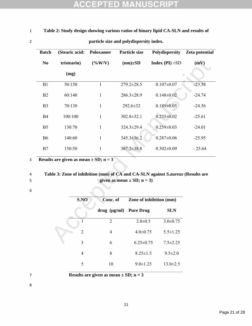
Composition of binary lipid CA-SLN and its results of particle size and PDI are 1
shown in table 2. Particle size and polydispersity index were ranging from 279.2±28.5 nm to 2
367.2±38.8 nm and 0.107 to 0.302 respectively. Particle size was decreased with increasing 3
TS concentration. This may be attributed to the fact that increase in TS content helps to 4
reduce the melting point of SA and leads to lattice crystal order disturbance in the lipid which 5
forms uni-model smaller particles. Zeta potential was measured only for binary lipid CA-6
SLN. Zeta potential of binary lipid CA-SLN was found to be varying from -23.58 to -25.95 7
mV. The highly negative zeta potential may be useful in maintaining the stability of CA-8
SLN. 9
4.2. Entrapment Efficiency (EE) 10
Entrapment efficiency was estimated for batches selected on the basis of lower particle size 11
of single lipid (S2 and S4) and all batches of binary lipid CA-SLN. The binary lipid CA-SLN 12
batches showed significantly higher entrapment efficiency compared to single lipid batches.. 13
The entrapment efficiency was in the range of 12.79±1.67% - 19.35±0.92% and 56.23±1.34% 14
- 70.62±0.82% for the selected batches of single lipid and all binary lipid CA-SLN, 15
respectively (Figure 1). The results are well support our earlier report on the application of 16
binary lipid mixture to enhance the entrapment efficiency [15]. The binary CA-SLN 17
comprising 50mg of SA; 150 mg of TS (1:3) showed the highest entrapment efficiency 18
(70.62±0.82 %). The higher entrapment efficiency with the binary lipid SLN formulations 19
was attributed to the greater deformation of crystal lattice of SA in the presence of TS and 20
formed enough space to accommodate drug molecules. This might also reduce the tendency 21
of the drug expulsion from the lipid [15]. CA-SLN batch B1 was finally selected for further 22
evaluations due to its lesser particle size, narrow polydispersity index and higher entrapment 23
efficiency. 24
Page 13 of 28
Accep
ted
Man
uscr
ipt
13
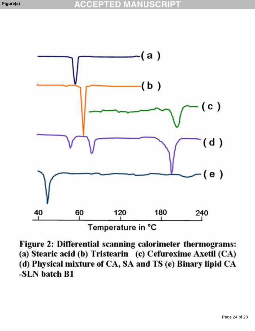
4.3. Differential scanning calorimetry (DSC) and FT-IR 1
The DSC thermograms of SA and TS showed a sharp melting point at 55 °C and 69 2
°C, respectively, indicating their crystalline nature (Figure 2). The pure CA showed a small 3
endothermic peak at 212 °C corresponding to its melting point. The physical mixture of CA, 4
SA and TS showed three sharp peaks at 56 oC, 71 oC and 200 oC. The sharp peak at 56 oC and 5
71 oC are corresponds to melting points of SA and TS whereas the peak at 200 oC 6
corresponds to melting point of CA. The DSC thermogram of binary lipid CA-SLN (B1) 7
showed melting point only at 45 °C. The decreased melting point of lipids and absence of 8
peak at 212 °C indicates that the drug is present in the amorphous form. The FT-IR spectra of 9
CA showed characteristic peaks at 1735.99 cm-1 (C=O stretching) together with peaks at 10
1074.39 cm-1 and 3481.63 cm-1 for C-O stretching of ester group and N-H stretching of amide 11
groups, respectively. The peak at 1330.93 cm-1 indicates the presence of aromatic ether in the 12
drug moiety. The FT-IR spectra of binary lipid CA-SLN showed all characteristic peaks of 13
CA which indicates absence of any interaction between drug and the lipids. 14
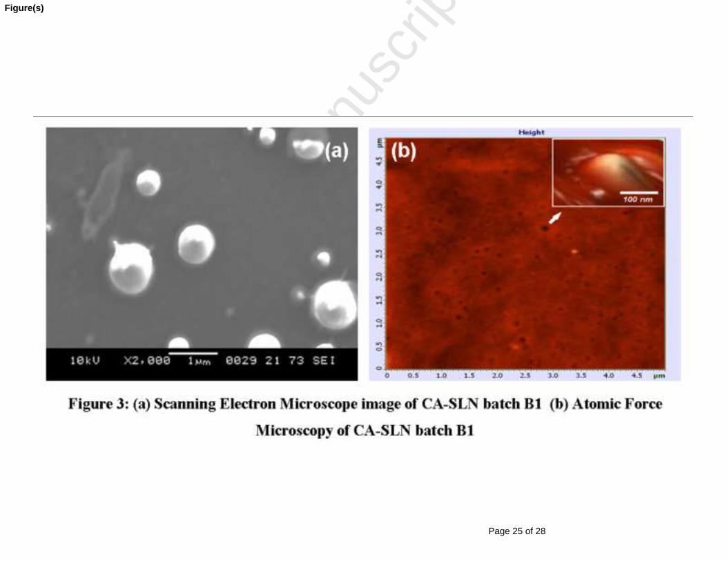
4.4. Scanning Electron Microscopy (SEM) and Atomic Force Microscopy (AFM) 15
The optimized SLN batch (B1) was studied for its shape and surface morphology by 16
SEM. The micrograph revealed that the particles are discrete, smooth and spherical in shape 17
(Figure 3a). The average particle size of batch B1 (triplicate) calculated from SEM study was 18
296.7 nm. The particle size measured from SEM was well correlated with the size obtained 19
by by DelsaTM Nano C particle analyzer (Beckman, USA). AFM image of nanoparticles also 20
confirmed discrete spherical particles with smooth surface (Figure 3b). The size of the 21
nanoparticles is below 300 nm. AFM images further confirmed the particle size obtained by 22
DelsaTM Nano C particle analyzer (Beckman, USA) and SEM. 23
24
Page 14 of 28
Accep
ted
Man
uscr
ipt
14
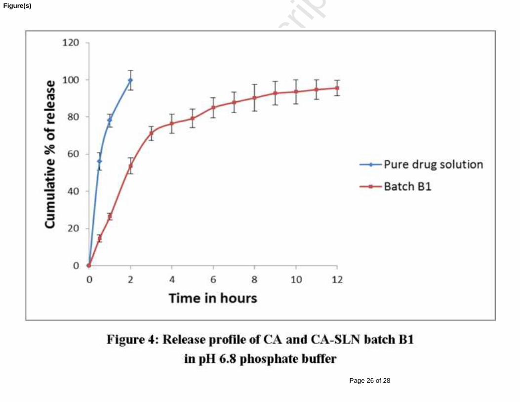
4.5. Invitro drug release 1
The cumulative percentage of drug release from CA solution and CA-SLN batch (B1) 2
is shown in Figure 4. CA solution showed 99.68% of release within 2 hours, whereas CA-3
SLN batch (B1) showed only 53.68% of drug release in 2 hours and 95.58% in 12 hours. This 4
indicates the prolonged release characteristics of CA-SLN (B1). Higuchi kinetics (r2=0.997) 5
was the best fit model for drug release. This indicates that the drug release is due to the 6
diffusion of drug through lipid matrix. 7
4.6. Stability studies 8
The stability of CA-SLN (B1) was assessed in terms of particle size, entrapment 9
efficiency and invitro drug release for 3 months. The particle size, entrapment efficiency and 10
cumulative percentage of drug release of batch B1 were found to be 279.2±28.5 nm, 11
70.62±0.82% and 95.58%, respectively at initial day of stability study. The same 12
characteristics were found to be 283.7±31.3 nm, 69.01±2.1% and 93.23%, respectively after 13
3 months of storage period. The obtained results clearly indicate insignificant change in 14
particle size, entrapment efficiency and invitro drug release pattern after 3 months of storage. 15
The results demonstrated that the concentration of stabilizer and optimized formulation 16
variables used to prepare SLN are sufficient for the stability of CA-SLN (B1). 17
4.7. Invitro antibiofilm study 18
4.7.1. Antibacterial susceptibility test: 19
Antibacterial susceptibility test was performed by Kirby Bauer (disk diffusion) 20
method against S. aureus. CA solution and CA-SLN (B1) containing 10 µg/ml were used in 21
the test. CA-SLN (B1) formulation showed zone of inhibition of 13 mm whereas CA solution 22
showed 9 mm as shown in figure 5a. This may be due to enhanced diffusion and 23
Page 15 of 28
Accep
ted
Man
uscr
ipt
15
accumulation of CA-SLN into the biofilm/microbial cells [16]. A placebo SLN was also used 1
to assess the anti microbial effect of fabrication materials SA, TS and poloxomer 188. The 2
placebo SLN showed negligible zone of inhibition (data not shown). This evidently indicates 3
SLN excipients did not exert any antibacterial activity against S. aureus. 4
4.7.2. MIC, anti biofilm activity by SEM and MBIC test 5
The MIC value of CA solution and CA-SLN (B1) were determined against S. 6
aureus. CA-SLN (B1) showed lower MIC value (4 µg/ml) than CA solution (10 µg/ml). The 7
lower MIC value of CA-SLN (B1) may be due to the impact of smaller size. The nano size 8
facilitates the penetration drug inside the cells and destroys the organism effectively [16, 17]. 9
A visible crystal violet stained blue colour film (indicated in arrows) confirmed 10
the formation of S. aureus biofilm (Figure 5b). Formation of biofilm was also confirmed 11
using SEM micrograph. Figure 5c represents SEM of S. aureus biofilm. Anti-biofilm activity 12
of CA solution and CA-SLN (B1) against S. aureus biofilm was characterized by SEM as 13
shown in Figures 5 d and e. The biofilm formed by incubation of the broth with CA-SLN 14
(B1) showed more cell damage (Figure 5e) than with pure CA solution (Figure 5d). The 15
higher bacterial cell damage by CA-SLN (B1) further confirms its potential inhibitory effect 16
than pristine CA solution. 17
The MBIC of CA solution and CA-SLN (B1) were determined against S. aureus 18
biofilm. CA and CA-SLN (B1) containing 10, 20, 40, 60, 80, 100, 120 and 140 µg/ml were 19
used for assessing MBIC. In case of CA solution bacterial growth was observed up to 60 20
µg/ml; it prevents growth of micro organism completely at 80 µg/ml and higher 21
concentrations. In CA-SLN (B1) bacterial growth was observed only in 10 and 20 µg/ml, not 22
in 40 µg/ml and higher concentrations. Therefore, MBIC of CA and CA-SLN (B1) were 80 23
µg/ml and 40 µg/ml, respectively. The lower MBIC of CA-SLN (B1) formulation (half of the 24

Page 16 of 28
Accep
ted
Man
uscr
ipt
16
pure drug) indicates increase in effectiveness of drug while it is formulated as SLN. The 1
reason behind the enhanced anti-biofilm activity of CA-SLN (B1) may be due to combined 2
effect of nano size and improved penetration of solid lipid nanoparticles into the biofilms. 3
Conclusion: 4
CA loaded SLNs were successfully fabricated with high entrapment efficiency using 5
binary lipid mixture of SA and TS and was found to be stable up to three months at 25 °C and 6
60% relative humidity. In vitro anti-biofilm activity was evaluated against S. aureus (ATCC-7
25923). Minimum biofilm inhibitory concentration (MBIC) value was found to be 80 µg/ml 8
and 40 µg/ml for CA and CA-SLN (B1), respectively. The superior anti biofilm activity may 9
be due to cumulative effect of nano size and use of lipid as carrier. This research work 10
suggests that the effectiveness of CA against S. aureus bacterial biofilm can be enhanced by 11
solid lipid nanoparticles. SLN technology has opened new approach to fight effectively 12
against biofilm and biofilm forming organisms. In the present work, binary lipid CA-SLN 13
exhibited productive results against S. aureus biofilm. In this view, as a complementary it has 14
been planned to assess efficacy of developed binary lipid CA-SLN against different bacterial 15
species which are potentially involved in formation of biofilm on live as well inanimate 16
matter. Eventually, in vivo evaluations will be carried for exploring therapeutic application of 17
binary lipid CA-SLN against biofilm. 18
19
4. References: 20
1. J.W. Costerton, K.J. Cheng, G.G. Geesey, T.I. Ladd, C.J. Nickel, M. Dasgupta and T.J. 21
Marrie, Bacterial biofilms in nature and disease. Annu. Rev. Microbiol. 41(1987) 435–22
464. 23
24
Page 17 of 28
Accep
ted
Man
uscr
ipt
17
2. S. Manuel, C.S. Lu cia and J.V. Maria. A review of current and emergent biofilm control 1
strategies. LWT - Food Science and Technology. 43 (2010) 573–583. 2
3. R.M. Donlan and J.W, Costerton, Biofilms: survival mechanisms of clinically relevant 3
microorganisms, Clin Microbiol Rev, 15 (2002) 167-193. 4
4. K. Lewis. Riddle of biofilm resistance. Antimicrobial Agents and Chemotherapy. 45 5
(2001) 999–1007. 6
5. M. Monzon, F. Alvarez Garcia, A. Lacleriga, E. Gracia, J. Leiva, C. Oteiza and B. 7
Amorena, A simple infection model using pre-colonized implants to reproduce rat 8
chronic Staphylococcus aureus osteomyelitis and study antibiotic treatment, Journal of 9
Orthopaedic research, 19 (2001) 820-826. 10
6. B. Sjostrom and B. Bergenstahl, Preparation of submicron drug particles in lecithin-11
stabilized o/w emulsions I. Model studies of the precipitation of cholesteryl acetate, Int. 12
J. Pharm, 88 (1992) 53–62. 13
7. A.K Kushwaha, P.R Vuddanda, K Priyanka, S.K Singh, S Singh, Development and 14
evaluation of solid lipid nanoparticles of raloxifene hydrochloride for enhanced 15
bioavailability, BioMed Research International, 2013 (2013) 1-9. 16
8. M.K. Rawat, A. Jain, A. Mishra, M.S. Muthu and S. Singh, Development of repaglinide 17
loaded solid lipid nanocarrier: selection of fabrication method, Current Drug Delivery, 7 18
(2010) 44-50. 19
9. M.K. Rawat, A. Jain and S. Singh, Studies on binary lipid matrix based solid lipid 20
nanoparticles of repaglinide: in vitro and in vivo evaluation, Journal of Pharmaceutical 21
Sciences, 100(6) (2011) 2366-78. 22

Page 18 of 28
Accep
ted
Man
uscr
ipt
18
10. R.S. Dhumal, S.V. Biradar, S. Yamamura, A.R. Paradkar, Peter York, Preparation of 1
amorphous cefuroxime axetil nanoparticles by sonoprecipitation for enhancement of 2
bioavailability, Eur. J Pharm. Biopharm, 70 (2008) 109–115. 3
11. W. Lawrence Drew, A.L. Barry, R.O. Toole and J.C. Sherris, Reliability of the Kirby-4
Bauer Disc Diffusion Method for Detecting Methicillin-Resistant Strains of 5
Staphylococcus aureus, Applied Microbiology, 24 (1972) 240-247. 6
12. S. Bose, M. Khodke, S. Basak and S.K. Mallick, Detection of biofilm producing 7
staphylococci: need of the hour, Journal of Clinical and Diagnostic Research, 3 (2009) 8
1915-1920. 9
13. W.S. Cheow, M.W. Chang and K. Hadinoto, The roles of lipid in anti-biofilm efficacy of 10
lipid–polymer hybrid nanoparticles encapsulating antibiotics, Colloids and Surfaces A: 11
Physicochem. Eng. Aspects, 389 (2011) 158– 165. 12
14. E.O. Merle, H. Ceri, W.D. Morck, G.A. Buret and R.R. Read, Biofilm bacteria: 13
formation and comparative susceptibility to antibiotics, The Canadian Journal of 14
Veterinary Research, 66 (2002) 86-92. 15
15. V. Jenning and S.H. Gohla, Comparison of wax and glyceride solid lipid nanoparticles 16
(SLN), Int. J. Pharm, 196 (2000) 219-222. 17
16. R. Misra, S. Acharya, F. Dilnawaz and S.K. Sahoo, Sustained antibacterial activity of 18
doxycycline-loaded poly (D,L-lactide-coglycolide) and poly(Iμ-caprolactone) 19
nanoparticles, Nanomedicine, 4 (2009) 519-30. 20
17. Y.I. Jeong, H.S. Na, D.H. Seo, D.G. Kim, H.C. Lee and M.K. Jang, Ciprofloxacin-21
encapsulated poly (DL-lactide-co-glycolide) nanoparticles and its antibacterial activity, 22
Int. J. Pharm, 352 (2008) 317-23. 23
Page 19 of 28
Accep
ted
Man
uscr
ipt
19
Acknowledgment 1
Mr. Bhupender Singh acknowledges the University Grant Commission (UGC), India for 2
providing the financial support in the form of the Junior Research Fellowship. The authors 3
also acknowledge Special Assistance Programme of UGC for providing of particle size 4
analysis instrumentation facility. 5
6

Page 20 of 28
Accep
ted
Man
uscr
ipt
20
Tables 1
Table 1: Study design showing the effect of formulation variables and process variables 2
on particle size and polydispersity index. (All the batches were prepared using single 3
lipid; stearic acid (200 mg) 4
Homogenization S.No. Batch
No.
Volume of
Dispersion
Medium (ml)
Speed
(rpm)
Time
(min)
Surfactant
Conc.
(% w/v)
Particle size
(nm) ± SD
Polydispersity
index ± SD
1. S1 50 10000 10 0.5 524.3±38.3 0.374±0.07
2. S2 50 10000 10 1 370.3±18.5 0.218±0.02
3. S3 50 10000 15 0.5 485.9±37.9 0.311±0.03
4. S4 50 10000 15 1 386.3±23.5 0.306±0.05
5. S5 50 15000 10 0.5 760.1±48.2 0.432±0.04
6. S6 50 15000 10 1 395.4±29.9 0.283±0.09
7. S7 50 15000 15 0.5 982.8±38.7 0.485±0.08
8. S8 50 15000 15 1 619.2±37.4 0.355±0.06
9. S9 100 10000 10 0.5 992.1±35.1 0.512±0.11
10. S10 100 10000 10 1 575.5±76.1 0.389±0.09
11. S11 100 10000 15 0.5 1532.0±62.2 0.573±0.12
12. S12 100 10000 15 1 698.5±49.7 0.403±0.11
13. S13 100 15000 10 0.5 782.1±45.3 0.452±0.07
14. S14 100 15000 10 1 1331.4±62.4 0.605±0.13
15. S15 100 15000 15 0.5 992.2±37.8 0.415±0.08
16. S16 100 15000 15 1 1065.6±49.9 0.485±0.07
Results are given as mean ± SD; n = 3 5
Page 21 of 28
Accep
ted
Man
uscr
ipt
21
Table 2: Study design showing various ratios of binary lipid CA-SLN and results of 1
particle size and polydispersity index. 2
Batch
No
(Stearic acid:
tristearin)
(mg)
Poloxamer
(%W/V)
Particle size
(nm)±SD
Polydispersity
Index (PI) ±SD
Zeta potential
(mV)
B1 50:150 1 279.2±28.5 0.107±0.07 -23.58
B2 60:140 1 286.3±28.9 0.148±0.02 -24.74
B3 70:130 1 292.6±32 0.189±0.05 -24.56
B4 100:100 1 302.8±32.1 0.235±0.02 -25.61
B5 130:70 1 324.3±29.4 0.259±0.03 -24.01
B6 140:60 1 345.3±36.2 0.287±0.06 -25.95
B7 150:50 1 367.2±38.8 0.302±0.09 - 25.64
Results are given as mean ± SD; n = 3 3
Table 3: Zone of inhibition (mm) of CA and CA-SLN against S.aureus (Results are 4 given as mean ± SD; n = 3) 5
6
Zone of inhibition (mm) S.NO Conc. of
drug (µg/ml) Pure Drug SLN
1 2 2.0±0.5 3.0±0.75
2 4 4.0±0.75 5.5±1.25
3 6 6.25±0.75 7.5±2.25
4 8 8.25±1.5 9.5±2.0
5 10 9.0±1.25 13.0±2.5
Results are given as mean ± SD; n = 3 7
8
Page 22 of 28
Accep
ted
Man
uscr
ipt
Graphical Abstract (for review)

Page 23 of 28
Accep
ted
Man
uscr
ipt
Figure(s)
Page 24 of 28
Accep
ted
Man
uscr
ipt
Figure(s)
Page 25 of 28
Accep
ted
Man
uscr
ipt
Figure(s)
Page 26 of 28
Accep
ted
Man
uscr
ipt
Figure(s)

Page 27 of 28
Accep
ted
Man
uscr
ipt
Figure(s)
Page 28 of 28
Accep
ted
Man
uscr
ipt
Figure 1: Entrapment efficiency of selected formulations (Error bars are SD, n=3)
Figure 2: Differential scanning calorimeter thermograms: (a) Stearic acid (b) Tristearin (c)
Cefuroxime Axetil (CA) (d) Physical mixture of CA, SA and TS (e) Binary lipid CA-SLN
batch B1
Figure 3: (a) Scanning Electron Microscope image of CA-SLN batch B1 (b) Atomic Force
Microscopy of CA-SLN batch B1
Figure 4: Release profile of CA and CA-SLN batch B1 in pH 6.8 phosphate buffer (Error
bars are SD, n=3)
Figure 5: (a) SLN formulation showing more zone of inhibition than pure drug in
antibacterial susceptibility test, (b) Detection of S.aureus biofilm by tube method (Biofilm
growth is confirmed by the crystal violet stain indicated by white arrows), (c) Scanning
electron micrograph of S.aureus biofilm, (d) Scanning electron micrograph showing
inhibitory effect of pure CA against biofilm produced by S. aureus, (e) Scanning electron
micrograph showing inhibitory effect of CA-SLN against biofilm produced by S. aureus