Novel Antibiotic-loaded Point-of-care Implant Coating Inhibits Biofilm
13
SYMPOSIUM: 2014 MUSCULOSKELETAL INFECTION SOCIETY Novel Antibiotic-loaded Point-of-care Implant Coating Inhibits Biofilm Jessica Amber Jennings PhD, Daniel P. Carpenter BS, Karen S. Troxel PhD, Karen E. Beenken PhD, Mark S. Smeltzer PhD, Harry S. Courtney PhD, Warren O. Haggard PhD Ó The Association of Bone and Joint Surgeons1 2015 Abstract Background Orthopaedic biomaterials are susceptible to biofilm formation. A novel lipid-based material has been developed that may be loaded with antibiotics and applied as an implant coating at point of care. However, this material has not been evaluated for antibiotic elution, biofilm inhibition, or in vivo efficacy. Questions/purposes (1) Do antibiotic-loaded coatings inhibit biofilm formation? (2) Is the coating effective in preventing biofilm in vivo? Methods Purified phosphatidylcholine was mixed with 25% amikacin or vancomycin or a combination of 12.5% of both. A 7-day elution study for coated titanium and stainless steel coupons was followed by turbidity and zone of inhibition assays against Staphylococcus aureus and Pseudomonas aeruginosa. Coupons were inoculated with bacteria and incubated 24 hours (N = 4 for each test group). Microscopic images of biofilm were obtained. After washing and vortexing, attached bacteria were counted. A mouse biofilm model was modified to include coated and uncoated stainless steel wires inserted into the lumens of catheters inoculated with a mixture of S aureus or P aeruginosa. Colony-forming unit counts (N = 10) and scanning electron microscopy imaging of implants were used to determine antimicrobial activity. Results Active antibiotics with colony inhibition effects were eluted for up to 6 days. Antibiotic-loaded coatings inhibited biofilm formation on in vitro coupons (log-fold reductions of 4.3 ± 0.4 in S aureus and 3.1 ± 0 for P aeruginosa in phosphatidylcholine-only coatings, 5.6 ± 0 for S aureus and 3.1 ± 0 for P aeruginosa for combination- loaded coatings, 5.5 ± 0.3 for S aureus in vancomycin- loaded coatings, and 3.1 ± 0 for P aeruginosa for amikacin- loaded coatings (p \ 0.001 for all comparisons of antibi- otic-loaded coatings against uncoated controls for both bacterial strains, p \ 0.001 for comparison of antibiotic- loaded coatings against phosphatidylcholine only for S aureus,p = 0.54 for comparison of vancomycin versus combination coating in S aureus,P = 0.99 for comparison of antibiotic- and unloaded phosphatidylcholine coatings in P aeruginosa). Similarly, antibiotic-loaded coatings reduced attachment of bacteria to wires in vivo (log-fold The institutions of one or more of the authors (JAJ, DC, KB, MS, HSC, WOH) have received, during the study period, funding from Biomet, LLC (Warsaw, IN, USA). One author (KT) is an employee of Biomet, LLC. All ICMJE Conflict of Interest Forms for authors and Clinical Orthopaedics and Related Research 1 editors and board members are on file with the publication and can be viewed on request. Clinical Orthopaedics and Related Research 1 neither advocates nor endorses the use of any treatment, drug, or device. Readers are encouraged to always seek additional information, including FDA- approval status, of any drug or device prior to clinical use. Each author certifies that his or her institution approved the animal protocol for this investigation and that all investigations were conducted in conformity with ethical principles of research. J. A. Jennings (&), D. P. Carpenter, W. O. Haggard Department of Biomedical Engineering, University of Memphis, 330 Engineering Technical Building, Memphis, TN 38152-3210, USA e-mail: [email protected] K. S. Troxel Biomet, Warsaw, IN, USA K. E. Beenken, M. S. Smeltzer University of Arkansas for Medical Sciences, Little Rock, AR, USA H. S. Courtney University of Tennessee Health Science Center, Veterans Affairs Medical Center, Memphis, TN, USA 123 Clin Orthop Relat Res DOI 10.1007/s11999-014-4130-8 Clinical Orthopaedics and Related Research ® A Publication of The Association of Bone and Joint Surgeons®
Transcript of Novel Antibiotic-loaded Point-of-care Implant Coating Inhibits Biofilm

SYMPOSIUM: 2014 MUSCULOSKELETAL INFECTION SOCIETY
Novel Antibiotic-loaded Point-of-care Implant Coating InhibitsBiofilm
Jessica Amber Jennings PhD, Daniel P. Carpenter BS, Karen S. Troxel PhD,
Karen E. Beenken PhD, Mark S. Smeltzer PhD, Harry S. Courtney PhD,
Warren O. Haggard PhD
� The Association of Bone and Joint Surgeons1 2015
Abstract
Background Orthopaedic biomaterials are susceptible to
biofilm formation. A novel lipid-based material has been
developed that may be loaded with antibiotics and applied
as an implant coating at point of care. However, this
material has not been evaluated for antibiotic elution,
biofilm inhibition, or in vivo efficacy.
Questions/purposes (1) Do antibiotic-loaded coatings
inhibit biofilm formation? (2) Is the coating effective in
preventing biofilm in vivo?
Methods Purified phosphatidylcholine was mixed with
25% amikacin or vancomycin or a combination of 12.5%
of both. A 7-day elution study for coated titanium and
stainless steel coupons was followed by turbidity and zone
of inhibition assays against Staphylococcus aureus and
Pseudomonas aeruginosa. Coupons were inoculated with
bacteria and incubated 24 hours (N = 4 for each test
group). Microscopic images of biofilm were obtained.
After washing and vortexing, attached bacteria were
counted. A mouse biofilm model was modified to include
coated and uncoated stainless steel wires inserted into the
lumens of catheters inoculated with a mixture of S aureus
or P aeruginosa. Colony-forming unit counts (N = 10) and
scanning electron microscopy imaging of implants were
used to determine antimicrobial activity.
Results Active antibiotics with colony inhibition effects
were eluted for up to 6 days. Antibiotic-loaded coatings
inhibited biofilm formation on in vitro coupons (log-fold
reductions of 4.3 ± 0.4 in S aureus and 3.1 ± 0 for P
aeruginosa in phosphatidylcholine-only coatings, 5.6 ± 0
for S aureus and 3.1 ± 0 for P aeruginosa for combination-
loaded coatings, 5.5 ± 0.3 for S aureus in vancomycin-
loaded coatings, and 3.1 ± 0 for P aeruginosa for amikacin-
loaded coatings (p \ 0.001 for all comparisons of antibi-
otic-loaded coatings against uncoated controls for both
bacterial strains, p \ 0.001 for comparison of antibiotic-
loaded coatings against phosphatidylcholine only for S
aureus, p = 0.54 for comparison of vancomycin versus
combination coating in S aureus, P = 0.99 for comparison
of antibiotic- and unloaded phosphatidylcholine coatings in
P aeruginosa). Similarly, antibiotic-loaded coatings
reduced attachment of bacteria to wires in vivo (log-fold
The institutions of one or more of the authors (JAJ, DC, KB, MS,
HSC, WOH) have received, during the study period, funding from
Biomet, LLC (Warsaw, IN, USA). One author (KT) is an employee of
Biomet, LLC.
All ICMJE Conflict of Interest Forms for authors and Clinical
Orthopaedics and Related Research1 editors and board members are
on file with the publication and can be viewed on request.
Clinical Orthopaedics and Related Research1 neither advocates nor
endorses the use of any treatment, drug, or device. Readers are
encouraged to always seek additional information, including FDA-
approval status, of any drug or device prior to clinical use.
Each author certifies that his or her institution approved the animal
protocol for this investigation and that all investigations were
conducted in conformity with ethical principles of research.
J. A. Jennings (&), D. P. Carpenter, W. O. Haggard
Department of Biomedical Engineering, University of Memphis,
330 Engineering Technical Building, Memphis, TN 38152-3210,
USA
e-mail: [email protected]
K. S. Troxel
Biomet, Warsaw, IN, USA
K. E. Beenken, M. S. Smeltzer
University of Arkansas for Medical Sciences, Little Rock, AR,
USA
H. S. Courtney
University of Tennessee Health Science Center, Veterans Affairs
Medical Center, Memphis, TN, USA
123
Clin Orthop Relat Res
DOI 10.1007/s11999-014-4130-8
Clinical Orthopaedicsand Related Research®
A Publication of The Association of Bone and Joint Surgeons®

reduction of 2.54 ± 0; p \ 0.001 for S aureus and
0.83 ± 0.3; p = 0.112 for P aeruginosa).
Conclusions Coatings deliver active antibiotics locally to
inhibit biofilm formation and bacterial growth in vivo.
Future evaluations will include orthopaedic preclinical
models to confirm therapeutic efficacy.
Clinical Relevance Clinical applications of local drug
delivery coating could reduce the rate of implant-associ-
ated infections.
Introduction
Musculoskeletal trauma often requires implanted bioma-
terials for fixation or joint replacement that are susceptible
to biofilm formation, which can lead to persistent infections
[11, 65, 68]. The primary strategy for prevention is aseptic
surgical technique followed by prophylactic systemic
administration of antibiotics [28, 71]. Antiinfective bio-
materials are increasingly used as an adjunctive strategy to
prevent implanted biomaterial infections and to inhibit
biofilm-forming microorganisms [13, 27, 44, 49, 55, 63,
64]. Commonly used local antibiotic delivery systems
range from antibiotic-loaded bone cement to calcium-based
drug delivery systems to sprinkling antibiotics within the
wound site [8, 24, 40, 42]. Recent developments have been
focused on degradable and customizable local delivery
strategies to ensure coverage of the wound, effective
antibiotic elution, and minimization of secondary proce-
dures [15, 30, 60]. These local delivery strategies include
degradable sponges [50], injectable biomaterials [48, 53,
80], and coatings of antimicrobial molecules on implanted
surfaces [25, 30, 43, 66]. These materials vary in degra-
dation rate and elution profile of antimicrobials and may
require prefabrication to attach antimicrobial molecules or
coating materials to implant surfaces.
We have developed a novel phosphatidylcholine-based
material that can be loaded with antibiotics and applied as a
coating on implants at point of care, acting as an ‘‘antibi-
otic crayon.’’ Here we provide the initial description of the
antibiotic-loaded lipid-based material, which is advanta-
geous because of its ability to be applied to the entire
implant surface with clinician-selected antibiotics and its
potential to elute antibiotics and prevent biofilm formation
over time. Although antibiotics have been incorporated into
phosphatidylcholine coatings, antibiotic elution and effi-
cacy in inhibiting biofilm-based microorganisms have not
yet been characterized.
The primary question we posed for our study was
whether lipid-based carriers elute active antibiotics in vitro
and in vivo to inhibit biofilm formation on metal substrates.
Additionally, is the antibiotic-loaded coating effective in
preventing biofilm formation in an in vivo model of
polymicrobial biofilm?
Materials and Methods
We evaluated coatings with and without antibiotics for
antimicrobial activity in vitro and in vivo compared with
uncoated controls (Fig. 1).
Fabrication and Coating
Four types of coatings were fabricated by mixing 6 g of
Phospholipon 90G (Lipoid Gmb, Ludwigshafen, Germany)
and 2 g of antibiotic in the following four groups: 25%
amikacin-loaded (Group A), 25% vancomycin-loaded
(Group V), combination of 12.5% amikacin and 12.5%
vancomycin-loaded (Group AV), unloaded phosphatidyl-
choline (Group P), and uncoated controls (Group U).
Through a process of warming to 37� C and kneading
powdered antibiotics into Phospholipon 90G, a uniform
mixture of powdered antibiotics and phosphatidylcholine
was obtained for each experimental group. In preliminary
formulation experiments, 25% of powdered antibiotic was
selected as the loading percentage because consistency of
the coating was negatively affected at concentrations
higher than this amount. For formulations with dual anti-
biotics, 25% of total antibiotic was incorporated by mixing
12.5% of each. After loading into open-ended syringes,
materials were sterilized by low-dose (25 kGy) gamma
irradiation. Titanium coupons (19-mm diameter; Ti-6Al-
4V Grade 5) and stainless steel coupons (17-mm diameter;
316L stainless steel) were machined and sanded with 100,
220, 400, 800, and 1000 grit sandpaper to a uniform finish.
The coupons were then washed with dish soap, sonicated
for 5 minutes in 20 mL of phosphate-buffered saline (PBS)
to remove any leftover residual particulates, and auto-
claved at 121� C for sterilization. Coupons were coated
with each one of the three types of antibiotic coatings by
exposing solid material by depressing the plunger of the
open-ended syringes and manually applying the mixtures
until all surfaces were visibly coated and the uncoated
coupons served as controls.
Elution and Eluate Activity Studies
Coupons were placed in 12-well cell culture plates with
2 mL of PBS. Samples were taken and the medium was
refreshed each day for 7 days. Concentration of vanco-
mycin in eluates was determined by measuring absorbance
at 270 nm with a BioTek Synergy H1 plate reader (BioTek,
Jennings et al. Clinical Orthopaedics and Related Research1
123

Winooski, VT, USA) [72]. Concentration of amikacin in
eluates was determined by reacting 10-fold diluted samples
with a mixture of acetylacetone and formaldehyde in a
buffer solution of boric acid, acetic acid, and phosphoric
acid at pH 2.7 and boiling for 20 minutes. Fluorescence of
the reacted product was measured at 471 nm (410 nm
excitation) with a BioTek Synergy plate reader [46]. All
concentrations were normalized to standard curves of
known concentrations of antibiotics. Eluates were diluted
1:10 in 5-mL tubes with 1.75 mL of tryptic soy broth
(TSB), 200 lL of eluate, and inoculated with 50 lL of
Staphylococcus aureus (UAMS-1 strain) or Pseudomonas
aeruginosa (PA01) at approximately 1 9 106 colony-
forming units (CFUs)/mL. After overnight incubation,
absorbance was determined at 530 nm as an indicator of
planktonic bacterial growth.
Zone of Inhibition Studies
Titanium and stainless steel coupons were coated as de-
scribed previously. One set of Petri dishes was inoculated
with 1 9 105 CFUs of S aureus, and another set was
inoculated with P aeruginosa. One coupon each of anti-
biotic-loaded coated groups was placed on plates with
controls groups of P and U. Coatings V and AV were
placed with P and C controls on each plate of S aureus.
One coupon of each coating (A, AV, P, or C) was placed on
plates inoculated with P aeruginosa. Each set of antibiotics
and controls was placed on three separate replicate plates.
Zones of inhibition were imaged and the diameters
measured using Image J software (National Institutes of
Health, Bethesda, MD, USA).
In Vitro Biofilm Inhibition
From preliminary studies and reports in the literature [9], it
was determined that a modified TSB, containing 0.5%
glucose and 3% salt, would be used as the biofilm medium
for S aureus to improve consistency. Coated coupons and
controls were placed into wells of 12-well plates and
inoculated with 106 CFUs of either S aureus or P aeru-
ginosa in biofilm media and TSB, respectively. After
overnight growth, bacterial media were removed from the
wells and washed gently twice with PBS to remove any
planktonic cells. One coupon from each group was then
carefully removed so as not to disturb the biofilm and
stained with FilmTracer live/dead stain (Invitrogen,
Eugene, OR, USA) to visualize patterns of live and dead
bacterial cell attachment. Another coupon from each group
was also removed and placed in fixative for imaging with a
scanning electron microscope. Fixed biofilm samples on
coupons were washed several times in phosphate buffer,
postfixed in 1% osmium tetroxide in phosphate buffer, and
rinsed. Biofilm was en bloc stained with 2% uranyl acetate,
rinsed with deionized water, dehydrated in ethanol, and
dried using a critical point dryer [32]. Coupons were
mounted, biofilm side up, onto standard electron micros-
copy stubs that were coated with double-sided, carbon
black sticky tabs. Specimens were conductively coated
before imaging with a thin sputtercoating of gold-
Fig. 1 Diagram shows the experimental design.
Point-of-care Implant Coating Inhibits Biofilm
123
palladium (20 nm). Images at various locations on the
coupons and at different magnifications were acquired
using a Philips XL30 environmental scanning electron
microscope (FEI, Hillsboro, OR, USA). Four coupons from
each group were placed in 50-mL centrifuge tubes in
10 mL of PBS and vortexed for approximately 1 minute to
remove biofilm. The live bacteria removed were then
serially diluted in 10-fold increments and plated for colony
counting.
In Vivo Biofilm Inhibition
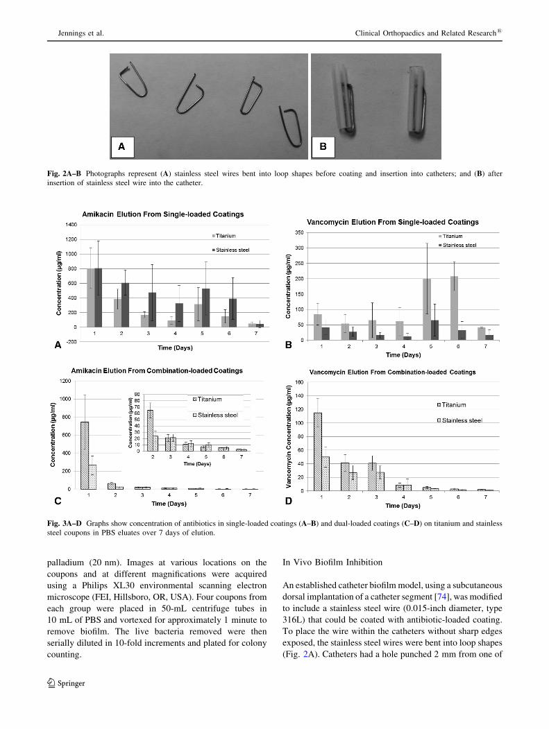
An established catheter biofilm model, using a subcutaneous
dorsal implantation of a catheter segment [74], was modified
to include a stainless steel wire (0.015-inch diameter, type
316L) that could be coated with antibiotic-loaded coating.
To place the wire within the catheters without sharp edges
exposed, the stainless steel wires were bent into loop shapes
(Fig. 2A). Catheters had a hole punched 2 mm from one of
Fig. 2A–B Photographs represent (A) stainless steel wires bent into loop shapes before coating and insertion into catheters; and (B) after
insertion of stainless steel wire into the catheter.
Fig. 3A–D Graphs show concentration of antibiotics in single-loaded coatings (A–B) and dual-loaded coatings (C–D) on titanium and stainless
steel coupons in PBS eluates over 7 days of elution.
Jennings et al. Clinical Orthopaedics and Related Research1
123

the edges to insert the bent end of the wire (Fig. 2B). Wires
and catheters were then washed, sterilized by autoclaving at
121� C for 20 minutes, and prepared for insertion into
catheters by coating with designated coatings. The five
coating-type groups (12 catheters in six mice) included
uncoated wires; uncoated wires with codelivery of antibiotic
solution (1 lg/mL vancomycin; 16 lg/mL amikacin);
phosphatidylcholine-only coated; AV-combination coated;
and A-coated. Amikacin was chosen as a single high-dose
antibiotic because of its activity against S aureus as well as
Gram-negative bacteria [58]. Two catheters from the same
experimental group were implanted into the dorsa of each
mouse bilaterally, one each on the right and left side of the
spine, and then inoculated with a polymicrobial mixture of
105 CFUs of S aureus (UAMS-1) and 104 CFUs of P aeru-
ginosa (Schroeter) Migula (ATCC1 27317TM, Manassas,
VA, USA) directly into the lumen of the catheter. After
48 hours, catheters and wires were retrieved, rinsed, and
separated for analysis of attached CFUs (10 catheters in five
mice/group) and scanning electron microscopy imaging
(two catheters in one mouse/group). Clearance rate was
calculated by determining the number of catheters with CFU
counts of zero and dividing by total number of catheters in
that experimental group.
Statistical Analysis
Four replicates in each group were chosen for the elution
and turbidity study based on activity and elution data from
previous studies of calcium sulfate and chitosan-based drug
delivery systems [8, 50] Based on a power analysis using
SigmaPlot (Systat Software, Inc, San Jose, CA, USA), it
was determined that a number of 4 provides approximately
80% power to detect a difference in proportions of solu-
tions with complete growth inhibition of 84% at a
significance level of 5% and provides over 80% power to
detect differences in concentration of 15 lg/mL or more in
antibiotic concentration given the standard deviation of
approximately 5 lg/mL. Three coupons in each group for
the zone of inhibition study gives over 80% power to detect
differences in clearing zones of 3 mm or more given the
approximate SD of 0.9 mm. For biofilm experiments,
assuming a SD of approximately 0.36, four coupons per
group gives over 80% power to detect log-fold reductions
in CFUs over 1 at a 5% significance level. One-way
Kruskal-Wallis analysis of variance with Student-New-
man-Keuls test post hoc were performed using Sigma Plot
(Systat Software, Inc, San Jose, CA, USA) to determine
whether colony counts in coated coupons or wires differed
from those in control groups. Using results of previous
studies determining log-fold reduction of CFUs in response
to antimicrobials [74], an a priori power analysis was
performed using SigmaPlot. Assuming a SD of 1 unit in
log-transformed CFU counts, five animals per group were
required for 80% power to detect a difference of 3 units
between the control group and each of the four experi-
mental treatment groups at a significance level of 5%.
Results
In Vitro Elution
For single loaded 25% antibiotic coatings, there was an
initial burst of 800 ± 371 lg/mL of amikacin or
84 ± 34 lg/mL of vancomycin during the first day with
decreasing elution until Days 5 and 6 when a second burst
of antibiotic was present in both antibiotic groups (Fig. 3
A–B). Similar first day elution values of 746 ± 300 lg/mL
of amikacin and 115 ± 21 lg/mL of vancomycin were
observed for coatings loaded with 12.5% of both antibiotics
(Fig. 3C–D). However, at the lower dual loading level, the
burst at 5 days seen in 25% loaded coatings was not
observed, and elution tapered off to below the minimum
inhibitory concentration by approximately Day 5.
Titanium Day 1 Day 2 Day 3 Day 4 Day 5 Day 6 Day 7A – – – – – – – – – † – † – † † † – † – – – – † – † † † †V – † – – – † – – – – – – – † – † – – † – – – † – – † † †AV – – – – – – – – † † † † † † † † † † † † † † † † † † † †P † † † † † † † † † † † † † † † † † † † † † † † † † † † †U † † † † † † † † † † † † † † † † † † † † † † † † † † † †
StainlessA – – – – – † – † – – – – – – – – – † – † † † † † † † † †V – – – – – † – † – † – † – † – † – – – – – † – † – † – †AV – – – – – – – – – † – † – † – † † † † † † † † † † † † †P † † † † † † † † † † † † † † † † † † † † † † † † † † † †U † † † † † † † † † † † † † † † † † † † † † † † † † † † †
Titanium Day 1 Day 2 Day 3 Day 4 Day 5 Day 6 Day 7A – – – – – – – – – † – † – † † † – † – – – – † – † † † †AV – – – – – – – – † † † † † † † † † † † † † † † † † † † †P † † † † † † † † † † † † † † † † † † † † † † † † † † † †U † † † † † † † † † † † † † † † † † † † † † † † † † † † †
Stainless Day 1 Day 2 Day 3 Day 4 Day 5 Day 6 Day 7A † – – – † – † – – † – † † – † † – – – – † – – – † † † †AV – – – – † † † – † – † † † † – – † † † † † † † † † † † †P † † † † † † † † † † † † † † † † † † † † † † † † † † † †U † † † † † † † † † † † † † † † † † † † † † † † † † † † †
A
B
S aureus
P aeruginosa
Fig. 4A–B Color-coded chart shows turbidity results for (A) S
aureus and (B) P aeruginosa over a 7-day period for amikacin 25%
(A); vancomycin 25% (V); combination amikacin and vancomycin,
12.5% each (AV); phosphatidylcholine only (P); and uncoated
controls (U). � � � � = no growth inhibition; – � � � = growth
inhibition in one sample; – � – � = growth inhibition in multiple
samples; – – – – = growth inhibition in all samples.
Point-of-care Implant Coating Inhibits Biofilm
123

In Vitro Colony Inhibition
For S aureus, 25% amikacin-loaded and 25% vancomycin-
loaded coatings had inhibitory effects for up to up to 3 days
and also on Days 5 and 6 (Fig. 4A). Combination-loaded
coatings with 12.5% of each antibiotic only inhibited
growth of S aureus for 2 days of elution. For P aeruginosa,
amikacin had inhibitory effects for up to 2 days of elution
with increased inhibitory effects on Days 5 and 6 (Fig. 4B).
Uncoated metal coupons had no effect on inhibition of
bacterial growth and phosphatidylcholine released into the
media without antibiotics also had no effect on bacterial
turbidity. The combination coating with amikacin reduced
to 12.5% only released inhibitory concentrations for 2 days
on both stainless steel and titanium coupons. Visual
inspection of coatings on coupons immersed in aqueous
solutions indicated that less coating was visible on stainless
steel coupons than titanium, starting at the 24-hour time
point. A minute amount of residual phosphatidylcholine
coating could be observed on titanium coupons for up to 5
to 7 days.
The antibiotic-loaded coatings had clear zones of inhi-
bition compared with P and U controls. Average diameter
of the zone of inhibition of vancomycin-loaded coating
against S aureus was 6 ± 1 mm (Fig. 5A) and average
zone of inhibition of amikacin-loaded coating against P
aeruginosa was 13 ± 2 mm (Fig. 5B). Results showed that
over the 24-hour period, all coatings, including nonantibi-
otic-loaded phosphatidylcholine, inhibited biofilm
formation by P aeruginosa on both titanium and stainless
steel coupons (3.1 ± 0 log-fold reduction, 95% confidence
interval for the difference in means, �3.7 to �2.5;
p \ 0.001). Although the phosphatidylcholine coating
alone significantly reduced S aureus biofilm formation on
coupons (4.3 ± 0.4 log-fold reduction, 95% confidence
interval for the difference in means, �5.1 to �3.6;
p \ 0.001), it did not completely inhibit attachment, and
coated titanium seemed to have a decreased attachment
compared with coated stainless steel (Fig. 6A). The anti-
biotic-loaded coatings resulted in a clearance rate of 100%
in coupons inoculated with P aeruginosa and a 56%
clearance rate in coupons inoculated with S aureus. On
titanium, there was a 3.1 ± 0.0 (95% confidence interval
for difference in means, �3.7 to �2.5; p \ 0.001) log-fold
reduction of P aeruginosa CFUs/coupon and a 5.6 ± 0.2
(95% confidence interval for difference in means, �6.175
to �4.827; p \ 0.001) log-fold reduction of S aureus on
coupons coated with antibiotic-loaded phosphatidylcholine
(Fig. 6). Images of biofilm formed on coupons demon-
strated that control groups without coating had attached
biofilm-forming bacterial colonies (Fig. 7), whereas few
colonies were found in coated groups. Of note is that
phosphatidylcholine and antibiotic-loaded phosphatidyl-
choline appeared to autofluoresce or bind to the dyes in the
green channel at high lighting levels, especially when
coated on titanium surfaces, giving an image of the surface
characteristics of the coating. The coating appeared as
‘‘globules’’ of material with an occasional ‘‘spider web’’
appearance when combined with both amikacin and van-
comycin. On titanium, coatings were observed to be
Fig. 5A–B Representative images show zone of inhibition results for
(A) S aureus and (B) P aeruginosa using titanium (top two images)
and stainless steel (bottom two images) coupons for vancomycin 25%
(V); combination amikacin and vancomycin, 12.5% each (AV);
phosphatidylcholine only (P); and uncoated controls (U).
Jennings et al. Clinical Orthopaedics and Related Research1
123

globular; coatings were sparse or absent on stainless steel
coupons (Fig. 8). Some S aureus bacteria were observed in
phosphatidylcholine-only-coated coupons in both groups,
confirming results of colony-counting studies.
In Vivo Inhibition and Clearance
Antibiotic-loaded coatings, both A alone and AV combi-
nation, were effective at inhibiting biofilm in vivo.
Fig. 6A–B Graph shows CFU counts from control and coated
titanium and stainless steel coupons (N = 4 each) for (A) S aureus
and (B) P aeruginosa. Data represented are mean ± SD. Asterisks
represent significant reduction in CFUs compared with the uncoated
control; # represents significant reduction in CFUs compared with the
P control (p \ 0.001).
Fig. 7A–B Photomicrographs show (A) UAMS-1 and (B) PA01. Live bacteria appear green; dead bacteria appear red. Phosphatidylcholine
coatings also demonstrate some autofluorescence in the green channel.
Point-of-care Implant Coating Inhibits Biofilm
123

Antibiotic-loaded coatings resulted in a 100% clearance
rate of wires from S aureus and a 90% clearance rate of P
aeruginosa in the polymicrobial in vivo model with one
breakthrough in one wire in the in vivo study (Fig. 9).
There was an average 2.5 ± 0 (95% confidence interval for
the difference in means, 1.342–3.743; p \ 0.001) log-fold
reduction of S aureus biofilm formation on wires and a
0.83 ± 0.3 (95% confidence interval for the difference in
means, �0.1 to 1.6; p = 0.112) log-fold reduction of P
aeruginosa for antibiotic-loaded coatings compared with
untreated controls. The 95% confidence interval for the
difference in proportion of clearance of S aureus was
�1.407 to �0.193 (p = 0.053) and the simultaneous
injection of antibiotics resulted in slightly higher clearance
S. aureus Titanium Stainless steel
Uncoated control
P coating
AV coating
V coating
A
P. aeruginosa Titanium Stainless steel
Uncoated control
P coating
AV coating
A coating
B
Fig. 8A–B Scanning electron micrographs show images at various magnifications of control and coated coupons inoculated with (A) UAMS-1.
(B) PA01.
Fig. 9A–B Graph shows log CFU counts of (A) S aureus and (B) P aeruginosa from wire implants retrieved from in vivo study. Data
represented are mean ± SD. Asterisks represent significant reduction in CFUs compared with the uncoated no treatment control.
Jennings et al. Clinical Orthopaedics and Related Research1
123
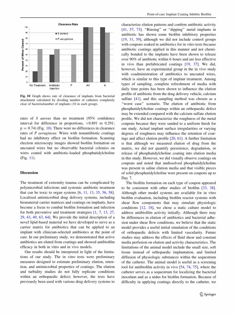
rates of S aureus than no treatment (95% confidence
interval for difference in proportions, �0.891 to 0.291;
p = 0.74) (Fig. 10). There were no differences in clearance
rates of P aeruginosa. Wires with nonantibiotic coatings
had no inhibitory effect on biofilm formation. Scanning
electron microscopy images showed biofilm formation on
uncoated wires but no observable bacterial colonies on
wires coated with antibiotic-loaded phosphatidylcholine
(Fig. 11).
Discussion
The treatment of extremity trauma can be complicated by
polymicrobial infections and systemic antibiotic treatment
that can be toxic to organ systems [6, 11, 13, 35, 56, 58].
Localized antimicrobial drug delivery systems, including
biomaterial carrier matrices and coatings on implants, have
become a focus to combat biofilm formation and infection
for both preventive and treatment strategies [1, 7, 13, 27,
29, 43, 49, 63, 64]. We provide the initial description of a
novel lipid-based material we have developed to serve as a
carrier matrix for antibiotics that can be applied to an
implant with clinician-selected antibiotics at the point of
care. In our preliminary study, we demonstrated that active
antibiotics are eluted from coatings and showed antibiofilm
efficacy in both in vitro and in vivo models.
Our results should be interpreted in light of the limita-
tions of our study. The in vitro tests were preliminary
measures designed to estimate preliminary elution, reten-
tion, and antimicrobial properties of coatings. The elution
and turbidity studies do not fully replicate conditions
within an orthopaedic defect; however, the tests have
previously been used with various drug delivery systems to
characterize elution patterns and confirm antibiotic activity
[41, 57, 73]. ‘‘Rinsing’’ or ‘‘dipping’’ metal implants in
antibiotic has shown some biofilm inhibitory properties
[19, 31, 59], although we did not include control groups
with coupons soaked in antibiotics for in vitro tests because
antibiotic coatings applied in this manner and not chemi-
cally bonded to the implants have been shown to release
over 90% of antibiotic within 6 hours and are less effective
in vivo than prefabricated coatings [19, 37]. We did,
however, have an experimental group in the in vivo study
with coadministration of antibiotics to uncoated wires,
which is similar to this type of implant treatment. Among
types of sampling, complete refreshment of media with
daily time points has been shown to influence the elution
profile of antibiotic from the drug delivery vehicle, calcium
sulfate [41], and this sampling method was chosen as a
‘‘worst case’’ scenario. The elution of antibiotic from
phosphatidylcholine coatings within an orthopaedic defect
may be extended compared with the calcium sulfate elution
profile. We did not characterize the roughness of the metal
coupons because they were sanded to a uniform finish for
our study. Actual implant surface irregularities or varying
degrees of roughness may influence the retention of coat-
ings and affect elution profile [20, 61]. A further limitation
is that although we measured elution of drug from the
matrix, we did not quantify persistence, degradation, or
release of phosphatidylcholine coating into elution media
in this study. However, we did visually observe coatings on
coupons and noted that undissolved phosphatidylcholine
was present in saline elution media and that visible pieces
of solid phosphatidylcholine were present on coupons up to
Day 7.
The biofilm formation on each type of coupon appeared
to be consistent with other studies of biofilm [33, 38].
Although other model systems are available for in vitro
biofilm evaluation, including biofilm reactor systems with
shear flow components that may simulate physiologic
conditions [12, 18], we chose a static culture model to
address antibiofilm activity initially. Although there may
be differences in elution of antibiotics and bacterial adhe-
sion under shear flow conditions, we believe that the static
model provides a useful initial simulation of the conditions
of orthopaedic defects with limited vascularity. Future
studies may address the effects of fluid shear and constant
media perfusion on elution and activity characteristics. The
limitations of the animal model include the small size, soft
tissue instead of orthopaedic implantation, and limited
diffusion of physiologic substances within the sequestrum
of the catheter. The animal model is useful as a screening
tool for antibiofilm activity in vivo [54, 74, 75], where the
catheter serves as a sequestrum for localizing the bacterial
inoculum and as a nidus for biofilm formation. Because of
difficulty in applying coatings directly to the catheter, we
Fig. 10 Graph shows rate of clearance of implants from bacterial
attachment calculated by dividing number of catheters completely
clear of bacteria/number of implants (10 in each group).
Point-of-care Implant Coating Inhibits Biofilm
123

modified the catheter-associated biofilm model by includ-
ing an easily coated stainless steel wire. Minimal diffusion
of fluid within the lumen of the catheter is a limitation of
this model in modeling physiological conditions to test
local drug delivery devices and the modification of this
model to include wires has not been previously validated.
Part of the wire was within the catheter lumen, and the
other part was exposed to the soft tissue and shear forces
resulting from fluid flow and animal movement. Despite
the limitations, our model provided a proof of principle that
the coatings inhibited biofilm formation on the wires.
Diffusion and elution were not characterized over a 48-
hour time course, but reduction in bacterial attachment
confirmed release of active antibiotics observed in our
in vitro studies. Future evaluations using coatings on
orthopaedic implants in more complex models should fol-
low our studies to confirm efficacy in clinically relevant
orthopaedic applications [8, 10, 62].
Elution of antibiotics from phosphatidylcholine coatings
applied to metal surfaces is initially described to follow a
burst release pattern with a second burst of antibiotic after
Day 5 when loaded at high concentration. We propose that
some of the crystals of antibiotic are entrapped within lipid
chains of phosphatidylcholine and that others are not bound
within lipid chains. Initial release of unbound antibiotic is
similar for different loading levels. Antibiotics not released
during this initial period may be released as the phospha-
tidylcholine molecules degrade or are hydrolyzed resulting
in the second burst effect. Amikacin, a smaller molecule
(molecular weight 585 g/mol) than vancomycin (molecular
weight 1449 g/mol), was released at a much higher level
during the first day of elution. Differences in molecular
weight and chemical structure may affect retention and
release of different antibiotics from phosphatidylcholine.
Antibiotics released from coatings were confirmed to be
active in inhibiting growth of microorganisms through
turbidity and zone of inhibition assays with similar activity
to other antimicrobial coatings [5, 14, 25, 43, 69]. Although
effective in inhibiting bacterial growth and biofilm for-
mation, several of the implant coating strategies have the
limitation of requiring prefabrication [25, 43] and chemical
modification with specific preselected antibiotics [4, 29].
Release of antibiotic resembles the burst release observed
in commonly used polymethylmethacrylate (PMMA) and
calcium sulfate drug delivery matrices [23, 47]. Unlike
PMMA, which requires secondary removal through surgi-
cal procedures, phosphatidylcholine coatings are
degradable and materials have been used in orthopaedic
applications [26]. Through application to the surface
directly, more coverage of the potentially contaminated
Fig. 11A–D Scanning electron micrographs show surfaces of wires from inner portion of catheter wire in untreated control group (A–B) and
antibiotic-loaded phosphatidylcholine-coated wires (C–D).
Jennings et al. Clinical Orthopaedics and Related Research1
123

implant site is provided compared with antibiotic-loaded
beads or sponges [34, 67, 70, 76].
Interestingly, we observed some inhibitory effect of
unloaded phosphatidylcholine on biofilm formation in vitro,
which may be a result of the hydrophobic nature of the
coating [21]. P aeruginosa biofilm was completely inhibited
by coatings in the 24-hour in vitro biofilm assay, which is
similar to the response of Gram-negative organisms to
hydrophobic surfaces in a study by Cheng et al. [17]. The
inhibitory effect of coating without antibiotic was not
observed in the in vivo study, likely as a result of the com-
peting effects of protein and cell adhesion [3, 78, 79]. Novel
nanoparticle systems releasing antibiotics or antimicrobial
silver have been shown to similarly inhibit bacterial growth
and biofilm formation, but balancing bactericidal efficacy
against cytotoxicity to the surrounding cells must be a con-
sideration before clinical use [2, 7, 16, 39].
The reduction in biofilm attachment in a challenging
polymicrobial in vivo model confirms the in vitro results
and is consistent with release of active concentrations of
antibiotic from various drug delivery vehicles [22, 45, 51,
54, 63]. Scanning electron microscopy images of biofilm
formation on implants resembled images from similar
retrieved infected implants [10, 52] with rounded S aureus
and rod-shaped P aeruginosa colonies visible in untreated
groups. Formation of fibrous tissue, to varying degrees,
along the outer portion of the wire exposed to soft tissue was
seen with morphology that may be indicative of fibroblast
or macrophage infiltration [3, 23, 36]. Material properties of
the coating may have contributed to this response [26, 77];
however, the majority of the fibrous tissue was formed at the
point of the wire that curved and extended from the catheter,
which may have been a location for rubbing and irritation as
the animals moved or were groomed.
In conclusion, phosphatidylcholine coatings are capable
of local delivery of active antibiotics and can be loaded
with antimicrobials to inhibit biofilm. Reduction of biofilm
on metal may be the result of a combination of lipid
material properties and localized antibiotic release. Anti-
biotic-loaded phosphatidylcholine coatings reduced
bacterial growth and biofilm formation on coated metal
substrates. We plan further development and testing,
including investigating efficacy in orthopaedic models and
use of different antimicrobial-loading strategies.
References
1. Adams CS, Antoci V Jr, Harrison G, Patal P, Freeman TA, Shapiro
IM, Parvizi J, Hickok NJ, Radin S, Ducheyne P. Controlled release
of vancomycin from thin sol-gel films on implant surfaces suc-
cessfully controls osteomyelitis. J Orthop Res. 2009;27:701–709.
2. Albers CE, Hofstetter W, Siebenrock KA, Landmann R, Klenke
FM. In vitro cytotoxicity of silver nanoparticles on osteoblasts
and osteoclasts at antibacterial concentrations. Nanotoxicology.
2013;7:30–36.
3. Anderson JM, Rodriguez A, Chang DT. Foreign body reaction to
biomaterials. Semin Immunol. 2008;20:86–100.
4. Antoci V Jr, Adams CS, Hickok NJ, Shapiro IM, Parvizi J.
Vancomycin bound to Ti rods reduces periprosthetic infection:
preliminary study. Clin Orthop Relat Res. 2007;461:88–95.
5. Antoci V Jr, King SB, Jose B, Parvizi J, Zeiger AR, Wickstrom E,
Freeman TA, Composto RJ, Ducheyne P, Shapiro IM, Hickok NJ,
Adams CS. Vancomycin covalently bonded to titanium alloy
prevents bacterial colonization. J Orthop Res. 2007;25:858–866.
6. Bailie GR, Neal D. Vancomycin ototoxicity and nephrotoxicity.
A review. Med Toxicol Adverse Drug Exp. 1988;3:376–386.
7. Barbour ME, Maddocks SE, Wood NJ, Collins AM. Synthesis,
characterization, and efficacy of antimicrobial chlorhexidine hexa-
metaphosphate nanoparticles for applications in biomedical materials
and consumer products. Int J Nanomedicine. 2013;8:3507–3519.
8. Beenken K, Smith J, Skinner R, MClaren S, Bellamy W,
Gruenwald M, Spencer HJ, Jennings J, Haggard W, Smeltzer M.
Chitosan coating to enhance the therapeutic efficacy of calcium
sulfate-based antibiotic therapy in the treatment of chronic oste-
omyelitis. J Biomater Appl. 2014;29:514–523.
9. Beenken KE, Blevins JS, Smeltzer MS. Mutation of sarA in
Staphylococcus aureus limits biofilm formation. Infect Immun.
2003;71:4206–4211.
10. Bernthal NM, Stavrakis AI, Billi F, Cho JS, Kremen TJ, Simon
SI, Cheung AL, Finerman GA, Lieberman JR, Adams JS, Miller
LS. A mouse model of post-arthroplasty Staphylococcus aureus
joint infection to evaluate in vivo the efficacy of antimicrobial
implant coatings. PLoS One. 2010;5:e12580.
11. Brady RA, Leid JG, Calhoun JH, Costerton JW, Shirtliff ME.
Osteomyelitis and the role of biofilms in chronic infection. FEMS
Immunol Med Microbiol. 2008;52:13–22.
12. Buckingham-Meyer K, Goeres DM, Hamilton MA. Comparative
evaluation of biofilm disinfectant efficacy tests. J Microbiol
Meth. 2007;70:236–244.
13. Campoccia D, Montanaro L, Arciola CR. The significance of
infection related to orthopedic devices and issues of antibiotic
resistance. Biomaterials. 2006;27:2331–2339.
14. Campoccia D, Montanaro L, Arciola CR. A review of the bio-
materials technologies for infection-resistant surfaces.
Biomaterials. 2013;34:8533–8554.
15. Campoccia D, Montanaro L, Arciola CR. A review of the clinical
implications of anti-infective biomaterials and infection-resistant
surfaces. Biomaterials. 2013;34:8018–8029.
16. Chavez de Paz LE, Resin A, Howard KA, Sutherland DS, Wejse
PL. Antimicrobial effect of chitosan nanoparticles on streptococcus
mutans biofilms. Appl Environ Microbiol. 2011;77:3892–3895.
17. Cheng G, Zhang Z, Chen S, Bryers JD, Jiang S. Inhibition of
bacterial adhesion and biofilm formation on zwitterionic surfaces.
Biomaterials. 2007;28:4192–4199.
18. Coenye T, Nelis HJ. In vitro and in vivo model systems to study
microbial biofilm formation. J Microbiol Meth. 2010;83:89–105.
19. Darouiche RO, Mansouri MD, Zakarevicz D, Alsharif A, Landon
GC. In vivo efficacy of antimicrobial-coated devices. J Bone
Joint Surg Am. 2007;89:792–797.
20. Dearnley PA. A review of metallic, ceramic and surface-treated
metals used for bearing surfaces in human joint replacements.
Proc Inst Mech Eng H. 1999;213:107–135.
21. Donlan RM. Biofilms: microbial life on surfaces. Emerg Infect
Dis. 2002;8:881–890.
22. Doty HA, Leedy MR, Courtney HS, Haggard WO, Bumgardner
JD. Composite chitosan and calcium sulfate scaffold for dual
Point-of-care Implant Coating Inhibits Biofilm
123

delivery of vancomycin and recombinant human bone morpho-
genetic protein-2. J Mater Sci Mater Med. 2014;25:1449–1459.
23. Gerhart TN, Roux RD, Horowitz G, Miller RL, Hanff P, Hayes
WC. Antibiotic release from an experimental biodegradable bone
cement. J Orthop Res. 1988;6:585–592.
24. Giavaresi G, Bertazzoni Minelli E, Sartori M, Benini A, Della
Bora T, Sambri V, Gaibani P, Borsari V, Salamanna F, Martini L,
Nicoli Aldini N, Fini M. Microbiological and pharmacological
tests on new antibiotic-loaded PMMA-based composites for the
treatment of osteomyelitis. J Orthop Res. 2012;30:348–355.
25. Greene AH, Bumgardner JD, Yang Y, Moseley J, Haggard WO.
Chitosan-coated stainless steel screws for fixation in contami-
nated fractures. Clin Orthop Relat Res. 2008;466:1699–1704.
26. Han B, Tang B, Nimni ME. Combined effects of phosphatidyl-
choline and demineralized bone matrix on bone induction.
Connect Tissue Res. 2003;44:160–166.
27. Jennings JA, Courtney HS, Haggard WO. Cis-2-decenoic acid
inhibits S. aureus growth and biofilm in vitro: a pilot study. Clin
Orthop Relat Res. 2012;470:2663–2670.
28. Kapadia BH, Pivec R, Johnson AJ, Issa K, Naziri Q, Daley JA,
Mont MA. Infection prevention methodologies for lower extremity
total joint arthroplasty. Expert Rev Med Devic. 2013;10:215–224.
29. Ketonis C, Barr S, Adams CS, Shapiro IM, Parvizi J, Hickok NJ.
Vancomycin bonded to bone grafts prevents bacterial coloniza-
tion. Antimicrob Agents Chemother. 2011;55:487–494.
30. Ketonis C, Parvizi J, Jones LC. Evolving strategies to prevent
implant-associated infections. J Am Acad Orthop Surg.
2012;20:478–480.
31. Koban I, Holtfreter B, Hubner NO, Matthes R, Sietmann R, Kindel
E, Weltmann KD, Welk A, Kramer A, Kocher T. Antimicrobial
efficacy of non-thermal plasma in comparison to chlorhexidine
against dental biofilms on titanium discs in vitro – proof of prin-
ciple experiment. J Clin Periodontol. 2011;38:956–965.
32. Kuo J. Electron Microscopy: Methods and Protocols. Totowa,
NJ, USA: Humana Press; 2007.
33. Lauderdale KJ, Malone CL, Boles BR, Morcuende J, Horswill
AR. Biofilm dispersal of community-associated methicillin-
resistant Staphylococcus aureus on orthopedic implant material. J
Orthop Res. 2010;28:55–61.
34. Lewis G, Brooks JL, Courtney HS, Li Y, Haggard WO. An
approach for determining antibiotic loading for a physician-
directed antibiotic-loaded PMMA bone cement formulation. Clin
Orthop Relat Res. 2010;468:2092–2100.
35. Logan TB, Prazma J, Thomas WG, Fischer ND. Tobramycin
ototoxicity. Arch Otolaryngol. 1974;99:190–193.
36. Lv W, Luo J, Deng Y, Sun Y. Biomaterials immobilized with
chitosan for rechargeable antimicrobial drug delivery. J Biomed
Mater Res A. 2013;101:447–455.
37. Mansouri MD, Boone TB, Darouiche RO. Comparative assess-
ment of antimicrobial activities of antibiotic-treated penile
prostheses. Eur Urol. 2009;56:1039–1045.
38. Marques SC, Rezende JDOS, Alves LAD, Silva BC, Alves E, de
Abreu LR, Piccoli RH. Formation of biofilms by Staphylococcus
aureus on stainless steel and glass surfaces and its resistance to some
selected chemical sanitizers. Braz J Microbiol. 2007;38:538–543.
39. Mathew TV, Kuriakose S. Photochemical and antimicrobial
properties of silver nanoparticle-encapsulated chitosan function-
alized with photoactive groups. Mater Sci Eng C Mater Biol
Appl. 2013;33:4409–4415.
40. McConoughey SJ, Howlin RP, Wiseman J, Stoodley P, Calhoun
JH. Comparing PMMA and calcium sulfate as carriers for the
local delivery of antibiotics to infected surgical sites. J Biomed
Mater Res B Appl Biomater. 2014 Aug 20 [Epub ahead of print].
41. McLaren AC, McLaren SG, Nelson CL, Wassell DL, Olsen KM.
The effect of sampling method on the elution of tobramycin from
calcium sulfate. Clin Orthop Relat Res. 2002;403:54–57.
42. Molinari RW, Khera OA, Molinari WJ, 3rd. Prophylactic intra-
operative powdered vancomycin and postoperative deep spinal
wound infection: 1,512 consecutive surgical cases over a 6-year
period. Eur Spine J. 2012;21(Suppl 4):S476–482.
43. Norowski PA, Courtney HS, Babu J, Haggard WO, Bumgardner
JD. Chitosan coatings deliver antimicrobials from titanium
implants: a preliminary study. Implant Dent. 2011;20:56–67.
44. Norowski PA Jr, Bumgardner JD. Biomaterial and antibiotic
strategies for peri-implantitis: a review. J Biomed Mater Res B
Appl Biomater. 2009;88:530–543.
45. Olson PD, Kuechenmeister LJ, Anderson KL, Daily S, Beenken
KE, Roux CM, Reniere ML, Lewis TL, Weiss WJ, Pulse M,
Nguyen P, Simecka JW, Morrison JM, Sayood K, Asojo OA,
Smeltzer MS, Skaar EP, Dunman PM. Small molecule inhibitors
of Staphylococcus aureus RnpA alter cellular mRNA turnover,
exhibit antimicrobial activity, and attenuate pathogenesis. PLoS
Pathog. 2011;7:e1001287.
46. Omar MA, Ahmed HM, Hammad MA, Derayea SM. Validated
spectrofluorimetric method for determination of selected amino-
glycosides. Spectrochim Acta A Mol Biomol Spectrosc.
2015;135:472–478.
47. Orellana BR, Hilt JZ, Puleo DA. Drug release from calcium
sulfate-based composites. J Biomed Mater Res B Appl Biomater.
2015;103:135–142.
48. Overstreet D, McLaren A, Calara F, Vernon B, McLemore R.
Local gentamicin delivery from resorbable viscous hydrogels is
therapeutically effective. Clin Orthop Relat Res. 2014 Sep 17
[Epub ahead of print].
49. Overstreet DJ, Huynh R, Jarbo K, McLemore RY, Vernon BL.
In situ forming, resorbable graft copolymer hydrogels providing
controlled drug release. J Biomed Mater Res A. 2013;101:1437–
1446.
50. Parker AC, Beenken KE, Jennings JA, Hittle L, Shirtliff ME,
Bumgardner JD, Smeltzer MS, Haggard WO. Characterization of
local delivery with amphotericin B and vancomycin from modi-
fied chitosan sponges and functional biofilm prevention
evaluation. J Orthop Res. 2014 Nov 18 [Epub ahead of print].
51. Parker AC, Jennings JA, Bumgardner JD, Courtney HS, Lindner
E, Haggard WO. Preliminary investigation of crosslinked chito-
san sponges for tailorable drug delivery and infection control. J
Biomed Mater Res B Appl Biomater. 2013;101:110–123.
52. Pribaz JR, Bernthal NM, Billi F, Cho JS, Ramos RI, Guo Y,
Cheung AL, Francis KP, Miller LS. Mouse model of chronic
post-arthroplasty infection: noninvasive in vivo bioluminescence
imaging to monitor bacterial burden for long-term study. J
Orthop Res. 2012;30:335–340.
53. Pritchard EM, Valentin T, Panilaitis B, Omenetto F, Kaplan DL.
Antibiotic-releasing silk biomaterials for infection prevention and
treatment. Adv Funct Mater. 2013;23:854–861.
54. Quave CL, Estevez-Carmona M, Compadre CM, Hobby G,
Hendrickson H, Beenken KE, Smeltzer MS. Ellagic acid deriv-
atives from Rubus ulmifolius inhibit Staphylococcus aureus
biofilm formation and improve response to antibiotics. PLoS One.
2012;7:e28737.
55. Ranall MV, Butler MS, Blaskovich MA, Cooper MA. Resolving
biofilm infections: current therapy and drug discovery strategies.
Curr Drug Targets. 2012;13:1375–1385.
56. Rathbone CR, Cross JD, Brown KV, Murray CK, Wenke JC.
Effect of various concentrations of antibiotics on osteogenic cell
viability and activity. J Orthop Res. 2011;29:1070–1074.
57. Richelsoph KC, Webb ND, Haggard WO. Elution behavior of
daptomycin-loaded calcium sulfate pellets: a preliminary study.
Clin Orthop Relat Res. 2007;461:68–73.
58. Sabath LD, Garner C, Wilcox C, Finland M. Susceptibility of
Staphylococcus aureus and Staphylococcus epidermidis to 65
antibiotics. Antimicrob Agents Chemother. 1976;9:962–969.
Jennings et al. Clinical Orthopaedics and Related Research1
123

59. Schwarz F, Sculean A, Romanos G, Herten M, Horn N,
Scherbaum W, Becker J. Influence of different treatment
approaches on the removal of early plaque biofilms and the
viability of SAOS2 osteoblasts grown on titanium implants. Clin
Oral Invest. 2005;9:111–117.
60. Shah SR, Tatara AM, D’Souza RN, Mikos AG, Kasper FK.
Evolving strategies for preventing biofilm on implantable mate-
rials. Mater Today. 2013;16:177–182.
61. Shalabi MM, Gortemaker A, Van’t Hof MA, Jansen JA, Creugers
NH. Implant surface roughness and bone healing: a systematic
review. J Dent Res. 2006;85:496–500.
62. Smeltzer MS, Thomas JR, Hickmon SG, Skinner RA, Nelson CL,
Griffith D, Parr TR Jr, Evans RP. Characterization of a rabbit model
of staphylococcal osteomyelitis. J Orthop Res. 1997;15:414–421.
63. Smith JK, Bumgardner JD, Courtney HS, Smeltzer MS, Haggard
WO. Antibiotic-loaded chitosan film for infection prevention: a
preliminary in vitro characterization. J Biomed Mater Res B Appl
Biomater. 2010;94:203–211.
64. Smith JK, Moshref AR, Jennings JA, Courtney HS, Haggard WO.
Chitosan sponges for local synergistic infection therapy: a pilot
study. Clin Orthop Relat Res. 2013;471:3158–3164.
65. Stewart PS, Costerton JW. Antibiotic resistance of bacteria in
biofilms. Lancet. 2001;358:135–138.
66. Stewart S, Barr S, Engiles J, Hickok NJ, Shapiro IM, Richardson
DW, Parvizi J, Schaer TP. Vancomycin-modified implant surface
inhibits biofilm formation and supports bone-healing in an
infected osteotomy model in sheep: a proof-of-concept study. J
Bone Joint Surg Am. 2012;94:1406–1415.
67. Stinner DJ, Noel SP, Haggard WO, Watson JT, Wenke JC. Local
antibiotic delivery using tailorable chitosan sponges: the future of
infection control? J Orthop Trauma. 2010;24:592–597.
68. Stoodley P, Ehrlich GD, Sedghizadeh PP, Hall-Stoodley L, Baratz
ME, Altman DT, Sotereanos NG, Costerton JW, Demeo P. Ortho-
paedic biofilm infections. Curr Orthop Pract. 2011;22:558–563.
69. Swanson TE, Cheng X, Friedrich C. Development of chitosan-
vancomycin antimicrobial coatings on titanium implants. J Bio-
med Mater Res A. 2011;97:167–176.
70. Turner TM, Urban RM, Hall DJ, Chye PC, Segreti J, Gitelis S.
Local and systemic levels of tobramycin delivered from calcium
sulfate bone graft substitute pellets. Clin Orthop Relat Res.
2005;437:97–104.
71. Uckay I, Hoffmeyer P, Lew D, Pittet D. Prevention of surgical
site infections in orthopaedic surgery and bone trauma: state-of-
the-art update. J Hosp Infect. 2013;84:5–12.
72. Vila MMDC, de Oliveira RM, Goncalves MM, Tubino M.
Analytical methods for vancomycin determination in biological
fluids and in pharmaceuticals. Quim Nova. 2007;30:395–399.
73. Weiss BD, Weiss EC, Haggard WO, Evans RP, McLaren SG,Smeltzer
MS. Optimized elution of daptomycin from polymethylmethacrylate
beads. Antimicrob Agents Chemother. 2009;53:264–266.
74. Weiss EC, Spencer HJ, Daily SJ, Weiss BD, Smeltzer MS.
Impact of sarA on antibiotic susceptibility of Staphylococcus
aureus in a catheter-associated in vitro model of biofilm forma-
tion. Antimicrob Agents Chemother. 2009;53:2475–2482.
75. Weiss EC, Zielinska A, Beenken KE, Spencer HJ, Daily SJ,
Smeltzer MS. Impact of sarA on daptomycin susceptibility of
Staphylococcus aureus biofilms in vivo. Antimicrob Agents
Chemother. 2009;53:4096–4102.
76. Wenke JC, Owens BD, Svoboda SJ, Brooks DE. Effectiveness of
commercially-available antibiotic-impregnated implants. J Bone
Joint Surg Br. 2006;88:1102–1104.
77. Whelan DM, van der Giessen WJ, Krabbendam SC, van Vliet
EA, Verdouw PD, Serruys PW, van Beusekom HM. Biocom-
patibility of phosphorylcholine coated stents in normal porcine
coronary arteries. Heart. 2000;83:338–345.
78. Wilson CJ, Clegg RE, Leavesley DI, Pearcy MJ. Mediation of
biomaterial-cell interactions by adsorbed proteins: a review.
Tissue Eng. 2005;11:1–18.
79. Xia Z, Triffitt JT. A review on macrophage responses to bio-
materials. Biomed Mater. 2006;1:R1–9.
80. Yazdi IK, Murphy MB, Loo C, Liu X, Ferrari M, Weiner BK,
Tasciotti E. Cefazolin-loaded mesoporous silicon microparticles
show sustained bactericidal effect against Staphylococcus aureus.
J Tissue Eng. 2014;5:2041731414536573.
Point-of-care Implant Coating Inhibits Biofilm
123