conVEntionaL FiXEd PartiaL dEnturE and imPLant ... - IBN
8
16 17 PROTEZA PARŢIALĂ FIXĂ CONVENŢIONALĂ şI COROANA PE SUPORT IMPLANTAR ÎN TRATAMENTUL EDENTAŢIEI UNIDENTARE Olga Cheptanaru, asist. univ.; Bajurea Nicolae, dr.şt.med., conf.univ., Uncuţa Diana, dr.hab.şt.med., conf.univ. Catedra de Propedeutică Stomatologică „Pavel Godoroja“ Universitatea de Stat de Medicină şi Farmacie „Nicolae Testemiţanu“ Chele Nicolae, dr.hab.şt.med., conf.univ. Catedra de Chirurgie oro-maxilo-facială şi implantologie orală „Arsenie Guţan“ Universitatea de Stat de Medicină şi Farmacie „Nicolae Testemiţanu“ Rezumat Edentaţiile unidentare în regiunea fron- tală a maxilarelor (43,3% și 6,7%; p<0,001), la nivelul incisivilor laterali (28,9% și 1,1%; p<0,001) și caninului (4,4% și 0%; p<0,05), erau semnificativ statistic mai frecvente la pacienţii trataţi cu proteze pe suport implan- tar, iar edentaţiile unidentare în regiunea posterioară a maxilarelor (93,3% și 56,7%; p<0,001), la nivelul premolarilor secunzi (41,1% și 12,2%; p<0,001), erau semnificativ statistic mai frecvente la pacienţii trataţi cu proteze parţiale fixe convenţionale. Rezultat estetic mai favorabil, rata de supravieţuire a coroanei, rata de complicaţii estetice și rata de satisfacţie a pacientului erau semnificativ statistic mai mari, rata totală de complicaţii și rata de complicaţii biologice erau semnificativ statistic mai mici la pacienţii cu edentaţii uni- dentare trataţi cu proteze pe suport implantar. Coroanele unitare pe suport implantar, com- parativ cu protezele parţiale fixe convenţiona- le, sunt mult mai favorabile și pot contribui la rezultate mai bune de tratament a edentaţiei unidentare, în special în situaţiile când există masă osoasă suficientă și dinţi adiacenţi in- tacţi sau minimal restauraţi. Cuvinte-cheie: edentaţie unidentară, pro- teză parţială fixă, implant, coroană, rată de supravieţuire, rată de succes, rezultat estetic, complicaţii Introducere Conservarea ţesuturilor moi și dure după extrac- ţia dinţilor în scopul restaurării funcţiei și esteticii prin intermediul tratamentului protetic este unul dintre obiectivele principale ale clinicienilor [1]. Tra- CONVENTIONAL FIXED PARTIAL DENTURE AND IMPLANT SUPPORTED CROWN IN THE TREATMENT OF SINGLE MISSING TOOTH Olga Cheptanaru, assistant professor, Bajurea Nicolae, PhD, Associate professor, Uncuța Diana, PhD, Doctor Habilitatus in Medical Science, Associate Professor „Pavel Godoroja“ Chair of Stomatological Propaedeutics State University of Medicine and Pharmacy „Nicolae Testemitanu“ Nicolae Chele, PhD, Doctor Habilitatus in Medical Science, Associate Professor “Arsenie Guțan” Chair of oro-maxillo-facial surgery and oral implantology State University of Medicine and Pharmacy “Nicolae Testemitanu” Summary Single missing tooth in the anterior region of the jaws (43.3% and 6.7%; p <0.001), at the level of the lateral incisors (28.9% and 1.1%; p <0.001) and of the canine (4.4% and 0%; p <0.05), were statistically significantly more frequent in patients treated with implant sup- ported prostheses, and single missing tooth in the posterior region of the jaws (93.3% and 56.7%; p <0.001), at the level of second premolars (41.1% and 12.2%; p <0.001), were statistically significantly more common in pa- tients treated with conventional fixed partial dentures. A more favorable aesthetic outcome, crown survival rate, aesthetic complication rate, and patient satisfaction rate were statisti- cally significantly higher, overall complication rate, and biological complication rate were statistically significantly lower in patients with single-tooth edentulism treated with implant supported prostheses. Single crowns on im- plant support, compared to conventional fixed partial dentures, are much more favorable and can contribute to better treatment results of single tooth replacement, especially in situa- tions where there is sufficient bone and adja- cent teeth are intact or minimally restored. Keywords: single missing tooth, fixed partial denture, implant, crown, survival rate, success rate, aesthetic result, complications Introduction Preservation of soft and hard tissues after tooth extraction in order to restore function and aesthet- ics through prosthetic treatment is one of the main
-
Upload
khangminh22 -
Category
Documents
-
view
7 -
download
0
Transcript of conVEntionaL FiXEd PartiaL dEnturE and imPLant ... - IBN
16 17
ProtEZa ParŢiaLĂ FiXĂ conVEnŢionaLĂ şi coroana PE
SuPort imPLantar În tratamEntuL EdEntaŢiEi unidEntarE
olga cheptanaru, asist. univ.; bajurea nicolae, dr.şt.med., conf.univ., uncuţa diana, dr.hab.şt.med., conf.univ.Catedra de Propedeutică Stomatologică „Pavel Godoroja“Universitatea de Stat de Medicină şi Farmacie „Nicolae Testemiţanu“chele nicolae, dr.hab.şt.med., conf.univ.Catedra de Chirurgie oro-maxilo-facială şi implantologie orală „Arsenie Guţan“Universitatea de Stat de Medicină şi Farmacie „Nicolae Testemiţanu“
rezumatEdentaţiile unidentare în regiunea fron-
tală a maxilarelor (43,3% și 6,7%; p<0,001), la nivelul incisivilor laterali (28,9% și 1,1%; p<0,001) și caninului (4,4% și 0%; p<0,05), erau semnificativ statistic mai frecvente la pacienţii trataţi cu proteze pe suport implan-tar, iar edentaţiile unidentare în regiunea posterioară a maxilarelor (93,3% și 56,7%; p<0,001), la nivelul premolarilor secunzi (41,1% și 12,2%; p<0,001), erau semnificativ statistic mai frecvente la pacienţii trataţi cu proteze parţiale fixe convenţionale. Rezultat estetic mai favorabil, rata de supravieţuire a coroanei, rata de complicaţii estetice și rata de satisfacţie a pacientului erau semnificativ statistic mai mari, rata totală de complicaţii și rata de complicaţii biologice erau semnificativ statistic mai mici la pacienţii cu edentaţii uni-dentare trataţi cu proteze pe suport implantar. Coroanele unitare pe suport implantar, com-parativ cu protezele parţiale fixe convenţiona-le, sunt mult mai favorabile și pot contribui la rezultate mai bune de tratament a edentaţiei unidentare, în special în situaţiile când există masă osoasă suficientă și dinţi adiacenţi in-tacţi sau minimal restauraţi.
Cuvinte-cheie: edentaţie unidentară, pro-teză parţială fixă, implant, coroană, rată de supravieţuire, rată de succes, rezultat estetic, complicaţii
introducere . Conservarea ţesuturilor moi și dure după extrac-
ţia dinţilor în scopul restaurării funcţiei și esteticii prin intermediul tratamentului protetic este unul dintre obiectivele principale ale clinicienilor [1]. Tra-
conVEntionaL FiXEd PartiaL dEnturE and imPLant SuPPortEd
croWn in tHE trEatmEnt oF SinGLE miSSinG tootH
olga cheptanaru, assistant professor, bajurea nicolae, PhD, Associate professor,uncuța diana, PhD, Doctor Habilitatus in Medical Science, Associate Professor„Pavel Godoroja“ Chair of Stomatological PropaedeuticsState University of Medicine and Pharmacy „Nicolae Testemitanu“nicolae chele, PhD, Doctor Habilitatus in Medical Science, Associate Professor“Arsenie Guțan” Chair of oro-maxillo-facial surgery and oral implantologyState University of Medicine and Pharmacy “Nicolae Testemitanu”
SummarySingle missing tooth in the anterior region
of the jaws (43.3% and 6.7%; p <0.001), at the level of the lateral incisors (28.9% and 1.1%; p <0.001) and of the canine (4.4% and 0%; p <0.05), were statistically significantly more frequent in patients treated with implant sup-ported prostheses, and single missing tooth in the posterior region of the jaws (93.3% and 56.7%; p <0.001), at the level of second premolars (41.1% and 12.2%; p <0.001), were statistically significantly more common in pa-tients treated with conventional fixed partial dentures. A more favorable aesthetic outcome, crown survival rate, aesthetic complication rate, and patient satisfaction rate were statisti-cally significantly higher, overall complication rate, and biological complication rate were statistically significantly lower in patients with single-tooth edentulism treated with implant supported prostheses. Single crowns on im-plant support, compared to conventional fixed partial dentures, are much more favorable and can contribute to better treatment results of single tooth replacement, especially in situa-tions where there is sufficient bone and adja-cent teeth are intact or minimally restored.
Keywords: single missing tooth, fixed partial denture, implant, crown, survival rate, success rate, aesthetic result, complications
introduction . Preservation of soft and hard tissues after tooth
extraction in order to restore function and aesthet-ics through prosthetic treatment is one of the main

18 19
tamentul edentaţiei unidentare poate fi realizat prin utilizarea unei proteze parţiale fixe convenţionale (PPFC) sau a unei coroane pe suport implantar. PPFC a fost metoda de elecţie o perioadă mai îndelungată de timp. Cu toate acestea, ratele ridicate de supravie-ţuire a implanturilor dentare osteointegrate au stabi-lit implantul ca alternativă preferată de tratament a edentaţiei unidentare în majoritatea situaţiilor. Am-bele abordări vizează pentru succesul pe termen lung, pentru cel mai bun rezultat estetic și funcţional [2].
Literatura de specialitate privind terapia optimală pentru edentaţia unidentară favorizează clar coroa-nele unitare pe suport implantar. În baza dovezilor aduse de revizuiri sistematice și meta-analize ale stu-diilor, în situaţiile clinice care implică dinţi adiacenţi cu restaurări minore sau fără restaurări și/sau con-diţii osoase favorabile, este recomandat tratamentul edentaţiei unidentare cu reconstrucţie pe suport im-plantar. Această metodă reprezintă cea mai conserva-toare, cea mai „biologică“ și cea mai favorabilă opţiu-ne de tratament cu reducerea costurilor, durabilitate pe termen mai lung și îmbunătăţire mai relevantă a calităţii vieţii, comparativ cu PPFC. În cazul deteri-orării sau necesităţii reconstrucţiei dinţilor adiacenţi edentaţiei unidentare, este de preferat o PPFC [2, 3].
Cu toate acestea, dezvoltarea și perfecţionarea permanentă a tehnologiilor și materialelor de con-fecţionare cu îmbunătăţirea preciziei de fabricare, rezistenţei mecanice, esteticii și ușurinţei de prelu-crare, integrarea chirurgiei parodontale plastice în procedurile dentare de recuperare estetică și bene-ficiile PPFC (volum redus, fixare permanentă, lipsa tulburărilor funcţionale) au îmbunătăţit semnifica-tiv pe termen lung ratele de supravieţuire și succes, rezultatele funcţionale și estetice prin dezvoltarea, susţinerea și menţinerea arhitecturii gingivale, au contribuit la folosirea pe larg a PPFC în ultimele 2-3 decenii [4, 5, 6].
Scopul studiului prezent constă în estimarea comparativă a rezultatelor tratamentului pacienţilor cu edentaţii unidentare cu proteze pe suport implan-tar sau cu proteze parţiale fixe convenţionale.
Material şi metode . În studiul clinic prospectiv controlat au fost in-
cluși consecutiv 180 de pacienţi în vârstă de 18-60 de ani cu edentaţii unidentare la maxilarul superior sau la maxilarul inferior, restabilite cu PPFC (90 de paci-enţi) sau cu coroane pe suport implantar (90 de pa-cienţi), care au semnat consimţământul scris pentru participare în studiu. Principalele motive pentru ex-tracţia dentară au fost complicaţiile cariilor dentare, periodontita, traumele dentare și cauzele ortodontice.
Pentru o mai bună acurateţe a cercetării, am luat în considerare o serie de criterii de includere și crite-rii de excludere, studiul fiind astfel mai bine delimitat și centrat pe un anumit grup reprezentativ. Criteriile de includere în studiu: 1) persoane sistemic și peri-odontal sănătoși cu vârsta cuprinsă între 18 și 60 de
ani și cu edentaţii unidentare la maxilarul superior sau inferior, 2) persoane cu dentaţie naturală pe ar-cada opusă, medial și distal de edentaţia unidentară, 3) persoane cu dinţi adiacenţi intacţi cu restaurări sau proteze cu funcţionalitate și estetică bune, 4) per-soane cu prezenţa ţesutului cheratinizat pe creasta alveolară (≥2 mm) și disponibilitatea volumului osos suficient pentru instalarea unui implant de cel puţin 10,0 mm în lungime și 3,5 mm în diametru, 5) per-soane cu capacitate cognitivă adecvată, 6) persoane care au semnat consimţământul informat și sunt dis-puse să participe la examinările periodice pe durata studiului.
Criteriile de excludere din studiu: 1) persoane cu edentaţii parţiale întinse sau extinse, 2) antecedente de boli sistemice, abuz de alcool sau de droguri, 3) status imunocompromis, 4) tulburări psihiatrice, 5) sarcina sau lactaţia, 6) maladie parodontală necon-trolată, 7) bruxism sever, 8) pierderea sau deteriora-rea crestei alveolare (>5 mm) după extracţia dintelui, 9) refuzul de participare în studiu sau indisponibili-tatea de urmărire pe parcursul studiului.
Pacienţii au fost examinaţi prin metode clinice și paraclinice și au fost supravegheaţi în dinamică cel puţin 2 ani de la tratamentul edentaţiei unidentare până la finalizarea studiului. Protocolul de studiu a fost aprobat de Comitetul de etică al Instituţiei Pu-blice Universitatea de Stat de Medicină și Farmacie „Nicolae Testemiţanu“.
Examenul clinic al pacienţilor s-a desfășurat în două etape: examenul subiectiv și examenul obiec-tiv intra- și extraoral [7]. Au fost evaluaţi următorii parametri clinici ai restaurărilor: supravieţuirea im-plantului, coroanei pe implant și a PPFC, succesul implantului, coroanei pe implant și a PPFC, scala de calitate a implantului, prezenţa plăcii dentare, prezenţa pungilor la sondarea gingivală, prezenţa sângerării la sondarea gingivală, biotipul gingival, lungimea coroanei clinice, pierderea de masă osoasă, rezultatele estetice, complicaţiile biologice, tehnice și estetice [8, 9].
Nivelul osos a fost definit ca distanţa de la inter-faţa implant-bont până la primul contact os-implant și a fost calculat pentru fiecare implant în baza ra-diografiei peri-apicale digitale. Modificarea nivelului osos între ziua tratamentului și examenul final al pa-cientului am considerat-o ca pierderea marginală de masă osoasă [10].
Supravieţuirea a fost definită ca implant, coroană pe suport implantar sau PPFC rămase in situ la vizita de examinare în perioada de supraveghere, indiferent de starea sa (cu sau fără modificări) [11, 12].
Succesul implantului a fost definit ca implant sta-bil clinic și funcţional, cu pierderi marginale de masă osoasă <1,5 mm în primul an de încărcare și ulterior ≤0,2 mm anual [3]. Succesul construcţiei pe suport implantar sau a PPFC a fost definit ca proteză care a rămas neschimbată, fără complicaţii și nu a necesitat nicio intervenţie pe întreaga perioadă de supraveghe-re [12].
goals of clinicians [1]. The treatment of single miss-ing tooth can be achieved by using a conventional fixed partial denture (CFPD) or an implant-support-ed crown. CFPD has been the method of choice for a longer period of time. However, the high survival rates of osseointegrated dental implants have estab-lished the implant as the preferred alternative for the treatment of single missing tooth in most situations. Both approaches aim for long-term success, for the best aesthetic and functional result [2].
The literature on optimal therapy for single miss-ing tooth clearly favors implant-supported crowns. Based on the evidence provided by systematic re-views and meta-analyzes of studies, in clinical situ-ations involving adjacent teeth with or without mi-nor restorations and / or favorable bone conditions, the treatment of single missing tooth with implant supported reconstructions is recommended. This method is the most conservative, the most „biologi-cal“ and the most favorable treatment option with reduced costs, longer-term durability and more relevant improvement in quality of life, compared to CFPD. In case of damage or need to reconstruct the adjacent teeth to the edentulous span, a CFPD is preferable [2, 3].
However, the development and continuous im-provement of manufacturing technologies and ma-terials with improved manufacturing accuracy, me-chanical strength, aesthetics and ease of processing, integration of plastic periodontal surgery in cosmetic dental procedures and the benefits of CFPD (reduced volume, permanent fixation, lack of functional disor-ders) have significantly improved long-term survival and success rates, functional and aesthetic results by developing, supporting and maintaining gingival ar-chitecture, have contributed to the widespread use of CFPD in the last 2-3 decades [4, 5, 6].
the aim of the present study is the comparative estimation of the treatment results of patients with single edentulism with implant supported prostheses or with conventional fixed partial dentures.
Material and methods . The prospective controlled clinical trial included
180 consecutive patients aged 18-60 years with sin-gle edentulism on the upper or lower jaw, restored with CFPD (90 patients) or with implant-supported crowns (90 patients). , who signed the written con-sent to participate in the study. The main reasons for tooth extraction were complications of tooth decay, periodontitis, dental trauma and orthodontic causes.
For a better accuracy of the research, there were considered a series of inclusion criteria and exclu-sion criteria, the study being thus better delimited and focused on a certain representative group. Cri-teria for inclusion in the study: 1) systemically and periodontally healthy persons aged between 18 and 60 years and with single missing tooth on the up-per or lower jaw, 2) persons with natural dentition
on the opposite arch, medial and distal to the eden-tulous span, 3 ) persons with intact adjacent teeth with restorations or prostheses with good func-tionality and aesthetics, 4) persons with the pres-ence of keratinized tissue on the alveolar ridge (≥2 mm) and the availability of sufficient bone volume to install an implant at least 10.0 mm in length; and 3.5 mm in diameter, 5) persons with adequate cog-nitive ability, 6) persons who have signed the in-formed consent and are willing to participate in the periodic examinations during the study. Exclusion criteria from the study: 1) people with medium or extended partial edentulism, 2) history of systemic disease, alcohol or drug abuse, 3) immunocompro-mised status, 4) psychiatric disorders, 5) pregnancy or lactation, 6) uncontrolled periodontal disease, 7) severe bruxism, 8) loss or damage of the the alveolar ridge (> 5 mm) after tooth extraction, 9) refusal to participate in the study or unavailability to follow up during the study.
Patients were examined by clinical and paraclini-cal methods and were dynamically monitored for at least 2 years from the treatment of single missing tooth until the end of the study. The study protocol was approved by the Ethics Committee of the Public Institution „Nicolae Testemitanu“ State University of Medicine and Pharmacy.
The clinical examination of the patients took place in two stages: the subjective examination and the objective intra- and extraoral examination [7]. The following clinical parameters of the restorations were evaluated: implant survival, implant crown and CFPD, implant success, implant crown and CFPD, implant quality scale, presence of dental plaque, pres-ence of gingival probing pocket, presence of gingival probing bleeding , gingival biotype, clinical crown length, bone loss, aesthetic results, biological, techni-cal and aesthetic complications [8, 9].
Bone level was defined as the distance from the implant-abutment interface to the first bone-implant contact and was calculated for each implant based on digital peri-apical radiography. The change in bone level between the day of treatment and the patient’s final examination was considered a marginal loss of bone mass [10].
Survival was defined as implant, crown on im-plant support or CFPD remaining in situ at the ex-amination visit during the surveillance period, re-gardless of its condition (with or without changes) [11, 12].
Implant success was defined as clinically and functionally stable implant, with marginal bone loss <1.5 mm in the first year of loading and subsequently ≤0.2 mm annually [3].
The success of implant construction or CFPD was defined as a prosthesis that remained unchanged without complications and did not require any inter-vention throughout the follow-up period [12].
Implant failure was defined as its removal due to technical or biological complications. Implant pros-

20 21
Eșecul implantului a fost definit ca îndepărtarea acestuia datorită complicaţiilor tehnice sau biologice. Proteza pe suport implantar sau PPFC au fost consi-derate un eșec dacă au fost îndepărtate sau deteriora-te și reparate în urma complicaţiilor tehnice [13, 14].
Lungimea coroanei pe suport implantar (distanţa de la marginea incizală până la platforma implantu-lui) și lungimea coroanei clinice a PPFC (distanţa de la marginea gingivală sau zenitul gingival până la marginea incizală) au fost determinate cu șuble-rul digital cu o precizie de 0,1 mm. Înălţimea papilei (distanţa dintre vârful papilei meziale și distale până la linia care unește coletele dinţilor, la nivelul gingiei marginale a celor doi dinţi adiacenţi) a fost măsurată cu o precizie de 0,5 mm cu o sondă parodontală [8, 15].
Am evaluat următorii parametri la dintele re-staurat și la dintele adiacent: scorul plăcii dentare, prezenţa pungilor gingivale, sângerarea la sondarea gingivală, biotipul gingival [8, 9] și scala de calitate a implantului conform Congresului Internaţional de Implantologie Orală (Pisa, Italia, Octombrie 2007) [16].
Evaluarea estetică obiectivă a implantului, co-roanei pe suport implantar și PPFC a fost realizată fo-losind următorii parametri: scorul estetic alb (SEA), scorul estetic roz (SER) și indicele papilar [8, 17, 18].
La pacienţii cu edentaţii unidentare trataţi cu sis-teme implanto-protetice am analizat complicaţiile bi-ologice (dereglări ocluzale, pierderea osteointegrării, complicaţiile ţesutului moale, pierdere de masă osoa-să >2 mm anual), complicaţiile tehnice (fractura im-plantului, slăbirea sau fractura șurubului sau bontului protetic, pierderea retenţiei, fractura sau deformările reconstrucţiei, slăbirea șurubului sau bontului prote-tic) și complicaţiile estetice (uzura coroanei, proces alveolar masiv, recesiune avansată mezio-vestibulară a papilei >1 mm, diferenţa de culoare cu dentiţia na-turală, absenţa parţială sau totală a papilei) [3].
La pacienţii cu edentaţii unidentare trataţi cu PPFC am analizat complicaţiile biologice (carii den-tare, pierderea vitalităţii pulpei, fractura dintelui stâlp, dereglări ocluzale și progresarea maladiei pa-rodontale), complicaţiile tehnice (pierderea retenţiei, fractura carcasei, fractura sau așchierea ceramicii de faţetare, decalajul/decolorarea marginală, decimen-tarea) și complicaţiile estetice (uzura coroanei, pro-ces alveolar masiv, recesiune avansată mezio-vestibu-lară a papilei >1 mm, diferenţa de culoare cu dentiţia naturală, absenţa parţială sau totală a papilei) [3].
Examenul paraclinic a inclus evaluarea imagisti-că prin radiografie panoramică digitală, efectuată la examenul iniţial și la examenul periodic (3 ani după tratament), la aparatul digital pentru radiografii pa-noramice Planmeca ProOne. Toate radiografiile au fost scanate (300 dpi) și digitalizate. Au fost apreciaţi următorii parametri: starea ţesuturilor moi care aco-pereau apofiza alveolară, oferta osoasă, integritatea, situaţia topografică și starea rădăcinilor dinţilor stâl-pi, starea dinţilor antagoniști [19].
Tomografia computerizată tridimensională, elec-troodontometria, studiul biometric, electromiografia mușchilor maseterici și a fasciculelor anterioare ai mușchilor temporali și determinarea eficienţei mas-ticatorii au fost realizate la necesitate doar la exame-nul iniţial.
Tratamentul pacienţilor cu edentaţii unidentare a fost efectuat în Clinica Stomatologică Universitară nr. 2 a Instituţiei Publice Universitatea de Stat de Me-dicină și Farmacie „Nicolae Testemiţanu“ și Clinica Stomatologică „MasterDent“.
Tratamentul pacienţilor cu proteze pe suport implantar a cuprins 2 etape — chirurgicală și pro-tetică. Procedurile chirurgicale au inclus terapie cu antibiotice și analgezice (amoxicilină/acid clavulanic 500 mg și ibuprofen 600 mg), ambele administrate cu o oră preoperatoriu. Dezinfectarea orală a fost efec-tuată cu apă de gură de digluconat de clorhexidină de 0,2%.
În ziua intervenţiei chirurgicale, pacientul a ad-ministrat 3 g de amoxicilină plus acid clavulanic cu 1 oră înainte de intervenţia chirurgicală și 1,5 g amoxi-cilină plus acid clavulanic la 8 ore după intervenţia chirurgicală pentru a reduce riscul de infecţie. Clă-tirea gurii cu clorhexidină digluconat de 2,0% a fost efectuată până la intervenţia chirurgicală și o dată la fiecare 8 ore timp de 7 zile după intervenţia chi-rurgicală. Pentru a reduce orice răspuns inflamator excesiv a fost administrat un preparat antiinflamator nesteroidian.
În studiul dat au fost folosite implante endoosoa-se de 2 tipuri: Alpha-Bio cu conexiune bont/implant prin hexagon intern și Implantium (Dentium, Core-ea) cu conexiune conică bont/implant.
Inserarea implanturilor a fost efectuată conform unui protocol standard chirurgical în alveola post-extracţională (în procesul alveolar format) cu în-cărcare funcţională imediată, precoce sau întârziată. În situaţiile clinice, în care un implant nu a putut fi inserat imediat, am aplicat procedura de conservare a crestei edentate cu implantare peste 4-5 luni. Sta-bilitatea implanturilor a fost determinată clinic ca absenţa absolută a mișcării axiale sau de rotaţie prin îndepărtarea implantului fără utilizarea cheii de sta-bilizare. Diametrul și lungimea implanturilor au fost selectate în dependenţă de dimensiunea edentaţiei pentru a sprijini coroana unitară, lungimea și lăţimea osului alveolar. Am folosit coroane metalo-ceramice în circa 75% din cazuri și coroane total-ceramice în circa 25%.
Tratamentul cu PPFC a fost efectuat conform protocoalelor standard în următoarele etape clinico-tehnice:1. Igienizarea cavităţii orale prin detartraj profesi-
onal urmat de periaj profesional și echilibrarea ocluzală.
2. Prepararea dinţilor stâlpi cu respectarea urmă-toarelor caracteristici: cel puţin doi pereţi opuși cu convergenţă convenţională de 6°; ax de in-serţie precis, pe cât posibil axial; acoperire ma-
theses or PPFCs were considered a failure if they were removed or damaged and repaired due to tech-nical complications [13, 14].
The length of the crown on the implant sup-port (distance from the incisal edge to the implant platform) and the clinical crown length of the PPFC (distance from the gingival margin or gingival zenith to the incisal edge) were determined with the digital caliper with an accuracy of 0.1 mm . The height of the papilla (the distance between the tip of the mesial and distal papilla to the line joining the tooth pack-ages, at the level of the marginal gum of the two adja-cent teeth) was measured with an accuracy of 0.5 mm with a periodontal probe [8, 15].
The following parameters were evaluated at the restored tooth and the adjacent tooth: dental plaque score, presence of gingival pockets, bleeding on gin-gival probe, gingival biotype [8, 9] and implant qual-ity scale according to the International Congress of Oral Implantology (Pisa, Italy, October 2007) [16].
Objective aesthetic evaluation of the implant, crown on implant support and CFPD was performed using the following parameters: white aesthetic score (WES), pink aesthetic score (PES) and papillary in-dex [8, 17, 18].
In patients with single edentulism treated with implant-prosthetic restorations we analyzed biologi-cal complications (occlusal disorders, loss of osse-ointegration, soft tissue complications, bone loss> 2 mm annually), technical complications (implant fracture, weakening or fracture of the screw or abut-ment loss of retention, fracture or deformation of the reconstruction, weakening of the prosthetic screw or abutment) and aesthetic complications (crown wear, advanced mesio-facial recession of the papilla > 1 mm, color difference with natural dentition, partial or total absence of papillae) [3].
In patients with single missing tooth treated with CFPD we analyzed biological complications (tooth decay, loss of pulp vitality, fracture of the abutment tooth, occlusal disorders and progression of peri-odontal disease), technical complications (loss of retention, fracture of the framework, fracture or chipping of the veneer / marginal discoloration, de-cementation) and aesthetic complications (crown wear, facial recession of the papilla> 1 mm, color difference with natural dentition, partial or total ab-sence of the papilla) [3].
The paraclinical examination included the im-aging evaluation by digital panoramic radiography, performed at the initial examination and at the pe-riodic examination (3 years follow -up ), at the digi-tal apparatus for panoramic radiographs Planmeca ProOne. All radiographs were scanned (300 dpi) and digitized. The following parameters were assessed: the condition of the soft tissues covering the alveo-lar process, the bone supply, the integrity, the topo-graphic situation and the condition of the roots of the abutment teeth, the condition of the antagonistic teeth [19].
Three-dimensional computed tomography, elec-troodontometry, biometric study, electromyography of the masseter muscles and anterior fascicles of the temporal muscles and the determination of mastica-tory efficiency were performed if necessary only at the initial examination.
The treatment of patients with single edentulism was performed in the University Dental Clinic no. 2 of the Public Institution “Nicolae Testemitanu” State University of Medicine and Pharmacy and the “Mas-terDent” Dental Clinic.
The treatment of patients with implant-sup-ported prostheses included 2 stages — surgical and prosthetic. Surgical procedures included antibiotic and analgesic therapy (amoxicillin / clavulanic acid 500 mg and ibuprofen 600 mg), both administered one hour preoperatively. Oral disinfection was per-formed with 0.2% chlorhexidine digluconate mouth-wash.
On the day of surgery, the patient was given 3 g of amoxicillin plus clavulanic acid 1 hour before sur-gery and 1.5 g of amoxicillin plus clavulanic acid 8 hours after surgery to reduce the risk of infection. Mouthwash with 2.0% chlorhexidine digluconate was performed until surgery and once every 8 hours for 7 days after surgery. A non-steroidal anti-inflam-matory drug was administered to reduce any exces-sive inflammatory response.
In this study, 2-type endosseous implants were used: Alpha-Bio with abutment / implant connection through internal hexagon and Implantium (Den-tium, Korea) with abutment / implant conical con-nection.
Implant insertion was performed according to a standard surgical protocol in the post-extraction alveolus (in the formed alveolar process) with immediate, early or delayed functional loading. In clinical situations, in which an implant could not be inserted immediately, we applied the con-servation procedure of the edentulous ridge with implantation after 4-5 months. Implant stability was clinically determined as the absolute absence of axial or rotational movement by removing the implant without using the torque wrench. The di-ameter and length of the implants were selected depending on the size of the edentulous to sup-port the single crown, the length and the width of the alveolar bone. We used metal-ceramic crowns in about 75% of cases and all-ceramic crowns in about 25%.
CFPD treatment was performed according to standard protocols in the following clinical-technical stages:1. Sanitation of the oral cavity by professional des-
caling followed by professional brushing and oc-clusal balancing.
2. Preparation of abutment teeth respecting the fol-lowing characteristics: at least two opposite walls with conventional convergence of 6 °; precise path of insertion, as axial as possible; maximum
22 23
ximă a smalţului integru; încercuire de minim 180°.
3. Amprentarea cu elastomeri de sinteză folosind tehnicile într-un timp sau în doi timpi și obţine-rea modelului de lucru. Modelarea din ceară a componentei metalice și turnarea.
4. Proba și adaptarea componentei metalice.5. Realizarea componentei fizionomice: ca material
fizionomic de restaurare a fost folosită ceramica VITA sau Noritake.
6. Proba suprastructurii, adaptarea în poziţie de in-tercuspidare maximă.
7. Cimentarea adezivă cu rășină compozită sau ce-ment ionomeric de sticlă. După polimerizare, s-a îndepărtat excesul de rășină, s-au lustruit supra-feţele, s-a instruit pacientul pentru întreţinerea artificială a PPFC. A urmat monitorizarea paci-entului [20].Toţi pacienţii din ambele loturi de studiu au pri-
mit un tratament complet parodontal, endodontic, cariologic, estetic și funcţional, îngrijiri standard și de monitorizare, inclusiv radiografia panoramică, conform protocoalelor naţionale de management în stomatologie. În plus, regulat au fost realizate inter-viuri de siguranţă la întrevederile personale sau prin telefon. La examenul final, pacienţii au fost rechemaţi pentru a evalua rezultatele estetice, supravieţuirea și succesul implantului, coroanei și PPFC, complicaţiile tehnice, biologice și estetice, satisfacţia cu rezultatele tratamentului.
Analiza statistică. Procesarea datelor primare a fost efectuată cu ajutorul funcţiilor și modulelor pro-gramului Statistical Package for the Social Sciences (SPSS), versiunea 21.0 pentru Windows (IBM SPSS Statistics, Armonk, NY, USA, 2012) la calculatorul personal prin proceduri statistice descriptive și in-ferenţiale. Pentru estimarea semnificaţiei diferenţe-lor dintre mediile a două grupuri s-a utilizat testul t pentru eșantioane independente, iar dintre mediile de grup la diferite etape de evaluare — testul t pentru eșantioane-pereche. Datele tabelelor de contingenţă au fost analizate prin metoda statisticii variaţionale (χ²). Semnificative statistic am considerat diferenţele cu valoarea bilaterală p<0,05 [21].
rezultate şi discuţii . Există diferite posibilităţi de tratament pentru re-
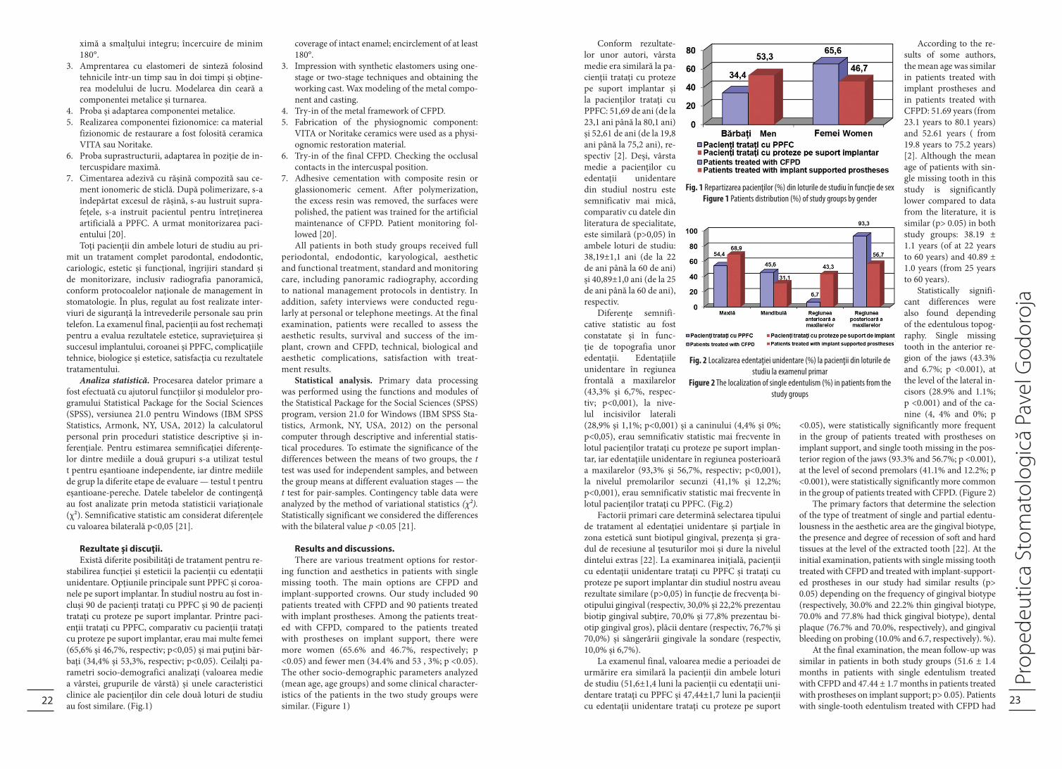
stabilirea funcţiei și esteticii la pacienţii cu edentaţii unidentare. Opţiunile principale sunt PPFC și coroa-nele pe suport implantar. În studiul nostru au fost in-cluși 90 de pacienţi trataţi cu PPFC și 90 de pacienţi trataţi cu proteze pe suport implantar. Printre paci-enţii trataţi cu PPFC, comparativ cu pacienţii trataţi cu proteze pe suport implantar, erau mai multe femei (65,6% și 46,7%, respectiv; p<0,05) și mai puţini băr-baţi (34,4% și 53,3%, respectiv; p<0,05). Ceilalţi pa-rametri socio-demografici analizaţi (valoarea medie a vârstei, grupurile de vârstă) și unele caracteristici clinice ale pacienţilor din cele două loturi de studiu au fost similare. (Fig.1)
Conform rezultate-lor unor autori, vârsta medie era similară la pa-cienţii trataţi cu proteze pe suport implantar și la pacienţilor trataţi cu PPFC: 51,69 de ani (de la 23,1 ani până la 80,1 ani) și 52,61 de ani (de la 19,8 ani până la 75,2 ani), re-spectiv [2]. Deși, vârsta medie a pacienţilor cu edentaţii unidentare din studiul nostru este semnificativ mai mică, comparativ cu datele din literatura de specialitate, este similară (p>0,05) în ambele loturi de studiu: 38,19±1,1 ani (de la 22 de ani până la 60 de ani) și 40,89±1,0 ani (de la 25 de ani până la 60 de ani), respectiv.
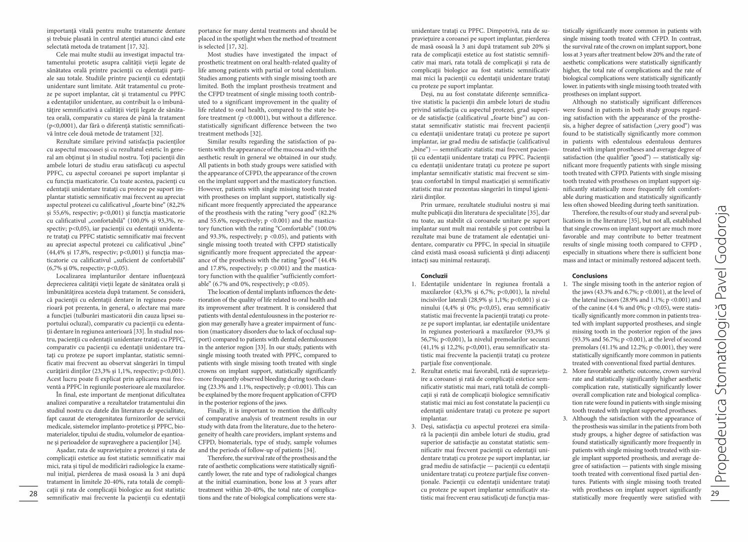
Diferenţe semnifi-cative statistic au fost constatate și în func-ţie de topografia unor edentaţii. Edentaţiile unidentare în regiunea frontală a maxilarelor (43,3% și 6,7%, respec-tiv; p<0,001), la nive-lul incisivilor laterali (28,9% și 1,1%; p<0,001) și a caninului (4,4% și 0%; p<0,05), erau semnificativ statistic mai frecvente în lotul pacienţilor trataţi cu proteze pe suport implan-tar, iar edentaţiile unidentare în regiunea posterioară a maxilarelor (93,3% și 56,7%, respectiv; p<0,001), la nivelul premolarilor secunzi (41,1% și 12,2%; p<0,001), erau semnificativ statistic mai frecvente în lotul pacienţilor trataţi cu PPFC. (Fig.2)
Factorii primari care determină selectarea tipului de tratament al edentaţiei unidentare și parţiale în zona estetică sunt biotipul gingival, prezenţa și gra-dul de recesiune al ţesuturilor moi și dure la nivelul dintelui extras [22]. La examinarea iniţială, pacienţii cu edentaţii unidentare trataţi cu PPFC și trataţi cu proteze pe suport implantar din studiul nostru aveau rezultate similare (p>0,05) în funcţie de frecvenţa bi-otipului gingival (respectiv, 30,0% și 22,2% prezentau biotip gingival subţire, 70,0% și 77,8% prezentau bi-otip gingival gros), plăcii dentare (respectiv, 76,7% și 70,0%) și sângerării gingivale la sondare (respectiv, 10,0% și 6,7%).
La examenul final, valoarea medie a perioadei de urmărire era similară la pacienţii din ambele loturi de studiu (51,6±1,4 luni la pacienţii cu edentaţii uni-dentare trataţi cu PPFC și 47,44±1,7 luni la pacienţii cu edentaţii unidentare trataţi cu proteze pe suport
coverage of intact enamel; encirclement of at least 180°.
3. Impression with synthetic elastomers using one-stage or two-stage techniques and obtaining the working cast. Wax modeling of the metal compo-nent and casting.
4. Try-in of the metal framework of CFPD.5. Fabrication of the physiognomic component:
VITA or Noritake ceramics were used as a physi-ognomic restoration material.
6. Try-in of the final CFPD. Checking the occlusal contacts in the intercuspal position.
7. Adhesive cementation with composite resin or glassionomeric cement. After polymerization, the excess resin was removed, the surfaces were polished, the patient was trained for the artificial maintenance of CFPD. Patient monitoring fol-lowed [20].All patients in both study groups received full
periodontal, endodontic, karyological, aesthetic and functional treatment, standard and monitoring care, including panoramic radiography, according to national management protocols in dentistry. In addition, safety interviews were conducted regu-larly at personal or telephone meetings. At the final examination, patients were recalled to assess the aesthetic results, survival and success of the im-plant, crown and CFPD, technical, biological and aesthetic complications, satisfaction with treat-ment results.
Statistical analysis. Primary data processing was performed using the functions and modules of the Statistical Package for the Social Sciences (SPSS) program, version 21.0 for Windows (IBM SPSS Sta-tistics, Armonk, NY, USA, 2012) on the personal computer through descriptive and inferential statis-tical procedures. To estimate the significance of the differences between the means of two groups, the t test was used for independent samples, and between the group means at different evaluation stages — the t test for pair-samples. Contingency table data were analyzed by the method of variational statistics (χ²). Statistically significant we considered the differences with the bilateral value p <0.05 [21].
results and discussions . There are various treatment options for restor-
ing function and aesthetics in patients with single missing tooth. The main options are CFPD and implant-supported crowns. Our study included 90 patients treated with CFPD and 90 patients treated with implant prostheses. Among the patients treat-ed with CFPD, compared to the patients treated with prostheses on implant support, there were more women (65.6% and 46.7%, respectively; p <0.05) and fewer men (34.4% and 53 , 3%; p <0.05). The other socio-demographic parameters analyzed (mean age, age groups) and some clinical character-istics of the patients in the two study groups were similar. (Figure 1)
According to the re-sults of some authors, the mean age was similar in patients treated with implant prostheses and in patients treated with CFPD: 51.69 years (from 23.1 years to 80.1 years) and 52.61 years ( from 19.8 years to 75.2 years)[2]. Although the mean age of patients with sin-gle missing tooth in this study is significantly lower compared to data from the literature, it is similar (p> 0.05) in both study groups: 38.19 ± 1.1 years (of at 22 years to 60 years) and 40.89 ± 1.0 years (from 25 years to 60 years).
Statistically signifi-cant differences were also found depending of the edentulous topog-raphy. Single missing tooth in the anterior re-gion of the jaws (43.3% and 6.7%; p <0.001), at the level of the lateral in-cisors (28.9% and 1.1%; p <0.001) and of the ca-nine (4, 4% and 0%; p
<0.05), were statistically significantly more frequent in the group of patients treated with prostheses on implant support, and single tooth missing in the pos-terior region of the jaws (93.3% and 56.7%; p <0.001), at the level of second premolars (41.1% and 12.2%; p <0.001), were statistically significantly more common in the group of patients treated with CFPD. (Figure 2)
The primary factors that determine the selection of the type of treatment of single and partial edentu-lousness in the aesthetic area are the gingival biotype, the presence and degree of recession of soft and hard tissues at the level of the extracted tooth [22]. At the initial examination, patients with single missing tooth treated with CFPD and treated with implant-support-ed prostheses in our study had similar results (p> 0.05) depending on the frequency of gingival biotype (respectively, 30.0% and 22.2% thin gingival biotype, 70.0% and 77.8% had thick gingival biotype), dental plaque (76.7% and 70.0%, respectively), and gingival bleeding on probing (10.0% and 6.7, respectively). %).
At the final examination, the mean follow-up was similar in patients in both study groups (51.6 ± 1.4 months in patients with single edentulism treated with CFPD and 47.44 ± 1.7 months in patients treated with prostheses on implant support; p> 0.05). Patients with single-tooth edentulism treated with CFPD had
Fig. 1 Repartizarea pacienţilor (%) din loturile de studiu în funcţie de sexFigure 1 Patients distribution (%) of study groups by gender
Fig. 2 Localizarea edentaţiei unidentare (%) la pacienţii din loturile de studiu la examenul primar
Figure 2 The localization of single edentulism (%) in patients from the study groups

24 25
implantar; p>0,05). Pacienţii cu edentaţii unidentare trataţi cu PPFC prezentau semnificativ statistic mai frecvent placă dentară (77,8% și 61,1%, respectiv; p<0,05) pe baza plăcii dentare sub formă de pelicu-lă subţire (63,3% și 47,8%, respectiv; p<0,05), pungi gingivale la sondare (12,2% și 0%, respectiv; p<0,01) și sângerări gingivale la sondare (42,2% și 6,7%, re-spectiv; p<0,001).
În scopul evaluării și analizei comparative a di-feritor opţiuni de tratament protetic sunt luaţi în considerare mai mulţi factori: costul tratamentului, rata de supravieţuire a protezelor, parametrii estetici și frecvenţa complicaţiilor [17]. Pentru evaluarea ţe-suturilor moi și obiectivizarea rezultatului estetic au fost elaboraţi mai mulţi indici estetici, cum ar fi SER, SEA și indicele papilar. Aspectul ţesuturilor moi a de-venit o componentă importantă a restaurării dentare, care necesită nu numai o formă și suprafaţă caracte-ristică adecvată, dar și o culoare care imită ţesuturile adiacente [23].
Evaluarea rezultatului estetic la pacienţii din stu-diul nostru a relevat valori medii semnificativ statis-tic mai mari în lotul pacienţilor trataţi cu proteze pe suport implantar, comparativ cu lotul pacienţilor tra-taţi cu PPFC: înălţimea papilei mezial la nivelul im-plantului/corpului de punte (2,9±0,1 mm și 2,3±0,7 mm, respectiv; p<0,001), înălţimea papilei distal la nivelul implantului/corpului de punte (2,8±0,1 mm și 2,2±0,7 mm, respectiv; p<0,001), lungimea coroa-nei clinice la nivelul implantului/corpului de punte (8,5±0,1 mm și 8,0±0,07 mm, respectiv; p<0,001), lungimea coroanei clinice la nivelul dinţilor adiacenţi (8,5±0,07 mm și 8,2±0,05 mm, respectiv; p<0,01) și SEA (9,36±0,07 puncte și 9,14±0,07 puncte, respec-tiv; p<0,05). (Fig.3)
Valoarea medie a SER (11,47±0,1 puncte și 11,41±0,1 puncte, respectiv), valoarea medie a sume-lor SEA și SER (20,77±0,2 puncte și 20,61±0,1 punc-te, respectiv) și pragurile clinice ale SEA și SER erau similare (p>0,05) în ambele loturi de studiu. Con-form SEA, toţi pacienţii din ambele loturi de studiu (100%) au avut un rezultat estetic clinic acceptat, iar 91,1% și 85,6% pacienţi, respectiv, au prezentat un re-zultat aproape perfect. Conform SER, 98,9% pacienţi din ambele loturi de studiu au avut un rezultat estetic clinic acceptat, iar 67,8% și 60,0% pacienţi, respectiv, au prezentat un rezultat aproape perfect.
Analiza rezultatelor indicelui papilar, care eva-luează înălţimea interproximală a papilei gingivale adiacente, a relevat semnificativ statistic mai frecvent papile care ocupau mai puţin de 1/2 din înălţimea pa-pilei (30,0% și 14,4%; p<0,05) în lotul pacienţilor cu edentaţii unidentare trataţi cu proteze pe suport im-plantar, iar papile hiperplastice (5,6% și 0%; p<0,05) — în lotul pacienţilor cu edentaţii unidentare trataţi cu PPFC.
Așadar, deși la examenul iniţial loturile de studiu erau similare în funcţie de vârstă, atitudinea faţă de fumat, localizarea edentaţiei unidentare la maxilare, biotipul gingival, placa dentară, scorul plăcii dentare, sângerarea gingivală la sondare și perioada de supra-veghere, rezultat estetic mai favorabil prezentau pa-cienţii cu edentaţie unidentară după tratamentul cu proteze pe suport implantar.
Restaurarea pe suport implantar pentru edentaţia unidentară este o alternativă acceptată de tratament pentru PPFC. O revizuire sistematică a literaturii a comparat coroanele unitare pe suport implantar cu PPFC pentru o perioadă de 5 ani de urmărire și a confirmat absenţa unor diferenţe semnificative între cele două tipuri de tratament în ceea ce privește rata de supravieţuire [24]. Mai multe studii au constatat că supravieţuirea PPFC pe o perioadă de 20 de ani este favorabilă și trebuie comparată cu alte opţiuni de tratament a edentaţiei unidentare [25].
Trei revizuiri sistematice ale literaturii și meta-analize au raportat rate de supravieţuire a implantu-rilor de 94,5-97,2% (de la 90,5% până la 100%) la 5 ani [26, 27, 28] și de 89,4-95,2% la 10 ani [26, 27] pentru reabilitarea unui dinte. Rata de supravieţuire a coroanelor unitare pe suport implantar a alcătuit 94,5-96,3% (de la 92,6% până la 98,1%) după 5 ani de funcţie și 89,4% după 10 ani de funcţie [27, 28], iar rata de supravieţuire a PPFC — 93,8% după 5 ani de funcţie și 89,1% după 10 ani de funcţie [3, 27, 28].
O meta-analiză, bazată pe 6 revizuiri sistematice ale studiilor prospective, retrospective, de cohortă și a seriilor de cazuri, publicată în 2012, a relevat că im-planturile au avut o rată de supravieţuire de 94,5% la 5 ani și de 89,4% la 10 ani de supraveghere, iar PPFC — de 93,8% și de 89,2%, respectiv. Rata anuală de eșec a constituit 1,12% pentru implanturi cu coroane unitare și 1,14% pentru PPFC [29].
În studiul respec-tiv, rata de supravie-ţuire a PPFC a fost semnificativ statistic mai mică, comparativ cu rata de supravie-ţuire a implantului (94,4% și 100,0%, respectiv; p<0,05) și rata de supravieţuire a coroanei pe suport implantar (94,4% și 100,0%, respectiv; p<0,05). Eșecuri cli-
statistically significantly more frequent dental plaque (77.8% and 61.1%, respectively; p <0.05) based on thin film dental plaque (63.3% and 47, 8%, respec-tively; p <0.05), gingival pockets at probing (12.2% and 0%, respectively; p <0.01) and gingival bleeding at probing (42.2% and 6.7%, respectively ; p <0.001).
For the purpose of evaluation and comparative analysis of different prosthetic treatment options, several factors are taken into account: the cost of treatment, the survival rate of prostheses, aesthetic parameters and the frequency of complications [17]. For the evaluation of soft tissues and objectification of the aesthetic result, several aesthetic indices were de-veloped, such as PES, WES and papillary index. The appearance of soft tissues has become an important component of dental restoration, which requires not only an appropriate characteristic shape and surface, but also a color that mimics adjacent tissues [23].
Evaluation of the aesthetic result in the patients in our study revealed statistically significantly higher mean values in the group of patients treated with res-torations on implant support, compared to the group of patients treated with CFPD: mesial papilla height at the level of the implant / bridge pontic (2.9 ± 0 , 1 mm and 2,3 ± 0,7 mm, respectively; p <0,001), the height of the distal papilla at the level of the implant / bridge pontic(2,8 ± 0,1 mm and 2,2 ± 0,7 mm, re-spectively ; p <0.001), the length of the clinical crown at the level of the implant / bridge pontic (8.5 ± 0.1 mm and 8.0 ± 0.07 mm, respectively; p <0.001), the length of the clinical crown at the level of the adja-cent teeth ( 8.5 ± 0.07 mm and 8.2 ± 0.05 mm, re-spectively; p <0.01) and WES (9.36 ± 0.07 points and 9.14 ± 0.07 points, respectively; p <0.05). (Figure 3)
The mean value of the PES (11.47 ± 0.1 points and 11.41 ± 0.1 points, respectively), the mean value of the WES and PES amounts (20.77 ± 0.2 points and 20.61 ± 0.1 points, respectively) and the clinical lev-els of WES and PES were similar (p> 0.05) in both study groups. According to the WES, all patients in both study groups (100%) had a clinically accepted aesthetic result, and 91.1% and 85.6% of patients, respectively, presented an almost perfect result. Ac-cording to PES, 98.9% of patients in both study groups had a clinically accepted aesthetic result, and 67.8% and 60.0% of patients, respectively, presented an almost perfect result.
Analysis of the results of the papillary index, which evaluates the interproximal height of the ad-jacent gingival papilla, revealed statistically signifi-cantly more frequently papillae that occupied less than 1/2 of the papilla height (30.0% and 14.4%; p <0.05) in the group of patients with single missing tooth treated with prostheses on implant support, and the hyperplastic papillae (5.6% and 0%; p <0.05) — in the group of patients with single missing tooth treated with CFPD.
Therefore, although at the initial examination the study groups were similar in terms of age, attitude towards smoking, location of unidentary edentulous jaw, gingival biotype, dental plaque, dental plaque score, gingival bleeding at probing and folow-up period, more favorable aesthetic result presented pa-tients with single missing tooth after treatment with prostheses on implant support.
Implant restoration for single missing tooth is an accepted treatment alternative for CFPD. A system-atic review of the literature compared single crowns on implant support with CFPD for a 5-year follow-up period and confirmed the absence of significant differences between the two types of treatment in terms of survival rate [24]. Several studies have found that the survival of CFPD over a period of 20 years is favorable and should be compared with other treat-ment options for single edentulous patients [25].
Three systematic literature reviews and meta-an-alyzes reported implant survival rates of 94.5-97.2% (90.5% to 100%) at 5 years [26, 27, 28], and 89 years, 4-95.2% at 10 years [26, 27] for the rehabilitation of a single missing tooth. The survival rate of single crowns on implant support was 94.5-96.3% (from 92.6% to 98.1%) after 5 years of function and 89.4% after 10 years of function [27 , 28], and the survival rate of CFPD — 93.8% after 5 years of function and 89.1% after 10 years of function [3, 27, 28].
A meta-analysis, based on 6 systematic reviews of prospective, retrospective, cohort and case series studies, published in 2012, revealed that the implants had a survival rate of 94.5% at 5 years and 89, 4% at 10 years of follow-up, and CFPD — 93.8% and 89.2%, respectively. The annual failure rate was 1.12% for single implant-supported crowns and 1.14% for CFPD [29].
In this study, the survival rate of CFPD was statistically sig-nificantly lower com-pared to the survival rate of the implant (94.4% and 100.0%, respectively; p <0.05) and the survival rate of the single implant supported crowns (94.4% and 100.0%, respectively; p <0.05). Clinical or absolute
Fig. 3 Evaluarea rezultatului estetic: 1 — papila dentară mezială; 2 — papila dentară distală; 3 — nivelul ţesutului moale; 4 — zenitul; 5 — procesul alveolar; 6 — culoarea gingiei; 7 — textura gingiei
Figure 3 Evaluation of the aesthetic result: 1 — mesial dental papilla; 2 — distal dental papilla; 3 — soft tissue level; 4 — the zenith; 5 — the alveolar process; 6 — gum color; 7 — gum texture
Fig. 4 Ratele de supravieţuire a PPFC şi a coroanei pe suport implantar (%) în funţie de sex, vârstă şi topografia edentaţiei unidentare
Figure 4. Survival rates of CFPD and single implant supported crown (%) by gender, age and topography of single edentulous area

26 27
nice sau absolute nu au fost constatate în ambele lo-turi de studiu. (Fig.4)
Examenul radiologic, realizat la 3 ani după tra-tament, a relevat pierdere de masă osoasă sub 20% semnificativ statistic mai frecvent în lotul pacienţilor cu edentaţii unidentare trataţi cu proteze pe suport implantar (91,1% și 68,9%, p<0,001), iar în lotul pa-cienţilor cu edentaţii unidentare trataţi cu PPFC — semnificativ statistic mai frecvent pierdere de masă osoasă în limitele 20-40% (26,7% și 7,8%, p<0,001).
Autorii unui studiu au constatat o predominare a complicaţiilor biologice (pierderea vitalităţii, com-plicaţii endodontice, fracturi ale rădăcinilor și carii dentare ale dinţilor stâlpi) la pacienţii cu PPFC, în timp ce fracturi ale materialului de faţetare au apărut doar în 3,8% cazuri. Dimpotrivă, complicaţiile tehni-ce (fracturi ale coroanei, slăbirea șuruburilor și pier-derea retenţiei) prezentau mult mai frecvent pacienţii cu reconstrucţii pe suport implantar [30].
Cele mai frecvente complicaţii precoce au fost iritarea gingivală localizată (1,9% pentru coroanele unitare și 2,5% pentru PPFC) și sensibilitatea denta-ră postoperatorie (0,4% și 3,3%, respectiv). Cea mai frecventă cauză de eșec pe termen scurt a fost frac-tura/ciobirea materialului de faţetare (0,8% și 0,8%, respectiv). Au eșuat 1 (0,4%) coroană din cauza pier-derii de retenţie și 2 (1,7%) PPFC din cauza fracturii carcasei [31].
În studiul nostru, rata totală de complicaţii era semnificativ statistic mai mare la pacienţii cu eden-taţii unidentare trataţi cu PPFC, comparativ cu pa-cienţii cu edentaţii unidentare trataţi cu proteze pe suport implantar (61,1% și 43,3%, respectiv; p<0,05) și printre femei (59,3% și 33,3%, respectiv; p<0,01). Acest indicator era similar în sublotul de bărbaţi (64,5% și 52,1, respectiv). Conform datelor literaturii de specialitate, unele studii au raportat o rată totală de complicaţi mult mai mică, comparativ cu rezulta-tele studiului nostru. Acest indicator era mai mare la pacienţii cu edentaţii unidentare trataţi cu proteze pe suport implantar supravegheaţi o perioadă medie de 27,6 luni (33,51%), comparativ cu pacienţii cu eden-taţii unidentare trataţi cu PPFC supravegheaţi o peri-oadă medie de 85,18 luni (20,2%), însă diferenţa nu a atins certitudine statistică [2]. (Fig.5)
Frecvenţa complicaţiilor tehnice (pierderea de retenţie a coroanei, fractura componentelor și deci-mentarea) în studiul nostru era statistic semnificativ mai mare în lotul pacienţilor cu edentaţii unidenta-re trataţi cu PPFC (5,6%), comparativ cu pacienţii cu edentaţii unidentare trataţi cu proteze pe suport implantar (0%; p<0,05). Aceiași tendinţă a fost con-statată printre femei (6,8% și 0%, respectiv; p<0,05) și printre pacienţii cu edentaţii unidentare în regi-unea posterioară a maxilarelor (4,8% și 0%, respec-tiv; p<0,05). În alte publicaţii, dimpotrivă, incidenţa complicaţiilor tehnice la pacienţii cu edentaţii uni-dentare trataţi cu coroane pe suport implantar peste 5 ani de supraveghere (12,7%) a fost de aproximativ 2 ori mai mare, comparativ cu pacienţii trataţi cu
PPFC (5,8%), iar incidenţa fracturilor materialului de faţetare a fost de aproximativ 3 ori mai mică (4,5% și 13,2%, respectiv) [28].
Complicaţii biologice au fost depistate statistic semnificativ mai frecvent (p<0,001) la pacienţii cu edentaţii unidentare trataţi cu PPFC (56,7% — com-plicaţii ale ţesutului moale, pierdere verticală de masă osoasă >2 mm și carie dentară), comparativ cu pacienţii cu edentaţii unidentare trataţi cu proteze pe suport implantar (16,7% — complicaţii ale ţesutului moale și pierdere verticală de masă osoasă >2 mm). Tendinţă similară a fost determinată printre băr-baţi (61,3% și 0%, respectiv; p<0,001), printre femei (54,2% și 7,1%, respectiv; p<0,001) și la pacienţii cu edentaţii unidentare în regiunea posterioară a ma-xilarelor (57,1% și 11,8%, respectiv; p<0,001). Rata de complicaţii biologice era similară la pacienţii cu edentaţii unidentare în regiunea frontală a maxila-relor (50,0% și 23,1%, respectiv; p>0,05). Rezultatele noastre coincid cu rezultatele altor studii. Printre pa-cienţii cu edentaţii unidentare trataţi cu PPFC sunt constatate mai multe complicaţii biologice. Apro-ximativ 10% dintre dinţii stâlpi își pierd vitalitatea după 10 ani și aproximativ la 9,1-9,5% dintre dinţii stâlpi au fost depistate carii dentare [28].
În studiul dat, complicaţii estetice au fost con-statate statistic semnificativ mai frecvent (p<0,01) la pacienţii cu edentaţii unidentare trataţi cu proteze pe suport implantar (43,3% — proces alveolar masiv, re-cesiune avansată mezio-vestibulară a papilei >1 mm, absenţa parţială a papilei și absenţa totală a papilei), comparativ cu pacienţii cu edentaţii unidentare tra-taţi cu PPFC (23,3% — uzura coroanei, recesiune avansată mezio-vestibulară a papilei >1 mm, diferen-ţă de culoare cu dentiţia naturală, absenţa parţială a papilei și absenţa totală a papilei). Aceiași tendinţă a fost relevată la pacienţii cu edentaţii unidentare în regiunea posterioară a maxilarelor (49,0% și 23,8%, respectiv; p<0,01). Rata de complicaţii estetice era similară printre bărbaţi (52,1% și 32,3%, respectiv; p>0,05), printre femei (33,3% și 18,6%, respectiv; p>0,05) și la pacienţii cu edentaţii unidentare în regi-unea frontală a maxilarelor (35,9% și 16,7%, respec-tiv; p>0,05).
Edentaţia uniden-tară agravează sănăta-tea generală și calitatea vieţii, afectează capa-citatea masticatorie, fonetică și aspectul estetic. În ultimii ani, evaluarea calităţii vie-ţii legate de sănătatea orală a devenit din ce în ce mai importantă, deoarece rezultatele raportate de pacient sunt esenţiale. Îmbu-nătăţirea satisfacţiei pacientului este de o
failures were not found in both study groups. (Fig-ure 4)
Radiological examination, performed 3 years after treatment, revealed statistically significant loss of bone mass below 20% more frequently in the group of patients with single tooth loss treated with prostheses on implant support (91.1% and 68.9%, p <0.001) , and in the group of patients treated with CFPD — statistically significant more frequent bone loss within 20-40% (26.7% and 7.8%, p <0.001).
The authors of a study found a predominance of biological complications (loss of vitality, endodon-tic complications, root fractures and tooth decay of abutment teeth) in patients with CFPD, while frac-tures of the veneer material occurred in only 3.8% of cases. On the contrary, technical complications (crown fractures, loosening of screws and loss of re-tention) were much more common in patients with reconstructions on implant support [30].
The most common early complications were localized gingival irritation (1.9% for single crowns and 2.5% for CFPD) and postoperative dental sen-sitivity (0.4% and 3.3%, respectively). The most common cause of short-term failure was fracture / chipping of veneer material (0.8% and 0.8%, respec-tively). 1 (0.4%) crown failed due to retention loss and 2 (1.7%) CFPD due to framework fracture [31].
In this study, the overall complication rate was statistically significantly higher in patients with sin-gle missing tooth treated with CFPD, compared to patients with single missing tooth treated with im-plant-supported prostheses (61.1% and 43.3%, re-spectively; p <0 .05) and among women (59.3% and 33.3%, respectively; p <0.01). This indicator was sim-ilar in the sublot of men (64.5% and 52.1, respective-ly). According to the literature, some studies report-ed a much lower total complication rate compared to the results of our study. This indicator was higher in patients with single missing tooth treated with crowns on implant support with a follow-up of an av-erage period of 27.6 months (33.51%), compared to patients with single missing tooth treated with CFPD folowed — up for an average period of 85.18 months (20.2%), but the difference did not reach statistical certainty [2].(Figure5)
The frequency of technical complications (loss of crown retention, fracture of components and decima-tion) in our study was statistically significantly higher in the group of patients with single missing tooth treated with CFPD (5.6%), compared to patients with single missing tooth treated with prostheses on implant support (0%; p <0.05). The same result was observed among women (6.8% and 0%, respective-ly; p <0.05) and among patients with single missing tooth in the posterior region of the jaws (4.8% and 0%, respectively; p <0, 05). In other publications, on the contrary, the incidence of technical complications in patients with single tooth replacement by implant supported prosthesis over 5 years of surveillance (12.7%) was approximately two times higher com-
pared to patients treated with CFPD (5.8%). %), and the incidence of veneer material was about 3 times lower (4.5% and 13.2%, respectively) [28].
Biological complications were statistically sig-nificantly more common (p <0.001) in patients with single tooth replacement by CFPD (56.7% — soft tissue complications, vertical loss of bone mass> 2 mm and tooth decay), compared with patients with sibgle missing tooth treated with prostheses on im-plant support (16.7% — complications of soft tissue and vertical loss of bone mass> 2 mm). A similar result was determined among men (61.3% and 0%, respectively; p <0.001), among women (54.2% and 7.1%, respectively; p <0.001) and in patients with single edentulism in the posterior regions of the jaws (57.1% and 11.8%, respectively; p <0.001). The rate of biological complications was similar in patients with single missing tooth in the anterior region of the jaws (50.0% and 23.1%, respectively; p> 0.05). Our results coincide with the results of other studies. Several bio-logical complications are found among patients with single-tooth edentulism treated with CFPD. Approx-imately 10% of abutment teeth lose their vitality af-ter 10 years and approximately 9.1-9.5% of abutment teeth have been detected tooth decay [28].
In this study, aesthetic complications were found to be statistically significantly more frequent (p <0.01) in patients with single tooth replacement by implant supported prosthesis (43.3% — massive al-veolar process, mesio-facial recession of the papilla> 1 mm, partial absence of the papilla and total absence of the papilla), compared to patients with single miss-ing tooth treated with CFPD (23.3% — crown wear, mesio-facial recession of the papilla> 1 mm, color difference with natural dentition, partial absence of the papilla and total absence of the papilla). The same result was found in patients with single missing tooth in the posterior region of the jaws (49.0% and 23.8%, respectively; p <0.01). The rate of aesthetic complications was similar among men (52.1% and 32.3%, respectively; p> 0.05), among women (33.3% and 18.6%, respectively; p> 0.05) and in patients with single missing tooth in the anterior region of the jaws (35.9% and 16.7%, respectively; p> 0.05).
Single tooth eden-tulism worsens the general health and quality of life, affects the masticatory capac-ity, phonetics and aes-thetic appearance. In recent years, assessing the quality of life re-lated to oral health has become increasingly important because the results reported by the patient are essential. Improving patient sat-isfaction is of vital im-
Fig. 5 Complicaţiile (%) tehnice, biologice şi estetice după tratamentul pacienţilor cu edentaţii unidentare în ambele loturi de studiu
Figure 5. Technical, biological and aesthetic complications (%) after replacement of single missing tooth in both study groups
28 29
importanţă vitală pentru multe tratamente dentare și trebuie plasată în centrul atenţiei atunci când este selectată metoda de tratament [17, 32].
Cele mai multe studii au investigat impactul tra-tamentului protetic asupra calităţii vieţii legate de sănătatea orală printre pacienţii cu edentaţii parţi-ale sau totale. Studiile printre pacienţii cu edentaţii unidentare sunt limitate. Atât tratamentul cu prote-ze pe suport implantar, cât și tratamentul cu PPFC a edentaţiilor unidentare, au contribuit la o îmbună-tăţire semnificativă a calităţii vieţii legate de sănăta-tea orală, comparativ cu starea de până la tratament (p<0,0001), dar fără o diferenţă statistic semnificati-vă între cele două metode de tratament [32].
Rezultate similare privind satisfacţia pacienţilor cu aspectul mucoasei și cu rezultatul estetic în gene-ral am obţinut și în studiul nostru. Toţi pacienţii din ambele loturi de studiu erau satisfăcuţi cu aspectul PPFC, cu aspectul coroanei pe suport implantar și cu funcţia masticatorie. Cu toate acestea, pacienţi cu edentaţii unidentare trataţi cu proteze pe suport im-plantar statistic semnificativ mai frecvent au apreciat aspectul protezei cu calificativul „foarte bine“ (82,2% și 55,6%, respectiv; p<0,001) și funcţia masticatorie cu calificativul „confortabilă“ (100,0% și 93,3%, re-spectiv; p<0,05), iar pacienţii cu edentaţii unidenta-re trataţi cu PPFC statistic semnificativ mai frecvent au apreciat aspectul protezei cu calificativul „bine“ (44,4% și 17,8%, respectiv; p<0,001) și funcţia mas-ticatorie cu calificativul „suficient de confortabilă“ (6,7% și 0%, respectiv; p<0,05).
Localizarea implanturilor dentare influenţează deprecierea calităţii vieţii legate de sănătatea orală și îmbunătăţirea acesteia după tratament. Se consideră, că pacienţii cu edentaţii dentare în regiunea poste-rioară pot prezenta, în general, o afectare mai mare a funcţiei (tulburări masticatorii din cauza lipsei su-portului ocluzal), comparativ cu pacienţii cu edenta-ţii dentare în regiunea anterioară [33]. În studiul nos-tru, pacienţii cu edentaţii unidentare trataţi cu PPFC, comparativ cu pacienţii cu edentaţii unidentare tra-taţi cu proteze pe suport implantar, statistic semni-ficativ mai frecvent au observat sângerări în timpul curăţării dinţilor (23,3% și 1,1%, respectiv; p<0,001). Acest lucru poate fi explicat prin aplicarea mai frec-ventă a PPFC în regiunile posterioare ale maxilarelor.
În final, este important de menţionat dificultatea analizei comparative a rezultatelor tratamentului din studiul nostru cu datele din literatura de specialitate, fapt cauzat de eterogenitatea furnizorilor de servicii medicale, sistemelor implanto-protetice și PPFC, bio-materialelor, tipului de studiu, volumelor de eșantioa-ne și perioadelor de supraveghere a pacienţilor [34].
Așadar, rata de supravieţuire a protezei și rata de complicaţii estetice au fost statistic semnificativ mai mici, rata și tipul de modificări radiologice la exame-nul iniţial, pierderea de masă osoasă la 3 ani după tratament în limitele 20-40%, rata totală de compli-caţii și rata de complicaţii biologice au fost statistic semnificativ mai frecvente la pacienţii cu edentaţii
unidentare trataţi cu PPFC. Dimpotrivă, rata de su-pravieţuire a coroanei pe suport implantar, pierderea de masă osoasă la 3 ani după tratament sub 20% și rata de complicaţii estetice au fost statistic semnifi-cativ mai mari, rata totală de complicaţii și rata de complicaţii biologice au fost statistic semnificativ mai mici la pacienţii cu edentaţii unidentare trataţi cu proteze pe suport implantar.
Deși, nu au fost constatate diferenţe semnifica-tive statistic la pacienţii din ambele loturi de studiu privind satisfacţia cu aspectul protezei, grad superi-or de satisfacţie (calificativul „foarte bine“) au con-statat semnificativ statistic mai frecvent pacienţii cu edentaţii unidentare trataţi cu proteze pe suport implantar, iar grad mediu de satisfacţie (calificativul „bine“) — semnificativ statistic mai frecvent pacien-ţii cu edentaţii unidentare trataţi cu PPFC. Pacienţii cu edentaţii unidentare trataţi cu proteze pe suport implantar semnificativ statistic mai frecvent se sim-ţeau confortabil în timpul masticaţiei și semnificativ statistic mai rar prezentau sângerări în timpul igieni-zării dinţilor.
Prin urmare, rezultatele studiului nostru și mai multe publicaţii din literatura de specialitate [35], dar nu toate, au stabilit că coroanele unitare pe suport implantar sunt mult mai rentabile și pot contribui la rezultate mai bune de tratament ale edentaţiei uni-dentare, comparativ cu PPFC, în special în situaţiile când există masă osoasă suficientă și dinţi adiacenţi intacţi sau minimal restauraţi.
Concluzii1. Edentaţiile unidentare în regiunea frontală a
maxilarelor (43,3% și 6,7%; p<0,001), la nivelulincisivilor laterali (28,9% și 1,1%; p<0,001) și ca-ninului (4,4% și 0%; p<0,05), erau semnificativstatistic mai frecvente la pacienţii trataţi cu prote-ze pe suport implantar, iar edentaţiile unidentareîn regiunea posterioară a maxilarelor (93,3% și56,7%; p<0,001), la nivelul premolarilor secunzi(41,1% și 12,2%; p<0,001), erau semnificativ sta-tistic mai frecvente la pacienţii trataţi cu protezeparţiale fixe convenţionale.
2. Rezultat estetic mai favorabil, rată de supravieţu-ire a coroanei și rată de complicaţii estetice sem-nificativ statistic mai mari, rată totală de compli-caţii și rată de complicaţii biologice semnificativstatistic mai mici au fost constatate la pacienţii cuedentaţii unidentare trataţi cu proteze pe suportimplantar.
3. Deși, satisfacţia cu aspectul protezei era simila-ră la pacienţii din ambele loturi de studiu, gradsuperior de satisfacţie au constatat statistic sem-nificativ mai frecvent pacienţii cu edentaţii uni-dentare trataţi cu proteze pe suport implantar, iargrad mediu de satisfacţie — pacienţii cu edentaţiiunidentare trataţi cu proteze parţiale fixe conven-ţionale. Pacienţii cu edentaţii unidentare trataţicu proteze pe suport implantar semnificativ sta-tistic mai frecvent erau satisfăcuţi de funcţia mas-
portance for many dental treatments and should be placed in the spotlight when the method of treatment is selected [17, 32].
Most studies have investigated the impact of prosthetic treatment on oral health-related quality of life among patients with partial or total edentulism. Studies among patients with single missing tooth are limited. Both the implant prosthesis treatment and the CFPD treatment of single missing tooth contrib-uted to a significant improvement in the quality of life related to oral health, compared to the state be-fore treatment (p <0.0001), but without a difference. statistically significant difference between the two treatment methods [32].
Similar results regarding the satisfaction of pa-tients with the appearance of the mucosa and with the aesthetic result in general we obtained in our study. All patients in both study groups were satisfied with the appearance of CFPD, the appearance of the crown on the implant support and the masticatory function. However, patients with single missing tooth treated with prostheses on implant support, statistically sig-nificant more frequently appreciated the appearance of the prosthesis with the rating “very good” (82.2% and 55.6%, respectively; p <0.001) and the mastica-tory function with the rating “Comfortable” (100.0% and 93.3%, respectively; p <0.05), and patients with single missing tooth treated with CFPD statistically significantly more frequent appreciated the appear-ance of the prosthesis with the rating “good” (44.4% and 17.8%, respectively; p <0.001) and the mastica-tory function with the qualifier “sufficiently comfort-able” (6.7% and 0%, respectively; p <0.05).
The location of dental implants influences the dete-rioration of the quality of life related to oral health and its improvement after treatment. It is considered that patients with dental edentulousness in the posterior re-gion may generally have a greater impairment of func-tion (masticatory disorders due to lack of occlusal sup-port) compared to patients with dental edentulousness in the anterior region [33]. In our study, patients with single missing tooth treated with PPFC, compared to patients with single missing tooth treated with single crowns on implant support, statistically significantly more frequently observed bleeding during tooth clean-ing (23.3% and 1.1%, respectively; p <0.001). This can be explained by the more frequent application of CFPD in the posterior regions of the jaws.
Finally, it is important to mention the difficulty of comparative analysis of treatment results in our study with data from the literature, due to the hetero-geneity of health care providers, implant systems and CFPD, biomaterials, type of study, sample volumes and the periods of follow-up of patients [34].
Therefore, the survival rate of the prosthesis and the rate of aesthetic complications were statistically signifi-cantly lower, the rate and type of radiological changes at the initial examination, bone loss at 3 years after treatment within 20-40%, the total rate of complica-tions and the rate of biological complications were sta-
tistically significantly more common in patients with single missing tooth treated with CFPD. In contrast, the survival rate of the crown on implant support, bone loss at 3 years after treatment below 20% and the rate of aesthetic complications were statistically significantly higher, the total rate of complications and the rate of biological complications were statistically significantly lower. in patients with single missing tooth treated with prostheses on implant support.
Although no statistically significant differences were found in patients in both study groups regard-ing satisfaction with the appearance of the prosthe-sis, a higher degree of satisfaction („very good“) was found to be statistically significantly more common in patients with edentulous edentulous dentures treated with implant prostheses and average degree of satisfaction (the qualifier “good”) — statistically sig-nificant more frequently patients with single missing tooth treated with CFPD. Patients with single missing tooth treated with prostheses on implant support sig-nificantly statistically more frequently felt comfort-able during mastication and statistically significantly less often showed bleeding during teeth sanitization.
Therefore, the results of our study and several pub-lications in the literature [35], but not all, established that single crowns on implant support are much more favorable and may contribute to better treatment results of single missing tooth compared to CFPD , especially in situations where there is sufficient bone mass and intact or minimally restored adjacent teeth.
Conclusions1. The single missing tooth in the anterior region of
the jaws (43.3% and 6.7%; p <0.001), at the level ofthe lateral incisors (28.9% and 1.1%; p <0.001) andof the canine (4.4 % and 0%; p <0.05), were statis-tically significantly more common in patients trea-ted with implant supported prostheses, and singlemissing tooth in the posterior region of the jaws(93.3% and 56.7%; p <0.001), at the level of secondpremolars (41.1% and 12.2%; p <0.001), they werestatistically significantly more common in patientstreated with conventional fixed partial dentures.
2. More favorable aesthetic outcome, crown survivalrate and statistically significantly higher aestheticcomplication rate, statistically significantly loweroverall complication rate and biological complica-tion rate were found in patients with single missing tooth treated with implant supported prostheses.
3. Although the satisfaction with the appearance ofthe prosthesis was similar in the patients from both study groups, a higher degree of satisfaction wasfound statistically significantly more frequently inpatients with single missing tooth treated with sin-gle implant supported prosthesis, and average de-gree of satisfaction — patients with single missingtooth treated with conventional fixed partial den-tures. Patients with single missing tooth treatedwith prostheses on implant support significantlystatistically more frequently were satisfied with
30 31
ticatorie și semnificativ statistic mai rar raportau sângerări în timpul igienizării dinţilor.
4. Coroanele unitare pe suport implantar, comparativ cu protezele parţiale fixe convenţionale, sunt multmai favorabile și pot contribui la rezultate maibune de tratament a edentaţiei unidentare, în spe-cial în situaţiile când există masă osoasă suficientăși dinţi adiacenţi intacţi sau minimal restauraţi.
bibliografie / bibliography
1. Del Fabbro M., Ceresoli V., Taschieri S. et al. Immediate loading of postextractionimplants in the esthetic area: systematicreview of the literature. Clin. Implant.Dent. Relat. Res. 2015; 17(1): 52-70.
2. Varga T.G. Economic evaluation of sin-gle-tooth replacement using fixed dentalprosthesis or implant-supported singlecrowns. Faculty of Medicine, Universityof Bern. http://dentiste-lausanne-vaud.ch/wp-content/uploads/2017/07/Disser-tation-Dr-Thomas-Varga-Economic.pdf.
3. Pjetursson B.E., Lang N.P. Prosthetictreatment planning on the basis of scien-tific evidence. J. Oral. Rehabil. 2008; 35Suppl 1: 72-79.
4. Wolff D., Wohlrab T., Saure D., KrisamJ., Frese C. Fiber-reinforced compositefixed dental prostheses: A 4-year pro-spective clinical trial evaluating survival,quality, and effects on surrounding peri-odontal tissues. J. Prosthet. Dent. 2018;119(1): 47-52.
5. Prabhu R., Prabhu G., Baskaran E., Aru-mugam E. Clinical acceptability of metal-ceramic fixed partial dental prosthesis fa-bricated with direct metal laser sinteringtechnique-5 year follow-up. J. Indian.Prosthodont. Soc. 2016; 16(2): 193-197.
6. Kern M., Sasse M., Wolfart S. Ten-yearoutcome of three-unit fixed dental pros-theses made from monolithic lithiumdisilicate ceramic. J. Am. Dent. Assoc.2012; 143(3): 234-240.
7. Bortolini S., Natali A., Franchi M. OTEquator Bont Protetic Biologic Un nouconcept în protezarea fixă și mobilă peimplanturi. Italy: DeMIR Editore — ViaEridania, 2015. 204 p.
8. Hof M., Pommer B., Ambros H., Jesch P., Vogl S., Zechner W. Does Timing of Im-plant Placement Affect Implant TherapyOutcome in the Aesthetic Zone? A Clini-cal, Radiological, Aesthetic, and Patient-Based Evaluation. Clin. Implant. Dent.Relat. Res. 2015; 17(6): 1188—1199.
9. Cuny-Houchmand M., Renaudin S., Le-roul M., Planche L., Le Guehennec L.,Soueidan A. Gingival biotype: The pro-be test utility. Open. J. Stomatol. 2013; 3:123-127.
10. Cosyn J., Eghbali A., Hermans A. et al. A 5-year prospective study on single imme-diate implants in the aesthetic zone. J.Clin. Periodontol. 2016; 43(8): 702-709.
11. Hjalmarsson L., Gheisarifar M., Jemt T.A systematic review of survival of sin-gle implants as presented in longitudi-nal studies with a follow-up of at least10 years. Eur. J. Oral. Implantol. 2016; 9Suppl 1: S155-162.
12. Pjetursson B., Tan K., Lang N. et al. Asystematic review of the survival andcomplication rates of fixed partial den-tures (FPDs) after an observation period of at least 5 years. I. Implant-supportedFPDs. Clin. Oral. Implants. Res. 2004;15(6): 625-642.
13. Pol C.W., Raghoebar G.M., Kerdijk W.,Boven G.C., Cune M.S., Meijer H.J. Asystematic review and meta-analysis of3-unit fixed dental prostheses: Are theresults of 2 abutment implants compara-ble to the results of 2 abutment teeth? J.Oral. Rehabil. 2018; 45(2): 147-160.
14. Anusavice K.J. Standardizing failure,success, and survival decisions in clinical studies of ceramic and metal-ceramic fi-xed dental prostheses. Dent. Mater. 2012; 28(1): 102-111.
15. Chang M. The peri-implant tissues from an esthetic perspective. Göteborg: Uni-versity of Gothenburg, Sweden, 2009. 71p.
16. Misch C.E., Perel M.L., Wang H.L. et al.Implant success, survival, and failure: the International Congress of Oral Implan-tologists (ICOI) Pisa Consensus Confe-rence. Implant. Dent. 2008; 17(1): 5-15.
17. Gjelvold B., Kisch J., Chrcanovic B. etal. Clinical and radiographic outcomefollowing immediate loading and de-layed loading of single-tooth implants:Randomized clinical trial. Clin. Implant.Dent. Relat. Res. 2017; 19(3): 549-558.
18. den Hartog L., Raghoebar G., Slater J. etal. Single-tooth implants with differentneck designs: a randomized clinical trialevaluating the aesthetic outcome. Clin.Implant. Dent. Relat. Res. 2013; 15(3):311-321.
19. Gumeniuc A., Topalo V., Mostovei A.Încărcarea funcţională precoce a implan-telor dentare de stadiul I în cadrul eden-taţiilor unidentare. Medicina Stomatolo-gică. 2013; (3): 41-46.
20. Zaharia A., Duţă M., Patroi G. et al. As-pecte clinice privind proteza parţială fixăadezivă tip Maryland. Craiova Medicală. 2008; 10(1): 42-45.
21. Spinei L., Lozan O., Badan V. Biostatisti-ca. Chișinău: Şcoala de Menegement înSănătate Piblică, 2009. 186 p.
22. Chu S.J., Hochman M.N., Tamow D.P.Loss of the Interdental Papilla. Preventi-on and management of dental implant-related complications in the esthetic zone. Inside. Dentistry. 2014; 10(1): 12-16.
23. Slagter K.W. Immediate dental implantplacement in the aesthetic zone. Thesis.Drukkerij van der Eems, Heerenveen,Netherlands. 2016, 156 p.
24. Salinas T.J., Eckert S.E. In patients requi-ring single-tooth replacement, what are
the outcomes of implant- as compared to tooth-supported restorations? Int. J. Oral. Maxillofac. Implants. 2007; 22 Suppl: 71-95.
25. De Backer H., Van Maele G., De MoorN. et al. Single-tooth replacement: is a3-unit fixed partial denture still an opti-on? A 20-year retrospective study. Int. J.Prosthodont. 2006; 19(6): 567-573.
26. Pjetursson B., Brägger U., Lang N. et al.Comparison of survival and complicati-on rates of tooth-supported fixed dentalprostheses (FDPs) and implant-supported FDPs and single crowns (SCs). Clin. Oral. Implants. Res. 2007; 18 Suppl 3: 97-113.
27. Jung R., Zembic A., Pjetursson B. et al.Systematic review of the survival rate and the incidence of biological, technical, and aesthetic complications of single crownson implants reported in longitudinalstudies with a mean follow-up of 5 years. Clin. Oral. Implants. Res. 2012; 23 Suppl6: 2-21.
28. Jung R., Pjetursson B., Glauser R. et al.A systematic review of the 5-year survi-val and complication rates of implant-supported single crowns. Clin. Oral. Im-plants. Res. 2008; 19(2): 119-130.
29. Pjetursson B., Zwahlen M., Lang N.Quality of reporting of clinical studiesto assess and compare performance ofimplant-supported restorations. J. Clin.Periodontol. 2012; 39 Suppl 12: 139-159.
30. Karl M. Outcome of bonded vs all-cera-mic and metal- ceramic fixed prosthesesfor single tooth replacement. Eur. J. Oral.Implantol. 2016; 9 Suppl 1: S25-44.
31. Pihlaja J., Näpänkangas R., Raustia A.Early complications and short-term fai-lures of zirconia single crowns and par-tial fixed dental prostheses. J. Prosthet.Dent. 2014; 112(4): 778-783.
32. Park S., Oh S, Kim J. et al. Single-ToothImplant Versus Three-Unit Fixed PartialDenture: A Study of Oral Health-Related Quality of Life. Int. J. Oral. Maxillofac.Implants. 2016; 31(2): 376-381.
33. Hara M., Matsumoto T., Yokoyama S.et al. Location of implant-retained fixeddentures affects oral health-related qua-lity of life. Clin. Implant. Dent. Relat. Res.2017; 19(4): 710-716.
34. Tan K., Pjetursson B., Lang N., Chan E.A systematic review of the survival andcomplication rates of fixed partial dentu-res (FPDs) after an observation period of at least 5 years. III. Conventional FPDs.Clin. Oral. Implants. Res. 2004; 15(6):654-666.
35. Moy P., Nishimura G., Pozzi A. et al. Sin-gle implants in dorsal areas — A syste-matic review. Eur. J. Oral. Implantol.2016; 9 Suppl 1: S163-172.
masticatory function and statistically significantly less reported bleeding during oral hygiene.
4. Single crowns on implant support, compared toconventional fixed partial dentures, are muchmore favorable and can contribute to better treat-ment of single missing tooth, especially in situati-ons where there is sufficient bone mass and intact or minimally restored adjacent teeth.