An Excitatory Loop with Astrocytes Contributes to Drive Neurons to Seizure Threshold
Upload
khangminh22Category
view
1download
0
Edinburgh Research Explorer
Mutant C9orf72 human iPSCderived astrocytes cause noncellautonomous motor neuron pathophysiology
Citation for published version:Zhao, C, Devlin, A, Chouhan, AK, Selvaraj, BT, Stavrou, M, Burr, K, Brivio, V, He, X, Mehta, AR, Story, D,Shaw, CE, Dando, O, Hardingham, GE, Miles, GB & Chandran, S 2019, 'Mutant C9orf72 human iPSCderived astrocytes cause noncell autonomous motor neuron pathophysiology', Glia.https://doi.org/10.1002/glia.23761
Digital Object Identifier (DOI):10.1002/glia.23761
Link:Link to publication record in Edinburgh Research Explorer
Document Version:Publisher's PDF, also known as Version of record
Published In:Glia
General rightsCopyright for the publications made accessible via the Edinburgh Research Explorer is retained by the author(s)and / or other copyright owners and it is a condition of accessing these publications that users recognise andabide by the legal requirements associated with these rights.
Take down policyThe University of Edinburgh has made every reasonable effort to ensure that Edinburgh Research Explorercontent complies with UK legislation. If you believe that the public display of this file breaches copyright pleasecontact [email protected] providing details, and we will remove access to the work immediately andinvestigate your claim.
Download date: 25. Mar. 2022

R E S E A R CH A R T I C L E
Mutant C9orf72 human iPSC-derived astrocytes cause non-cellautonomous motor neuron pathophysiology
Chen Zhao1,2 | Anna-Claire Devlin1,3 | Amit K. Chouhan1,3 |
Bhuvaneish T. Selvaraj1,2,4 | Maria Stavrou1,2,4 | Karen Burr1,2,4 |
Veronica Brivio1,3 | Xin He4,5 | Arpan R. Mehta1,2,4 | David Story1,2,4 |
Christopher E. Shaw6,7 | Owen Dando4,5 | Giles E. Hardingham4,5 |
Gareth B. Miles1,3 | Siddharthan Chandran1,2,4,8
1Euan MacDonald Centre for MND Research, The University of Edinburgh, Edinburgh, UK
2Centre for Clinical Brain Sciences, The University of Edinburgh, Edinburgh, UK
3School of Psychology and Neuroscience, University of St Andrews, St Andrews, Fife, UK
4Dementia Research Institute at the University of Edinburgh, Edinburgh, UK
5Centre for Discovery Brain Sciences, The University of Edinburgh, Edinburgh, UK
6MRC Centre for Neurodegeneration Research, King's College London, Institute of Psychiatry, London, UK
7Dementia Research Institute at Kings College London, Maurice Wohl Clinical Neuroscience Institute, London, UK
8Centre for Brain Development and Repair, Institute for Stem Cell Biology and Regenerative Medicine, Bangalore, India
Correspondence
Siddharthan Chandran, Euan MacDonald
Centre for MND Research, The University of
Edinburgh, Edinburgh EH16 4SB, UK.
Email: [email protected]
Gareth B. Miles, School of Psychology and
Neuroscience, University of St Andrews, St
Andrews, Fife, UK.
Email: [email protected]
Present address
Chen Zhao, Department of Neurology, Peking
University Third Hospital, Beijing, China.
Funding information
Euan MacDonald Centre for MND Research;
Medical Research Council; Motor Neurone
Disease Association, Grant/Award Numbers:
Miles/Oct 2014/878-792, Miles/
Oct12/862-792; MND Scotland; UK Dementia
Research Institute; China Scholarship Council;
MS Society
Abstract
Mutations in C9orf72 are the most common genetic cause of amyotrophic lateral sclero-
sis (ALS). Accumulating evidence implicates astrocytes as important non-cell autonomous
contributors to ALS pathogenesis, although the potential deleterious effects of astrocytes
on the function of motor neurons remains to be determined in a completely humanized
model of C9orf72-mediated ALS. Here, we use a human iPSC-based model to study the
cell autonomous and non-autonomous consequences of mutant C9orf72 expression by
astrocytes. We show that mutant astrocytes both recapitulate key aspects of C9orf72-
related ALS pathology and, upon co-culture, cause motor neurons to undergo a progres-
sive loss of action potential output due to decreases in the magnitude of voltage-
activated Na+ and K+ currents. Importantly, CRISPR/Cas-9 mediated excision of the
C9orf72 repeat expansion reverses these phenotypes, confirming that the C9orf72 muta-
tion is responsible for both cell-autonomous astrocyte pathology and non-cell autono-
mous motor neuron pathophysiology.
K E YWORD S
ALS, C9orf72, iPSCs, motor neuron, non-cell autonomous
Chen Zhao, Anna-Claire Devlin, and Amit K. Chouhan contributed equally to this work.
Received: 7 March 2018 Revised: 24 November 2019 Accepted: 25 November 2019
DOI: 10.1002/glia.23761
This is an open access article under the terms of the Creative Commons Attribution License, which permits use, distribution and reproduction in any medium,
provided the original work is properly cited.
© 2019 The Authors. Glia published by Wiley Periodicals, Inc.
Glia. 2019;1–19. wileyonlinelibrary.com/journal/glia 1

1 | INTRODUCTION
Although amyotrophic lateral sclerosis (ALS) is characterized by loss of
motor neurons (MNs), accumulating experimental and pathological
evidence reveal the involvement of other cell types that are impli-
cated in non-cell autonomous toxic effects on MN health (Boillee,
Vande Velde, & Cleveland, 2006; Ilieva, Polymenidou, & Cleveland,
2009). Astrocyte pathology is prominent, with an emerging consensus,
particularly from SOD1 based studies, that astrocytes appear critical
to disease progression (Papadeas, Kraig, O'Banion, Lepore, &
Maragakis, 2011; Wang, Gutmann, & Roos, 2011; Yamanaka et al.,
2008). It has also been shown that astrocytes derived from sporadic
or familial cases can, upon co-culture or upon exposure to astrocyte
conditioned media (ACM), be directly toxic to MNs leading to cell
death (Cassina et al., 2008; Di Giorgio, Carrasco, Siao, Maniatis, &
Eggan, 2007; Fritz et al., 2013; Haidet-Phillips et al., 2011; Kia,
McAvoy, Krishnamurthy, Trotti, & Pasinelli, 2018; Madill et al., 2017;
Marchetto et al., 2008; Nagai et al., 2007; Phatnani et al., 2013; Re
et al., 2014; Rojas, Cortes, Abarzua, Dyrda, & van Zundert, 2014). In
contrast, the influence of astrocytes on MN function is unclear and
comparatively understudied. Importantly, altered MN function, specif-
ically perturbations in excitability that may initiate or contribute to a
cascade of excitotoxic disease mechanisms, represent the earliest
observed phenotype in animal models of ALS (Delestree et al., 2014;
Kuo et al., 2004; Pieri et al., 2003; Quinlan, Schuster, Fu, Siddique, &
Heckman, 2011; van Zundert et al., 2008). Changes in MN excitability
have also been reported in ALS patients (Kanai et al., 2012; Vucic &
Kiernan, 2006) and more recently in human induced pluripotent stem
cell (iPSC)-based models of ALS (Devlin et al., 2015; Naujock et al.,
2016; Sareen et al., 2013; Wainger et al., 2014). Studies of MN dys-
function in ALS have primarily focused on cell autonomous mecha-
nisms, whereas a recent study has shown that changes in the
physiological properties of MNs can be mediated by ALS-affected
mouse astrocytes (Fritz et al., 2013). Together, these studies highlight
the need to better understand the nature and functional conse-
quences of astrocyte pathology in ALS.
The finding that sporadic ALS (sALS) is phenotypically and patho-
logically indistinguishable from familial ALS (fALS) (Ajroud-Driss &
Siddique, 2015; Freischmidt, Muller, Ludolph, Weishaupt, & Andersen,
2017; Sreedharan et al., 2008) highlights the value of studying fALS
to determine common pathogenic pathways (Hardiman et al., 2017).
The intronic hexanucleotide repeat expansion GGGGCC (G4C2) in the
chromosome 9 open reading frame 72 (C9orf72) gene is the most
common genetic cause of ALS, accounting for ~40% of fALS
and ~10% of sALS (DeJesus-Hernandez et al., 2011; Renton et al.,
2011). In contrast to extensive studies focused on MN biology and
pathology, the consequences of G4C2 expansion at physiological levels
on astrocytes is understudied (Meyer et al., 2014). Moreover, the func-
tional interaction between MNs and astrocytes is yet to be investigated
in a completely humanized model of C9orf72-mediated ALS.
We and others have previously shown that ALS patient-derived
iPSC lines recapitulate key aspects of ALS pathology and MN dysfunc-
tion (Bilican et al., 2012; Devlin et al., 2015; Donnelly et al., 2013;
Serio et al., 2013; Shi et al., 2018). An important advance in human
iPSC-based disease modeling is the use of paired isogenic control lines
which help establish causality between a given mutation and pheno-
types (Sandoe & Eggan, 2013; Selvaraj, Livesey, & Chandran, 2017;
Wang et al., 2017). Using CRISPR/Cas9-mediated genome editing to
selectively excise G4C2 repeats we have recently shown selective
human MN vulnerability to AMPA receptor mediated excitotoxicity
that is mutation dependent (Selvaraj et al., 2018). In this study, we
have utilized this system to further explore the cell-autonomous and
non-cell autonomous consequences of C9orf72 mutation on iPSC-
derived astrocytes and MNs. We report C9orf72-dependent cell
autonomous astrocyte pathology and astrocyte mediated loss of MN
function independent of overt effects on MN viability. Furthermore,
we suggest possible molecular pathways, highlighted from RNA-Seq
data, which may underlie loss of MN function.
2 | MATERIALS & METHODS
2.1 | Generation of iPSC lines and isogenic controliPSC lines
Eight iPSC lines were used in this study, including two healthy con-
trols (Con-1 and Con-2), three ALS patient lines carrying the G4C2
repeat expansion in the C9orf72 gene (C9-1, C9-2 and C9-3) and 3 iso-
genic control lines C9-1 (C9-Δ1), C9-2 (C9-Δ2), and C9-3 (C9-Δ3).
IPSCs were generated from fibroblasts using either Sendai virus or
retrovirus expressing the Yamanaka transcription factors (OCT3/4,
SOX2, c-Myc, and KLF4) (Bilican et al., 2012; Devlin et al., 2015;
Selvaraj et al., 2018; Takahashi et al., 2007). These were conducted
under full Ethical/Institutional Review Board approval at the Univer-
sity of Edinburgh (Con-1 and Con-2), at University College London
(C9-1) and at King's College of London (C9-2 and C9-3). The isogenic
control lines of C9-1, C9-2, and C9-3, named C9-Δ1, C9-Δ2, and
C9-Δ3 respectively, were generated by using CRISPR/Cas9-mediated
genome editing to selectively excise the G4C2 repeat expansion as
described in the following section (Ran et al., 2013; Selvaraj et al., 2018).
2.2 | CRISPR/Cas9 gene correction
The C9-Δ1, C9-Δ2, and C9-Δ3, were generated from their respective
parental patient line C9-1, C9-2, and C9-3 which have multiple G4C2
repeats in the wild-type allele and in the mutant allele (Selvaraj et al.,
2018). Two guide RNAs (gRNA-1:50GACTCAGGAGTCGCGCGCTA-30
and gRNA-2: GGCCCGCCCCGACCACGCCC) flanking the G4C2
repeat expansion were cloned into the pSpCas9 (BB)-2A-GFP vector.
C9ORF72 patient iPSCs were transfected with these vectors to induce
double strand break in the DNA sequence at a precise locus resulting in
the deletion of G4C2 repeats. Individual iPSC clones were screened for
deletion of G4C2 using the repeat-primed PCR. One positive clone for
each isogenic control line C9-Δ1, C9-Δ2, and C9-Δ3 was selected, and
Sanger sequencing for the C9orf72 G4C2 locus in these clone
2 ZHAO ET AL.

demonstrated complete deletion of G4C2 repeats in the mutant allele
and one remaining G4C2 in the wild-type allele (Selvaraj et al., 2018).
2.3 | Generation of MNs from iPSCs
Differentiation of iPSCs into a neuronal and MN lineage was per-
formed using minor modifications of previously established and vali-
dated protocols (Amoroso et al., 2013; Bilican et al., 2012; Devlin
et al., 2015). The iPSCs were neutralized to neuroectoderm using dual
SMAD inhibition in Phase I medium for 4–10 days. Neurospheres
were patterned to a caudal, spinal cord identity in Phase II medium for
4–10 days. Caudalized neural stem cells (NSCs) were next ventralized
in Phase III medium for 4–10 days, and then cultured in Phase III-FGF
for another 4–14 days. Caudalized and ventralized NSCs were trans-
itioned to MN maturation medium for 2–6 weeks. These MN spheres
were dissociated into single cells which were plated onto monolayers
of astrocytes for co-culture as described below. Complete media
changes were conducted every 2–3 days during the MN generation
process. Components of the media used are as follows. CDM contains
50% Iscove's Modified Dulbecco's Medium (IMDM) (Invitrogen), 50%
F12, 5 mg/ml bovine serum albumin (BSA, Europa), 1% chemically
defined Lipid 100x (Invitrogen), 450 mM Monothioglycerol (Sigma),
7 mg/ml Insulin (Roche), 15 mg/ml Transferrin (Roche), 1% penicillin/
streptomycin (Invitrogen). Phase I medium contains CDM, 1 mM N-
acetyl cysteine (Sigma), 10 μM Activin Inhibitor (R&D Systems) and
100 μM LDN (Merck Millipore). Phase II medium contains CDM,
1 mM N-acetyl cysteine, 5 ng/ml fibroblast growth factor (FGF;
PeproTech)/heparin (Sigma; 20 μg/ml), and 0.1 μM retinoic acid (Sigma).
Phase III medium contains Advanced Dulbecco's Modified Eagle Medium
(DMEM) Nutrient Mixture F12 (Invitrogen), 1% penicillin/streptomycin,
0.5% GlutaMAX, 1% B-27, 0.5% N-2 supplement, 5 ng/ml FGF
+ Heparin, 1 μM retinoic acid, and 1 μM purmorphamine (Merck Mil-
lipore). Phase III-FGF contains Advanced Dulbecco's Modified Eagle
Medium (DMEM) Nutrient Mixture F12 (Invitrogen), 1% penicillin/strep-
tomycin, 0.5% GlutaMAX, 1% B-27, 0.5% N-2 supplement, 1 μM retinoic
acid 0.5 μM purmorphamine (Merck Millipore). MN maturation medium
contains advanced DMEM/F12, 1% penicillin/streptomycin, 0.5% B-27,
0.5% N-2 supplement, 2 ng/ml Heparin, 10 ng/ml brain-derived neuro-
trophic factor (BDNF; R&D systems), 10 ng/ml glial cell line-derived neu-
rotrophic factor (GDNF; R&D systems), 10 μM forskolin (R&D systems),
0.1 μM retinoic acid, and 0.1 μM purmorphamine. At least 4 iPSC differ-
entiations were performed for each experiment.
2.4 | Generation of astrocytes from iPSCs
IPSCs were neutralized and then converted to spheres as described in
the previous section (Bilican et al., 2012; Serio et al., 2013). Next,
spheres were cultured in MN maturation medium for 2–4 weeks
before being chopped and cultured in NSCR EL20 medium for
4–6 weeks to induce astrogliogenesis. At the end of this conversion
phase, medium was switched to NSCR EF20 medium to maintain the
proliferation of astrocyte progenitor cells (APCs) in spheres. These
astrospheres were dissociated into single cells using the Papain Disso-
ciation System (Worthington Biochemical) and plated onto 6-well
Matrigel (BD Biosciences, 1:80 diluted) coated plates at a density of
7.5 × 10⁵ to obtain monolayers of APCs, which were subsequently
differentiated into astrocytes by switching the medium to AstroMed
CNTF medium for 2 weeks. All media were changed every 2–3 days
during the astrocyte generation process. NSCR EF20 contains Advanced
DMEM/F12 (Invitrogen), 1% N2 supplement (Invitrogen), 1% B-27 sup-
plement (Invitrogen), 1% penicillin/streptomycin (Invitrogen), 1% Glu-
taMAX solution (Invitrogen), 20 ng/ml fibroblast-growth factor 2 (FGF-2;
PeproTech), and 20 ng/ml epidermal growth factor (EGF; R&D Systems).
AstroMED CNTF contains Neurobasal medium (Invitrogen), 2% B-27
supplement (Invitrogen), 1% NEAA (Invitrogen), 1% penicillin/streptomy-
cin (Invitrogen), 1% GlutaMAX (Invitrogen), and 10 ng/ml ciliary neuro-
trophic factor (CNTF; R&D Systems). At least 4 iPSC differentiations
were performed for each line with control and patient iPSC differentia-
tions always run in parallel. For co-culture with enriched populations of
MNs, astrocyte media was modified to contain 0.1% B-27 supplement in
NSCR EF-20 and to 0.2% in AstroMED CNTF.
2.5 | Generation of enriched MN culturesfrom iPSCs
Differentiation of iPSCs into a neuronal and MN lineage was per-
formed using a modification of a previously published protocol
(Maury et al., 2015) which allows for generation of an enriched culture
with approximately 50–60% MNs, as evidenced by Isl-1/2+ immuno-
staining within 14–16 days (Selvaraj et al., 2018) and minimal labeling
for the astrocytic marker GFAP (Selvaraj et al., 2018, Figure S2). Neu-
rospheres were dissociated, plated at a density of 30–40,000 cells per
well in 24-well plates (Fisher Scientific) on 13 mm glass coverslips
(VWR) treated overnight with poly-ornithine (0.01%, Sigma-Aldrich), and
further coated with Matrigel (1 in 10 dilution, VWR), fibronectin
(10 μg/ml, Sigma-Aldrich), and laminin (5 μg/ml, Sigma-Aldrich). Plate
down medium consisted of Neurobasal medium, 1% penicillin/strepto-
mycin, 1% GlutaMAX, 1% NEAA, 1% B-27, 1% N-2, 2-Mercaptoethanol
(BME, 0.1 mM, Thermo Fisher Scientific), 10 ng/ml GDNF, 10 ng/ml
BDNF, 10 ng/ml CNTF, 10 ng/ml IGF-1(R&D Systems), 1 μM retinoic
acid, 2.27 μM ascorbic acid (Sigma), 25 μM L-glutamic acid (Sigma), and
1 μM Uridine/ 5-Fluoro-20-deoxyuridine U/FDU (Sigma). Twelve hours
post-plating, fresh media without addition of L-Glutamic acid was added
to cells. From this point onwards media, which contained no L-glutamic
acid but contained U/FDU, was changed on alternate days.
2.6 | Generation of cortical neuron cultures fromhuman stem cells
A complete and systematic description of the derivation of human corti-
cal neurons from human stem cells can be found in Bilican et al. (Bilican
et al., 2014). Briefly, human cortical neurons were differentiated from
ZHAO ET AL. 3

anterior neural precursors, which were derived from the H9 human
embryonic stem cell line (WiCell), obtained under ethical/IRB approval of
the University of Edinburgh. Experiments were carried out on human cor-
tical neurons that had been differentiated and maintained in culture for at
least 30 days in vitro (DIV). At these time points, around 70% of cells
were neuronal (β3-tubulin+), with little contamination from neural precur-
sor cells (nestin+), astrocytes (GFAP+) or GABAergic (GAD65/67+) inter-
neurons (Bilican et al., 2014; Livesey, Magnani, Hardingham, Chandran, &
Wyllie, 2015). Neurons exhibited markers (VGLUT1+) consistent with an
excitatory identity and also exhibited properties of neurones of the upper
and lower layers of the cortex (Bilican et al., 2014; Livesey et al., 2015).
2.7 | Immunofluorescence
Cells were fixed in 4% (wt/vol) paraformaldehyde for 10 min, perme-
abilized with 0.2% Triton X-100 for 5 min and blocked in 3% (vol/vol)
goat serum (Dako) or donkey serum (Sigma) for 45 min. They were
then incubated in primary antibodies for 45 min followed by second-
ary antibodies for 30 min (Alexa Fluor dyes, 1:1000, Invitrogen). All
antibodies were diluted in the blocking buffer. Nuclei were counter-
stained with DAPI (Sigma) for 5 min and coverslips were mounted on
slides with FluorSave (Merck). All procedures were performed at room
temperature. Primary antibodies used in this study were Vimentin
(1:100, Millipore), NFIA (1:250, abcam), GFAP (1:500, Dako), GFAP
(1:500, Sigma), S100B (1:500, Dako), βIII-tubulin (1:1000, Sigma),
TDP-43 (1:250, Abnova), NANOG (1:250, R&D Systems), SOX2
(1:250, Millipore), TRA-1-60 (1:250, Santa Cruz), OCT3/4 (1:250,
Santa Cruz), SOX1 (1:100, R&D Systems), Nestin (1:1000, Millipore),
Brachyury (1:100, R&D Systems), EOMES (1:600, abcam), FOXA2
(1:100, R&D Systems), GATA-4 (1:100, Santa Cruz), SMI32 (1:250,
Covance), and Caspase-3 (1:500, Abcam).
Fluorescent imaging was performed on fields of view containing
uniform DAPI staining using either an Axio Observer.Z1 (Zeiss) epi-
fluorescence microscope or an LSM710 confocal microscope (Carl
Zeiss). Images were processed and blindly analyzed by using the
ImageJ64 (v 1.47) software.
2.8 | Glutamate uptake assay
2-week old differentiated astrocytes were dissociated into single cells
using Accutase and plated on 96-well plates coated with Matrigel
(1:80 diluted) at a density of 2.5x10⁴ cells per well in AstroMedCNTF
medium. Five days later, astrocytes were exposed to 100 μM L-
glutamic acid (ATT Bioquest), and supernatants were collected at 300,
600 , and 1200, respectively. A negative control was also setup by
exposing astrocytes to 100 μM L-glutamic acid supplemented with
2 mM L-trans-Pyrrolidine-2,4-dicarboxylic acid (PDC, Sigma) for 1200.
Amplite™ Fluorimetric Glutamic Acid Assay Kit (ATT Bioquest) was
used to determine the residual concentrations of L-glutamic acid in
supernatants following the manufacturer's instructions. The glutamate
uptake was calculated by subtracting the remaining concentration
from 100 μM. The cell number in each well was determined by using a
CyQUANT® NF Cell Proliferation Assay Kit (Life Technologies,
C35006) following the manufacturer's instructions. The glutamate
uptake was normalized to the cell number and presented as uptake
concentration per 1,000 cells.
2.9 | Calcium imaging
2-week old differentiated astrocytes were dissociated into single
cells using Accutase and plated on μ-Slide 8 Well Glass Bottom (ibidi)
chambers coated with Matrigel (1:80 diluted) at a density of
1.5 × 10⁵ cells per well. Fourdays later, astrocytes were loaded with
fluo-4 acetoxymethyl ester (Fluor-4 AM) (Life Technologies) diluted
in Neurobasal® Medium (Invitrogen) for 1 hour at 37�C. Astrocytes
were then washed for three times and left in Neurobasal® Medium
for 30 min at 37�C. A negative control was set up by applying 50 μM
2-APB (Calbiochem), an inhibitor of the IP3-dependent calcium
release, at this stage. The medium was switched to Dulbecco's
Phosphate-Buffered Saline (Life Technologies) prior to imaging.
Glass beads (200 μm diameter) were dropped on top of astrocyte
cultures as a mechanical stimulus. Time-lapse imaging was per-
formed using an Axio Observer.Z1 (Carl Zeiss) epifluorescence
microscope at 10× magnification with a 488 nm excitation filter at
37�C and 5% CO2.
2.10 | qRT-PCR
Total RNA was isolated from 2-week old differentiated astrocytes
using an RNeasy Mini Kit (Qiagen) following the manufacturer's
instructions. Five hundred nanograms of RNA was reverse transcribed
to complementary DNA (cDNA) using a DyNAmocDNA Synthesis Kit
(Thermo Scientific) following manufacturers' instructions. RT-PCR
reactions were performed in triplicate using a DyNAmo™ ColorFlash
SYBR® Green qPCR Kit (Thermo Scientific, F-416) following the
manufacturers' instructions, and a C1000™ Thermal Cycler with a
CFX96 Real-time System (Bio-Rad) was used to conduct the cycling.
Primer sequences (50à30) are C9orf72 total F TGTGACAGTTGGA-
ATGCAGTGA, C9orf72 total R GCCACTTAAAGCAATCTCTGTCTTG,
Beta-Actin F GTTACAGGAAGTCCCTTGCCATCC, and Beta-Actin R
CACCTCCCCTGTGTGGACTTGGG. The CFX Manager™ Software (Bio-
Rad) with the 2-ΔΔCt method was used to calculate relative gene expres-
sion levels.
2.11 | Western blot
2-week old differentiated astrocytes were lysed in cold radio-
immunoprecipitation assay (RIPA) buffer (50 mM Trizma® base,
150 mM NaCl, 1% TritonX-100, 0.5% sodium deoxycholate, 0.1%
SDS, and 2 mM EDTA, all from Sigma) supplemented with 1× prote-
ase inhibitor (Roche) and 1× phosphatase inhibitor (Roche), and
4 ZHAO ET AL.
incubated for 30 min on ice. Lysate was then centrifuged at
13000 rpm for 30 min at 4�C, and the resulting supernatant was col-
lected as the RIPA-soluble fraction. RIPA-insoluble pellets were fur-
ther washed with RIPA buffer once and then dissolved in Urea buffer
(7 M Urea, 2 M Thiourea, 4% CHAPS, and 30 mM Trizma® base, all
from Sigma) supplemented with 1× protease inhibitor and 1× phos-
phatase inhibitor of a volume in proportion to the soluble fraction.
Sonication was performed to further dissolve the protein. After centri-
fugation at 13000 rpm for 30 min at 4�C, the supernatant was col-
lected as the RIPA-insoluble fraction.
Protein concentrations in the RIPA-soluble fraction were deter-
mined using a Pierce™ BCA Protein Assay Kit (Thermo Scientific) fol-
lowing the manufacturer's instructions, and 10 μg of protein of each
sample was loaded in an 8–20% Precise™ Protein Gel (Thermo Scien-
tific) for SDS-PAGE. The amount of insoluble protein was adjusted
based on Coomassie Brilliant Blue staining on a duplicate gel to ensure
equal loading across samples. Separated proteins were transferred to
an Immobilon-FL PVDF membrane (Millipore) and blocked in Odyssey
Blocking Buffer (LI-COR) for an hour at room temperature. Primary
antibodies diluted in Odyssey Blocking Buffer (LI-COR) were applied
at 4�C overnight. After three washes with 0.1% PBS-Tween, IRDye®
Secondary Antibodies (LI-COR) (1:15000 diluted in Odyssey Blocking
Buffer) were applied for 1 hour at room temperature followed by
another 3 washes with 0.1% PBS-Tween. Membranes were imaged
using an Odyssey® Fc Imager (LI-COR), and images were processed
and analyzed using the Image Studio™ software (LI-COR). Primary
antibodies used in this study were TDP-43 (Proteintech 1:2000),
GAPDH (Calbiochem, 1:15000), C9ORF72 (Santa Cruz, 1:2000), and
poly-GP (Proteintech, 1:2000).
2.12 | RNA-fish
Astrocytes on glass coverslips were fixed with 4% paraformaldehyde
(Agar Scientific) for 15 min at room temperature followed by
permeabilization in 70% ethanol at 4�C overnight. Cells were then re-
hydrated in 50% formamide (Sigma)/2x SSC (Sigma) for 10 min at
room temperature and blocked in hybridization buffer (50% Formam-
ide (Sigma), 2×SSC (Sigma), 10% Dextran Sulfate (Millipore), 1 mg/ml
Yeast tRNA (Invitrogen) and 1 mg/ml Salmon Sperm DNA (Invitrogen))
for 30 min at 45�C. 50 ng of an Alexa Fluor® 546-conjugated
(GGCCCC)4 probe (IDT) diluted in the hybridization buffer was
applied on cells for 2 hours at 45�C in a humidified chamber. After
the hybridization, cells were washed twice with 50% formamide/2×
SSC for 30 min at 45�C and then once with 2× SSC for 30 min at
room temperature. After another three washes with PBS at room
temperature, immunofluorescence imaging was performed as described
previously.
As controls, cells were treated with either 3 U/ml DNase (Life
Technologies) or 100 μg/ml RNase (Sigma) diluted in 2x SSC for 1 hour
at 37�C prior to the hybridization step. In addition, an anti-sense RNA
probe against the CCTG repeat expansion was also applied on cells to
assess the specificity of the (GGCCCC)4 probe.
2.13 | LDH assay
Two-week old differentiated astrocytes were dissociated into single
cells using Accutase and plated on 96-well plates coated with Matrigel
(1:80 diluted) at a density of 2.5 × 10⁴ cells per well. Cells were
washed once with Neurobasal® Medium prior to replacement with
fresh AstroMed CNTF medium. Twenty-four hours later, conditioned
medium was collected and the concentration of lactate dehydroge-
nase (LDH) was measured using a CytoTox-ONE™ Homogeneous
Membrane Integrity Assay (Promega) following the manufacturer's
instructions.
2.14 | Population viability assay
APCs were plated at a density of 1.5x10⁵ cells per well in 24-well
plates on plastic coverslips (Thermo Scientific) coated with laminin
(Sigma), fibronectin (Sigma), and Matrigel (BD Biosciences) in NSCR
EF20 medium for 5–7 days followed by differentiation into astrocytes
for a further 2 weeks in AstroMED CNTF medium. Control iPSC-
derived MN progenitors were dissociated with the Papain Dissocia-
tion System (Worthington Biochemical) and plated at a density of
5 × 10⁴ cells per well on top of the astrocytes once the astrocyte
medium had been removed. MN plate down medium consisted of
Neurobasal medium, 1% penicillin/streptomycin, 0.5% GlutaMAX,
0.5% B-27, 0.5% N-2 Supplement, 20 ng/ml basic FGF, 1 μM retinoic
acid, 1 μM purmorphamine, 1 μM mouse Smo agonist SAG (Merck
Millipore). Twenty-four hours post MN plating, 20 ng/ml CNTF; R&D,
10 ng/ml GDNF, and 10 μM forskolin were added, with this medium
used until day 14, feeding every 3 days. From day 14, RA, SAG,
purmorphamine and forskolin was removed from the medium, with
cells then maintained for up to 10 weeks.
Population cell viability of control iPSC-derived MNs was per-
formed with the observer blinded to the cell lines by counting the
number of MNs on astrocytes stained with the MN marker SMI-32,
the apoptotic marker caspase-3 and the nuclear marker DAPI. Twenty
images were taken from each line at weeks 5–6 and 7–10 post-
plating.
2.15 | RNA-Seq
Total RNA from mature, iPSC-derived astrocytes was assessed for
quality (Agilent Bionalyzer) and quantity (Invitrogen Qubit) before
library preparation. Illumina libraries were prepared from 1 μg of total
RNA using TruSeq RNA Sample Prep Kit v2 with a 10 cycle enrich-
ment step as per the manufacturer's recommendations. Final libraries
were pooled in equimolar proportions before Illumina sequencing on a
HiSeq 2500 platform using 100 base paired-end reads. Reads were
mapped to the primary assembly of the human (hg38) reference
genome contained in Ensembl release 90 (Zerbino et al., 2018). Align-
ment was performed with STAR, version 2.5.3a (Dobin et al., 2013).
Tables of per-gene read counts were generated from the mapped
ZHAO ET AL. 5

reads with featureCounts v1.5.2 (Liao, Smyth, & Shi, 2014). Differen-
tial expression analysis was then performed using DESeq2 (R package
version 1.18.1) (Love, Huber, & Anders, 2014). Gene ontology enrich-
ment analysis was performed using topGO (R package version 2.30.1)
(Alexa, Rahnenfuhrer, & Lengauer, 2006).
2.16 | Electrophysiology
Whole-cell patch-clamp recordings were used to assess the function-
ality of iPSC-derived MNs. Voltage-clamp mode was used to investi-
gate intrinsic membrane properties. Current-clamp mode was used to
investigate the firing properties of MNs. Experiments were carried out
in a recording chamber which was perfused continuously with oxy-
genated artificial cerebral spinal fluid (aCSF) at room temperature
(22–24�C). Whole-cell patch-clamp recordings were made from cells
visualized by infrared-differential interference contrast (IR-DIC)
microscopy using an Olympus upright BX51WI microscope with a
40X submersion lens. Patch electrodes (4.0–5.0 MΩ resistance) were
pulled on a Sutter P-97 horizontal puller (Sutter Instrument Company,
Novato, CA) from borosilicate glass capillaries (World Precision Instru-
ments, Sarasota, FL). Recorded signals were amplified and filtered
(4 kHz low-pass Bessel filter) using a MultiClamp 700B amplifier
(Axon Instruments, Union City, CA) and acquired at ≥10 kHz using a
Digidata 1440A analog-to-digital board and pClamp10 software (Axon
Instruments). Whole-cell capacitance (Cm), input resistance (RN),
series resistance (RS), and resting membrane potential (RMP) values
were calculated using pClamp10 software. Only cells with an
Rs < 20 MΩ, a RMP more hyperpolarized than −20 mV and
RN > 100 MΩ were included in data analysis. Rs values were not sig-
nificantly different between control iPSC-derived MNs co-cultured
with control, C9orf72 or gene edited iPSC-derived astrocytes. Cells
were defined as neurons if they had clear fast-inactivating inward cur-
rents (≥50 pA). Recordings from glial cells were excluded from all ana-
lyses. An on-line P4 leak subtraction protocol was used for all
recordings of voltage-activated currents. Descriptions of voltage and
current-clamp protocols are provided in the results section.
The aCSF used for all electrophysiological recordings contained
the following in mM; 127 NaCl, 3 KCl, 2 CaCl2, 1 MgSO4, 26 NaHCO3,
1.25 NaH2PO4, 10 D-glucose (equilibrated with 95% O2 and 5% CO2
at room temperature, pH 7.45; osmolarity, ~ 310 mOsm). The pipette
solution contained (in mM): 140 potassium methane-sulfonate,
10 NaCl, 1 CaCl2, 10 HEPES, 1 EGTA, 3 ATP-Mg, 0.4 GTP,
(pH 7.2–7.3, adjusted with KOH; osmolarity adjusted to ~ 300 mOsm
with sucrose).
Electrophysiological data were analyzed using Clampfit10 soft-
ware (Axon Instruments). Data from control iPSC-derived MNs co-
cultured with C9orf72 iPSC-derived astrocytes lines (3 lines) were
pooled for all analyses. Peak Na+ currents and peak K+ currents (log10
transformed), Cm, RN, and RMP were compared across the three dif-
ferent co-culture groups using one-way ANOVAs.
For the purposes of statistical comparisons, action potential gen-
eration was classified as either present or absent. These binary data
were fitted with a general linear model and contrasts were made using
Wald's tests and p values adjusted using a Bonferroni correction.
2.17 | Statistics
At least three independent derivations of astrocytes and MNs were
used in each assay. All data are presented as mean ± S.E.M. Differ-
ence between means of two groups was analyzed by two-sided Stu-
dent's t-test, whereas difference between means of three or more
groups were analyzed by one-way ANOVA with Bonferroni correction
or Turkey's post-hoc test. Two-way ANOVA was performed where
two independent factors were involved. For all analyses, the null
hypothesis was rejected at 0.05.
3 | RESULTS
3.1 | Generation of functional astrocytes fromiPSCs of healthy controls and C9orf72 ALS patients
Dermal fibroblast derived iPSCs were generated from two healthy
individuals (Con-1 and Con-2), three ALS patients carrying the
C9orf72 hexanucleotide repeat expansion (C9-1, C9-2, and C9-3) as
well as isogenic control lines for C9-1, C9-2, and C9-3 wherein the
G4C2 repeat expansion was corrected using CRISPR/Cas9 mediated
genome editing (C9-Δ) (Selvaraj et al., 2018). All iPSC lines were kar-
yotypically normal, expressed pluripotent stem cell markers and were
able to be differentiated in vitro into three germ layers (Figure S1a,b).
Repeat-primed PCR was used to confirm both the presence of G4C2
repeat expansion in all three C9orf72 mutant lines and its absence in
control and gene corrected lines (Figure S1c).
We next generated astrocyte progenitor cells (APCs) from iPSC
lines and differentiated them into astrocytes using a previously
established protocol (Serio et al., 2013). Immunocytochemistry
showed high expression of APC markers vimentin and nuclear factor
I-A (NFIA), and quantitative immunolabeling at 2 weeks post differen-
tiation revealed >90% of cells positive for astrocyte markers, S100
calcium-binding protein B (S100B) and glial fibrillary acidic protein
(GFAP). Comparable differentiation efficiency was observed across all
six iPSC lines (Figure 1a,b). Functional evaluation of the iPSC-derived
astrocytes was next undertaken. All lines demonstrated propagation
of calcium waves upon mechanical stimulation that was blocked by
application of 2-aminoethoxydiphenyl borate (2-APB), an inhibitor of
ionisitol-3-phosphate (IP3)-dependent calcium release (Figure 1c).
Astrocytes from all lines also exhibited the ability to take up extra-
cellular L-glutamic acid in a time-dependent manner with no differ-
ences observed between lines (Figure 1d). Clearance of glutamate
was reversed by the glutamate transporter inhibitor L-trans-
pyrrolidine-2,4-dicarboxylic acid (PDC) (Figure 1d). These data dem-
onstrate that the presence of the G4C2 repeat expansion does not
affect differentiation efficiency or basic functional properties of
astrocytes.
6 ZHAO ET AL.

3.2 | Mutant astrocytes manifest RNA foci anddipeptide repeats that are reversed upon genecorrection
C9orf72 is believed to cause disease by three putative mechanism(s);
haploinsufficiency, sequestration of RNA binding proteins by RNA foci
and/or di-peptide repeat (DPR) mediated toxicity (Mizielinska &
Isaacs, 2014; Shi et al., 2018; Tabet et al., 2018). As intranuclear RNA
foci are observed in astrocytes in post-mortem derived material from
C9orf72 patients (Lagier-Tourenne et al., 2013), we first used fluo-
rescent in situ hybridization (FISH) to confirm the presence of abun-
dant intranuclear RNA foci in mutant astrocytes that were absent in
controls (Figure 2a). Foci were absent upon RNase treatment but
observed with DNase treatment validating that they are bona-fide
RNA foci (Figure S2). In addition, no foci were detected in C9orf72
mutant astrocytes when using a probe against the myotonic dystro-
phy type 2 (DM2) repeat expansion (CCTG)n, confirming the speci-
ficity of the G4C2 anti-sense probe (Figure S2). Quantification of
RNA FISH revealed up to 60% of mutant astrocytes contained
nuclear foci with no foci observed in control astrocytes (Figure 2b,c).
Notably, RNA foci were absent in astrocytes derived from the gene-
corrected C9-Δ line (Figure 2a–c), demonstrating a direct causal
link between C9orf72 mutation and the formation of RNA foci in
astrocytes.
We next examined the transcript levels of total C9orf72 in iPSC-
derived control and mutant astrocytes. C9orf72 transcripts detected
in astrocytes were almost four-fold less compared to cortical neurons
derived from a control human embryonic stem cell line (Figure 2d).
These data are in agreement with the in vivo finding that C9orf72 is
more highly expressed in neurons compared to astrocytes (Jiang et al.,
2016; Suzuki et al., 2013). Although no difference between control
and mutant astrocytes was evident when data from all lines were
pooled, a significant reduction was detected when comparing C9-3
astrocytes directly to its isogenic control C9-Δ astrocytes (Figure 2d;
C9-3, 0.482 ± 0.039, n = 5; C9-Δ, 0.868 ± 0.129, n = 4; p < .05, Stu-
dent's t-test). We further performed western blot analysis in iPSC
F IGURE 1 Generation and characterisation of astrocytes from iPSCs. (a) Representative images of vimentin and NFIA immunostaining inastrocyte progenitors (upper panel) and S100B and GFAP immunostaining in 2-week old astrocyte cultures (lower panel). (Scale bars: 50 μm).(b) Percentage of S100B+, GFAP+ and βIII-tubulin+ cells in 2-week old astrocyte cultures derived from various iPSC lines (N = 3–7, at least400 cells per cell line per experiment, one-way ANOVA with Bonferroni correction). (c) Representative calcium imaging showed that iPSC-derivedastrocytes could propagate calcium waves under mechanical stimulation (upper panel), which was absent in the presence of 2-APB (lower panel).(Arrows: location of mechanical stimulation at time 0 s; scale bars: 50 μm). (d) The glutamate uptake assay confirmed the capability of glutamateclearance in iPSC-derived astrocytes without significant difference across various iPSC lines. Application of PDC from 00 to 1200 was used as anegative control (N = 3–6, two-way ANOVA with Bonferroni correction) [Color figure can be viewed at wileyonlinelibrary.com]
ZHAO ET AL. 7

derived astrocytes and did not observe reduction in C9ORF72 protein
levels (Figure S3).
Recent reports have shown that the G4C2 repeat expansions are
translated by repeat-associated non-ATG (RAN) translation generat-
ing five different di-peptide repeats (Ash et al., 2013; Donnelly et al.,
2013; Mori et al., 2013). Using a commercially available antibody
detecting poly-GP DPR, we performed western blot analysis on
urea-soluble protein fractions isolated from control and mutant
astrocyte samples. Two bands of ~60 kD were detected only in C9-2
and C9-3 astrocytes but not in control astrocytes (Figure 2e). Impor-
tantly, the bands found in C9-3 astrocytes were absent in astrocytes
derived from its isogenic control C9-Δ (Figure 2e). However, no
F IGURE 2 Rescue of mutant specific astrocyte pathology via gene editing. (a) RNA FISH showed nuclear RNA foci (arrow heads) in mutantastrocytes which were abolished in gene edited astrocytes C9-Δ (cells were co-stained with an astrocyte marker GFAP; the lower panel showsenlarged images of areas indicated by squares in the upper panel; scale bars, 10 μm—upper panel, 2.5 μm—lower panel). (b) Percentage of 2-weekold astrocytes positive for nuclear RNA foci across various iPSC lines. (c) Average number of RNA foci per cell in 2-week old astrocytes derivedfrom various iPSC lines. (d) Differentiated human astrocytes had significantly lower C9orf72 transcript levels compared to cortical neurons, and asignificant reduction of C9orf72 transcript levels was detected when comparing C9-3 astrocytes to its isogenic control C9-Δ astrocytes. (ns, notsignificant, between control and C9; #, p < .05, between C9-3 and C9-Δ; Student's t-test). (e) A western blot of urea-soluble protein fractionshowed presence of the poly-GP DPR (indicated by red arrow heads) in C9-2 and C9-3 astrocytes, which was absent in the gene edited C9-Δastrocytes. (f) A population-based LDH release assay revealed no differences in viability under basal culture conditions either between controland mutant astrocytes or between the isogenic pair (ns, not significant; Student's t-test) [Color figure can be viewed at wileyonlinelibrary.com]
8 ZHAO ET AL.

poly-GA and poly-PA DPR was detected in mutant astrocytes (data
not shown).
TDP-43 proteinopathy is a pathological hallmark of ALS with
cytoplasmic misaccumulation of TDP-43 found in MNs and glial cells
(Arai et al., 2006; Ling, Polymenidou, & Cleveland, 2013; Neumann
et al., 2006). However, using immunocytochemical labeling of TDP-43
we found predominantly nuclear localization (Figure S4a) with no dif-
ference in nuclear or cytoplasmic TDP-43 intensity upon densitomet-
ric analysis between control and mutant astrocytes, or between C9-3
and C9-Δ astrocytes (Figure S4b,c). In addition, quantitative immuno-
blot analysis showed equivalent protein levels of soluble TDP-43 in
astrocytes derived from all six iPSC lines (Figure S4d,e).
3.3 | Mutant astrocytes cause control MNs to losefunctional output without overtly effecting cellviability
Accumulating evidence from pathological and experimental studies
suggests that astrocytes may both undergo degeneration in ALS
(Serio et al., 2013; Tong et al., 2013) and exert toxic effects on MNs
(Ilieva et al., 2009). To first address whether G4C2 expansion adversely
affects the viability of isolated astrocytes, we undertook LDH assays
that showed no difference between control and C9orf72 mutant
astrocytes (Figure 2f; ns, not significant; student t-test). We next co-
cultured mutant astrocytes with wild-type MNs and determined MN
viability by quantitative caspase-3 and SMI-32 counts. No difference
in MN survival was found even in co-cultures maintained for up to
10 weeks post-plating (Figure 3a–c; ns, not significant; one-way
ANOVA).
In view of the absence of any viability differences in co-cultures
and our previous finding of mutant iPSC-derived MNs demonstrating
physiological dysfunction prior to any changes in survival (Devlin
et al., 2015), we next examined whether mutant astrocytes affect MN
function. To facilitate comparisons with our previous study (Devlin
et al., 2015), MNs derived from the same control iPSC line (Con-2)
that we used in our previous work were co-cultured with astrocytes
for up to 10 weeks post-plating. Electrophysiological analyses were
used to investigate whether patient iPSC-derived astrocytes had any
effect on the function of control MNs. Whole-cell patch-clamp
recordings were obtained from the largest neurons visualized via IR-
DIC microscopy in the co-cultures from 3 to 10 weeks post MN plat-
ing. Selecting the largest neurons ensured recordings were predomi-
nantly obtained from MNs (Devlin et al., 2015).
We first compared the passive membrane properties of control
MNs co-cultured with astrocytes from a healthy individual (Con-2),
three ALS patients carrying the C9orf72 hexanucleotide repeat expan-
sion (C9-1, C9-2, and C9-3) as well as an isogenic control line for
C9-3 (C9-Δ). For these and all other electrophysiological analyses,
data were pooled for control MNs co-cultured with mutant astrocytes
(see Figure S5 for data from individual lines). At weeks 3–4 post-plat-
ing, whole-cell capacitance (Cm) values were similar across MNs plated
on control, mutant and gene edited-astrocytes (see Table 1 for
x ± SEM and sample sizes; one-way ANOVA with Tukey's honest sig-
nificant difference). From weeks 5–10, MNs plated on mutant astro-
cytes had smaller Cm values compared to those on gene-edited
astrocytes and from weeks 7–10 compared to MNs on control
F IGURE 3 Mutant astrocytes have no clear impact on MNviability. (a,b) Quantitative analysis of the apoptotic marker caspase-3(a) and the MN marker SMI-32 (b) reveal no effect of mutantastrocytes on MN viability in long-term cultures (20 fields of view [F.O.V] per time-point per line for minimum of 3 differentiations, one-way ANOVA). (c) Representative images of Caspase-3 and SMI-32immunostaining in MNs co-cultured with control, mutant and gene-edited astrocytes at 6 and 9 weeks post MN plating (scale bars:20 μm) [Color figure can be viewed at wileyonlinelibrary.com]
ZHAO ET AL. 9

astrocytes (Table 1). Input resistance (RN) values were similar in MNs
co-cultured with control, mutant and gene-edited astrocytes through-
out the time period studied (Table 1). MNs co-cultured with mutant
astrocytes had a more depolarized resting membrane potential (RMP)
at weeks 3–4 compared to MNs co-cultured with gene-edited astro-
cytes (Table 1; p< .05, one-way ANOVA with Tukey's honest signifi-
cant difference). However, resting membrane potential did not differ
at other time points in co-culture. These findings indicate that
C9orf72 patient iPSC-derived astrocytes cause time-dependent
changes to some of the passive membrane properties of control iPSC-
derived MNs.
As reported previously (Devlin et al., 2015), current injection
elicited four output patterns described as repetitive, adaptive, single,
and no firing, in MNs cultured on astrocytes (Figure 4a). Repetitive fir-
ing was defined as a train of action potentials that lasted for the dura-
tion of the square current injection (1 s), while adaptive firing was
defined as multiple action potentials that stopped before the end of
the current stimuli. Cells defined as having an adaptive output pattern
were unable to repetitively fire in response to any of the series of cur-
rent steps applied. In order to compare the excitability of repetitively
firing MNs co-cultured with control, mutant or gene-edited astro-
cytes, frequency-current (f-I) relationships were generated from
responses to a series of injected current steps (0 to 145 pA, in 10 pA
increments, 1 s duration). Comparisons were performed on data
pooled from recordings of repetitively firing cells at weeks 2–6 post
MN plating. Analyses of the slope of the combined f-I relationship
TABLE 1 Passive membrane properties
Passive membrane properties MNs on control astros MNs on C9 astros MNs on C9-Δ astros
Cm (pF)
Weeks 3–4 23.8 ± 0.9 (n = 50) 26.2 ± 0.8 (n = 153) 27.3 ± 1.1 (n = 74)
Weeks 5–6 25.6 ± 1.4 (n = 53)†††† 33.4 ± 1.6 (n = 49)
Weeks 7–10 27.2±1.6 (n=43) 20.2 ± 1.1* (n = 61)***†††† 31.2 ± 2.6 (n = 33)
RN (MΩ)
Weeks 3–4 540 ± 45 579 ± 38 564 ± 46
Weeks 5–6 377 ± 38 391 ± 44
Weeks 7–10 457 ± 48 514 ± 55 470 ± 62
RMP (mV)
Weeks 3–4 −48.3 ± 1.9 −49.5 ± 1.2 −42.0 ± 1.7
Weeks 5-6 −44.0 ± 1.8† −50.2 ± 2.2
Weeks 7-10 −49.8 ± 2.1 −43.1 ± 1.5 −43.4 ± 2.4
*Significantly different to controls (*** p < .001; one way ANOVA with Tukey's honest significant difference).; †Significantly different to the gene-edited
line C9-Δ († p < .05; †††† p < .0001; one way ANOVA with Tukey's honest significant difference).
F IGURE 4 iPSC-derived astrocytes from ALS patients cause loss of functional output in healthy control iPSC-derived MNs. (a) Representativeexamples of the four categories of firing observed in iPSC-derived MNs (repetitive, adaptive, single or no firing). (b) Percentage of cells exhibitingeach firing category in MNs co-cultured with astrocytes derived from various iPSC lines across weeks 3–10 post plating (Weeks 3–4: Control,n = 47; C9, n = 151; C9-Δ, n = 73; Weeks 5–6: C9, n = 38; C9-Δ, n = 48; Weeks 7–10: Control, n = 40; C9, n = 58; C9-Δ n = 34; ****, p < .0001,significantly different to MNs on control astrocytes; ††††, p < .0001, significantly different to MNs on the gene-edited C9-Δ astrocytes; generallinear model with multiple Wald's tests and Bonferroni correction) [Color figure can be viewed at wileyonlinelibrary.com]
10 ZHAO ET AL.

found no differences between MNs co-cultured with control, mutant
or gene-edited astrocytes. However, rheobase current was greater in
MNs plated on mutant astrocytes compared to MNs plated on control
astrocytes (control, x 15.6 ± SEM 1.4 pA, n = 16; mutant, 26.1 ± 3.2,
n = 26; gene-edited, 22.3 ± 1.2, n = 15; p< .05, one-way ANOVA),
suggesting some degree of hypoexcitability in MNs co-cultured with
mutant astrocytes (data not shown). Clear evidence of hypo-
excitability (reduced output) was next revealed when the firing pat-
terns of control iPSC-derived MNs co-cultured with control, mutant
or gene-edited astrocytes were compared. The relative proportions of
firing versus non-firing cells were similar in all cultures from weeks
3–4 post MN plating (Figure 4b; control firing, 82.9%, n = 47; mutant
firing, 78.1%, n = 151; gene-edited firing, 84.9%, n = 73). However, at
weeks 5–6 and 7–10 post MN plating, the number of cells able to fire
action potentials decreased significantly in MNs co-cultured with
mutant astrocytes while the ratio of firing versus non-firing cells
remained unchanged in MNs co-cultured with control or gene-edited
astrocytes throughout these time-points (Figure 4b; Weeks 5–6:
mutant firing, 40.3%, n = 38; gene-edited firing, 93.7%, n = 48;
p< .0001; Weeks 7–10: control firing, 85%, n = 40; mutant firing,
12.0%, n = 58; gene-edited firing, 79.4%, n = 34; p< .0001, general lin-
ear model with multiple Wald's tests and Bonferroni correction).
These data demonstrate a clear loss of functional output in control
MNs co-cultured with mutant astrocytes compared to control or gene
edited astrocytes, consistent with the idea that mutant astrocytes
alone are sufficient to cause physiological toxicity to MNs.
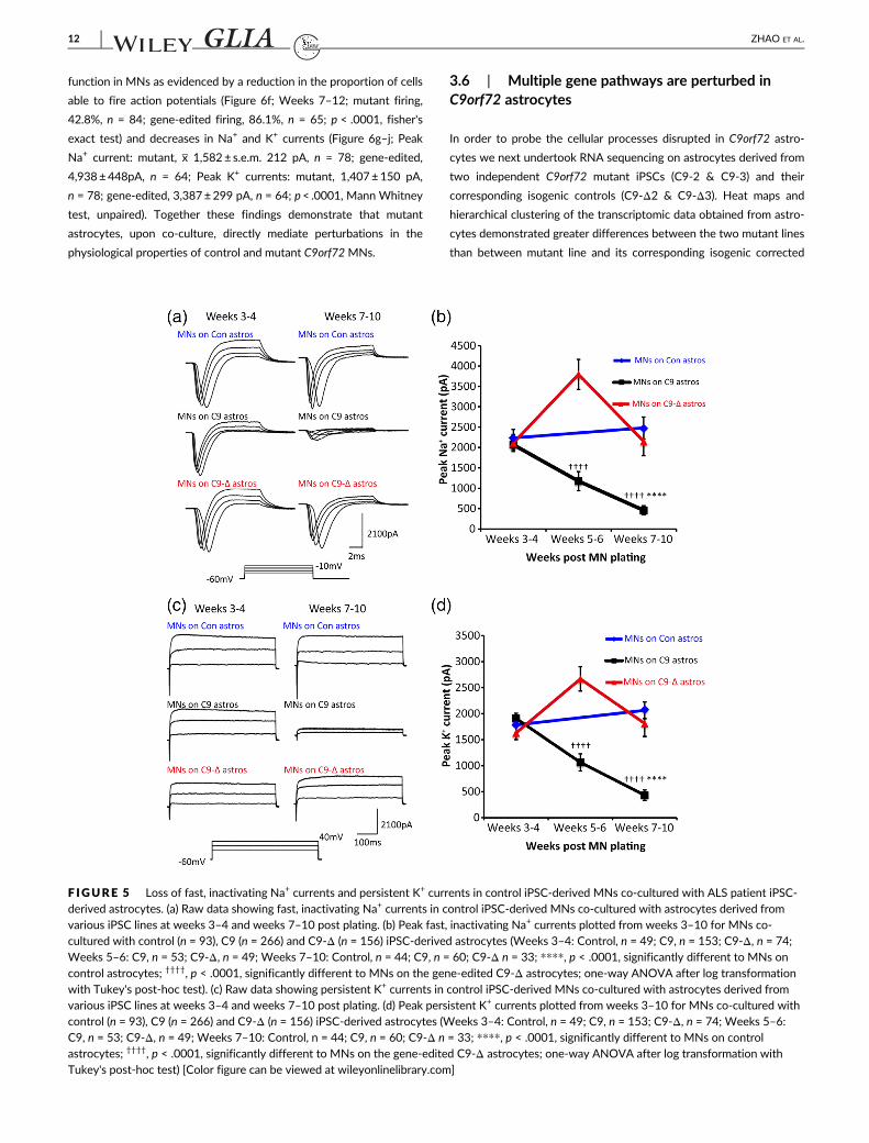
3.4 | Mutant astrocytes cause loss of voltage-activated currents in control MNs
To investigate the mechanisms underlying the progressive loss of
action potential output in control MNs co-cultured with mutant astro-
cytes, voltage-clamp recordings were performed to assess voltage-
activated currents involved in action potential generation. Fast
inactivating Na+ currents were first investigated by using a series of
voltage steps (−70 to 20 mV, 2.5 mV increments, 10 ms duration)
from a holding potential of −60 mV (Figure 5a). We found no differ-
ences in the current (I–V) relationships (Figure S6a) or peak Na+ cur-
rents between MNs co-cultured with control, mutant or gene-edited
astrocytes at 3–4 weeks post MN plating (Figure 5b; peak current:
control, x 2,232± s.e.m. 211 pA, n = 49; mutant, 2,067± 120 pA,
n = 153; gene-edited 2,110±209 pA, n = 74). However, from weeks
5–10 post MN plating, there was a progressive decrease in peak Na+
currents in MNs co-cultured with mutant astrocytes compared to co-
cultures with control or gene-edited astrocytes (Figure 5b and
Figure S6b; Weeks 5–6: mutant x 1,174± s.e.m. 233 pA, n = 53; gene-
edited, 3,795±371, n = 49; Weeks 7–10: control, 2,480±268 pA,
n = 44; mutant, 458±113 pA, n = 60; gene-edited 2,147±345 pA,
n = 33; p< .0001, one-way ANOVA after log transformation with
Tukey's post-hoc test).
We next investigated whether the progressive loss of Na+ cur-
rents reflected a more general decrease in voltage-activated currents
in control MNs co-cultured with mutant astrocytes. Persistent K+ cur-
rents were measured by using a series of voltage steps (−70 to
40 mV, 10 mV increments, 500 ms duration) from a holding potential
of −60 mV (Figure 5c). At weeks 3–4 post MN plating, peak K+ cur-
rents were comparable in MNs co-cultured with control, mutant or
gene-edited astrocytes (Figure 5d and Figure S6c; Peak current: con-
trol, x 1,788± s.e.m. 148 pA, n = 46; mutant, 1913±98 pA, n = 147;
gene-edited, 1,634±127 pA, n = 74). Similar to Na+ currents, peak K+
currents progressively declined in control MNs co-cultured with
mutant astrocytes from weeks 5–10 compared to MNs co-cultured
with control or gene-edited astrocytes (Figure 5d and Figure S6d;
Weeks 5–6: mutant, x 1,070± SEM 165 pA, n = 53; gene-edited,
2,673±233 pA, n = 43; Weeks 7–10: control, 2069±160 pA, n = 41;
mutant, 438 ±99 pA, n = 60; gene-edited, 1816±248 pA, n = 33;
p< .0001, one-way ANOVA after log transformation with Tukey's
post-hoc test).
3.5 | Functional perturbations in C9orf72 mutantMNs are mediated by mutant astrocytes
We, and others, have previously shown that mutant C9orf72 MN cul-
tures demonstrate functional perturbations (Devlin et al., 2015;
Naujock et al., 2016; Sareen et al., 2013). However, all these studies,
including our own (Devlin et al., 2015), used MN generation protocols
that also resulted in the production of a significant fraction of astro-
cytes (up to approximately 20%). One interpretation of these earlier
findings is therefore that the observed pathophysiological phenotype
was a consequence of contaminant astrocytes and not cell processes
intrinsic to MNs. To address this possibility, we next used a recently
published method to generate highly enriched mutant C9orf72 MN
cultures with negligible astrocyte contamination (Maury et al., 2015;
Selvaraj et al., 2018). In cultures derived from the C9orf72 ALS patient
lines C9-1 & C9-3 and their respective gene edited controls (C9-Δ1 &
C9- Δ3), we assessed firing output and voltage-gated Na+ and K+ cur-
rents using the same protocols described above for co-culture
experiments.
Even after 7–12 weeks of culture, we found no difference in the
proportion of cells able to fire action potentials in C9orf72 versus
gene-edited MN cultures (Figure 6a; Weeks 7–12: mutant firing,
94.8%, n = 115; gene-edited firing, 96.7%, n = 92; fisher's exact test).
We also observed equivalent voltage gated Na+ and K+ currents in
recordings from C9orf72, and gene-edited MN cultures at weeks
7–12 (Figure 6b–e; Peak Na+ current: mutant, x 2,641± s.e.m. 201 pA,
n = 110; gene-edited, 3,043±267 pA, n = 82; Peak K+ currents:
mutant, 1,844±108pA, n = 110; gene-edited, 1,772±126 pA, n = 82;
two tailed, equal variance, unpaired student t-test).
Next, to ensure that the lack of pathophysiology was not related
to differences in differentiation protocols, we co-cultured these same
highly enriched C9orf72 (C9-2 & C9-3) and gene-edited (C9-Δ2 &
C9-Δ3) MNs with mutant (C9-2, C9-3) and gene-corrected (C9-Δ2,
C9-Δ3) astrocytes respectively. The presence of mutant, but not
gene-corrected astrocytes, was again sufficient to induce altered
ZHAO ET AL. 11
function in MNs as evidenced by a reduction in the proportion of cells
able to fire action potentials (Figure 6f; Weeks 7–12; mutant firing,
42.8%, n = 84; gene-edited firing, 86.1%, n = 65; p < .0001, fisher's
exact test) and decreases in Na+ and K+ currents (Figure 6g–j; Peak
Na+ current: mutant, x 1,582± s.e.m. 212 pA, n = 78; gene-edited,
4,938±448pA, n = 64; Peak K+ currents: mutant, 1,407±150 pA,
n = 78; gene-edited, 3,387±299 pA, n = 64; p< .0001, Mann Whitney
test, unpaired). Together these findings demonstrate that mutant
astrocytes, upon co-culture, directly mediate perturbations in the
physiological properties of control and mutant C9orf72MNs.
3.6 | Multiple gene pathways are perturbed inC9orf72 astrocytes
In order to probe the cellular processes disrupted in C9orf72 astro-
cytes we next undertook RNA sequencing on astrocytes derived from
two independent C9orf72 mutant iPSCs (C9-2 & C9-3) and their
corresponding isogenic controls (C9-Δ2 & C9-Δ3). Heat maps and
hierarchical clustering of the transcriptomic data obtained from astro-
cytes demonstrated greater differences between the two mutant lines
than between mutant line and its corresponding isogenic corrected
F IGURE 5 Loss of fast, inactivating Na+ currents and persistent K+ currents in control iPSC-derived MNs co-cultured with ALS patient iPSC-derived astrocytes. (a) Raw data showing fast, inactivating Na+ currents in control iPSC-derived MNs co-cultured with astrocytes derived fromvarious iPSC lines at weeks 3–4 and weeks 7–10 post plating. (b) Peak fast, inactivating Na+ currents plotted from weeks 3–10 for MNs co-cultured with control (n = 93), C9 (n = 266) and C9-Δ (n = 156) iPSC-derived astrocytes (Weeks 3–4: Control, n = 49; C9, n = 153; C9-Δ, n = 74;Weeks 5–6: C9, n = 53; C9-Δ, n = 49; Weeks 7–10: Control, n = 44; C9, n = 60; C9-Δ n = 33; ****, p < .0001, significantly different to MNs on
control astrocytes; ††††, p < .0001, significantly different to MNs on the gene-edited C9-Δ astrocytes; one-way ANOVA after log transformationwith Tukey's post-hoc test). (c) Raw data showing persistent K+ currents in control iPSC-derived MNs co-cultured with astrocytes derived fromvarious iPSC lines at weeks 3–4 and weeks 7–10 post plating. (d) Peak persistent K+ currents plotted from weeks 3–10 for MNs co-cultured withcontrol (n = 93), C9 (n = 266) and C9-Δ (n = 156) iPSC-derived astrocytes (Weeks 3–4: Control, n = 49; C9, n = 153; C9-Δ, n = 74; Weeks 5–6:C9, n = 53; C9-Δ, n = 49; Weeks 7–10: Control, n = 44; C9, n = 60; C9-Δ n = 33; ****, p < .0001, significantly different to MNs on controlastrocytes; ††††, p < .0001, significantly different to MNs on the gene-edited C9-Δ astrocytes; one-way ANOVA after log transformation withTukey's post-hoc test) [Color figure can be viewed at wileyonlinelibrary.com]
12 ZHAO ET AL.

lines owing to heterogeneity across different iPSC lines. Therefore, to
overcome this transcriptional heterogeneity we performed compari-
sons between each mutant and its corresponding isogenic control
(Figure 7a). Differentially expressed genes were filtered using the fol-
lowing criteria: (a) genes must have significant differential expression
in the 2 independent mutant lines when compared to respective inde-
pendent isogenic controls (false discovery rate < 0.1 for each isogenic
pair), (b) genes must be dysregulated in the same direction across both
lines, (c) genes were also filtered to only retain those with mean frag-
ments per kilobase per million (FPKM) > 1 (measured across all sam-
ples, which approximates to 0.5–1 mRNA per cell). Using this
approach 698 dysregulated genes were identified (Figure 7b,c &
Figures S8 and S9). Gene ontology analysis revealed that genes
including those involved in ionotropic glutamate receptor signaling
(GRIA1, GRIA4), complement activation, ribosomal subunit assembly
(large and small) and nuclear RNA export were significantly
upregulated in mutant C9orf72 astrocytes. Downregulated genes in
mutant C9orf72 astrocytes included genes involved in cell adhesion
(L1CAM, TSP1, NTN1), synapse assembly (BDNF, NRG1, THBS2),
cell-to-cell signaling (GPC6), regulation of sodium ion transport
(SLC8A1, ATP1B2, NKAIN4) and potassium ion import (DLG1,
ATP1B2). These novel transcriptomic data reveal changes in multiple
pathways that may contribute to the deleterious effects of C9orf72
astrocytes on MN function.
F IGURE 6 MN-enriched cultures alone fail to recapitulate the pathophysiology observed when MNs are co-cultured with mutant astrocytes.(a) Percentage of cells exhibiting each firing category in MN-enriched cultures derived from mutant and gene-edited patient iPSC lines acrossweeks 7–12 post plating (C9, n = 115; C-1, n = 49; C-3, n = 66, C9-Δ, n = 92; C-Δ1, n = 19; C-Δ3, n = 73, fisher's exact test). (b) Raw datashowing fast, inactivating Na+ currents in mutant and gene-edited iPSC-derived MNs in MN-enriched cultures at weeks 7–12. (c) Peak fast,inactivating Na+ currents plotted at weeks 7–12 post-plating for mutant and gene-edited iPSC-derived MNs in MN-enriched cultures (C9,n = 110; C-1, n = 48; C-3, n = 62, C9-Δ, n = 82; C-Δ1, n = 17; C-Δ3, n = 65; two tailed, equal variance, non-paired, student t-test). (d) Raw datashowing persistent K+ currents in mutant and gene-edited iPSC-derived MNs in MN-enriched cultures at weeks 7–12. (e) Peak persistent K+
currents plotted at weeks 7–12 post-plating for mutant and gene-edited iPSC-derived MNs in MN-enriched cultures (C9, n = 110; C-1, n = 48; C-3, n = 62, C9-Δ, n = 82; C-Δ1, n = 17; C-Δ3, n = 65; two tailed, equal variance, non-paired, student t-test). (f) Percentage of cells exhibiting eachfiring category in mutant and gene- edited MNs co-cultured with astrocytes derived from mutant and gene-edited patient iPSC lines respectively,across weeks 7–12 weeks post plating (C9, n = 84; C-2, n = 34; C-3, n = 50, C9-Δ, n = 65; C-Δ2, n = 29; C-Δ3, n = 36; ††††, p < .0001, significantlydifferent to gene-edited MNs on gene-edited C9-Δ astrocytes; fisher's exact test). (g) Raw data showing fast, inactivating Na+ currents in mutantand gene-edited iPSC-derived MNs from MN-enriched cultures co-cultured with mutant and gene-edited astrocytes respectively at weeks 7–12.(h) Peak fast, inactivating Na+ currents plotted at weeks 7–12 post-plating for MN-enriched mutant and gene-edited cultures co-cultured withmutant and gene-edited astrocytes respectively (C9, n = 78; C-2, n = 31; C-3, n = 47, C9-Δ, n = 64; C-Δ2, n = 27; C-Δ3, n = 37; ††††, p < .0001,significantly different to gene-edited MNs on gene-edited C9-Δ astrocytes; student t test, two tailed, non-paired, unequal variance). (i) Raw datashowing persistent K+ currents in mutant and gene-edited iPSC-derived MNs from MN-enriched cultures co-cultured with mutant and gene-edited astrocytes respectively at weeks 7–12. (j) Peak persistent K+ currents plotted at weeks 7–12 post-plating for MN-enriched mutant andgene-edited MN cultures co-cultured with mutant and gene-edited astrocytes respectively (C9, n = 78; C-2, n = 31; C-3, n = 47, C9-Δ, n = 64; C-Δ2, n = 27; C-Δ3, n = 37; ††††, p < .0001, significantly different to gene-edited MNs on gene-edited C9-Δ astrocytes; student t test, two tailed,non-paired, unequal variance) [Color figure can be viewed at wileyonlinelibrary.com]
ZHAO ET AL. 13

4 | DISCUSSION
Here, we show that expression of the C9orf72 mutation in astrocytes
recapitulates key aspects of C9orf72-related ALS pathology and
directly results in physiological dysfunction of control and C9orf72
MNs upon co-culture, thus highlighting both cell-autonomous astro-
cyte pathology and non-cell autonomous MN pathophysiology.
C9orf72 mutant iPSC derived astrocytes displayed key pathologi-
cal features of RNA foci and poly-GP DPR. The loss of foci and DPR
in gene-edited astrocytes directly links the G4C2 repeat expansion
F IGURE 7 RNA Seq analysis ofC9ORF72 astrocytes. (a) Heat mapand dendrogram depictinghierarchical clustering of RNAsequencing reads from control (Con-1), C9 mutant (C9-2, C9-3), andrespective isogenic control (C9-Δ2,C9-Δ3) astrocytes. (b) Scatter plotshowing comparison of
transcriptome reads between2 different C9 mutants and isogeniccontrols. Red crosses denotesignificantly upregulated genes andblue crosses denote significantlydownregulated genes (p < 0.05,analysis performed using DESeq 2).(c) Venn diagram indicating totalno. of genes differentially expressedbetween each C9ORF72 mutantand isogenic control astrocyte outof which 698 genes were found tobe common between both C9 andisogenic pair. (d, e) Go ontologystudies predicted pathwaysincluding significantlydownregulated (d) and upregulated(e) genes in C9orf72 astrocytes[Color figure can be viewed atwileyonlinelibrary.com]
14 ZHAO ET AL.

with formation of RNA foci and poly-GP DPR in human astrocytes. It
remains to be determined whether other DPRs, in addition to poly-
GP, are also produced in patient iPSC-derived astrocytes. In line with
previous in vivo findings we observed relatively low levels of C9orf72
transcripts and protein in astrocytes (Jiang et al., 2016). Although TDP-
43 proteinopathies are the pathological hallmark of ALS regardless of
patients' genotypes, with TDP-43 deposition observed in astrocytes in
post-mortem tissues (Yamanaka & Komine, 2018) no post-mortem stud-
ies have yet reported TDP-43 proteinopathies in astrocytes in C9orf72-
related ALS. Our previous study has shown that iPSC-derived astrocytes
carrying a TARDBP mutation do not display TDP-43 aggregates or loss
of nuclear TDP-43 despite increased cytoplasmic TDP-43 expression
(Serio et al., 2013), nor do we detect TDP-43 proteinopathies in astro-
cytes harboring a C9orf72 mutation in the present study, suggesting that
iPSC-derived astrocytes may not manifest all TDP-43 proteinopathies
in vitro. Additionally, in the AAV-G4C2-66 mice, only 7–8% of cells in
cortex and hippocampus display phosphorylated TDP-43 inclusions
(Chew et al., 2015), and no TDP-43 aggregations were observed in BAC-
C9 (100–1,000) mice (O'Rourke et al., 2015).
Although there is consistent evidence of non-cell autonomous
toxicity mediated by astrocytes harboring SOD1 mutations (Ilieva
et al., 2009; Marchetto et al., 2008; Nagai et al., 2007; Papadeas et al.,
2011; Tripathi et al., 2017; Tyzack et al., 2017), data are either lacking
or conflicting for other ALS-related mutations (Haidet-Phillips et al.,
2013; Serio et al., 2013; Tong et al., 2013). For example, human iPSC-
derived astrocytes from a patient harboring an TARDBP M337V muta-
tion did not affect the survival of control iPSC-derived MNs (Serio
et al., 2013). This finding was supported by an independent study
where astrocytes lacking TDP-43 or overexpressing mutant TARDBP
failed to cause the death of control MNs in co-culture or when
implanted into wild-type rat spinal cords (Haidet-Phillips et al., 2013).
Conversely, wild-type MNs in transgenic rats, where the TARDBP
M337V mutation was restricted to astrocytes, progressively degener-
ate (Tong et al., 2013). Furthermore, cell culture experiments have
shown that control MNs degenerate when exposed to astrocyte-
conditioned medium (ACM) collected from cultures of mouse astro-
cytes harboring mutant TDP-43 (Rojas et al., 2014) or sporadic ALS
patient astrocytes (Haidet-Phillips et al., 2011).
Our finding, that C9orf72 mutant astrocytes cause progressive
dysfunction of control MNs, strongly support non-cell autonomous
disease mechanisms in C9orf72-mediated ALS. Although we did not
find an effect of C9orf72 mutant astrocytes on MN survival, Meyer
et al. previously reported that, upon co-culture, mutant human
C9orf72 astrocytes led to the loss of control mouse MNs (Meyer
et al., 2014). However, important differences between the studies
limit direct comparison. These include the method of generation and
regional identity of astrocytes. Meyer et al. derived astrocytes
through direct conversion of adult skin fibroblasts into neural precur-
sors, which were subsequently differentiated into astrocytes in
serum-containing media without patterning. In the current study,
fibroblasts were first reprogrammed into iPSCs before generating spi-
nal astrocytes in chemically defined media. These differences in gen-
eration methods can lead to distinct epigenetic and transcriptional
patterns with functional consequences (Chandrasekaran, Avci, Leist,
Kobolak, & Dinnyes, 2016). Critically, Meyer et al. also studied mouse
ESC-derived MNs in contrast to an entirely humanized co-culture
model as reported in this study. This is an important consideration,
given that astrocytes derived from human versus rodent iPSCs exhibit
different transcriptomic profiles and subtle functional differences
(Y. Zhang et al., 2016). Notwithstanding experimental differences, both
the present study and that of Meyer and colleagues demonstrate
adverse effects of C9orf72 mutant astrocytes on MNs, strongly
supporting involvement of non-cell autonomous disease mechanisms in
C9orf72-mediated ALS. Furthermore, our findings highlight the impor-
tance of investigating function as well as cell survival when determining
whether non-cell autonomous processes contribute to pathology.
Recent studies of human iPSC-derived MNs from our group and
others have demonstrated physiological changes in MNs harboring
ALS-related mutations (Devlin et al., 2015; Guo et al., 2017; Naujock
et al., 2016; Sareen et al., 2013; Wainger et al., 2014; Z. Zhang et al.,
2013). The most commonly reported physiological change in ALS-
affected MNs is a reduction in output, or hypoexcitability similar to
that revealed in the present study (Devlin et al., 2015; Guo et al.,
2017; Naujock et al., 2016; Sareen et al., 2013; Z. Zhang et al., 2013).
Such perturbations in the function of iPSC-derived MNs were previ-
ously assumed to reflect cell autonomous disease mechanisms in cul-
tures consisting of approximately 80% neurons, 50% of which were
MNs (Devlin et al., 2015). However, in the present study we found
that patient iPSC-derived astrocytes caused a reduction in the func-
tional output of control and patient iPSC-derived MNs, supporting
non-cell autonomous mechanisms. Our findings therefore implicate
“contaminant” astrocytes present in previous studies of enriched
motor neuron mixed cultures as key mediators of MN dysfunction.
Interestingly, a previous rodent-based study has also shown non-cell
autonomous effects of astrocytes on the electrophysiological proper-
ties of control MNs (Fritz et al., 2013). Fritz and colleagues showed
that astrocyte conditioned medium, taken from primary cultures of
mutant SOD1 expressing mouse astrocytes, induced changes in the
output of wild-type mouse MNs, thus implicating toxic factors
released by astrocytes as mediators of altered MN function
(Yamanaka & Komine, 2018). Factors released by astrocytes which
may alter MN function include the effectors of necroptosis: receptor-
integrating serine/threonine-protein kinase 1 (RIP1) and mixed lineage
kinase domain-like (MLKL) (Re et al., 2014), proinflammatory cyto-
kines and inflammatory mediators (Aebischer et al., 2011; Kia et al.,
2018; Phatnani et al., 2013; Tripathi et al., 2017) as well as reactive
oxygen species (ROS) (Marchetto et al., 2008; Rao & Weiss, 2004).
The toxic effects of the astrocyte secretome have also been dem-
onstrated in the field of C9orf72-mediated ALS, although this area
remains grossly unexplored. Madill and colleagues showed that
C9-ALS patient iPSC-derived astrocytes modulate the autophagy
pathway in a non-cell autonomous manner (Madill et al., 2017). Cells
treated with patient conditioned medium demonstrated decreased
expression of LC3-II, a key adapter autophagy protein, with a concom-
itant accumulation of p62 and increased SOD1 expression. Addition-
ally, micro-RNAs secreted through astrocyte-derived extracellular
ZHAO ET AL. 15

vesicles cause increased neuronal death and deficits in neurite out-
growth in control mouse MNs (Varcianna et al., 2019). Through path-
way analysis, they identified that hsa-miR-494-3p regulates axonal
maintenance, with its primary target being Semaphorin 3A (SEM3A).
Treatment of the MNs with a miR-494-3p mimic in the presence of
C9 iAstrocyte conditioned medium significantly reduced the levels of
SEM3A by 25% in the MNs, and increased branching and neurite
length, and survival.
An additional hypothesis to explain the toxic effect of ALS astro-
cytes on MN function is the loss or reduction of normal supportive roles
fulfilled by astrocytes, including homeostatic regulation of extracellular
glutamate (Foran & Trotti, 2009; Sasabe et al., 2012). This hypothesis is
supported by a recent study that showed a reduction in the ability of
VCPmutant astrocytes to support MN survival (Hall et al., 2017).
RNA-Seq analysis carried out on C9orf72 astrocytes in this study
further highlighted alterations in multiple new gene pathways which
may be causative towards both cell autonomous and non-cell autono-
mous pathophysiology. We observed upregulation of many genes
involved in ribosome biogenesis and assembly. This is of interest in
view of recent interactome studies and yeast genetic modifier screens
that show toxic di-peptide repeat proteins play a role in ribosomal
processing/biogenesis and reduce overall cell translation (Chai &
Gitler, 2018; Hartmann et al., 2018). These studies thus provide indi-
rect support for astrocyte DPRs having a role in the observed dys-
regulation of ribosomal processing genes. Na+/K+ ATPase is a
membrane bound pump that exchanges Na+ and K+ across the plasma
membrane to maintain ionic concentration gradients, whilst also mod-
ulating neuronal excitability in an activity dependent manner (Picton,
Nascimento, Broadhead, Sillar, & Miles, 2017). In our transcriptome
analysis we observed upregulation of glial specific Na+/K+ ATPase
(ATP1B2). ATP1B2 knock-out mice exhibit deficits in motor co-
ordination and develop tremors leading to premature death due to
osmotic imbalance (Magyar et al., 1994). Furthermore, ATP1A2 is found
to be upregulated in astrocytes expressing mutant SOD1 and contrib-
utes to non-cell autonomous toxicity to motor neurons (Gallardo et al.,
2014). Furthermore, astrocytic focal adhesion molecules have been
implicated in modulating neuronal excitability in seizure paradigms (Cho,
Muthukumar, Stork, Coutinho-Budd, & Freeman, 2018) and dys-
regulation of cell-adhesion by L1CAM deficiency leads to impairment of
action potential initiation (Valente et al., 2016). Taken together, the
transcriptomic data are consistent with the possibility that impairments
in astrocytic cellular processes could lead to a Na+/K+ ionic imbalance in
the synaptic cleft, leading to pathological changes in neuronal excitability.
Given the complex and dynamic interplay between astrocytes and MNs,
it is likely that mechanisms underlying non-cell autonomous dysfunction
and neurodegeneration are varied and reflect an imbalance between loss
of homeostatic function and gain of toxic effects. It will therefore be
important in future studies to comprehensively evaluate these newly dis-
covered gene pathways and determine the consequences of perturba-
tions in the transcriptome and proteome of astrocytes expressing the
human C9orf72 mutation in order to fully define the mechanism(s) that
underlie the observed pathological consequences of human C9orf72
astrocytes on human MN function.
In summary, our study provides the first report of the direct molecu-
lar and cellular impact of the C9orf72 mutation on human astrocytes and
their interaction with human MNs. Findings here demonstrate that astro-
cytes, in addition to MNs, are affected by expression of mutant C9orf72,
which leads to the development of pathological changes. In addition,
expression of mutant C9orf72 in astrocytes induces progressive dysfunc-
tion of MNs due to the loss of voltage-activated currents. These data
suggest that non-cell autonomous disease mechanisms are a contributor
to C9orf72-mediated ALS. Furthermore, our study demonstrates the
value of combining gene-editing with sensitive physiological studies in
human iPSC-based neurodegenerative disease modeling.
ACKNOWLEDGMENTS
This work has been supported by: Motor Neurone Disease Associa-
tion (Miles/Oct 2014/878-792), Euan MacDonald Centre, Motor
Neurone Disease Scotland, Dementias Platform UK (DPUK)-MRC
stem cell partnership, MS Society and UK Dementia Research Insti-
tute. C.Z. was funded by China Scholarship Council. A.R.M. and M.S
are funded by the Medical Research Council and the Motor Neurone
Disease Association. They also acknowledge support from the Row-
ling Scholars scheme, administered by the Anne Rowling Regenerative
Neurology Clinic, University of Edinburgh, Edinburgh, UK.
CONFLICT OF INTEREST
The authors declare no potential conflict of interest.
AUTHOR CONTRIBUTIONS
C.Z, A.C.D., A.K.C., S.C., and G.B.M. conceived and designed the
experiments. C.Z, A.C.D, A.K.C, M.S, B.T.S, and V.B performed experi-
ments and analyzed the data. K.B. maintained iPSC lines. K.B. and A.R.
M differentiated and validated iPSC lines. C.E.S generated and pro-
vided iPSC lines. C.Z, A.C.D, A.K.C, G.B.M, and S.C. interpreted the
data and wrote the manuscript. C.Z, A.C.D., A.K.C, M.S, B.T.S, G.B.M.,
and S.C. and edited and revised the manuscript.
DATA AVAILABILITY STATEMENT
The data that support the findings of this study are available from the
corresponding author upon reasonable request.
ORCID
Anna-Claire Devlin https://orcid.org/0000-0001-7368-1722
Arpan R. Mehta https://orcid.org/0000-0002-5715-5025
Owen Dando https://orcid.org/0000-0002-6269-6408
Gareth B. Miles https://orcid.org/0000-0002-8624-4625
REFERENCES
Aebischer, J., Cassina, P., Otsmane, B., Moumen, A., Seilhean, D.,
Meininger, V., … Raoul, C. (2011). IFNgamma triggers a LIGHT-
dependent selective death of motoneurons contributing to the non-
cell-autonomous effects of mutant SOD1. Cell Death and Differentia-
tion, 18(5), 754–768. https://doi.org/10.1038/cdd.2010.143Ajroud-Driss, S., & Siddique, T. (2015). Sporadic and hereditary
amyotrophic lateral sclerosis (ALS). Biochimica et Biophysica Acta, 1852
(4), 679–684. https://doi.org/10.1016/j.bbadis.2014.08.010
16 ZHAO ET AL.

Alexa, A., Rahnenfuhrer, J., & Lengauer, T. (2006). Improved scoring of
functional groups from gene expression data by decorrelating GO
graph structure. Bioinformatics, 22(13), 1600–1607. https://doi.org/10.1093/bioinformatics/btl140
Amoroso, M. W., Croft, G. F., Williams, D. J., O'Keeffe, S., Carrasco, M. A.,
Davis, A. R., … Wichterle, H. (2013). Accelerated high-yield generation
of limb-innervating motor neurons from human stem cells. The Journal
of Neuroscience, 33(2), 574–586. https://doi.org/10.1523/
JNEUROSCI.0906-12.2013
Arai, T., Hasegawa, M., Akiyama, H., Ikeda, K., Nonaka, T., Mori, H., …Oda, T. (2006). TDP-43 is a component of ubiquitin-positive tau-
negative inclusions in frontotemporal lobar degeneration and
amyotrophic lateral sclerosis. Biochemical and Biophysical Research
Communications, 351(3), 602–611. https://doi.org/10.1016/j.bbrc.
2006.10.093
Ash, P. E., Bieniek, K. F., Gendron, T. F., Caulfield, T., Lin, W. L., Dejesus-
Hernandez, M., … Petrucelli, L. (2013). Unconventional translation of
C9ORF72 GGGGCC expansion generates insoluble polypeptides spe-
cific to c9FTD/ALS. Neuron, 77(4), 639–646. https://doi.org/10.1016/j.neuron.2013.02.004
Bilican, B., Livesey, M. R., Haghi, G., Qiu, J., Burr, K., Siller, R., …Chandran, S. (2014). Physiological normoxia and absence of EGF is
required for the long-term propagation of anterior neural precursors
from human pluripotent cells. PLoS One, 9(1), e85932. https://doi.org/
10.1371/journal.pone.0085932
Bilican, B., Serio, A., Barmada, S. J., Nishimura, A. L., Sullivan, G. J.,
Carrasco, M., … Chandran, S. (2012). Mutant induced pluripotent stem
cell lines recapitulate aspects of TDP-43 proteinopathies and reveal
cell-specific vulnerability. Proceedings of the National Academy of Sci-
ences of the United States of America, 109(15), 5803–5808. https://doi.org/10.1073/pnas.1202922109
Boillee, S., Vande Velde, C., & Cleveland, D. W. (2006). ALS: A disease of
motor neurons and their nonneuronal neighbors. Neuron, 52(1),
39–59. https://doi.org/10.1016/j.neuron.2006.09.018Cassina, P., Cassina, A., Pehar, M., Castellanos, R., Gandelman, M., de
Leon, A., … Radi, R. (2008). Mitochondrial dysfunction in SOD1G93A-
bearing astrocytes promotes motor neuron degeneration: Prevention
by mitochondrial-targeted antioxidants. The Journal of Neuroscience,
28(16), 4115–4122. https://doi.org/10.1523/jneurosci.5308-07.2008Chai, N., & Gitler, A. D. (2018). Yeast screen for modifiers of C9orf72
poly(glycine-arginine) dipeptide repeat toxicity. FEMS Yeast Research,
18(4). https://doi.org/10.1093/femsyr/foy024
Chandrasekaran, A., Avci, H. X., Leist, M., Kobolak, J., & Dinnyes, A. (2016).
Astrocyte differentiation of human pluripotent stem cells: New tools
for neurological disorder research. Frontiers in Cellular Neuroscience,
10, 215. https://doi.org/10.3389/fncel.2016.00215
Chew, J., Gendron, T. F., Prudencio, M., Sasaguri, H., Zhang, Y. J.,
Castanedes-Casey, M., … Petrucelli, L. (2015). Neurodegeneration.
C9ORF72 repeat expansions in mice cause TDP-43 pathology, neuro-
nal loss, and behavioral deficits. Science, 348(6239), 1151–1154.https://doi.org/10.1126/science.aaa9344
Cho, S., Muthukumar, A. K., Stork, T., Coutinho-Budd, J. C., &
Freeman, M. R. (2018). Focal adhesion molecules regulate astrocyte
morphology and glutamate transporters to suppress seizure-like
behavior. Proceedings of the National Academy of Sciences of the United
States of America, 115(44), 11316–11321. https://doi.org/10.1073/pnas.1800830115
DeJesus-Hernandez, M., Mackenzie, I. R., Boeve, B. F., Boxer, A. L.,
Baker, M., Rutherford, N. J., … Rademakers, R. (2011). Expanded
GGGGCC hexanucleotide repeat in noncoding region of C9ORF72
causes chromosome 9p-linked FTD and ALS. Neuron, 72(2), 245–256.https://doi.org/10.1016/j.neuron.2011.09.011
Delestree, N., Manuel, M., Iglesias, C., Elbasiouny, S. M., Heckman, C. J., &
Zytnicki, D. (2014). Adult spinal motoneurones are not hyperexcitable
in a mouse model of inherited amyotrophic lateral sclerosis. The
Journal of Physiology, 592(7), 1687–1703. https://doi.org/10.1113/
jphysiol.2013.265843
Devlin, A. C., Burr, K., Borooah, S., Foster, J. D., Cleary, E. M., Geti, I., …Miles, G. B. (2015). Human iPSC-derived motoneurons harbouring
TARDBP or C9ORF72 ALS mutations are dysfunctional despite
maintaining viability. Nature Communications, 6, 5999. https://doi.org/
10.1038/ncomms6999
Di Giorgio, F. P., Carrasco, M. A., Siao, M. C., Maniatis, T., & Eggan, K.
(2007). Non-cell autonomous effect of glia on motor neurons in an
embryonic stem cell-based ALS model. Nature Neuroscience, 10(5),
608–614. https://doi.org/10.1038/nn1885Dobin, A., Davis, C. A., Schlesinger, F., Drenkow, J., Zaleski, C., Jha, S., …
Gingeras, T. R. (2013). STAR: ultrafast universal RNA-seq aligner. Bioinfor-
matics, 29(1), 15–21. https://doi.org/10.1093/bioinformatics/bts635
Donnelly, C. J., Zhang, P. W., Pham, J. T., Haeusler, A. R., Mistry, N. A.,
Vidensky, S., … Rothstein, J. D. (2013). RNA toxicity from the ALS/FTD
C9ORF72 expansion is mitigated by antisense intervention. Neuron,
80(2), 415–428. https://doi.org/10.1016/j.neuron.2013.10.015Foran, E., & Trotti, D. (2009). Glutamate transporters and the excitotoxic
path to motor neuron degeneration in amyotrophic lateral sclerosis.
Antioxidants & Redox Signaling, 11(7), 1587–1602. https://doi.org/10.1089/ars.2009.2444
Freischmidt, A., Muller, K., Ludolph, A. C., Weishaupt, J. H., &
Andersen, P. M. (2017). Association of Mutations in TBK1 with spo-
radic and familial amyotrophic lateral sclerosis and frontotemporal
dementia. JAMA Neurology, 74(1), 110–113. https://doi.org/10.1001/jamaneurol.2016.3712
Fritz, E., Izaurieta, P., Weiss, A., Mir, F. R., Rojas, P., Gonzalez, D., … van
Zundert, B. (2013). Mutant SOD1-expressing astrocytes release toxic
factors that trigger motoneuron death by inducing hyperexcitability.
Journal of Neurophysiology, 109(11), 2803–2814. https://doi.org/10.1152/jn.00500.2012
Gallardo, G., Barowski, J., Ravits, J., Siddique, T., Lingrel, J. B.,
Robertson, J., … Bonni, A. (2014). An alpha2-Na/K ATPase/alpha-
adducin complex in astrocytes triggers non-cell autonomous neu-
rodegeneration. Nature Neuroscience, 17(12), 1710–1719. https://doi.org/10.1038/nn.3853
Guo, W., Naujock, M., Fumagalli, L., Vandoorne, T., Baatsen, P., Boon, R., …Van Den Bosch, L. (2017). HDAC6 inhibition reverses axonal transport
defects in motor neurons derived from FUS-ALS patients. Nature Com-
munications, 8(1), 861. https://doi.org/10.1038/s41467-017-00911-y
Haidet-Phillips, A. M., Gross, S. K., Williams, T., Tuteja, A., Sherman, A.,
Ko, M., … Maragakis, N. J. (2013). Altered astrocytic expression of TDP-
43 does not influence motor neuron survival. Experimental Neurology,
250, 250–259. https://doi.org/10.1016/j.expneurol.2013.10.004Haidet-Phillips, A. M., Hester, M. E., Miranda, C. J., Meyer, K., Braun, L.,
Frakes, A., … Kaspar, B. K. (2011). Astrocytes from familial and spo-
radic ALS patients are toxic to motor neurons. Nature Biotechnology,
29(9), 824–828. https://doi.org/10.1038/nbt.1957Hall, C. E., Yao, Z., Choi, M., Tyzack, G. E., Serio, A., Luisier, R., … Patani, R.
(2017). Progressive motor neuron pathology and the role of astrocytes
in a human stem cell model of VCP-related ALS. Cell Reports, 19(9),
1739–1749. https://doi.org/10.1016/j.celrep.2017.05.024Hardiman, O., Al-Chalabi, A., Chio, A., Corr, E. M., Logroscino, G.,
Robberecht, W., … van den Berg, L. H. (2017). Amyotrophic lateral
sclerosis. Nature Reviews. Disease Primers, 3, 17071. https://doi.org/
10.1038/nrdp.2017.71
Hartmann, H., Hornburg, D., Czuppa, M., Bader, J., Michaelsen, M., Farny, D.,…Edbauer, D. (2018). Proteomics and C9orf72 neuropathology identify ribo-
somes as poly-GR/PR interactors driving toxicity. Life Sci Alliance, 1(2),
e201800070. https://doi.org/10.26508/lsa.201800070
Ilieva, H., Polymenidou, M., & Cleveland, D. W. (2009). Non-cell autono-
mous toxicity in neurodegenerative disorders: ALS and beyond. The
Journal of Cell Biology, 187(6), 761–772. https://doi.org/10.1083/jcb.200908164
ZHAO ET AL. 17

Jiang, J., Zhu, Q., Gendron, T. F., Saberi, S., McAlonis-Downes, M.,
Seelman, A., … Lagier-Tourenne, C. (2016). Gain of toxicity from ALS/-
FTD-linked repeat expansions in C9ORF72 is alleviated by antisense
oligonucleotides targeting GGGGCC-containing RNAs. Neuron, 90(3),
535–550. https://doi.org/10.1016/j.neuron.2016.04.006Kanai, K., Shibuya, K., Sato, Y., Misawa, S., Nasu, S., Sekiguchi, Y., …
Kuwabara, S. (2012). Motor axonal excitability properties are strong
predictors for survival in amyotrophic lateral sclerosis. Journal of Neu-
rology, Neurosurgery, and Psychiatry, 83(7), 734–738. https://doi.org/10.1136/jnnp-2011-301782
Kia, A., McAvoy, K., Krishnamurthy, K., Trotti, D., & Pasinelli, P. (2018).
Astrocytes expressing ALS-linked mutant FUS induce motor neuron
death through release of tumor necrosis factor-alpha. Glia, 66,
1016–1033. https://doi.org/10.1002/glia.23298Kuo, J. J., Schonewille, M., Siddique, T., Schults, A. N., Fu, R., Bar, P. R., …
Kroese, A. B. (2004). Hyperexcitability of cultured spinal motoneurons
from presymptomatic ALS mice. Journal of Neurophysiology, 91(1),
571–575. https://doi.org/10.1152/jn.00665.2003Lagier-Tourenne, C., Baughn, M., Rigo, F., Sun, S., Liu, P., Li, H. R., …
Ravits, J. (2013). Targeted degradation of sense and antisense
C9orf72 RNA foci as therapy for ALS and frontotemporal degenera-
tion. Proceedings of the National Academy of Sciences of the United
States of America, 110(47), E4530–E4539. https://doi.org/10.1073/pnas.1318835110
Liao, Y., Smyth, G. K., & Shi, W. (2014). featureCounts: An efficient
general purpose program for assigning sequence reads to genomic fea-
tures. Bioinformatics, 30(7), 923–930. https://doi.org/10.1093/
bioinformatics/btt656
Ling, S. C., Polymenidou, M., & Cleveland, D. W. (2013). Converging
mechanisms in ALS and FTD: Disrupted RNA and protein homeostasis.
Neuron, 79(3), 416–438. https://doi.org/10.1016/j.neuron.2013.
07.033
Livesey, M. R., Magnani, D., Hardingham, G. E., Chandran, S., &
Wyllie, D. J. (2015). Functional properties of in vitro excitatory cortical
neurons derived from human pluripotent stem cells. The Journal of
Physiology, 594, 6573–6582. https://doi.org/10.1113/JP270660Love, M. I., Huber, W., & Anders, S. (2014). Moderated estimation of fold
change and dispersion for RNA-seq data with DESeq2. Genome Biol-
ogy, 15(12), 550. https://doi.org/10.1186/s13059-014-0550-8
Madill, M., McDonagh, K., Ma, J., Vajda, A., McLoughlin, P., O'Brien, T., …Shen, S. (2017). Amyotrophic lateral sclerosis patient iPSC-derived astro-
cytes impair autophagy via non-cell autonomous mechanisms. Molecular
Brain, 10(1), 22. https://doi.org/10.1186/s13041-017-0300-4
Magyar, J. P., Bartsch, U., Wang, Z. Q., Howells, N., Aguzzi, A.,
Wagner, E. F., & Schachner, M. (1994). Degeneration of neural cells in
the central nervous system of mice deficient in the gene for the adhe-
sion molecule on glia, the beta 2 subunit of murine Na,K-ATPase. The
Journal of Cell Biology, 127(3), 835–845. https://doi.org/10.1083/jcb.127.3.835
Marchetto, M. C., Muotri, A. R., Mu, Y., Smith, A. M., Cezar, G. G., &
Gage, F. H. (2008). Non-cell-autonomous effect of human SOD1
G37R astrocytes on motor neurons derived from human embryonic
stem cells. Cell Stem Cell, 3(6), 649–657. https://doi.org/10.1016/j.stem.2008.10.001
Maury, Y., Come, J., Piskorowski, R. A., Salah-Mohellibi, N., Chevaleyre, V.,
Peschanski, M., … Nedelec, S. (2015). Combinatorial analysis of devel-
opmental cues efficiently converts human pluripotent stem cells into
multiple neuronal subtypes. Nature Biotechnology, 33(1), 89–96.https://doi.org/10.1038/nbt.3049
Meyer, K., Ferraiuolo, L., Miranda, C. J., Likhite, S., McElroy, S., Renusch, S.,
… Kaspar, B. K. (2014). Direct conversion of patient fibroblasts demon-
strates non-cell autonomous toxicity of astrocytes to motor neurons
in familial and sporadic ALS. Proceedings of the National Academy of
Sciences of the United States of America, 111(2), 829–832. https://doi.org/10.1073/pnas.1314085111
Mizielinska, S., & Isaacs, A. M. (2014). C9orf72 amyotrophic lateral sclero-
sis and frontotemporal dementia: Gain or loss of function? Current
Opinion in Neurology, 27(5), 515–523. https://doi.org/10.1097/wco.
0000000000000130
Mori, K., Arzberger, T., Grasser, F. A., Gijselinck, I., May, S., Rentzsch, K., …Edbauer, D. (2013). Bidirectional transcripts of the expanded C9orf72
hexanucleotide repeat are translated into aggregating dipeptide repeat
proteins. Acta Neuropathologica, 126(6), 881–893. https://doi.org/10.1007/s00401-013-1189-3
Nagai, M., Re, D. B., Nagata, T., Chalazonitis, A., Jessell, T. M.,
Wichterle, H., & Przedborski, S. (2007). Astrocytes expressing ALS-linked
mutated SOD1 release factors selectively toxic to motor neurons. Nature
Neuroscience, 10(5), 615–622. https://doi.org/10.1038/nn1876Naujock, M., Stanslowsky, N., Bufler, S., Naumann, M., Reinhardt, P.,
Sterneckert, J., … Petri, S. (2016). 4-Aminopyridine induced activity
rescues Hypoexcitable motor neurons from amyotrophic lateral sclero-
sis patient-derived induced pluripotent stem cells. Stem Cells, 34(6),
1563–1575. https://doi.org/10.1002/stem.2354
Neumann, M., Sampathu, D. M., Kwong, L. K., Truax, A. C., Micsenyi, M. C.,
Chou, T. T., … Lee, V. M. (2006). Ubiquitinated TDP-43 in frontotemporal
lobar degeneration and amyotrophic lateral sclerosis. Science, 314(5796),
130–133. https://doi.org/10.1126/science.1134108O'Rourke, J. G., Bogdanik, L., Muhammad, A., Gendron, T. F., Kim, K. J.,
Austin, A., … Baloh, R. H. (2015). C9orf72 BAC transgenic mice display
typical pathologic features of ALS/FTD. Neuron, 88(5), 892–901.https://doi.org/10.1016/j.neuron.2015.10.027
Papadeas, S. T., Kraig, S. E., O'Banion, C., Lepore, A. C., & Maragakis, N. J.
(2011). Astrocytes carrying the superoxide dismutase 1 (SOD1G93A)
mutation induce wild-type motor neuron degeneration in vivo. Proceed-
ings of the National Academy of Sciences of the United States of America,
108(43), 17803–17808. https://doi.org/10.1073/pnas.1103141108Phatnani, H. P., Guarnieri, P., Friedman, B. A., Carrasco, M. A., Muratet, M.,
O'Keeffe, S., … Maniatis, T. (2013). Intricate interplay between astro-
cytes and motor neurons in ALS. Proceedings of the National Academy
of Sciences of the United States of America, 110(8), E756–E765.https://doi.org/10.1073/pnas.1222361110
Picton, L. D., Nascimento, F., Broadhead, M. J., Sillar, K. T., & Miles, G. B.
(2017). Sodium pumps mediate activity-dependent changes in mam-
malian motor networks. The Journal of Neuroscience, 37(4), 906–921.https://doi.org/10.1523/jneurosci.2005-16.2016
Pieri, M., Albo, F., Gaetti, C., Spalloni, A., Bengtson, C. P., Longone, P., …Zona, C. (2003). Altered excitability of motor neurons in a transgenic
mouse model of familial amyotrophic lateral sclerosis. Neuroscience Let-
ters, 351(3), 153–156. https://doi.org/10.1016/j.neulet.2003.07.010Quinlan, K. A., Schuster, J. E., Fu, R., Siddique, T., & Heckman, C. J. (2011).
Altered postnatal maturation of electrical properties in spinal moto-
neurons in a mouse model of amyotrophic lateral sclerosis. The Journal
of Physiology, 589(Pt 9), 2245–2260. https://doi.org/10.1113/jphysiol.2010.200659
Ran, F. A., Hsu, P. D., Wright, J., Agarwala, V., Scott, D. A., & Zhang, F.
(2013). Genome engineering using the CRISPR-Cas9 system. Nature
Protocols, 8(11), 2281–2308. https://doi.org/10.1038/nprot.2013.143Rao, S. D., & Weiss, J. H. (2004). Excitotoxic and oxidative cross-talk
between motor neurons and glia in ALS pathogenesis. Trends in Neuro-
sciences, 27(1), 17–23. https://doi.org/10.1016/j.tins.2003.11.001Re, D. B., Le Verche, V., Yu, C., Amoroso, M. W., Politi, K. A., Phani, S., …
Przedborski, S. (2014). Necroptosis drives motor neuron death in
models of both sporadic and familial ALS. Neuron, 81(5), 1001–1008.https://doi.org/10.1016/j.neuron.2014.01.011
Renton, A. E., Majounie, E., Waite, A., Simon-Sanchez, J., Rollinson, S.,
Gibbs, J. R., … Traynor, B. J. (2011). A hexanucleotide repeat expansion
in C9ORF72 is the cause of chromosome 9p21-linked ALS-FTD. Neu-
ron, 72(2), 257–268. https://doi.org/10.1016/j.neuron.2011.09.010Rojas, F., Cortes, N., Abarzua, S., Dyrda, A., & van Zundert, B. (2014). Astro-
cytes expressing mutant SOD1 and TDP43 trigger motoneuron death
18 ZHAO ET AL.

that is mediated via sodium channels and nitroxidative stress. Frontiers in
Cellular Neuroscience, 8, 24. https://doi.org/10.3389/fncel.2014.00024
Sandoe, J., & Eggan, K. (2013). Opportunities and challenges of pluripotent
stem cell neurodegenerative disease models. Nature Neuroscience, 16
(7), 780–789. https://doi.org/10.1038/nn.3425Sareen, D., O'Rourke, J. G., Meera, P., Muhammad, A. K., Grant, S.,
Simpkinson, M., … Baloh, R. H. (2013). Targeting RNA foci in iPSC-
derived motor neurons from ALS patients with a C9ORF72 repeat
expansion. Science Translational Medicine, 5(208), 208ra149. https://
doi.org/10.1126/scitranslmed.3007529
Sasabe, J., Miyoshi, Y., Suzuki, M., Mita, M., Konno, R., Matsuoka, M., …Aiso, S. (2012). D-amino acid oxidase controls motoneuron degenera-
tion through D-serine. Proceedings of the National Academy of Sciences
of the United States of America, 109(2), 627–632. https://doi.org/10.1073/pnas.1114639109
Selvaraj, B. T., Livesey, M. R., & Chandran, S. (2017). Modeling the
C9ORF72 repeat expansion mutation using human induced pluripo-
tent stem cells. Brain Pathology, 27(4), 518–524. https://doi.org/10.1111/bpa.12520
Selvaraj, B. T., Livesey, M. R., Zhao, C., Gregory, J. M., James, O. T.,
Cleary, E. M., … Chandran, S. (2018). C9ORF72 repeat expansion cau-
ses vulnerability of motor neurons to ca(2+)-permeable AMPA
receptor-mediated excitotoxicity. Nature Communications, 9(1), 347.
https://doi.org/10.1038/s41467-017-02729-0
Serio, A., Bilican, B., Barmada, S. J., Ando, D. M., Zhao, C., Siller, R., …Chandran, S. (2013). Astrocyte pathology and the absence of non-cell
autonomy in an induced pluripotent stem cell model of TDP-43
proteinopathy. Proceedings of the National Academy of Sciences of the
United States of America, 110(12), 4697–4702. https://doi.org/10.
1073/pnas.1300398110
Shi, Y., Lin, S., Staats, K. A., Li, Y., Chang, W. H., Hung, S. T., … Ichida, J. K.
(2018). Haploinsufficiency leads to neurodegeneration in C9ORF72
ALS/FTD human induced motor neurons. Nature Medicine, 24(3),
313–325. https://doi.org/10.1038/nm.4490
Sreedharan, J., Blair, I. P., Tripathi, V. B., Hu, X., Vance, C., Rogelj, B., …Shaw, C. E. (2008). TDP-43 mutations in familial and sporadic
amyotrophic lateral sclerosis. Science, 319(5870), 1668–1672. https://doi.org/10.1126/science.1154584
Suzuki, N., Maroof, A. M., Merkle, F. T., Koszka, K., Intoh, A., Armstrong, I., …Eggan, K. (2013). The mouse C9ORF72 ortholog is enriched in neurons
known to degenerate in ALS and FTD. Nature Neuroscience, 16(12),
1725–1727. https://doi.org/10.1038/nn.3566Tabet, R., Schaeffer, L., Freyermuth, F., Jambeau, M., Workman, M.,
Lee, C. Z., … Lagier-Tourenne, C. (2018). CUG initiation and
frameshifting enable production of dipeptide repeat proteins from
ALS/FTD C9ORF72 transcripts. Nature Communications, 9(1), 152.
https://doi.org/10.1038/s41467-017-02643-5
Takahashi, K., Tanabe, K., Ohnuki, M., Narita, M., Ichisaka, T.,
Tomoda, K., & Yamanaka, S. (2007). Induction of pluripotent stem cells
from adult human fibroblasts by defined factors. Cell, 131(5),
861–872. https://doi.org/10.1016/j.cell.2007.11.019Tong, J., Huang, C., Bi, F., Wu, Q., Huang, B., Liu, X., … Xia, X. G. (2013).
Expression of ALS-linked TDP-43 mutant in astrocytes causes non-
cell-autonomous motor neuron death in rats. The EMBO Journal, 32
(13), 1917–1926. https://doi.org/10.1038/emboj.2013.122
Tripathi, P., Rodriguez-Muela, N., Klim, J. R., de Boer, A. S., Agrawal, S.,
Sandoe, J., … Zhou, Q. (2017). Reactive astrocytes promote ALS-like
degeneration and intracellular protein aggregation in human motor
neurons by disrupting autophagy through TGF-beta1. Stem Cell
Reports, 9(2), 667–680. https://doi.org/10.1016/j.stemcr.2017.06.008
Tyzack, G. E., Hall, C. E., Sibley, C. R., Cymes, T., Forostyak, S., Carlino, G., …Lakatos, A. (2017). A neuroprotective astrocyte state is induced by neu-
ronal signal EphB1 but fails in ALS models. Nature Communications, 8(1),
1164. https://doi.org/10.1038/s41467-017-01283-z
Valente, P., Lignani, G., Medrihan, L., Bosco, F., Contestabile, A.,
Lippiello, P., … Baldelli, P. (2016). Cell adhesion molecule L1 contrib-
utes to neuronal excitability regulating the function of voltage-gated
Na+ channels. Journal of Cell Science, 129(9), 1878–1891. https://doi.org/10.1242/jcs.182089
van Zundert, B., Peuscher, M. H., Hynynen, M., Chen, A., Neve, R. L.,
Brown, R. H., Jr., … Bellingham, M. C. (2008). Neonatal neuronal cir-
cuitry shows hyperexcitable disturbance in a mouse model of the
adult-onset neurodegenerative disease amyotrophic lateral sclerosis.
The Journal of Neuroscience, 28(43), 10864–10874. https://doi.org/10.1523/jneurosci.1340-08.2008
Varcianna, A., Myszczynska, M. A., Castelli, L. M., O'Neill, B., Kim, Y.,
Talbot, J., … Ferraiuolo, L. (2019). Micro-RNAs secreted through
astrocyte-derived extracellular vesicles cause neuronal network
degeneration in C9orf72 ALS. eBioMedicine, 40, 626–635. https://doi.org/10.1016/j.ebiom.2018.11.067
Vucic, S., & Kiernan, M. C. (2006). Axonal excitability properties in
amyotrophic lateral sclerosis. Clinical Neurophysiology, 117(7),
1458–1466. https://doi.org/10.1016/j.clinph.2006.04.016Wainger, B. J., Kiskinis, E., Mellin, C., Wiskow, O., Han, S. S., Sandoe, J., …
Woolf, C. J. (2014). Intrinsic membrane hyperexcitability of
amyotrophic lateral sclerosis patient-derived motor neurons. Cell
Reports, 7(1), 1–11. https://doi.org/10.1016/j.celrep.2014.03.019Wang, L., Gutmann, D. H., & Roos, R. P. (2011). Astrocyte loss of mutant
SOD1 delays ALS disease onset and progression in G85R transgenic
mice. Human Molecular Genetics, 20(2), 286–293. https://doi.org/10.1093/hmg/ddq463
Wang, L., Yi, F., Fu, L., Yang, J., Wang, S., Wang, Z., … Liu, G. H. (2017).
CRISPR/Cas9-mediated targeted gene correction in amyotrophic lat-
eral sclerosis patient iPSCs. Protein & Cell, 8(5), 365–378. https://doi.org/10.1007/s13238-017-0397-3
Yamanaka, K., Chun, S. J., Boillee, S., Fujimori-Tonou, N., Yamashita, H.,
Gutmann, D. H., … Cleveland, D. W. (2008). Astrocytes as determinants
of disease progression in inherited amyotrophic lateral sclerosis. Nature
Neuroscience, 11(3), 251–253. https://doi.org/10.1038/nn2047Yamanaka, K., & Komine, O. (2018). The multi-dimensional roles of astro-
cytes in ALS. Neuroscience Research, 126, 31–38. https://doi.org/10.1016/j.neures.2017.09.011
Zerbino, D. R., Achuthan, P., Akanni, W., Amode, M. R., Barrell, D., Bhai, J.,
… Flicek, P. (2018). Ensembl 2018. Nucleic Acids Research, 46(D1),
D754–D761. https://doi.org/10.1093/nar/gkx1098
Zhang, Y., Sloan, S. A., Clarke, L. E., Caneda, C., Plaza, C. A.,
Blumenthal, P. D., … Barres, B. A. (2016). Purification and characteriza-
tion of progenitor and mature human astrocytes reveals transcriptional
and functional differences with mouse. Neuron, 89(1), 37–53. https://doi.org/10.1016/j.neuron.2015.11.013
Zhang, Z., Almeida, S., Lu, Y., Nishimura, A. L., Peng, L., Sun, D., …Gao, F. B. (2013). Downregulation of microRNA-9 in iPSC-derived
neurons of FTD/ALS patients with TDP-43 mutations. PLoS One, 8
(10), e76055. https://doi.org/10.1371/journal.pone.0076055
SUPPORTING INFORMATION
Additional supporting information may be found online in the
Supporting Information section at the end of this article.
How to cite this article: Zhao C, Devlin A-C, Chouhan AK,
et al. Mutant C9orf72 human iPSC-derived astrocytes cause
non-cell autonomous motor neuron pathophysiology. Glia.
2019;1–19. https://doi.org/10.1002/glia.23761
ZHAO ET AL. 19
Copyright © 2022 FDOKUMEN