Astrocytes increase ATP exocytosis mediated calcium signaling in response to microgroove structures
Upload
independentCategory
view
2download
0
Neuroscience Vol. 22, No. I, pp. 255-266, 1987 Printed in Great Britain
0306-4522/87 $3.00 + 0.00 Pergamon Journals Ltd
0 1987 IBRO
ORGANIZATION OF MICROFILAMENTS IN ASTROCYTES THAT FORM IN THE PRESENCE OF DIBUTYRYL
CYCLIC AMP IN CULTURES, AND WHICH ARE SIMILAR TO REACTIVE ASTROCYTES IN VIVO
S. FEDOROFF,*~ I. AHMED,* M. OPAS$ and V. I. KALNINS~ *Department of Anatomy, University of Saskatchewan, Saskatoon, Saskatchewan, Canada S7N OWO;
IDepartment of Anatomy, University of Toronto, Canada
Abstract-This paper deals with changes in the arrangement of microfilaments at various stages during the transformation of astroblasts into reactive astrocytes in the presence of dibutyryl3’,5’-cyclic adenosine monophosphate in vitro. When cultures of two-week-old mouse astroblasts are treated with dibutyryl cyclic AMP, drastic changes occur in cell shape and in the organization of microfilaments, resulting in cells that closely resemble reactive astrocytes in vivo. A thick, prominent ring of microfilaments in such cells which stains strongly with 7-nitrobenz-2 oxa-1,3-diazole-phallacidin, delineates the perikaryon. Electron microscope examination showed that the ring is composed of many smaller bundles of microfilaments running parallel to each other. Prominent bundles of microfilaments radiate from the cell body into the cell processes.
Based on the observation of intermediate forms, we propose that the microfilament ring may be important in the development of cell processes in reactive astrocytes.
Normally the central nervous system (CNS) contains two major types of astrocytes: fibrous and proto- plasmic. After trauma in the CNS a new type of astrocyte which is larger, has many processes and is strongly positive for glial fibrillary acidic protein (GFP) appears. These cells have been referred to as reactive astrocytes and are generally believed to form through hypertrophy and hyperplasia of astrocytes or their precursor cells.2~‘9~2’~2*~29~36
Astrocyte precursor cells isolated from brain can be grown as monolayers in culture.4.‘8 After addition of dibutyryl 3’,5’-cyclic adenosine monophosphate (dBcAMP) those astrocyte precursor cells which have reached the astroblast stage of maturation transform into large stellate cells IO-12.21.22.24.2S,37.39-41 which have
an increased number of intermediate filaments ar- ranged in tightly packed bundles,“~‘2*26~39 an increased amount of GFP,‘6,39 ultrastructural features charac- teristic of secretory and metabolically active cells,” an increased rate of respirationI and demonstrated pinocytic” and phagocytic7 activity. The changes induced by the dBcAMP treatment depended on the media used.25*2’ With some media, rapid but transient transformation-more like a modulation- is induced, while with others the transformation resembles cell maturation in vim in that it proceeds more slowly, and is irreversible.13 Initially, these cells were thought to correspond to fibrous astrocytes in vivo. Subsequently, morphometric analysis showed
tTo whom correspondence should be addressed. Abbreviations: dBcAMP, dibutyryl 3’,5’-cyclic adenosine
monophosphate; GFP, glial fibrillary acidic protein: MEM, minimum essential medium; NBD, 7-nitrobenz-2 oxa-1,3-diazole; PBS, phosphate-buffered saline.
that the dBcAMP transformed cells are considerably larger than the normal fibrous astrocytes in vivo and that instead they are very similar to reactive astro- cytes which characteristically form in the vicinity of stab wounds in the brain.”
In the meantime, a different population of small stellate cells had been observed in cell cultures, which form independently of the presence of dBcAMP. These cells also have both vimentin- and GFP- containing intermediate filaments and are similar in size to fibrous astrocytes in vivo.‘“*” We conclude, therefore, that the small stellate cells in culture are similar to fibrous astrocytes in vivo and the large stellate cells induced by dBcAMP to reactive astro- cytes in vivo.‘O
We have previously shown that the maturation of astrocyte precursor cells into fibrous astrocytes is accompanied by profound changes in the organiz- ation of the cytoskeleton, particularly the micro- filaments, as well as in cell adhesion patterns, reflecting different functional demands at various stages of maturation. 2o A characteristic organization of microfilaments is seen in cells at each stage of maturation along the astrocytic lineage. The microfilament pattern typical of stationary epithelial cells seen in glioblasts changes to one which is characteristic of motile cells as the glioblasts change into astroblasts. The extensive, complex array of microfilaments seen in astroblasts is then lost as the cells acquire many processes and become non-motile fibrous astrocytes.
In this paper we examine the changes in the organization of microfilaments (4-5 nm actin- containing filaments) in cells at various stages during the transformation of astroblasts into reactive astro-
255

256 S. FEDOROFF et al.
cytes which occurs in the presence of dBcAMP in
vitro.
EXPERIMENTAL PROCEDURES
Cell cultures
The neopallia of newborn DBA/I J or C,H/HeJ mice were removed and gently forced through a 74 pm Nitex mesh to form a suspension of disaggregated cells in medium (modified Eagle’s minimum essential medium (MEM) with 5% (v/v) horse serum added).9 The MEM medium con- tained a 4-fold concentration of vitamins, a double concen- tration of amino acids (except glutamine which was kept at the 2 mM level) and 7.5 mM glucose. Culture plates were preincubated for 24 h with culture medium before 4 x IO4 nigrosine dye excluding cells were added to each 6Omm Falcon plastic culture dish. The cultures were incubated in a humidified atmosphere of 5% CO, in air and the medium was changed every second day. After 14days in vitro the culture dishes received medium containing 0.25 mM dB- CAMP and were incubated for an additional 7days with daily changes of this medium.
Microscopy
For investigation of microfilaments, cells were fixed for 10 min with 3.7% formaldehyde in phosphate-buffered saline (PBS) and then extracted for 5 min with a solution containing 5 mM ethyleneglycolbis(aminoethylether)tetra- acetate. 27 mM CKI, 10 mM piperazine-N,N’-bis (ethane- sulfonic acid), 10% glycerine, 4% polyethylene glycol 6000 and 0.16 mM Triton X-100. After washing with PBS the cells were treated with 0.3 mM NBD-phallaidin (Molecular Probes Inc., Johnston City, OR) for 30min at room tem- perature.’ After a brief wash with PBS, cells were mounted in 50% glycerol in PBS, pH 7.8. A Zeiss Photomicroscope II equipped with epifluorescence optics and standard fluorescein isothiocyanate filter set was used for obser- vations and photography.
Adhesion patterns of cells in cultures were visualized by surface reflection interference microscopy’5,‘0 using a Photo- microscope II equipped with epi-condenser III RS, semi- . __ transparent mirror, 546 nm narrow band interference filter and Amofex-Neofluar 63X/l .25 oil immersion obiectives. Fluorescence and SRI micrographs were recorded on Ilford XPI 400 and Kodak Tech Pan 2415films, respectively.
For electron microscopy ‘the cultures were fixed in situ by removing the nutrient medium and adding 2.5% glutar- aldehyde in 0.1 M phosphate buffer (pH 7.4), at room temperature for 15 min. After a buffer rinse the cells were postfixed in 0.5% 0~0, in 0.1 M phosphate buffer for IOmin. The cultures were passed through 30% and 50% alcohol, stained en bloc with uranyl acetate in 70% alcohol, further dehydrated using alcohol, and embedded in Epon 812. After polymerization at 55°C was completed, the plastic dish was broken off from the Epon while it was still
warm and the colonies were stained with Methylene Blue and Azure II to facilitate viewing with the light microscope. The colonies selected for study by electron microscopy were
then punched out of Epon, mounted with epoxy glue onto blocks and sectioned. The thin sections obtained were stained with alcoholic uranyl acetate and lead citrate and viewed with Philips EM 200 operated at 60 or 40 kV.
RESULTS
When two-week-old astroblast colonies are treated with dBcAMP in modified Eagle’s MEM, dramatic changes occur in the cell shape and in the organ- ization of microfilaments. Flat pleomorphic cells increase in size and acquire numerous long processes. Once acquired these changes persist even in the absence of dBcAMP in the culture medium.
Before the addition of dBcAMP the flat, pleomor- phic astroblasts have a complex organization of
microfiiaments (Figs 1 and 2) which was described previously in detail. 2o In astroblasts some of the microfilaments are organized into fine bundles in a concentric arrangement along the peripheral part of the cells, others are found in stress fibers which insert into cell membranes on the substratum side of the cell. Microfilament bundles are also found in the fine
processes by which the cells remain attached to the neighboring cells and in star-shaped structures from which microfilaments radiate to form networks in the cytoplasm.
In contrast, the dBcAMP transformed cells (reac- tive astrocytes) in culture contain a thick, prominent, strongly stained ring of microfilaments which delin- eates the cell body (Figs 3 and 4). Finer bundles of microfilaments in a circular arrangement are found in the region between this ring and the nucleus. In addition, prominent bundles of microfilaments radi- ate from the cell body into the cell processes. These bundles are more prominent along the edges of the processes than in their central part (Fig. 4).
In some cells intermediate patterns in the organ- ization of microfilaments are present, suggesting how the pattern seen in reactive astrocytes is acquired (Figs S-9). The circumferentially oriented bundles of microfilaments gradually become more prominent either through assembly of new microfilaments, re- organization of pre-existing microfilaments, or both (Figs S-7). The increase in staining is not syn- chronous around the circumference of the cell since cells in which some parts of the ring are more condensed and strongly stained than others are com- mon (Figs 5 and 6). Also, these prominent rings of
Fig. I. Distribution of microfilaments in astroblasts in a colony from a IO-day-old culture. The microfilament bundles in this and subsequent fluorescence micrographs were visualized by NBD- phallacidin staining and fluorescence microscopy. Some microfilament bundles are arranged concentrically around the periphery of the cell. Other microfilament bundles are linear and arranged radially or tangentially in relation to the circular bundles of microfilaments. A cell with foci (arrows) from which
bundles of microfilament bundles radiate is also seen (x 1371).
Fig. 2. Distribution of microfilaments in astroblasts in a colony similar to that shown in Fig. I. Numerous fine bundles of microfilaments are arranged concentrically around the nucleus. Note also the microfilament
bundles in the fine processes through which the cells interact with their neighbours (x 1451).

Microfilaments in reactive astrocytes 25-l
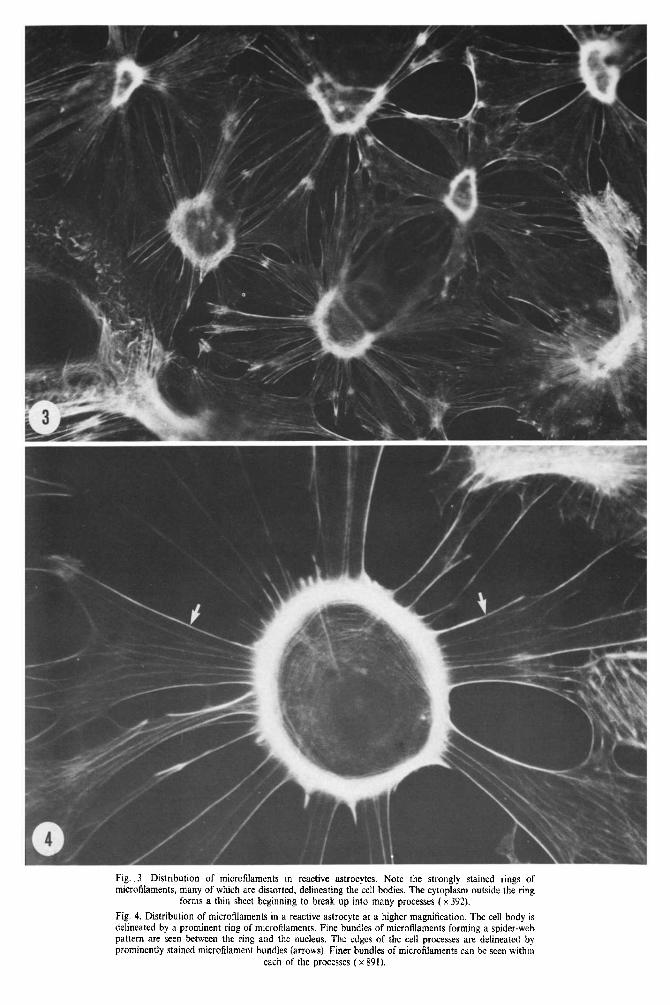
Fig. 3. Distribution of microfilaments in reactive astrocytes. Note the strongly stained rings of microfilaments, many of which are distorted. delineating the cell bodies. The cytoplasm outside the ring
forms a thin sheet canning to break up into many processes (x 392).
Fig. 4. Distribution of microfilaments in a reactive astrocyte at a higher magnification. The cell body is delineated by a prominent ring of microfilaments. Fine bundles of microfilaments forming a spider-web pattern are seen between the ring and the nucleus. The edges of the cell processes are delineated by prominently stained micro~lament bundles (arrows). Finer bundles of microfilam~ts can be seen within
each of the processes ( x 891).
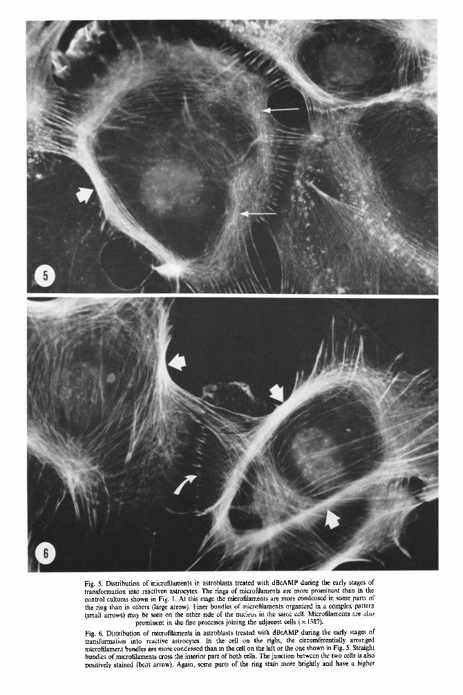
Fig. 5. Distribution of microtilaments in astroblasts treated with dBcAMP during the early stages of transformation into reactiven astrocytes. The rings of microfilaments are more prominent than in the control cultures shown in Fig. I. At this stage the microfilaments are more condensed in some parts of the ring than in others (large arrow). Finer bundles of micro~l~ents organized in a complex pattern (small arrows) may be seen on the other side of the nucleus in the same cell. ~icrofilaments are also
prominent in the fine processes joining the adjacent cells ( x 1387).
Fig 6. Distribution of microfilaments in astroblasts treated with dBcAMP during the early stages of transformation into reactive astrocytes. In the cell on the right, the circumferentially arranged microfilament bundles are more condensed than in the cell on the left or the one shown in Fig. 5. Straight bundles of microfilaments cross the interior part of both cells. The junction between the two cells is also positively stained (bent arrow}. Again, some parts of the ring stain more b~ghtly and have a higher

Fig. 7. Reactive astrocytc? with prominent circumfcrentlal rings afm~crofilamcnts The rings aredeformed to some extent in mos of the cells. possibly as a result of tension generated through the interaction of their processes aith adjacent cells. &tight bundles of mi~rofil~men~s CIOSS the cytoplasm ins& &he ring.
Tension generated by these miero~l~ment bundles could also deform the rmg (x920).
Fig. 8. A ma&c astrocyte with a prominent circumfcrcntial ring of microfilaments. The cell is attached to an adJaceIIt cell on the right by a number of processes. Larger longitundal bundles of microfilaments are present alotlg &r edge5 of the processes while finer bundles are present within Ihcm. The processes ramify at their tips into many fine ones in which microfilament bundb extend in a pattern resembling
the spread fingers of a hand ( x 920).

Microfilaments in reactive astrocytes 261
microfilaments can be deformed to some extent, possibly as a result of forces generated through interaction with adjacent cells (Figs 6-8). Inside the ring several arrangements of the organi~tion of microfilaments were observed. In some cells a spiderweb-like arrangement with a few radial micro- filament bundles crossing the circumferential ones was observed (Fig. 4). In others straight bundles of microfilaments cross from one side of the ring to the other (Fig. 7). These seem to counteract the pull on the ring from the bundles of microfilaments in the processes outside and may also contribute to the deformation of the ring. A third pattern in which the microfilaments are organized in a loose network inside the ring was also sometimes observed.
In the initial stages of ring formation the cells are well spread and a thin sheet of cytoplasm extends in all directions outside the ring. The process formation begins with the appearance of a series of perforations in a linear array radiating centripetally (Fig. 9). The coalescence of these perforations results in the for- mation of processes which vary in width (Figs 3, 4 and 8). In the initial stages of process formation the
sheet of cytoplasm outside the ring contains many fine radially arranged bundles of microfilaments (Fig. 9). As the processes form they become more clearly delineated by strongly staining microfilament bundles along their edges and less prominent ones within the processes (Figs 3 and 4). When the process formation occurs in well separated cells small patches of more prominent staining were seen at the distal ends of the developing processes (Fig. 9). These patches are interconnected by finer bundles of microfilaments running parallel to each other and to the cell periphery. The transforming cells which are interconnected are attached to neighboring cells by processes containing longitudinal bundles of micro- filaments. These bundles ramify at the tips of the processes into many finer bundles of microfilaments extended in a pattern resembling the spread fingers of a hand (Fig. 8).
When the attachment of the reactive astrocyte to the substratum was examined, prominent adhesion foci were not obseived. A few weakly reflective areas repre~nting small focal contacts were, however, seen at the ends of the processes in the more immature
Fig. 9. Distribution of microtilaments in a developing reactive astrocyte before processes have formed. A thin sheet of cytoplasm surrounds the prominent microfilament ring. Radially oriented microfilament bundles form rays from the ring toward the periphery of the cell (long arrows). At the distal ends of the more prominent rays, stained patches are visible along the periphery of the cell (short arrows). The adjacent patches are interconnected by fine bundles of mieroflaments. Perforations (arrowheads) in the cytoplasm are visible between the rays. A series of smaller performations are arranged in rows parallel
to the rays. The edges of larger perforations are strongly stained (x 784).

S. FEDOROFF et al.
Fig. 10. Phase contrast image of a developing reactive astrocyte (RA) and some astroblasts (AB) ( x 450).
Fig. 11. SRI image of the cells shown in Fig. IO. Note focal contacts (large arrow) in the peripheral parts of astroblasts which are in contact with the reactive astrocyte. The reactive astrocytes (RA) have a few
very small focal contacts near the ends of some of the processes (small arrow) ( x 600).
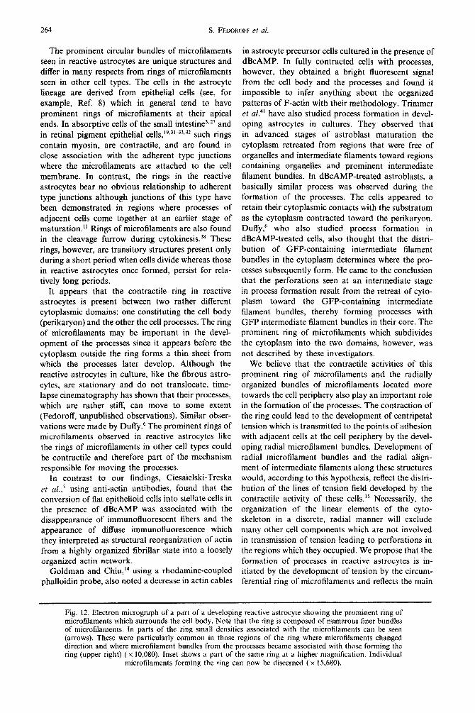
stages of reactive astrocyte formation (Figs 10 and 11). Examination of the developing reactive astro- cytes by electron microscopy confirmed that the prominently stained ring consists of microfilaments (Fig. 12). It also showed that the ring is composed of smaller bundles of microfilaments which run parallel to each other. In some regions of the ring denser areas associated with microfilaments could be discerned. These tended to be particularly common in those parts of the ring where the direction in the orientation of the microfilaments was changing and where microfilament bundles from the periphery of the cell joined those forming the ring.
DISCUSSION
The results obtained show that reactive astrocytes in culture have a very elaborate pattern of micro- filaments that is distinctly different from that seen in their precursor cells, the astroblasts. It is also different from the pattern seen in fibrous astrocytes.*’ When astroblasts develop into stationary fibrous astrocytes the complex pattern of microfilaments seen in the highly motile astrobiasts is lost.*’ In contrast, when astroblasts develop into reactive astrocytes in the presence of dBcAMP they acquire a characteristic pattern of their own.

Microfilaments in reactive astrocytes 263
264 S. FED~ROFF et al.
The prominent circular bundles of microfilaments seen in reactive astrocytes are unique structures and differ in many respects from rings of microfilaments seen in other cell types. The cells in the astrocyte lineage are derived from epithelial cells (see, for example, Ref. 8) which in general tend to have prominent rings of microfilaments at their apical ends. In absorptive cells of the small intestine”*’ and in retinal pigment epithelial cells,‘9.3’-33*42 such rings contain myosin, are contractile, and are found in close association with the adherent type junctions where the microfilaments are attached to the cell membrane. In contrast, the rings in the reactive astrocytes bear no obvious relationship to adherent type junctions although junctions of this type have been demonstrated in regions where processes of adjacent cells come together at an earlier stage of maturation.” Rings of micro~laments are abo found in the cleavage furrow during cytokinesis.38 These rings, however, are transitory structures present only during a short period when cells divide whereas those in reactive astrocytes once formed, persist for rela- tively long periods.
It appears that the contractile ring in reactive astrocytes is present between two rather different cytoplasmic domains: one constituting the cell body (perikaryon) and the other the cell processes. The ring of microfilaments may be important in the devel- opment of the processes since it appears before the cytoplasm outside the ring forms a thin sheet from which the processes later develop. Although the reactive astrocytes in culture, like the fibrous astro- cytes, are stationary and do not translocate, time- lapse cinematography has shown that their processes, which are rather stiff, can move to some extent (Fedoroff, unpublished observations). Similar obser- vations were made by D~ffy.~ The prominent rings of microfilaments observed in reactive astrocytes like the rings of microfilaments in other cell types could be contractile and therefore part of the mechanism responsible for moving the processes.
In contrast to our findings, Ciesaielski-Treska
et aLs using anti-actin antibodies, found that the conversion of flat epithelioid cells into stellate cells in the presence of dBcAMP was associated with the disappearance of immunofluorescent fibers and the appearance of diffuse immunofluorescence which they interpreted as structural reorganization of actin from a highly organized fibrillar state into a loosely organized actin network.
Goldman and Chiu,14 using a rhodamine-coupled phalloidin probe, also noted a decrease in actin cables
in astrocyte precursor cells cultured in the presence of dBcAMP. In fully contracted cells with processes, however, they obtained a bright fluorescent signal from the cell body and the processes and found it impossible to infer anything about the organized patterns of F-actin with their methodology. Trimmer et al.*’ have also studied process formation in devel- oping astrocytes in cultures. They observed that in advanced stages of astroblast maturation the cytoplasm retreated from regions that were free of organelies and intermediate filaments toward regions containing organelles and prominent intermediate filament bundles. In dBcAMP-treated astroblasts, a basically similar process was observed during the formation of the processes. The cells appeared to retain their cytoplasmic contacts with the substratum as the cytoplasm contracted toward the perikaryon. Duffy, who also studied process formation in dBcAMP-treated ceils, also thought that the distri- bution of GFP-containing intermediate filament bundles in the cytoplasm determines where the pro- cesses subsequently form. He came to the conclusion that the perforations seen at an intermediate stage in process formation result from the retreat of cyto- plasm toward the GFP-containing intermediate filament bundles, thereby forming processes with GFP intermediate filament bundles in their core. The prominent ring of microfilaments which subdivides the cytoplasm into the two domains, however, was not described by these investigators.
We believe that the contractile activities of this prominent ring of microfilaments and the radially organized bundles of microfilaments located more towards the cell periphery also play an important role in the formation of the processes. The contraction of the ring could lead to the development of centri~tal tension which is transmitted to the points of adhesion with adjacent cells at the ceil periphery by the devel- oping radial microfilament bundles. Development of radial microfilament bundles and the radial align- ment of intermediate filaments along these structures would, according to this hypothesis, reflect the distri- bution of the lines of tension field developed by the contractile activity of these cells.” Necessarily. the organization of the linear elements of the cyto- skeleton in a discrete, radial manner will exclude many other cell components which are not involved in transmission of tension leading to perforations in the regions which they occupied. We propose that the formation of processes in reactive astrocytes is in- itiated by the development of tension by the circum- ferential ring of microfilaments and reflects the main
Fig. 12. Efectron micrograph of a part of a developing reactive astrocyte showing the prominent ring of mi~ro~laments which surrounds the cell body. Note that the ring is composed of numerous finer bundles of microfilaments. In parts of the ring small densities associated with the microfilaments can be seen (arrows). These were particularly common in those regions of the ring where microfilaments changed direction and where microfilament bundles from the processes became associated with those forming the ring (upper right) (x 10,080). Inset shows a part of the same ring at a higher magnification. Individual
microfilaments forming the ring can now be discerned (x 15.680).

Microfilaments in reactive astrocytes 265
lines along which this tension is transmitted to the attachment points with adjacent cells or to points of attachment with the substratum where neighboring cells are absent. The symmetry and number of pro- cesses may, therefore, be regulated by contacts with adjacent cells. In our view the bundles of intermediate filaments play a passive role in the process formation
by providing regions which are more resistant to deformation. Duffy’s time-lapse photography studies6 support this view. These studies showed reduction of rapid and undulating movements where intermediate filaments were concentrated and formation of pro- cesses around the intermediate filament bundles.
REFERENCES
1. Barak L. L., Yocum R. R., Northnagel E. A. and Webb W. (1980) Fluorescence staining of the actin cytoskeleton in livine cells with 7-nitrobenz-2 oxa-1.3-diazole Phallacidin. Proc. natn. Acad. Sci. U.S.A. 77, 980-984.
2. Barrett C. P., Guth L., Donat E. J. and Krikorian J. G. (1981) Astroglial reaction with gray matter of lumbar segments after midthoracic transection of the adult rat spinal cord. Expl Neural. 73, 365-377.
3. Bretscher A. (1983) Microfilament organization in the cytoskeleton of the intestinal brush border. Cell and Muscle Motility 4, 239-268.
4. Booher J. and Sensenbrenner M. (1972) Growth and cultivation of dissociated neurons and glial cells from embryonic chick, rat and human brain in flask cultures. Neurobiol. 2, 977105.
5.
6. 7.
8.
9. 10.
11.
12.
13.
14.
15. 16.
17.
18.
19.
20.
21.
22.
23.
24.
25.
26.
27. 28.
29.
30.
31.
Ciesielski-Treska J., Guerold B. and Aunis D. (1982) Immunofluorescence study on the organization of actin in astroghal cells in primary cultures. Neuroscience 7, 509-522. Duffv P. E. (1983) Astrocvtes: Normal. Reactive and NeoDlastic. Raven Press, New York. Fed&off S. (1985) Cells resembling reactive astrocytes in’cultures. In Proc. Safeliife Symp. of the 10th Znternafional Society for Neurochemistry on Biochemistry of Glial Cells, p. 70, Liege, Belgium. Fedoroff S. (1985) Macroglial cell lineages. In Molecular Bases of Neural Developmenr (eds Edelman G. M., Gall E. and Cowan W. M.), pp. 91-117. Neuroscience Research Foundation Incorporated. Fedoroff S. and Hall C. (1979) Effect of horse serum on neural cell differentiation in tissue culture. In Vitro l&641&648. Fedoroff S., McAuley W. A. J., Houle J. D. and Devon R. M. (1984) Astrocyte cell lineage. V. Similarity of astrocytes that form in the presence of dBcAMP in cultures to reactive astrocytes in oiuo. J. Neurosci. Res. 12, 15-27. Fedoroff S., Neal J., Opas M. and Kalnins V. I. (1984) Astrocyte cell lineage. III. The morphology of differentiating mouse astrocytes in colony culture. J. Neurocytol. 13, I-20. Fedoroff S., White R. V., Neal J., Subrahmanyan L. and Kalnins V. I. (1983) Astrocyte cell lineage. II. Mouse fibrous astrocytes and reactive astrocytes in cultures have vimentin- and GFP-containing intermediate filaments. Dell/ Brain Res. 7, 303-315. Fedoroff S., White R. V., Subrahmanyan L. and Kalnins V. I. (1981) Properties of putative astrocytes in colony cultures of mouse neopalhum. In Gliul and Neuronal Cell Biology (ed. Fedoroff S.), pp. I-19. Alan Liss, New York. Goldman J. E. and Chiu F. C. (1984) Dibutvrvl cvclic AMP causes intermediate filament accumulation and actin reorganization in astrocytes. Brain Rei. 306, 85195.- Greenspan H. P. and Folkman J. (1977) Hypothesis on cell adhesion and actin cables. J. theor. Biol. 65, 397-398. Hertz L.. Bock E. and Schousboe A. (1978) GFA content, glutamate uptake and activity of glutamate metabolizing enzymes ‘in differentiating mouse astrdcytes’ in primary cultures. Devl ieurosci. 1, 226238. _ Hertz E. and Hertz L. (1979) Polarographic measurement of oxygen uptake by astrocytes in primary cultures using the tissue-culture flask as the respirometer chamber. In Vitro 15, 4299436. Hertz L., Juurlink B. H. J., Fosmark H. and Schousboe A. (1982) Astrocytes in primary culture. In Neuroscience Approach Through Cell Culture, Vol. 1 (ed. Pfeifer S. E.), pp. 1755186, CRC Press, Boca Raton, Florida. Horteea P. del Rio and Penfield W. (1927) Cerebral cicatrix. The reaction of neuroglia and microglia to brain wounds. Bull. Johns Hopkins Hosp. 31, 278-303. ’ Kalnins V. I., Opas M., Ahmed I. and Fedoroff S. (1984) Astrocyte cell lineage. IV. Distribution of microfilaments and adhesion patterns in astrocytes and their precursor cells in culture. J. Neuroqrol. 13, 867-882. Kimelberg H. K., Narumi S. and Bourke R. S. (1978) Enzymatic and morphological properties of primary rat brain astrocyte cultures, and enzymatic development in uiuo. Brain Res. 153, 55-77. Korinkova P. and Lodin 2. (1977) A transitional differentiation of glial cells of cultured corpus callosum caused by dibutyryl cyclic adenosine monophosphate. Neuroscience 2, I103~1114. Latov N., Nilaver G. and Zimmerman A. (1979) Fibrillary astrocytes proliferate in response to brain injury. Decal Biol. 72, 38 I-384. Lim R., Mitsunobu K. and Li W. K. P. (1973) Maturation-stimulating effect of brain extract and dibutyryl cyclic AMP on dissociated embryonic brain cells in culture. Expl Cell Res 79, 243-246. Moonen G., Cam Y., Sensenbrenner M. and Mandel P. (1975) Variability of the effects of serum-free medium, dibutyryl-cyclic AMP or theophyline on the morphology of cultured newborn rat astroblasts. Cell Tiss. Res. 163, 365-372. Moonen G., Heinan E. and Goessens G. (1976) Comparative ultrastructural study of the effects of serum-free medium and dibutytyl-cyclic AMP on newborn rat astroblasts. Cell Tbs. Res. 167, 221-227. Mooseker M. S. (1983) Actin binding proteins of the brush border. CeN 35, 1 I-13. Nathanial E. J. H. and Nathaniel D. R. (1977) Astroglial response to degeneration of dorsal root fibers in adult rat spinal cord. Expl Neural. 54, 60-76. Oehmichen M. (1980) Enzvme-histochemical differentiation of neuroglia and microgha: A contribution to the cytogenesis of microgha and globoid cells. Path. Res. Pratt. 168, 334-373. Opas M. and Kalnins V. I. (1984) Surface reflection interference microscopy: a new method for visualizing cytoskeletal components by light microscopy. J. Microsc. 133, 291L300. Opas M. and Kalnins V. I. (1985) Spatial distribution of cortical protein in cells of epitbelial sheets, Cell Tiss. Res. 239. 451-454.

266 S. FED~ROFF er al.
32. Owaribe K., Kodama R. and Eguchi G. (1981) Demonstration of contractility of circumferential actin bundles and its morphogenetic significance in pigmented epithelium in vitro and in uiuo. J. Cell Biol. 90, 507-514.
33. Owaribe K. and Masuda H. (1982) Isolation and characterization of circumferential bundles from retinal pigmented epithelial cells. J. Cell Biol. 95, 31c315.
34. Philp N. J. and Nachmias V. T. (1985) Components of the cytoskeleton in the retinal pigmented epithelium of the chick. J. Cell Biol. 101, 358-362.
35. Ploem J. S. (1975) General introduction. In Fifrh Inrernational Conference on Immunofluorescence and Related Staining Techniques, Ann. N.Y. Acad Sci. 254, 420.
36. Polak M., Haymaker W., Johnson J. E. Jr. and D’Amelio F. (1982) Neuroglia and their reactions. In Hisfology and Histopathology of the Nervous System (eds Haymaker W. and Adams R. D.), pp. 363-480. Charles C. Thomas, Springfield, Illinois.
37. Schousboe A. (1980) Primary cultures of astrocytes from mammalian brain as a tool in neurochemical research. CeN Molec. Biol. 26, 505-513.
38. Schroeder T. E. (1973) Actin in dividing cells: contractile ring filaments bind heavy meromysin. Proc. natn. Acad. Sci. U.S.A. 70, 168881692.
39. Sensenbrenner M., Devilliers G., Bock E. and Porte A. (1980) Biochemical and ultrastructural studies of cultured rat astroglial cells: Effect of brain extract and dBcAMP on glial fibrillary acid protein and glial filaments. Differenriafion 17, 51-61.
40. Tardy M., Fager C., Rolland B., Bardakdjian F. and Gonnard P. (1981) Effect of prostaglandins and dibutyryl cyclic AMP on the morphology of cells in primary astroglial cultures and on metabolic enzymes of GABA and glutamate metabolism. Experienfia 37, 19-21.
41. Trimmer P. A., Reier P. J., Oh T. H. and Eng L. F. (1982) An ultrastructural and immunocytochemical study of astrocytic differentiation in vitro. J. Neuroimmun. 2, 2355260.
42. Turksen K., Opas M., Aubin J. E. and Kalnins V. I. (1983) Microtubules, microfilaments and adhesion patterns in differentiating chick retinal pigment epithelial (RPE) cells in vitro. Expl Cell Res. 147, 3799391.
(Accepted 17 September 1986)
Copyright © 2022 FDOKUMEN