Morphological changes in hippocampal astrocytes induced by environmental enrichment in mice
31
1 Research Report Morphological changes in hippocampal astrocytes induced by environmental enrichment in mice Giordano G. Viola 1 , Letícia Rodrigues 1,2 , João C. Américo 1,3 , Gisele Hansel 1 , Rafael S. Vargas 1 , Regina Biasibetti 1 , Alessandra Swarowsky 1,2 , Carlos A. Gonçalves 1 , Léder L. Xavier 4 , Matilde Achaval 1,5 , Diogo O. Souza 1 , Olavo B. Amaral 1 1 Departamento de Bioquímica, Instituto de Ciências Básicas da Saúde, Universidade Federal do Rio Grande do Sul, Ramiro Barcelos, 2600-Anexo, 90035-003 Porto Alegre, RS, Brazil. 2 Programa de Pós- Graduação em Neurociências, Instituto de Ciências Básicas da Saúde, Universidade Federal do Rio Grande do Sul, Sarmento Leite, 500, 90050-170, Porto Alegre, RS, Brazil. 3 Instituto de Informática da Universidade Federal do Rio Grande do Sul, Av. Bento Gonçalves 9500, Campus do Vale Bloco 4. 91501-970, Porto Alegre, RS, Brazil. 4 Laboratório de Biologia Tecidual, Departamento de Ciências Morfofisiológicas, PUCRS, Av. Ipiranga 6681, 90 619-900 Porto Alegre, RS, Brazil. 5 Departamento de Ciências Morfológicas, Instituto de Ciências Básicas da Saúde, Universidade Federal do Rio Grande do Sul, Sarmento Leite, 500, 90050-170, Porto Alegre, RS, Brazil. Number of text pages: 25 Number of figures : 5 Corresponding author Olavo B. Amaral Departamento de Bioquímica, ICBS Universidade Federal do Rio Grande do Sul R. Ramiro Barcelos, 2600 (anexo) Porto Alegre, RS, Brazil CEP 90035-003 Phone: 55-51-33085557 FAX: 55-51-33085540 E-mail: [email protected] * 3. Manuscript-title pg, abst, fig lgnd, refs, tbls
-
Upload
independent -
Category
Documents
-
view
4 -
download
0
Transcript of Morphological changes in hippocampal astrocytes induced by environmental enrichment in mice
1
Research Report
Morphological changes in hippocampal astrocytes induced by
environmental enrichment in mice
Giordano G. Viola1, Letícia Rodrigues1,2, João C. Américo1,3, Gisele Hansel1, Rafael S.
Vargas1, Regina Biasibetti1, Alessandra Swarowsky1,2, Carlos A. Gonçalves1, Léder L.
Xavier4, Matilde Achaval1,5, Diogo O. Souza1, Olavo B. Amaral1
1Departamento de Bioquímica, Instituto de Ciências Básicas da Saúde, Universidade Federal do Rio
Grande do Sul, Ramiro Barcelos, 2600-Anexo, 90035-003 Porto Alegre, RS, Brazil. 2Programa de Pós-
Graduação em Neurociências, Instituto de Ciências Básicas da Saúde, Universidade Federal do Rio
Grande do Sul, Sarmento Leite, 500, 90050-170, Porto Alegre, RS, Brazil. 3Instituto de Informática da
Universidade Federal do Rio Grande do Sul, Av. Bento Gonçalves 9500, Campus do Vale Bloco 4.
91501-970, Porto Alegre, RS, Brazil. 4Laboratório de Biologia Tecidual, Departamento de Ciências
Morfofisiológicas, PUCRS, Av. Ipiranga 6681, 90 619-900 Porto Alegre, RS, Brazil. 5Departamento de
Ciências Morfológicas, Instituto de Ciências Básicas da Saúde, Universidade Federal do Rio Grande do
Sul, Sarmento Leite, 500, 90050-170, Porto Alegre, RS, Brazil.
Number of text pages: 25
Number of figures: 5
Corresponding author
Olavo B. Amaral
Departamento de Bioquímica, ICBS
Universidade Federal do Rio Grande do Sul
R. Ramiro Barcelos, 2600 (anexo)
Porto Alegre, RS, Brazil
CEP 90035-003
Phone: 55-51-33085557
FAX: 55-51-33085540
E-mail: [email protected]
* 3. Manuscript-title pg, abst, fig lgnd, refs, tbls
2
Abstract
Environmental enrichment is known to induce plastic changes in the brain, including
morphological changes in hippocampal neurons, with increases in synaptic and spine
densities. In recent years, the evidence for a role of astrocytes in regulating synaptic
transmission and plasticity has increased, and it is likely that morphological and
functional changes in astrocytes play an important role in brain plasticity. Our study
was designed to evaluate changes in astrocytes induced by environmental enrichment in
the CA1 region of the hippocampus, focusing on astrocytic density and on
morphological changes in astrocytic processes. After 8 weeks of environmental
enrichment starting at weaning, male CF-1 mice presented no significant changes in
astrocyte number or in the density of GFAP immunoreactivity in the stratum radiatum.
However, they did present changes in astrocytic morphology in the same region, as
expressed by a significant increase in the ramification of astrocytic processes measured
by the Sholl concentric circles method, as well as by an increase in the number and
length of primary processes extending in a parallel orientation to CA1 nerve fibers. This
led astrocytes to acquire a more stellate morphology, a fact which could be related to the
increase in hippocampal synaptic density observed in previous studies. These findings
corroborate the idea that structural changes in astrocytic networks are an integral part of
plasticity processes occurring in the brain.
Classification Terms
Section: Structural organization of the brain
Keywords: Environmental enrichment, astrocyte, GFAP, hippocampus, stratum
radiatum, morphology.
3
Introduction
Neuroplasticity refers to the changes that occur in the functional and anatomical
organization of the brain as a result of experience (Spolidoro et al., 2008).
Environmental enrichment (EE) is an experimental model that allows the study of
neuroplasticity by providing increases in physical activity, learning experiences,
somatosensorial and visual inputs and social interaction among animals in their home
environment (van Praag et al., 2000, Mohammed et al., 2002). EE promotes plasticity
and neuronal protection through increased levels of neurotrophins (Ickes et al., 2000,
Rossi et al., 2006), changes in cell proliferation (Kempermann et al., 1997, Steiner et
al., 2004), induction of signaling cascades (Li et al., 2006), and enhanced dendritic
branching and synaptogenesis (Moser et al., 1994, Rampon et al., 2000b). It also leads
to chromatin remodeling and histone acetylation, which regulate the susceptibility of
DNA to transcription and therefore control protein formation (Fischer et al., 2007).
These changes lead to morphological alterations in neuronal morphology and
physiology, such as altered synaptic responses and increased synaptic number and size
(Moser et al., 1994, Rampon et al., 2000b).
Astrocytic and neuronal changes in response to various stimuli generally occur
in parallel (Theodosis et al., 2008), and increases in astrocytic volume and number have
been reported after EE in cortical regions (Szeligo and Leblond, 1977, Sirevaag and
Greenough, 1991). In the visual cortex, exposure to a complex environment also
increases the contact between astrocytic processes and synapses (Jones and Greenough,
1996). This enhanced morphological synergism may be a mechanism by which
astrocytes can better regulate the synaptic microenvironment in response to increased
4
neural activity (Jones and Greenough, 1996, Dong and Greenough, 2004). However,
most of the work demonstrating changes in astrocytic morphology after EE has been
performed in the neocortex. Data on whether such changes also occur in the
hippocampus are scarcer and more controversial, with studies demonstrating either an
increase (Briones et al., 2006) or no change (Sirevaag et al., 1991) in astrocytic surface
density. Moreover, no data on astrocytic morphology was provided in either one of
these studies.
Protoplasmic astrocytes play many important roles in the brain, including
guiding synapse development (Ullian et al., 2001), regulating extracellular
concentrations of various molecules (Vernadakis, 1996, Theodosis et al., 2008),
supporting neuronal energetic metabolism (Simpson et al., 2007) and modulating
neuronal and synaptic function (Perea and Araque, 2005, Santello and Volterra, 2008,
Fiacco et al., 2009). Astrocytic networks establish astrocyte-specific anatomical
domains in the hippocampus (Bushong et al., 2002) and are susceptible to plasticity,
controlled by mediators secreted by neurons and other cell types in the brain including
other astrocytes, microglia and endothelial cells (Theodosis et al., 2008). Since EE
promotes synaptic changes in neurons (Foster et al., 1996, Rampon et al., 2000a),
modulation of astrocytic shape is probably involved in the effects of enrichment, as
plastic changes in synapses involve the interaction between new spines and astrocytic
processes (Ullian et al., 2001, Haber et al., 2006).
EE has been shown to increase synaptic and spine density in the hippocampus
(Moser et al., 1994, Rampon et al., 2000a), with morphological changes observed in
neurons in CA1 and dentate gyrus (Faherty et al., 2003). The hippocampus is involved
in several important functions including learning and memory (Squire et al., 2004),
which have been widely shown to be positively modulated by environmental
5
enrichment (Kempermann et al., 1997, Tang et al., 2001, Bruel-Jungerman et al., 2005).
It also contains numerous astrocytes that are located predominantly in Ammon’s horn,
dentate gyrus and subiculum (Catalani et al., 2002). In the presence of long-term
potentiation (LTP) in the dentate gyrus, astrocytes around potentiated synapses have
been shown to increase the ramification of their processes to accompany neural
plasticity (Wenzel et al., 1991).
However, even though astrocytic plasticity is probably involved in the effects of
EE in the hippocampus, there is little data on whether EE can induce changes in
hippocampal astrocytic networks (Sirevaag et al., 1991, Briones et al., 2006). We have
therefore decided to study whether EE induces morphologic changes in astrocytes in the
mouse hippocampus, focusing on the stratum radiatum of the CA1 region, a region
where synapses between CA3 Schaffer collaterals and CA1 neurons are found (Megías
et al., 2001), and where astrocytic calcium currents have been shown to be induced by
neuronal activity (Porter and McCarthy, 1996). For this means, we performed GFAP
immunohistochemical analysis of hippocampal slices in animals weaned under enriched
or control conditions, followed by quantitative analysis of astrocytic density and
morphology.
Results
Male CF1 albino mice were weaned at P21 and housed under environmental
enrichment or control conditions for 8 weeks. After this period, animals were sacrificed
and their brains were prepared for immunohistochemical analysis using an anti-GFAP
antibody. The stratum radiatum of the CA1 region was analyzed in these sections with
6
the use of an automated software in order to evaluate the number of astrocytes, regional
GFAP optical density and GFAP optical density in individual astrocytes. Astrocytic
morphology was evaluated in the same region by analyzing the degree of astrocytic
process ramification with the Sholl concentric circles method, as well as the number,
orientation and length of astrocytic primary processes.
Astrocytic cell bodies and processes were identified in the CA1 stratum
radiatum as shown in Fig. 1. Those observed in the control group usually had rod or
fusiform shapes, while stellate shapes were observed less often. In the EE group, on the
other hand, qualitative analysis showed an increase in astrocytic branching, with more
astrocytes acquiring stellate shapes.
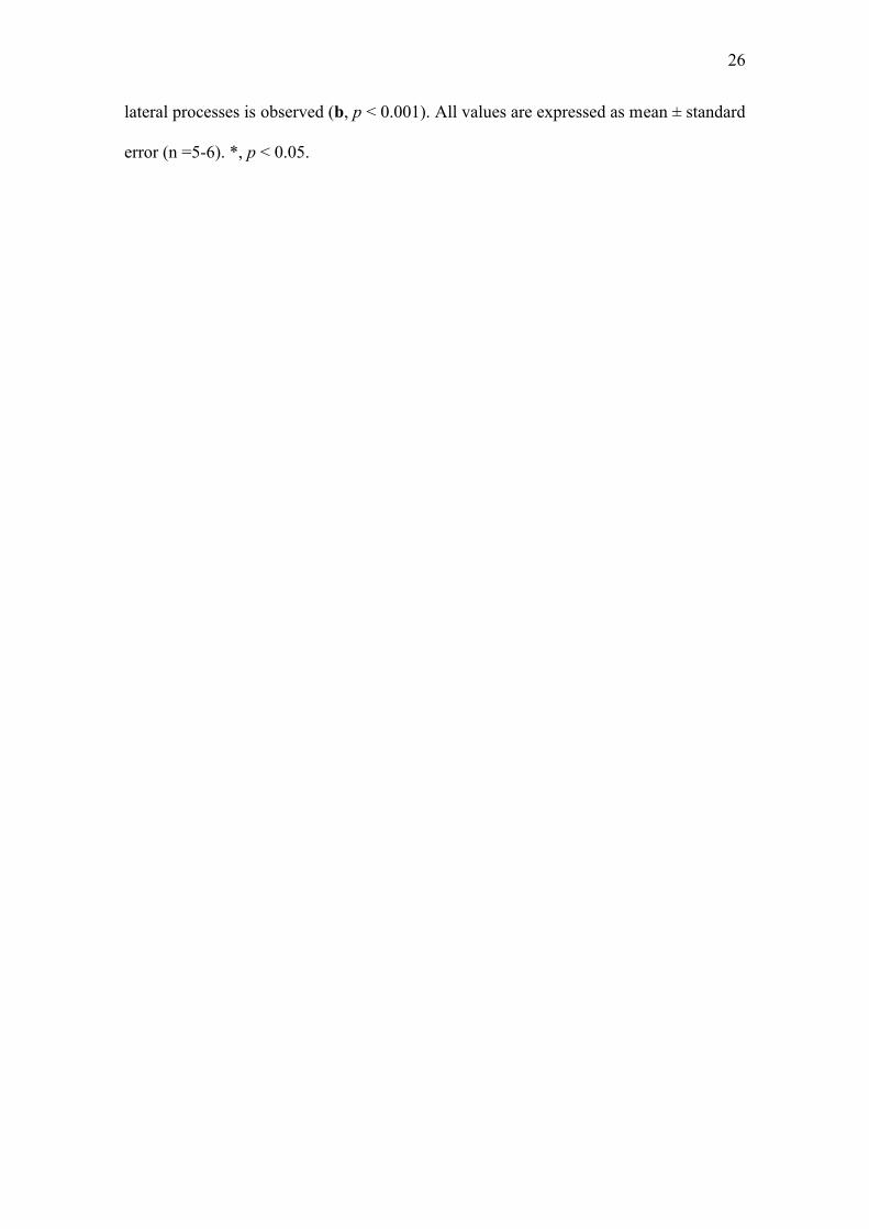
Quantitative results for number of astrocytes (Fig. 2a), regional GFAP optical
density (OD) (Fig. 2b) and GFAP OD within individual astrocytes (Fig. 2c) are shown
in Fig. 2. There was no significant difference in any of these parameters between the
enriched and control groups (unpaired t test, p = 0.98 for astrocyte number, 0.79 for
regional OD and 0.74 for astrocytic OD), demonstrating that the number of astrocytes
and GFAP density were not affected by EE.
The degree of astrocyte process ramification was measured through an
adaptation of the Sholl concentric circles method (Sholl, 1953, Dall'Oglio et al., 2008),
as shown in Fig. 3a. Environmental enrichment induced an increase in astrocyte
ramification (Fig. 3b), as measured by counting the intersection of astrocytic processes
with the concentric circles (p = 0.0116). This increase was observed both in processes
oriented perpendicularly to CA3-CA1 projections (“central” processes, Fig. 3c, p =
0.0339), which is the usual orientation of astrocytic processes in this region (Nixdorf-
Bergweiler et al., 1994), and in those oriented parallel to these fibers (“lateral”
7
processes, Fig. 3d, p = 0.0212); however, it was more prominent in lateral processes
(46.6% increase vs. 21.6% in central processes).
Since the above-mentioned increase in the number of intersections could be due
either to an increase in the number of primary processes, to an increase in the
ramification of each primary process, or to an increase in process length, we proceeded
to count the number of primary astrocytic processes in both groups. As shown in Fig. 4,
EE did not induce significant differences in the total number of primary processes (Fig.
4a, p = 0.18) or in the number of central processes (i.e. those oriented perpendicularly to
CA3-CA1 axons; fig. 4b, p = 0.22). However, the number of lateral primary processes
(those oriented in parallel to axons) was significantly increased by enrichment (Fig. 4c,
p = 0.038), a finding which was counterbalanced by the slight and non-significant
decrease in the number of central processes shown in Fig. 4b.
Finally, we measured the length of the longest central and lateral primary
processes of each astrocyte included in the analysis, to examine whether astrocytic
process size could also be involved in the increased ramification observed with the
Sholl method. As shown in Fig. 5, central processes were distinctly longer than lateral
ones in the control group, and their length was not significantly affected by EE
(although there was a non-significant trend for decreased length in EE animals; Fig. 5a,
p=0.053). On the other hand, the longest lateral processes in the EE group were
markedly increased in length when compared with those in control animals (Fig. 5b,
p<0.001), becoming about as long as the central processes in these animals. The greater
number and increased length of lateral primary processes observed in the EE group,
therefore, probably account at least partially for the greater number of intersections
observed with the Sholl method in the lateral quadrants of astrocytes, as well as for the
more stellate morphology observed qualitatively in Fig. 1.
8
Discussion
Evidence shows that synaptic organization in the hippocampus is influenced by
environmental experience, both during initial postnatal development and after adulthood
(van Praag et al., 2000). Plastic changes induced by EE include morphological
alterations in hippocampal neurons, both in CA1 and in the dentate gyrus (Faherty et al.,
2003). In our study, we describe changes in astrocytic morphology in the stratum
radiatum of CA1, which could be functionally related to the synaptogenesis induced by
EE in this region (Moser et al., 1994, Rampon et al., 2000b), as changes in astrocytic
processes have been shown to correlate with alterations in hippocampal synapses
induced both by physiologic (Klintsova et al., 1995) and pathologic conditions
(Hawrylak et al., 1993) in vivo, as well as with LTP induction in slices (Wenzel et al.
1991).
The changes in astrocytic morphology described in our study seem to reflect
both an increase in astrocytic branching and a change in the orientation of astrocytic
processes. The number and length of primary processes were increased by EE in the
lateral axis of astrocytes, but not in the central axis (which is their usual orientation in
this region), leading astrocytes to change from their usually fusiform morphology
(Nixdorf-Bergweiler et al., 1994, Bushong et al. 2002) to a more stellate shape.
Additionally, both central and lateral processes in EE animals were found to form a
greater number of intersections with concentric circles when analyzed by the Sholl
method. Since the number and length of primary processes in the central axis did not
appear to increase significantly, this suggests that at least in this axis, an increased
branching of individual processes probably occurs in response to EE.
9
Such enrichment-induced changes are somewhat analogous to those observed in
the stratum radiatum over the course of postnatal development. Between P16 and P30,
astrocyte cell bodies become smaller and develop longer processes with thin and
ramified branches, a fact which has been proposed to be related to maturation of
synapses between hippocampal neurons (Catalani et al., 2002). It is possible that post-
weaning EE can make such changes more extensive, by stimulating further plasticity in
the hippocampus during later postnatal development. However, unlike what happens in
normal postnatal development (Catalani et al., 2002), the enrichment-induced
morphological changes in our study were not accompanied by an increase in the number
of astrocytes.
Unlike what has been described in cortical regions after EE (Szeligo and
Leblond, 1977, Sirevaag and Greenough, 1991), we found no increase in the number of
astrocytes or GFAP immunoreactivity in the hippocampus after EE, a fact which is in
agreement with the results of Sirevaag et al. (1991). On the other hand, an increase in
total GFAP reactive surface with EE has been recently reported (Briones et al., 2006).
The variation among these results might be due to differences in the enrichment
protocols – for example, Briones et al.’s study started enrichment at 3-4 months of age,
as opposed to after weaning as in ours. Our results also seem to be somewhat at odds
with studies showing evidence of gliogenesis in the hippocampus, as shown by
increases in BrdU-labeled GFAP positive cells in the subgranular zone (Steiner et al.,
2004) and an increase in nestin-expressing astrocytes in CA1 (Kronenberg et al., 2007).
It is possible that these precursor cells are either short-lived or too few in number to
cause a significant increase in the total number of astrocytes; it is also possible that
protocol differences could be involved in this apparent discrepancy between studies, as
the animals used in these studies were also slightly older than ours. Finally, a study of
10
EE in which animals were sacrificed at old age reported a decrease in the number of
astrocytes in the hippocampus of animals undergoing enrichment (Soffié et al., 1999);
however, those results could be related to reactive astrocytes responding to neuronal
injury, and cannot be directly compared to our data.
Previous studies demonstrate that plastic changes in astrocytes are controlled by
products secreted by neurons and other cell types in the brain including astrocytes,
microglial and endothelial cells (Stevens, 2008). Astrocytes in the stratum radiatum of
CA1 have also been shown to respond to the stimulation of Schaffer collaterals with
increases in the intracellular calcium concentration (Porter and McCarthy, 1996), which
could potentially be involved in the initial induction of plastic changes. Signaling
molecules which could play a role in inducing astrocytic morphological responses
include neurotransmitters such as glutamate and ATP, nitric oxide (De Seranno et al.,
2004), ephrins (Nestor et al., 2007) and growth factors such as brain-derived
neurotrophic factor (BDNF) (Ohira et al., 2007), which could presumably set in motion
plastic cascades culminating in changes in the astrocytic cytoskeleton. BDNF in
particular seems a likely candidate for such a role, as it has been shown both to induce
increases in astrocytic ramification in brain slices (Ohira et al., 2007) and to have its
levels increased by EE in the hippocampus (Ickes et al., 2000, Gobbo and O’Mara,
2004). Conversely, astrocytic morphological changes could also lead to modifications in
hippocampal synapses through the release of mediators such as glutamate, ATP,
adenosine, D-serine, and others (Volterra and Meldolesi, 2005, Fiacco et al., 2009), as
well as through changes in the uptake of neurotransmitters in the synaptic cleft
(Theodosis et al., 2008).
In recent years, knowledge about reciprocal signaling between astrocytes and
synaptic terminals has dramatically increased, leading to the notion of a ‘tripartite
11
synapse’ which includes pre- and post-synaptic terminals and astrocytic processes
(Araque et al., 1999, Perea and Araque, 2005, Santello and Volterra, 2008). This has led
to an increasing recognition of the role of glial cells as active elements in synaptic
transmission. Our results demonstrate morphological changes in astrocytes in the
stratum radiatum of CA1 after environmental enrichment, a finding which is probably
relevant for the interaction between astrocytes and synaptic terminals and related to
hippocampal synaptic plasticity. These results emphasize the need for studies on
biochemical and physiological changes in astrocytes induced by environmental
enrichment and other plastic events in the brain.
Experimental Procedures
Animals
Male albino CF1 mice were obtained from State Foundation for Health Science
Research (FEPPS, Porto Alegre/RS, Brasil). All experimental procedures were
performed according to the NIH Guide for Care and Use of Laboratory Animals and the
Brazilian Society for Neuroscience and Behavior (SBNeC) Recommendations for
Animal Care.
Housing conditions
Mice were weaned on P21 and assigned to enriched or standard housing
conditions immediately after weaning. All animals were kept in a temperature-
controlled colony room with food and water available ad libitum, and maintained on a
12
12-h light/dark cycle (lights on at 7:00 A.M.). Over the course of the study, animals
were kept in either standard or enriched housing conditions for 8 weeks. Standard
housing consisted of a 27 cm × 16 cm × 12 cm acrylic box with sawdust containing
groups of 5 mice. Enriched housing consisted of a 38 cm × 32 cm ×16 cm acrylic box
connected to a 28 cm × 21 cm × 50 cm three-story metal cage with sawdust at the
bottom, housing 10 mice at a time. The enrichment housing apparatus contained two
running wheels and a variety of objects, including toys, tunnels, hiding places and
nesting material, which were changed three times per week, taking care not to repeat
objects during the same week. The metal cage also allowed full view of the colony
room, as well as of other enrichment cages. The same enrichment protocol has been
previously shown to induce long-lasting behavioral alterations in this strain of mice
(Amaral et al., 2008).
Immunohistochemistry
For the immunohistochemical study, animals (5 controls, 6 EE) were deeply
anesthetized with sodium thiopental (50 mg/kg, i.p.) and injected with 400 IU of
heparin. They were then transcardially perfused through the left cardiac ventricle using
a peristaltic pump (10 mL/min) with 50 mL of saline solution followed by 50 mL of 4%
paraformaldehyde in 0.1 M phosphate buffer (pH 7.4; PB). The brains were removed
and post-fixed in the same fixative solution at room temperature for 4 h, then
cryoprotected in a 30% sucrose solution in PB at 4 °C until they sank, and then frozen
in isopentane and liquid nitrogen.
Coronal sections (50 μm) were obtained using a cryostat (Leica, Germany).
Sections were collected in phosphate buffer saline solution (PBS, pH=7.4) and
processed for GFAP immunohistochemistry following the unlabeled antibody
13
peroxidase–antiperoxidase (PAP) procedure (Sternberger, 1979). Free-floating sections
were pretreated with 10% methanol in 3% H2O2 for 30 min, then carefully washed and
blocked with 2% bovine serum albumin (BSA) in PBS containing 0.4% Triton X-100
(PBS-Tx, Sigma Chemical Co., USA) for 30 min. They were then incubated with
polyclonal GFAP antiserum raised in rabbit (Dako, UK) diluted 1: 200 in PBS-Tx
containing 2% BSA for 48 h at 4 °C. After being washed several times with PBS-Tx,
sections were incubated in a PAP-conjugated anti-rabbit IgG (Amersham, UK) diluted
1:50 in PBS-Tx at room temperature for 2 h. The reaction was developed by incubating
the sections in a histochemical medium that contained 0.06% 3,3′-diaminobenzidine
(DAB, Sigma Chemical Co., USA) dissolved in PBS for 10 min and in the same
solution containing 1 μL of 3% H2O2 per mL of DAB medium for an additional 10 min.
Finally, sections were rinsed in PBS, dehydrated in ethanol, cleared with xylene and
covered with Permount and coverslips. A few sections were prepared omitting the
primary antibody by replacing it with PBS as a control to rule out unspecific binding.
The brains in both experimental groups were fixed and post-fixed for the same time in
identical solutions, processed at the same time, and incubated at the same
immunostaining medium for the same period of time, in order to minimize differences
in the staining of astrocytes and in background levels.
Estimation of GFAP density and astrocyte number
The intensity of GFAP immunoreactivity was measured by semi-quantitative
densitometric analysis (Ferraz et al., 2003, Xavier et al., 2005, Martinez et al., 2006)
using a Nikon Eclipse E-600 microscope (500×, Japan) coupled to a Nikon DXM
1200C CCD camera and to NIS Elements AR 2.30. Digitized images obtained from the
14
selected areas were converted to an 8-bit gray scale (0–255 gray levels). All lighting
conditions and magnifications were kept constant during the capture process. Picture
elements (pixels) employed to measure optical density were obtained from region of
interest (ROI) squares with 5827.78 μm2 overlaid on the gray scale image, with
background correction. Obvious blood vessels and other artifacts were avoided. 15
images were analyzed per stratum of hippocampus from each animal (3 fields per
section). Background correction was performed according to the formula previously
described in Xavier et al. (2005). This and all other analyses were performed in the
stratum radiatum of the CA1 region, within an area of the hippocampus extending
between 1.58 mm and 1.74 mm posteriorly and between 1.74 mm to 2.22 mm laterally
from bregma (Franklin and Paxinos, 1997).
The number of GFAP-immunoreactive astrocytes per mm2 in the stratum
radiatum of CA1 was also estimated, using the same software employed to measure
optical density. A ROI square of 5827.78 μm2 was overlaid on the analyzed regions, and
GFAP-reactive astrocytes located inside this square or intersected by the lower and/or
right edges of the square were counted. Astrocytes intersected by the upper and/or left
edges of the square were not counted. Fifteen fields were analyzed in each animal. The
intensity of GFAP immunoreactivity within astrocytes was estimated using the same
software, but with ROI squares measuring 10.37 μm2, which were overlaid in the
astrocytic cell bodies located within the larger square used to measure regional optical
density.
Morphological analysis of astrocytes
Morphologic analysis was performed on 15 astrocytes in each animal (3
sections, each including a single focal plane, with 5 astrocytes analyzed per section).
15
Astrocytes within the same ROI used for estimating the number of astrocytes were used
for morphological analysis, taking care to avoid those which were superimposed upon
other astrocytes or blood vessels. Morphological analysis was performed by two
separate observers who were blind to the experimental groups, and results for both
observers were averaged in the final results.
For a general analysis of astrocytic ramification, we used an adaptation of
Sholl’s concentric circles technique (Sholl, 1953, Dall'Oglio et al., 2008). Briefly, seven
virtual circles with 3.91 μm intervals were drawn around each astrocyte, and the number
of intersections of astrocytic processes with each virtual circle was quantified in both
the lateral (i.e. right/left) and central (i.e. superior/inferior) quadrants around astrocytes.
Primary process quantification was performed by counting the processes extending
directly from the soma in both the lateral and central quadrants of astrocytes in the same
sections. The longest primary process in each quadrant was measured by tracing the
process with an automated measurement tool included in the NIS Elements AR 2.30
package.
Statistical analysis
Data for all variables is expressed either as mean ± standard error. An unpaired
(Student’s) t test was used for all comparisons between groups, and a p value of less
than 0.05 was considered to indicate statistical significance.
16
Acknowledgements
This work was supported by the FINEP research grant "Rede Instituto Brasileiro
de Neurociência (IBN.Net)" # 01.06.0842-00, CNPq, PRONEX, CAPES, FAPERGS
and by the INCT for Excitotoxicity and Neuroprotection grant from MCT, Brazil. We
thank Débora Guerini de Souza for assisting in morphologic analysis during the
manuscript review process.
17
References
Amaral, O.B., Vargas, R.S., Hansel, G., Izquierdo, I., Souza, D.O., 2008. Duration of
environmental enrichment influences the magnitude and persistence of its
behavioral effects on mice. Physiol. Behav. 93, 388-394.
Araque, A., Parpura, V., Sanzgiri, R.P., Haydon, P.G., 1999. Tripartite synapses: glia,
the unacknowledged partner. Trends Neurosci. 22, 208-215.
Briones, T.L., Woods, J., Wadowska, M., Rogozinska, M., Nguyen, M., 2006.
Astrocytic changes in the hippocampus and functional recovery after cerebral
ischemia are facilitated by rehabilitation training. Behav. Brain Res. 171, 17-25.
Bruel-Jungerman, E., Laroche, S., Rampon, C., 2005. New neurons in the dentate gyrus
are involved in the expression of enhanced long-term memory following
environmental enrichment. Eur. J. Neurosci. 21, 513-521.
Bushong, E.A., Martone, M.E., Jones, Y.Z., Ellisman, M.H., 2002. Protoplasmic
astrocytes in CA1 stratum radiatum occupy separate anatomical domains. J.
Neurosci. 22, 183-192.
Catalani, A., Sabbatini, M., Consoli, C., Cinque, C., Tomassoni, D., Azmitia, E.,
Angelucci, L., Amenta, F., 2002. Glial fibrillary acidic protein immunoreactive
astrocytes in developing rat hippocampus. Mech. Ageing Dev. 123, 481-490.
Dall'Oglio, A., Gehlen, G., Achaval, M., Rasia-Filho, A.A., 2008. Dendritic branching
features of Golgi-impregnated neurons from the "ventral" medial amygdala
subnuclei of adult male and female rats. Neurosci. Lett. 439, 287-292.
18
De Seranno, S., Estrella, C., Loyens, A., Cornea, A., Ojeda, S.R., Beauvillain, J.C.,
Prevot, V., 2004. Vascular endothelial cells promote acute plasticity in
ependymoglial cells of the neuroendocrine brain. J. Neurosci. 24, 10353-10363.
Dong, W.K., Greenough, W.T., 2004. Plasticity of nonneuronal brain tissue: roles in
developmental disorders. Ment. Retard. Dev. Disabil. Res. Rev. 10, 85-90.
Faherty, C.J., Kerley, D., Smeyne, R.J., 2003. A Golgi-Cox morphological analysis of
neuronal changes induced by environmental enrichment. Brain Res. Dev. Brain
Res. 141, 55-61.
Ferraz, A.C., Xavier, L.L., Hernandes, S., Sulzbach, M., Viola, G.G., Anselmo-Franci,
J.A., Achaval, M., da Cunha, C., 2003. Failure of estrogen to protect the
substantia nigra pars compacta of female rats from lesion induced by 6-
hydroxydopamine. Brain Res. 986, 200-205.
Fiacco, T.A., Agulhon, C., McCarthy, K.D., 2009. Sorting out astrocyte physiology
from pharmacology. Annu. Rev. Pharmacol. Toxicol. 49, 151-174.
Fischer, A., Sananbenesi, F., Wang, X., Dobbin, M., Tsai, L.H., 2007. Recovery of
learning and memory is associated with chromatin remodelling. Nature 447,
178-182.
Foster, T.C., Gagne, J., Massicotte, G., 1996. Mechanism of altered synaptic strength
due to experience: relation to long-term potentiation. Brain Res. 736, 243-250.
Franklin, K.B.J., Paxinos, G., 1997. The mouse brain in stereotaxic coordinates.
Academic Press, San Diego.
Gobbo, O.L., O’Mara, S.M., 2004. Impact of enriched-environment housing on brain
derived neurotrophic factor and on cognitive performance after a transient global
ischemia. Behav. Brain Res. 152, 231-241.
19
Haber, M., Zhou, L., Murai, K.K., 2006. Cooperative astrocyte and dendritic spine
dynamics at hippocampal excitatory synapses. J. Neurosci. 26, 8881-8891.
Hawrylak, N., Chang, F.L., Greenough, W.T., 1993. Astrocytic and synaptic response to
kindling in hippocampal subfield CA1. II. Synaptogenesis and astrocytic process
increases to in vivo kindling. Brain Res. 19, 309-316.
Ickes, B.R., Pham, T.M., Sanders, L.A., Albeck, D.S., Mohammed, A.H., Granholm,
A.C., 2000. Long-term environmental enrichment leads to regional increases in
neurotrophin levels in rat brain. Exp. Neurol. 164, 45-52.
Jones, T.A., Greenough, W.T., 1996. Ultrastructural evidence for increased contact
between astrocytes and synapses in rats reared in a complex environment.
Neurobiol. Learn. Mem. 65, 48-56.
Kempermann, G., Kuhn, H.G., Gage, F.H., 1997. More hippocampal neurons in adult
mice living in an enriched environment. Nature 386, 493-495.
Klintsova, A., Levy, W.B., Desmond, N.L., 1995. Astrocytic volume fluctuates in the
hippocampal CA1 region across the estrous cycle. Brain Res. 690, 269-274.
Kronenberg, G., Wang, L.P., Geraerts, M., Babu, H., Synowitz, M., Vicens, P., Lutsch,
G., Glass, R., Yamaguchi, M., Baekelandt, V., Debyser, Z., Kettenmann, H.,
Kempermann, G., 2007. Local origin and activity-dependent generation of
nestin-expressing protoplasmic astrocytes in CA1. Brain Struct. Funct. 212, 19-
35.
Li, S., Tian, X., Hartley, D.M., Feig, L.A., 2006. The environment versus genetics in
controlling the contribution of MAP kinases to synaptic plasticity. Curr. Biol.
16, 2303-2313.
Martinez, F.G., Hermel, E.E., Xavier, L.L., Viola, G.G., Riboldi, J., Rasia-Filho, A.A.,
Achaval, M., 2006. Gonadal hormone regulation of glial fibrillary acidic protein
20
immunoreactivity in the medial amygdala subnuclei across the estrous cycle and
in castrated and treated female rats. Brain Res. 1108, 117-126.
Megías, M., Emri, Z.S., Freund, T.F., Gulyás, A.I., 2001. Total number and distribution
of inhibitory and excitatory synapses on hippocampal CA1 pyramidal cells.
Neuroscience 102, 527-540.
Mohammed, A.H., Zhu, S.W., Darmopil, S., Hjerling-Leffler, J., Ernfors, P., Winblad,
B., Diamond, M.C., Eriksson, P.S., Bogdanovic, N., 2002. Environmental
enrichment and the brain. Prog. Brain Res. 138, 109-133.
Moser, M.B., Trommald, M., Andersen, P., 1994. An increase in dendritic spine density
on hippocampal CA1 pyramidal cells following spatial learning in adult rats
suggests the formation of new synapses. Proc. Natl. Acad. Sci. U. S. A. 91,
12673-12675.
Nestor, M.W., Mok, L.P., Tulapurkar, M.E., Thompson, S.M., 2007. Plasticity of
neuron-glial interactions mediated by astrocytic EphARs. J. Neurosci. 27,
12817-12828.
Nixdorf-Bergweiler, BE., Albrecht, D., Heinemann, U., 1994. Developmental changes
in the number, size, and orientation of GFAP-positive cells in the CA1 region of
rat hippocampus. Glia 12, 180-195.
Ohira, K., Funatsu, N., Homma, K.J., Sahara, Y., Hayashi, M., Kaneko, T., Nakamura,
S., 2007. Truncated TrkB-T1 regulates the morphology of neocortical layer I
astrocytes in adult rat brain slices. Eur. J. Neurosci. 25, 406-416.
Perea, G., Araque, A., 2005. Synaptic information processing by astrocytes. J. Physiol.
(Paris) 99, 92-97.
Porter, J.T., McCarthy, K.D., 1996. Hippocampal astrocytes in situ respond to
glutamate released from synaptic terminals. J. Neurosci. 16, 5073-5081.
21
Rampon, C., Jiang, C.H., Dong, H., Tang, Y.P., Lockhart, D.J., Schultz, P.G., Tsien,
J.Z., Hu, Y., 2000a. Effects of environmental enrichment on gene expression in
the brain. Proc. Natl. Acad. Sci. U. S. A. 97, 12880-12884.
Rampon, C., Tang, Y.P., Goodhouse, J., Shimizu, E., Kyin, M., Tsien, J.Z., 2000b.
Enrichment induces structural changes and recovery from nonspatial memory
deficits in CA1 NMDAR1-knockout mice. Nat. Neurosci. 3, 238-244.
Rossi, C., Angelucci, A., Costantin, L., Braschi, C., Mazzantini, M., Babbini, F., Fabbri,
M.E., Tessarollo, L., Maffei, L., Berardi, N., Caleo, M., 2006. Brain-derived
neurotrophic factor (BDNF) is required for the enhancement of hippocampal
neurogenesis following environmental enrichment. Eur. J. Neurosci. 24, 1850-
1856.
Santello, M., Volterra, A., 2008. Synaptic modulation by astrocytes via Ca2+-dependent
glutamate release. Neuroscience, in press.
Sholl, D.A., 1953. Dendritic organization in the neurons of the visual and motor cortices
of the cat. J. Anat. 87, 387-406.
Simpson, I.A., Carruthers, A., Vannucci, S.J., 2007. Supply and demand in cerebral
energy metabolism: the role of nutrient transporters. J. Cereb. Blood Flow
Metab. 27, 1766-1791.
Sirevaag, A.M., Black, J.E., Greenough, W.T., 1991. Astrocyte hypertrophy in the
dentate gyrus of young male rats reflects variation of individual stress rather
than group environmental complexity manipulations. Exp. Neurol. 111, 74-79.
Sirevaag, A.M., Greenough, W.T., 1991. Plasticity of GFAP-immunoreactive astrocyte
size and number in visual cortex of rats reared in complex environments. Brain
Res. 540, 273-278.
22
Soffié, M., Hahn, K., Terao, E., Eclancher, F., 1999. Behavioural and glial changes in
old rats following environmental enrichment. Behav. Brain Res. 101, 37-49.
Spolidoro, M., Sale, A., Berardi, N., Maffei, L., 2008. Plasticity in the adult brain:
lessons from the visual system. Exp. Brain Res.
Squire, L.R., Stark, C.E.L., Clark, R.E., 2004. The medial temporal lobe. Annu. Rev.
Neurosci. 27, 279-306.
Steiner, B., Kronenberg, G., Jessberger, S., Brandt, M.D., Reuter, K., Kempermann, G.,
2004. Differential regulation of gliogenesis in the context of adult hippocampal
neurogenesis in mice. Glia 46, 41-52.
Stevens, B., 2008. Neuron-astrocyte signaling in the development and plasticity of
neural circuits. Neurosignals 16, 278-288.
Szeligo, F., Leblond, C.P., 1977. Response of the three main types of glial cells of
cortex and corpus callosum in rats handled during suckling or exposed to
enriched, control and impoverished environments following weaning. J. Comp.
Neurol. 172, 247-263.
Tang, Y.P., Wang, H., Feng, R., Kyin, M., Tsien, J.Z., 2001. Differential effects of
enrichment on learning and memory function in NR2B transgenic mice.
Neuropharmacology. 41, 779-790.
Theodosis, D.T., Poulain, D.A., Oliet, S.H.R., 2008. Activity-dependent structural and
functional plasticity of astrocyte-neuron interactions. Physiol. Rev. 88, 983-
1008.
Ullian, E.M., Sapperstein, S.K., Christopherson, K.S., Barres, B.A., 2001. Control of
synapse number by glia. Science 291, 657-661.
van Praag, H., Kempermann, G., Gage, F.H., 2000. Neural consequences of
environmental enrichment. Nat. Rev. Neurosci. 1, 191-198.
23
Vernadakis, A., 1996. Glia-neuron intercommunications and synaptic plasticity. Prog.
Neurobiol. 49, 185-214.
Volterra, A., Meldolesi, J., 2005. Astrocytes, from brain glue to communication
elements: the revolution continues. Nat. Rev. Neurosci. 6, 626-640.
Wenzel, J., Lammert, G., Meyer, U., Krug, M., 1991. The influence of long-term
potentiation on the spatial relationship between astrocyte processes and
potentiated synapses in the dentate gyrus neuropil of rat brain. Brain Res. 560,
122-131.
Xavier, L.L., Viola, G.G., Ferraz, A.C., da Cunha, C., Deonizio, J.M., Netto, C.A.,
Achaval, M., 2005. A simple and fast densitometric method for the analysis of
tyrosine hydroxylase immunoreactivity in the substantia nigra pars compacta
and in the ventral tegmental area. Brain Res. Brain Res. Protoc. 16, 58-64.
24
Figure legends
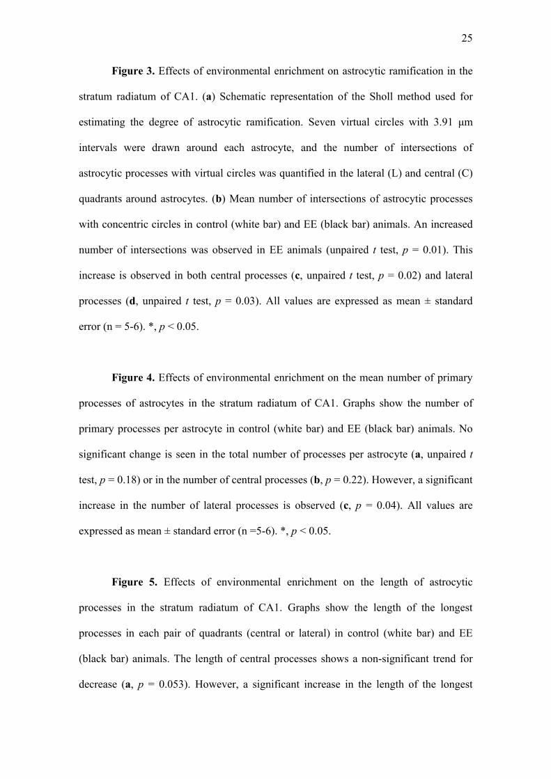
Figure 1. Digitized images of GFAP immunohistochemistry in the CA1 region
of the hippocampus in animals weaned in control and enriched environments. In the
stratum radiatum of control animals (a, top line), astrocytes tended to display a fusiform
shape (top central and right images), while astrocytes in animals which underwent
enrichment (b, bottom line) had more stellate shapes (lower central and right images).
In the right image, primary processes extending in the central (dot) and lateral (asterisk)
quadrants of astrocytes are shown; lateral processes tended to be more numerous and
longer in animals undergoing enrichment. (c) The orientation and location of the images
are shown on a schematic drawing of the brain slice (adapted from Franklin and
Paxinos, 1997). P, stratum pyramidale; R, stratum radiatum; LM, stratum lacunosum
moleculare; M, stratum moleculare of the dentate gyrus; Scale bars: left, 100 µm;
center, 50 µm; right, 10 µm.
Figure 2. Effects of environmental enrichment on astrocyte number and GFAP
density in the stratum radiatum of CA1. (a) number of astrocytes per mm² in sections
from control (white bar) and EE (black bar) animals (unpaired t test, p = 0.98); (b)
regional GFAP optical density measurements in sections from control and EE animals
(unpaired t test, p = 0.79); (c) astrocytic GFAP optical density measurements in
astrocytes within sections from control and EE animals (unpaired t test, p = 0.74). All
values are expressed as mean ± standard error (n = 5-6). A.U., arbitrary units.
25
Figure 3. Effects of environmental enrichment on astrocytic ramification in the
stratum radiatum of CA1. (a) Schematic representation of the Sholl method used for
estimating the degree of astrocytic ramification. Seven virtual circles with 3.91 μm
intervals were drawn around each astrocyte, and the number of intersections of
astrocytic processes with virtual circles was quantified in the lateral (L) and central (C)
quadrants around astrocytes. (b) Mean number of intersections of astrocytic processes
with concentric circles in control (white bar) and EE (black bar) animals. An increased
number of intersections was observed in EE animals (unpaired t test, p = 0.01). This
increase is observed in both central processes (c, unpaired t test, p = 0.02) and lateral
processes (d, unpaired t test, p = 0.03). All values are expressed as mean ± standard
error (n = 5-6). *, p < 0.05.
Figure 4. Effects of environmental enrichment on the mean number of primary
processes of astrocytes in the stratum radiatum of CA1. Graphs show the number of
primary processes per astrocyte in control (white bar) and EE (black bar) animals. No
significant change is seen in the total number of processes per astrocyte (a, unpaired t
test, p = 0.18) or in the number of central processes (b, p = 0.22). However, a significant
increase in the number of lateral processes is observed (c, p = 0.04). All values are
expressed as mean ± standard error (n =5-6). *, p < 0.05.
Figure 5. Effects of environmental enrichment on the length of astrocytic
processes in the stratum radiatum of CA1. Graphs show the length of the longest
processes in each pair of quadrants (central or lateral) in control (white bar) and EE
(black bar) animals. The length of central processes shows a non-significant trend for
decrease (a, p = 0.053). However, a significant increase in the length of the longest
26
lateral processes is observed (b, p < 0.001). All values are expressed as mean ± standard
error (n =5-6). *, p < 0.05.
Figure 1Click here to download high resolution image
Figure 2Click here to download high resolution image
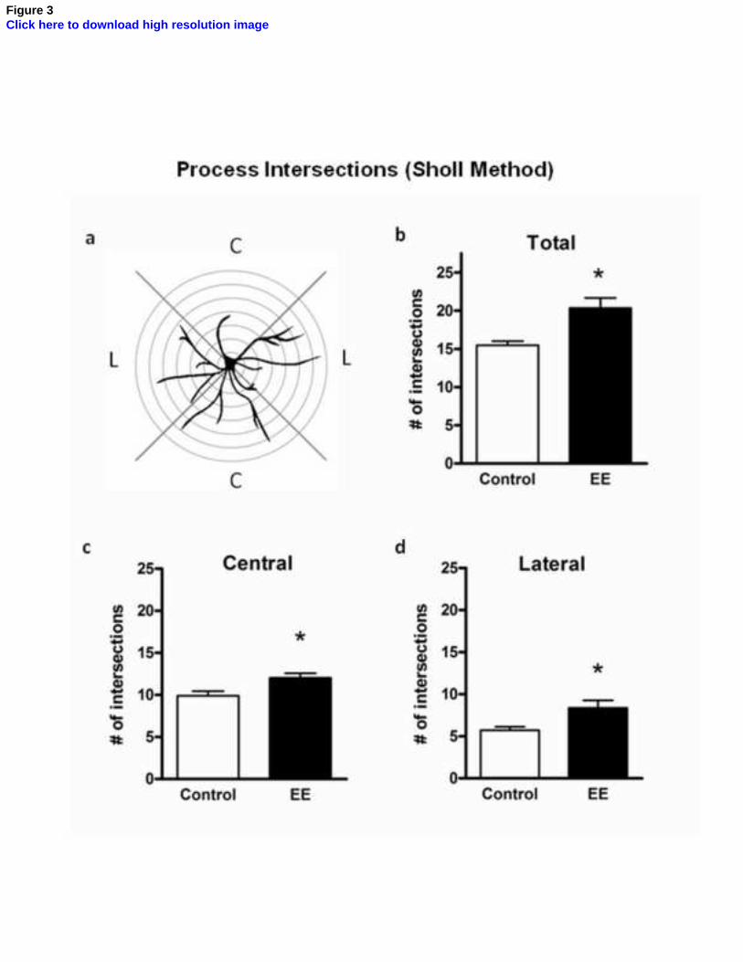
Figure 3Click here to download high resolution image
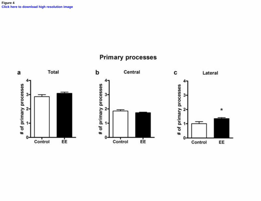
Figure 4Click here to download high resolution image
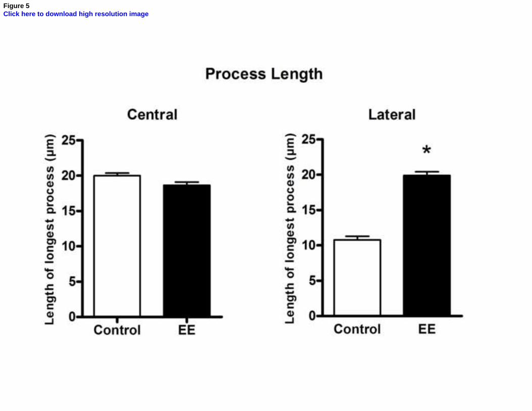
Figure 5Click here to download high resolution image