Identification of thyrotropin-releasing hormone as hippocampal glutaminyl cyclase substrate in...
10
Identification of thyrotropin-releasing hormone as hippocampal glutaminyl cyclase substrate in neurons and reactive astrocytes Alexander Waniek a , Maike Hartlage-Rübsamen a , Corinna Höfling a , Astrid Kehlen c , Stephan Schilling b , Hans-Ulrich Demuth b, ⁎, Steffen Roßner a, ⁎⁎ a Paul Flechsig Institute for Brain Research, University of Leipzig, Germany b Fraunhofer Institute of Cell Therapy and Immunology IZI Leipzig, Department of Drug Design and Target Validation MWT Halle, Germany c Institute for Medical Microbiology, Martin-Luther-University Halle-Wittenberg, Germany abstract article info Article history: Received 4 August 2014 Received in revised form 29 October 2014 Accepted 11 November 2014 Available online 18 November 2014 Keywords: Glutaminyl cyclase Hippocampus Thyrotropin-releasing hormone Amyloid pathology Recently, Aβ peptide variants with an N-terminal truncation and pyroglutamate modification were identified and shown to be highly neurotoxic and prone to aggregation. This modification of Aβ is catalyzed by glutaminyl cyclase (QC) and pharmacological inhibition of QC diminishes Aβ deposition and accompanying gliosis and ameliorates memory impairment in transgenic mouse models of Alzheimer's disease (AD). QC expression was initially described in the hypothalamus, where thyrotropin-releasing hormone (TRH) is one of its physiological substrates. In addition to its hormonal role, a novel neuroprotective function of TRH following excitotoxicity and Aβ-mediated neurotoxicity has been reported in the hippocampus. Functionally matching this finding, we recently demonstrated QC expression by hippocampal interneurons in mouse brain. Here, we detected neuronal co-expression of QC and TRH in the hippocampus of young adult wild type mice using double immunofluorescence labeling. This provides evidence for TRH being a physiological QC substrate in hippocampus. Additionally, in neocortex of aged but not of young mice transgenic for amyloid precursor protein an increase of QC mRNA levels was found compared to wild type littermates. This phenomenon was not observed in hippocampus, which is later affected by Aβ pathology. However, in hippocampus of transgenic — but not of wild type mice — a correlation between QC and TRH mRNA levels was revealed. This co-regulation of the enzyme QC and its substrate TRH was reflected by a co-induction of both proteins in reactive astrocytes in proximity of Aβ deposits. Also, in primary mouse astrocytes a co-induction of QC and TRH was demonstrated upon Aβ stimulation. © 2014 Elsevier B.V. All rights reserved. 1. Introduction Alzheimer's disease (AD) is characterized by the formation of Aβ plaques and neurofibrillary tangles [1,2]. In particular neocortex and hippocampus are affected by AD pathology leading to clinical symptoms like cognitive decline and loss of memory function [2–4]. The knowledge about the structural diversity of Aβ peptides was extended in the 1990s by the identification of N-terminally truncated and pyroglutamate (pGlu)-modified Aβ peptides (pGlu-Aβ) in post mortem brains of AD patients [5,6]. These pGluAβ-peptides exhibit altered biochemical prop- erties causing increased neurotoxicity, resistance to proteolysis and accelerated tendency to aggregate [7–10]. Subsequently, the enzyme glutaminyl cyclase (QC) was demonstrated to catalyze this post-translational modification of Aβ in vitro [11] and in vivo [12]. Furthermore, pharmacological inhibition of QC leads to a significant reduction of overall Aβ aggregation in transgenic animal models of AD and to better performance in experimental tasks of learning and memory [12]. Recently, we reported QC expression in a subset of hippocampal interneurons of wild type mice [13] as well as deposition of pGlu-Aβ in the core of Aβ plaques in human amyloid precursor protein (APP) transgenic Tg2576 mice [14]. An understanding of the physiological function and substrate specificity of QC in brain regions such as the hippocampus appears important in order to (i) gain insight into the in- volvement of QC in AD-related pathological changes and (ii) estimate the effects of pharmacological inhibition of QC in the course of a possible therapeutic intervention. Biochimica et Biophysica Acta 1852 (2015) 146–155 Abbreviations: AD, Alzheimer's disease; APP, amyloid precursor protein; BACE1, beta- site APP-cleaving enzyme 1; HPT, hypothalamus–pituitary–thyroid; IFN-γ, interferon-γ; LPS, lipopolysaccharide; pGlu, pyroglutamate; QC, glutaminyl cyclase; TRH, thyrotropin- releasing hormone ⁎ Correspondence to: H.-U. Demuth, Fraunhofer Institute of Cell Therapy and Immunology IZI Leipzig, Department of Drug Design and Target Validation MWT Halle, Biocenter Weinbergweg 22, 06120 Halle (Saale), Germany. Tel.: +49 345 13142800; fax: +49 345 13142801. ⁎⁎ Correspondence to: S. Roßner, Paul Flechsig Institute for Brain Research, Jahnallee 59, 04109 Leipzig, Germany. Tel.: +49 341 9725758; fax: +49 341 9725749. E-mail addresses: [email protected] (H.-U. Demuth), [email protected] (S. Roßner). http://dx.doi.org/10.1016/j.bbadis.2014.11.011 0925-4439/© 2014 Elsevier B.V. All rights reserved. Contents lists available at ScienceDirect Biochimica et Biophysica Acta journal homepage: www.elsevier.com/locate/bbadis
-
Upload
independent -
Category
Documents
-
view
1 -
download
0
Transcript of Identification of thyrotropin-releasing hormone as hippocampal glutaminyl cyclase substrate in...

Biochimica et Biophysica Acta 1852 (2015) 146–155
Contents lists available at ScienceDirect
Biochimica et Biophysica Acta
j ourna l homepage: www.e lsev ie r .com/ locate /bbad is
Identification of thyrotropin-releasing hormone as hippocampalglutaminyl cyclase substrate in neurons and reactive astrocytes
Alexander Waniek a, Maike Hartlage-Rübsamen a, Corinna Höfling a, Astrid Kehlen c, Stephan Schilling b,Hans-Ulrich Demuth b,⁎, Steffen Roßner a,⁎⁎a Paul Flechsig Institute for Brain Research, University of Leipzig, Germanyb Fraunhofer Institute of Cell Therapy and Immunology IZI Leipzig, Department of Drug Design and Target Validation MWT Halle, Germanyc Institute for Medical Microbiology, Martin-Luther-University Halle-Wittenberg, Germany
Abbreviations:AD, Alzheimer's disease; APP, amyloidsite APP-cleaving enzyme 1; HPT, hypothalamus–pituitarLPS, lipopolysaccharide; pGlu, pyroglutamate; QC, glutamreleasing hormone⁎ Correspondence to: H.-U. Demuth, Fraunhofer I
Immunology IZI Leipzig, Department of Drug Design andBiocenter Weinbergweg 22, 06120 Halle (Saale), Germfax: +49 345 13142801.⁎⁎ Correspondence to: S. Roßner, Paul Flechsig Institute04109 Leipzig, Germany. Tel.: +49 341 9725758; fax: +4
E-mail addresses: [email protected]@medizin.uni-leipzig.de (S. Roßner).
http://dx.doi.org/10.1016/j.bbadis.2014.11.0110925-4439/© 2014 Elsevier B.V. All rights reserved.
a b s t r a c t
a r t i c l e i n f oArticle history:Received 4 August 2014Received in revised form 29 October 2014Accepted 11 November 2014Available online 18 November 2014
Keywords:Glutaminyl cyclaseHippocampusThyrotropin-releasing hormoneAmyloid pathology
Recently, Aβ peptide variantswith anN-terminal truncation andpyroglutamatemodificationwere identifiedandshown to be highly neurotoxic and prone to aggregation. This modification of Aβ is catalyzed by glutaminylcyclase (QC) and pharmacological inhibition of QC diminishes Aβ deposition and accompanying gliosis andameliorates memory impairment in transgenic mouse models of Alzheimer's disease (AD). QC expression wasinitially described in the hypothalamus, where thyrotropin-releasing hormone (TRH) is one of its physiologicalsubstrates. In addition to its hormonal role, a novel neuroprotective function of TRH following excitotoxicityand Aβ-mediated neurotoxicity has been reported in the hippocampus. Functionally matching this finding, werecently demonstrated QC expression by hippocampal interneurons in mouse brain.Here, we detected neuronal co-expression of QC and TRH in the hippocampus of young adult wild type miceusing double immunofluorescence labeling. This provides evidence for TRH being a physiological QC substratein hippocampus. Additionally, in neocortex of aged but not of young mice transgenic for amyloid precursorprotein an increase of QC mRNA levels was found compared to wild type littermates. This phenomenon wasnot observed in hippocampus, which is later affected by Aβ pathology. However, in hippocampus of transgenic —but not of wild type mice — a correlation between QC and TRH mRNA levels was revealed. This co-regulation ofthe enzyme QC and its substrate TRH was reflected by a co-induction of both proteins in reactive astrocytes inproximity of Aβ deposits. Also, in primary mouse astrocytes a co-induction of QC and TRH was demonstratedupon Aβ stimulation.
© 2014 Elsevier B.V. All rights reserved.
1. Introduction
Alzheimer's disease (AD) is characterized by the formation of Aβplaques and neurofibrillary tangles [1,2]. In particular neocortex andhippocampus are affected by AD pathology leading to clinical symptomslike cognitive decline and loss of memory function [2–4]. The knowledgeabout the structural diversity of Aβ peptides was extended in the 1990sby the identification of N-terminally truncated and pyroglutamate
precursor protein; BACE1, beta-y–thyroid; IFN-γ, interferon-γ;inyl cyclase; TRH, thyrotropin-
nstitute of Cell Therapy andTarget Validation MWT Halle,
any. Tel.: +49 345 13142800;
for Brain Research, Jahnallee 59,9 341 9725749.e (H.-U. Demuth),
(pGlu)-modified Aβ peptides (pGlu-Aβ) in post mortem brains of ADpatients [5,6]. These pGluAβ-peptides exhibit altered biochemical prop-erties causing increased neurotoxicity, resistance to proteolysis andaccelerated tendency to aggregate [7–10].
Subsequently, the enzyme glutaminyl cyclase (QC)was demonstratedto catalyze this post-translational modification of Aβ in vitro [11] andin vivo [12]. Furthermore, pharmacological inhibition of QC leads to asignificant reduction of overall Aβ aggregation in transgenic animalmodels of AD and to better performance in experimental tasks of learningand memory [12].
Recently, we reported QC expression in a subset of hippocampalinterneurons of wild type mice [13] as well as deposition of pGlu-Aβin the core of Aβ plaques in human amyloid precursor protein (APP)transgenic Tg2576 mice [14]. An understanding of the physiologicalfunction and substrate specificity of QC in brain regions such as thehippocampus appears important in order to (i) gain insight into the in-volvement of QC in AD-related pathological changes and (ii) estimatethe effects of pharmacological inhibition of QC in the course of a possibletherapeutic intervention.
147A. Waniek et al. / Biochimica et Biophysica Acta 1852 (2015) 146–155
There is substantial knowledge about QC function in the hypothala-mus, where the enzyme catalyzes the pGlu modification of neuropep-tides like neurotensin and thyrotropin-releasing hormone (TRH)[15–17]. Notably, QC-mediated pGlu modification of TRH is known tooccur as the latest step of a processing cascade which is essential forthe biological activity of TRH enabling its interaction with specific TRHreceptors in pituitary gland and decreasing its degradation rate[18–21]. In this hypothalamus–pituitary–thyroid (HPT) axis, activationof TRH receptors increases the release of TSH, which in turn stimulatesthe secretion of thyroid hormones [22]. Accordingly, QC knock-outmice show reduced plasma thyroxine concentration [23].
However, there is evidence for a function of TRH independent of theHPT axis. In this context, TRHwas shown to exhibit neuromodulatory aswell as neuroprotective effects. The expression of TRH in hippocampus,raphe nuclei and the hypothalamic nuclei [24–27] as well as the co-localization with neurotransmitters like choleocystokinin, galanin,neuropeptide Y and serotonin point towards a function of TRH asneuromodulator [25,28–30]. More recently, the potential of TRH to actas a neuroprotective compound has been discussed. TRH was reportedto prevent neurodegenerative effects of excitotoxic substances likekainate, glutamate and NMDA in cell culture studies of organotypicbrain slices and primary neurons [31–33]. In this context, it is also notablethat TRH decreases the release of excitatory glutamate [34] but increasessecretion of inhibitoryGABA [35] as shown in electrophysiological studiesusing organotypic brain slice cultures and primary neurons.
Interestingly, TRH-mediated neuroprotection has not only beenreported with respect to excitotoxicity [31–33], Parkinson's disease [36,37] and following brain injury [38,39], but also in response to Aβ neuro-toxicity [39,40]. Furthermore, in post mortem hippocampal tissue of ADpatients diminished levels of TRH were demonstrated and depletion ofTRH in cell culture studies was shown to result in hyperphosphorylationof tau [41], another hallmark of AD.
In summary, due to its presumedneuroprotective effects, hippocam-pal TRH expression and function deserves a thorough investigation,especially since the TRH-modifying enzyme QC also contributes to Aβpathology. In order to study a possible spatial and temporal relationshipbetweenhippocampal QC and TRHexpression, quantitativemRNA anal-ysis and immunohistochemical labeling were performed in young andaged wild type and Tg2576 mice with distinct hippocampal Aβ pathol-ogy. Additionally, activated astrocytes were used as an in vitromodel tostudy a possible co-regulation of QC and TRH under pathogenic condi-tions. The relevance of TRH in the hippocampus, a brain area of particu-lar importance in learning andmemorywhich is strongly affected in AD,is discussed.
2. Materials and methods
2.1. Experimental animals
In order to analyze hippocampal QC and TRH expression, transgenicTg2576 mice and wild type littermates at postnatal ages of 4, 8, 10, 13,17 and 21 months were used for immunohistochemical and for qPCRexperiments (N = 3–6 per age group). Due to overexpression ofhuman APP comprising Swedish double mutation (Lys670→Asn,Met671→Leu) Tg2576 mice develop Aβ pathology first detectable by10 months of age in entorhinal cortex and by about 13 months inhippocampus.
2.2. Preparation of mouse brains for immunohistochemistry and qPCR
The mice were deeply anesthetized with pentobarbital andtranscardially perfused with 50 ml 0.9% saline containing 0.1% heparinfollowed by perfusion with 80 ml 4% paraformaldehyde in PBS (0.1 M;pH 7.4). The brains were removed from the skull and post-fixed byimmersion in the same fixative overnight at 4 °C. After cryoprotectionin 30% sucrose in 0.1 M PBS for 3 days, the brains were snap-frozen in
n-hexane at −68 °C and stored at −20 °C. Coronal sections (30 μm)were cut on a sliding microtome and collected in 0.1 M PBS.
In order to perform qPCRmicewere sacrificed, brainswere removedfrom the skull and subsequently neocortex, hippocampus and ventralbrainwere dissected. Dissection of the ventral brainwasdoneby cuttingoff the rostral part of the brain at the level of the anterior commissure,followed by removal of the overlaying neocortex, hippocampus andcerebellum. Thus, it contains subcortical nuclei and hypothalamuswith well-described QC and TRH expression.
2.3. Immunohistochemical labeling
In order to perform immunohistochemistry, mouse brain slices werepre-treated with 60% methanol (30 min), followed by washes in 0.1 MTBS and blocked in TBS containing 0.3% TritonX-100 and 5% normaldonkey serum for 30 min to reduce unspecific binding of antibodies.Incubation with primary antibodies was performed in TBS containing0.1% TritonX-100 and 5% normal donkey serum over 1–3 days at 4 °C.For detection of QC a rabbit anti-mouse QC antibody (AB1301,Probiodrug AG, Halle/Saale, Germany) or goat anti-mouse QC antibody(10269, Probiodrug AG)was used as described earlier [13,14]. Immuno-histochemical labeling of TRHwasobtainedwith different commerciallyavailable rabbit anti-TRH antibodies raised either against proTRH(BP5066, Acris) or against synthetic pGlu-His-Pro conjugated to KLH(PAB13482, Abnova; TRH, BioLogo). Staining with these antibodiesdisplayed a very similar pattern of fibers and single neurons in mousehypothalamus indicating specific labeling since this brain region is de-scribed to show robust TRH immunoreactivity [22]. pGlu-Aβ peptidesin mouse hippocampus were detected using the mouse monoclonalantibody mab2-48 (Synaptic Systems, Göttingen, Germany), whichhas been well characterized [42]. A goat anti-GFAP antibody (SantaCruz) was used for labeling of astroglia. According to the host speciesof the primary antibody appropriate secondary antibodies (Dianova)were used to visualize respective antigens either with chromogen(3,3′DAB) or fluorescent dyes (Cy2, Cy3, Cy5).
2.4. Confocal laser scanning microscopy
Laser scanning microscopy (LSM 510, Zeiss, Oberkochen, Germany)was performed to analyze co-localization of QC and TRH in mousebrain tissue. For Cy2-labeled antigens (green fluorescence), an argonlaser with 488 nm excitation was used and emission from Cy2 wasrecorded at 510 nm applying a low-range band pass (505–550 nm).For Cy3-labeled antigens (red fluorescence), a helium–neon laser with543 nm excitation was used and emission from Cy3 at 570 nmwas de-tected applying high-range band pass (560–615 nm). The Cy5-labeledantigens were visualized using excitation at 650 nm and emission at670 nm.
2.5. Stimulation of cultured primary astrocytes
Astrocyte primary cell cultures were started with brains of newbornmice according to Löffner et al. [43] and were maintained in DMEM-based medium at 37 °C in a humidified atmosphere with 95% air/5%CO2. The medium was renewed once a week.
Activation of astrocyteswas inducedwhen cells had reached 80–90%confluency by incubationwith (i) Aβ1–42 (5 μM) or (ii) lipopolysaccha-ride derived from Escherichia coli (LPS; 1 μg/ml; O55:B5, Sigma) andinterferon-γ (IFN-γ; 20 ng/ml; Preprotech) for 48 h. Cell viability wasanalyzed with a commercially available lactate dehydrogenase assaykit from Promega.
2.6. mRNA quantification by qPCR
RNA was isolated using the NucleoSpin RNA II kit (Macherey Nagel,Düren, Germany) according to the manufacturer's instructions. RNA

148 A. Waniek et al. / Biochimica et Biophysica Acta 1852 (2015) 146–155
was obtained from different brain regions as well as from culturedprimary astrocytes. The RNA concentration was measured using aNanoDrop 2000 spectrophotometer (Peqlab, Erlangen, Germany). RNA(0.1–1 mg) was reverse transcribed into cDNA using random primers(Roche, Penzberg, Germany) and Superscript III (Life Technologies).Quantitative PCR was performed in a Rotorgene 3000 (CorbettResearch, Sydney, Australia) using the Rotor-Gene SYBR Green PCR kitand specific primers for QC (NM_027455.2, 5′-GGGAGGCAGACACAATCAAT and 3′-TCAGATTCCCAGCTGTCAGA), TRH (NM_009426.2, 5′-GTGCCAACCAAGACAAGGAT and 3′-TTCTTCCCAGCTTCTTTGGA) andGFAP (NM_010277.3, 5′-ACATCGAGATCGCCACCTAC and 3′-TCACATCACCACGTCCTTGT) synthesized by Metabion (Martinsried, Germany).Relative gene expressionwas determined using the Rotorgene Softwareversion 6.1 in comparative quantitation mode. TTl (NM_027192.2),Umps (NM_009471.2) and Sys1 (NM_025575.3) were used as referencegenes for expression analysis. The PCR was verified by product meltingcurves and single amplicons were confirmed by agarose gelelectrophoresis.
2.7. Quantification of immunocytochemistry
After treatment of primary astrocytes, cells were fixed in 4%paraformaldehyde, labeled with antibodies against TRH and QC asdescribed in 2.3 and cell nuclei were counterstained with Hoechstdye (1:10,000; Invitrogen). From each sample 5 images were takenby means of fluorescence microscope (Keyence Biorevo BZ-9000)and approx. 10 cells/sample were densitometrically analyzed byKeyence BZ-9000 Analyzer software. Values obtained from controlastrocytes were set to 100%.
2.8. Statistical analyses
Data that were obtained from qPCR experiments were analyzedby GraphPad Prism4 Software with respect to statistical significance inStudent's t-test and correlation analysis of data was performed withPearson's correlation coefficient.
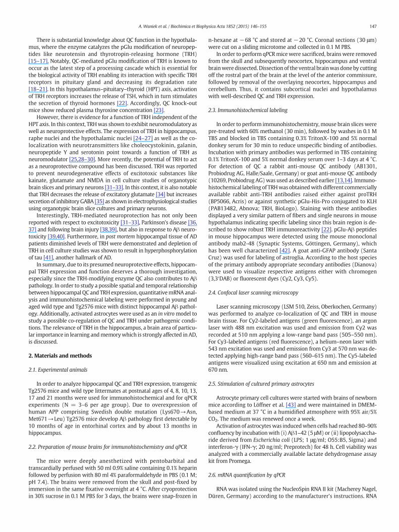
Fig. 1. QC and TRH co-localization in hippocampal neurons of adult wild type mice. Immunohismunoreactive neurons in distinct layers of the hippocampus. The staining displayed a similar dstratum radiatum (rad) of the hippocampus. In the polymorphic cell layer of dentate gyrus (po)cell layer of the hippocampus (pyr) aswell as in the granular cell layer of dentate gyrus (gr) no sand TRH in a subpopulation of hippocampal interneurons (C). Abbreviations or, stratum oriensmolecular layer of dentate gyrus; po, polymorphic cell layer of dentate gyrus; gr, granular cell la
3. Results
3.1. Co-localization of QC and TRH in hippocampus of wild type mice
In order to obtain insight into the biological significance of the QCexpression in the hippocampus as reported by Hartlage-Rübsamenet al. [13], we focused on the hypothalamic QC substrate TRH, sincethis well known releasing hormone of the HPT axis has also beenshown to play a role in the hippocampus [33–35]. Therefore, immuno-histochemistry was used to reveal the cellular expression pattern ofTRH in the hippocampus of young adult postnatal day 42 wild typemice.
Immunohistochemical labelings displayed a considerable number ofTRH positive interneurons dispersed over different layers of the hippo-campus (Fig. 1B). The regional expression pattern strongly resembledthe distribution of QC positive neurons, particularly in stratumlacunosum moleculare and in stratum radiatum of hippocampus proper(Fig. 1A). In the polymorphic cell layer of dentate gyrus TRH immunore-activity was low in labeling intensity compared to the relatively strongQC labeling. Double immunofluorescent staining demonstrated co-localization of QC and its substrate TRH in a subpopulation of hippocam-pal interneurons (Fig. 1C).
3.2. Temporal expression pattern of QC and TRH mRNA in different brainregions during aging of Tg2576 mice
QCwas shown to be involved in the development of Aβpathology bymodifying N-terminally truncated Aβ peptides. The resulting pGlu-Aβpeptides exhibit altered biochemical properties which lead to increasedneurotoxicity. TRH, a known hypothalamic substrate of QC, was demon-strated to have neuromodulatory properties in the hippocampus and toprovide protection towards Aβ-mediated neurotoxicity aswell as gluta-matergic excitotoxicity. In order to study the interrelationship betweenTRH and QC we investigated the temporal gene expression pattern ofthese two proteins in Tg2576 mice before and after the onset of Aβpathology and compared to non-transgenic littermates.
tochemistry for QC (A) and TRH (B) in hippocampal slices of wild type mice revealed im-istribution of QC- and TRH-positive neurons in stratum lacunosum moleculare (lac) and instrong labeling for QC occurred whereas TRH immunoreactivity was low. In the pyramidalpecific labelingwas detected (A, B). Double immunolabeling revealed co-localization of QC; lac, stratum lacunosum moleculare; rad, stratum radiatum; pyr, pyramidal cell layer; mol,yer of dentate gyrus.
149A. Waniek et al. / Biochimica et Biophysica Acta 1852 (2015) 146–155
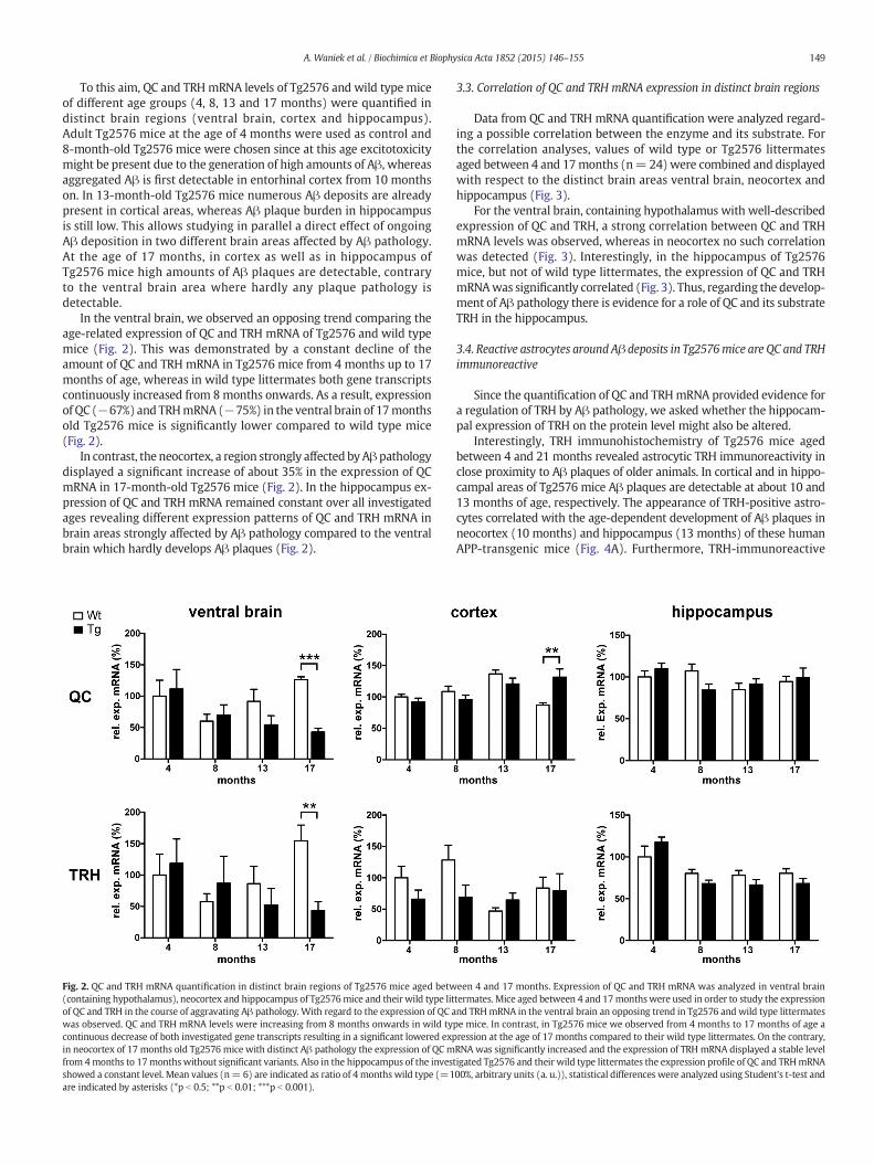
To this aim, QC and TRHmRNA levels of Tg2576 and wild type miceof different age groups (4, 8, 13 and 17 months) were quantified indistinct brain regions (ventral brain, cortex and hippocampus).Adult Tg2576 mice at the age of 4 months were used as control and8-month-old Tg2576 mice were chosen since at this age excitotoxicitymight be present due to the generation of high amounts of Aβ, whereasaggregated Aβ is first detectable in entorhinal cortex from 10 monthson. In 13-month-old Tg2576 mice numerous Aβ deposits are alreadypresent in cortical areas, whereas Aβ plaque burden in hippocampusis still low. This allows studying in parallel a direct effect of ongoingAβ deposition in two different brain areas affected by Aβ pathology.At the age of 17 months, in cortex as well as in hippocampus ofTg2576 mice high amounts of Aβ plaques are detectable, contraryto the ventral brain area where hardly any plaque pathology isdetectable.
In the ventral brain, we observed an opposing trend comparing theage-related expression of QC and TRH mRNA of Tg2576 and wild typemice (Fig. 2). This was demonstrated by a constant decline of theamount of QC and TRH mRNA in Tg2576 mice from 4 months up to 17months of age, whereas in wild type littermates both gene transcriptscontinuously increased from 8months onwards. As a result, expressionof QC (−67%) and TRHmRNA (−75%) in the ventral brain of 17monthsold Tg2576 mice is significantly lower compared to wild type mice(Fig. 2).
In contrast, the neocortex, a region strongly affected byAβpathologydisplayed a significant increase of about 35% in the expression of QCmRNA in 17-month-old Tg2576 mice (Fig. 2). In the hippocampus ex-pression of QC and TRH mRNA remained constant over all investigatedages revealing different expression patterns of QC and TRH mRNA inbrain areas strongly affected by Aβ pathology compared to the ventralbrain which hardly develops Aβ plaques (Fig. 2).
Fig. 2. QC and TRH mRNA quantification in distinct brain regions of Tg2576 mice aged betw(containing hypothalamus), neocortex and hippocampus of Tg2576mice and their wild type litof QC and TRH in the course of aggravating Aβ pathology. With regard to the expression of QC awas observed. QC and TRH mRNA levels were increasing from 8 months onwards in wild tycontinuous decrease of both investigated gene transcripts resulting in a significant lowered expin neocortex of 17 months old Tg2576 mice with distinct Aβ pathology the expression of QC mfrom 4months to 17monthswithout significant variants. Also in the hippocampus of the invesshowed a constant level. Mean values (n= 6) are indicated as ratio of 4 months wild type (=1are indicated by asterisks (*p b 0.5; **p b 0.01; ***p b 0.001).
3.3. Correlation of QC and TRH mRNA expression in distinct brain regions
Data from QC and TRH mRNA quantification were analyzed regard-ing a possible correlation between the enzyme and its substrate. Forthe correlation analyses, values of wild type or Tg2576 littermatesaged between 4 and 17months (n= 24)were combined and displayedwith respect to the distinct brain areas ventral brain, neocortex andhippocampus (Fig. 3).
For the ventral brain, containing hypothalamus with well-describedexpression of QC and TRH, a strong correlation between QC and TRHmRNA levels was observed, whereas in neocortex no such correlationwas detected (Fig. 3). Interestingly, in the hippocampus of Tg2576mice, but not of wild type littermates, the expression of QC and TRHmRNAwas significantly correlated (Fig. 3). Thus, regarding the develop-ment of Aβ pathology there is evidence for a role of QC and its substrateTRH in the hippocampus.
3.4. Reactive astrocytes around Aβ deposits in Tg2576mice are QC and TRHimmunoreactive
Since the quantification of QC and TRHmRNA provided evidence fora regulation of TRH by Aβ pathology, we asked whether the hippocam-pal expression of TRH on the protein level might also be altered.
Interestingly, TRH immunohistochemistry of Tg2576 mice agedbetween 4 and 21 months revealed astrocytic TRH immunoreactivity inclose proximity to Aβ plaques of older animals. In cortical and in hippo-campal areas of Tg2576 mice Aβ plaques are detectable at about 10 and13 months of age, respectively. The appearance of TRH-positive astro-cytes correlated with the age-dependent development of Aβ plaques inneocortex (10 months) and hippocampus (13 months) of these humanAPP-transgenic mice (Fig. 4A). Furthermore, TRH-immunoreactive
een 4 and 17 months. Expression of QC and TRH mRNA was analyzed in ventral braintermates. Mice aged between 4 and 17monthswere used in order to study the expressionnd TRHmRNA in the ventral brain an opposing trend in Tg2576 and wild type littermatespe mice. In contrast, in Tg2576 mice we observed from 4 months to 17 months of age aression at the age of 17 months compared to their wild type littermates. On the contrary,RNA was significantly increased and the expression of TRH mRNA displayed a stable leveltigated Tg2576 and their wild type littermates the expression profile of QC and TRHmRNA00%, arbitrary units (a. u.)), statistical differences were analyzed using Student's t-test and

Fig. 3. Correlation of QC and TRHmRNA expression in distinct brain regions of Tg2576mice. Correlation analysis revealed a strong interdependence of QC and TRHmRNA expression in theventral brain of, both, Tg2576mice and their non transgenic littermates as well as a significant correlation in the hippocampus of Tg2576 but not of wild type animals. No correlation wasmeasured in the neocortex. For correlation analysis mRNA values of all mice of the indicated genotype aged between 4 and 17months were combined (n= 24 each). Pearson correlationcoefficient r and p values (*p b 0.5; **p b 0.01; ***p b 0.001) are indicated in the diagrams.
150 A. Waniek et al. / Biochimica et Biophysica Acta 1852 (2015) 146–155
glia-like cells surrounded Aβ plaques and showed typical morphologi-cal characteristics of activated astroglia such as enlarged somata andprocesses (Fig. 4A). This distinct glia-like TRH stainingwas not observeddistant from Aβ plaques.
3.5. QC and TRH are co-localized in activated astrocytes in Tg2576 mice
Double immunofluorescent labelings for GFAP and pGlu-Aβ inhippocampal brain slices of 17 months old Tg2576 mice confirmed theactivation of astrocytes around Aβ plaques (Fig. 4B). Reactive astrocytesin proximity to plaques but not in astrocytes away from Aβ depositswere found to be pGlu-Aβ immunoreactive (Fig. 4B). Similarly, Aβplaque-associated reactive astrocytes also displayed TRH immunoreactiv-ity (Fig. 4C). Both products of the enzymatic activity of QC, TRH and pGlu-Aβ, were found to be co-localized with QC in Aβ plaque-associated reac-tive astrocytes (Fig. 4D). Thus, there is a distinct induction of QC, TRH andpGlu-Aβ in plaque-associated reactive astrocytes.
3.6. Expression of QC and TRH in cultures of primary astrocytes upon Aβtreatment
As described above, we observed activated astrocytes surroundingAβ plaques of Tg2576 mice which show co-expression of QC and TRH.In order to reveal whether Aβ peptides directly mediate the inductionof QC and TRH in reactive astrocyteswe used a primarymouse astrocytecell model. Primary astrocytes were stimulated with Aβ or a generalpro-inflammatory mixture of LPS/IFN-γ and the expression of QC andTRH mRNA as well as immunoreactivity for QC and TRH protein wasstudied after a 48 h stimulation period. Compared to vehicle-treated as-trocytes, the expression of QC mRNA was increased by 23% (p b 0.05)upon Aβ stimulation, whereas the 15% increase in TRH mRNA levelswas not statistically significant (Fig. 5A). Immunocytochemical labelingof QC and TRH also suggested increased protein levels (Fig. 5B). Quanti-tative image analysis demonstrated increased QC (+65%; p b 0.01) andfor TRH (+57%; p b 0.05) protein levels (Fig. 5A). This indicates that a
primary Aβ-induced increase in QC mRNA and protein expressionresults in the stabilization and, therefore, increased concentration ofthe QC substrate TRH without significant change in its mRNA level. Incontrast, a general pro-inflammatory stimulation of astrocytes withLPS/IFN-γ did not induce QC or TRH mRNA or protein levels (notshown).
4. Discussion
In the 1990s, new Aβ peptide variants were identified comprisingN-terminally truncated andpGlu-modified species. TheseAβ variants dis-play altered biochemical, biophysical and cell biological properties leadingto enhanced neurotoxicity and resistance to proteolytical degradation.The enzyme QC was demonstrated to catalyze the pGlu modification ofAβ andpharmacological QC inhibition in Tg2576mice resulted in a reduc-tion of Aβ plaque burden [12]. However, when using QC inhibition aspharmacological tool for AD treatment, it is important to understandthe physiological function of QC expression.
In a previous report, we alreadymapped regional and cellular QC ex-pression in wild type mouse brain and identified QC-immunoreactiveinterneurons in distinct layers of the hippocampus [13]. The neurohor-mone TRH is known to be an important physiological QC substrate indiencephalon where both, enzyme and substrate, are expressed byneurons of the paraventricular nucleus of the hypothalamus. Studiesconcerning an alternative TRH function as neurotransmitter andneuromodulator in the hippocampus [25,28,34,35] prompted us torelate TRH to QC expression in this brain region.
Here, we report for the first time co-localization of QC and TRH in aneuronal subpopulation of the hippocampus in adult wild type mice.However, since not all QC expressing neuronswere positive for TRH im-munoreactivity, it is very likely that other, yet unknown QC substratesexist in the hippocampus. Our finding confirms previous reports thatdemonstrated intrinsic TRH expression in the hippocampus, albeitmost hippocampal TRH is supposed to derive from extrinsic sources[26]. When pathological amounts of Aβ peptides are present in AD, QC

Fig. 4.Age-dependent increase of astrocytic QC and TRH immunoreactivity in the vicinity of Aβ-deposits in the neocortex and hippocampus of Tg2576mice. Tg2576mice develop depositsof aggregated Aβ in cortex and hippocampus from 10 and 17 months onwards, respectively. For analysis of TRH immunoreactivity Tg2576mice aged between 4 and 21months were ex-amined. (A) Immunohistochemistry revealed TRH staining in astrocytes that encircle Aβ deposits in cortex and hippocampus of Tg2576mice at 10 and 17months, respectively (asterisks).The increasing number of TRH-positive astrocytes was associated with age and thereby the quantity of Aβ deposits. Tg2576 mice without apparent Aβ aggregates (4 months old) weredevoid of TRH-positive astrocytes. (B) The density of GFAP-immunoreactive astrocytes (red) around pGlu-Aβ deposits (green, asterisk) is increased and their morphology is altered toa reactive phenotype. Some of the Aβ plaque-associated astrocytes (within the white circle) display pGlu-Aβ immunoreactivity (arrows), as indicated by yellow/orange color. Astrocytesdistant from Aβ deposits (outside thewhite circle) do not display pGlu-Aβ immunoreactivity. (C) Aβ plaque-associated astrocytes (red) are also immunoreactive for TRH (green, arrows).The location of Aβ deposits is marked by asterisks. Astrocytes distant from Aβ deposits, outside the circle, are not TRH-immunoreactive (arrowheads). (D) Both products of the enzymaticactivity of QC, TRH (red) and pGlu-Aβ (blue, asterisk), and QC itself (green) are co-localized in Aβ plaque-associated reactive astrocytes (arrows) as indicated bywhite color in the overlaychannel.
151A. Waniek et al. / Biochimica et Biophysica Acta 1852 (2015) 146–155
expressing neurons might be at particular risk of degeneration due tothe intracellular modification of Aβ resulting in highly neurotoxicpGlu-Aβ. Such a degeneration of QC expressing interneurons could re-sult in the release of pGlu-Aβ acting as a seed for further Aβ aggregation
[9] and to a decrease of TRH concentration in the hippocampus as it wasdemonstrated in post mortem brains of AD [41]. Regarding the neuro-protective ability of TRH to counteract Aβ-induced neurodegeneration[39,40] as well as excitotoxicity in cell culture models [31–33], the

Fig. 5. Expression of QC and TRH in primary astrocytes. Cultured primary astrocytes wereactivated by incubation with Aβ (5 μM) for 48 h. (A) Quantitative RT-PCR revealed an in-crease in QC mRNA in Aβ-stimulated astrocytes (black bars) by 23% (p b 0.05), whereasTRH mRNA levels remained unchanged (n = 6). Moreover, protein levels for QC andTRHwere increased by 65 and 57%, respectively as revealed by quantitative immunocyto-chemistry. Mean values for treated astrocytes are given as per cent of untreated value(=100%). Statistical differences were analyzed using Student's t-test and are indicatedby asterisks (*p b 0.5). (B) Examples of immunocytochemical fluorescent labelings of as-trocytes for QC (green) and TRH (red) under control conditions and after Aβ stimulation.Note the robust increase in the staining intensity after Aβ stimulation in a high proportionof the astrocytes (arrows). Cellular nuclei are stained with Hoechst dye (blue).
152 A. Waniek et al. / Biochimica et Biophysica Acta 1852 (2015) 146–155
degeneration of hippocampal TRH-expressing neurons could aggravatepathological processes in AD. Also, in Parkinson's disease [36,37] and inthe context of CNS trauma [38,39] neuroprotective effects of TRH werereported.
In order to study the regional expression of QC and TRHwith regardto Aβ pathology, we compared Tg2576 mice with wild type littermatesat different postnatal ages using quantitative PCR aswell as immunohis-tochemistry. We detected a significantly higher expression of QCmRNAonly in the neocortex of 17-month-old transgenic mice with substantialAβ pathology which could account for the enhanced formation of
pGlu-Aβ deposits as revealed by immunohistochemistry. In contrast,unchanged QC mRNA levels in the hippocampus of Tg2576 mice couldbe related to the delayed development of Aβ pathology in this brain re-gion. In the hippocampus aswell as in the neocortexwe did not observesignificant differences in the expression of TRH mRNA in Tg2576 micecompared to wild type littermates. In the light of the induction ofQC and TRH protein in reactive astrocytes of aged Tg2576 mice (seebelow), increased levels of these transcripts might be expected. Thisdiscrepancymay be explained by a dissociation between the regulationof mRNA and protein expression and/or a reduction of neuronal QC andTRHmRNAexpression in parallelwith the induction of these transcriptsin reactive astrocytes.
On the other hand, diminished TRH protein levels were reported forhippocampus in AD [41]. Since we observed constant hippocampal TRHmRNA levels and an induction of TRH protein in reactive astrocytes ofaged Tg2576 mice, our findings point towards differences between thetransgenic animal model and the human disease. For example, humanbrain samples analyzed always reflect the final stage of the disease,the post mortem delay is much longer than in animal studies and themouse model used only mimics aspects of the human disease.
A correlation analysis revealed a significant correlation between QCand TRHmRNA in the hippocampus of Tg2576mice but not ofwild typemice pointing to an effect of developing Aβ pathology on the expressionof QC and TRH.
To study the expression of QC and TRH in a part of the brain,which isless affected byAβ pathology,we used tissue of the ventral brain includ-ing the hypothalamus amongst other subcortical nuclei. In this part ofthe brain, the mRNA expression of both, QC and its substrate TRH, wassignificantly decreased in 17-month-old Tg2576 mice compared towild type littermates. Most TRH is synthesized in the paraventricularnucleus of the hypothalamus, which projects— apart from the pituitarygland — also to the hippocampus [44], brain stem [45] and spinal cord[46]. A decrease in the release of hypothalamus-derived TRH couldtherefore impair TRH-mediated neuromodulation as well as HPT axis,the latter being affected in AD patients as reported by Yong-Honget al. [47]. Vice versa, hippocampal Aβ pathology may in a retrogrademanner affect hypothalamic afferents. In Tg2576 mice devoid of hypo-thalamic Aβ pathology, hypothalamicmetabolic andhormonal dysfunc-tion have been reported [48,49] which may by the basis for the effectsobserved here on TRH and QC mRNA expression.
Since determination of mRNA expression is not sufficient to gaininsight in cellular expression, immunohistochemical stainings wereperformed. In order to obtain more information about pronouncedstate of pathology in hippocampus, we decided to use brain slices ofTg2576 mice aged between 4 and 21 months for immunohistochemis-try, because qPCR experiments revealed significant differences inmRNA expression levels particularly in 17-month-oldmicewith distinctAβ pathology.
Interestingly, we did not observe any obvious alteration in the num-ber of QC and TRHpositive neurons (not shown), but detected inductionof strong TRH labeling of astrocytes in close proximity to Aβ plaques.Furthermore, QC, the enzyme which converts TRH into its bioactivestate, was co-localized with its substrate in activated astrocytes. Weconsider this as being strong evidence for an intracellular productionof biologically active TRH in astrocytes. However, we cannot completelyrule out the possibility that the observed immunoreactivity is causedby endocytosis of neuronally secreted TRH. Although the expressionand processing of neuropeptides by astroglia is still controversiallydiscussed, there is some evidence supporting this hypothesis. For exam-ple, astrocyteswere shown to express enzymes that are involved in pro-TRH processing like carboxypeptidase C/D [50,51] and peptidyl-glycinealpha-amidating monooxygenase [52]. Recently, spinal astrocytes weredemonstrated to process prodynorphin and release dynorphin [53]indicating that astrocytes are principally able to process pro-peptides.In addition, a glioblastoma cell line was shown to release TRH [54]and with respect to cultured astrocytes, earlier publications already

Fig. 6. Potential relevance of hippocampal QC and TRH under physiological and pathological conditions. Under physiological conditions hippocampal interneurons co-express QC and TRHand contribute to LTP modulation and neuroprotection by releasing TRH. However, under pathological conditions in AD, QC-catalyzed pGlu-Aβ generation induces degeneration of theseinterneurons. This results in diminished release of neuroprotective TRH and presumably in altered neuromodulation and increased excitotoxicity. Furthermore, the formation of Aβplaques as well as neurodegeneration induces the activation of astrocytes. The co-induction of QC and TRH in reactive astrocytes could contribute to TRH-mediated neuroprotection aswell as to further neurodegeneration by generation of astrocyte-derived pGlu-Aβ.
153A. Waniek et al. / Biochimica et Biophysica Acta 1852 (2015) 146–155
mentioned an increased phosphoinositol turnover after administrationof TRH in medium [55].
Furthermore, in performing qPCR on mouse primary astrocyteswe detected substantial expression of TRH mRNA and QC mRNA. Thetranscript levels of QC and protein concentrations of QC and TRH wereincreased upon stimulation with Aβ. The elevated TRH protein concen-tration without induction of the corresponding mRNA suggests post-translational pGlu modification and, thereby, stabilization of TRH byQC. On the other hand, the stimulation of cultured astrocytes with LPSand IFN-γ did not modulate QC or TRH mRNA or protein expression.This is strong evidence for Aβ peptides themselves — and not otherfactors present in or around Aβ deposits — being triggers for astrocyticQC and TRH expression. Since we also observed strong TRH immunore-activity in astrocytes enclosing Aβ plaques in Tg2576mice, it is conceiv-able, that it is due to a chronic, Aβ-mediated activation of astrocytes inthese AD model mice.
The finding of QC and TRH in activated astrocytesmight be of impor-tance with regard to a role of astroglia contributing to AD pathogenesis.Since astrocytes were demonstrated to express the β-secretase BACE1[56,57], the additional expression of QC provides the possibility ofastroglial derived toxic pGlu-Aβ leading to astrogliosis and the forma-tion of Aβ plaques. Moreover, astrocytes are involved in maintaining astable extracellular milieu for proper neuronal functioning and excit-ability. For example, astrocytes express specific glutamate transportersto avoid excessive extracellular concentrations of glutamate that couldinduce excitotoxicity [58]. In this context, a function of TRH is discussedin reducing the neuronal release of glutamate [34], thereby modulatingneuronal activity andpresumably diminishing excitotoxicity. Astrocyteswere also shown to express receptors for leptin [59,60] which is well-known to increase TRH expression [61]. For leptin, a role in AD pathol-ogy has been discussed by influencing the composition of membranelipid rafts and thereby the activity of BACE1 [62–64].
5. Conclusions
This is the first demonstration of (i) a neuronal co-localization of QCand TRH in hippocampus, (ii) distinct differences in QC and TRH mRNAexpression in brain regions with and without Aβ pathology in Tg2576mice and (iii) astroglial co-expression of QC and TRH in the vicinity ofAβ plaques in the hippocampus. Functionally, the expression of QC inastrocytes could play a role in neuroprotection by the activation andrelease of TRH, thereby reducing excitotoxicity but also in neurodegen-eration due to the formation of pGlu-Aβ (Fig. 6). With regard to QC as atherapeutical target for pharmacological inhibition in AD, our results
suggest that a potential decrease of a neuroprotective TRH effectswould be compensated by the benefits of a reduction in Aβ pathology.
Acknowledgments
The authors like to express their gratitude to Dr. Karen Hsiao,Department of Neurology, University of Minnesota, USA, for kindlyproviding the Tg2576 F1 mice which were backcrossed to breed N2generation mice used in this study. We thank R. Jendrek (Paul FlechsigInstitute for Brain Research) for the technical assistance. This work wassupported by the German Federal Department of Education, Science andTechnology, BMBF [grant #0316033A to HUD and grant #0316033Bto SR] and by a grant of the Deutsche Forschungsgemeinschaft, DFG[grant # RO 2226/13-1 to SR].
References
[1] D.J. Selkoe, D. Schenk, Alzheimer's disease: molecular understanding predictsamyloid-based therapeutics, Annu. Rev. Pharmacol. Toxicol. 43 (2003) 545–584,http://dx.doi.org/10.1146/annurev.pharmtox.43.100901.140248.
[2] H.W. Querfurth, F.M. LaFerla, Alzheimer's disease, N. Engl. J. Med. 362 (2010)329–344, http://dx.doi.org/10.1056/NEJMra0909142.
[3] E.R. Kandel, The molecular biology of memory storage: a dialogue between genesand synapses, Science 294 (2001) 1030–1038, http://dx.doi.org/10.1126/science.1067020.
[4] K. Blennow, M.J. de Leon, H. Zetterberg, Alzheimer's disease, Lancet 368 (2006)387–403, http://dx.doi.org/10.1016/S0140-6736(06)69113-7.
[5] T.C. Saido, T. Iwatsubo, D.M. Mann, H. Shimada, Y. Ihara, S. Kawashima, Dominantand differential deposition of distinct beta-amyloid peptide species, A betaN3(pE), in senile plaques, Neuron 14 (1995) 457–466, http://dx.doi.org/10.1016/0896-6273(95)90301-1.
[6] T.C. Saido, W. Yamao-Harigaya, T. Iwatsubo, S. Kawashima, Amino- and carboxyl-terminal heterogeneity of beta-amyloid peptides deposited in human brain,Neurosci. Lett. 215 (1996) 173–176.
[7] W. He, C.J. Barrow, The A beta 3-pyroglutamyl and 11-pyroglutamyl peptides foundin senile plaque have greater beta-sheet forming and aggregation propensitiesin vitro than full-length A beta, Biochemistry 38 (1999) 10871–10877, http://dx.doi.org/10.1021/bi990563r.
[8] C. Russo, E. Violani, S. Salis, V. Venezia, V. Dolcini, G. Damonte, U. Benatti, C. D'Arrigo,E. Patrone, P. Carlo, G. Schettini, Pyroglutamate-modified amyloid beta-peptides-AbetaN3(pE)-strongly affect cultured neuron and astrocyte survival, J. Neurochem.82 (2002) 1480–1489, http://dx.doi.org/10.1046/j.1471-4159.2002.01107.x.
[9] S. Schilling, T. Lauber, M. Schaupp, S. Manhart, E. Scheel, G. Böhm, H.U. Demuth, Onthe seeding and oligomerization of pGlu-amyloid peptides (in vitro), Biochemistry45 (2006) 12393–12399, http://dx.doi.org/10.1021/bi0612667.
[10] D. Schlenzig, S. Manhart, Y. Cinar, M. Kleinschmidt, G. Hause, D.Willbold, S.A. Funke,S. Schilling, H.U. Demuth, Pyroglutamate formation influences solubility andamyloidogenicity of amyloid peptides, Biochemistry 48 (2009) 7072–7078, http://dx.doi.org/10.1021/bi900818a.
[11] S. Schilling, T. Hoffmann, S. Manhart, M. Hoffmann, H.U. Demuth, Glutaminylcyclases unfold glutamyl cyclase activity under mild acid conditions, FEBS Lett.563 (2004) 191–196, http://dx.doi.org/10.1016/S0014-5793(04)00300-X.
[12] S. Schilling, U. Zeitschel, T. Hoffmann, U. Heiser, M. Francke, A. Kehlen, M. Holzer,B. Hutter-Paier, M. Prokesch, M. Windisch, W. Jagla, D. Schlenzig, C. Lindner, T.

154 A. Waniek et al. / Biochimica et Biophysica Acta 1852 (2015) 146–155
Rudolph, G. Reuter, H. Cynis, D. Montag, H.U. Demuth, S. Roßner, Glutaminyl cy-clase inhibition attenuates pyroglutamate Abeta and Alzheimer's disease-likepathology, Nature Med 14 (2008) 1106–1111.
[13] M. Hartlage-Rübsamen, K. Staffa, A. Waniek, M. Wermann, T. Hoffmann, H. Cynis, S.Schilling, H.U. Demuth, S. Roßner, Developmental expression and subcellular local-ization of glutaminyl cyclase in mouse brain, Int. J. Dev. Neurosci. 27 (2009)825–835, http://dx.doi.org/10.1016/j.ijdevneu.2009.08.007.
[14] M. Hartlage-Rübsamen, M. Morawski, A. Waniek, C. Jäger, U. Zeitschel, B. Koch, H.Cynis, S. Schilling, R. Schliebs, H.U. Demuth, S. Roßner, Glutaminyl cyclase contrib-utes to the formation of focal and diffuse pyroglutamate (pGlu)-Aβ deposits in hip-pocampus via distinct cellular mechanisms, Acta Neuropathol. 121 (2011) 705–719,http://dx.doi.org/10.1007/s00401-011-0806-2.
[15] W.H. Busby, G.E. Quackenbush, J. Humm, W.W. Youngblood, J.S. Kizer, Anenzyme(s) that converts glutaminyl-peptides into pyroglutamyl-peptides. Presencein pituitary, brain, adrenal medulla, and lymphocytes, J. Biol. Chem. 262 (1987)8532–8536.
[16] W.H. Fischer, J. Spiess, Identification of a mammalian glutaminyl cyclase convertingglutaminyl into pyroglutamyl peptides, Proc. Natl. Acad. Sci. U. S. A. 84 (1987)3628–3632.
[17] A.C. Awadé, P. Cleuziat, T. Gonzalès, J. Robert-Baudouy, Pyrrolidone carboxyl pepti-dase (Pcp): an enzyme that removes pyroglutamic acid (pGlu) from pGlu-peptidesand pGlu-proteins, Proteins 20 (1994) 34–51, http://dx.doi.org/10.1002/prot.340200106.
[18] K. Folkers, J.K. Chang, B.L. Currie, C.Y. Bowers, A. Weil, A.V. Schally, Synthesis and re-lationship of L-glutaminyl–L-histidyl–L-prolinamide to the thyrotropin releasinghormone, Biochem. Biophys. Res. Commun. 39 (1970) 110–113, http://dx.doi.org/10.1016/0006-291X(70)90764-3.
[19] G.N. Abraham, D.N. Podell, Pyroglutamic acid. Non-metabolic formation, function inproteins and peptides, and characteristics of the enzymes effecting its removal, Mol.Cell. Biochem. 38 (1981) 181–190, http://dx.doi.org/10.1007/978-94-009-8027-3_11.
[20] E.A. Nillni, Neuroregulation of ProTRH biosynthesis and processing, Endocrine 10(1999) 185–199, http://dx.doi.org/10.1007/BF02738618.
[21] V. Monga, C.L. Meena, N. Kaur, R. Jain, Chemistry and biology of thyrotropin-releasing hormone (TRH) and its analogs, Curr. Med. Chem. 15 (2008) 2718–2733,http://dx.doi.org/10.2174/092986708786242912.
[22] E.A. Nillni, Regulation of the hypothalamic thyrotropin releasing hormone (TRH)neuron by neuronal and peripheral inputs, Front. Neuroendocrinol. 31 (2010)134–156, http://dx.doi.org/10.1016/j.yfrne.2010.01.001.
[23] S. Schilling, S. Kohlmann, C. Bäuscher, R. Sedlmeier, B. Koch, R. Eichentopf, A. Becker,H. Cynis, T. Hoffmann, S. Berg, E.J. Freyse, S. von Hörsten, S. Roßner, S. Graubner, H.U.Demuth, Glutaminyl cyclase knock-out mice exhibit slight hypothyroidism but nohypogonadism: implications for enzyme function and drug development, J. Biol.Chem. 286 (2011) 14199–14208, http://dx.doi.org/10.1074/jbc.M111.229385.
[24] I. Merchenthaler, V. Csernus, C. Csontos, P. Petrusz, B. Mess, New data on the immu-nocytochemical localization of thyrotropin releasing hormone in the rat centralnervous system, Am. J. Anat. 181 (1988) 359–376, http://dx.doi.org/10.1002/aja.1001810404.
[25] T. Hökfelt, Y. Tsuruo, B. Ulfhake, S. Cullheim, U. Arvidsson, G.A. Foster, M.Schultzberg, M. Schalling, L. Arborelius, J. Freedman, Distribution of TRH-like immu-noreactivity with special reference to coexistence with other neuroactivecompounds, Ann. N. Y. Acad. Sci. 553 (1989) 76–105, http://dx.doi.org/10.1111/j.1749-6632.1989.tb54479.x.
[26] W.C. Low, S.D. Farber, T.G. Hill, A. Sattin, M.J. Kubek, Evidence for extrinsic andintrinsic sources of thyrotropin-releasing hormone (TRH) in the hippocampalformation as determined by radioimmunoassay and immunocytochemistry, Ann.N. Y. Acad. Sci. 553 (1989) 574–578.
[27] W.C. Low, J. Roepke, S.D. Farber, T.G. Hill, A. Sattin, M.J. Kubek, Distribution ofthyrotropin-releasing hormone (TRH) in the hippocampal formation as determinedby radioimmunoassay, Neurosci. Lett. 103 (1989) 314–319, http://dx.doi.org/10.1016/0304-3940(89)90119-5.
[28] J.E. Morley, Extrahypothalamic thyrotropin releasing hormone (TRH) — its distribu-tion and its functions, Life Sci. 25 (1979) 1539–1550.
[29] Y. Tsuruo, S. Ceccatelli, M.J. Villar, T. Hökfelt, T.J. Visser, L. Terenius, M. Goldstein, J.C.Brown, A. Buchan, J. Walsh, Coexistence of TRH with other neuroactive substancesin the rat central nervous system, J. Chem. Neuroanat. 1 (1988) 235–253.
[30] G. Wittmann, T. Füzesi, Z. Liposits, R.M. Lechan, C. Fekete, Distribution and axonalprojections of neurons coexpressing thyrotropin-releasing hormone and urocortin3 in the rat brain, J. Comp. Neurol. 517 (2009) 825–840, http://dx.doi.org/10.1002/cne.22180.
[31] M. Pizzi, F. Boroni, M. Benarese, C. Moraitis, M. Memo, P. Spano, Neuroprotectiveeffect of thyrotropin-releasing hormone against excitatory amino acid-inducedcell death in hippocampal slices, Eur. J. Pharmacol. 370 (1999) 133–137, http://dx.doi.org/10.1016/S0014-2999(99)00139-9.
[32] L. Jaworska-Feil, M. Kajta, B. Budziszewska, M. Leśkiewicz, W. Lasoń, Protectiveeffects of TRH and its stable analogue, RGH-2202, on kainate-induced seizures andneurotoxicity in rodents, Epilepsy Res. 43 (2001) 67–73, http://dx.doi.org/10.1016/S0920-1211(00)00178-9.
[33] M.C. Veronesi, M. Yard, J. Jackson, D.K. Lahiri, M.J. Kubek, An analog of thyrotropin-releasing hormone (TRH) is neuroprotective against glutamate-induced toxicity infetal rat hippocampal neurons in vitro, Brain Res. 1128 (2007) 79–85, http://dx.doi.org/10.1016/j.brainres.2006.10.047.
[34] Y. Nie, D.D. Schoepp, J.E. Klaunig, M. Yard, D.K. Lahiri, M.J. Kubek, Thyrotropin-releasing hormone (protirelin) inhibits potassium-stimulated glutamate and aspar-tate release from hippocampal slices in vitro, Brain Res. 1054 (2005) 45–54, http://dx.doi.org/10.1016/j.brainres.2005.06.077.
[35] P.Y. Deng, J.E. Porter, H.S. Shin, S. Lei, Thyrotropin-releasing hormone increasesGABA release in rat hippocampus, J. Physiol. 577 (2006) 497–511, http://dx.doi.org/10.1113/jphysiol.2006.118141.
[36] F. Crespi, P.E. Keane, M. Morre, In vivo evaluation by differential pulse voltammetryof the effect of thyrotropin-releasing hormone (TRH) on dopaminergic and seroto-ninergic synaptic activity in the striatum and nucleus accumbens of the rat, Exp.Brain Res. 62 (1986) 329–334.
[37] A. Ogata, K. Nagashima, K. Yasui, T. Matsuura, K. Tashiro, Sustained release dosage ofthyrotropin-releasing hormone improves experimental Japanese encephalitis virus-induced parkinsonism in rats, J. Neurol. Sci. 159 (1998) 135–139, http://dx.doi.org/10.1016/S0022-510X(98)00150-6.
[38] A.I. Faden, S. Salzman, Pharmacological strategies in CNS trauma, Trends Pharmacol.Sci. 13 (1992) 29–35.
[39] A.I. Faden, V.A. Movsesyan, S.M. Knoblach, F. Ahmed, I. Cernak, Neuroprotectiveeffects of novel small peptides in vitro and after brain injury, Neuropharmacology49 (2005) 410–424, http://dx.doi.org/10.1016/j.neuropharm.2005.04.001.
[40] L. Jaworska-Feil, D. Jantas, M. Leskiewicz, B. Budziszewska, M. Kubera, A. Basta-Kaim,A.W. Lipkowski, W. Lason, Protective effects of TRH and its analogues against variouscytotoxic agents in retinoic acid (RA)-differentiated human neuroblastoma SH-SY5Ycells, Neuropeptides 44 (2010) 495–508, http://dx.doi.org/10.1016/j.npep.2010.08.004.
[41] L. Luo, N. Yano, Q. Mao, I.M.D. Jackson, E.G. Stopa, Thyrotropin releasing hormone(TRH) in thehippocampus of Alzheimer patients, J. AlzheimersDis. 4 (2002) 97–103.
[42] O.Wirths, T. Bethge, A. Marcello, A. Harmeier, S. Jawhar, P.J. Lucassen, G. Multhaup, D.L.Brody, T. Esparza, M. Ingelsson, H. Kalimo, L. Lannfelt, T.A. Bayer, Pyroglutamate Abetapathology in APP/PS1KI mice, sporadic and familial Alzheimer's disease cases, J.Neural Transm. 117 (2010) 85–96, http://dx.doi.org/10.1007/s00702-009-0314-x.
[43] F. Löffner, S.M. Lohmann, B.Walckhoff, U.Walter, B. Hamprecht, Immunocytochem-ical characterization of neuron-rich primary cultures of embryonic rat brain cells byestablished neuronal and glial markers and by monospecific antisera against cyclicnucleotide-dependent protein kinases and the synaptic vesicle protein synapsin I,Brain Res. 363 (1986) 205–221.
[44] Z. Cui, C.R. Gerfen, W.S. Young, Hypothalamic and other connections with dorsalCA2 area of the mouse hippocampus, J. Comp. Neurol. 521 (2013) 1844–1866,http://dx.doi.org/10.1002/cne.23263.
[45] J.C. Geerling, J.-W. Shin, P.C. Chimenti, A.D. Loewy, Paraventricular hypothalamic nu-cleus: axonal projections to the brainstem, J. Comp. Neurol. 518 (2010) 1460–1499,http://dx.doi.org/10.1002/cne.22283.
[46] L.W. Swanson, H.G. Kuypers, The paraventricular nucleus of the hypothalamus:cytoarchitectonic subdivisions and organization of projections to the pituitary, dor-sal vagal complex, and spinal cord as demonstrated by retrograde fluorescencedouble-labeling methods, J. Comp. Neurol. 194 (1980) 555–570.
[47] L. Yong-Hong, P. Xiao-Dong, H. Chang-Quan, Y. Bo, L. Qing-Xiu, Hypothalamic–pitu-itary–thyroid axis in patients with Alzheimer disease (AD), J. Investig. Med. 61(2013) 578–581, http://dx.doi.org/10.231/JIM.0b013e318280aafb.
[48] H. Dong, C.M. Yuede, H.S. Yoo, M.V. Martin, C. Deal, A.G. Mace, J.G. Csernansky,Corticosterone and related receptor expression are associated with increasedbeta-amyloid plaques in isolated Tg2576 mice, Neuroscience 155 (2008) 154–163,http://dx.doi.org/10.1016/j.neuroscience.2008.05.017.
[49] M. Ishii, G. Wang, G. Racchumi, J.P. Dyke, C. Iadecola, Transgenic mice overexpress-ing amyloid precursor protein exhibit early metabolic deficits and a pathologicallylow leptin state associated with hypothalamic dysfunction in arcuate neuropeptideY neurons, J. Neurosci. 34 (2014) 9096–9106, http://dx.doi.org/10.1523/JNEUROSCI.0872-14.2014.
[50] R.S. Klein, B. Das, L.D. Fricker, Secretion of carboxypeptidase E from culturedastrocytes and from AtT-20 cells, a neuroendocrine cell line: implications for neuro-peptide biosynthesis, J. Neurochem. 58 (1992) 2011–2018.
[51] M. Eddleston, L. Mucke, Molecular profile of reactive astrocytes—implications fortheir role in neurologic disease, Neuroscience 54 (1993) 15–36, http://dx.doi.org/10.1016/0306-4522(93)90380-X.
[52] R.S. Klein, L.D. Fricker, Cultured astrocytes express mRNA for peptidylglycine-alpha-amidating monooxygenase, a neuropeptide processing enzyme, Brain Res. 596(1992) 202–208.
[53] A. Wahlert, L. Funkelstein, B. Fitzsimmons, T. Yaksh, V. Hook, Spinal astrocytesproduce and secrete dynorphin neuropeptides, Neuropeptides 47 (2013) 109–115,http://dx.doi.org/10.1016/j.npep.2012.10.006.
[54] S.I. García, P.I. Porto, V.N. Martinez, A.L. Alvarez, S. Finkielman, C.J. Pirola, Expressionof TRH and TRH-like peptides in a human glioblastoma–astrocytoma cell line(U-373-MG), J. Endocrinol. 166 (2000) 697–703.
[55] A.M. McDermott, S.L. Dickinson, G.P. Wilkin, Thyrotropin releasing hormone (TRH)and a degradation stabilized analogue (RX77368) stimulate phosphoinositide turn-over in cultured astrocytes in a regionally specific manner, Neurochem. Int. 20(1992) 307–313, http://dx.doi.org/10.1016/0197-0186(92)90045-S.
[56] M. Hartlage-Rübsamen, U. Zeitschel, J. Apelt, U. Gärtner, H. Franke, T. Stahl, A.Günther, R. Schliebs, M. Penkowa, V. Bigl, S. Roßner, Astrocytic expression of theAlzheimer's disease beta-secretase (BACE1) is stimulus-dependent, Glia 41 (2003)169–179, http://dx.doi.org/10.1002/glia.10178.
[57] S. Roßner, C. Lange-Dohna, U. Zeitschel, J.R. Perez-Polo, Alzheimer's disease beta-secretase BACE1 is not a neuron-specific enzyme, J. Neurochem. 92 (2005)226–234, http://dx.doi.org/10.1111/j.1471-4159.2004.02857.x.
[58] A. Verkhratsky, M. Olabarria, H.N. Noristani, C.-Y. Yeh, J.J. Rodriguez, Astrocytes inAlzheimer's disease, Neurotherapeutics 7 (2010) 399–412, http://dx.doi.org/10.1016/j.nurt.2010.05.017.
[59] H. Hsuchou, W. Pan, M.J. Barnes, A.J. Kastin, Leptin receptor mRNA in rat brain astro-cytes, Peptides 30 (2009) 2275–2280, http://dx.doi.org/10.1016/j.peptides.2009.08.023.

155A. Waniek et al. / Biochimica et Biophysica Acta 1852 (2015) 146–155
[60] B. Jayaram, R.S. Khan, A.J. Kastin, H. Hsuchou, X. Wu, W. Pan, Protective role ofastrocytic leptin signaling against excitotoxicity, J. Mol. Neurosci. 49 (2013)523–530, http://dx.doi.org/10.1007/s12031-012-9924-0.
[61] D.L. Carbone, D.G. Zuloaga, A.F. Lacagnina, R.J. Handa, Prepro-thyrotropin releasinghormone expressing neurons in the juxtaparaventricular region of the lateral hypo-thalamus are activated by leptin and altered by prenatal glucocorticoid exposure,Brain Res. 1477 (2012) 19–26, http://dx.doi.org/10.1016/j.brainres.2012.08.020.
[62] N. Tezapsidis, J.M. Johnston, M.A. Smith, J.W. Ashford, G. Casadesus, N.K. Robakis, B.Wolozin, G. Perry, X. Zhu, S.J. Greco, S. Sarkar, Leptin: a novel therapeutic strategyfor Alzheimer's disease, J. Alzheimers Dis. 16 (2009) 731–740, http://dx.doi.org/10.3233/JAD-2009-1021.
[63] S.J. Greco, K.J. Bryan, S. Sarkar, X. Zhu, M.A. Smith, J.W. Ashford, J.M. Johnston, N.Tezapsidis, G. Casadesus, Leptin reduces pathology and improves memory in atransgenic mouse model of Alzheimer's disease, J. Alzheimers Dis. 19 (2010)1155–1167, http://dx.doi.org/10.3233/JAD-2010-1308.
[64] G. Marwarha, B. Dasari, J.R.P. Prasanthi, J. Schommer, O. Ghribi, Leptin reduces theaccumulation of Abeta and phosphorylated tau induced by 27-hydroxycholesterolin rabbit organotypic slices, J. Alzheimers Dis. 19 (2010) 1007–1019, http://dx.doi.org/10.3233/JAD-2010-1298.