Biochemical characterization of MLC1 protein in astrocytes and its association with the...
14
Biochemical characterization of MLC1 protein in astrocytes and its association with the dystrophin–glycoprotein complex Elena Ambrosini, a, ⁎ Barbara Serafini, a Angela Lanciotti, a Fabio Tosini, b Flavia Scialpi, a Rossana Psaila, a Carla Raggi, c Francesco Di Girolamo, b Tamara Corinna Petrucci, a and Francesca Aloisi a a Department of Cell Biology and Neurosciences, Istituto Superiore di Sanità, Viale Regina Elena 299, 00161 Rome, Italy b Department of Infectious, Parasitic and Immunomediated Diseases, Istituto Superiore di Sanità, Viale Regina Elena 299, 00161 Rome, Italy c Department of Pharmacology, Istituto Superiore di Sanità, Viale Regina Elena 299, 00161 Rome, Italy Received 17 July 2007; revised 31 October 2007; accepted 7 November 2007 Available online 17 November 2007 MLC1 gene mutations have been associated with megalencephalic leukoencephalopathy with subcortical cysts (MLC), a rare neurologic disorder in children. The MLC1 gene encodes a membrane protein (MLC1) with unknown function which is mainly expressed in astrocytes. Using a newly developed anti-human MLC1 polyclonal antibody, we have investigated the biochemical properties and localization of MLC1 in cultured astrocytes and brain tissue and searched for evidence of a relationship between MLC1 and proteins of the dystrophin–glycoprotein complex (DGC). Cultured astrocytes express two MLC1 components showing different solubilisation properties and subcellular distribution. Most importantly, we show that the membrane-associated component of MLC1 (60–64 kDa) localizes in astrocytic lipid rafts together with dystroglycan, syntrophin and caveolin-1, and co-fractionates with the DGC in whole rat brain tissue. In the human brain, MLC1 protein is expressed in astrocyte processes and ependymal cells, where it colocalizes with dystroglycan and syntrophin. These data indicate that the DGC may be involved in the organization and function of the MLC1 protein in astrocyte membranes. © 2007 Elsevier Inc. All rights reserved. Keywords: Leukoencephalopathy; Astrocytes; MLC; Dystroglycan; Syn- trophin; Glial cells Introduction Vacuolating megalencephalic leukoencephalopathy with sub- cortical cysts (MLC; MIM 604004) is a rare inherited, autosomal recessive form of childhood-onset spongiform leukodystrophy, which was first described by Van der Knaap et al. (1995). Clinically, MLC is characterized by macrocephaly, slowly progressive deterioration of motor functions, ataxia and spasticity, epileptic seizures and mental decline. Magnetic resonance imaging analysis of the brain indicates diffuse white matter swelling and invariant bilateral subcortical cysts, in the temporal and fronto-parietal regions (Van der Knaap et al., 1995, 1996). Analysis of brain biopsies showed that spongiform white matter changes are related to the presence of vacuoles between the outer lamellae of the myelin sheaths, probably generated by splitting of these lamellae along the intraperiod line or their incomplete compaction. Astrogliosis, enlargement of extracellular spaces and alterations in the structure of some blood vessels were also reported (Van der Knaap et al., 1996; Pascual-Castroviejo et al., 2005). The first gene responsible for MLC disease was mapped to chromosome 22q-tel in Turkish families (Topku et al., 2000) and then identified as MLC1 gene (also named KIAA0027)(Leegwater et al., 2001). A broad spectrum of pathogenic mutations (missense, splice site, insertions and deletions) was found in this gene without a clear correlation with the severity of the phenotype (Leegwater et al., 2001, 2002; Ben-Zeev et al., 2002; Saijo et al., 2003; Blattner et al., 2003; Patrono et al., 2003; Gorospe et al., 2004; Boor et al., 2006). However, some patients with the characteristic features of MLC do not harbour mutations in MLC1, supporting the existence of at least one other MLC locus (Leegwater et al., 2001; Blattner et al., 2003; Patrono et al., 2003). The human MLC1 gene encodes for a 377-amino acid protein (MLC1) with eight predicted transmembrane domains (Boor et al., 2005), which is highly expressed in the brain (Nomura et al., 1994). In situ hybridization and immunohistochemical studies performed in the mouse and human brain showed that MLC1 protein is preferentially expressed in astrocyte processes in perivascular, subependymal and subpial regions, in astrocyte– www.elsevier.com/locate/ymcne Mol. Cell. Neurosci. 37 (2008) 480 – 493 ⁎ Corresponding author. Fax: +39 064957821. E-mail address: [email protected] (E. Ambrosini). Available online on ScienceDirect (www.sciencedirect.com). 1044-7431/$ - see front matter © 2007 Elsevier Inc. All rights reserved. doi:10.1016/j.mcn.2007.11.003
-
Upload
independent -
Category
Documents
-
view
6 -
download
0
Transcript of Biochemical characterization of MLC1 protein in astrocytes and its association with the...

www.elsevier.com/locate/ymcne
Mol. Cell. Neurosci. 37 (2008) 480–493Biochemical characterization of MLC1 protein in astrocytes and itsassociation with the dystrophin–glycoprotein complex
Elena Ambrosini,a,⁎ Barbara Serafini,a Angela Lanciotti,a Fabio Tosini,b Flavia Scialpi,a
Rossana Psaila,a Carla Raggi,c Francesco Di Girolamo,b
Tamara Corinna Petrucci,a and Francesca Aloisia
aDepartment of Cell Biology and Neurosciences, Istituto Superiore di Sanità, Viale Regina Elena 299, 00161 Rome, ItalybDepartment of Infectious, Parasitic and Immunomediated Diseases, Istituto Superiore di Sanità, Viale Regina Elena 299, 00161 Rome, ItalycDepartment of Pharmacology, Istituto Superiore di Sanità, Viale Regina Elena 299, 00161 Rome, Italy
Received 17 July 2007; revised 31 October 2007; accepted 7 November 2007Available online 17 November 2007
MLC1 gene mutations have been associated with megalencephalicleukoencephalopathy with subcortical cysts (MLC), a rare neurologicdisorder in children. The MLC1 gene encodes a membrane protein(MLC1) with unknown function which is mainly expressed inastrocytes. Using a newly developed anti-human MLC1 polyclonalantibody, we have investigated the biochemical properties andlocalization of MLC1 in cultured astrocytes and brain tissue andsearched for evidence of a relationship between MLC1 and proteins ofthe dystrophin–glycoprotein complex (DGC). Cultured astrocytesexpress two MLC1 components showing different solubilisationproperties and subcellular distribution. Most importantly, we showthat the membrane-associated component of MLC1 (60–64 kDa)localizes in astrocytic lipid rafts together with dystroglycan, syntrophinand caveolin-1, and co-fractionates with the DGC in whole rat braintissue. In the human brain, MLC1 protein is expressed in astrocyteprocesses and ependymal cells, where it colocalizes with dystroglycanand syntrophin. These data indicate that the DGC may be involved inthe organization and function of the MLC1 protein in astrocytemembranes.© 2007 Elsevier Inc. All rights reserved.
Keywords: Leukoencephalopathy; Astrocytes; MLC; Dystroglycan; Syn-trophin; Glial cells
Introduction
Vacuolating megalencephalic leukoencephalopathy with sub-cortical cysts (MLC; MIM 604004) is a rare inherited, autosomalrecessive form of childhood-onset spongiform leukodystrophy,
⁎ Corresponding author. Fax: +39 064957821.E-mail address: [email protected] (E. Ambrosini).Available online on ScienceDirect (www.sciencedirect.com).
1044-7431/$ - see front matter © 2007 Elsevier Inc. All rights reserved.doi:10.1016/j.mcn.2007.11.003
which was first described by Van der Knaap et al. (1995).Clinically, MLC is characterized by macrocephaly, slowlyprogressive deterioration of motor functions, ataxia and spasticity,epileptic seizures and mental decline. Magnetic resonanceimaging analysis of the brain indicates diffuse white matterswelling and invariant bilateral subcortical cysts, in the temporaland fronto-parietal regions (Van der Knaap et al., 1995, 1996).Analysis of brain biopsies showed that spongiform white matterchanges are related to the presence of vacuoles between the outerlamellae of the myelin sheaths, probably generated by splitting ofthese lamellae along the intraperiod line or their incompletecompaction. Astrogliosis, enlargement of extracellular spaces andalterations in the structure of some blood vessels were alsoreported (Van der Knaap et al., 1996; Pascual-Castroviejo et al.,2005).
The first gene responsible for MLC disease was mapped tochromosome 22q-tel in Turkish families (Topku et al., 2000) andthen identified as MLC1 gene (also named KIAA0027) (Leegwateret al., 2001). A broad spectrum of pathogenic mutations(missense, splice site, insertions and deletions) was found in thisgene without a clear correlation with the severity of the phenotype(Leegwater et al., 2001, 2002; Ben-Zeev et al., 2002; Saijo et al.,2003; Blattner et al., 2003; Patrono et al., 2003; Gorospe et al.,2004; Boor et al., 2006). However, some patients with thecharacteristic features of MLC do not harbour mutations inMLC1, supporting the existence of at least one other MLC locus(Leegwater et al., 2001; Blattner et al., 2003; Patrono et al.,2003).
The human MLC1 gene encodes for a 377-amino acid protein(MLC1) with eight predicted transmembrane domains (Boor et al.,2005), which is highly expressed in the brain (Nomura et al.,1994). In situ hybridization and immunohistochemical studiesperformed in the mouse and human brain showed that MLC1protein is preferentially expressed in astrocyte processes inperivascular, subependymal and subpial regions, in astrocyte–

Fig. 1. Western blot analysis of recombinant and endogenous MLC1 protein.(A) Human six histidine-tagged recombinantMLC1 protein revealedwith thein-house-generated affinity-purified anti-MLC1 pAb (lane 1, arrow), anti-RGS histidines (lane 3) and anti penta-histidines mAbs (lane 4), but not withrabbit preimmune serum (lane 2). The asterisk indicates a band recognised byanti-MLC1 pAb and most likely corresponding to degradation products thathave lost the histidine tag. The arrowhead indicates some aspecific bandsrecognised by anti penta-His Ab. (B) In total rat brain extract, anti-MLC1pAb detects two bands of apparent molecular weight of 36 kDa and 60–64 kDa, respectively. No signals were observed with the preimmune serum.
481E. Ambrosini et al. / Mol. Cell. Neurosci. 37 (2008) 480–493
astrocyte contacts, and in Bergmann glia, but not in oligoden-drocytes and microglia (Schmitt et al., 2003; Teijido et al., 2004;Boor et al., 2005). In mouse brain, but not in human brain, MLC1mRNA and protein were also detected in ependymal cells and inneurons (Schmitt et al., 2003; Teijido et al., 2004, 2007).
To date, nothing is known about the function of the MLC1protein. MLC1 shows no similarities with proteins of knownfunction with the exception of a very low homology with a voltagegated potassium (K) channel (Kv 1.1) and an ABC type oftransporter (Meyer et al., 2001; Leegwater et al., 2001). Moreover,MLC1 contains an internal amino acid repeat that is found in severalion channel proteins (Teijido et al., 2004). A search in the Swissprotdatabase indicated that most of the human proteins containing eighttransmembrane domains have a transporter or channel function(Boor et al., 2005). Taken together with the predominant localizationof MLC1 in astrocytic processes in perivascular, subpial andsubependymal regions, these observations suggest that MLC1 maybe involved in the transport of ions and/or other molecules across theblood–brain barrier and brain–cerebrospinal fluid interfaces, therebyparticipating in extracellular fluid homeostasis.
Recently, it has been hypothesized that the dystrophin–glycoprotein complex (DGC), which plays a major role in linkingthe cell cytoskeleton to the extracellular matrix and in blood–brainbarrier formation (Cohn, 2005), might be implicated in MLC1function (Teijido et al., 2004; Boor et al., 2005). This idea is mainlybased on the observation that, similar to MLC1, DGC is highlyexpressed in astrocytic end-feet at the blood–brain barrier, inastrocyte–astrocyte contact regions and in the subpial glia limitans(Zaccaria et al., 2001;Moukhles and Carbonetto, 2001; Haenggi andFritschy, 2006). In addition, components of the DGC were shownrecently to be responsible for the localization and function in specificareas of the glial cell membrane of proteins and ion channels that areinvolved in extracellular fluid homeostasis, like the water channelaquaporin-4 (AQP-4) and the inward rectifier potassium channelKir4.1 (Neely et al., 2001; Connors et al., 2004). Experimentalevidence supporting a relationship between MLC1 and DGCproteins in astrocytes is, however, still lacking.
To date, the biochemical properties, subcellular localization andpresumed ion channel activity of MLC1 have been investigated inHeLa and CHO cell lines or in Xenopus oocytes transientlytransfected with mouse or human MLC1 (Kaganovich et al., 2004;Teijido et al., 2004; Boor et al., 2005). However, in theseheterologous systems, no ion channel activity could be assigned toMLC1 (Kaganovich et al., 2004; Teijido et al., 2004), likely becauseof the inability to identify appropriate experimental conditions toopen the channel or to activate a transporter, and/or to the lack ofproteins that are essential for MLC1 function. Although a previousstudy reported that adult human astrocytes and a SV40-transformedastroglial cell line do not express MLC1 (Boor et al., 2005), inpreliminary experiments we detectedMLC1mRNA in rat astrocytesand human tumoral glial cell lines. Hence, we considered these invitro systems as suitable models to investigate the biochemistry andfunction of the endogenous MLC1 protein, with the ultimate goal ofbetter understanding its role in MLC pathogenesis.
In this study, we generated a polyclonal antibody against theentire human recombinant MLC1 protein and used it to study theexpression, biochemical properties and subcellular distribution ofMLC1 in cultured astrocytes and its localization in human braintissue. We also performed biochemical and immunohistochemicalstudies to test the hypothesis that MLC1 associates with componentsof the DGC.
Results
Characterization of anti-human MLC1 polyclonal antibody
An immune serum against the whole human recombinantMLC1 protein produced in Escherichia coli was generated inrabbit (see Experimental methods). After affinity purification usingthe 6His-tagged human recombinant MLC1 protein, the polyclonalantibody (pAb) was tested by western blot (WB) on the recom-binant MLC1 protein. Anti-MLC1 pAb, but not the preimmuneserum, detected a band of apparent molecular weight of 30–35 kDa(Fig. 1A, arrow). The same molecular weight band was alsodetected by two anti-penta-His and anti-RGS-His Abs, whichrecognize the N-terminal six histidine and the RGS-histidineepitopes fused to recombinant MLC1, respectively (Fig. 1A).Because of the discrepancy between the predicted molecularweight of the MLC1 protein (41 kDa) and that observed in SDS–PAGE and western blot (~10 kDa lower), the protein detected byanti-MLC1 pAb was analysed by MALDI-TOF. It was found thatthe tryptic peptides derived from the 30–35 kDa band spanned theentire recombinant MLC1 protein (data not shown), which is likelyto migrate faster in SDS gel because of its high hydrophobicity.
On WB of total rat brain homogenates, anti-MLC1 pAb, but notpreimmune serum, detected two bands of approximately 36 and60–64 kDa (Fig. 1B), which likely correspond to the monomericand oligomeric form of MLC1, as previously described for mousebrain tissue (Teijido et al., 2004).
Expression, biochemical characterization and subcellularlocalization of MLC1 protein in cultured astrocytes
Having found that anti-MLC1 pAb recognizes both humanrecombinant and endogenous rat brain MLC1, we used it to studyMLC expression in highly purified rat cortical astrocytes and intwo human tumoral glial cell lines, the glioblastoma cell line U251MG and the astrocytoma cell line U373 MG. By RT–PCR, we firstverified the expression of MLC1 mRNA in the different cellcultures. Fig. 2A shows that MLC1 mRNA is expressed in primaryrat and human tumoral astrocytes, whereas no MLC1 mRNA was

Fig. 2. MLC1 mRNA and protein expression in cultured rat and human astrocytes. (A) RT–PCR analysis of MLC1 mRNA in cultured astrocytes of differentorigin: rat brain-derived astrocytes (lane 1), human glioblastoma (U251) and astrocytoma (U373) cell lines (lanes 2 and 3, respectively). HeLa cells were used asnegative control (lane 4). mRNA levels in different cell cultures were normalized using β-actin as control gene. Histograms represent the ratio between theamount of MLC1 and β-actin mRNAs quantified by densitometric analysis. Data are expressed as mean values±SEM from three independent experiments.(B) Western blot analysis of endogenous MLC1 protein in cultured astrocytes. In the absence of detergent, the MLC1 protein was detected as a 36-kDa band incultured rat and human tumoral astrocyte extracts; HeLa cells were negative. (C) The MLC1 protein was detected in human tumoral U251 cell extracts as a bandat 36 kDa and as a doublet representing both the endogenous and the recombinant MLC1 proteins (arrows) in U251 cells stably transfected with histidine-taggedMLC1. In transfected cells, the upper band revealed by anti-MLC1 pAb is also stained by the anti-His antibody. Equal amounts of proteins were loaded on the gelas indicated by β-actin normalization.
482 E. Ambrosini et al. / Mol. Cell. Neurosci. 37 (2008) 480–493
detected in HeLa cells, which were included in the experiment as anegative control (Nomura et al., 1994). Following normalizationwith the housekeeping gene β-actin, it was found that rat astrocytesexpress higher levels of MLC1 than human tumoral cells (Fig. 2A).
Fig. 3. Solubilisation properties of different forms of MLC1. (A) MLC1 detection iand solubilisation in the presence of detergents. The 60–64 kDa and the 36 kDa babrain samples. In primary rat astrocytes, a band with lower apparent molecular weigastrocyte protein extracts in the absence or presence of the reducing agent (dithiothdisappeared (lane 2 arrow) and a higher molecular weight band of approximately 1were observed in samples loaded with or without DTT. (C) Western blot analysis oastrocytes in the presence of different detergents: lane 1, protein fraction soluble in 4cytosolic fraction containing the 36 kDa protein; lane 2, protein fraction soluble inthe membrane-associated fraction containing the 30 and 36 kDa proteins; lane 3, p50 mM DTT and 10 mM NaCl, representing the integral membrane fraction conta
Expression of MLC1 protein in cultured astrocytes was thenanalysed by WB. When protein samples from cultured cells wereobtained using the same protocol utilized for rat brain tissue, anti-MLC1 pAb identified a single band of apparent molecular weight of
n cultured rat and human tumoral astrocyte extracts obtained after extractionnds are detected in human (U251) and rat astrocytic extracts as well as in ratht (30 kDa) is also observed. (B) Western blot analysis of MLC1 protein in ratreitol, DTT) in the sample buffer. When DTTwas omitted, the 36 kDa band50 kDa was observed (lane 1 arrow). No differences in the 60–64 kDa bandf MLC1 protein distribution in subcellular fractions derived from primary rat0 mM Tris–HCl pH 7.4, 0.5% Tween-20 and 10 mM NaCl, representing the40 mM Tris–HCl pH 7.4, 0.5% Triton X-100 and 10 mM NaCl, representingrotein fraction soluble in 40 mM Tris–HCl pH 7.4, 8 M urea, 4% CHAPS,ining the 60–64 kDa component.

483E. Ambrosini et al. / Mol. Cell. Neurosci. 37 (2008) 480–493
36 kDa in primary rat astrocytes, U251 and U373 cells, but not inHeLa cells (Fig. 2B). To evaluate the specificity of the bandrecognised by our anti-MLC1 pAb, we analysed by WB the proteinextracts derived from the stably transfected U251 cell line over-expressing MLC1 tagged with six histidines. Panel C of Fig. 2shows that anti-MLC1 pAb recognizes two close bands of about36 kDa representing endogenous MLC1 and recombinant His-MLC1 proteins in His-MLC1 transfected U251 cells and one singleband in the parental cell line U251. Due to the presence of histidinetags, the recombinant proteins migrate slower in SDS gels than thenative ones. The identity of the proteins recognised by our pAb wasconfirmed by staining with an anti-His antibody, which recognisedthe upper band representing the recombinant His-tagged protein inHis-MLC1 transfected U251 cells.
Because we did not observe the 64 kDa band in astrocyteprotein extracts, we hypothesized that the 60–64 kDa band was notdetected due to lower levels of this MLC1 component in culturedcells compared to total rat brain tissue. However, when cells weresolubilised with 1% Triton X-100 and 1% SDS to improve mem-brane protein solubilisation, on WB anti-MLC1 pAb recognised a60–64 kDa band in addition to the 36 kDa band, as shown for ratbrain tissue (Fig. 3A). In highly concentrated rat astrocyte-derivedprotein extracts, anti-MLC1 pAb also detected a lower molecularweight protein, which migrated at about 30 kDa (Fig. 3A).MALDI-TOF analysis of the 30, 36 and 60–64 kDa bands detectedby the anti-MLC1 pAb in extracts of rat brain and primary
Fig. 4. Association of MLC1 and dystrophin–glycoprotein complex (DGC) compoof equal amounts of proteins from the detergent (Triton X-100)-resistant membrfractions (8–11) derived from rat astrocyte cell lysates following density gradient cefractions, whereas the 36 kDa band is detected only in the soluble fractions togetblotted with anti-caveolin-1 Ab to track the position of caveolae-enriched membraTriton X-100-soluble pool (fractions 8–11) were analysed for the presence of caveband was observed in the insoluble pool together with caveolin, syntrophin and β-Dtogether with a portion of the 64 kDa MLC1 protein. One representative experimeblotted with the different antibodies.
astrocytes indicated that all three bands analyzed give rise to trypticpeptides spanning the entire MLC1 protein (Supplementary data,Figs. 1 and 2). Treatment of astrocyte extracts with the glyco-sidases EndoH and PNGase indicated that the 36 kDa protein didnot result from a process of post-translational glycosylation of the30 kDa protein (data not shown). Although other post-translationalmodifications cannot be excluded, we hypothesize that the 30 kDaband observed in the overloaded samples could result from degra-dative processes. The 36 kDa band most likely represents themonomeric MLC1 protein whereas the 60–64 kDa band may resultfrom a process of homodimerization or heterodimerization with astill unknown partner.
To investigate further the relationship between the 36 and the60–64 kDa MLC1 components, we performed SDS–PAGE ofprotein extracts from rat astrocytes in reducing and non-reducingconditions. In the absence of DTT, the 36 kDa protein disappearedand a higher molecular weight protein complex of approximately150 kDa was detected by anti-MLC1 pAb (Fig. 3B, arrows in lanes1 and 2), whereas no change in the intensity of the 60–64 kDaprotein was noted (Fig. 3B). These data indicate that the mono-meric 36 kDa protein is prone to associate in multimeric aggregates(most likely tetramers) by means of disulphide bridges, whereasthe 60–64 kDa component may represent a more stable multi-protein complex possibly integrated in the cellular membrane. Totest this idea, we used a solubilisation protocol that allows toseparate cytosolic and membrane-associated proteins from highly
nents with DMRs/lipid raft cell compartments. (A) Immunoblotting analysisanes (DRMs)/lipid raft fractions (3–6) and from the Triton X-100 solublentrifugation. The 60–64 kDa MLC1 component is associated with the DRMher with a portion of the 60–64 kDa component. The same membrane wasnes in the DRM fractions. (B, C) The insoluble pool (fractions 3–6) and theolin-1, MLC1, syntrophin (syn) and β-dystroglycan (β-DG). MLC1 64 kDaG, while the 36 kDaMLC1 component was detected in the soluble pool onlynt out of three performed is shown; the same nitrocellulose membrane was

484 E. Ambrosini et al. / Mol. Cell. Neurosci. 37 (2008) 480–493
hydrophobic integral membrane proteins (Herbert, 1999). By WBanalysis of the different fractions, we found that the cytosolicfraction contained only the 36 kDa protein (Fig. 3C, lane 1), andthat the membrane-associated fraction, which is likely to includethe organelle fraction, contained both the 30 and 36 kDa protein(Fig. 3C, lane 2). Notably, the 60–64 kDa protein complex wasdetected only in the integral membrane protein fraction (Fig. 3C,lane 3). These data indicate that the two pools of endogenousMLC1 protein identified in cultured astrocytes exhibit differentsolubilisation properties and subcellular distribution.
Association of MLC1 and dystroglycan with DMRs/lipid raft cellcompartments
We next investigated the possibility that the 60–64 kDa,membrane-associated MLC1 component associates with detergent-resistant membranes (DRMs)/lipid raft microdomains in culturedastrocytes. The DMRs/lipid rafts are specialized cellular micro-domains of the plasma membrane rich in cholesterol and glyco-sphingolipid that are resistant to solubilisation in cold, non-ionicdetergents and are involved in protein trafficking and formation ofcell signalling complexes (Simons and Ikonen, 1997; Brown,2006). Recent biochemical and functional evidence indicates thatdifferent types of ion channels are distributed in lipid raft com-partments where their localization and function can be modulated
Fig. 5. Distribution of MLC1 and α-DG immunoreactivities in rat cultured aspermeabilization shows that MLC1 localizes in discrete clusters on the cell membrshows the negative control exposed to preimmune serum. The same patchy distribuprotein after staining with a pAb recognizing the anti-α-DG COOH terminal (Bdemonstrates colocalization of MLC1 and α-DG in discrete clusters in the cellularprocesses (asterisk in panel D). Bars: A, B, C=50 μm; D and E=20 μm.
by the interaction with lipid raft components (Martens et al., 2004).To investigate the localization of MLC1 in raft compartments,DMRs/lipid raft microdomains were purified by Triton X-100solubilisation at 4 °C followed by sucrose density gradient ultra-centrifugation of rat astrocyte cell lysates (Lisanti et al., 1993).After sucrose gradient centrifugation, DMRs/lipid rafts float tolow-density fractions because of their high lipid content. TheDRMs derived from cultured rat astrocytes (visible as a whiteopalescent band in fractions 3, 4 and 5) were compared with thesoluble fractions (from 8 to 11) for the presence of MLC1. InFig. 4A, immunoblotting analysis performed by SDS–PAGEloaded with equal amounts of proteins from the DRMs/lipid raftand soluble fractions showed that only the MLC1 60–64 kDacomponent was associated with the DRM fractions. The MLC136 kDa band was exclusively detected in the soluble fractionstogether with part of the 60–64 kDa component (Fig. 4A). Westernblot analysis of DRM insoluble and soluble fractions was performedwith anti-caveolin-1 Ab. As expected, caveolin-1 (Fig. 4A), whichis used to specifically track the position of caveolae-enriched raftmembranes, and another specific marker for lipid rafts, flotillin (notshown), were also detected in specific DRM fractions.
Some of the DGC components, including the membrane proteinbeta-dystroglycan (β-DG), have been co-immunoprecipitatedwith caveolin from caveolar lipid rafts of cardiac muscle or co-fractionated with caveolin in skeletal muscle (Song et al., 1996,
trocytes. Immunofluorescence staining with anti-MLC1 pAb without cellane in the cell body and processes of rat astrocytes (A). The inset in panel Ation along the cell membrane of rat astrocytes is observed for the α-DG core). Double staining with anti-MLC1 pAb and anti α-DG mAb (VIA4-1)membrane along astrocytic cell bodies (arrows in panel C and panel E) and

Fig. 6. Co-fractionation of MLC1 with dystrophin-glycoprotein complex(DGC). For DGC preparation, solubilised rat brain was enriched with sWGAand DEAE chromatography. Protein fractions eluted from sWGA and,sequentially, from DEAE resin with increasing NaCl concentrations (100,150, 500, and 750 mM) were collected, precipitated with acetone and probedby WB for α/β-DG, MLC1, syntrophin (syn) and caveolin-1. The 60–64 kDa MLC1 component co-fractionates with DGC proteins in the 500 and750 mM NaCl DEAE-eluted fractions. A representative experiment out oftwo performed is shown.
485E. Ambrosini et al. / Mol. Cell. Neurosci. 37 (2008) 480–493
Doyle et al., 2000). To assess whether also in astrocytes the DGcomplex was localized in DRMs/lipid raft compartments, weperformed WB analysis of the soluble fractions (fractions from 8to 11) and insoluble caveolin+ astrocyte-derived fractions (fractionsfrom 3 to 6), that were pooled together using Abs specific for DGCcomponents (Figs. 4B, C). We found that β-DG and syntrophin wereenriched in astrocyte DRMs/lipid rafts along with the 60–64 kDaMLC1 protein component (Figs. 4B, C), indirectly supporting arelationship between MLC1 and DGC components.
We also compared the distribution of MLC1 and the extra-cellular subunit of dystroglycan, alpha-dystroglycan (α-DG), in themembranes of cultured astrocytes. Dystroglycan, the centralorganizing element of the DGC, is a glycosylated protein producedas a unique polypeptide and then post-translationally cleaved intwo subunits linked together at the plasmalemma: the highly
Fig. 7. Immunohistochemical detection of MLC1 in the normal human brain. Immu(frontal lobe) shows strong immunoreactivity around blood vessels (asterisks) in thstains the molecular layer under the pial membrane (arrowhead in panel B) and ep
glycosylated extracellular component α-DG, which binds extra-cellular matrix components, and the transmembrane spanningsubunit β-DG, which binds dystrophin and links the extracellularmatrix to the actin cytoskeleton (Ervasti and Campbell, 1993).Immunofluorescence stainings performed on fixed (but notpermeabilized) primary astrocytes using anti-MLC1 pAb and apAb raised against the recombinant COOH terminal region of α-DG (Zaccaria et al., 2001), revealed a similar distribution of MLC1and α-DG immunoreactivities in discrete areas of the cell mem-brane in the cell body and along astrocytic processes (Figs. 5A, B).Double immunostainings with anti-MLC1 pAb and anti-α-DG(VIA4-1) mAb confirmed the membrane colocalization of MLC1and α-DG (Figs. 5C–E).
Cofractionation of MLC1 with DG
We next sought to obtain more direct evidence for anassociation of MLC1 with DGC. Since our anti-MLC1 pAb wasnot able to efficiently immunoprecipitate the 60–64 kDa MLC1component, we performed a multistep biochemical enrichment,specifically set up for DGC, which allows to extract and purify thewhole DGC complex. In previous studies, this procedure was usedto demonstrate association among different DGC proteins(Cavaldesi et al., 1999; Durbeej and Campbell, 1999; Beedleet al., 2007). Due to the high amounts of proteins required for thesequential DGC fractionation, this set of experiments wasperformed with rat brain tissue rather than cultured astrocytes.By performing succinyl–wheat germ agglutinin (sWGA), ionexchange chromatography followed by sequential ionic strengthelutions and WB analysis, the DGC components α-DG, β-DG andsyntrophin were specifically localized in fractions eluted with 500and 750 mM of NaCl. After blotting the same membranes withanti-MLC1 pAb, we observed that the 60–64 kDa MLC1component co-purified with α- and β-DG and syntrophin in thesame eluted fractions, indicating that MLC1 may coexist withdystroglycan and syntrophin in the native DGC (Fig. 6). Caveolin-1, a protein interacting with DGC, was also eluted from DEAE butits enrichment in a different elution fraction (150 mM NaCl)suggests that not all the proteins belonging to the DGC areassociated in a similar manner (Fig. 6). This finding reinforces theconcept that MLC1 is part of or associates with the DGC, aspreviously demonstrated for other proteins (Beedle et al., 2007).
nofluorescence staining with anti-MLC1 pAb in autopsy human brain tissuee periventricular white matter (A), and cerebral cortex (B). MLC1 pAb alsoendymal cells (inset in panel A). Bars: A=50 μm, B=20 μm.

486 E. Ambrosini et al. / Mol. Cell. Neurosci. 37 (2008) 480–493
Colocalization of MLC1 and DGC components in the human brain
The relationship betweenMLC1 and DGC components was theninvestigated in post-mortem human brain tissue. In agreement with aprevious study (Boor et al., 2005), strong MLC1 immunoreactivitywas found in perivascular, subependymal (Fig. 7A) and subpialregions (Fig. 7B). In addition, MLC1 immunoreactivity was presentin the baso-lateral membranes of ependymal cells lining the lateralventricles (Fig. 7A, inset). Double immunofluorescence stainingswith anti-GFAP mAb and anti-MLC1 pAb confirmed that MLC1 isexpressed in astrocyte end-feet contacting or projecting toward thepial membrane (Fig. 8A), ependymal cells (not shown) and bloodvessels (Fig. 8B). Double immunofluorescence staining with MLC1pAb and anti-CD31 mAb, a marker for endothelial cells, ruled outexpression of MLC1 in the latter cells (Figs. 8C, D). Contrary towhat observed in the mouse brain (Teijido et al., 2007), nocolocalization betweenMLC1 and the neuronal markersMAP-2 andNF200was observed (Figs. 8E, F). A similarMLC1 distribution wasobserved with a recently commercially available anti-MLC1 pAb(ATLAS AB) (data not shown).
Fig. 8. Characterization of cells expressing MLC1 in the normal human brain. Do(green) reveals that MLC1 colocalizes with GFAP in astrocyte end-feet surroundingthe cerebral hemispheres (B). Double immunofluorescence staining shows no colocells, in subependymal (C) and cortical regions (D). MLC1 does not colocalize witpanel F point to MLC1+ blood vessels in the cerebral cortex. In several GFAP+
cytoplasmic vesicular structures in the cell body (G) most of which are identifiedE=50 μm; B, C, D, F, G and H=20 μm.
Interestingly, in some grey matter astrocytes that apparently didnot contact blood vessels, MLC1 immunoreactivity was confinedto vesicular-like structures in the cell bodies whereas the cellprocesses were negative (Fig. 8G). We hypothesize that vesicle-associated MLC1 protein could correspond to the 36 kDacomponent identified in the non-membrane associated MLC1 poolof cultured astrocytes. Double immunostaining with anti-MLC1pAb and anti-EAA1 mAb demonstrated that in cortical astrocytesmost of the MLC1 positive vesicles are endosomes (Fig. 8H).
Similarly to MLC1, β-DG immunoreactivity was mainly local-ized around blood vessels and in ependymal cells (Fig. 9). Bydouble immunostaining, we observed an almost complete overlapof β-DG and MLC1 immunoreactivities in astrocyte perivascularend-feet and at contact sites between ependymal cells (Fig. 9B,inset).
We next evaluated whether MLC1 colocalizes with syntrophin.Syntrophins are a family of scaffolding proteins that link signallingproteins to dystrophin family members and directly bind differentproteins, including ion and water channels (Bragg et al., 2006). Bystaining human brain sections with an anti-syntrophin mAb which
uble immunofluorescence with anti-MLC1 pAb (red) and anti-GFAP mAbthe blood vessels in the subpial region (A) and periventricular white matter ofcalization between MLC1 (red) and CD31 (green), a marker of endothelialh neurofilament+ (E) and MAP2+ neurons (black arrow, F). White arrows inastrocytes scattered throughout the cerebral cortex, MLC1 is localized inas endosomes by immunostaining with anti-EEA1 mAb (H). Bars: A and

Fig. 9. Colocalization ofMLC1 andβ-dystroglycan in the normal human brain. Double immunofluorescence staining forMLC1 (red) andβ-DG (green) in human braintissue shows that immunoreactivities forMLC1 andβ-DG colocalize in perivascular astrocyte end-feet in the periventricular white matter (A, B).β-DG immunoreactivityis distributed in the baso-lateral and apical membranes of ependymal cells but colocalizes withMLC1 in the lateral ependymal contacts (arrow and inset in panel B). In thesubependymal zone, MLC1 is strongly expressed, whereas β-DG immunoreactivity is much weaker (asterisk and inset in panel B). Bars: A=20 μm; B=50 μm.
487E. Ambrosini et al. / Mol. Cell. Neurosci. 37 (2008) 480–493
recognizes all the three syntrophin isoforms expressed in the brain(α, β1 and β2), we observed that syntrophin immunoreactivitymainly localizes in the membrane of ependymal cells (Fig. 10A),around some blood vessels (Fig. 10B) and in the subpial region(not shown). By double immunostaining, colocalization of MLC1and syntrophin immunoreactivities was observed in ependymal celllateral contacts (Fig. 10A) and around blood vessels, particularly inthe periventricular white matter (Fig. 10B).
Fig. 10. Colocalization of MLC1 and syntrophin in the normal human brain.Double immunofluorescence staining for MLC1 (red) and syntrophin (green) inhuman brain shows that MLC1 and syntrophin colocalize in the lateral contactsbetween ependymal cells (A) and in astrocyte perivascular end-feet in theperiventricular white matter (B). Bars: A, B=20 μm.
Discussion
The finding that MLC1 is mainly expressed in astrocytes, butnot oligodendrocytes (Schmitt et al., 2003; Teijido et al., 2004;Boor et al., 2005), has led to hypothesize that the myelinopathyassociated with the rare neurologic disease MLC might result fromalterations in astrocytic function, similarly to what happens inanother congenital leukoencephalopathy, Alexander disease(Brenner et al., 2001; Mignot et al., 2004; Gorospe andMaletkovic, 2006). In this study, we provide new insights onthe biochemical properties and subcellular localization ofendogenous MLC1 in cultured astrocytes and brain tissue. Inparticular, we characterize two different pools of MLC1 andprovide the first evidence that the integral membrane componentof MLC1 is localized in lipid rafts and associates with the DGCcomplex. These results represent a first step in the comprehensionof the pathophysiological role of MLC1 as they allow to link aprotein with still unknown function with a well characterizedmultiproteic complex involved in cellular interactions and fluidhomeostasis in the CNS (Montanaro and Carbonetto, 2003;Higginson and Winder, 2005).

488 E. Ambrosini et al. / Mol. Cell. Neurosci. 37 (2008) 480–493
Two pools of endogenous MLC1 are present in astrocytes
Using a newly developed anti-MLC1 pAb, we first show that inrat brain tissue and cultured rat and human tumoral astrocytes, theendogenous MLC1 protein is expressed as two distinct bands ofapparent molecular weight of 36 kDa and 60–64 kDa, respectively.Similar data were reported for recombinant MLC1 expressed inheterologous cell systems and for mouse brain-derived MLC1(Teijido et al., 2004, 2007). In this study, the existence of two poolsof endogenous MLC1 is also supported by MALDI-TOF analysis.Most importantly, by using differential extraction, solubilisationand subcellular fractionation procedures, we provide evidence thatthe 60–64 kDa, but not the 36 kDa, MLC1 component is stronglyassociated with the plasma membrane and part of it localizes in theDRMs/lipid rafts. Due to its solubilisation properties, it is likelythat the 36 kDa fraction, representing the monomeric molecularmoiety, is the protein localized in small cytoplasmic vesicles incultured astrocytes (Fig. 3; Supplementary material) and in someastrocytes in the brain tissue. The different intracellular distributionand protein associations suggest that the two MLC1 componentsmight have a different functional role. The exact molecularcomposition of the 60–64 kDa MLC1 component remains elusive.It is possible that this fraction results from a process of associationnot mediated by disulfide bridges between two MLC1 monomericmoieties, post-translational modifications and/or heterodimeriza-tion with a still unknown partner.
60–64 kDa MLC1 is present together with DGC components inDMRs/lipid rafts from cultured astrocytes and cofractionates withrat brain DGC
We also show that in cultured astrocytes, the 60–64 kDa MLC1fraction is localized in the same lipid raft compartments where theDGC components β-DG, syntrophin and caveolin-1 are alsopresent. It is now well established that lipid rafts and caveolae, aspecific class of lipid rafts, are specialised microdomains thatprovide a matrix for protein support and play an active role insignal transduction and regulation of membrane trafficking(Brown, 2006). Proteins recruited to these organizational platformscan modify their phosphorylation state by means of raft-associatedkinases and phosphatases resulting in downstream signalling(Simons and Toomre, 2000). Biochemical and functional evidenceindicates that different kinds of ion channels localize in lipid raftsmicrodomains in the cell membrane (Martens et al., 2004).
The presence of caveolins and caveolar rafts in rat astrocytesand astrocytoma cells has been reported previously (Cameronet al., 1997, 2002; Ikezu et al., 1998). Recently, it has been shownthat in isolated membranes of rat oligodendrocyte progenitor cellsand astrocytes calcium signalling proteins segregate into lipid raft-like microdomains and that such organization is essential for long-distance Ca2+ wave propagation (Weerth et al., 2007). In ratcardiac myocytes and muscle, DGC proteins have been identifiedamong the proteins associated with lipid rafts and caveolarmicrodomains (Song et al., 1996; Doyle et al., 2000, Halaykoand Stelmack, 2005,) and a direct interaction between β-DG andcaveolin-3 has been demonstrated in transfected cells (Sotgia et al.,2000). The findings that in astrocytes MLC1 localizes in lipid raftstogether with DGC components and that MLC1 cofractionates withrat brain DGC suggest that in these cells DGC components couldstabilize MLC1 to specific membrane compartments where it may
perform its presumed ion channel or transporter function (Meyeret al., 2001; Leegwater et al., 2002). Alternatively, β-DG could beinvolved in MLC1 processing and recycling since β-DG canassociate with dynamin and regulate endocytosis (Zhan et al.,2005). A more detailed examination of MLC1 association withincaveolar rafts and the identification of MLC1-associated proteinswill provide further insights on the membrane organization ofMLC1. By using two hybrid-system and pull-down assays, we planto investigate whether MLC1 might interact with DGC compo-nents directly or through a still unknown adaptor protein.
In the human brain MLC1 is expressed in astrocytes andependymal cells and colocalizes with dystroglycan and syntrophin
Immunohistochemical analysis of the human brain confirms therelationship between MLC1 and DGC components in the CNS byshowing that MLC1 colocalizes with dystroglycan and syntrophinin perivascular astrocyte end-feet and in ependymal inter-cellularcontacts. Ependymal cells are highly specialized ciliated cellslining the cerebral ventricles, which are involved in regulating thetransport of ions, small molecules and water between thecerebrospinal fluid (CSF) and the neuropil, and have an importantbarrier function to protect the neural tissue from potentiallyharmful substances (Del Bigio, 1998; Bruni, 1998). MLC1expression in cells contributing to the structure of brain barriersreinforces the idea that MLC1 might have a transporter or channelfunction and participate in brain-fluid homeostasis processes.
Interestingly, we have observed that in some astrocytes scatteredin the grey matter, and apparently not contacting blood vessels,MLC1 immunoreactivity localizes within vesicles in the cell body,being undetectable in the astrocyte processes. Most of the MLC1vesicular fraction appeared to be localized in endosomes. Thedifferent distribution ofMLC1 in intraparenchymal astrocytes and inastrocytes contacting the ependyma and blood vessels or formingthe glia limitans suggests that MLC1 localization might beinfluenced by interactions with extracellular matrix components ofthe perivascular and subpial basal lamina and/or soluble factorsreleased by endothelial, ependymal or pial cells. Of note, analysis ofa brain biopsy from a patient with MLC showed limited structuralalterations of the cerebrovascular component, indicating that MLC1mutations may also affect to some extent blood–brain barrierintegrity (Van der Knaap et al., 1996). Co-culturing of astrocyteswith vascular endothelia, ependymal or meningeal cells shouldallow to identify the factors regulating MLC1 expression andmembrane localization. Addressing this issue may be relevant for abetter understanding of MLC molecular pathogenesis since MLC1-mutated proteins fail to attain a membrane localization whenexpressed in heterologous systems (Teijido et al., 2004; Montagnaet al., 2006).
Implications of MLC1–DGC association
Both the in vitro and in situ evidences obtained in this studyindicate that a structural and/or functional relationship betweenMLC1 and DGC components exists. In muscle cells, the DGCconfers structural stability to the plasma membrane and is involvedin ion homeostasis and transmembrane signalling (Higginson andWinder, 2005). Interestingly, mutations in some of the DGCcomponents lead to muscular dystrophies (Cohn and Campbell,2000). In the CNS, there is compelling evidence for theinvolvement of the DGC in brain development, synapse formation
489E. Ambrosini et al. / Mol. Cell. Neurosci. 37 (2008) 480–493
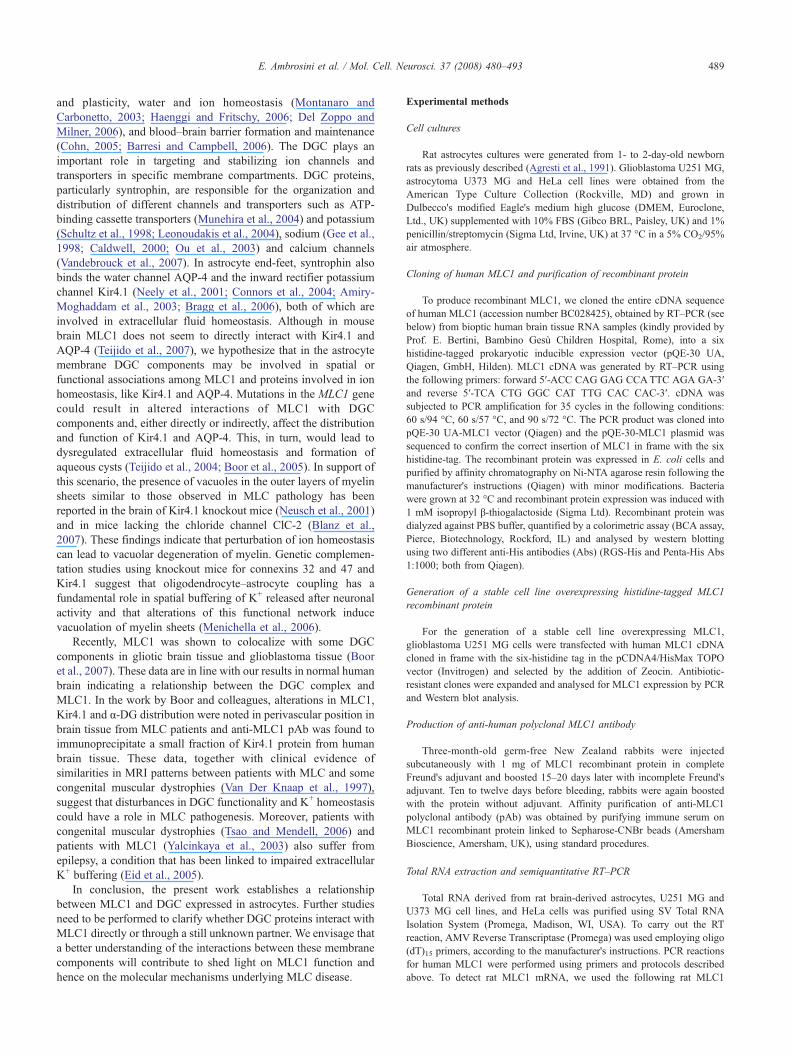
and plasticity, water and ion homeostasis (Montanaro andCarbonetto, 2003; Haenggi and Fritschy, 2006; Del Zoppo andMilner, 2006), and blood–brain barrier formation and maintenance(Cohn, 2005; Barresi and Campbell, 2006). The DGC plays animportant role in targeting and stabilizing ion channels andtransporters in specific membrane compartments. DGC proteins,particularly syntrophin, are responsible for the organization anddistribution of different channels and transporters such as ATP-binding cassette transporters (Munehira et al., 2004) and potassium(Schultz et al., 1998; Leonoudakis et al., 2004), sodium (Gee et al.,1998; Caldwell, 2000; Ou et al., 2003) and calcium channels(Vandebrouck et al., 2007). In astrocyte end-feet, syntrophin alsobinds the water channel AQP-4 and the inward rectifier potassiumchannel Kir4.1 (Neely et al., 2001; Connors et al., 2004; Amiry-Moghaddam et al., 2003; Bragg et al., 2006), both of which areinvolved in extracellular fluid homeostasis. Although in mousebrain MLC1 does not seem to directly interact with Kir4.1 andAQP-4 (Teijido et al., 2007), we hypothesize that in the astrocytemembrane DGC components may be involved in spatial orfunctional associations among MLC1 and proteins involved in ionhomeostasis, like Kir4.1 and AQP-4. Mutations in the MLC1 genecould result in altered interactions of MLC1 with DGCcomponents and, either directly or indirectly, affect the distributionand function of Kir4.1 and AQP-4. This, in turn, would lead todysregulated extracellular fluid homeostasis and formation ofaqueous cysts (Teijido et al., 2004; Boor et al., 2005). In support ofthis scenario, the presence of vacuoles in the outer layers of myelinsheets similar to those observed in MLC pathology has beenreported in the brain of Kir4.1 knockout mice (Neusch et al., 2001)and in mice lacking the chloride channel ClC-2 (Blanz et al.,2007). These findings indicate that perturbation of ion homeostasiscan lead to vacuolar degeneration of myelin. Genetic complemen-tation studies using knockout mice for connexins 32 and 47 andKir4.1 suggest that oligodendrocyte–astrocyte coupling has afundamental role in spatial buffering of K+ released after neuronalactivity and that alterations of this functional network inducevacuolation of myelin sheets (Menichella et al., 2006).
Recently, MLC1 was shown to colocalize with some DGCcomponents in gliotic brain tissue and glioblastoma tissue (Booret al., 2007). These data are in line with our results in normal humanbrain indicating a relationship between the DGC complex andMLC1. In the work by Boor and colleagues, alterations in MLC1,Kir4.1 and α-DG distribution were noted in perivascular position inbrain tissue from MLC patients and anti-MLC1 pAb was found toimmunoprecipitate a small fraction of Kir4.1 protein from humanbrain tissue. These data, together with clinical evidence ofsimilarities in MRI patterns between patients with MLC and somecongenital muscular dystrophies (Van Der Knaap et al., 1997),suggest that disturbances in DGC functionality and K+ homeostasiscould have a role in MLC pathogenesis. Moreover, patients withcongenital muscular dystrophies (Tsao and Mendell, 2006) andpatients with MLC1 (Yalcinkaya et al., 2003) also suffer fromepilepsy, a condition that has been linked to impaired extracellularK+ buffering (Eid et al., 2005).
In conclusion, the present work establishes a relationshipbetween MLC1 and DGC expressed in astrocytes. Further studiesneed to be performed to clarify whether DGC proteins interact withMLC1 directly or through a still unknown partner. We envisage thata better understanding of the interactions between these membranecomponents will contribute to shed light on MLC1 function andhence on the molecular mechanisms underlying MLC disease.
Experimental methods
Cell cultures
Rat astrocytes cultures were generated from 1- to 2-day-old newbornrats as previously described (Agresti et al., 1991). Glioblastoma U251 MG,astrocytoma U373 MG and HeLa cell lines were obtained from theAmerican Type Culture Collection (Rockville, MD) and grown inDulbecco's modified Eagle's medium high glucose (DMEM, Euroclone,Ltd., UK) supplemented with 10% FBS (Gibco BRL, Paisley, UK) and 1%penicillin/streptomycin (Sigma Ltd, Irvine, UK) at 37 °C in a 5% CO2/95%air atmosphere.
Cloning of human MLC1 and purification of recombinant protein
To produce recombinant MLC1, we cloned the entire cDNA sequenceof human MLC1 (accession number BC028425), obtained by RT–PCR (seebelow) from bioptic human brain tissue RNA samples (kindly provided byProf. E. Bertini, Bambino Gesù Children Hospital, Rome), into a sixhistidine-tagged prokaryotic inducible expression vector (pQE-30 UA,Qiagen, GmbH, Hilden). MLC1 cDNA was generated by RT–PCR usingthe following primers: forward 5′-ACC CAG GAG CCATTC AGA GA-3′and reverse 5′-TCA CTG GGC CAT TTG CAC CAC-3′. cDNA wassubjected to PCR amplification for 35 cycles in the following conditions:60 s/94 °C, 60 s/57 °C, and 90 s/72 °C. The PCR product was cloned intopQE-30 UA-MLC1 vector (Qiagen) and the pQE-30-MLC1 plasmid wassequenced to confirm the correct insertion of MLC1 in frame with the sixhistidine-tag. The recombinant protein was expressed in E. coli cells andpurified by affinity chromatography on Ni-NTA agarose resin following themanufacturer's instructions (Qiagen) with minor modifications. Bacteriawere grown at 32 °C and recombinant protein expression was induced with1 mM isopropyl β-thiogalactoside (Sigma Ltd). Recombinant protein wasdialyzed against PBS buffer, quantified by a colorimetric assay (BCA assay,Pierce, Biotechnology, Rockford, IL) and analysed by western blottingusing two different anti-His antibodies (Abs) (RGS-His and Penta-His Abs1:1000; both from Qiagen).
Generation of a stable cell line overexpressing histidine-tagged MLC1recombinant protein
For the generation of a stable cell line overexpressing MLC1,glioblastoma U251 MG cells were transfected with human MLC1 cDNAcloned in frame with the six-histidine tag in the pCDNA4/HisMax TOPOvector (Invitrogen) and selected by the addition of Zeocin. Antibiotic-resistant clones were expanded and analysed for MLC1 expression by PCRand Western blot analysis.
Production of anti-human polyclonal MLC1 antibody
Three-month-old germ-free New Zealand rabbits were injectedsubcutaneously with 1 mg of MLC1 recombinant protein in completeFreund's adjuvant and boosted 15–20 days later with incomplete Freund'sadjuvant. Ten to twelve days before bleeding, rabbits were again boostedwith the protein without adjuvant. Affinity purification of anti-MLC1polyclonal antibody (pAb) was obtained by purifying immune serum onMLC1 recombinant protein linked to Sepharose-CNBr beads (AmershamBioscience, Amersham, UK), using standard procedures.
Total RNA extraction and semiquantitative RT–PCR
Total RNA derived from rat brain-derived astrocytes, U251 MG andU373 MG cell lines, and HeLa cells was purified using SV Total RNAIsolation System (Promega, Madison, WI, USA). To carry out the RTreaction, AMV Reverse Transcriptase (Promega) was used employing oligo(dT)15 primers, according to the manufacturer's instructions. PCR reactionsfor human MLC1 were performed using primers and protocols describedabove. To detect rat MLC1 mRNA, we used the following rat MLC1

490 E. Ambrosini et al. / Mol. Cell. Neurosci. 37 (2008) 480–493
primers: forward 5′-ATG ACA CGG GAG GGG CAG TTC-3′; reverse 5′-TCA CTG GGC CAT TTG TAC CAC-3′. To normalize MLC1 mRNAlevels in the different cell cultures, RT–PCR reactions were also performedfor β-actin as housekeeping gene using the following primers: for human β-actin, forward 5′-AGC AAG AGA GGC ATC CTC AC-3′; reverse 5′-CACGGA GTA CTT GCG CTC AG-3′; for rat β-actin: forward 5′-TAA AGACCT CTATGC CAA CAC-3′; reverse 5′-CTC CTG CTT GCT GAT CCACAT-3′. PCR reactions were performed for 25 cycles in the followingconditions: 30 s/94 °C, 30 s/54 °C, and 40 s/72 °C.
Protein extract preparation
Protein extracts from rat brain tissue and cellular pellets were obtained aspreviously described (Teijido et al., 2004; Schindler et al., 2006), with minormodifications. Briefly, brains were dissected from 1- to 2-day-oldWistar rats,immediately frozen in liquid nitrogen and stored at −80 °C. The brains wereresuspended in buffer containing 250 mM sucrose, 5 mM EDTA, 5 mMEGTA, 10 mM Hepes (pH 7.4) and a protease inhibitor cocktail (Sigma Ltd)in a ratio of 4.5 ml buffer for 1.5 g of brain, and then homogenized on ice.Homogenates were incubated in ice for 20 min and then centrifuged at3000×g for 10 min at 4 °C, to pellet nuclei. Supernatants were centrifuged at10,000×g for 12 min at 4 °C to pellet mitochondria and then ultracentrifugedat 100,000×g for 1 h at 4 °C. Samples were then analyzed with SDS–PAGEand Western Blotting (WB). The same procedure was applied to extractproteins from cellular pellets obtained from approximately 4.0×10⁷ culturedastrocytes or HeLa cells. In some experiments, membrane proteins wereextracted from cultured cells with detergents using the following procedure:approximately 2.0×10⁷ cells were scraped from plates using buffer Acontaining 50mMTris–HCl pH 7.4, 1 mMEDTA, 1mMEGTA, 1mMDTT,protease inhibitor cocktail (Sigma Ltd); samples were centrifuged at 2000×gfor 10 min and pellets were resuspended in lysis buffer B containing 1%Triton X-100, 0.5% sodium deoxycholate, 150 mM NaCl, 20 mM Hepes(pH 7.4), 1% SDS and protease inhibitor cocktail. Lysates were passedthrough a 0.45-mM syringe needle, incubated in ice for 45 min andcentrifuged at 15,000×g for 30 min at 4 °C. The supernatants were analyzedby SDS–gel electrophoresis and WB.
Sequential extraction and solubilisation of membrane proteins
To improve solubilisation and to separate integral membrane proteinsfrom cytosolic and membrane-associated proteins, sequential extraction wasperformed as previously described (Herbert, 1999) with minor modifica-tions. The cellular pellet obtained from 4.0×10⁷ primary rat astrocytes wasresuspended in buffer 1 containing 40 mM Tris–HCl pH 7.4, 0.5% Tween20, 10 mM NaCl, and inhibitor protein cocktail, and incubated for 1 h at RTin rotation. After 30 min of centrifugation at 20,000×g at 4 °C, supernatant1 containing cytosolic protein was recovered and cellular pellet wasresuspended in buffer 2 containing 40 mM Tris–HCl pH 7.4, 0.5% TritonX-100, 10 mM NaCl and inhibitor protein cocktail and centrifuged asdescribed above. Supernatant 2 containing membrane-associated proteinswas recovered and to solubilise integral membrane proteins residual pelletwas resuspended in buffer 3 containing 40 mM Tris–HCl pH 7.4, 8 M urea,4% CHAPS, 50 mM DTT and inhibitor protein cocktail, incubated andcentrifuged as described above. Supernatant 3 obtained in the last step ishighly enriched in integral membrane proteins. All the supernatantsobtained were precipitated with a solution containing 50% ethanol, 25%methanol and 25% acetone over night at −20 °C. Pellets obtained aftercentrifugation were washed in the same solution, air-dried and resuspendedin Laemmli buffer.
Detergent-resistant microdomain (DMR/lipid rafts) preparation by sucrosegradients
DRMs from cultured astrocytes were prepared as previously described(Lisanti et al., 1993). Briefly, primary cultures of rat astrocytes and thehuman glioma cell line (U251 MG) were grown to confluence in 150-mmdishes, harvested in PBS and lysed on ice with 0.75 ml of Mes-buffered
saline (25 mM Mes pH 6.5, 0.15 M NaCl) containing 1% (v/v) Triton X-100 and protease inhibitors. Cell lysate was homogenized with 10 strokes ofa Dounce homogenizer, adjusted to 40% sucrose and placed at the bottomof an ultracentrifuge tube. A 5–30% linear sucrose gradient was placedabove the homogenate and the mixture was centrifuged at 260,000×g for16 h at 4 °C in a SW 61 rotor (Beckman Instruments). Twelve 0.375-mlfractions were harvested from the top of the gradient. The DMR fractionsare visible as a light-scattering band migrating at approximately 20%sucrose (fractions 3, 4 and 5). Samples were precipitated over night withacetone (1:4 v/v) and proteins analysed by SDS–PAGE and WB.
Dystrophin–glycoprotein complex preparation
DGC enrichment was performed as described (Durbeej and Campbell,1999; Beedle et al., 2007). Briefly, rat brain (5 g) was solubilised in 1%digitonin, 50 mM Tris–HCl pH 7.4, 0.5 M NaCl and protease inhibitorsand centrifuged at 38,000×g for 30 min at 4 °C. Supernatant was enrichedin DGC with succinyl-WGA-agarose (Vector Laboratories, Burlingame,CA) and eluted with 0.3 M N-acetylglucosamine, 50 mM Tris–HCl pH 7.4,0.1% digitonin, 0.5 M NaCl and protease inhibitors. Pooled fractionswere diluted 10-fold with 50 mM Tris–HCl (pH 7.4), 0.1% digitonin andprotease inhibitors and applied to DEAE resin (Whatman). After ex-tensive washes, elution of bound proteins were carried out with 4 ml of50 mM Tris–HCl, pH 7.4, 0.1% digitonin containing 100, 150, 500 and750 mM of NaCl, respectively. Aliquots (0.5 ml) of DEAE-eluted fractionswere precipitated with acetone (1:4 v/v) and analysed by SDS–PAGE andWB.
Electrophoresis and Western blotting
Protein samples were separated by 12% SDS–polyacrylamide gel or ingradient (4–12%) precasted gels (Invitrogen) and gels were transferred topolyvinylidene difluoride (PVDF) membranes or nitrocellulose (AmershamBiosciences, Europe, GmbH) for 30 min in a semi-dry apparatus. After 1 hof incubation with blocking buffer, membranes were incubated over night at4 °C with primary anti-MLC1 pAb, anti-Penta-His (1:1000; Qiagen), anti-RGS-His (1:1000; Qiagen), anti-caveolin-1(1:1000; Upstate Laboratory),anti-syntrophin (1:2000; MA-1-745, Affinity Bioreagents, Co., USA), antiβ-DG (1:25; NCL-43 DAG, Novocastra Lab, Ltd., UK) mAbs, or an inhouse generated mouse anti α-DG serum (dilution 1:250) (Pavoni E, BarcaS, Brancaccio A and Petrucci TC, manuscript in preparation), in PBS–3%BSA followed by extensive washings and then incubated for 1 h withhorseradish peroxidase-conjugated anti-mouse or anti-rabbit Abs (1:500;Pierce), for 1 h at RT. Immunoreactive bands were visualized using anenhanced chemiluminescence reagent (Pierce), according to the manufac-turer's instructions, and were exposed on X-ray films.
Immunofluorescence
Cells were grown on polylysinated coverslips, fixed for 10 min with 4%paraformaldehyde and extensively washed with PBS. After 1 h ofincubation with blocking solution (5% BSA in PBS), cells were incubated1 h at RT with the following primary Abs diluted in PBS: affinity-purifiedanti-MLC1 rabbit pAb (1:40); α-DG rabbit pAb, recognizing the COOH-terminal globular region of α-DG (1:100) (Zaccaria et al., 2001). For doubleimmunostaining, anti-α-DG mAb (1:30) (clone VIA4-1, Upstate) or anti-EEA1 mAb (1:30; BD Biosciences) was used in combination with anti-MLC1 pAb. For intracellular stainings with anti-EEA1 mAb, after fixationcells were permeabilized with 0.025% Triton X-110 in PBS for 5 min. Cellswere then incubated for 45 min at RT with the secondary antibodies ata concentration of 4.3 μg/ml (Biotin-SP-AffiniPure Goat Anti-Rabbit IgGH+L, goat anti-mouse FITC-conjugated, all from Jackson ImmunoresearchLaboratories, Cambridgeshire, UK) in PBS+2% normal goat serum,followed by a 30-min incubation at RT with 2 μg/ml streptavidin–TRITC inPBS (Jackson, UK). Coverslips were washed, sealed in Vectashield medium(Vector Laboratories) and analyzed with a laser scanning confocalmicroscope (LSM 5 Pascal, Carl Zeiss, Jena, Germany).

491E. Ambrosini et al. / Mol. Cell. Neurosci. 37 (2008) 480–493
Immunohistochemistry
Indirect immunofluorescence techniques were used to detect MLC1,glial fibrillary astrocytic protein (GFAP), β-DG and syntrophin in autopsybrain tissue from a patient who died for cardiac failure without evidence ofneurological disease or neuropathological alterations (obtained from UCSCPoliclinico A. Gemelli, Rome). Brain tissue was fixed in 4% paraformal-dehyde in PBS, cryoprotected in 30% sucrose for 1 week, frozen in dry ice-cooled isopentane and stored at −75 °C. Air-dried, acetone-fixed (4 °C), 10-μm-thick cryosections were rehydrated with PBS and post-fixed sectionswere subjected to the antigen retrieval procedure with microwave in citratebuffer 10 mM (pH 6.0). For single stainings, sections were incubated for 1 hwith 10% of normal goat serum (NGS) containing 0.05% Triton X-100(Jackson, UK), and then over night at 4 °C with affinity purified anti-MLC1pAb (1:50). After washing in PBS, sections were incubated for 1 h at roomtemperature with TRITC-conjugated goat anti-rabbit IgG (Jackson, UK)diluted in PBS+5% NGS and then washed and sealed in Vectashieldmedium (Vector Laboratories). For double immunofluorescence stainings,sections were incubated overnight at 4 °Cwith a mixture of rabbit anti-MLC1pAb and one of the followingmAbs: anti-GFAP (1:300; Biogenex Lab), anti-CD31 (1:20; Dako), anti-EEA1 (1:20; BD Biosciences), anti-NF200 (1:50;Boeringer), anti-β-DG (1:100; NCL-43 DAG, Novocastra Lab, Ltd., UK)and anti-syntrophin (1:50; Affinity BioReagents, Co, USA) in PBS, 2%BSAand 0.05% Triton X-100. After washing, sections were incubated for 1 h atRTwith a mixture of fluorescein-conjugated goat anti-mouse and rhodamine-conjugated goat anti-rabbit Abs diluted in PBS containing 10%NGS. Imageswere analysed with a laser scanning confocal microscope, as above.
For double immunohistochemical staining for MLC1 and MAP2, post-fixedsections were subjected to the antigen retrieval procedure and incubated for20 min with 0.1% H2O2 in PBS to eliminate endogenous peroxidase activity,for 1 h with 10% normal donkey serum, and overnight at 4 °C with anti-MLC1pAb diluted in PBS+0.2% Triton X-100. The binding of biotinylated secondaryAb (donkey anti-rabbit, Jackson) was visualized with avidin–biotin horseradishperoxidase complex technique (ABC Vectastain Elite Vector Laboratories,Burlingame, CA) and 3,3′-diaminobenzidine containing 0.01% H2O2 assubstrate. Sections were then treated with 2% Triton X-100 in PBSfor 15 min at RT and incubated overnight with anti-MAP2 mAb (1:50;Chemicon), 1 h with the biotinylated secondary antibody donkey anti-mouseand 1 h with the Standard ABC-Alkaline Phosphatase kit. Immunoreactivitywas visualized with Fast Red (SIGMA). All sections were analyzed with a ZeissAxiophot microscope. Negative controls included the use of IgG isotype controland preimmune serum.
Acknowledgments
We thank Dr. Egidio Stigliano (UCSC, Policlinico A. Gemelli,Rome, Italy) for providing the human brain tissue used forimmunohistochemical experiments and Dr. Antonietta Bernardo forproviding primary cultures of rat astrocytes. We thank Drs. MassimoSargiacomo, Gianfranco Macchia and Federica Fratini for technicaladvice about subcellular fractionations. This study was supported bygrants of the ItalianMinistry of Health and ELA Foundation (France).CRwas a recipient of a PhD fellowship fromFIRB ItalianMinistry forUniversity and Research, grant year 2003(Grant RBNE03FMCJ).
Appendix A. Supplementary data
Supplementary data associated with this article can be found, inthe online version, at doi:10.1016/j.mcn.2007.11.003.
References
Agresti, C., Aloisi, F., Levi, G., 1991. Heterotypic and homotypic cellularinteractions influencing the growth and differentiation of bipotentialoligodendrocyte-type-2 astrocyte progenitors in culture. Dev. Biol. 144,16–29.
Amiry-Moghaddam, M., Otsuka, T., Hurn, P.D., Traystman, R.J., Haug,F.M., Froehner, S.G., Adams, M.E., Neely, J.D., Agre, P., Ottersen,O.P., Bhardway, A., 2003. An alpha-syntrophin-dependent pool ofAQP4 in astroglial end-feet confers bidirectional water flow betweenblood and brain. PNAS 18, 2106–2111.
Barresi, R., Campbell, K.P., 2006. Dystroglycan: from biosynthesis topathogenesis of human disease. J. Cell Sci. 15, 199–207.
Beedle, A.M., Nienaber, P.M., Campbell, K.P., 2007. Fukutin-related proteinassociates with the sarcolemmal dystrophin–glycoprotein complex.J. Biol. Chem. 282, 16713–16717.
Ben-Zeev, B., Levy-Nissenbaum, E., Lahat, H., Anikster, Y., Shinar, Y.,Brand, N., Gross-Tzur, V., MacGregor, D., Sidi, R., Kleta, R.,Frydman, M., Pras, E., 2002. Megalencephalic leukoencephalopathywith subcortical cysts; a founder effect in Israeli patients and a higherthan expected carrier rate among Libyan Jews. Hum. Genet. 111,214–218.
Blanz, J., Schweizer, M., Auberson, M., Maier, H., Muenscher, A., Hubner,C.A., Jentsch, T.J., 2007. Leukoencephalopathy upon disruption of thechloride channel ClC-2. J. Neurosci. 13, 6581–6589.
Blattner, R., Von Moers, A., Leegwater, P.A., Hanefeld, F.A., Van DerKnaap, M.S., Kohler, W., 2003. Clinical and genetic heterogeneity inmegalencephalic leukoencephalopathy with subcortical cysts (MLC).Neuropediatrics 34, 215–218.
Boor, P.K.I., DeGroot, K., Waisfisz, Q., Kamphrost, W., Oudejans, C.B.M.,Powers, J.M., Pronk, J.C., Shepert, G.C., Van Der Knaap, M.S., 2005.MLC1 a novel protein in distal astroglial processes. J. Neuropathol. Exp.Neurol. 64, 412–419.
Boor, P.K.I., DeGroot, K., Mejaski-Bosnjak, V., Brenner, C., Van der Knaap,M.S., Scheper, G.C., Pronk, J.C., 2006. Megalencephalic leukoence-phalopathy with subcortical cysts: an update and extended mutationanalysis of MLC1. Hum. Mutat. 27, 505–512.
Boor, I., Nagtegaal, M., Kamphorst, W., van der Valk, P., Pronk, J.C., vanHorssen, J., Dinopoulos, A., Bove, K.E., Pascual-Castroviejo, I.,Muntoni, F., Estevez, R., Scheper, G.C., van der Knaap, M.S., 2007.MLC1 is associated with the dystrophin–glycoprotein complex atastrocytic endfeet. Acta Neuropathol. (Berl.) 114, 403–410.
Bragg, A.D., Amiry Moghaddam, M., Ottersen, O.P., Adams, M.E.,Frohener, S.C., 2006. Assembly of a perivascular astrocyte proteinscaffold at the mammalian blood–brain barrier is dependent on alpha-syntrophin. Glia 53, 879–890.
Brenner, M., Johnson, A.B., Boespflug-Tanguy, O., Rodriguez, D., Gold-man, J.E., Messing, A., 2001. Mutations in GFAP, encoding glialfibrillary acidic protein, are associated with Alexander disease. Nat.Genet. 27, 117–120.
Brown, D.A., 2006. Lipid rafts, detergent-resistant membranes, and rafttargeting signals. Physiology 21, 430–439.
Bruni, J.E., 1998. Ependymal development, proliferation, and functions: areview. Microsc. Res. Tech. 4, 12–13.
Caldwell, J.H., 2000. Clustering of sodium channels at the neuromuscularjunction. Microsc. Res. Tech. 49, 84–89.
Cameron, P.L., Ruffin, J.W., Bollag, R., Rasmussen, H., Cameron, R.S.,1997. Identification of caveolin and caveolin-related proteins in thebrain. J. Neurosci. 17, 9520–9535.
Cameron, P.L., Liu, C., Smart, D.K., Hantus, S.T., Fick, J.R., Cameron, R.S.,2002. Caveolin-1 expression is maintained in rat and human astrogliomacell lines. Glia 37, 275–290.
Cavaldesi, M., Macchia, G., Barca, S., Defilippi, P., Tarone, G., Petrucci,T.C., 1999. Association of the dystroglycan complex isolated frombovine brain synaptosomes with proteins involved in signal transduc-tion. J. Neurochem. 72, 1648–1655.
Cohn, R.D., 2005. Dystroglycan: important player in skeletal muscle andbeyond. Neuromuscul. Disord. 15, 207–217.
Cohn, R.D., Campbell, K.P., 2000. Molecular basis of muscular dystrophies.Muscle Nerve 23, 1456–1471.
Connors, N.C., Adams, M.E., Froehner, S.C., Kofuji, P., 2004. Thepotassium channel Kir4.1 associates with the dystrophin–glycoproteincomplex via alpha-syntrophin in glia. J. Biol. Chem. 279, 28387–28392.

492 E. Ambrosini et al. / Mol. Cell. Neurosci. 37 (2008) 480–493
Del Bigio, M.R., 1998. The ependyma: a protective barrier between brainand cerebrospinal fluid. Glia 14, 1–13.
Del Zoppo, G.J., Milner, R., 2006. Integrin–matrix interactions in the cere-bral microvasculature. Arterioscler. Thromb. Vasc. Biol. 26, 1966–1975.
Doyle, D.D., Goings, G., Upshaw-Earley, J., Ambler, S.K., Mondul, A.,Palfrey, H.C., Page, E., 2000. Dystrophin associates with caveolae ofrat cardiac myocytes: relationship to dystroglycan. Circ. Res. 87,480–488.
Durbeej, M., Campbell, K.P., 1999. Biochemical characterization of theepithelial dystroglycan complex. J. Biol. Chem. 274, 26609–26616.
Eid, T., Lee, T.S., Thomas, M.J., Amiry-Moghaddam, M., Bjornsen, L.P.,Spencer, D.D., Agre, P., Ottersen, O.P., De Lanerolle, N.C., 2005. Lossof perivascular aquaporin 4 may underlie deficient water and K+
homeostasis in the human epileptogenic hippocampus. Proc. Natl. Acad.Sci. U. S. A. 102 (4), 1193–1198.
Ervasti, J.M., Campbell, K.P., 1993. A role for the dystrophin–glycoproteincomplex as a transmembrane linker between laminin and actin. J. CellBiol. 122, 809–823.
Gee, S.H., Madhavan, R., Levinson, S.R., Caldwell, J.H., Sealock, R.,Froehner, S.C., 1998. Interaction of muscle and brain sodium channelswith multiple members of the syntrophin family of dystrophin-associated proteins. J. Neurosci. 18, 128–137.
Gorospe, J.R., Maletkovic, J., 2006. Alexander disease and megalencephalicleukoencephalopathy with subcortical cysts: leukodistrophies arisingfrom astrocytes dysfunction. Ment. Retard. Dev. Disabil. Res. Rev. 12,13–22.
Gorospe, J.R., Singhal, B.S., Kainu, T., Wu, F., Stephan, D., Trent, J.,Hoffman, E.P., Naidu, S., 2004. Indian Agarwal megalencephalicleukodystrophy with cysts is caused by a common MLC1 mutation.Neurology 23, 878–882.
Haenggi, T., Fritschy, J.M., 2006. Role of dystrophin and utrophin forassembly and function of the dystrophin glycoprotein complex in non-muscle tissue. Cell. Mol. Life Sci. 63, 1614–1631.
Halayko, A.J., Stelmack, G.L., 2005. The association of caveolae, actin, andthe dystrophin–glycoprotein complex: a role in smooth musclephenotype and function? Can. J. Physiol. Pharm. 83, 877–891.
Herbert, B., 1999. Advances in protein solubilisation for two-dimensionalelectrophoresis. Electrophoresis 20, 660–663.
Higginson, J.R., Winder, S.J., 2005. Dystroglycan: a multifunctional adaptorprotein. Biochem. Soc. Trans. 33, 1254–1255.
Ikezu, T., Ueda, H., Trapp, B.D., Nishiyama, K., Sha, J.F., Volonte, D.,Galbiati, F., Byrd, A.L., Bassell, G., Serizawa, H., Lane, W.S., Lisanti,M.P., Okamoto, T., 1998. Affinity-purification and characterizationof caveolins from the brain: differential expression of caveolin-1, -2,and -3 in brain endothelial and astroglial cell types. Brain Res. 7,177–192.
Kaganovich, M., Peretz, A., Ritsner, M., Bening Abu-Shach, U., Attali, B.,Navon, R., 2004. Is theWKL1 gene associated with schizophrenia? Am.J. Med. Genet. 125B, 31–37.
Leegwater, P.A., Yuan, B.Q., Van Der Steen, J., Mulders, J., Konst, A.A.,Boor, P.K., Mejaski-Bosnjak, V., Van Der Maarel, S.M., Frants, R.R.,Oudejans, C.B., Schutgens, R.B., Pronk, J.C., Van Der Knaap, M.S.,2001. Mutations of MLC1 (KIAA0027), encoding a putative membraneprotein, cause megalencephalic leukoencephalopathy with subcorticalcysts. Am. J. Hum. Genet. 68, 831–838.
Leegwater, P.A., Boor, P.K., Yuan, B.Q., Van Der Steen, J., Visser, A.,Konst, A.A., Oudejans, C.B., Schutgens, R.B., Pronk, J.C., Van DerKnaap, M.S., 2002. Identification of novel mutations in MLC1responsible for megalencephalic leukoencephalopathy with subcorticalcysts. Hum. Genet. 110, 279–283.
Leonoudakis, D., Conti, L.R., Anderson, S., Radeke, C.M., McGuire, L.M.,Adams, M.E., Froehner, S.C., Yates, J.R., Vandenberg, C.A., 2004.Protein trafficking and anchoring complexes revealed by proteomicanalysis of inward rectifier potassium channel (Kir2.x)-associatedproteins. J. Biol. Chem. 279, 22331–22346.
Lisanti, M.P., Tang, Z.L., Sargiacomo, M., 1993. Caveolin forms a hetero-oligomeric protein complex that interacts with an apical GPI-linked
protein: implications for the biogenesis of caveolae. J. Cell Biol. 123,595–604.
Martens, J.R., O'Connell, K., Tamkun, M., 2004. Targeting of ion channelsto membrane microdomains: localization of KV channels to lipid rafts.Trends Pharmacol. Sci. 25, 16–21.
Menichella, D.M., Majdan, M., Awatramani, R., Goodenough, D.A.,Sirkowski, E., Scherer, S.S., Paul, D.L., 2006. Genetic and physiologicalevidence that oligodendrocyte gap junctions contribute to spatialbuffering of potassium released during neuronal activity. J. Neurosci.25, 10984–10991.
Meyer, J., Huberth, A., Ortega, G., Syagailo, Y.V., Jatzke, S., Mossner, R.,Strom, T.M., Ulzheimer-Teuber, I., Stober, G., Schmitt, A., Lesch, K.P.,2001. A missense mutation in a novel gene encoding a putative cationchannel is associated with catatonic schizophrenia in a large pedigree.Mol. Psychiatry 6, 302–306.
Mignot, C., Boespflug-Tanguy, O., Gelot, A., Dautigny, A., Pham-Dinh, D.,Rodriguez, D., 2004. Alexander disease: putative mechanisms of anastrocytic encephalopathy. Cell. Mol. Life Sci. 61, 369–385.
Montagna, G., Teijido, O., Eymard-Pierre, E., Muraki, K., Cohen, B.,Loizzo, A., Grosso, P., Tedeschi, G., Palacin, M., Boespflug-Tanguy, O.,Bertini, E., Santorelli, F.M., Estevez, R., 2006. Vacuolating mega-lencephalic leukoencephalopathy with subcortical cysts: functionalstudies of novel variants in MLC1. Hum. Mutat. 27, 292.
Montanaro, F., Carbonetto, S., 2003. Targeting dystroglycan in the brain.Neuron 37, 193–196.
Moukhles, H., Carbonetto, S., 2001. Dystroglycan contributes to theformation of multiple dystrophin-like complexes in brain. J. Neurochem.78, 824–834.
Munehira, Y., Ohnishi, T., Kawamoto, S., Furuya, A., Shitara, K., Imamura,M., Yokota, T., Takeda, S., Amachi, T., Matsuo, M., Kioka, N., Ueda, K.,2004. Alpha1-syntrophin modulates turnover of ABCA1. J. Biol. Chem.279, 15091–15095.
Neely, J.D., Amiry-Moghaddam, M., Ottersen, O.P., Froehner, S.C., Agre,P., Adams, M.E., 2001. Syntrophin-dependent expression and localiza-tion of aquaporin-4 water channel protein. Proc. Natl. Acad. Sci. U. S. A.98, 14108–14113.
Neusch, C., Rozengurt, N., Jacobs, R.E., Lester, H.A., Kofuji, P., 2001.Kir4.1 potassium channel subunit is crucial for oligodendrocytedevelopment and in vivo myelination. J. Neurosci. 1, 5429–5438.
Nomura, N., Miyajima, N., Sazuka, T., Tanaka, A., Kawarabayasi, Y., Sato,S., Nagase, T., Seki, N., Ishikawa, K., Tabata, S., 1994. Prediction of thecoding sequences of unidentified human genes: I. The coding sequencesof 40 new genes (KIAA0001–KIAA0040) deduced by analysis ofrandomly sampled cDNA clones from human immature myeloid cellline KG-1 (supplement). DNA Res. 1, 47–56.
Ou, Y., Strege, P., Miller, S.M., Makielski, J., Ackerman, M., Gibbons,S.J., Farrugia, G., 2003. Syntrophin gamma 2 regulates SCN5Agating by a PDZ domain-mediated interaction. J. Biol. Chem. 278,1915–1923.
Pascual-Castroviejo, I., Van der Knaap, M.S., Pronk, J.C., Garcia-Segura,J.M., Gutierez-Molina, M., Pascual-Pascual, S.I., 2005. Vacuolatingmegalencephalic leukoencephalopathy: 24 year follow-up of two sib-lings. Neurologia 20, 33–40.
Patrono, C., Di Giacinto, G., Eymard-Pierre, E., Santorelli, F.M., Rodriguez,D., De Stefano, N., Federico, A., Gatti, R., Benigno, V., Megarbane, A.,Tabarri, B., Boespflug-Tanguy, O., Bertini, E., 2003. Genetic hetero-geneity of megalencephalic leukoencephalopathy and subcortical cysts.Neurology 61, 534–537.
Saijo, H., Nakayama, H., Ezoe, T., Araki, K., Sone, S., Hamaguchi, H.,Suzuki, H., Shiroma, N., Kanazawa, N., Tsujino, S., Hirayama, Y.,Arima, M., 2003. A case of megalencephalic leukoencephalopathy withsubcortical cysts (van der Knaap disease): molecular genetic study. BrainDevelop. 25, 362–366.
Schindler, J., Lewandrowski, U., Sickmann, A., Friauf, E., Nothwang, H.G.,2006. Proteomic analysis of brain plasma membranes isolated by affinitytwo-phase partitioning. Mol. Cell. Proteomics 5, 390–400.
Schmitt, A., Gofferje, V., Weber, M., Meyer, J., Mossner, R., Lesch, K.P.,

493E. Ambrosini et al. / Mol. Cell. Neurosci. 37 (2008) 480–493
2003. The brain-specific protein MLC1 implicated in megalencephalicleukoencephalopathy with subcortical cysts is expressed in glial cells inthe murine brain. Glia 44, 283–295.
Schultz, J., Hoffmuller, U., Krause, G., Ashurst, J., Macias, M.J., Schmieder,P., Schneider-Mergener, J., Oschkinat, H., 1998. Specific interactionsbetween the syntrophin PDZ domain and voltage-gated sodiumchannels. Nat. Struct. Biol. 5, 19–24.
Simons, K., Ikonen, E., 1997. Functional rafts in cell membranes. Nature387, 569–572.
Simons, K., Toomre, D., 2000. Lipid rafts and signal transduction. Nat. Rev.,Mol. Cell Biol. 1, 31–39.
Song, K.S., Scherer, P.E., Tang, Z., Okamoto, T., Li, S., Chafel, M., Chu, C.,Kohtz, D.S., Lisanti, M.P., 1996. Expression of caveolin-3 in skeletal,cardiac, and smooth muscle cells. Caveolin-3 is a component of thesarcolemma and co-fractionates with dystrophin and dystrophin-associated glycoproteins. J. Biol. Chem. 271, 15160–15165.
Sotgia, F., Lee, J.K., Das, K., Bedford, M., Petrucci, T.C., Macioce, P.,Sargiacomo, M., Bricarelli, F.D., Minetti, C., Sudol, M., Lisanti, M.P.,2000. Caveolin-3 directly interacts with the C-terminal tail of beta-dystroglycan. Identification of a central WW-like domain withincaveolin family members. J. Biol. Chem. 275, 38048–38058.
Teijido, O., Martinez, A., Pusch, M., Zorzano, A., Soriano, E., Del Rio, J.A.,Palacin, M., Estevez, R., 2004. Localization and functional analyses ofthe MLC1 protein involved in megalencephalic leukoencephalopathywith subcortical cysts. Hum. Mol. Genet. 13, 2581–2594.
Teijido, O., Casaroli-Marano, R., Kharkovets, T., Aguado, F., Zorzano, A.,Palacín, M., Soriano, E., Martínez, A., Estévez, R., 2007. Expressionpatterns of MLC1 protein in the central and peripheral nervous systems.Neurobiol. Dis. 26 (3), 532–545.
Topku, M., Gartioux, C., Ribierre, F., Yalçinkaya, C., Tokus, E., Oztekin, N.,Beckmann, J.S., Ozguc, M., Seboun, E., 2000. Vacuolating mega-
lencephalic leukoencephalopathy with subcortical cysts mapped tochromosome 22qtel. Am. J. Hum. Genet. 66, 733–739.
Tsao, C.Y., Mendell, J.R., 2006. Coexisting muscular dystrophies andepilepsy in children. J. Child. Neurol. 21, 148–150.
Van der Knaap, M.S., Barth, P.G., Stroink, H., Van Nieuwenhuizen, O., Arts,W.F., Hoogenraad, F., Valk, J., 1995. Leukoncephalopathy with swellingand a discrepantly mild clinical course in eight children. Ann. Neurol. 7,324–334.
Van derKnaap,M.S., Barth, P.G., Vrensen, G.F., Valk, J., 1996. Histopathologyof an infantile-onset spongiform leukoencephalopathy with a discrepantlymild clinical course. Acta Neuropathol. (Berl.) 92, 206–212.
Van Der Knaap, M.S., Smit, L.M., Bart, P.G., 1997. Magnetic resonanceimaging in classification of congenital muscular dystrophies with brainabnormalities. Ann. Neurol. 42, 50–59.
Vandebrouck, A., Sabourin, J., Rivet, J., Balghi, H., Sebille, S., Kitzis, A.,Raymond, G., Cognard, C., Bourmeyster, N., Constantin, B., 2007.Regulation of capacitative calcium entries by {alpha}1-syntrophin: associa-tion of TRPC1 with dystrophin complex and the PDZ domain of {alpha}1-syntrophin. FASEB J. 21, 608–617.
Weerth, S.H., Holtzclaw, L.A., Russell, J.T., 2007. Signaling proteins in raft-like microdomains are essential for Ca2+ wave propagation in glial cells.Cell Calcium 41, 155–167.
Yalcinkaya, C., Yuksel, A., Comu, S., Kilic, G., Cokar, O., Dervent, A.,2003. Epilepsy in vacuolating megalencephalic leukoencephalopathywith subcortical cysts. Seizure 12, 388–396.
Zaccaria, M.L., Di Tommaso, F., Brancaccio, A., Paggi, P., Petrucci, T.C.,2001. Dystroglycan distribution in adult mouse brain: a light andelectron microscopy study. Neuroscience 104, 311–324.
Zhan, Y., Tremblay, M.R., Melian, N., Carbonetto, S., 2005. Evidence thatdystroglycan is associated with dynamin and regulates endocytosis.J. Biol. Chem. 280, 18015–18024.