DOT1L regulates dystrophin expression and is critical for cardiac function
12
DOT1L regulates dystrophin expression and is critical for cardiac function Anh T. Nguyen, 1,2 Bin Xiao, 3 Ronald L. Neppl, 4 Eric M. Kallin, 1,2,6 Juan Li, 3 Taiping Chen, 5 Da-Zhi Wang, 4 Xiao Xiao, 3 and Yi Zhang 1,2,7 1 Howard Hughes Medical Institute, University of North Carolina at Chapel Hill, North Carolina 27599, USA; 2 Department of Biochemistry and Biophysics, Lineberger Comprehensive Cancer Center, University of North Carolina at Chapel Hill, North Carolina 27599, USA; 3 Division of Molecular Pharmaceutics, University of North Carolina at Chapel Hill, North Carolina 27599, USA; 4 Department of Cardiology, Children’s Hospital Boston, Harvard Medical School, Boston, Massachusetts 02115, USA; 5 Novartis Institutes for Biomedical Research, Cambridge, Massachusetts 02139, USA Histone methylation plays an important role in regulating gene expression. One such methylation occurs at Lys 79 of histone H3 (H3K79) and is catalyzed by the yeast DOT1 (disruptor of telomeric silencing) and its mammalian homolog, DOT1L. Previous studies have demonstrated that germline disruption of Dot1L in mice resulted in embryonic lethality. Here we report that cardiac-specific knockout of Dot1L results in increased mortality rate with chamber dilation, increased cardiomyocyte cell death, systolic dysfunction, and conduction abnormalities. These phenotypes mimic those exhibited in patients with dilated cardiomyopathy (DCM). Mechanistic studies reveal that DOT1L performs its function in cardiomyocytes through regulating Dystrophin (Dmd) transcription and, consequently, stability of the Dystrophin–glycoprotein complex important for cardiomyocyte viability. Importantly, expression of a miniDmd can largely rescue the DCM phenotypes, indicating that Dmd is a major target mediating DOT1L function in cardiomyocytes. Interestingly, analysis of available gene expression data sets indicates that DOT1L is down-regulated in idiopathic DCM patient samples compared with normal controls. Therefore, our study not only establishes a critical role for DOT1L-mediated H3K79 methylation in cardiomyo- cyte function, but also reveals the mechanism underlying the role of DOT1L in DCM. In addition, our study may open new avenues for the diagnosis and treatment of human heart disease. [Keywords: DOT1L; H3K79 methylation; cardiomyopathy; dystrophin] Supplemental material is available for this article. Received April 19, 2010; revised version accepted December 23, 2010. Chromatin is subject to reversible post-translational modifications that may alter chromatin structure and function directly, or indirectly through the recruitment of effector proteins at heterochromatic (silenced) and eu- chromatic (active) DNA. Histone methylation plays an important role in regulating transcription at target loci and is important for X inactivation, cell fate mainte- nance, and terminal differentiation (Peterson and Laniel 2004; Martin and Zhang 2005; Kouzarides 2007). One particular histone methylation event occurs at Lys 79 within the globular domain of histone H3 (H3K79) and is catalyzed by yeast DOT1 (disruptor of telomeric silenc- ing) and its mammalian homolog, DOT1L (Feng et al. 2002; Lacoste et al. 2002; Ng et al. 2002a; van Leeuwen et al. 2002). Although DOT1 was originally identified as a regulator of telomeric silencing (Singer et al. 1998), more recent studies suggest that DOT1-mediated H3K79 methylation is linked to euchromatic gene transcrip- tion (Schubeler et al. 2004; Barski et al. 2007; Steger et al. 2008). In yeast, DOT1 activity is positively regulated during transcription elongation through Rad6-Bre1 monoubiqui- tination of H2B (Ng et al. 2002b; Krogan et al. 2003; Wood et al. 2003). Additionally, DOT1 has been linked to the meiotic pachytene checkpoint control (San-Segundo and Roeder 2000) and DNA damage repair (Giannattasio et al. 2005; Wysocki et al. 2005; Conde et al. 2009). However, the biological function of mammalian DOT1L, particu- larly in the context of the animal, is less characterized. A recent study indicates that DOT1L exists in a large pro- tein complex and regulates the expression of Wingless target genes (Mohan et al. 2010). We and others demon- strated previously that mistargeting of DOT1L and sub- sequent H3K79 hypermethylation play an important role in leukemic transformation (Okada et al. 2005, 2006; Mueller et al. 2007; Krivtsov et al. 2008). Most recently, DOT1L has been shown to regulate the erythroid and 6 Present address: Department of Differentiation and Cancer, Centre de Regulacio Genomica, Barcelona 08003, Spain. 7 Corresponding author. E-MAIL [email protected]; FAX (919) 966-4330. Article is online at http://www.genesdev.org/cgi/doi/10.1101/gad.2018511. GENES & DEVELOPMENT 25:263–274 Ó 2011 by Cold Spring Harbor Laboratory Press ISSN 0890-9369/11; www.genesdev.org 263
-
Upload
independent -
Category
Documents
-
view
2 -
download
0
Transcript of DOT1L regulates dystrophin expression and is critical for cardiac function
DOT1L regulates dystrophin expressionand is critical for cardiac function
Anh T. Nguyen,1,2 Bin Xiao,3 Ronald L. Neppl,4 Eric M. Kallin,1,2,6 Juan Li,3 Taiping Chen,5
Da-Zhi Wang,4 Xiao Xiao,3 and Yi Zhang1,2,7
1Howard Hughes Medical Institute, University of North Carolina at Chapel Hill, North Carolina 27599, USA; 2Departmentof Biochemistry and Biophysics, Lineberger Comprehensive Cancer Center, University of North Carolina at Chapel Hill, NorthCarolina 27599, USA; 3Division of Molecular Pharmaceutics, University of North Carolina at Chapel Hill, North Carolina27599, USA; 4Department of Cardiology, Children’s Hospital Boston, Harvard Medical School, Boston, Massachusetts 02115,USA; 5Novartis Institutes for Biomedical Research, Cambridge, Massachusetts 02139, USA
Histone methylation plays an important role in regulating gene expression. One such methylation occurs at Lys79 of histone H3 (H3K79) and is catalyzed by the yeast DOT1 (disruptor of telomeric silencing) and its mammalianhomolog, DOT1L. Previous studies have demonstrated that germline disruption of Dot1L in mice resultedin embryonic lethality. Here we report that cardiac-specific knockout of Dot1L results in increased mortality ratewith chamber dilation, increased cardiomyocyte cell death, systolic dysfunction, and conduction abnormalities.These phenotypes mimic those exhibited in patients with dilated cardiomyopathy (DCM). Mechanistic studiesreveal that DOT1L performs its function in cardiomyocytes through regulating Dystrophin (Dmd) transcriptionand, consequently, stability of the Dystrophin–glycoprotein complex important for cardiomyocyte viability.Importantly, expression of a miniDmd can largely rescue the DCM phenotypes, indicating that Dmd is a majortarget mediating DOT1L function in cardiomyocytes. Interestingly, analysis of available gene expression data setsindicates that DOT1L is down-regulated in idiopathic DCM patient samples compared with normal controls.Therefore, our study not only establishes a critical role for DOT1L-mediated H3K79 methylation in cardiomyo-cyte function, but also reveals the mechanism underlying the role of DOT1L in DCM. In addition, our study mayopen new avenues for the diagnosis and treatment of human heart disease.
[Keywords: DOT1L; H3K79 methylation; cardiomyopathy; dystrophin]
Supplemental material is available for this article.
Received April 19, 2010; revised version accepted December 23, 2010.
Chromatin is subject to reversible post-translationalmodifications that may alter chromatin structure andfunction directly, or indirectly through the recruitment ofeffector proteins at heterochromatic (silenced) and eu-chromatic (active) DNA. Histone methylation plays animportant role in regulating transcription at target lociand is important for X inactivation, cell fate mainte-nance, and terminal differentiation (Peterson and Laniel2004; Martin and Zhang 2005; Kouzarides 2007). Oneparticular histone methylation event occurs at Lys 79within the globular domain of histone H3 (H3K79) and iscatalyzed by yeast DOT1 (disruptor of telomeric silenc-ing) and its mammalian homolog, DOT1L (Feng et al.2002; Lacoste et al. 2002; Ng et al. 2002a; van Leeuwenet al. 2002). Although DOT1 was originally identified asa regulator of telomeric silencing (Singer et al. 1998),
more recent studies suggest that DOT1-mediated H3K79methylation is linked to euchromatic gene transcrip-tion (Schubeler et al. 2004; Barski et al. 2007; Steger et al.2008).
In yeast, DOT1 activity is positively regulated duringtranscription elongation through Rad6-Bre1 monoubiqui-tination of H2B (Ng et al. 2002b; Krogan et al. 2003; Woodet al. 2003). Additionally, DOT1 has been linked to themeiotic pachytene checkpoint control (San-Segundo andRoeder 2000) and DNA damage repair (Giannattasio et al.2005; Wysocki et al. 2005; Conde et al. 2009). However,the biological function of mammalian DOT1L, particu-larly in the context of the animal, is less characterized. Arecent study indicates that DOT1L exists in a large pro-tein complex and regulates the expression of Winglesstarget genes (Mohan et al. 2010). We and others demon-strated previously that mistargeting of DOT1L and sub-sequent H3K79 hypermethylation play an important rolein leukemic transformation (Okada et al. 2005, 2006;Mueller et al. 2007; Krivtsov et al. 2008). Most recently,DOT1L has been shown to regulate the erythroid and
6Present address: Department of Differentiation and Cancer, Centre deRegulacio Genomica, Barcelona 08003, Spain.7Corresponding author.E-MAIL [email protected]; FAX (919) 966-4330.Article is online at http://www.genesdev.org/cgi/doi/10.1101/gad.2018511.
GENES & DEVELOPMENT 25:263–274 � 2011 by Cold Spring Harbor Laboratory Press ISSN 0890-9369/11; www.genesdev.org 263
myeloid lineage switch during differentiation (Feng et al.2010). In addition, loss-of-function studies revealed a crit-ical role of DOT1L during mouse embryogenesis, asgermline Dot1l knockout (KO) causes lethality at embry-onic day 10.5 (E10.5) with growth impairment, yolk sacangiogenesis defects, and cardiac dilation (Jones et al.2008).
Congestive heart failure (CHF) is a common manifes-tation of cardiomyopathy, a disease caused by malfunc-tion of the heart muscle (Seidman and Seidman 2001;Liew and Dzau 2004). Dilated cardiomyopathy (DCM) ischaracterized by dilation of the left or both ventricles andreduced contractile function (systolic dysfunction), and isthe most prevalent form of cardiomyopathy (Seidman andSeidman 2001; Liew and Dzau 2004). Recent studiessuggest that, in addition to genetic alterations, epigeneticfactors also contribute to DCM. For example, severalstudies have linked histone acetylation to cardiac hyper-trophy and DCM (Zhang et al. 2002; Kook et al. 2003;Montgomery et al. 2007; Ha et al. 2010; Hang et al. 2010).However, whether histone methylation contributes toDCM is not clear, although dysregulation of histonemethylation has been linked to a number of human dis-eases (Feinberg et al. 2002; Handel et al. 2009).
To further characterize the function of DOT1L in themouse heart, we generated a cardiomyocyte-specific KOmouse model using the a-MHC (a-myosin heavy chain)-Cre line and demonstrate that DOT1L plays an importantrole in heart function. We provide evidence suggestingthat dysregulation of Dystrophin in cardiomyocytes islargely responsible for the phenotypes exhibited in theDot1L cardiac conditional KO mice.
Results
Dot1L deficiency in cardiomyocytes does not causeembryonic lethality
Previous studies demonstrate that germline Dot1l KOcauses lethality at E10.5 with diverse impairments thatinclude growth retardation, yolk sac angiogenesis defects,and cardiac dilation (Jones et al. 2008). To understand themolecular mechanism underlying the embryonic pheno-types, we took advantage of the fact that the Dot1Lconditional allele contains a promoterless b-geo cassette(Jones et al. 2008), and analyzed Dot1L expression byX-gal staining. This study revealed that the heart is one ofthe highest Dot1L-expressing organs (Supplemental Fig.1A). RT-qPCR analysis also indicates that cardiac expres-sion of Dot1L peaks after birth (Supplemental Fig. 1B).This Dot1L expression pattern in combination with thetiming of lethality suggests that heart defects mightcontribute to the embryonic lethality phenotype.
To explore a role for DOT1L in the heart, we gener-ated a cardiac-specific conditional KO mouse model byfirst crossing DOT1L2lox/+ and DOT1L1lox/+ with thea-MHC-Cre line (Supplemental Fig. 2A; Abel et al.1999). Cardiac conditional KO (referred to as CKO here-after), DOT1L2lox/1lox;a-MHC-Cre, mice were then ob-tained by crossing DOT1L2lox/+;a-MHC-Cre mice with
DOT1L1lox/+;a-MHC-Cre mice. Cre-mediated deletionresults in removal of 108 amino acids in the catalyticdomain of DOT1L, rendering an enzymatically inactiveDOT1L (Supplemental Fig. 2B). CKO mice were born atMendelian ratio (Supplemental Fig. 2C), and recombina-tion efficiency was verified by RT-qPCR using heartsderived from newborn, postnatal day 1 (P1) mice (Supple-mental Fig. 2D). Consistent with loss-of-function ofDOT1L in the CKO hearts, Western blot analysis andimmunostaining using an antibody that recognizes bothdi- and trimethylation of H3K79 (H3K79me2/3) demon-strate loss of H3K79me2/3 in the CKO hearts (Supplemen-tal Fig. 2E,F). These results suggest that loss-of-function ofDOT1L in cardiomyocytes alone is not sufficient to causeembryonic lethality.
Dot1L deficiency in cardiomyocytes causes heartdilation and postnatal lethality
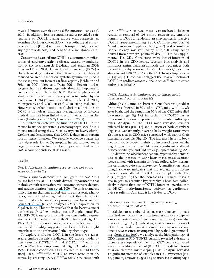
Although CKO mice are born at Mendelian ratio, suddendeath was observed in 50% of the CKO mice within 2 wkafter birth, and the remaining 50% of the CKO mice diedby 6 mo of age (Fig. 1A), indicating that DOT1L has animportant function in postnatal and adult cardiomyo-cytes. Analysis of the CKO mice revealed severelyenlarged hearts (Fig. 1B) and dilation of both chambers(Fig. 1C). Consistently, heart to body weight ratios werealso increased in CKO mice compared with that of theirlittermate controls (Fig. 1D). The increased heart to bodyweight ratio is caused mainly by increased heart weight(Fig. 1E), as the body weight is not significantly alteredbetween wild-type and CKO mice (Supplemental Fig. 3A).
To determine whether concentric hypertrophy contrib-utes to the increase in CKO heart mass, tissue sectionswere stained with Laminin antibody followed by measur-ing cardiomyocyte circumference. Quantification usingImageJ software indicates that the average cell circum-ference is not altered in CKO mice (Supplemental Fig.3B,C), suggesting that the increase in CKO heart mass isdue in part to eccentric hypertrophy. These data collec-tively indicate that loss of DOT1L function—particularlyits H3K79 methyltransferase activity—in cardiomyo-cytes results in CHF that is likely due to DCM.
CKO hearts exhibit similar cardiac remodelingobserved in DCM patients
In addition to chamber dilation, gross changes in heartmorphology (such as deviation from an elliptical shape toa more spherical one and increased heart mass) were alsoobserved (Fig. 1C,E), indicating that loss-of-function ofDOT1L in cardiomyocytes caused cardiac remodeling.Since DCM is often accompanied by pathologic remodel-ing (Cohn et al. 2000), we analyzed the histopathology ofCKO hearts at P10. TUNEL staining revealed a dramaticincrease in apoptotic cell death in CKO hearts comparedwith the wild-type control (Fig. 2A). In addition, trans-mission electron microscopic (TEM) analysis revealeda significant increase of vacuoles in CKO myocytes (Fig.2B, panel ii, arrows), suggesting an increase in autophagic
Nguyen et al.
264 GENES & DEVELOPMENT
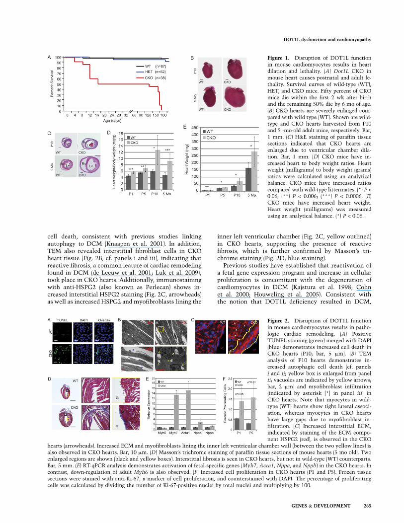
cell death, consistent with previous studies linkingautophagy to DCM (Knaapen et al. 2001). In addition,TEM also revealed interstitial fibroblast cells in CKOheart tissue (Fig. 2B, cf. panels i and iii), indicating thatreactive fibrosis, a common feature of cardiac remodelingfound in DCM (de Leeuw et al. 2001; Luk et al. 2009),took place in CKO hearts. Additionally, immunostainingwith anti-HSPG2 (also known as Perlecan) shows in-creased interstitial HSPG2 staining (Fig. 2C, arrowheads)as well as increased HSPG2 and myofibroblasts lining the
inner left ventricular chamber (Fig. 2C, yellow outlined)in CKO hearts, supporting the presence of reactivefibrosis, which is further confirmed by Masson’s tri-chrome staining (Fig. 2D, blue staining).
Previous studies have established that reactivation ofa fetal gene expression program and increase in cellularproliferation is concomitant with the degeneration ofcardiomyocytes in DCM (Kajstura et al. 1998; Cohnet al. 2000; Houweling et al. 2005). Consistent withthe notion that DOT1L deficiency resulted in DCM,
Figure 1. Disruption of DOT1L functionin mouse cardiomyocytes results in heartdilation and lethality. (A) Dot1L CKO inmouse heart causes postnatal and adult le-thality. Survival curves of wild-type (WT),HET, and CKO mice. Fifty percent of CKOmice die within the first 2 wk after birthand the remaining 50% die by 6 mo of age.(B) CKO hearts are severely enlarged com-pared with wild type (WT). Shown are wild-type and CKO hearts harvested from P10and 5 -mo-old adult mice, respectively. Bar,1 mm. (C) H&E staining of paraffin tissuesections indicated that CKO hearts areenlarged due to ventricular chamber dila-tion. Bar, 1 mm. (D) CKO mice have in-creased heart to body weight ratios. Heartweight (milligrams) to body weight (grams)ratios were calculated using an analyticalbalance. CKO mice have increased ratioscompared with wild-type littermates. (*) P <
0.06; (**) P < 0.006; (***) P < 0.0006. (E)CKO mice have increased heart weight.Heart weight (milligrams) was measuredusing an analytical balance. (*) P < 0.06.
Figure 2. Disruption of DOT1L functionin mouse cardiomyocytes results in patho-logic cardiac remodeling. (A) PositiveTUNEL staining (green) merged with DAPI(blue) demonstrates increased cell death inCKO hearts (P10; bar, 5 mm). (B) TEManalysis of P10 hearts demonstrates in-creased autophagic cell death (cf. panelsi and ii; yellow box is enlarged from panelii; vacuoles are indicated by yellow arrows;bar, 2 mm) and myofibroblast infiltration(indicated by asterisk [*] in panel iii) inCKO hearts. Note that myocytes in wild-type (WT) hearts show tight lateral associ-ation, whereas myocytes in CKO heartshave large gaps due to myofibroblast in-filtration. (C) Increased interstitial ECM,indicated by staining of the ECM compo-nent HSPG2 (red), is observed in the CKO
hearts (arrowheads). Increased ECM and myofibroblasts lining the inner left ventricular chamber wall (between the two yellow lines) isalso observed in CKO hearts. Bar, 10 mm. (D) Masson’s trichrome staining of paraffin tissue sections of mouse hearts (5 mo old). Twoenlarged regions are shown (black and yellow boxes). Interstitial fibrosis is seen in CKO hearts, but not in wild-type (WT) counterparts.Bar, 5 mm. (E) RT-qPCR analysis demonstrates activation of fetal-specific genes (Myh7, Acta1, Nppa, and Nppb) in the CKO hearts. Incontrast, down-regulation of adult Myh6 is also observed. (F) Increased cell proliferation in CKO hearts (P1 and P5). Frozen tissuesections were stained with anti-Ki-67, a marker of cell proliferation, and counterstained with DAPI. The percentage of proliferatingcells was calculated by dividing the number of Ki-67-positive nuclei by total nuclei and multiplying by 100.
DOT1L dysfunction and cardiomyopathy
GENES & DEVELOPMENT 265

RT-qPCR demonstrated that expression of the fetal genesMyh7, Acta1, Nppa, and Nppb is up-regulated in CKOhearts (Fig. 2E). In contrast, adult gene Myh6 is down-regulated. Mouse cardiomyocytes retain a small capacityto proliferate after birth (Ahuja et al. 2007; Banerjee et al.2007). To determine whether DOT1L deficiency resultsin an increased cell proliferation, as exhibited in DCM,heart tissue sections were immunostained for Ki-67 at P1and P5. Results shown in Figure 2F demonstrate that thepercentage of proliferating cells (ratio of Ki-67-positivenuclei to total nuclei, multiplied by 100) is significantlyincreased in the CKO hearts compared with the control,which may contribute to the observed increase in theCKO heart mass. We note that this increased cell pro-liferation in the DOT1L-deficient heart is in contrast toprevious studies showing a requirement for DOT1L inembryonic stem cell cycle progression (Jones et al. 2008;Barry et al. 2009), suggesting cell type specificity. Takentogether, the above data support the notion that CKOhearts exhibit multiple phenotypes similar to thoseobserved in DCM.
CKO hearts exhibit similar functional defectsobserved in DCM patients
To gain further support that DOT1L deficiency in car-diomyocytes results in DCM, we asked whether themorphological changes and cardiac remodeling observedin CKO hearts affect their function. To this end, weperformed echocardiography (ECHO) analysis at differentmouse age groups. Conscious ECHOs performed on P10pups during the first stage of lethality (n = 5 per genotype)demonstrated that CKO mice have increased left ventric-ular internal dimensions and volume. Analysis of cardiacoutput by measuring ejection fraction (EF) and fractionalshortening (FS) revealed that both EF and FS is reduced byalmost half in CKO mice when compared with those ofwild-type mice (Table 1). These results are indicative ofleft ventricular systolic dysfunction and are consistentwith clinical DCM outcome (Karkkainen and Peuhkurinen2007; Luk et al. 2009). Similar results were obtained at2 and 5 mo of age (Table 1). Interestingly, the smaller
difference between wild-type and CKO mice at 2 mo mayreflect a compensation that allowed these mice to bypassthe first stage of lethality.
Cardiac conduction abnormalities are frequently ob-served in DCM heart failure patients with left ventricularsystolic dysfunction (Olson 2004). During heart contrac-tion, an electrical impulse transmits from atria (P-wave)to ventricles (QRS-wave) at the atrioventricular node(AVN). The time delay for electrical propagation can bemeasured directly by electrocardiography (EKG) (Hatcherand Basson 2009). To determine whether the conductionsystem is perturbed in CKO mice, EKG was performed at5 mo of age (n = 8 per genotype). All CKO mice displayedminimally a first-degree heart block at the AVN, with an80% penetration of either nonsustained ventriculartachycardia (n = 1 of 8) (Fig. 3A, CKOa), periodic third-degree heart block (n = 3 of 8), or second-degree Type IIheart block (n = 3 of 8) (Fig. 3A, CKOb). Overall, CKOmice have a significant increase in RR interval (Fig. 3B),PR interval (Fig. 3C), P-wave duration (Fig. 3D), and QRSinterval (Fig. 3E). These EKG data from CKO mice areconsistent with EKG findings in human DCM patients(Seidman and Seidman 2001; Towbin and Bowles 2006;Luk et al. 2009). The physiological studies further supportthe notion that DOT1L deficiency in cardiomyocytesconfers phenotypes similar to those observed in patientswith DCM.
Dot1L deficiency in cardiomyocytes down-regulatesdystrophin expression
Having established that DOT1L deficiency in cardiomyo-cytes causes phenotypes similar to those observed inDCM, we next attempted to understand the molecularmechanism. To date, mutations in >30 genes have beenlinked to human DCM (Supplemental Table 1) (Towbinand Bowles 2006; Karkkainen and Peuhkurinen 2007;Kimura 2008; Luk et al. 2009). Given that DOT1L-mediated H3K79 methylation is associated with activelytranscribed genes (Martin and Zhang 2005; Z Wang et al.2008), we anticipated that one or more of the DCM-associated genes might be down-regulated due to loss of
Table 1. Heart function of wild-type and CKO mice as measured by ECHO
EF FSLVID;d(mm)
LVID;s(mm)
LV Vol:d(mL)
LV Vol;s(mL) LV mass n
P10Wild type 89.20% 6 1.86% 56.50% 6 2.69% 1.59 6 0.13 0.70 6 0.08 7.23 6 1.58 0.79 6 0.28 18.70 6 3.01 5CKO 55.80% 6 3.83% 27.30% 6 2.15% 2.16 6 0.25 1.57 6 0.23 15.79 6 4.82 7.15 6 2.87 21.41 6 3.47 5P-value *** *** * *** * ** ns
2 moWild type 85.15% 6 4.63% 53.47% 6 6.03% 2.73 6 0.19 1.27 6 0.18 27.97 6 4.66 4.11 6 1.46 76.61 6 11.88 6CKO 72.56% 6 5.77% 40.81% 6 4.73% 3.12 6 0.13 1.85 6 0.20 38.83 6 4.12 10.87 6 2.91 98.51 6 18.0 4P-value * * * ** * ** ns
5 moWild type 84.73% 6 3.60% 52.74% 6 4.43% 2.98 6 0.28 1.41 6 0.19 35.01 6 7.72 5.36 6 1.81 87.16 6 13.89 10CKO 47.02% 6 18.48% 24.44% 6 10.84% 4.91 6 1.43 3.85 6 1.60 127.58 6 83.67 80.83 6 70.08 205.86 6 101.11 7P-value *** *** ** *** ** ** **
(EF) Ejection fraction; (FS) fractional shortening; (LVID) left ventricular internal diameter; (LV Vol) left ventricular volume; (LV mass) leftventricular mass (AW) corrected; (d) end diastolic; (s) end systolic. (***) <0.0006; (**) <0.006; (*) <0.06; (ns) $0.06.
Nguyen et al.
266 GENES & DEVELOPMENT

H3K79 methylation in the CKO heart. To this end, weperformed four independent gene expression microarraysusing the dual-color Agilent 4X44K Whole Mouse Ge-nome Array system. Data analysis revealed 751 down-regulated probes representing 471 genes that are statisti-cally significant, with a false-positive rate of 0.06%(Supplemental Table 2). Comparison of the microarraydata with known DCM-associated genes identified twocommon genes: Titin (Ttn) and Dystrophin (Dmd).
Ttn is a giant myofilament protein important formaintaining sarcomere structure and elasticity (Kostinet al. 2000). Mutations in Ttn have been reported inautosomal dominant forms of familial DCM (Gerull et al.2002). Mouse models expressing M-line-deficient Ttnexhibit widened M-lines and gradual disassembly ofsarcomeres, which lead to cardiac failure (Gotthardtet al. 2003; Weinert et al. 2006). If Ttn down-regulationis responsible for the DCM in CKO mice, we anticipatean abnormal sarcomere structure in DOT1L CKO hearts.However, TEM analysis revealed that sarcomere integrityis maintained in DOT1L CKO hearts (Supplemental Fig.4), suggesting that down-regulation of Ttn is not a majorcontributing factor for the DCM in DOT1L CKO mice.
Dmd was the first discovered DCM-associated genethat can cause both DCM and muscular dystrophy. Dmdis a membrane-associated protein that forms a dystro-phin–glycoprotein complex (DGC), which connects con-tractile sarcomeres to the sarcolemma and extracellularmatrix (ECM). This connection is vital for lateral forcetransduction between cardiomyocytes, as well as forrelieving mechanical stress on sarcolemma during con-traction (Kostin et al. 2000; Kimura 2008). Since a loss ofDmd expression may be the cause of cell death andcardiac remodeling observed in CKO hearts, we firstconfirmed the microarray results by RT-qPCR. Datapresented in Figure 4A demonstrate that the Dmd mRNAlevels are down-regulated to ;25% of the wild-type levelin the CKO hearts. In contrast, expression of otherrandomly selected DCM-relevant genes (Actn2, Ldb3,Des, and Taz) was not significantly altered by DOT1Ldeficiency (Fig. 4A). Consistent with a reduction at theRNA level, immunostaining revealed that Dmd proteinlevel is also greatly diminished in CKO hearts (Fig. 4B,C).
Previous studies have demonstrated that mutations af-fecting expression of Dmd or any sarcoglycan (Sgc) genelead to DGC instability and reduced levels of all com-plex proteins (Deconinck et al. 1997; Grady et al. 1997).Consistently, immunostaining revealed a great loss ofb-dystroglycan (bDG) and a-sarcoglycan (SGCA) proteinsin CKO mice (Fig. 4B,C), although none of the DGCcomponents is altered at the RNA level by DOT1Ldeficiency (Fig. 4A). These results suggest that loss ofDmd caused degradation of the DGC components, whichin turn affects cardiomyocyte viability.
We next sought to determine whether DOT1L directlyregulates Dmd expression in the mouse heart by chro-matin immunoprecipitation (ChIP). Despite extensiveefforts, none of the homemade or commercial DOT1Lantibodies (Abgent AP1198a and AP1198b; Cell SignalingD8891 and D8890; and Abcam ab7295) was able to de-tect endogenous DOT1L protein (data not shown), andthus they are unsuitable for ChIP. Therefore, we per-formed ChIP assays across the Dmd locus using an anti-H3K79me2/3 antibody. Results shown in Figure 4Ddemonstrate that relatively less H3K79me2/3 is observedupstream of the Dmd transcription start site (TSS), but itgreatly increases downstream, continues to rise at 20 kbdownstream from the TSS, and is still present as far as 59kb downstream from the TSS. This H3K79me2/3 distri-bution pattern is consistent with published ChIP-seqresults using various cell lines (Barski et al. 2007; Z Wanget al. 2008). Importantly, the H3K79me2/3 enrichment onthe Dmd gene depends on functional DOT1L, as theenrichment is abolished when samples derived fromDot1L CKO hearts are used. In addition, the detectedsignals are specific, as enrichment was not observed whenIgG was used in a parallel ChIP assay. Previous studieshave demonstrated that DOT1L deficiency leads to acomplete loss of H3K79 methylation (Jones et al. 2008),indicating that DOT1L is the only H3K79 methyltrans-ferase. The demonstration that H3K79 methylation of theDmd gene is dependent on functional DOT1L supportsthe notion that Dmd is a direct DOT1L target.
It has been reported previously that Dmd expression ispositively regulated by the binding of the transcriptionactivator SRF (serum response factor) to a CArG-box
Figure 3. Disruption of DOT1L functionin mouse cardiomyocytes results in conduc-tion abnormalities. (A) RepresentativeEKGs for wild-type (Wt) and CKO mice.The analysis was performed using 5-mo-oldmice (n = 8 per group). CKOa has completeAV dissociation, as evidence by nonsus-tained ventricular tachycardia, while CKObhas a Type II second-degree heart block. Bar,200 msec. (B–E) Quantification of EKG dataindicated an overall significant increase inRR interval (B), PR interval (C), P-waveduration (D), and QRS interval (E). P-valueswere calculated by Student t-test.
DOT1L dysfunction and cardiomyopathy
GENES & DEVELOPMENT 267
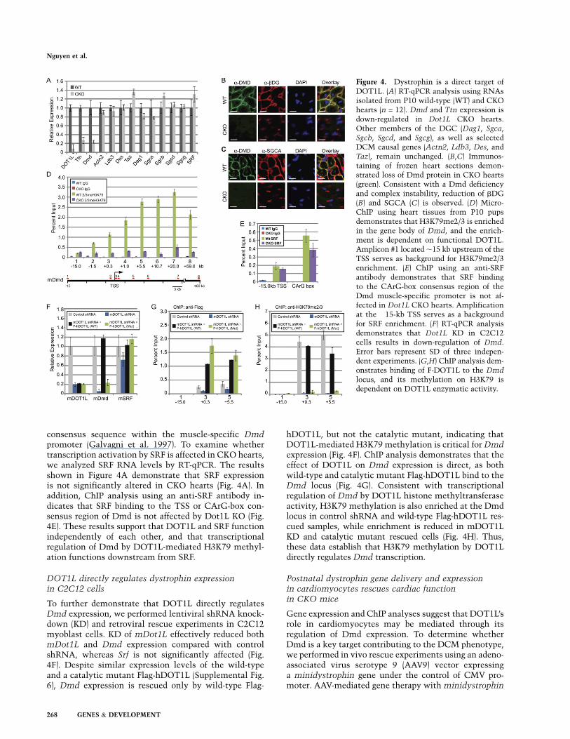
consensus sequence within the muscle-specific Dmdpromoter (Galvagni et al. 1997). To examine whethertranscription activation by SRF is affected in CKO hearts,we analyzed SRF RNA levels by RT-qPCR. The resultsshown in Figure 4A demonstrate that SRF expressionis not significantly altered in CKO hearts (Fig. 4A). Inaddition, ChIP analysis using an anti-SRF antibody in-dicates that SRF binding to the TSS or CArG-box con-sensus region of Dmd is not affected by Dot1L KO (Fig.4E). These results support that DOT1L and SRF functionindependently of each other, and that transcriptionalregulation of Dmd by DOT1L-mediated H3K79 methyl-ation functions downstream from SRF.
DOT1L directly regulates dystrophin expressionin C2C12 cells
To further demonstrate that DOT1L directly regulatesDmd expression, we performed lentiviral shRNA knock-down (KD) and retroviral rescue experiments in C2C12myoblast cells. KD of mDot1L effectively reduced bothmDot1L and Dmd expression compared with controlshRNA, whereas Srf is not significantly affected (Fig.4F). Despite similar expression levels of the wild-typeand a catalytic mutant Flag-hDOT1L (Supplemental Fig.6), Dmd expression is rescued only by wild-type Flag-
hDOT1L, but not the catalytic mutant, indicating thatDOT1L-mediated H3K79 methylation is critical for Dmdexpression (Fig. 4F). ChIP analysis demonstrates that theeffect of DOT1L on Dmd expression is direct, as bothwild-type and catalytic mutant Flag-hDOT1L bind to theDmd locus (Fig. 4G). Consistent with transcriptionalregulation of Dmd by DOT1L histone methyltransferaseactivity, H3K79 methylation is also enriched at the Dmdlocus in control shRNA and wild-type Flag-hDOT1L res-cued samples, while enrichment is reduced in mDOT1LKD and catalytic mutant rescued cells (Fig. 4H). Thus,these data establish that H3K79 methylation by DOT1Ldirectly regulates Dmd transcription.
Postnatal dystrophin gene delivery and expressionin cardiomyocytes rescues cardiac functionin CKO mice
Gene expression and ChIP analyses suggest that DOT1L’srole in cardiomyocytes may be mediated through itsregulation of Dmd expression. To determine whetherDmd is a key target contributing to the DCM phenotype,we performed in vivo rescue experiments using an adeno-associated virus serotype 9 (AAV9) vector expressinga minidystrophin gene under the control of CMV pro-moter. AAV-mediated gene therapy with minidystrophin
Figure 4. Dystrophin is a direct target ofDOT1L. (A) RT-qPCR analysis using RNAsisolated from P10 wild-type (WT) and CKOhearts (n = 12). Dmd and Ttn expression isdown-regulated in Dot1L CKO hearts.Other members of the DGC (Dag1, Sgca,Sgcb, Sgcd, and Sgcg), as well as selectedDCM causal genes (Actn2, Ldb3, Des, andTaz), remain unchanged. (B,C) Immunos-taining of frozen heart sections demon-strated loss of Dmd protein in CKO hearts(green). Consistent with a Dmd deficiencyand complex instability, reduction of bDG(B) and SGCA (C) is observed. (D) Micro-ChIP using heart tissues from P10 pupsdemonstrates that H3K79me2/3 is enrichedin the gene body of Dmd, and the enrich-ment is dependent on functional DOT1L.Amplicon #1 located ;15 kb upstream of theTSS serves as background for H3K79me2/3enrichment. (E) ChIP using an anti-SRFantibody demonstrates that SRF bindingto the CArG-box consensus region of theDmd muscle-specific promoter is not af-fected in Dot1L CKO hearts. Amplificationat the �15-kb TSS serves as a backgroundfor SRF enrichment. (F) RT-qPCR analysisdemonstrates that Dot1L KD in C2C12cells results in down-regulation of Dmd.Error bars represent SD of three indepen-dent experiments. (G,H) ChIP analysis dem-onstrates binding of F-DOT1L to the Dmd
locus, and its methylation on H3K79 isdependent on DOT1L enzymatic activity.
Nguyen et al.
268 GENES & DEVELOPMENT
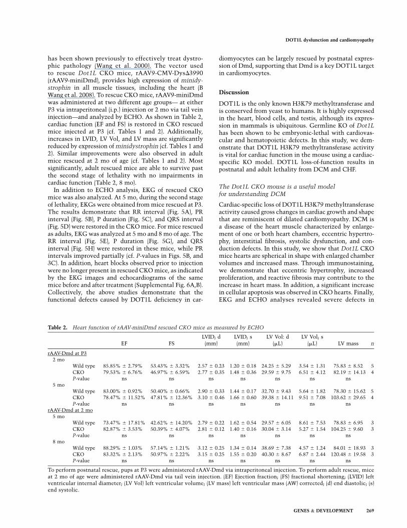
has been shown previously to effectively treat dystro-phic pathology (Wang et al. 2000). The vector usedto rescue Dot1L CKO mice, rAAV9-CMV-DysD3990(rAAV9-miniDmd), provides high expression of minidy-strophin in all muscle tissues, including the heart (BWang et al. 2008). To rescue CKO mice, rAAV9-miniDmdwas administered at two different age groups— at eitherP3 via intraperitoneal (i.p.) injection or 2 mo via tail veininjection—and analyzed by ECHO. As shown in Table 2,cardiac function (EF and FS) is restored in CKO rescuedmice injected at P3 (cf. Tables 1 and 2). Additionally,increases in LVID, LV Vol, and LV mass are significantlyreduced by expression of minidystrophin (cf. Tables 1 and2). Similar improvements were also observed in adultmice rescued at 2 mo of age (cf. Tables 1 and 2). Mostsignificantly, adult rescued mice are able to survive pastthe second stage of lethality with no impairments incardiac function (Table 2, 8 mo).
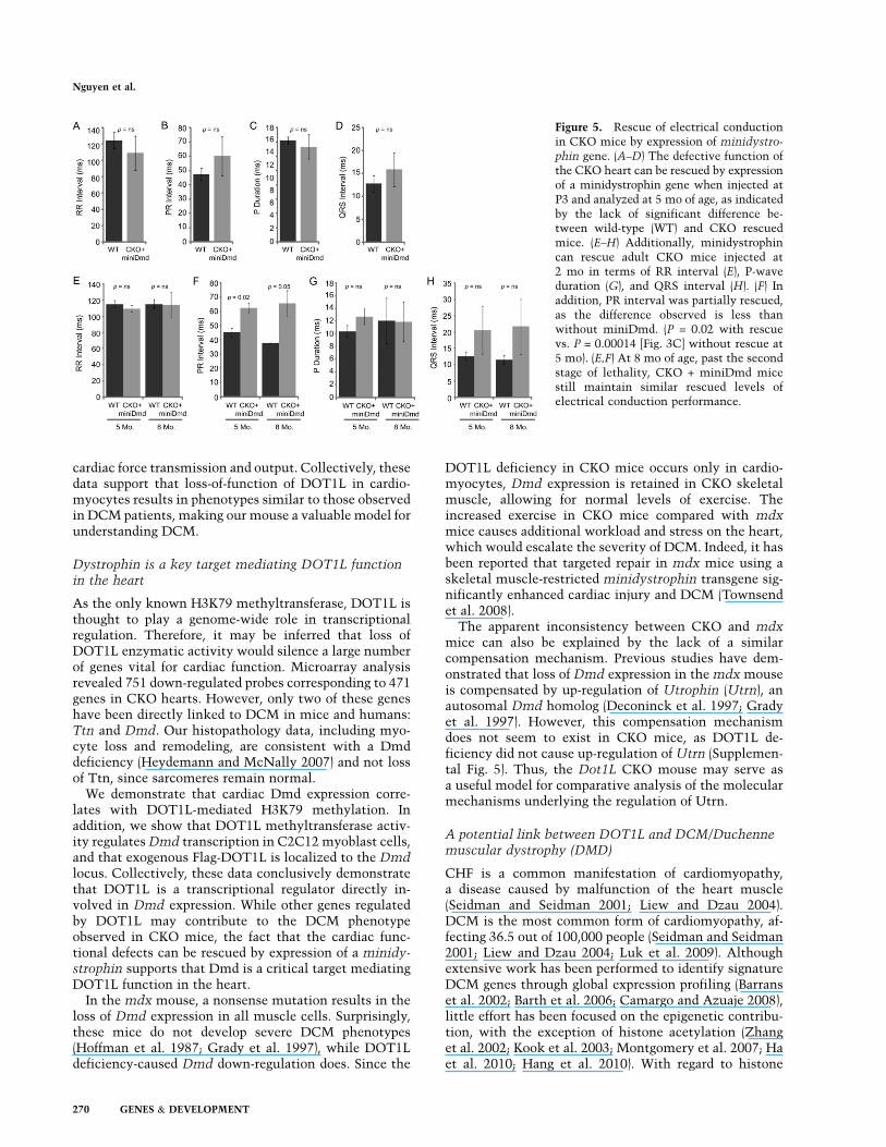
In addition to ECHO analysis, EKG of rescued CKOmice was also analyzed. At 5 mo, during the second stageof lethality, EKGs were obtained from mice rescued at P3.The results demonstrate that RR interval (Fig. 5A), PRinterval (Fig. 5B), P duration (Fig. 5C), and QRS interval(Fig. 5D) were restored in the CKO mice. For mice rescuedas adults, EKG was analyzed at 5 mo and 8 mo of age. TheRR interval (Fig. 5E), P duration (Fig. 5G), and QRSinterval (Fig. 5H) were restored in these mice, while PRintervals improved partially (cf. P-values in Figs. 5B, and3C). In addition, heart blocks observed prior to injectionwere no longer present in rescued CKO mice, as indicatedby the EKG images and echocardiograms of the samemice before and after treatment (Supplemental Fig. 6A,B).Collectively, the above studies demonstrate that thefunctional defects caused by DOT1L deficiency in car-
diomyocytes can be largely rescued by postnatal expres-sion of Dmd, supporting that Dmd is a key DOT1L targetin cardiomyocytes.
Discussion
DOT1L is the only known H3K79 methyltransferase andis conserved from yeast to humans. It is highly expressedin the heart, blood cells, and testis, although its expres-sion in mammals is ubiquitous. Germline KO of Dot1Lhas been shown to be embryonic-lethal with cardiovas-cular and hematopoietic defects. In this study, we dem-onstrate that DOT1L H3K79 methyltransferase activityis vital for cardiac function in the mouse using a cardiac-specific KO model. DOT1L loss-of-function results inpostnatal and adult lethality from DCM and CHF.
The Dot1L CKO mouse is a useful modelfor understanding DCM
Cardiac-specific loss of DOT1L H3K79 methyltransferaseactivity caused gross changes in cardiac growth and shapethat are reminiscent of dilated cardiomyopathy. DCM isa disease of the heart muscle characterized by enlarge-ment of one or both heart chambers, eccentric hypertro-phy, interstitial fibrosis, systolic dysfunction, and con-duction defects. In this study, we show that Dot1L CKOmice hearts are spherical in shape with enlarged chambervolumes and increased mass. Through immunostaining,we demonstrate that eccentric hypertrophy, increasedproliferation, and reactive fibrosis may contribute to theincrease in heart mass. In addition, a significant increasein cellular apoptosis was observed in CKO hearts. Finally,EKG and ECHO analyses revealed severe defects in
Table 2. Heart function of rAAV-miniDmd rescued CKO mice as measured by ECHO
EF FSLVID; d
(mm)LVID; s(mm)
LV Vol: d(mL)
LV Vol; s(mL) LV mass n
rAAV-Dmd at P32 mo
Wild type 85.85% 6 2.79% 53.43% 6 3.32% 2.57 6 0.23 1.20 6 0.18 24.25 6 5.29 3.54 6 1.31 75.83 6 8.52 5CKO 79.53% 6 6.76% 46.97% 6 6.59% 2.77 6 0.35 1.48 6 0.36 29.59 6 9.75 6.51 6 4.12 82.19 6 14.13 4P-value ns ns ns ns ns ns ns
5 moWild type 83.00% 6 0.92% 50.40% 6 0.66% 2.90 6 0.33 1.44 6 0.17 32.70 6 9.43 5.64 6 1.82 78.30 6 15.62 5CKO 78.47% 6 11.52% 47.81% 6 12.36% 3.10 6 0.46 1.66 6 0.60 39.38 6 14.11 9.51 6 7.08 103.62 6 29.65 4P-value ns ns ns ns ns ns ns
rAAV-Dmd at 2 mo5 mo
Wild type 73.47% 6 17.81% 42.62% 6 14.20% 2.79 6 0.22 1.62 6 0.54 29.57 6 6.05 8.61 6 7.53 78.83 6 6.95 3CKO 82.87% 6 3.53% 50.39% 6 4.07% 2.81 6 0.12 1.40 6 0.16 30.04 6 3.14 5.27 6 1.54 104.25 6 9.60 3P-value ns ns ns ns ns ns ns
8 moWild type 88.29% 6 1.03% 57.14% 6 1.21% 3.12 6 0.25 1.34 6 0.14 38.69 6 7.38 4.57 6 1.24 84.01 6 18.93 3CKO 83.32% 6 2.13% 50.97% 6 2.22% 3.15 6 0.25 1.55 6 0.20 40.30 6 8.67 6.87 6 2.44 120.48 6 19.58 3P-value ns ns ns ns ns ns ns
To perform postnatal rescue, pups at P3 were administered rAAV-Dmd via intraperitoneal injection. To perform adult rescue, miceat 2 mo of age were administered rAAV-Dmd via tail vein injection. (EF) Ejection fraction; (FS) fractional shortening; (LVID) leftventricular internal diameter; (LV Vol) left ventricular volume; (LV mass) left ventricular mass (AW) corrected; (d) end diastolic; (s)end systolic.
DOT1L dysfunction and cardiomyopathy
GENES & DEVELOPMENT 269
cardiac force transmission and output. Collectively, thesedata support that loss-of-function of DOT1L in cardio-myocytes results in phenotypes similar to those observedin DCM patients, making our mouse a valuable model forunderstanding DCM.
Dystrophin is a key target mediating DOT1L functionin the heart
As the only known H3K79 methyltransferase, DOT1L isthought to play a genome-wide role in transcriptionalregulation. Therefore, it may be inferred that loss ofDOT1L enzymatic activity would silence a large numberof genes vital for cardiac function. Microarray analysisrevealed 751 down-regulated probes corresponding to 471genes in CKO hearts. However, only two of these geneshave been directly linked to DCM in mice and humans:Ttn and Dmd. Our histopathology data, including myo-cyte loss and remodeling, are consistent with a Dmddeficiency (Heydemann and McNally 2007) and not lossof Ttn, since sarcomeres remain normal.
We demonstrate that cardiac Dmd expression corre-lates with DOT1L-mediated H3K79 methylation. Inaddition, we show that DOT1L methyltransferase activ-ity regulates Dmd transcription in C2C12 myoblast cells,and that exogenous Flag-DOT1L is localized to the Dmdlocus. Collectively, these data conclusively demonstratethat DOT1L is a transcriptional regulator directly in-volved in Dmd expression. While other genes regulatedby DOT1L may contribute to the DCM phenotypeobserved in CKO mice, the fact that the cardiac func-tional defects can be rescued by expression of a minidy-strophin supports that Dmd is a critical target mediatingDOT1L function in the heart.
In the mdx mouse, a nonsense mutation results in theloss of Dmd expression in all muscle cells. Surprisingly,these mice do not develop severe DCM phenotypes(Hoffman et al. 1987; Grady et al. 1997), while DOT1Ldeficiency-caused Dmd down-regulation does. Since the
DOT1L deficiency in CKO mice occurs only in cardio-myocytes, Dmd expression is retained in CKO skeletalmuscle, allowing for normal levels of exercise. Theincreased exercise in CKO mice compared with mdxmice causes additional workload and stress on the heart,which would escalate the severity of DCM. Indeed, it hasbeen reported that targeted repair in mdx mice using askeletal muscle-restricted minidystrophin transgene sig-nificantly enhanced cardiac injury and DCM (Townsendet al. 2008).
The apparent inconsistency between CKO and mdxmice can also be explained by the lack of a similarcompensation mechanism. Previous studies have dem-onstrated that loss of Dmd expression in the mdx mouseis compensated by up-regulation of Utrophin (Utrn), anautosomal Dmd homolog (Deconinck et al. 1997; Gradyet al. 1997). However, this compensation mechanismdoes not seem to exist in CKO mice, as DOT1L de-ficiency did not cause up-regulation of Utrn (Supplemen-tal Fig. 5). Thus, the Dot1L CKO mouse may serve asa useful model for comparative analysis of the molecularmechanisms underlying the regulation of Utrn.
A potential link between DOT1L and DCM/Duchennemuscular dystrophy (DMD)
CHF is a common manifestation of cardiomyopathy,a disease caused by malfunction of the heart muscle(Seidman and Seidman 2001; Liew and Dzau 2004).DCM is the most common form of cardiomyopathy, af-fecting 36.5 out of 100,000 people (Seidman and Seidman2001; Liew and Dzau 2004; Luk et al. 2009). Althoughextensive work has been performed to identify signatureDCM genes through global expression profiling (Barranset al. 2002; Barth et al. 2006; Camargo and Azuaje 2008),little effort has been focused on the epigenetic contribu-tion, with the exception of histone acetylation (Zhanget al. 2002; Kook et al. 2003; Montgomery et al. 2007; Haet al. 2010; Hang et al. 2010). With regard to histone
Figure 5. Rescue of electrical conductionin CKO mice by expression of minidystro-
phin gene. (A–D) The defective function ofthe CKO heart can be rescued by expressionof a minidystrophin gene when injected atP3 and analyzed at 5 mo of age, as indicatedby the lack of significant difference be-tween wild-type (WT) and CKO rescuedmice. (E–H) Additionally, minidystrophincan rescue adult CKO mice injected at2 mo in terms of RR interval (E), P-waveduration (G), and QRS interval (H). (F) Inaddition, PR interval was partially rescued,as the difference observed is less thanwithout miniDmd. (P = 0.02 with rescuevs. P = 0.00014 [Fig. 3C] without rescue at5 mo). (E,F) At 8 mo of age, past the secondstage of lethality, CKO + miniDmd micestill maintain similar rescued levels ofelectrical conduction performance.
Nguyen et al.
270 GENES & DEVELOPMENT
methylation, only two studies have investigated changesin methylation patterns in heart failure (Kaneda et al.2009; Movassagh et al. 2010). However, whether it di-rectly contributes to the development of DCM is notknown.
The demonstration that loss of DOT1L enzymaticactivity results in DCM not only establishes a connectionbetween dysregulation of histone methylation to DCM,but also raises the possibility that malfunction of DOT1Lmight account for some DCM patients. To explore thispossibility, we analyzed hDOT1L expression level inidiopathic DCM (n = 27) and normal (n = 11) myocardialsamples from publically available microarray data (http://cardiogenomics.med.harvard.edu/project-detail?project_id=229). This analysis indicates that DOT1L is down-regulated in idiopathic DCM patients with all Affymetrixprobe sets (n = 11 probes per set) (Supplemental Fig. 8),supporting the notion that dysfunction of DOT1L may bea contributing factor to human idiopathic DCM. Muta-tions in Dmd is also the cause of both DMD and Beckermuscular dystrophy (BMD), affecting one out of 3500males (Hoffman et al. 1987). Up to 90% of those patientsmanifest cardiomyopathies, and many die of heart failure(Connuck et al. 2008). In this study, we established thatDOT1L regulates Dmd expression in both cardiac andC2C12 cells, suggesting that a DOT1L deficiency maycontribute to DCM and human muscular dystrophy.Future studies should reveal whether DOT1L is geneti-cally linked to DCM, DMD, and BMD in human patients.
Materials and methods
Generation of cardiac-specific Dot1L CKO mice
The targeting vector and generation of DOT1L chimeric micehave been described previously (Jones et al. 2008). DOT1L2lox/+
and DOT1L1lox/+ mice were mated with a-MHC-Cre+/+
transgenic mice to obtain DOT1L2lox/+;a-MHC-Cre+/+ andDOT1L1lox/+;a-MHC-Cre+/+ mating pairs. Mice were kept ona 129SvJ, C57BL/6J mixed background. DOT1L2lox/1lox;a-MHC-Cre+/+ mice and DOT1L2lox/2lox; a-MHC-Cre+/+ mice were usedas CKO mice. Upon Cre recombination, exons 5 and 6 wereexcised from the DOT1L locus. After splicing of the DOT1Ltranscript from the CKO allele, exons 4 and 7 of the maturemRNA were translated in-frame, generating a mutant DOT1Lprotein lacking a portion of the SAM-binding motif. All miceprocedures were performed following the guidelines set by theInstitutional Animal Care and Use Committee.
ECHO and EKG
Experiments were performed at the Mouse CardiovascularModels Core Facility at University of North Carolina at ChapelHill (UNC-CH) or in the laboratory of Dr. Xiao Xiao (UNC-CH).To restrain postnatal pups, paws were taped to a plastic board.For adult mice, soft cotton thread loops were placed around eachleg, just proximal to the paw, and gently snugged with a plasticslider. The distal ends of the threads were placed in notches cutinto a plastic board and gently tightened to hold the animal ina supine position to prevent self-mutilation of the forelimbs.Warmed Aquasonic gel was applied over the thorax and a 30-MHz probe was positioned over the chest in a parasternalposition. Long and short axis B-mode and M-mode images were
recorded. Upon completion of the procedure, the gel was wipedoff and the animal was returned to its cage housed in a warmchamber. Time of restraint was 5 min or less. For EKG, micewere anesthetized with inhaled isoflurane. Mice were taped toa warmed mouse board and their body temperature was moni-tored with a rectal probe and maintained at 37°C 6 1°Cthroughout the procedure. Thin (29 gauge) sharpened needleelectrodes were passed subcutaneously into the area at theventral base of each limb. After three leads were recorded, theneedles were removed and the animals were allowed to recover.
Histology and immunofluorescence staining
For all tissue sectioning, beating hearts were harvested fromeuthanized mice and immediately transferred to ice-cold PBScontaining 1 M KCl until the hearts stopped beating at diastolestate. For H&E staining, hearts were fixed in 4% paraformalde-hyde overnight and paraffin-embedded. Serial sections at 5-mmthickness were used for staining. Hearts used for all immuno-fluorescence staining were fixed in 4% PFA and subjected tosequential incubation with 10%, 20%, and 30% sucrose in PBSat 4°C. Hearts were then flash-frozen in OCT medium usingliquid nitrogen, and frozen serial sections of 5-mm thicknesswere prepared using a Leica Cryostat. Primary antibodies usedfor staining include anti-Dystrophin (Abcam, ab15277-500), anti-HSPG2 (Neomarkers, RT-794), anti-a-Laminin (Chemicon,AB2034), anti-H3K79me2/3 (Abcam, ab2621-100), and anti-Ki-67 (Abcam, ab15580). Secondary antibodies used were AlexaFluor 594 goat anti-rat IgG (Invitrogen, A21212), Alexa Fluor 594donkey anti-rabbit IgG (Invitrogen, A21207), and Alexa Fluor 488donkey anti-rabbit IgG (Invitrogen, A21206). Sections werecounterstained with DAPI.
TUNEL assay
TUNEL staining was performed on frozen sections using theApopTag Fluorescein In Situ Apoptosis Detection kit (Millipore,S7110) and counterstained with DAPI.
Masson’s trichrome staining
Paraffin sections, 5 mm thick, of hearts from 5-mo-old mice wereused for staining. Masson’s Trichrome Stain kit was purchasedfrom Dako (AR173), and procedures were followed according tothe manufacturer’s specifications, manually, without an Artisanstaining system.
TEM
Wild-type and CKO mice were injected with 25 U of heparinintramuscular prior to euthanasia by isofluorane overdose. Thehearts were quickly exposed and perfused with 3 mg/mL2,3-Butanedione monoxime in Hepes-buffered Krebs solution,followed by perfusion with 2% glutaraldehyde plus 6% sucrosein 75 mM Na-Cacodylate buffer (pH 7.4) supplemented with3 mg/mL BDM and 0.1% tannic acid, followed by 2% osmiumtetroxide. Samples were stained with uranyl acetate en bloc. Imageswere acquired on a Zeiss EM 910 transmission electron microscopeusing a Gatan SC1000 digital camera.
Microarray and RT-qPCR analysis
Hearts were flash-frozen in liquid nitrogen and ground to a finepowder. Total RNA was purified from tissue powder using theQiagen RNeasy kit. RNA from three hearts, 1 ug each, waspooled together. Four pairs of pooled RNA, representing a total of
DOT1L dysfunction and cardiomyopathy
GENES & DEVELOPMENT 271
12 wild-type and 12 CKO hearts, were used for gene expressionanalysis. Samples were submitted to the UNC Genomics andBioinformatics Core Facility for RNA labeling, amplification,hybridization, and scanning. The dual-color Agilent 4X44KWhole Mouse Genome Array system was used. All reagentswere purchased from Agilent, and procedures were followedaccording to Agilent’s protocols. Raw data were uploaded intothe UNC Microarray Database (Agi-Scanner-Reg-MM-4X44K-D20060807-BARCODE14868; Slide Run US82800149). Datawere analyzed using the SAM algorithm (tail strength 51.4%,SE 63.8%) to yield 1379 significant probes with a median numberof false positives (0.81) and a false discovery rate of 0.06%. ForRT-qPCR analysis, RNA prepared above was treated with DNaseI, and first strand DNA synthesis was performed using Improm II(Promega). SYBR GreenER qPCR SuperMix (Invitrogen) was usedfor qPCR. Relative expression was normalized to gapdh. Primersare shown in Supplemental Table 3.
Micro-ChIP and Western blot
Micro-ChIP from frozen heart biopsies was performed as de-scribed previously (Dahl and Collas 2008) with the followingmodifications. Frozen hearts from P10 pups were ground to a finepowder prior to formaldehyde cross-linking. DNA was frag-mented into 300–500 base pairs (bp) by sonication at 15% power(2 3 15 sec, 0.5 sec on and 2 sec off). Immunoprecipitation wasperformed using anti-H3K79me2/3 (Abcam) and anti-rabbit IgG(Santa Cruz Biotechnology, sc-2027). Chromatin immunoprecip-itated samples were washed twice with low-salt (140 mM NaCl)RIPA buffer, once with high-salt (500 mM NaCl) RIPA buffer, andtwice with TE buffer. DNA was purified using the Chelex-100method, and qPCR was performed using the ChIP primers listedin Supplemental Table 3. For Western blot analysis, P1 frozenhearts were ground to a fine powder for histone extraction andWestern blot as described previously (Fang et al. 2002).
C2C12 KD and rescue
C2C12 cells were maintained in Dulbecco’s modified Eagle’smedium supplemented with 10% FBS and 1% penicillin/strep-tomycin. To establish stable KD cell lines, the lentivirus pTY-EF1a system was used as described previously (Cao et al. 2008;He et al. 2008). To knock down mDOT1L, a shRNA 19mer wasdesigned targeting the coding region (59-GGAGCCAGATCTCAGAGAA-39). The control shRNA is targeted against a bacterialprotein with no mouse or human homology (59-GTTCAGATGTGCGGCGAGT-39). KD cells were selected and maintained inmedium containing 2 mg/mL puromycin. For rescue experiments,KD cells were infected with retrovirus expressing wild-type andcatalytic mutant Flag-tagged human DOT1L (Flag-hDOT1L) asdescribed previously (Okada et al. 2005). Retrovirus-infected cellswere selected and maintained in medium containing 2 mg/mLblasticidin. RNA was isolated using RNeasy kit from Qiagen.The same micro-ChIP procedure described above was followedfor ChIP using 50,000 cells per sample. Dynabeads Protein A andM2 Flag antibody (Sigma, F3165) were also used.
Postnatal rescue of CKO mice with rAAV-miniDmd
The functional miniature version of human dystrophin geneD3990 (miniDmd) under the transcriptional control of CMVpromoter has been described previously (Wang et al. 2000). TheminiDmd gene expression cassette was packaged into AAV9vector using the helper-free, triple plasmids transfection methodand was purified by double CsCl density ultracentrifugation(Xiao et al. 1998). The rAAV9-CMV-miniDmd titers were de-
termined by DNA dot blot at ;1 3 1013 viral genome (v.g.)particles per milliliter. For 3-d-old neonatal CKO mice, a singledose of 1 3 1011 v.g. per mouse in 50 mL was injected i.p. For2-mo-old CKO mice, a single dose of 1 3 1012 v.g. per mouse in600 mL was injected via tail vein.
Statistics
Indicated P-values were calculated using a two-tailed t-test.
Acknowledgments
We thank Jackie Kylander, Kristine Porter, and Mauricio Rojas atthe UNC-CH Mouse Cardiovascular Models Core Facility forperforming ECHO on P10 pups and EKG on adult control mice;Kai Xia for help with the microarray data analysis; Jin He for helpin EKG data analysis; and Kwon-Ho Hong for critical reading ofthe manuscript. The work is supported by an NIH grant(CA119133). A.T.N is a recipient of the Predoctoral Fellowshipfrom the American Heart Association. Y.Z. is an Investigator ofthe Howard Hughes Medical Institute.
References
Abel ED, Kaulbach HC, Tian R, Hopkins JC, Duffy J, DoetschmanT, Minnemann T, Boers ME, Hadro E, Oberste-Berghaus C,et al. 1999. Cardiac hypertrophy with preserved contractilefunction after selective deletion of GLUT4 from the heart.J Clin Invest 104: 1703–1714.
Ahuja P, Sdek P, MacLellan WR. 2007. Cardiac myocyte cellcycle control in development, disease, and regeneration.Physiol Rev 87: 521–544.
Banerjee I, Fuseler JW, Price RL, Borg TK, Baudino TA. 2007.Determination of cell types and numbers during cardiacdevelopment in the neonatal and adult rat and mouse. Am
J Physiol Heart Circ Physiol 293: H1883–H1891. doi:10.1152/ajpheart.00514.2007.
Barrans JD, Allen PD, Stamatiou D, Dzau VJ, Liew CC. 2002.Global gene expression profiling of end-stage dilated cardio-myopathy using a human cardiovascular-based cDNA micro-array. Am J Pathol 160: 2035–2043.
Barry ER, Krueger W, Jakuba CM, Veilleux E, Ambrosi DJ,Nelson CE, Rasmussen TP. 2009. ES cell cycle progressionand differentiation require the action of the histone methyl-transferase Dot1L. Stem Cells 27: 1538–1547.
Barski A, Cuddapah S, Cui K, Roh TY, Schones DE, Wang Z, WeiG, Chepelev I, Zhao K. 2007. High-resolution profiling ofhistone methylations in the human genome. Cell 129: 823–837.
Barth AS, Kuner R, Buness A, Ruschhaupt M, Merk S, ZwermannL, Kaab S, Kreuzer E, Steinbeck G, Mansmann U, et al. 2006.Identification of a common gene expression signature indilated cardiomyopathy across independent microarray stud-ies. J Am Coll Cardiol 48: 1610–1617.
Camargo A, Azuaje F. 2008. Identification of dilated cardiomy-opathy signature genes through gene expression and networkdata integration. Genomics 92: 404–413.
Cao R, Wang H, He J, Erdjument-Bromage H, Tempst P, ZhangY. 2008. Role of hPHF1 in H3K27 methylation and Hox genesilencing. Mol Cell Biol 28: 1862–1872.
Cohn JN, Ferrari R, Sharpe N. 2000. Cardiac remodeling—Conceptsand clinical implications: A consensus paper from an interna-tional forum on cardiac remodeling. Behalf of an InternationalForum on Cardiac Remodeling. J Am Coll Cardiol 35: 569–582.
Conde F, Refolio E, Cordon-Preciado V, Cortes-Ledesma F,Aragon L, Aguilera A, San-Segundo PA. 2009. The Dot1
Nguyen et al.
272 GENES & DEVELOPMENT
histone methyltransferase and the Rad9 checkpoint adaptorcontribute to cohesin-dependent double-strand break repairby sister chromatid recombination in Saccharomyces cer-
evisiae. Genetics 182: 437–446.Connuck DM, Sleeper LA, Colan SD, Cox GF, Towbin JA, Lowe
AM, Wilkinson JD, Orav EJ, Cuniberti L, Salbert BA, et al.2008. Characteristics and outcomes of cardiomyopathy inchildren with Duchenne or Becker muscular dystrophy: Acomparative study from the Pediatric Cardiomyopathy Reg-istry. Am Heart J 155: 998–1005.
Dahl JA, Collas P. 2008. A rapid micro chromatin immu-noprecipitation assay (microChIP). Nat Protoc 3: 1032–1045.
Deconinck AE, Rafael JA, Skinner JA, Brown SC, Potter AC,Metzinger L, Watt DJ, Dickson JG, Tinsley JM, Davies KE.1997. Utrophin-dystrophin-deficient mice as a model forDuchenne muscular dystrophy. Cell 90: 717–727.
de Leeuw N, Ruiter DJ, Balk AH, de Jonge N, Melchers WJ,Galama JM. 2001. Histopathologic findings in explantedheart tissue from patients with end-stage idiopathic dilatedcardiomyopathy. Transpl Int 14: 299–306.
Fang J, Feng Q, Ketel CS, Wang H, Cao R, Xia L, Erdjument-Bromage H, Tempst P, Simon JA, Zhang Y. 2002. Purificationand functional characterization of SET8, a nucleosomalhistone H4-lysine 20-specific methyltransferase. Curr Biol
12: 1086–1099.Feinberg AP, Oshimura M, Barrett JC. 2002. Epigenetic mecha-
nisms in human disease. Cancer Res 62: 6784–6787.Feng Q, Wang H, Ng HH, Erdjument-Bromage H, Tempst P,
Struhl K, Zhang Y. 2002. Methylation of H3-lysine 79 ismediated by a new family of HMTases without a SETdomain. Curr Biol 12: 1052–1058.
Feng Y, Yang Y, Ortega MM, Copeland JN, Zhang M, Jacob JB,Fields TA, Vivian JL, Fields PE. 2010. Early mammalianerythropoiesis requires the Dot1L methyltransferase. Blood116: 4483–4491.
Galvagni F, Lestingi M, Cartocci E, Oliviero S. 1997. Serumresponse factor and protein-mediated DNA bending contrib-ute to transcription of the dystrophin muscle-specific pro-moter. Mol Cell Biol 17: 1731–1743.
Gerull B, Gramlich M, Atherton J, McNabb M, Trombitas K,Sasse-Klaassen S, Seidman JG, Seidman C, Granzier H,Labeit S, et al. 2002. Mutations of TTN, encoding the giantmuscle filament titin, cause familial dilated cardiomyopa-thy. Nat Genet 30: 201–204.
Giannattasio M, Lazzaro F, Plevani P, Muzi-Falconi M. 2005.The DNA damage checkpoint response requires histone H2Bubiquitination by Rad6-Bre1 and H3 methylation by Dot1.J Biol Chem 280: 9879–9886.
Gotthardt M, Hammer RE, Hubner N, Monti J, Witt CC,McNabb M, Richardson JA, Granzier H, Labeit S, Herz J.2003. Conditional expression of mutant M-line titins resultsin cardiomyopathy with altered sarcomere structure. J BiolChem 278: 6059–6065.
Grady RM, Teng H, Nichol MC, Cunningham JC, Wilkinson RS,Sanes JR. 1997. Skeletal and cardiac myopathies in micelacking utrophin and dystrophin: A model for Duchennemuscular dystrophy. Cell 90: 729–738.
Ha CH, Kim JY, Zhao J, Wang W, Jhun BS, Wong C, Jin ZG. 2010.PKA phosphorylates histone deacetylase 5 and prevents itsnuclear export, leading to the inhibition of gene transcriptionand cardiomyocyte hypertrophy. Proc Natl Acad Sci 107:15467–15472.
Handel AE, Ebers GC, Ramagopalan SV. 2009. Epigenetics:Molecular mechanisms and implications for disease. TrendsMol Med 16: 7–16.
Hang CT, Yang J, Han P, Cheng HL, Shang C, Ashley E, Zhou B,Chang CP. 2010. Chromatin regulation by Brg1 underliesheart muscle development and disease. Nature 466: 62–67.
Hatcher CJ, Basson CT. 2009. Specification of the cardiacconduction system by transcription factors. Circ Res 105:620–630.
He J, Kallin EM, Tsukada Y, Zhang Y. 2008. The H3K36demethylase Jhdm1b/Kdm2b regulates cell proliferationand senescence through p15(Ink4b). Nat Struct Mol Biol15: 1169–1175.
Heydemann A, McNally EM. 2007. Consequences of disruptingthe dystrophin-sarcoglycan complex in cardiac and skeletalmyopathy. Trends Cardiovasc Med 17: 55–59.
Hoffman EP, Brown RH Jr, Kunkel LM. 1987. Dystrophin: Theprotein product of the Duchenne muscular dystrophy locus.Cell 51: 919–928.
Houweling AC, van Borren MM, Moorman AF, Christoffels VM.2005. Expression and regulation of the atrial natriureticfactor encoding gene Nppa during development and disease.Cardiovasc Res 67: 583–593.
Jones B, Su H, Bhat A, Lei H, Bajko J, Hevi S, Baltus GA, KadamS, Zhai H, Valdez R, et al. 2008. The histone H3K79methyltransferase Dot1L is essential for mammalian devel-opment and heterochromatin structure. PLoS Genet 4:e1000190. doi: 10.1371/journal.pgen.1000190.
Kajstura J, Leri A, Finato N, Di Loreto C, Beltrami CA, AnversaP. 1998. Myocyte proliferation in end-stage cardiac failure inhumans. Proc Natl Acad Sci 95: 8801–8805.
Kaneda R, Takada S, Yamashita Y, Choi YL, Nonaka-SarukawaM, Soda M, Misawa Y, Isomura T, Shimada K, Mano H. 2009.Genome-wide histone methylation profile for heart failure.Genes Cells 14: 69–77.
Karkkainen S, Peuhkurinen K. 2007. Genetics of dilated cardio-myopathy. Ann Med 39: 91–107.
Kimura, A. 2008. Molecular etiology and pathogenesis of hered-itary cardiomyopathy. Circ J 72: A38–A48.
Knaapen MW, Davies MJ, De Bie M, Haven AJ, Martinet W,Kockx MM. 2001. Apoptotic versus autophagic cell death inheart failure. Cardiovasc Res 51: 304–312.
Kook H, Lepore JJ, Gitler AD, Lu MM, Wing-Man Yung W,Mackay J, Zhou R, Ferrari V, Gruber P, Epstein JA. 2003.Cardiac hypertrophy and histone deacetylase-dependenttranscriptional repression mediated by the atypical homeo-domain protein Hop. J Clin Invest 112: 863–871.
Kostin S, Hein S, Arnon E, Scholz D, Schaper J. 2000. Thecytoskeleton and related proteins in the human failing heart.Heart Fail Rev 5: 271–280.
Kouzarides T. 2007. Chromatin modifications and their func-tion. Cell 128: 693–705.
Krivtsov AV, Feng Z, Lemieux ME, Faber J, Vempati S, SinhaAU, Xia X, Jesneck J, Bracken AP, Silverman LB, et al. 2008.H3K79 methylation profiles define murine and human MLL-AF4 leukemias. Cancer Cell 14: 355–368.
Krogan NJ, Dover J, Wood A, Schneider J, Heidt J, Boateng MA,Dean K, Ryan OW, Golshani A, Johnston M, et al. 2003. ThePaf1 complex is required for histone H3 methylation byCOMPASS and Dot1p: Linking transcriptional elongation tohistone methylation. Mol Cell 11: 721–729.
Lacoste N, Utley RT, Hunter JM, Poirier GG, Cote J. 2002.Disruptor of telomeric silencing-1 is a chromatin-specifichistone H3 methyltransferase. J Biol Chem 277: 30421–30424.
Liew CC, Dzau VJ. 2004. Molecular genetics and genomics ofheart failure. Nat Rev Genet 5: 811–825.
Luk A, Ahn E, Soor GS, Butany J. 2009. Dilated cardiomyopathy:A review. J Clin Pathol 62: 219–225.
DOT1L dysfunction and cardiomyopathy
GENES & DEVELOPMENT 273

Martin C, Zhang Y. 2005. The diverse functions of histonelysine methylation. Nat Rev Mol Cell Biol 6: 838–849.
Mohan M, Herz HM, Takahashi YH, Lin C, Lai KC, Zhang Y,Washburn MP, Florens L, Shilatifard A. 2010. Linking H3K79trimethylation to Wnt signaling through a novel Dot1-containing complex (DotCom). Genes Dev 24: 574–589.
Montgomery RL, Davis CA, Potthoff MJ, Haberland M, Fielitz J,Qi X, Hill JA, Richardson JA, Olson EN. 2007. Histonedeacetylases 1 and 2 redundantly regulate cardiac morpho-genesis, growth, and contractility. Genes Dev 21: 1790–1802.
Movassagh M, Choy MK, Goddard M, Bennett MR, Down TA,Foo RS. 2010. Differential DNA methylation correlateswith differential expression of angiogenic factors in humanheart failure. PLoS ONE 5: e8564. doi: 10.1371/journal.pone.0008564.
Mueller D, Bach C, Zeisig D, Garcia-Cuellar MP, Monroe S,Sreekumar A, Zhou R, Nesvizhskii A, Chinnaiyan A, HessJL, et al. 2007. A role for the MLL fusion partner ENL intranscriptional elongation and chromatin modification. Blood
110: 4445–4454.Ng HH, Feng Q, Wang H, Erdjument-Bromage H, Tempst P,
Zhang Y, Struhl K. 2002a. Lysine methylation within theglobular domain of histone H3 by Dot1 is important fortelomeric silencing and Sir protein association. Genes Dev16: 1518–1527.
Ng HH, Xu RM, Zhang Y, Struhl K. 2002b. Ubiquitination ofhistone H2B by Rad6 is required for efficient Dot1-mediatedmethylation of histone H3 lysine 79. J Biol Chem 277:34655–34657.
Okada Y, Feng Q, Lin Y, Jiang Q, Li Y, Coffield VM, Su L, Xu G,Zhang Y. 2005. hDOT1L links histone methylation toleukemogenesis. Cell 121: 167–178.
Okada Y, Jiang Q, Lemieux M, Jeannotte L, Su L, Zhang Y. 2006.Leukaemic transformation by CALM-AF10 involves upregu-lation of Hoxa5 by hDOT1L. Nat Cell Biol 8: 1017–1024.
Olson EN. 2004. A decade of discoveries in cardiac biology. Nat
Med 10: 467–474.Peterson CL, Laniel MA. 2004. Histones and histone modifica-
tions. Curr Biol 14: R546–R551. doi: 10.1016/j.cub.2004.07.007.
San-Segundo PA, Roeder GS. 2000. Role for the silencing proteinDot1 in meiotic checkpoint control. Mol Biol Cell 11: 3601–3615.
Schubeler D, MacAlpine DM, Scalzo D, Wirbelauer C, KooperbergC, van Leeuwen F, Gottschling DE, O’Neill LP, Turner BM,Delrow J, et al. 2004. The histone modification pattern ofactive genes revealed through genome-wide chromatin anal-ysis of a higher eukaryote. Genes Dev 18: 1263–1271.
Seidman JG, Seidman C. 2001. The genetic basis for cardiomy-opathy: From mutation identification to mechanistic para-digms. Cell 104: 557–567.
Singer MS, Kahana A, Wolf AJ, Meisinger LL, Peterson SE,Goggin C, Mahowald M, Gottschling DE. 1998. Identifica-tion of high-copy disruptors of telomeric silencing in Sac-
charomyces cerevisiae. Genetics 150: 613–632.Steger DJ, Lefterova MI, Ying L, Stonestrom AJ, Schupp M, Zhuo
D, Vakoc AL, Kim JE, Chen J, Lazar MA, et al. 2008. DOT1L/KMT4 recruitment and H3K79 methylation are ubiquitouslycoupled with gene transcription in mammalian cells. Mol
Cell Biol 28: 2825–2839.Towbin JA, Bowles NE. 2006. Dilated cardiomyopathy: A tale of
cytoskeletal proteins and beyond. J Cardiovasc Electrophy-
siol 17: 919–926.Townsend D, Yasuda S, Li S, Chamberlain JS, Metzger JM. 2008.
Emergent dilated cardiomyopathy caused by targeted repairof dystrophic skeletal muscle. Mol Ther 16: 832–835.
van Leeuwen F, Gafken PR, Gottschling DE. 2002. Dot1pmodulates silencing in yeast by methylation of the nucleo-some core. Cell 109: 745–756.
Wang B, Li J, Xiao X. 2000. Adeno-associated virus vectorcarrying human minidystrophin genes effectively amelio-rates muscular dystrophy in mdx mouse model. Proc Natl
Acad Sci 97: 13714–13719.Wang B, Li J, Fu FH, Chen C, Zhu X, Zhou L, Jiang X, Xiao X.
2008. Construction and analysis of compact muscle-specificpromoters for AAV vectors. Gene Ther 15: 1489–1499.
Wang Z, Zang C, Rosenfeld JA, Schones DE, Barski A, CuddapahS, Cui K, Roh TY, Peng W, Zhang MQ, et al. 2008.Combinatorial patterns of histone acetylations and methyl-ations in the human genome. Nat Genet 40: 897–903.
Weinert S, Bergmann N, Luo X, Erdmann B, Gotthardt M. 2006.M line-deficient titin causes cardiac lethality through im-paired maturation of the sarcomere. J Cell Biol 173: 559–570.
Wood A, Schneider J, Dover J, Johnston M, Shilatifard A. 2003.The Paf1 complex is essential for histone monoubiquitina-tion by the Rad6–Bre1 complex, which signals for histonemethylation by COMPASS and Dot1p. J Biol Chem 278:34739–34742.
Wysocki R, Javaheri A, Allard S, Sha F, Cote J, Kron SJ. 2005.Role of Dot1-dependent histone H3 methylation in G1 and Sphase DNA damage checkpoint functions of Rad9. Mol Cell
Biol 25: 8430–8443.Xiao X, Li J, Samulski RJ. 1998. Production of high-titer
recombinant adeno-associated virus vectors in the absenceof helper adenovirus. J Virol 72: 2224–2232.
Zhang CL, McKinsey TA, Chang S, Antos CL, Hill JA, OlsonEN. 2002. Class II histone deacetylases act as signal-responsive repressors of cardiac hypertrophy. Cell 110: 479–488.
Nguyen et al.
274 GENES & DEVELOPMENT