Cardiac cycle mls
36
Cardiac cycle, cardiac output & dynamics of blood flow Dr H Waidyasekera Dept. of Physiology, Faculty of Medical Sciences, University of Sri Jayawardenepura
-
Upload
independent -
Category
Documents
-
view
1 -
download
0
Transcript of Cardiac cycle mls
Cardiac cycle, cardiac output & dynamics of blood flow
Dr H Waidyasekera
Dept. of Physiology,
Faculty of Medical Sciences,
University of Sri Jayawardenepura

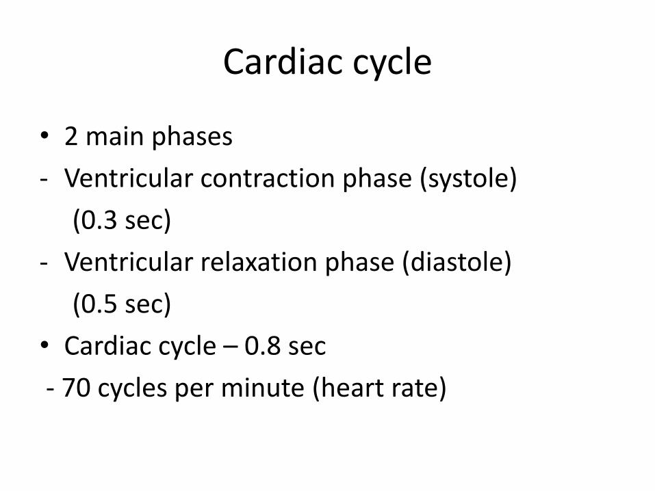
Cardiac cycle
• 2 main phases
- Ventricular contraction phase (systole)
(0.3 sec)
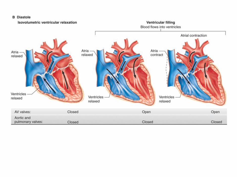
- Ventricular relaxation phase (diastole)
(0.5 sec)
• Cardiac cycle – 0.8 sec
- 70 cycles per minute (heart rate)
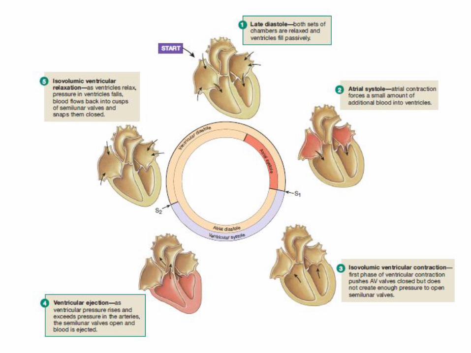
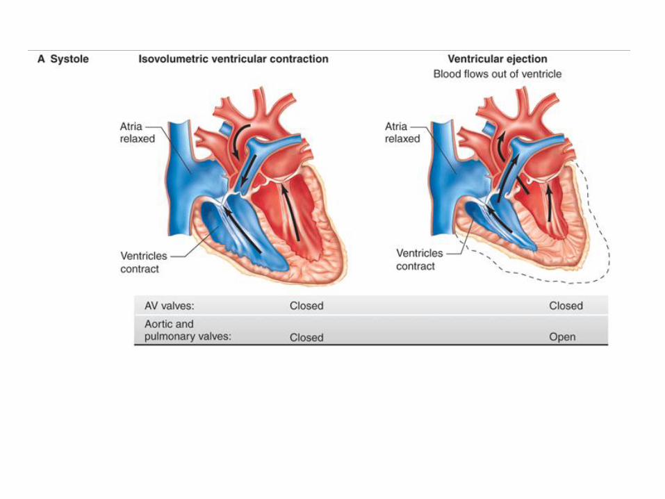
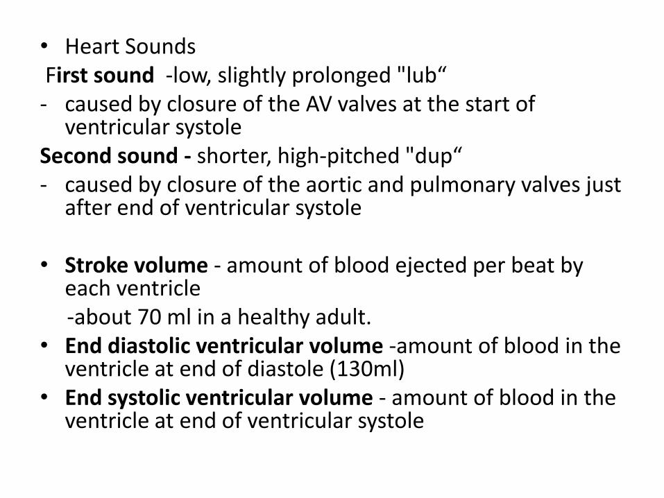
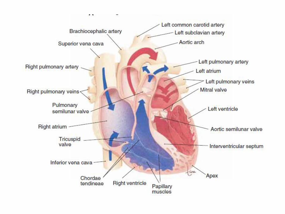
• Heart SoundsFirst sound -low, slightly prolonged "lub“- caused by closure of the AV valves at the start of
ventricular systole Second sound - shorter, high-pitched "dup“- caused by closure of the aortic and pulmonary valves just
after end of ventricular systole
• Stroke volume - amount of blood ejected per beat by each ventricle-about 70 ml in a healthy adult.
• End diastolic ventricular volume -amount of blood in the ventricle at end of diastole (130ml)
• End systolic ventricular volume - amount of blood in the ventricle at end of ventricular systole
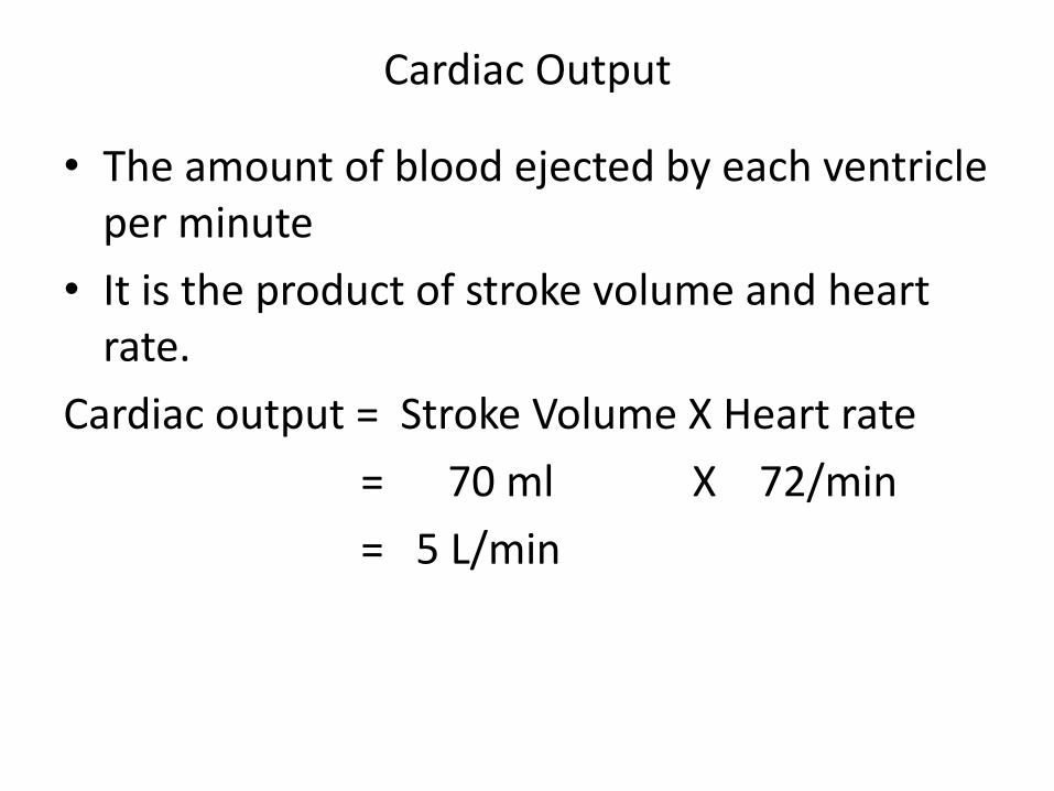
Cardiac Output
• The amount of blood ejected by each ventricle per minute
• It is the product of stroke volume and heart rate.
Cardiac output = Stroke Volume X Heart rate
= 70 ml X 72/min
= 5 L/min
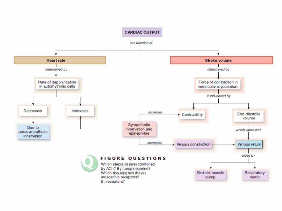
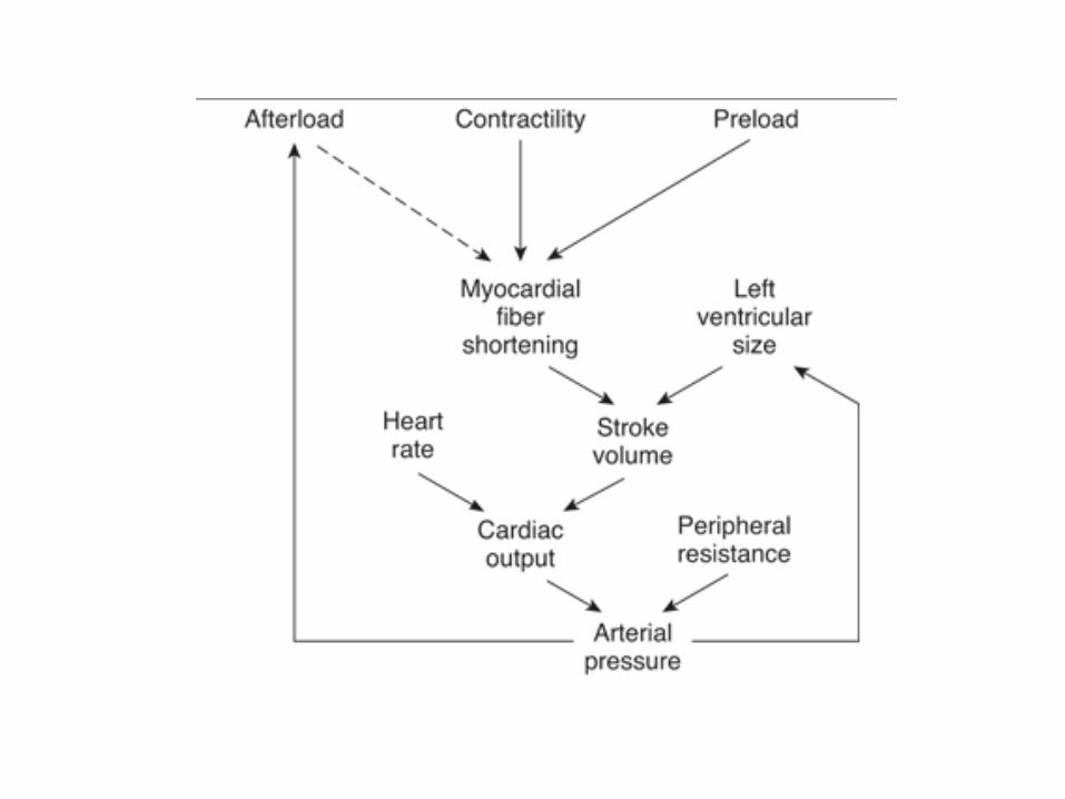
Factors regulating the cardiac output.
1. Heart rate
2. Preload - degree to which the myocardium is stretched before it contracts
3. Inotropic state of the heart (contractility)
4. Afterload - resistance against which blood is expelled.
1. Heart rate
- regulated by cardiac innervation (sympathetic / parasympathetic) and hormones (epinephrine, thyroxine)
2. Preload (venous return)
- the degree to which the myocardium is stretched before it contracts.
- It depends on the ventricular end diastolic volume (EDV)
Starling's law of the heart - "energy of contraction is proportional to the initial length of the cardiac muscle fiber"
- For the heart, the length of the muscle fibers (preload) is proportional to the end-diastolic volume
- Greater the preload (EDV) – increased force of contraction – increased stroke volume
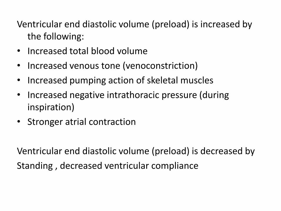
Ventricular end diastolic volume (preload) is increased by the following:
• Increased total blood volume
• Increased venous tone (venoconstriction)
• Increased pumping action of skeletal muscles
• Increased negative intrathoracic pressure (during inspiration)
• Stronger atrial contraction
Ventricular end diastolic volume (preload) is decreased by
Standing , decreased ventricular compliance
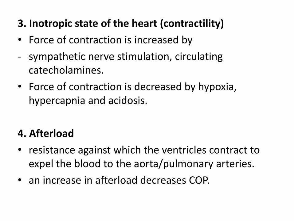
3. Inotropic state of the heart (contractility)
• Force of contraction is increased by
- sympathetic nerve stimulation, circulating catecholamines.
• Force of contraction is decreased by hypoxia, hypercapnia and acidosis.
4. Afterload
• resistance against which the ventricles contract to expel the blood to the aorta/pulmonary arteries.
• an increase in afterload decreases COP.
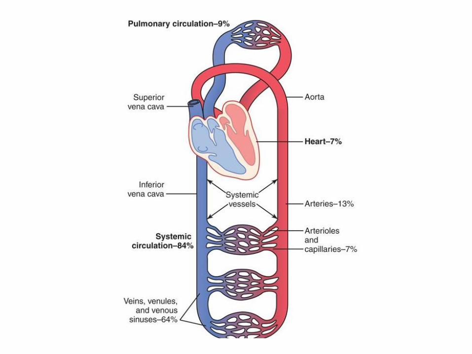
Dynamics of blood flow
• Factors that affect the blood flow through vessels
1. Pumping of the heart, is the primary factor
2. Diastolic recoil of arteries
3. Compression of veins by skeletal muscles during exercise.
4. Negative pressure in the thorax
5. Resistance to flow – diameter of vessel & velocity

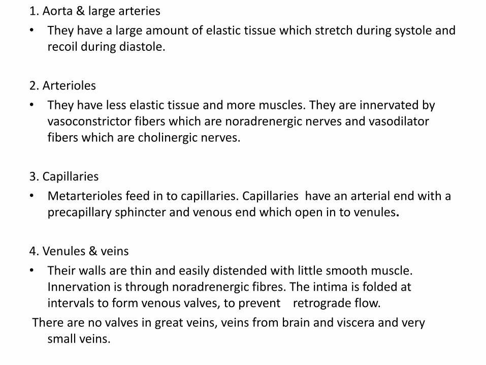
1. Aorta & large arteries
• They have a large amount of elastic tissue which stretch during systole and recoil during diastole.
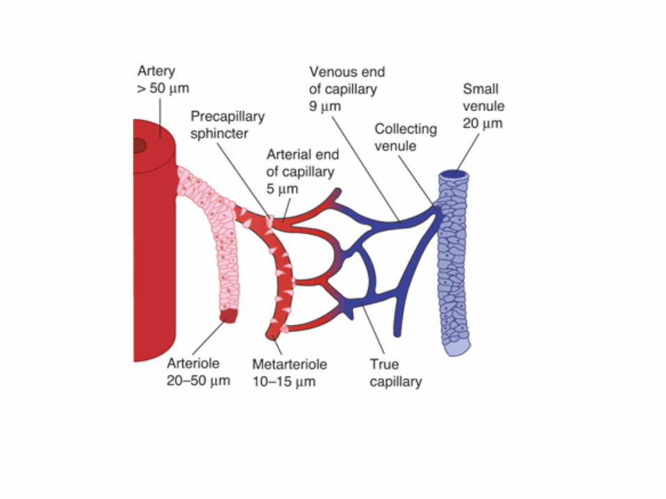
2. Arterioles
• They have less elastic tissue and more muscles. They are innervated by vasoconstrictor fibers which are noradrenergic nerves and vasodilator fibers which are cholinergic nerves.
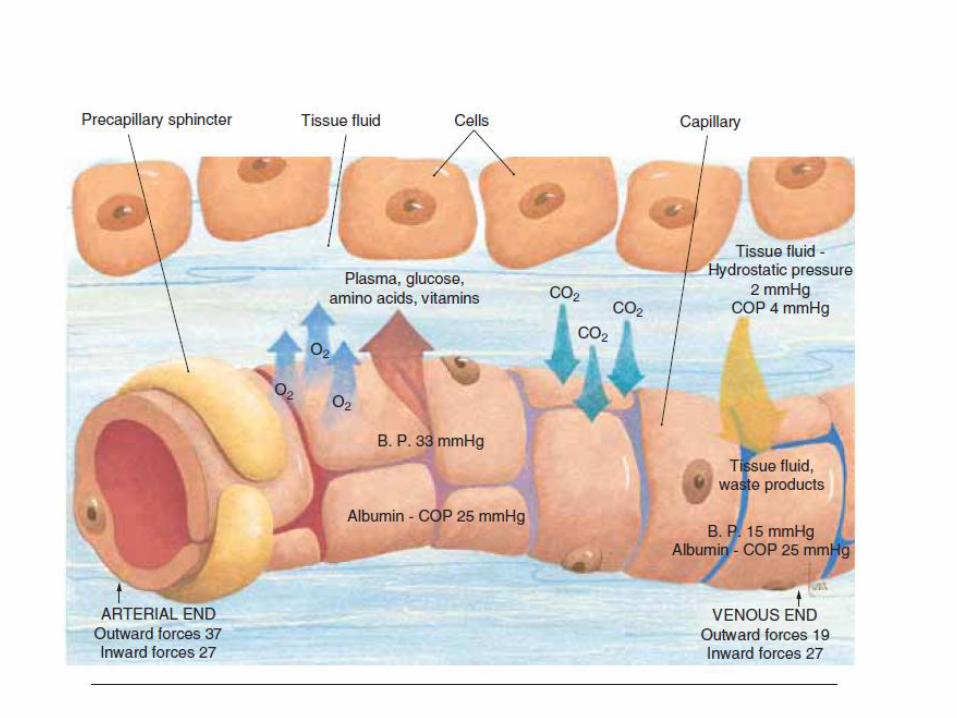
3. Capillaries
• Metarterioles feed in to capillaries. Capillaries have an arterial end with a precapillary sphincter and venous end which open in to venules.
4. Venules & veins
• Their walls are thin and easily distended with little smooth muscle. Innervation is through noradrenergic fibres. The intima is folded at intervals to form venous valves, to prevent retrograde flow.
There are no valves in great veins, veins from brain and viscera and very small veins.
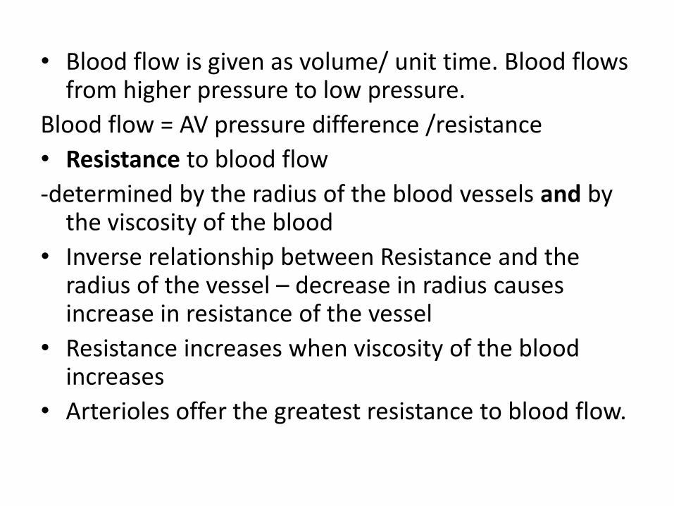
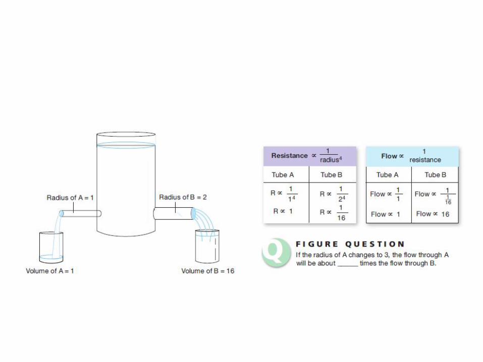
• Blood flow is given as volume/ unit time. Blood flows from higher pressure to low pressure.
Blood flow = AV pressure difference /resistance
• Resistance to blood flow
-determined by the radius of the blood vessels and by the viscosity of the blood
• Inverse relationship between Resistance and the radius of the vessel – decrease in radius causes increase in resistance of the vessel
• Resistance increases when viscosity of the blood increases
• Arterioles offer the greatest resistance to blood flow.
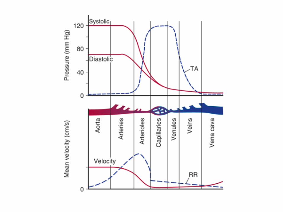
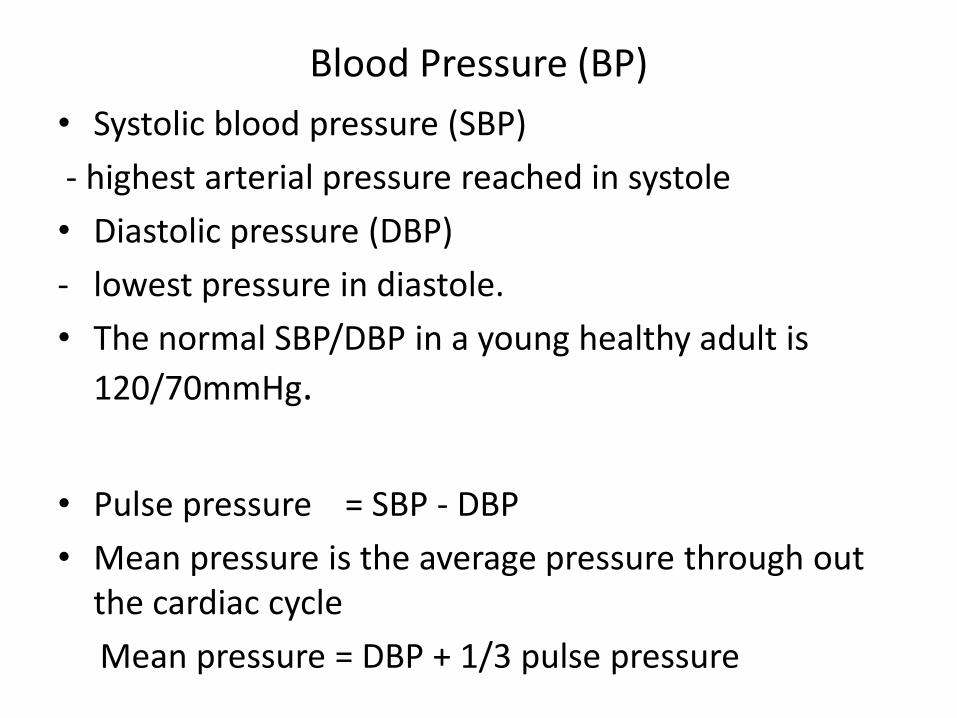
Blood Pressure (BP)
• Systolic blood pressure (SBP)
- highest arterial pressure reached in systole
• Diastolic pressure (DBP)
- lowest pressure in diastole.
• The normal SBP/DBP in a young healthy adult is
120/70mmHg.
• Pulse pressure = SBP - DBP
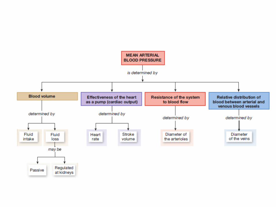
• Mean pressure is the average pressure through out the cardiac cycle
Mean pressure = DBP + 1/3 pulse pressure
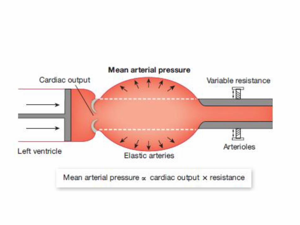
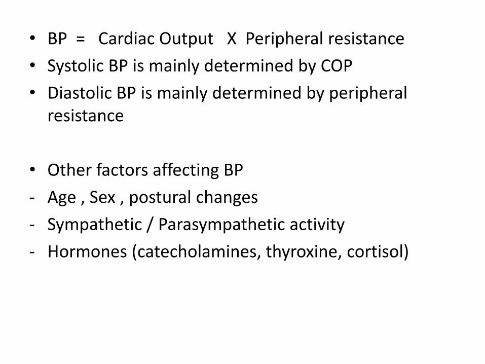
• BP = Cardiac Output X Peripheral resistance
• Systolic BP is mainly determined by COP
• Diastolic BP is mainly determined by peripheral resistance
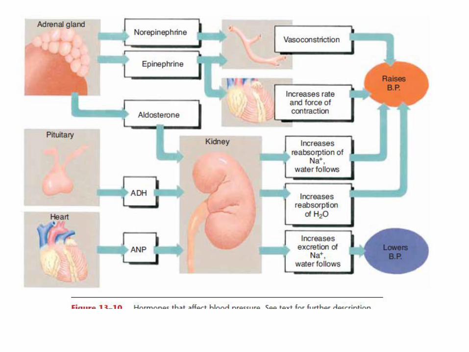
• Other factors affecting BP
- Age , Sex , postural changes
- Sympathetic / Parasympathetic activity
- Hormones (catecholamines, thyroxine, cortisol)
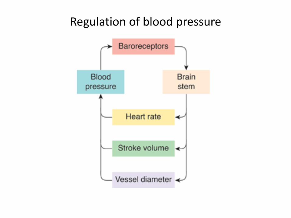
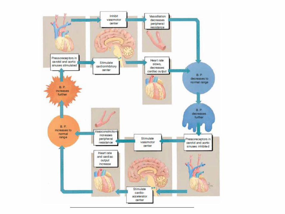
Regulation of blood pressure
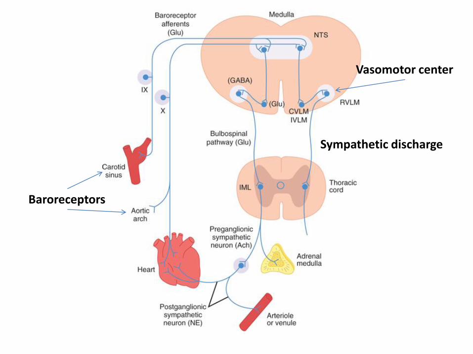
Vasomotor center
Sympathetic discharge
Baroreceptors
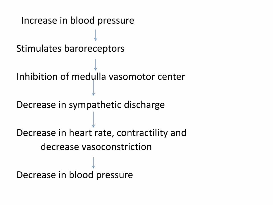
Increase in blood pressure
Stimulates baroreceptors
Inhibition of medulla vasomotor center
Decrease in sympathetic discharge
Decrease in heart rate, contractility and
decrease vasoconstriction
Decrease in blood pressure

Increase in blood pressure
Stimulates baroreceptors
Stimulation of medulla cardiac inhibitory center
Increased parasympathetic (vagal) discharge
Decrease in heart rate, vasodilation
Decrease in blood pressure
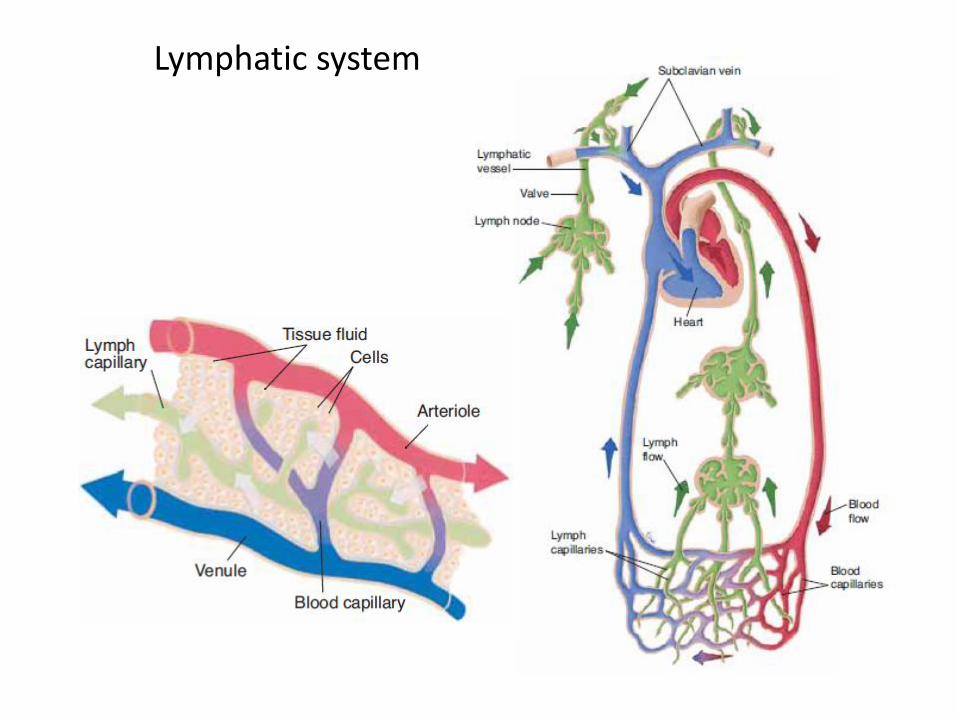
Lymphatic system