Deconstructing “reconciliation”: ideas on how to critically approach peacebuilding

Cardiovascular, Pulmonary and Renal Pathology
Interleukin-1 Receptor Type I Signaling CriticallyRegulates Infarct Healing and Cardiac Remodeling
Marcin Bujak, Marcin Dobaczewski,Khaled Chatila, Leonardo H. Mendoza, Na Li,Anilkumar Reddy, and Nikolaos G. FrangogiannisFrom the Section of Cardiovascular Sciences, Baylor College of
Medicine, Houston, Texas
The proinflammatory cytokine interleukin (IL)-1 sig-nals exclusively through the type I IL-1 receptor (IL-1RI). IL-1 expression is markedly induced in the in-farcted heart; however, its role in cardiac injury andrepair remains controversial. We examined the ef-fects of disrupted IL-1 signaling on infarct healing andcardiac remodeling using IL-1RI�/� mice. After reper-fused infarction IL-1RI-null mice exhibited decreasedinfiltration of the infarcted myocardium with neutro-phils and macrophages and reduced chemokine andcytokine expression. In the absence of IL-1 signaling,suppressed inflammation was followed by an attenu-ated fibrotic response. Infarcted IL-1RI�/� mice haddecreased myofibroblast infiltration and reduced col-lagen deposition in the infarcted and remodelingmyocardium. IL-1RI deficiency protected against thedevelopment of adverse remodeling; however, infarctsize was comparable between groups suggesting thatthe beneficial effects of IL-1RI gene disruption were notattributable to decreased cardiomyocyte injury. Re-duced chamber dilation in IL-1RI-null animals was asso-ciated with decreased collagen deposition and attenu-ated matrix metalloproteinase (MMP)-2 and MMP-3expression in the peri-infarct area, suggesting de-creased fibrotic remodeling of the noninfarcted heart.IL-1� stimulated MMP mRNA synthesis in wild-type, butnot in IL-1RI-null cardiac fibroblasts. In conclusion, IL-1signaling is essential for activation of inflammatory andfibrogenic pathways in the healing infarct, playing animportant role in the pathogenesis of remodeling afterinfarction. Thus, interventional therapeutics targetingthe IL-1 system may have great benefits in myocardialinfarction. (Am J Pathol 2008, 173:57–67; DOI:10.2353/ajpath.2008.070974)
Infarct healing is dependent on induction of an inflamma-tory cascade that ultimately results in formation of a collag-
en-based scar.1 The healing response is closely intertwinedwith ventricular remodeling, a complex process that in-volves both the infarcted and noninfarcted myocardiumresulting in dilation, hypertrophy, and enhanced sphericityof the ventricle.2 The extent of remodeling after infarction isan important predictor of mortality and adverse outcomeafter infarction,3,4 and depends on the size of the infarct andon the mechanical and structural characteristics of the heal-ing wound. Inflammatory mediators may be critically in-volved in the pathogenesis of cardiac remodeling by mod-ulating cell behavior in the infarcted heart and by regulatingextracellular matrix metabolism.5–7
Interleukin (IL)-1 plays a central role in regulating inflam-matory and fibrotic responses by inducing synthesis ofproinflammatory mediators, by promoting leukocyte infiltra-tion and activation, and by modulating fibroblast function.IL-1 binds to two distinct receptors on the cell membrane:the type I IL-1 receptor (IL-1RI) is sufficient to mediate allIL-1 actions,8 whereas the type II receptor (IL-1RII) servesas a decoy target,9 trapping and scavenging IL-1 mole-cules,10 thus reducing IL-1 concentration available for inter-action with the IL-1RI signaling receptor.
Although several studies have demonstrated that IL-1�is markedly induced in healing infarcts,11,12 the role ofIL-1 signaling in myocardial infarction remains poorlyunderstood and controversial. Administration of an anti-IL-1� neutralizing antibody in the acute phase of non-reperfused murine myocardial infarction was detrimental,resulting in reduced collagen accumulation in the scarand attenuated adverse remodeling.13 In contrast, inhi-bition of IL-1-mediated effects through overexpression ofIL-1R antagonist (IL-1Ra) decreased cardiomyocyte ap-optosis and reduced inflammation decreasing myocar-dial injury after reperfused infarction.14 Our study inves-tigates for the first time the effects of disruption of IL-1signaling on infarct healing and cardiac remodeling afterinfarction. We found that IL-1RI-null mice exhibit attenu-ated ventricular dilation after reperfused infarction. The
Supported by the National Institutes of Health (grants R01 HL-76246 andHL-85440).
Accepted for publication April 11, 2008.
Address reprint requests to Nikolaos G. Frangogiannis, M.D., Section ofCardiovascular Sciences, One Baylor Plaza BCM620, Baylor College ofMedicine, Houston, TX 77030. E-mail: [email protected].
The American Journal of Pathology, Vol. 173, No. 1, July 2008
Copyright © American Society for Investigative Pathology
DOI: 10.2353/ajpath.2008.070974
57

protective effects of disrupted IL-1 signaling on the re-modeling heart are not attributable to decreased cardio-myocyte injury, but are associated with a markedly sup-pressed cardiac inflammatory response and reduced fi-brosis of the noninfarcted myocardium. The IL-1 systemmay be a promising therapeutic target for patients withmyocardial infarction.
Materials and Methods
Murine Ischemia/Reperfusion Protocols
All animal studies were approved by the animal protocolreview committee at Baylor College of Medicine. IL-1RI�/� mice15 and WT C57/BL/6 controls (purchasedfrom The Jackson Laboratories, Bar Harbor, ME) wereused for myocardial infarction experiments. Female mice,8 to 12 weeks of age (18.0 to 22.0 g body weight) wereanesthetized by an intraperitoneal injection of sodiumpentobarbital (60 �g/g). A closed-chest mouse model ofreperfused myocardial infarction was used to avoid theconfounding effects of surgical trauma and inflammation,which may influence the baseline levels of chemokinesand cytokines.16 The left anterior descending coronaryartery was occluded for 1 hour then reperfused for 6hours to 7 days. At the end of the experiment, the chestwas opened and the heart was immediately excised,fixed in zinc-formalin, and embedded in paraffin for his-tological studies, or snap-frozen and stored at �80°C forRNA isolation. Sham animals were prepared identicallywithout undergoing coronary occlusion/reperfusion. Ani-mals used for histology underwent 24-hour, 72-hour, and7-day reperfusion protocols (eight animals per group).Mice used for RNA extraction underwent 6 hours, 24hours, and 72 hours of reperfusion (eight animals pergroup). To examine matrix metalloproteinase (MMP)and TIMP mRNA expression in the infarcted and re-modeling myocardium hearts from animals undergoing1 hour of ischemia and 7 days of reperfusion were used(eight mice per group). mRNA was extracted sepa-rately from the infarct and the remote noninfarctedmyocardium. Additional animals [knockout (KO), n �10; wild type (WT), n � 8] were used for perfusion-fixation after 7 days of reperfusion to assess remodel-ing-associated parameters.
Immunohistochemistry and QuantitativeHistology
Murine hearts were fixed in zinc-formalin (Z-fix; Anatech,Battle Creek, MI), and embedded in paraffin. Sectionswere cut at 5 �m and stained immunohistochemicallywith the following antibodies: anti-� smooth muscle actin(�-SMA) antibody (Sigma, St. Louis, MO), rat anti-mousemacrophage antibody Mac-2 (Cedarlane, Burlington,Canada), and rat anti-neutrophil antibody (Serotec, Ra-leigh, NC). Staining was performed using a peroxidase-based technique with the Vectastain ELITE rat, rabbit, orgoat kit (Vector Laboratories, Burlingame, CA) and de-veloped with diaminobenzidine plus nickel (Vector Lab-
oratories).17 The mouse on mouse (MOM) kit (VectorLaboratories) was used for �-SMA immunohistochemis-try.16 Quantitative assessment of neutrophil and macro-phage density was performed by counting the number ofneutrophils and Mac-2-immunoreactive cells, respec-tively, in the infarcted area.16 Myofibroblasts were iden-tified as extravascular �-SMA-positive cells and countedin the infarcted myocardium.6 Macrophage, neutrophil,and myofibroblast density was expressed as cells/mm2.The collagen network was identified using Picrosirius redstaining.16 Picrosirius red-stained slides from each in-farcted heart were scanned using a digital camera. Thepercentage of the collagen-stained area was assessed inthe infarcted myocardium, peri-infarct area, and remoteremodeling myocardium using ImagePro software (Me-dia Cybernetics, Bethesda, MD). Ten distinct fields fromtwo different sections were used for quantitative analysisof collagen content in each area.
Perfusion Fixation and Assessment ofVentricular Volumes
For assessment of remodeling after infarction, in-farcted hearts after 7 days of reperfusion were used forperfusion-fixation as previously described.16 The entireheart from base to apex was cross-sectioned at250-�m intervals. Ten serial 5-�m sections were ob-tained at each interval. The first section from eachinterval was stained with hematoxylin and eosin andwas used for morphometric assessment of left ventric-ular volumes and scar size. The left ventricular end-diastolic volume (LVEDV), left ventricular volume, sep-tal volume, and scar size were assessed with ImageProsoftware using methods developed in our laboratory.18–20
The size of the infarct was expressed as a percentage ofthe left ventricular volume.
Echocardiography
Short axis M-mode echocardiographic studies were per-formed before instrumentation and after 7 days of reper-fusion (WT, n � 7; IL-1RI�/�, n � 8) using an 8 MHz probe(Sequoia C256; Acuson, Mountain View, CA). The follow-ing parameters were measured as indicators of functionand remodeling: left ventricular end-diastolic diameter(LVEDD), left ventricular end-systolic diameter (LVESD),and fractional shortening (FS � [LVEDD � LVESD] �100/LVEDD). The percent change in these parametersafter infarction was quantitatively assessed using the fol-lowing formulas: �LVEDD � (LVEDD 7 days � LVEDDpre) � 100/LVEDD pre, �LVESD � (LVESD 7 days �LVESD pre) � 100/LVESD pre, �FS � (FS pre � FS 7days) � 100/FS pre.
RNA Extraction and Ribonuclease ProtectionAssay
Inflammatory gene expression in murine hearts wasassessed using a ribonuclease protection assay as
58 Bujak et alAJP July 2008, Vol. 173, No. 1

previously described.12,16 The mRNA expression levelof the chemokines MIP-1�, MIP-1�, MIP-2, MCP-1, andinterferon-�-inducible protein (IP)-10; the cytokines tu-mor necrosis factor (TNF)-�, IL-1�, IL-6, and IL-10; thegrowth factors transforming growth factor (TGF)-�1, -2,and -3, and M-CSF; the MMPs MMP-2, -3, -8, -9; andthe TIMPs, TIMP-1, -2, -3, and -4 were determinedusing a ribonuclease protection assay (RiboQuant;Pharmingen, Franklin Lakes, NJ) according to the man-ufacturer’s protocol. Phosphorimaging of the gels wasperformed (Storm 860; Molecular Dynamics, Sunny-vale, CA) and signals were quantified using ImageQuaNT software and normalized to the ribosomal pro-tein L32 mRNA.
Protein Extraction and Western Blotting
Protein was isolated from the infarct and the noninfarctedremodeling myocardium of WT and IL-1RI-null heartsafter 7 days of reperfusion. Western blotting was per-formed using a goat anti-MMP-2 and a rat anti-MMP-3antibody (both from R&D Systems, San Diego, CA) aspreviously described.6
Cardiac Fibroblast Isolation and Stimulation
Fibroblasts were isolated from murine WT and IL-1RI-null hearts, cultured as previously described,21,22 andstimulated with rIL-1� (10 ng/ml) (R&D Systems) for 2to 16 hours. At the end of the experiment total RNA wasisolated from the fibroblast cell lysates. MMP-2, -3, -8,-9, and TIMP-1, -2, -3, and -4 mRNA expressions wereassessed using a ribonuclease protection assay.
Statistical Analysis
Statistical analysis was performed using analysis ofvariance followed by t-test corrected for multiple com-parisons (Student-Newman-Keuls). Data were ex-pressed as mean � SEM. Statistical significance wasset at 0.05.
Results
IL-1RI Gene Disruption Did Not Affect CardiacHomeostasis
IL-1RI�/� and WT mice had comparable cardiac mor-phology. IL-1RI gene disruption did not affect macro-
phage density and the morphology of the extracellularmatrix network in the mouse heart (not shown). Further-more, in the absence of injury, IL-1RI and WT hearts hadcomparable cardiac function and chamber dimensions(Table 1).
IL-1RI-Null Animals Showed DecreasedMortality after Reperfusion
IL-1RI-null mice showed a trend toward decreased mor-tality after myocardial infarction (WT, 14.7%, versus IL-1RI�/�, 6.1%; P � 0.09). WT and IL-1RI-null mice hadcomparable mortality during coronary occlusion (WT,8.1%, versus IL-1RI�/�, 6.1%; P � NS); however, IL-1RI-null mice showed significantly decreased mortality duringreperfusion (WT, 7.2%, versus IL-1RI�/�, 1.1%; P �0.05). The mortality rate during the initial instrumentationsurgery was comparable between groups (WT,: 4.9%,versus IL-1RI�/�, 5.8%; P � NS).
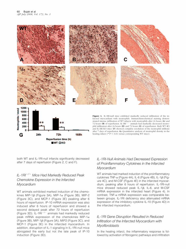
IL-1RI-Null Mice Exhibited Markedly ReducedNeutrophil Infiltration in the InfarctedMyocardium
WT infarcts showed intense neutrophil infiltration, peak-ing after 24 hours of reperfusion (Figure 1A). IL-1RI genedisruption markedly reduced neutrophil density after 24hours (IL-1RI�/�, 223.5 � 38.92 cells/mm2, versus WT,1037 � 55.32 cells/mm2; P � 0.001) and 72 hours (IL-1RI�/�, 156.0 � 25.28 cells/mm2, versus WT, 629.1 �57.67 cells/mm2; P � 0.001) of reperfusion (Figure 1).Both IL-1RI-null and WT animals had almost completeresolution of the neutrophilic infiltrate after 7 days ofreperfusion (Figure 1, C, F, and G).
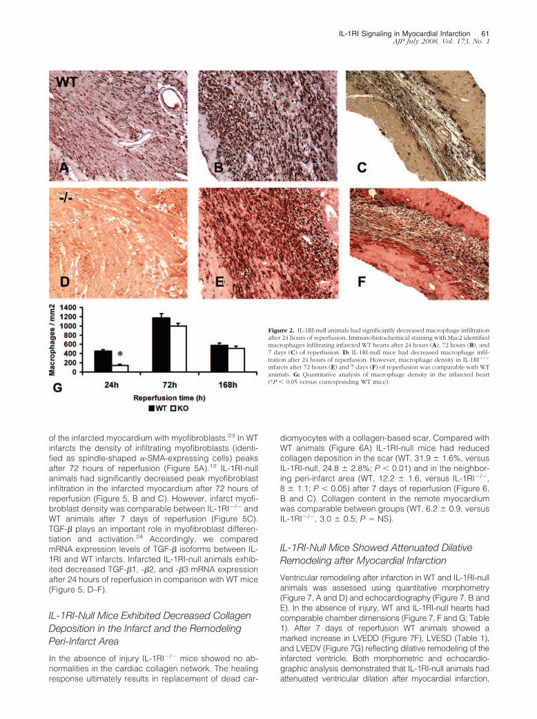
IL-1RI Gene Disruption Delayed Infiltration of theInfarcted Myocardium with Macrophages
Macrophages were identified in the infarcted mouseheart using Mac-2 immunohistochemistry (Figure 2). Incomparison with WT mice (Figure 2, A–C), IL-1RI-nullanimals had significantly lower macrophage density inthe infarcted myocardium after 24 hours of reperfusion(Figure 2, D and G). However, peak macrophage densityafter 72 hours of reperfusion was comparable betweengroups (Figure 2, B, E, and G). Macrophage density in
Table 1. Assessment of Echocardiographic Parameters in the Infarcted Heart
n � 14 WT pre WT 168h IL1RKO pre IL1RKO 168h
LVEDD 3.37 � 0.057 3.74 � 0.067* 3.35 � 0.054 3.56 � 0.05*†
LVESD 1.97 � 0.052 2.42 � 0.066* 1.93 � 0.067 2.32 � 0.064*FS 0.42 � 0139 0.35 � 0.010* 0.41 � 0.016 0.35 � 0.013*�LVEDD (%) 10.98 � 2.247 6.601 � 1.168 (P � 0.09)�FS (%) 17.79 � 3.470 17.2 � 3.507
*P � 0.01 versus corresponding pre.†P � 0.01 versus corresponding WT.
IL-1RI Signaling in Myocardial Infarction 59AJP July 2008, Vol. 173, No. 1
both WT and IL-1RI-null infarcts significantly decreasedafter 7 days of reperfusion (Figure 2, C and F).
IL-1RI�/� Mice Had Markedly Reduced PeakChemokine Expression in the InfarctedMyocardium
WT animals exhibited marked induction of the chemo-kines MIP-1� (Figure 3A), MIP-1� (Figure 3B), MIP-2(Figure 3C), and MCP-1 (Figure 3E) peaking after 6hours of reperfusion. IP-10 mRNA expression was alsoinduced after 6 hours of reperfusion and showed asecond delayed peak after 72 hours of reperfusion(Figure 3D). IL-1RI�/� animals had markedly reducedpeak mRNA expression of the chemokines MIP-1�
(Figure 3B), MIP-1� (Figure 3A), MIP2 (Figure 3C), andMCP-1 (Figure 3E) in the infarcted myocardium. Inaddition, disruption of IL-1 signaling in IL-1RI-null miceabrogated the early but not the late peak of IP-10induction (Figure 3D).
IL-1RI-Null Animals Had Decreased Expressionof Proinflammatory Cytokines in the InfarctedMyocardium
WT animals had marked induction of the proinflammatorycytokines TNF-� (Figure 4A), IL-6 (Figure 4B), IL-1� (Fig-ure 4C), and M-CSF (Figure 4E) in the infarcted myocar-dium, peaking after 6 hours of reperfusion. IL-1RI-nullmice showed reduced peak IL-1�, IL-6, and M-CSFmRNA expression in the infarcted heart (Figure 4). Incontrast, TNF-� mRNA expression was comparable be-tween groups. IL-1RI deficiency also attenuated mRNAexpression of the inhibitory cytokine IL-10 (Figure 4D) inthe infarcted myocardium.
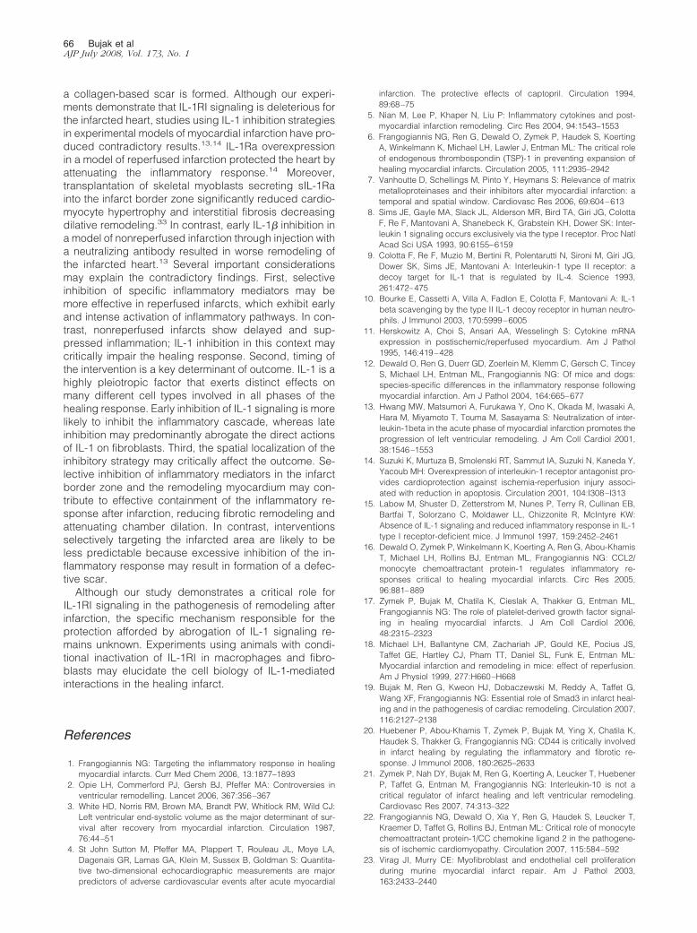
IL-1RI Gene Disruption Resulted in ReducedInfiltration of the Infarcted Myocardium withMyofibroblasts
In the healing infarct, the inflammatory response is fol-lowed by activation of fibrogenic pathways and infiltration
Figure 1. IL-1RI-null mice exhibited markedly reduced infiltration of the in-farcted myocardium with neutrophils. Immunohistochemical staining demon-strated intense infiltration of WT infarcts with neutrophils after 24 hours (A) and72 hours (B) of reperfusion. IL-1RI�/� animals had markedly decreased neutro-phil infiltration after 24 hours (D) and 72 hours (E) of reperfusion. Both WT (C)and IL-1RI KO mice (F) showed complete resolution of the neutrophil infiltrateafter 7 days of reperfusion. G: Quantitative analysis of neutrophil density in thehealing infarct (**P � 0.01 versus corresponding WT mice).
60 Bujak et alAJP July 2008, Vol. 173, No. 1
of the infarcted myocardium with myofibroblasts.23 In WTinfarcts the density of infiltrating myofibroblasts (identi-fied as spindle-shaped �-SMA-expressing cells) peaksafter 72 hours of reperfusion (Figure 5A).12 IL-1RI-nullanimals had significantly decreased peak myofibroblastinfiltration in the infarcted myocardium after 72 hours ofreperfusion (Figure 5, B and C). However, infarct myofi-broblast density was comparable between IL-1RI�/� andWT animals after 7 days of reperfusion (Figure 5C).TGF-� plays an important role in myofibroblast differen-tiation and activation.24 Accordingly, we comparedmRNA expression levels of TGF-� isoforms between IL-1RI and WT infarcts. Infarcted IL-1RI-null animals exhib-ited decreased TGF-�1, -�2, and -�3 mRNA expressionafter 24 hours of reperfusion in comparison with WT mice(Figure 5, D–F).
IL-1RI-Null Mice Exhibited Decreased CollagenDeposition in the Infarct and the RemodelingPeri-Infarct Area
In the absence of injury IL-1RI�/� mice showed no ab-normalities in the cardiac collagen network. The healingresponse ultimately results in replacement of dead car-
diomyocytes with a collagen-based scar. Compared withWT animals (Figure 6A) IL-1RI-null mice had reducedcollagen deposition in the scar (WT, 31.9 � 1.6%, versusIL-1RI-null, 24.8 � 2.8%; P � 0.01) and in the neighbor-ing peri-infarct area (WT, 12.2 � 1.6, versus IL-1RI�/�,8 � 1.1; P � 0.05) after 7 days of reperfusion (Figure 6,B and C). Collagen content in the remote myocardiumwas comparable between groups (WT, 6.2 � 0.9, versusIL-1RI�/�, 3.0 � 0.5; P � NS).
IL-1RI-Null Mice Showed Attenuated DilativeRemodeling after Myocardial Infarction
Ventricular remodeling after infarction in WT and IL-1RI-nullanimals was assessed using quantitative morphometry(Figure 7, A and D) and echocardiography (Figure 7, B andE). In the absence of injury, WT and IL-1RI-null hearts hadcomparable chamber dimensions (Figure 7, F and G; Table1). After 7 days of reperfusion WT animals showed amarked increase in LVEDD (Figure 7F), LVESD (Table 1),and LVEDV (Figure 7G) reflecting dilative remodeling of theinfarcted ventricle. Both morphometric and echocardio-graphic analysis demonstrated that IL-1RI-null animals hadattenuated ventricular dilation after myocardial infarction,
Figure 2. IL-1RI-null animals had significantly decreased macrophage infiltrationafter 24 hours of reperfusion. Immunohistochemical staining with Mac2 identifiedmacrophages infiltrating infarcted WT hearts after 24 hours (A), 72 hours (B), and7 days (C) of reperfusion. D: IL-1RI-null mice had decreased macrophage infil-tration after 24 hours of reperfusion. However, macrophage density in IL-1RI�/�
infarcts after 72 hours (E) and 7 days (F) of reperfusion was comparable with WTanimals. G: Quantitative analysis of macrophage density in the infarcted heart(*P � 0.05 versus corresponding WT mice).
IL-1RI Signaling in Myocardial Infarction 61AJP July 2008, Vol. 173, No. 1

showing significantly lower LVEDD and LVEDV (Figure 7,Table 1) than their WT littermates. Attenuated adverse re-modeling of the infarcted ventricle was not attributable to adifference in infarct size (IL-1RI�/�, 16.1 � 1.7%, versus
WT, 15.0 � 1.3%; P � NS) (Figure 7C). WT and IL-1RI-nullanimals exhibited comparable deterioration in systolic func-tion after 7 days of reperfusion (FSWT, 0.353 � 0.01, versusIL-1RI�/�, 0.346 � 0.01; P � NS) (Table 1).
Figure 3. IL-1RI�/� mice exhibited markedly reduced chemokine up-regulation in the infarcted heart. WT mice showed marked early mRNA induction of thechemokines MIP-1� (A), MIP-1� (B), MIP2 (C), and MCP-1 (E) in the infarcted myocardium. D: IP-10 induction showed a biphasic response. IL-1RI gene disruptionprevented the early chemokine response in the infarcted heart suggesting a key role for IL-1 signaling in mediating chemokine synthesis after ischemia andreperfusion (***P � 0.001, **P � 0.01, *P � 0.05 versus corresponding WT mice).
Figure 4. IL-1RI�/� mice had decreased cytokine up-regulation in the infarcted heart. mRNA expression of the proinflammatory cytokines TNF-� (A), IL-6 (B),IL-1� (C), and M-CSF (E) peaked after 6 hours of reperfusion in WT mouse infarcts. IL-1RI KO mice exhibited decreased peak IL-6 (B), IL-1� (C), and M-CSF (E)mRNA expression. A: In contrast, TNF-� expression levels were comparable between groups. D: Furthermore, IL-1RI-deficient mice had significantly attenuatedinduction of the inhibitory cytokine IL-10 after 24 hours of reperfusion (*P � 0.05, **P � 0.01 versus corresponding WT mice).
62 Bujak et alAJP July 2008, Vol. 173, No. 1

Disrupted IL-1 Signaling Results in DecreasedMMP-2 and MMP-3 mRNA and ProteinExpression in the Infarcted Heart
Because MMPs are critically involved in remodeling ofthe infarcted ventricle, we examined mRNA expression ofMMPs and their inhibitors in infarcted and remote remod-eling areas of the heart after 7 days of reperfusion. IL-1RIKO mice exhibited significantly reduced MMP-2 mRNAlevels in the infarcted and remodeling areas of the ven-tricle (Table 2). In addition, MMP-3 mRNA expressionwas lower in IL-1RI-null infarcts. Western blotting exper-iments demonstrated that IL-1RI deficiency was associ-ated with reduced MMP-2 and MMP-3 protein levels inthe infarcted myocardium. In contrast, TIMP-4 mRNAlevels were higher in infarcted and noninfarcted IL-1RIcardiac segments (Table 2). MMP-8, MMP-9, TIMP-1,
and TIMP-2 mRNA expression was comparable betweengroups. The findings indicated that IL-1RI deficiency al-tered the MMP:TIMP expression profile in the infarct andthe remote remodeling heart reducing MMP-2 and -3expression.
IL-1� Induces MMP Synthesis in IsolatedMurine Cardiac Fibroblasts through anIL-1RI-Dependent Mechanism
IL-1� stimulation significantly enhanced MMP-3,MMP-8, and MMP-9 mRNA synthesis and reducedTIMP-2 and TIMP-4 mRNA expression by isolated WTmouse cardiac fibroblasts (Table 3). However, fibro-blast MMP-2, TIMP-1, and TIMP-3 mRNA expressionwas not significantly affected by IL-1� stimulation. In
Figure 5. IL-1RI deficiency is associated with reduced myofibroblast infiltration and decreased TGF-� isoform expression in the infarcted heart. Myofibroblastswere identified as spindle-shaped, �-SMA-expressing cells, predominantly located in the border zone of WT (A) and IL-1RI-null (B) infarcts (arrows). C:Quantitative analysis demonstrated that peak myofibroblast density in the infarcted myocardium was significantly lower in IL-1RI-null animals. D–F: TGF-� is akey mediator involved in myofibroblast differentiation and fibrous tissue deposition. TGF-�1 (D), TGF-�2 (E), and TGF-�3 (F) mRNA levels were significantlylower in infarcted IL-1RI KO hearts after 24 hours of reperfusion (*P � 0.05 versus corresponding WT mice).
Figure 6. IL-1RI-null mice exhibited attenuated fibrotic remodeling of the infarcted ventricle. Picrosirius red staining was used to label the collagen network inWT (A) and IL-1RI-null (B) infarcts. Collagen content was quantitatively assessed in the infarct (arrowheads), the peri-infarct area (Peri-I, arrows), and the remotemyocardium. C: IL-1RI KO mice had significantly lower collagen percent content in the infarct and the peri-infarct area (**P� 0.01, *P� 0.05 versus correspondingWT).
IL-1RI Signaling in Myocardial Infarction 63AJP July 2008, Vol. 173, No. 1

contrast, fibroblasts isolated from IL-1RI-null mice didnot respond to IL-1� stimulation, exhibiting MMP andTIMP mRNA expression levels comparable with un-stimulated fibroblasts (Table 3).
Discussion
Our findings demonstrate a critical role for IL-1-mediatedinteractions in the pathogenesis of adverse cardiac re-modeling after myocardial infarction. Disruption of IL-1signaling in genetically targeted IL-1RI-null mice pro-tected from the development of ventricular dilation after
reperfused infarction. The protective effect of IL-1RI de-ficiency was not attributable to a reduction in the extent ofcardiomyocyte injury, but was associated with a mark-edly suppressed inflammatory response, attenuated fi-brous tissue deposition in the peri-infarct area and analtered MMP:TIMP expression profile.
IL-1� and IL-1� are pleiotropic cytokines that activateinflammatory pathways, signaling exclusively via the typeI receptor.8 IL-1� is markedly induced in the infarctedheart,11,12 and is rapidly released in the plasma of pa-tients with acute myocardial infarction.25 Our findingsdemonstrate that IL-1 signaling plays a key role in trig-
Figure 7. IL-1RI�/� animals had attenuated adverse remodeling after myocardial infarc-tion. Remodeling after infarction was assessed using morphometric (A: WT; D: IL-1RI�/�)and echocardiographic techniques (B: WT; E: IL-1RI�/�). Although WT and KO animalshad comparable infarct size after 7 days of reperfusion (C), IL-1RI deficiency protectedfrom the development of ventricular dilation. Both echocardiographically derived LVEDD(F) and morphometrically assessed LVEDV (G) were lower in infarcted IL-1RI-null mice 7days after reperfusion in comparison with WT animals (##P � 0.01 versus pre or sham,*P � 0.05 versus corresponding WT).
Table 2. MMP and TIMP Expression Profile in the Infarcted and Remote Remodeling Myocardium after 7 Days of Reperfusion
WT infarct KO infarct WT remote KO remote
MMP/TIMP mRNA (ratio to L32)MMP2 0.515 � 0.022 0.388 � 0.028* 0.179 � 0.013 0.153 � 0.0034*MMP3 0.037 � 0.004 0.024 � 0.004* 0.016 � 0.002 0.022 � 0.002*MMP8 0.024 � 0.002 0.034 � 0.005 0.028 � 0.003 0.038 � 0.006MMP9 0.037 � 0.004 0.028 � 0.004 0.025 � 0.002 0.025 � 0.003TIMP1 0.264 � 0.0142 0.282 � 0.030 0.150 � 0.017 0.126 � 0.016TIMP2 0.395 � 0.016 0.456 � 0.036 0.350 � 0.025 0.316 � 0.021TIMP3 0.310 � 0.017 0.236 � 0.028* 0.118 � 0.017 0.268 � 0.016†
TIMP4 0.024 � 0.0054 0.051 � 0.007† 0.086 � 0.001 0.113 � 0.008*MMP protein (ratio to GAPDH)
MMP-2 0.95 � 0.12 0.61 � 0.06† 0.21 � 0.04 0.14 � 0.02MMP-3 1.05 � 0.16 0.6 � 0.08† 0.68 � 0.06 0.35 � 0.03
*P � 0.05 and †P � 0.01 versus corresponding WT.
64 Bujak et alAJP July 2008, Vol. 173, No. 1

gering the inflammatory cascade in the infarcted myocar-dium; these observations are consistent with a previousinvestigation demonstrating an attenuated inflammatoryreaction in IL-1RI-null animals after hepatic ischemia.26
Peak expression of cytokines and chemokines was sig-nificantly decreased in IL-1RI-null myocardial infarcts(Figures 3 and 4). Furthermore, IL-1RI gene disruptionmarkedly reduced neutrophil infiltration and delayedmacrophage recruitment in the infarcted myocardium(Figures 1 and 2). The greatly diminished neutrophil den-sity in IL-1RI-null infarcts may reflect both decreasedrecruitment of neutrophils and their increased suscepti-bility to apoptosis. IL-1 strongly prolongs neutrophil sur-vival by inhibiting their apoptotic death.27
Suppressed inflammation in ischemic IL-1RI-nullhearts was not associated with less extensive infarction,suggesting that endogenous IL-1 does not exacerbatecardiomyocyte injury. Suzuki and co-workers14 have pre-viously demonstrated that IL-1Ra overexpression bygene transfection resulted in reduced cardiomyocyte ap-optosis and protection from ischemic injury in heterotopi-cally transplanted rat hearts undergoing coronary occlu-sion and reperfusion. Although our findings also supporta beneficial effect of disrupted IL-1 signaling in healinginfarction, the mechanisms of protection do not involveattenuation of ischemic cardiomyocyte injury.
The suppressed inflammatory reaction in IL-1RI-nullinfarcts was followed by an attenuated fibrotic response.Myofibroblast accumulation in the infarcted area wassignificantly lower in IL-1RI�/� infarcts in comparison withWT animals (Figure 5). In addition, expression of the keyprofibrotic mediator TGF-�28 was significantly reduced,and collagen deposition was markedly decreased, inboth the healing scar and the peri-infarct area of IL-1RI�/� hearts (Figures 5 and 6). In the absence of IL-1signaling, reduced fibrotic remodeling of the infarctedventricle may be attributable to an attenuated inflamma-tory reaction and to the loss of direct stimulatory IL-1-mediated effects on cardiac fibroblast phenotype andfunction. IL-1� directly enhances fibrous tissue deposi-tion by up-regulating expression of angiotensin II type 1(AT1) receptors on cardiac fibroblasts29 and by stimulat-ing fibroblast migration.30
Beyond its proinflammatory and fibrogenic properties,IL-1 also promotes extracellular matrix remodeling byenhancing cardiac fibroblast MMP expression.31 We
found that IL-1� stimulation induced MMP-3, MMP-8, andMMP-9 mRNA synthesis by isolated mouse cardiac fibro-blasts, while down-regulating TIMP-2 and TIMP-4 expres-sion levels (Table 3). In the complex and dynamic envi-ronment of the infarct, where cellular behavior isregulated by a variety of mediators, the contribution ofdirect IL-1-mediated actions on fibroblast protease ex-pression and extracellular matrix remodeling is difficult toassess. In comparison with WT animals, IL-1RI-null miceexhibited a decrease in MMP-2 and MMP-3 expression inboth the infarcted and remote remodeling myocardium,supporting the in vivo relevance of IL-1-mediated effectson synthesis of matrix-degrading proteases.
Our experiments demonstrated that the cellular andmolecular alterations observed in IL-1RI-null infarcts re-sult in significant attenuation of dilative remodeling afterinfarction (Figure 7, Table 2). Protection from adverseremodeling in the absence of IL-1 signaling was notattributable to enhanced cardiomyocyte survival. Despitethe marked suppression of the inflammatory responseafter infarction in infarcted IL-1RI-null animals, infarct sizewas comparable with WT mice, suggesting that IL-1-mediated inflammatory activity does not accentuate isch-emic injury. However, suppression of inflammation maybe protective by reducing fibrotic remodeling of the in-farcted ventricle. Decreased interstitial fibrosis and re-duced MMP synthesis in the remodeling noninfarctedmyocardium of IL-1RI-null hearts may indicate attenuatedinterstitial remodeling. Fibrosis is often associated withenhanced matrix degradation indicating active remodel-ing of the interstitial space.32 IL-1RI gene disruption ap-pears to abrogate both events resulting in decreasedactivity of the remodeling interstitial space and attenu-ated ventricular dilation. The beneficial effects of defec-tive IL-1 signaling in remodeling after infarction may bemediated both through suppression of the inflammatoryresponse and through the loss of direct IL-1-mediatedeffects on cardiac fibroblasts.
Our investigation identifies the IL-1 signaling pathwayas a potential therapeutic target for the treatment of pa-tients with acute myocardial infarction. Disruption of IL-1signaling prevents the development of maladaptive fibro-sis after myocardial infarction resulting in attenuated ad-verse remodeling. Despite the suppression of the inflam-matory response after infarction, clearance of the infarctfrom dead cardiomyocytes occurs in a timely manner and
Table 3. Effects of IL-1� Stimulation (10 ng/ml) on MMP and TIMP mRNA Expression by Isolated WT and IL-1RI-Null CardiacFibroblasts (-Fold Increase Versus Corresponding Control Unstimulated Fibroblasts)
2 hours 6 hours 16 hours
WT �/� WT �/� WT �/�
MMP-2 1.06 � 0.19 0.84 � 0.11 1.16 � 0.12 0.73 � 0.13 1.40 � 0.17 0.81 � 0.13MMP-3 1.69 � 0.25 1.01 � 0.06 3.47 � 0.32† 0.92 � 0.10 5.74 � 0.32† 1.00 � 0.08MMP-8 1.22 � 0.05 1.07 � 0.08 1.85 � 0.36 0.95 � 0.06 3.98 � 0.82† 0.97 � 0.06MMP-9 1.59 � 0.01* 0.87 � 0.07 3.45 � 0.13† 0.92 � 0.03 5.86 � 0.12† 0.88 � 0.03TIMP-1 0.96 � 0.15 0.88 � 0.18 0.80 � 0.27 0.90 � 0.17 0.70 � 0.21 0.89 � 0.17TIMP-2 0.84 � 0.07 1.14 � 0.11 0.56 � 0.12* 1.15 � 0.02 0.54 � 0.08† 1.11 � 0.20TIMP-3 1.09 � 0.24 0.91 � 0.05 1.16 � 0.73 0.76 � 0.11 0.8 � 0.28 0.83 � 0.11TIMP-4 0.63 � 0.02† 0.88 � 0.07 0.58 � 0.03† 0.84 � 0.17 0.45 � 0.01† 0.98 � 0.14
*P � 0.05 and †P � 0.01 versus corresponding control.
IL-1RI Signaling in Myocardial Infarction 65AJP July 2008, Vol. 173, No. 1
a collagen-based scar is formed. Although our experi-ments demonstrate that IL-1RI signaling is deleterious forthe infarcted heart, studies using IL-1 inhibition strategiesin experimental models of myocardial infarction have pro-duced contradictory results.13,14 IL-1Ra overexpressionin a model of reperfused infarction protected the heart byattenuating the inflammatory response.14 Moreover,transplantation of skeletal myoblasts secreting sIL-1Rainto the infarct border zone significantly reduced cardio-myocyte hypertrophy and interstitial fibrosis decreasingdilative remodeling.33 In contrast, early IL-1� inhibition ina model of nonreperfused infarction through injection witha neutralizing antibody resulted in worse remodeling ofthe infarcted heart.13 Several important considerationsmay explain the contradictory findings. First, selectiveinhibition of specific inflammatory mediators may bemore effective in reperfused infarcts, which exhibit earlyand intense activation of inflammatory pathways. In con-trast, nonreperfused infarcts show delayed and sup-pressed inflammation; IL-1 inhibition in this context maycritically impair the healing response. Second, timing ofthe intervention is a key determinant of outcome. IL-1 is ahighly pleiotropic factor that exerts distinct effects onmany different cell types involved in all phases of thehealing response. Early inhibition of IL-1 signaling is morelikely to inhibit the inflammatory cascade, whereas lateinhibition may predominantly abrogate the direct actionsof IL-1 on fibroblasts. Third, the spatial localization of theinhibitory strategy may critically affect the outcome. Se-lective inhibition of inflammatory mediators in the infarctborder zone and the remodeling myocardium may con-tribute to effective containment of the inflammatory re-sponse after infarction, reducing fibrotic remodeling andattenuating chamber dilation. In contrast, interventionsselectively targeting the infarcted area are likely to beless predictable because excessive inhibition of the in-flammatory response may result in formation of a defec-tive scar.
Although our study demonstrates a critical role forIL-1RI signaling in the pathogenesis of remodeling afterinfarction, the specific mechanism responsible for theprotection afforded by abrogation of IL-1 signaling re-mains unknown. Experiments using animals with condi-tional inactivation of IL-1RI in macrophages and fibro-blasts may elucidate the cell biology of IL-1-mediatedinteractions in the healing infarct.
References
1. Frangogiannis NG: Targeting the inflammatory response in healingmyocardial infarcts. Curr Med Chem 2006, 13:1877–1893
2. Opie LH, Commerford PJ, Gersh BJ, Pfeffer MA: Controversies inventricular remodelling. Lancet 2006, 367:356–367
3. White HD, Norris RM, Brown MA, Brandt PW, Whitlock RM, Wild CJ:Left ventricular end-systolic volume as the major determinant of sur-vival after recovery from myocardial infarction. Circulation 1987,76:44–51
4. St John Sutton M, Pfeffer MA, Plappert T, Rouleau JL, Moye LA,Dagenais GR, Lamas GA, Klein M, Sussex B, Goldman S: Quantita-tive two-dimensional echocardiographic measurements are majorpredictors of adverse cardiovascular events after acute myocardial
infarction. The protective effects of captopril. Circulation 1994,89:68–75
5. Nian M, Lee P, Khaper N, Liu P: Inflammatory cytokines and post-myocardial infarction remodeling. Circ Res 2004, 94:1543–1553
6. Frangogiannis NG, Ren G, Dewald O, Zymek P, Haudek S, KoertingA, Winkelmann K, Michael LH, Lawler J, Entman ML: The critical roleof endogenous thrombospondin (TSP)-1 in preventing expansion ofhealing myocardial infarcts. Circulation 2005, 111:2935–2942
7. Vanhoutte D, Schellings M, Pinto Y, Heymans S: Relevance of matrixmetalloproteinases and their inhibitors after myocardial infarction: atemporal and spatial window. Cardiovasc Res 2006, 69:604–613
8. Sims JE, Gayle MA, Slack JL, Alderson MR, Bird TA, Giri JG, ColottaF, Re F, Mantovani A, Shanebeck K, Grabstein KH, Dower SK: Inter-leukin 1 signaling occurs exclusively via the type I receptor. Proc NatlAcad Sci USA 1993, 90:6155–6159
9. Colotta F, Re F, Muzio M, Bertini R, Polentarutti N, Sironi M, Giri JG,Dower SK, Sims JE, Mantovani A: Interleukin-1 type II receptor: adecoy target for IL-1 that is regulated by IL-4. Science 1993,261:472–475
10. Bourke E, Cassetti A, Villa A, Fadlon E, Colotta F, Mantovani A: IL-1beta scavenging by the type II IL-1 decoy receptor in human neutro-phils. J Immunol 2003, 170:5999–6005
11. Herskowitz A, Choi S, Ansari AA, Wesselingh S: Cytokine mRNAexpression in postischemic/reperfused myocardium. Am J Pathol1995, 146:419–428
12. Dewald O, Ren G, Duerr GD, Zoerlein M, Klemm C, Gersch C, TinceyS, Michael LH, Entman ML, Frangogiannis NG: Of mice and dogs:species-specific differences in the inflammatory response followingmyocardial infarction. Am J Pathol 2004, 164:665–677
13. Hwang MW, Matsumori A, Furukawa Y, Ono K, Okada M, Iwasaki A,Hara M, Miyamoto T, Touma M, Sasayama S: Neutralization of inter-leukin-1beta in the acute phase of myocardial infarction promotes theprogression of left ventricular remodeling. J Am Coll Cardiol 2001,38:1546–1553
14. Suzuki K, Murtuza B, Smolenski RT, Sammut IA, Suzuki N, Kaneda Y,Yacoub MH: Overexpression of interleukin-1 receptor antagonist pro-vides cardioprotection against ischemia-reperfusion injury associ-ated with reduction in apoptosis. Circulation 2001, 104:I308–I313
15. Labow M, Shuster D, Zetterstrom M, Nunes P, Terry R, Cullinan EB,Bartfai T, Solorzano C, Moldawer LL, Chizzonite R, McIntyre KW:Absence of IL-1 signaling and reduced inflammatory response in IL-1type I receptor-deficient mice. J Immunol 1997, 159:2452–2461
16. Dewald O, Zymek P, Winkelmann K, Koerting A, Ren G, Abou-KhamisT, Michael LH, Rollins BJ, Entman ML, Frangogiannis NG: CCL2/monocyte chemoattractant protein-1 regulates inflammatory re-sponses critical to healing myocardial infarcts. Circ Res 2005,96:881–889
17. Zymek P, Bujak M, Chatila K, Cieslak A, Thakker G, Entman ML,Frangogiannis NG: The role of platelet-derived growth factor signal-ing in healing myocardial infarcts. J Am Coll Cardiol 2006,48:2315–2323
18. Michael LH, Ballantyne CM, Zachariah JP, Gould KE, Pocius JS,Taffet GE, Hartley CJ, Pham TT, Daniel SL, Funk E, Entman ML:Myocardial infarction and remodeling in mice: effect of reperfusion.Am J Physiol 1999, 277:H660–H668
19. Bujak M, Ren G, Kweon HJ, Dobaczewski M, Reddy A, Taffet G,Wang XF, Frangogiannis NG: Essential role of Smad3 in infarct heal-ing and in the pathogenesis of cardiac remodeling. Circulation 2007,116:2127–2138
20. Huebener P, Abou-Khamis T, Zymek P, Bujak M, Ying X, Chatila K,Haudek S, Thakker G, Frangogiannis NG: CD44 is critically involvedin infarct healing by regulating the inflammatory and fibrotic re-sponse. J Immunol 2008, 180:2625–2633
21. Zymek P, Nah DY, Bujak M, Ren G, Koerting A, Leucker T, HuebenerP, Taffet G, Entman M, Frangogiannis NG: Interleukin-10 is not acritical regulator of infarct healing and left ventricular remodeling.Cardiovasc Res 2007, 74:313–322
22. Frangogiannis NG, Dewald O, Xia Y, Ren G, Haudek S, Leucker T,Kraemer D, Taffet G, Rollins BJ, Entman ML: Critical role of monocytechemoattractant protein-1/CC chemokine ligand 2 in the pathogene-sis of ischemic cardiomyopathy. Circulation 2007, 115:584–592
23. Virag JI, Murry CE: Myofibroblast and endothelial cell proliferationduring murine myocardial infarct repair. Am J Pathol 2003,163:2433–2440
66 Bujak et alAJP July 2008, Vol. 173, No. 1

24. Desmouliere A, Geinoz A, Gabbiani F, Gabbiani G: Transforminggrowth factor-beta 1 induces alpha-smooth muscle actin expressionin granulation tissue myofibroblasts and in quiescent and growingcultured fibroblasts. J Cell Biol 1993, 122:103–111
25. Guillen I, Blanes M, Gomez-Lechon MJ, Castell JV: Cytokine signal-ing during myocardial infarction: sequential appearance of IL-1 betaand IL-6. Am J Physiol 1995, 269:R229–R235
26. Kato A, Gabay C, Okaya T, Lentsch AB: Specific role of interleukin-1in hepatic neutrophil recruitment after ischemia/reperfusion. Am JPathol 2002, 161:1797–1803
27. Colotta F, Re F, Polentarutti N, Sozzani S, Mantovani A: Modulation ofgranulocyte survival and programmed cell death by cytokines andbacterial products. Blood 1992, 80:2012–2020
28. Bujak M, Frangogiannis NG: The role of TGF-beta signaling in myo-cardial infarction and cardiac remodeling. Cardiovasc Res 2007,74:184–195
29. Gurantz D, Cowling RT, Varki N, Frikovsky E, Moore CD, Greenberg
BH: IL-1beta and TNF-alpha upregulate angiotensin II type 1 (AT1)receptors on cardiac fibroblasts and are associated with increasedAT1 density in the post-MI heart. J Mol Cell Cardiol 2005, 38:505–515
30. Mitchell MD, Laird RE, Brown RD, Long CS: IL-1beta stimulates ratcardiac fibroblast migration via MAP kinase pathways. Am J Physiol2007, 292:H1139–H1147
31. Siwik DA, Chang DL, Colucci WS: Interleukin-1beta and tumor necro-sis factor-alpha decrease collagen synthesis and increase matrixmetalloproteinase activity in cardiac fibroblasts in vitro. Circ Res2000, 86:1259–1265
32. Berk BC, Fujiwara K, Lehoux S: ECM remodeling in hypertensiveheart disease. J Clin Invest 2007, 117:568–575
33. Murtuza B, Suzuki K, Bou-Gharios G, Beauchamp JR, SmolenskiRT, Partridge TA, Yacoub MH: Transplantation of skeletal myo-blasts secreting an IL-1 inhibitor modulates adverse remodeling ininfarcted murine myocardium. Proc Natl Acad Sci USA 2004,101:4216–4221
IL-1RI Signaling in Myocardial Infarction 67AJP July 2008, Vol. 173, No. 1
Copyright © 2022 FDOKUMEN