Management of Peritonitis in the Critically Ill Patient
29
Management of Peritonitis in the Critically Ill Patient Carlos A. Ordoñez, MD a and Juan Carlos Puyana, MD b,* a Universidad del Valle, Fundación Clínica Valle del Lili, Autopista Simón Bolívar, Carrera 98 No. 18-49, Cali, Colombia b Division of Trauma and General Surgery, University of Pittsburgh Medical Center Presbyterian, Suite F-1265, 200 Lothrop Street, Pittsburgh, P A 15213, USA The terms peritonitis, intra-abdominal infection, and abdominal sepsis are not synonymous, yet sometimes they are used indistinctly to define similar clinical states. Peritonitis is defined as an inflammatory process of the peritoneum caused by any irritant/agent such as bacteria, fungi, virus, talc, drugs, granulomas, and foreign bodies. Intra-abdominal infection is defined as the local manifestations that occur as a consequence of peritonitis. Intra- abdominal sepsis entails a systemic manifestation of a severe peritoneal inflammation. The clinical spectrum of peritonitis may also be classified according to the pathogenesis as primary, secondary, or tertiary peritonitis. Alternatively, a more localized phenomenon in peritonitis is the formation of abscesses, a condition characterized by the isolation and walling off of the infectious process from the rest of abdominal cavity [1–3]. The mortality of an intra-peritoneal infection in the early 1900s was close to 90%. This condition was managed nonoperatively until Kishner introduced the basic principles of surgery in intra-abdominal infections: (1) elimination of the septic foci, (2) removal of necrotic tissue, and (3) drainage of purulent material. By the 1930s, mortality had been reduced to 50%. With the introduction of antibiotics, the mortality continued to decrease slowly. The use of cephalosporins by the early 1970s was associated with a reduction of mortality to less than 30% to 40%. Subsequent advances in the understanding of physiology, the monitoring and support of the cardiopulmonary systems, the rational use of new drugs, and ICU care aided in stabilizing mortality at around 30% [1,4]. There is no controversy regarding the standard treatment that includes control of the source and intra-abdominal lavage (washing); however, in patients who have advanced peritonitis, the source of the infection may not be completely eradicated with a single operation. Thus controversy arises, specifically regarding issues such as time and frequency of repetitive laparotomies, and management of the open wound/abdomen. Furthermore, the aggressive resuscitation required in these patients causes gut and abdominal wall edema that may be associated with increased intra-abdominal pressure, worsened by a premature closing of the abdominal wall. To date, it is clear that the reduction of mortality below 20% has been the result of a better understanding of the role of damage control, prevention of intra-abdominal compartment syndrome, and improved antibiotic alternatives with broad-spectrum newer medications [5–15]. Anatomy and physiology of the peritoneum The peritoneum is a single layer of mesothelial cells resting on a basal membrane, and a bed of conjunctive tissue formed by adipose cells, macrophages, fibroblasts, lymphocytes, and * Corresponding author. [email protected] (J.C. Puyana). NIH Public Access Author Manuscript Surg Clin North Am. Author manuscript; available in PMC 2012 August 7. Published in final edited form as: Surg Clin North Am. 2006 December ; 86(6): 1323–1349. doi:10.1016/j.suc.2006.09.006. NIH-PA Author Manuscript NIH-PA Author Manuscript NIH-PA Author Manuscript
-
Upload
independent -
Category
Documents
-
view
6 -
download
0
Transcript of Management of Peritonitis in the Critically Ill Patient
Management of Peritonitis in the Critically Ill Patient
Carlos A. Ordoñez, MDa and Juan Carlos Puyana, MDb,*
aUniversidad del Valle, Fundación Clínica Valle del Lili, Autopista Simón Bolívar, Carrera 98 No.18-49, Cali, ColombiabDivision of Trauma and General Surgery, University of Pittsburgh Medical Center Presbyterian,Suite F-1265, 200 Lothrop Street, Pittsburgh, P A 15213, USA
The terms peritonitis, intra-abdominal infection, and abdominal sepsis are not synonymous,yet sometimes they are used indistinctly to define similar clinical states. Peritonitis isdefined as an inflammatory process of the peritoneum caused by any irritant/agent such asbacteria, fungi, virus, talc, drugs, granulomas, and foreign bodies. Intra-abdominal infectionis defined as the local manifestations that occur as a consequence of peritonitis. Intra-abdominal sepsis entails a systemic manifestation of a severe peritoneal inflammation.
The clinical spectrum of peritonitis may also be classified according to the pathogenesis asprimary, secondary, or tertiary peritonitis. Alternatively, a more localized phenomenon inperitonitis is the formation of abscesses, a condition characterized by the isolation andwalling off of the infectious process from the rest of abdominal cavity [1–3].
The mortality of an intra-peritoneal infection in the early 1900s was close to 90%. Thiscondition was managed nonoperatively until Kishner introduced the basic principles ofsurgery in intra-abdominal infections: (1) elimination of the septic foci, (2) removal ofnecrotic tissue, and (3) drainage of purulent material. By the 1930s, mortality had beenreduced to 50%. With the introduction of antibiotics, the mortality continued to decreaseslowly. The use of cephalosporins by the early 1970s was associated with a reduction ofmortality to less than 30% to 40%. Subsequent advances in the understanding of physiology,the monitoring and support of the cardiopulmonary systems, the rational use of new drugs,and ICU care aided in stabilizing mortality at around 30% [1,4].
There is no controversy regarding the standard treatment that includes control of the sourceand intra-abdominal lavage (washing); however, in patients who have advanced peritonitis,the source of the infection may not be completely eradicated with a single operation. Thuscontroversy arises, specifically regarding issues such as time and frequency of repetitivelaparotomies, and management of the open wound/abdomen. Furthermore, the aggressiveresuscitation required in these patients causes gut and abdominal wall edema that may beassociated with increased intra-abdominal pressure, worsened by a premature closing of theabdominal wall. To date, it is clear that the reduction of mortality below 20% has been theresult of a better understanding of the role of damage control, prevention of intra-abdominalcompartment syndrome, and improved antibiotic alternatives with broad-spectrum newermedications [5–15].
Anatomy and physiology of the peritoneumThe peritoneum is a single layer of mesothelial cells resting on a basal membrane, and a bedof conjunctive tissue formed by adipose cells, macrophages, fibroblasts, lymphocytes, and
* Corresponding author. [email protected] (J.C. Puyana).
NIH Public AccessAuthor ManuscriptSurg Clin North Am. Author manuscript; available in PMC 2012 August 7.
Published in final edited form as:Surg Clin North Am. 2006 December ; 86(6): 1323–1349. doi:10.1016/j.suc.2006.09.006.
NIH
-PA Author Manuscript
NIH
-PA Author Manuscript
NIH
-PA Author Manuscript

some elastic fibers of collagen. The abdominal cavity is covered by the parietal peritoneumand it turns into the visceral peritoneum to cover the abdominal viscera. The total surface ofthe peritoneum is approximately 1.7 m2. In normal conditions it is sterile, and it contains 50mL of yellow fluid, which contains a few macrophages; mesothelial cells generally andlymphocytes. Most of the peritoneal membrane behaves like a passive barrier,semipermeable to the bidirectional diffusion of water and most solutes. The total surface ofinterchange of the peritoneal cavity is approximately 1 m2. Unlike liquids and most solutes,larger particles are eliminated through the larger orifices that exist between the specializedmesothelial cells that cover the lymphatic conduits on the diaphragmatic surface of theperitoneal cavity. These intracellular orifices correspond to fenestrations of the basalmembrane, and together serve as conduits of the peritoneal cavity to the underlyinglymphatic drainage system of the diaphragm, called “lakes” or “lagoons.” The reabsorptionof particles or bacteria is only possible in the subdiaphragmatic peritoneal surface throughnumerous stomas or intracellular lagoons, to which a network of lymphatic vessels flowsinto the diaphragmatic lacunas. These lacunas have a diameter of 8 to 12 microns, subject tovariations depending on the diaphragmatic movements and the changes of thoraco-abdominal pressure. Smaller particles, such as bacteria that by general are approximately 2microns in diameter, are readily absorbed through diaphragmatic lacunes into the thoracicduct. Intra-peritoneal fluid and exudates circulate constantly in the cavity toward thedecanting zones via gravity, and toward the subphrenic spaces by the suction caused bydiaphragmatic contraction. This works like a suction pump. It accelerates the flow duringinspiration, and diminishes or restrains it during expiration, and it is probably the mostimportant mechanism in charge of the “defensive” cleansing of the peritoneum [1,4].
After peritoneal inoculation of bacteria in dogs, the micro-organisms can be identifiedwithin 6 minutes in the thoracic duct and within 12 minutes in the bloodstream. This is thefirst mechanism of peritoneal defense, and after the insaturation of a peritoneal infection,bacteremia can occur. If the host is healthy or bacteremia is not massive, it will be controlledwithout any other systemic repercussion. If, on the contrary, the host is jeopardized orbacteremia is very great, a systemic inflammatory response with commitment of the patient(sepsis) can be produced [16,17].
Simultaneously to the first physical contact between the bacteria and the peritoneum, there isan associated injury to the mesothelial cells, with subsequent activation of inflammatorymediators, which will activate both cellular and humoral immunological responses.
The initial response of the peritoneum against the bacterial contamination is characterizedby hyperemia and increased exudates of fluid with phagocytes into the peritoneal cavity. Inthis initial stage these are predominately macrophages. Neutrophils arrive within 2 to 4hours, and become the predominant cells of the peritoneal cavity by the first 48 to 72 hours.These cells release great amount of cytokines such as interleukin (IL)-1, IL-6, and tumornecrosis factor (TNF), leukotriens, platelet activating factor, C3A and C5A, that promoteseven more local inflammation. The combined effect of these mediators contributes toobserved inflammatory response during peritonitis. As there is destruction of the bacteria,the lipo-polysacharids of gram-negative Enterobacteria constitute a powerful stimulus forfurther generation of inflammatory cytokines [18,19].
As a consequence of this inflammation there is production of fibrinogen in the septic foci,with fast formation of fibrin, creating a mesh of fibrin that temporarily reduces and blocksthe reabsorption of fluid from peritoneal cavity and “traps” bacteria. This phenomenon maygenerate the formation of an abscess. In addition, the omentum migrates toward the inflamedarea and aids in delivering mediators and cells to facilitate the abscess formation. The mostcommon location is the subphrenic areas.
Ordoñez and Puyana Page 2
Surg Clin North Am. Author manuscript; available in PMC 2012 August 7.
NIH
-PA Author Manuscript
NIH
-PA Author Manuscript
NIH
-PA Author Manuscript

This response can be controlled and the peritonitis can be resolved, or it may continue toproduce a residual or persistent peritonitis or may form an abscess. Therefore, there are threedefense mechanisms in the peritoneum against the bacteria that have contaminated it, butthese three mechanisms also have paradoxical effects in the organism. The first one, theearly mechanical elimination of the bacteria through the diaphragmatic gaps, may in factproduce bacteremia, that if massive, will generate septic shock and eventually death. Thesecond, the liberation of large exudates, rich in phagocytic cells and opsoninas, may alsoproduce a severe displacement of fluids and proteins toward this “third space,” which wouldcause a state of hipovolemia and shock with loss of albumen toward the peritoneal cavity.
The systemic response of to a severe bacterial peritonitis will then include the liberation ofcatecholamines, and an increase of the secretion of hormones adrenocorticoids, as well asthe secretion of aldosterone and antidiuretic hormone. The hemodynamic alterations that areobserved in patients who have peritonitis have several causes. The hypovolemia decrease theextracellular volume by the massive shift of fluids toward the peritoneal cavity, producing adecrease in cardiac index, increase of the resistance vascular peripheral, and oxygenconsumption increase in the periphery. IL-2 and IL-8 favor cell recruitment; this stimulus ofthe cell abduction is so profound that pancytopenia can be observed some 4 to 6 hours afterthe initial stimulus. It has been difficult, however, to show the direct correlation between themagnitude of the septic answer and the concentration of circulating cytokines [20–23].
Classification of intra-abdominal infectionPrimary peritonitis
Primary peritonitis is an inflammation of the peritoneum by an extraperitoneal source,frequently occurring from hematogens dissemination. It occurs in children and adults, andcan endanger life, particularly in patients who have cirrhosis or in children who havenephrosis. Primary or spontaneous peritonitis relates to the deterioration of the immunedefenses of the guest. It is generally produced by a single micro-organism, and the mainpathogens in adults are the coliforms. In fact, 70% of these infections they are caused byEscherichia coli, 10% to 20% by gram-positive cocci, and 10% by anaerobes. Themanagement includes antibiotics and fluid resuscitation. Occasionally the patient may haveto undergo surgery, usually as a diagnostic laparotomy characterized by finding purulentmaterial with gram-positive cultures without obvious perforation or abscesses in solidorgans [1,24].
Secondary peritonitisThis is an infection that results from an inflammation or mechanical break of the integrity ofthe intestinal or the urogenital tract or solid organs, thus exposing the peritoneal cavity to theresident flora of the gastrointestinal (GI) tract.
Secondary peritonitis is classified as acute peritonitis by perforation, postoperativeperitonitis, or post-traumatic peritonitis.
Acute peritonitis by perforationThis is the most common type of acute intra-abdominal. Almost 80% of cases result fromnecrosis of the digestive conduit [1–4]. The perforation of the small intestine caused by theinflammation and necrosis of the intestine, such as in typhoid fever and mesenteric ischemiasecondary to the intestinal obstruction, occurs initially as a paralytic ileus, with subsequentprogression to necrosis and perforation. Peritonitis caused by appendicitis in most casespresents as a localized peritonitis that if left long enough, may become a generalizedperitonitis. Close to 22% of peritonitis is secondary to inflammation of the colon
Ordoñez and Puyana Page 3
Surg Clin North Am. Author manuscript; available in PMC 2012 August 7.
NIH
-PA Author Manuscript
NIH
-PA Author Manuscript
NIH
-PA Author Manuscript

(diverticulitis and colitis); less common causes are perforations of the colon by cancer,incarcerated hernia, or intussusceptions. Necrotizing pancreatitis may be associated withsubsequent peritonitis if there is contamination or infection of the necrotic pancreas and orexudates.
Postoperative peritonitisThe incidence of postoperative peritonitis occurs in 1% to 20% of patients undergoinglaparotomy, and its occurrence is related to the primary reason for laparotomy and other riskfactors that are discussed below. The most common cause of postoperative peritonitis isanastomotic failure/leak. In the majority of the cases there is a delay in diagnosis. As ageneral rule, symptoms are evident between the fifth and the seventh postoperative days,contributing to a very high mortality. The infected intestinal content and the proteolyticenzymes leak into the peritoneal cavity, inflaming it and producing the local and systemicresponse described above just as the diagnosis is performed. Morbidity and mortality alsodepend on the anatomical location and the magnitude of the leak. Thus, for example, it ismore difficult to repair a duodenum that is fixed and leaks toward the retro-peritoneum thana leak in the distal small bowel or colon that can be easily externalized. In some instances ofpostoperative peritonitis, the anastomosis may be intact; however, the patient may remainsick because of residual peritonitis from diverse causes. Among them is the inadequatedrainage of the initial septic focus, in which the surgeon failed to drain completely, or morecommonly, the peritoneum does not have the sufficient defense capacity to control theproblem [1,2,25].
Post-traumatic peritonitisPeritonitis in trauma patients may occur because of missed injures, such as mesenteric tearwith loss of blood supply and subsequent ischemia and intestinal perforation. This type ofintra-abdominal infection is usually serious because of the delay in diagnosis, especially inpatients who have multiple injuries and associated traumatic brain injury. Patients whosuffer a penetrating trauma by sharp weapon or by firearm, with an acute abdomen, areoperated on immediately and the injury is controlled. Generally, the intra-abdominalinfection secondary to contamination seen in penetrating traumatism is a function of the timelapsed between the injury and the surgery. Only a third of the patients who have penetratingtrauma of the colon have contamination of the cavity peritoneal that requires treatment withantibiotics [26–28].
Tertiary peritonitisTertiary peritonitis is defined as a persistent or recurrent intra-abdominal infection, after anapparently adequate treatment of a primary or secondary peritonitis. The standard treatmentof secondary peritonitis consists of draining the septic focus to remove the necrotic materialand to prevent the reaccumulation of pus with an adequate management of antibiotics from 5to 7 days. If, having completed adequate surgical and antibiotic treatment, the infectionpersists or recurs after 48 hours, then it can be considered as a tertiary peritonitis.Furthermore, the term tertiary or recurrent peritonitis may also be used when there ispersistent infection after a third reintervention for secondary peritonitis that has beenmanaged with staged or planned relaparotomies. The microbiology of this infection ischaracterized by low virulence germs or sterile peritonitis. The flora changes with time, andthe local mechanisms of defense of the abdominal cavity are overwhelmed. Ongoing organdysfunction should indicate inadequate drainage or unidentified focus. This presentation isclassically seen in immune-compromised patients. The patient is septic, showing ahyperdynamic cardiovascular state, fever, and failure to thrive. The operative findings arecharacterized by lack of formation of abscess and rather diffuse exudates without fibrin-purulent membranes, and the appearance is of a liquid more or less clear. Cultures of this
Ordoñez and Puyana Page 4
Surg Clin North Am. Author manuscript; available in PMC 2012 August 7.
NIH
-PA Author Manuscript
NIH
-PA Author Manuscript
NIH
-PA Author Manuscript

fluid reveal coagulase negative Staphylococcus, enterococcus, Pseudomonas, yeast, andEnterobacter among others. The septic foci are rarely amenable to percutaneous drainage,and they are in difficult locations within the abdomen. An inexperienced surgeon maydescribe a sero-sanguineous fluid, and may conclude that the abdominal cavity is clean andeven think that that the laparotomy was unnecessary; however, these patients usuallyexperience a severe systemic response immediately after the procedure, with tachycardia,hypotension, and bacteremia. It is therefore necessary to aggressively manage the immediatepostoperative resuscitation in the ICU with fluids combined with inotropic and vasopressorsupport. Progressive multiple organ dysfunction with death may follow if this cycle is notcontrolled [29–33].
The management of tertiary peritonitis should be done in the ICU, and should be performedby a multidisciplinary team. The patient will require metabolic and nutritional support, aswell as hemodynamic and respiratory care; early physical therapy is ideal. An opportune andtimely decision to adjust antibiotics according to the most recent culture report of blood andof the abdominal cavity should be made in conjunction with the surgeon and the intensivist.From the surgical viewpoint, the decision for on-demand relaparotomy should be made inadvance, and decisions for reoperation should not be delayed. If the decision has been madefor management with an open abdomen with planned laparotomy using a mesh, then itshould be decided when to suspend the repeated lavages of the abdominal cavity, becausethese can cause more damage than benefit after the fifth or sixth procedure. Antimicrobialtherapy should not exceed 14 days, except in patients who have fungal infections [34–38].
DiagnosisThe diagnosis of peritonitis is a clinical diagnosis, based mostly on history and physicalexamination. The main symptom in all cases is abdominal pain. The pain can be sharp orinsidious; often the pain is constant and intense, and is aggravated with movement. Themajority of patients lie still, with their knees bent and the head raised; these maneuversdiminish the tension of the abdominal wall and alleviate the pain. Anorexia, nausea, andvomiting are frequent symptoms. Nevertheless, depending on the etiology of the peritonitisand of their time of evolution, the symptoms can vary. The majority of patients are seen inpoor general conditions, showing a sharp and severe illness. The temperature is usuallyabove 38° centigrade, but patients who have septic shock may have hypothermia.Tachycardia and the decrease in the amplitude of the pulse are indicatives of hypovolemia,and they are common in the majority of patients. Patients present with a high cardiac outputand decreased systemic vascular resistance. They may have increase pulse pressure. Pain topalpation is the most characteristic sign of peritonitis, to both deep and superficial touch.Initially there is voluntary guarding; subsequently the muscular wall undergoes aninvoluntary and severe spasm. Bowel sounds may or may not be present, and they mayresemble an early ileus. Localized peritonitis generates pain localized toward the incitingorgan. Percussion of the abdomen may aid in accurately localizing the place of maximumperitoneal irritation. Rectal examination, although indispensable in the physicalexamination, rarely orients toward the origin of the peritonitis. In the first hours ofperitoneal irritation the pain may be intense, but to the extent that time elapses, the painbecomes more insidious and more difficult to assess. A high index of suspicion may be thedifference in making an early rather than a late diagnosis with dire consequences. Thepatient may have an elevated white cell count greater than 11,000 cells per mL with leftshift. Leucopenia suggests generalized sepsis and is associated with a poor prognosis. Bloodchemistry may be normal, but in serious cases it may indicate severe dehydration, such asincreased blood ureal nitrogen (BUN) and hypernatremia. Metabolic acidosis helps in theconfirmation of the diagnosis. A urinalysis is indispensable to rule out urinary tractinfection, pyelonephritis, and nephrolithiasis. Plain film of the abdomen is not ordered
Ordoñez and Puyana Page 5
Surg Clin North Am. Author manuscript; available in PMC 2012 August 7.
NIH
-PA Author Manuscript
NIH
-PA Author Manuscript
NIH
-PA Author Manuscript

routinely. When obtained, however, it could reveal paralytic ileus with bowel distension orair fluid levels. An upright chest radiograph is useful if perforated viscera is suspected. Freeair in the abdomen may occur in 80% of cases of duodenal ulcer perforation, but it isobserved with less frequency when there is colon, small bowel, or intra-peritoneal rectumperforation [1,39–42].
When the diagnosis is made clinically, an abdominal CT only delays the surgicalintervention. Nevertheless, an abdominal CT can be useful in for suspected recurrent orundrained infection in the postoperative period. Velmahos and colleagues [43] recommendobtaining an abdominal CT in critically ill post-trauma patients who have sepsis of unknownorigin. The CT usually aids in guiding therapy in two of every three cases. Abdominalultrasound (US) may also aid in working up patients who have postoperative septiccomplications. Depending on the operator, fluid collection may be identified; however, thisfinding by itself may be nonspecific. The greatest advantage of US is that it can be done atthe bedside. Bowel loops may be identified by their peristalsis, and bedside percutaneousdrainage may be done in some cases, thus facilitating the procurement of samples forcultures. Go and coworkers [44] performed a comparative study to validate the use of USversus CT in patients who had postoperative intra-abdominal sepsis. They showed that CT isthe procedure of choice in these patients, and that US may used in selected cases.
Diagnostic peritoneal lavageDiagnostic peritoneal lavage (DPL) is a trustworthy and safe method for the diagnosis ofgeneralized peritonitis, specifically in patients who do not have conclusive signs on physicalexamination, who have poor medical history, or who have sedation or post brain injury,advanced age, or spinal cord injury. Patients on steroids or immune-compromised patientsmay or may not give conclusive results in the DPL. A positive DPL (greater than 500leukocytes/mL) suggests peritonitis.
Laparoscopy has been used, and there have been recent reports about its efficacy. It is usefulin selected cases that have similar characteristics as those described for DPL. The limitingstep is the ability of creating an adequate optical cavity in patients who have abdominaldistension, anasarca, or previous surgery/scars. Finally, the gold standard diagnosticintervention continues to be exploratory laparotomy. The risks of an added surgery versusthe benefits of obtaining a diagnosis are critical to aid in making an individual decision. Insummary, the clinical assessment takes precedence over diagnostic tests that may delay alife-saving intervention. One must avoid unnecessary delays that will ultimately compromisethe patient's physiology and will impact the ability to successfully resuscitate the patientafter surgery.
TreatmentThe management of severe peritonitis is complex and requires a multidisciplinary approach.The surgeons and intensivists must work together with practitioners in nutritional support,personal respiratory therapy, infectious disease, and radiology. The use of standard protocolsfor resuscitation and hemodynamic/ventilatory support to facilitate overall managementshould have a positive impact on outcome. The authors have developed a strict surgicalmanagement protocol with aggressive interventional approach to eradicate septic foci in theabdomen (AAST).
When the decision for re-exploration is made because of patient deterioration or failure tothrive associated with early organ dysfunction, an aggressive preoperative resuscitation isimplemented, including management with mechanical ventilation with low tidal volumes(6–8 mL/kg), placement of a pulmonary artery catheter (PAC), and judicious fluid
Ordoñez and Puyana Page 6
Surg Clin North Am. Author manuscript; available in PMC 2012 August 7.
NIH
-PA Author Manuscript
NIH
-PA Author Manuscript
NIH
-PA Author Manuscript
resuscitation. Elderly patients require more aggressive cardiac monitoring, and may needperioperative support to maintain an adequate cardiac output. These patients may alsorequire monitoring of intra-abdominal pressure to prevent and identify abdominalcompartment syndrome. Once the patient is adequately resuscitated, he is taken to theoperating room.
Goals of resuscitationAs a general guideline, the authors use the following parameters to guide resuscitation: (1)central venous pressure (CVP) and pulmonary occlusion pressure (POP) between 8 and 12mmHg, (2) mean arterial pressure (MAP) greater than 65 mmHg, (3) urine output greaterthan 0.5 mL/Kg/h, and (4) a mixed venous O2 saturation greater than 70%.
The authors attempt to correct the patient's compromised hemodynamic and respiratorystatus, and in most cases achieve these parameters within 6 hours of ICU care. Effectivecirculatory volume and inotropic/vasopressor support are often needed. We use dobutamine,and we aim for a hemoglobin above 7 gr/dL. Transfusions of packed red cells are givenwhen there is active bleeding or if the mixed oxygen saturation is less than 70% and thehemoglobin is less than 7. We use crystalloids, and a patient may easily require three to sixliters in the first hours, depending on the state of the patient and the illness. Blood glucose isalso closely monitored, aiming to maintain values around 220 mg/dL. All patients receiveprophylactic medications, including ranitidina or omeprazol for stress ulcer prevention andsubcutaneous heparin for prevention of thromboembolic disease if there is no evidence ofcoagulopathy. Enteral nutrition is preferred to intravenous as soon as possible. The use oflow-dose steroids for 7 days is indicated if the state of shock persists despite adequateresuscitation, or if there is poor response to vasopressors or adrenal insufficiency [45–57].
Antibiotic therapy should be initiated as soon as possible. The initial therapy is administeredon an empirical basis. The selection of antimicrobial agents should be based on the suspicionof the responsible micro-organisms and the capacity of the antibiotics to reach adequatelevels in the peritoneal cavity. Generally upper GI tract perforations are associated withgram-positive bacteria, which are sensitive to cephalosporin and penicillins. Perforations ofthe distal small bowel and colon generally present with polimicrobial aerobic and anaerobicbacteria.
Secondary peritonitis is characterized by positive cultures with combinations of thefollowing flora: E Coli, Streptococcus, Enterobacter spp, Klebsiella spp, enterococci,Pseudomona aeruginosa, Proteus spp, Staphylococci aurous, and epidermides. The areBacteroides fragilis, Clostridium spp and Peptostreptococci. Antifungal therapy should beused if their presence is documented in the peritoneal cavity [58–60].
In cases of moderate or community-acquired peritonitis, monotherapy may be sufficient. Inthese cases the authors suggest the use of ampicillin/sulbactam or ertapenem. Combinedtherapy is best guided by the local ICU/hospital antibiogram. The first line of therapy maydiffer from one institution to the next, and may include anti-anaerobic medications such asmetronidazol or clindamicina, combined with aminoglycsides in selected cases (gentamycinor amikacyn), ciprofloxacyn and third- or fourth-generation cephalosporins (ceftriaxona orcefotaxime).
In severe cases and patients who have high risk of nosocomial infections, the authors useinitial monotherapy with piperacillin—tazobactam, or carbapenem type (imipenem ormeropenem). The alternative is the use of fourth-generation cephalosporin andmetronidazole.
Ordoñez and Puyana Page 7
Surg Clin North Am. Author manuscript; available in PMC 2012 August 7.
NIH
-PA Author Manuscript
NIH
-PA Author Manuscript
NIH
-PA Author Manuscript
Careful attention to dosing must be given and corrective measurements implemented dailyaccording to drug levels, because many of these patients have unsteady volume ofdistribution caused by marked fluid shifts [61–65]. The duration of therapy should beestablished and guided by the surgical findings. The absence of fever, elevated white count,or left shift may aid in discontinuing antibiotic therapy because the incidence of recurrenceis low when these parameters have been met. If leukocytes and rectal temperature arenormal for 48 hours, they can be suspended by postoperative day 4, depending on thepathology that produced the peritonitis [66,67]. The incidence of recurrent intra-abdominalsepsis has been reported as high as 33% to 50% in patients who remain febrile and havepersistent leukocytosis. The 1996 European consensus recommends short as possibleduration of antibiotic use, depending on the condition that caused the peritonitis, withoutexceeding 5 days [68].
Surgical managementThe goals of surgical treatment are eliminating the cause of the contamination, reducing thebacterial inoculum, and preventing persistent or recurrent sepsis. The surgical approach isbest made via a midline incision to ensure adequate and complete exploration of theabdominal cavity. Diligent hemostasis and thorough exploration are of primary importance.Suctioning of all fluid cavities is done and quantified. Samples are taken for Gram's stainfungal studies and culture. In general terms, control is achieved by excluding or resectingthe perforated viscera. If a colonic perforation is found, the proximal segment is externalizedwith a col and a mucous fistula is made on the distal segment. In cases that have small bowelperforation, resection is followed by primary anastomosis whenever possible. Notinfrequently, the small bowel may require entry both of the proximal and distal segment,especially if the peritoneal contamination is severe and the viability of the bowel is doubtful.The other fundamental objective in the surgical treatment of peritonitis consists in reducingthe size of the bacterial load to prevent sepsis and recurrent reaccumulation of purulentmaterial. A thorough lavage of the abdominal cavity removing all detritus and particlesshould be done. Special attention should be given to areas where abscess may form such asthe pelvis, the gutters, and the subphrenic spaces. These areas should be carefully exposedand debrided, avoiding bleeding by excessive peeling of the fibrin [1–4]. Warm salinesolution is used until a clear return is obtained. The authors do not recommend using iodineor other chemical agents to lavage the peritoneal cavity, because they may compromise thelocal inflammatory response. Excess fluid must be suctioned as well.
In closing the abdominal cavity, ideally the fascia should be reapproximated withnonabsorbable material. The skin should be left open and covered with wet gauze dressingsfor a period from 48 to 72 hours. The incidence of wound infection is close to 42% in thesecases; however, if the wound is clean after 3 to 4 days, a secondary closure of the skinmaybe attempted, depending of the underlying cause of peritonitis [8,23,69–71].
Some patients may not improve despite adequate initial operative management. Thesepatients may require several laparotomies to achieve the goals listed above. Therefore thesurgeon should decide during the first or “index” laparotomy whether or not the patient maybenefit from a reoperation, which could be done either as needed or through a planned/scheduled reintervention [72–76]. Postoperative peritonitis has a very high mortality,ranging from 30% to 50%. High mortality may be caused in part by delayed diagnoses anddelayed reintervention of complications with associated organ dysfunction. The mortalityand morbidity associated with aggressive re-operation strategy in complicated patients whohave peritonitis has been reported in the literature. Because the decision to reoperate maynot always be done soon enough, several authors suggest the use of open abdomen or strictdaily re-exploration to avoid delays in management. During the 1980s this was a procedure
Ordoñez and Puyana Page 8
Surg Clin North Am. Author manuscript; available in PMC 2012 August 7.
NIH
-PA Author Manuscript
NIH
-PA Author Manuscript
NIH
-PA Author Manuscript

recommend often. More recent reviews indicate that laparotomy on demand may be asbeneficial and may have lower morbidity; however, there is no definitive evidenceindicating which strategy is better [77–85].
Re-laparotomy “on demand”The authors refer to relaparotomy on demand (ROD) when the decision for furtherintervention is not planned in a fixed or scheduled manner, but is rather based on the clinicalprogress of the patient during the immediate postoperative course. These patients manifestclinical evidence of an intra-abdominal complication characterized by generalized peritonitisor abscess. These complications can manifest at any time, as early as during the first weekpostoperative. The most common scenario is a patient who has ongoing septic picture,caused by a persistent peritonitis with reaccumulation of pus. Some suggest that thesepatients may have ongoing immunosupression and are more susceptible to endotoxemia ortrue “translocation.” Most commonly these patients develop new pathogens and manifest avariety of symptoms suggesting ongoing infection. The surgeon must be very attentive andhave a high index of suspicion. The clinical presentation may include persisting pain,prolonged ileus, abdominal distension, or intolerance to enteral nutrition. Patients may havepersistent fever and leucocytosis, as mentioned earlier. The surgeon should make thediagnosis within the first 48 hours after any of these nonspecific findings occur and avoidwaiting until the fifth or seventh postoperative day, even though some authors suggest thatanastomotic failure or leaks may take that long to manifest themselves. Koperna and Schulz[86] showed that patients reoperated after 48 hours had mortality significantly higher thanthose operated earlier (76.5% versus 28%; P = .001); however, the timing of therelaparotomy did not have any repercussion on survival in those patients who had an acutephysiology and chronic health (APACHE) II score greater than 26. This finding suggestedthat under these circumstances of severely impaired physiology the early operation had littleeffect.
Unfortunately, the symptoms described above may be a manifestation of systemicinflammatory response (SIRS). Furthermore, patients may manifest respiratory distresssyndrome and renal dysfunction as well. The most challenging dilemma for the surgeon is todecide whether or not these findings are the result of an untreated abdominal source.Therefore the decision to reoperate is extremely difficult. The clinical criteria are subjective.The authors frequently obtain feedback from all those involved in the daily care of thepatient, including the intensivist, infectious disease consultant, and the radiologists. Earlypostoperative changes in the CT may be difficult to interpret. After a laparotomy there isdistortion of the planes, and there may be fluid collections that can be blood, serum, salinesolution, intestinal liquid or pus that can complicate the diagnosis even further. Yet theradiologist is key to guide the surgeon to perform a limited exploration, or to avoid thesurgery if a transcutaneous approach is safe and effective. Occasionally the surgeon isobliged to carry out a complete exploration [6]. The advantage of deciding to do alaparotomy on demand at the time of the index laparotomy is that the patient is notsubmitted to unnecessary surgery or anesthesia. The most serious disadvantage is when aneeded reintervention is done late, when the patient's underlying physiology is compromisedand has progressed to deterioration and multiple organ dysfunction. Lamme and colleagues[87] compared these two strategies in a retrospective study of 278 patients. The ROD had amortality of 21.8% (n = 197) compared with 36% in the planned relaparotomy (PR) group(P = 0.016). The average numbers of surgeries in the ROD group was 0.9/patient comparedwith 1.3 in the PR group. Fifty-four percent of the patients assigned to ROD did not need arelaparotomy.
Ordoñez and Puyana Page 9
Surg Clin North Am. Author manuscript; available in PMC 2012 August 7.
NIH
-PA Author Manuscript
NIH
-PA Author Manuscript
NIH
-PA Author Manuscript

A meta-analysis of all reports on ROD and PR for secondary peritonitis showed that there isnot a significant difference in mortality when these two modalities are compared [88].Hutchins and coworkers [89] found that the combined positive predictive value of theclinical assessment made in concert with the input from the laboratory and radiological dataas well as the assessment made by the intensivist and the surgeons is close to 83%. In thisstudy the hospital mortality was 43% and the average time for execution of the relaparotomywas 5 days. Not surprisingly, outcome was highly associated to age and organ dysfunction[89].
Planned re-laparotomyThe decision to re-explore the abdomen is made during the initial surgery. The patient isprogrammed to undergo a repeat laparotomy every 24 hours until the septic focus is fullycontrolled. Daily debridement and lavage is performed removing all necrotic tissue via a“laparostomy” or open abdomen. To have easy access to the abdomen, a protectivenonadhesive mesh is use to cover the bowel. The indications for PR at the authors'institutions are to cover the bowel. The indications for PR at universidad del valle,Fundación Clínica valle del Lili and the University of Pittsburgh are
Failure in obtaining an adequate control of the source during the laparotomy index
Inadequate or poor drainage
Diffuse fecal peritonitis
Hemodynamic instability
Reassessment of the anastomosis (tenuous)
Intra-abdominal hypertension
The authors use a mesh for staged abdominal repair [7]. We achieve adequate control aftertwo to three reinterventions planned every 24 hours. The advantages of PR in our opinionare the ability to clean and treat the whole abdominal cavity as a giant abscess. By placing amesh with Velcro (Velcro, Manchester, New Hampshire) we do not add unnecessarystraining on the skin or the fascia that otherwise would be necessary in some cases of ROD.The very approach of PR should avoid the occurrence of abdominal compartment syndromeand intra-abdominal hypertension. The technique itself requires that the surgeon beextremely gentle; ideally the same surgeon who performed the index laparotomy should dothe subsequent procedures. He or she decides whether to break all the interloop adhesionsand possible collections, and should know how accessible some of the areas in theperitoneum may be. The amelioration of the inflammatory response, the presence of cleangranulation tissue, and the reduction of the systemic response should all aid in decidingwhen to discontinue the PR and when to proceed with removing the mesh and closing theabdomen. In the authors' experience, we accomplish this stage with two to threelaparotomies. At this point, we remove the Velcro or zipper that we use occasionally, and inmost patients the wound is then covered with a Bogota bag, allowing for formation ofgranulation tissue underneath. In most of our cases, the authors carry on the subsequentlaparostomies in the ICU under general anesthesia using intravenous agents [6,8].Alternatively, the PR strategy can be modified by using a vacuum pack system as anothertechnique that may facilitate reduction of the size of the wound, and in some cases facilitateclosure of the fascia. Additional advantages of the vacuum pack include managing thepossible intra-abdominal hypertension and better control of drainage and fluid productionfrom the wound. Several reports have been published regarding the use of the vacuum packsystem [90–92]. In the authors' institutions we prefer the method described by Barker andcoworkers [93]. In the initial step during the index laparotomy when a decision for PR is
Ordoñez and Puyana Page 10
Surg Clin North Am. Author manuscript; available in PMC 2012 August 7.
NIH
-PA Author Manuscript
NIH
-PA Author Manuscript
NIH
-PA Author Manuscript

made, a large sheet of polyethylene is used that is displayed laterally all the way into thecolic gutters bilaterally, thus preventing the bowel from adhering to the abdominal cavity.Then several small cuts are made on the plastic sheet and the plastic sheet is covered withthree or four abdominal surgical towels. On top of the surgical towels, two Jackson-Prattdrains are placed and these are then covered with two more abdominal surgical towels. Insome patients the authors close the skin partially with 1-0 vicryl and a large adhesivedressing is used to cover the abdominal wall. We place a negative pressure close to 100 to150 mm Hg.
The disadvantages of PR include higher requirements of anesthesia and surgery; increasedmanipulation of the viscera that may result in fistula formation, evisceration, increased lossof fluid, electrolyte, and proteins; and potential contamination with exogenous flora. This isa more labor-intensive approach and requires great dedication from the multidisciplinaryteam. Several studies suggest the use of APACHE II to assess the level of physiologiccompromise on a daily bases. There have been reports indicating that PR reducessignificantly the mortality in patients who have APACHE II scores ranging between 10 to25. Patients who have initial APACHE II scores greater than 26 have a high mortalityregardless of the strategy used, and patients who have APACHE II scores less than 10 havea good prognosis regardless of the approach used, PR or ROD [7,94–96].
Our multidisciplinary team in Cali, Colombia has been working with a protocolizedapproach to these patients over the last 10 years, and we have accumulated experience of267 patients who had severe peritonitis managed with the PR approach. The demographicand clinical characteristics of our population are summarized in Tables 1, 2 and 3. Thenumber of laparotomies used in the PR strategy has diminished in our hands, because wehave realized over time that most times an adequate lavage and debridement is completed bythe third procedure (see Table 2).
The source of infection in this group of patients was colon pathology in 33% of the cases:22.5% from the small bowel, 22% from hepatobiliary and pancreas sources, 10.5% fromstomach and duodenum, and 5% originated from appendicitis. Cultures obtained during theindex laparotomy yielded negative results in 25% of the cases, most likely because ofprevious use of antibiotics. E coli was found in the 23% of the patients, enterococci in the11.6%, Pseudomona in 7.7%, Klebsiella in 6%, Staphylococcus in 5.1%, and other bacteriain 21%. The authors used combination metronidazol-cefotaxime in 43% of the cases,imipenem in 11.3%, carbapenem alone in 20.6%, and combination carbapenem withfluconazol or vancomiyn in 8.6%. The overall mortality was 19.9%, (95% confidenceinterval [CI],15.1–24.7) and multiple regression analyses revealed age older than 50 years,APACHE II greater than 25, and septic as independent predictors of death (see Table 3).
In the authors' hands, PR is a safe and viable technique. We experienced a reduction inmortality from 28.4% seen during the period 1995 to 1999 to a mortality of 14.5% (95% CI,9.1–19.1) from the year 2000 until now. We were able to diminish the number ofrelaparotomies to 3.4 ± 2.3. It is likely, however, that the reason for this lower mortality isnot merely the result of diminishing the number of laparotomies, but rather the combinedeffect of a protocolized management that included aggressive resuscitation and improvedmetabolic, ventilatory, and hemodynamic support [45,53,54,97].
Intraoperative management and surgical approach to bowel anastomosis:the role of deferred early anastomosis
Control of the source of infection and prevention of further contamination can be achievedquickly by performing an ostomy. This procedure is strongly indicated, especially in the face
Ordoñez and Puyana Page 11
Surg Clin North Am. Author manuscript; available in PMC 2012 August 7.
NIH
-PA Author Manuscript
NIH
-PA Author Manuscript
NIH
-PA Author Manuscript
of massive contamination, hemodynamic instability, and hypoperfusion. The bowel can beexteriorized at any level, most commonly as a colostomy or an ileostomy, and in some casesas a distal or even proximal jejunostomy. Yet these ostomies could became a source ofsignificant morbidity in those patients who survive the acute phase of peritonitis.Complications range from electrolyte and fluid imbalance to skin and wound complicationssuch as hernias and stenosis, as well as serious psychological problems. Furthermore, thereis a significant morbidity associated with the surgical approach to re-establish bowelcontinuity [98,99]. Primary anastomoses of the small bowel are done routinely in patientswho have peritonitis caused by small bowel pathology. In patients who have peritonitissecondary to perforated diverticulitis, the decision to perform a primary anastomosis shouldbe based on an individual basis, according to the patient's stability and the surgeon'sjudgment [100–103]. More recently, primary colonic anastomosis has been advocated in thesetting of acute trauma of the colon [104–111]. In trauma, the risk of intra-abdominalinfection is independent of the surgical procedure, as shown by Demetríades and colleagues[112], who recommend primary anastomosis in most cases. These data, however, cannot beextrapolated to cases of advanced peritonitis.
De Graaf and coworkers [113] described the use of primary anastomosis as a feasibletechnique in the management of severe secondary peritonitis in critically ill patients; its use,however, has been limited because of the high risk of failure and leak, which in thesepatients may result in worsening sepsis, multiple organ dysfunction syndrome (MODS), anddeath. The determinants that dictate whether or not a patient should undergo primary repairor undergo exteriorization of the bowel are
Hemodynamic stability
Extent of inflammation of the peritoneal cavity
Viability of the bowel
The bowel should not be anastomosed in the face of severe contamination or diffuseperitonitis; in the setting of a failed anastomosis (postoperative peritonitis); in cases whohave severe edema of the bowel, severe malnutrition, chronic use of steroids, mesentericischemia; and during the initial phases of damage control surgery.
The concept of performing a deferred primary anastomosis (DPA) in septic patients whohave peritonitis stems from the authors' experience with the concept of staged laparotomyand damage control in trauma patients. Despite the use of damage control in trauma patients,the mortality-associated damage control has been reported above 40% in some series [114–120].
The authors implemented a protocol of damage control in septic patients who had peritonitisand followed our patients in a prospective observational and descriptive study at theFundación Valle de Lilí in Cali, Colombia, from November 2000 to May 2004 [121]. Ourprotocol was designed with the objective of performing definitive management of the bowelwhenever possible. Specific criteria were defined and patients were submitted to DPA, thusminimizing the numbers of ostomies. The inclusion criteria were
• Severe peritonitis with hemodynamic instability and sepsis
• Septic shock
• Aged 18 years and older
Patients were excluded if they died in the first 24 hours, or if the bowel anastomosis anddefinitive closure of the abdomen was accomplished during the first-index laparotomy.
Ordoñez and Puyana Page 12
Surg Clin North Am. Author manuscript; available in PMC 2012 August 7.
NIH
-PA Author Manuscript
NIH
-PA Author Manuscript
NIH
-PA Author Manuscript

All patients were operated within 6 hours of arrival to the emergency room. They underwentaggressive resuscitation, and were submitted to bowel resection and ligature. The abdominalcavity was lavaged and the pus drained. They were placed in the authors' plannedlaparotomy schedule, explained above, and managed in the ICU as described earlier. Whenthe peritonitis was controlled, a DPA was performed using either a GIA 80 automatic suturedevice or a hand-sewn anastomosis with vicryl 3-0 continuous suture in a single plan (Figs.1–4) [122–125].
The demographic data of 26 patients submitted to DPA are depicted in Table 4. Fifteenpatients underwent small bowel resection in the initial surgery (57.7%), and 11 (42.3%) hada colon resection. The abdominal wall was closed with a Velcro mesh as described earlier.The average number of relaparotomies until peritonitis was controlled was four. Therelaparotomies were performed every 24 hours. The authors performed 14 anastomoses inthe small bowel, five colo-colonic anastomoses, and four ileo-colonic anastomoses. Threepatients could not undergo bowel anastomosis because of severe protracted inflammation.Eighteen anastomoses were done with stapler; 5 were hand- sewn. All patients underwentone-more-look relaparotomy to ascertain the integrity of the anastomosis before closing theabdomen. Twenty patients had septic shock, and 4 patients developed acute respiratorydistress syndrome (ARDS). The most frequent micro-organisms were E coli, followed byCandida albicans and Pseudomona aeruginosa. Only 3 patients developed fistulas. The firstpatient had a colonic leak colon that closed in the first 5 days without surgery. Two fistulasof the small bowel were difficult to manage and required prolonged support with totalparenteral nutrition (TPN). One patient closed spontaneously in 3 months and the third onewas taken to the surgery for resection of the fistula 4 months later. Twenty patients receivedTPN while their bowel was ligated, and enteral nutrition was given to 24 patients about 30hours after the anastomosis was accomplished. We closed the fascia in 14 patients, 11underwent skin closure only, and one died before closing.
The mortality was 11.6% (3 patients). Twenty three patients left the hospital alive. Thecauses of death were line sepsis in one patient, a cardiac event in the second, and the thirdpatient died of multiple organ dysfunction and gut failure with untreatable fistulas (Table 5).
The results of this study indicate that damage control and DPA in severe peritonitis is aviable technique. The numbers of ostomies was reduced, and this was accomplished with alow mortality.
Closure of the abdominal wallThe technique to close the abdomen is decided on during the last relaparotomy when theVelcro mesh is removed. Ideally by this time there is formation of granulation tissue, and inmost patients the bowel is adhered to the abdominal wall.
The authors contemplate the following options:
• Remove the Velcro mesh, bring the fascia together with no tension, and leave theskin open.
• Place a plastic bag (Bogota bag) on the abdominal cavity and suture it to theabdominal wall (skin or fascia); wait a few more days for granulation tissue toform.
• Close the skin only because the fascia is adhered and cannot be closed. Anincisional hernia repair is done 6 months later.
Ordoñez and Puyana Page 13
Surg Clin North Am. Author manuscript; available in PMC 2012 August 7.
NIH
-PA Author Manuscript
NIH
-PA Author Manuscript
NIH
-PA Author Manuscript

• When the granulation has been completed and is not possible to close the fascia orthe skin, then a split-thickness skin graft is placed over the laparostomy withsubsequent correction of the incisional hernia.
• In some cases is possible to “shrink” the defect by bringing the Velcro closer andcloser until the last relaparotomy, at which point the foci can be sutured. Theauthors achieved closing of the fascia in the 50% of the cases of 267 patientsmanaged with Velcro [8,126].
• Using a vacuum pack system in 33 of our patients, the authors were also able toclose the fascia in 50% of them and skin in the other 50%. Clearly the vacuum packhas the added advantages of maintaining sterility of the cavity, facilitating promptaccess to the abdomen, preventing loss of domain, preserving the quality of thefascia, facilitating wound care by sucking the drainage around the wound, anddiminishing the chance of intra-abdominal hypertension and the syndrome ofabdominal compartment (Sistema vacuum pack en peritonitis severa. Carlos A.Ordóñez, MD, personal communication, 2006).
The authors do not advise closing by doing an extensive dissection of the fascia, nor do weuse a mesh at this time to prevent colonization. [8,126]. When the patient is totallyrecovered, in approximately 6 to 8 months, a repair of the incisional hernia is performed.
SummarySevere secondary peritonitis is an entity that continues to carry a significant mortality,despite advancements in critical care support and antibiotic therapies. Surgical managementis pivotal and requires a multidisciplinary approach to guide the timing and the number ofinterventions that may be necessary to eradicate the septic foci and create the optimalconditions for healing with the fewest possible complications. Further research is needed todetermine the best surgical strategy for very severe cases. The use of DPA seems to be asafe option in patients presenting with hemodynamic instability and hypoperfusion. Thesepatients have a high-risk anastomotic failure and fistula formation. Allowing for aggressiveresuscitation and judicious assessment of the progression of the local inflammation are safestrategies to achieve the highest success and minimize serious and protracted complicationsin patients who survive the initial septic insult.
AcknowledgmentsFunded in part by Fogarty International Center NIH Grant No. 1 D43 TW007560-01.
References1. Wittmann, DH.; Walker, AP.; Condon, RE. Peritonitis, intra-abdominal infection, and intra-
abdominal abscess. In: Schwartz, SI.; Shires, GT.; Spencer, FC., editors. Principles of surgery. 6th.New York: McGraw-Hill; 1994. p. 1449-84.
2. Wittmann DH, Shein M, Condon RE, et al. Management of secondary peritonitis. Ann Surg. 1996;224(1):10–8. [PubMed: 8678610]
3. Schein, M.; Saadia, R. Peritonitis: contamination and infection, principles of treatment. In: Schein,M.; Rogers, P., editors. Schein's common sense emergency abdominal surgery. 2nd. New York:Springer; 2005. p. 95-101.
4. Wittmann, DH. Intra abdominal infections: pathophysiology and treatment. New York: MarcelDekker Publisher; 1991. p. 8-75.
5. Rotstein, OR.; Nathens, AB. Peritonitis and intra-abdominal abscesses. In: Wilmore, D., et al.,editors. ACS surgery principles and practice. New York: WedMD Inc; 2002. p. 1239-62.
Ordoñez and Puyana Page 14
Surg Clin North Am. Author manuscript; available in PMC 2012 August 7.
NIH
-PA Author Manuscript
NIH
-PA Author Manuscript
NIH
-PA Author Manuscript

6. Schein, M.; Saadia, R.; Rosin, D. Re-laparotomy and laparostomy for infection. In: Schein, M.;Rogers, P., editors. Schein's common sense emergency abdominal surgery. 2nd. New York:Springer; 2005. p. 395-410.
7. Wittmann, DH. Newer methods of operative therapy for peritonitis: open abdomen, plannedrelaparotomy or staged abdominal repair (STAR). In: Tellado, JM.; Christou, NV., editors. Intra-abdominal infections. Madrid (Spain): Harcourt; 2000. p. 153-92.
8. Ordónez, CA.; Franco, JE. Peritonis y sepsis intra-abdominal. In: Ordónez, CA.; Ferrada, R.;Buitrago, R., editors. Cuidado intensivo y trauma. Bogotá (Colombia): Editorial Distribuna; 2003.p. 667-84.in Spanish
9. Marshall J, Innes M. Intensive care unit management of intra-abdominal infection. Crit Care Med.2003; 31(8):2228–37. [PubMed: 12973184]
10. Schein M. Surgical management of intra-abdominal infection: is there any evidence? LangenbecksArch Surg. 2002; 387(1):1–7. [PubMed: 11981677]
11. Holzheimer RG, Gathof B. Re-operation for complicated secondary peritonitis—how to identifypatients at risk for persistent sepsis. Eur J Med Res. 2003; 8(3):125–34. [PubMed: 12730034]
12. Malangoni M. Contributions to the management of intra-abdominal infections. Am J Surg. 2005;190(2):255–9. [PubMed: 16023441]
13. Malbrain M, Deerenb D, De Potterc T, et al. Intra-abdominal hypertension in the critically ill: it istime to pay attention. Curr Opin Crit Care. 2005; 11(2):156–71. [PubMed: 15758597]
14. Sugrue M. Abdominal compartment syndrome. Curr Opin Crit Care. 2005; 11(4):333–8. [PubMed:16015111]
15. Malbrain M, Chiumello D, Pelosi P, et al. Incidence and prognosis of intra-abdominal hypertensionin a mixed population of critically ill patients: a multiple-center epidemiological study. Crit CareMed. 2005; 33(2):315–22. [PubMed: 15699833]
16. Holzheirmer RE, Shein M, Wittmann DH. Inflammatory response in peritoneal exudates andplasma of patients undergoing planned relaparotomy for severe secondary peritonitis. Arch Surg.1995; 130(12):1314–9. discussion: 1319–20. [PubMed: 7492280]
17. Schein M, Wittmann DH, Holzeimer R, et al. Hypothesis: compartmentalization of cytokines inintra abdominal infection. Surgery. 1996; 119(6):694–700. [PubMed: 8650611]
18. Tang GL, Kuo CD, Yen T, et al. Perioperative plasma concentrations of tumor necrosis factoralpha and interleukin-6 infected patients. Crit Care Med. 1996; 24(3):423–8. [PubMed: 8625629]
19. Riche F, Cholley B, Panis Y, et al. Inflammatory cytokine response in patients with septic shocksecondary to generalized peritonitis. Crit Care Med. 2000; 28(2):433–7. [PubMed: 10708179]
20. Baue AE. Multiple organ dysfunction syndrome. Arch Surg. 1997; 132(7):703–7. [PubMed:9230852]
21. Merrell RC. The abdomen as source of sepsis in critically ill patients. Crit Care Clin. 1995; 11(2):255–72. [PubMed: 7788531]
22. León, A.; Torres, M.; Montenegro, G. Infección intra-abdominal. In: Gómez, A.; Alvarez, C.;León, A., editors. Enfermedades infecciosas en UCI Una aproximación basada en la evidencia.Bogotá (Colombia): Editorial Distribuna; 2004. p. 293-353.in Spanish
23. Patino, JF.; Quintero, G.; Baptiste, S. Infección quirúrgica. In: Patino, JF., editor. Lecciones encirugía. Bogotá (Colombia): Editorial Médica Panamericana; 2000. p. 105-17.in Spanish
24. Laroche M, Harding G. Primary and secondary peritonitis: an update. Eur J Clin Microbiol InfectDis. 1998; 17(8):542–50. [PubMed: 9796651]
25. Johnson CC, Baldessarre J, Levison ME, et al. Peritonitis: update on pathophysiology, clinicalmanifestations, and management. Clin Infect Dis. 1997; 24(6):1035–45. quiz: 1046–7. [PubMed:9195055]
26. Rogers PN, Wright IH. Postoperative intra-abdominal sepsis. Br J Surg. 1987; 74(11):973–5.[PubMed: 3319028]
27. Bartlett JG. Intra abdominal sepsis. Med Clin North Am. 1995; 79(3):599–617. [PubMed:7752731]
28. Martin RF, Rossi RL. The acute abdomen an overview and algorithms. Surg Clin North Am. 1997;77(6):1227–43. [PubMed: 9431337]
Ordoñez and Puyana Page 15
Surg Clin North Am. Author manuscript; available in PMC 2012 August 7.
NIH
-PA Author Manuscript
NIH
-PA Author Manuscript
NIH
-PA Author Manuscript
29. Wittmann, DH. Tertiary peritonitis. In: Tellado, JM.; Christou, NV., editors. Intra-abdominalinfections. Madrid (Spain): Harcourt; 2000. p. 143-52.
30. Cercenado, E.; Garcia-Garrote, F. Therapeutic challenges of tertiary peritonitis. In: Tellado, JM.;Christou, NV., editors. Intra-abdominal infections. Madrid (Spain): Harcourt; 2000. p. 179-92.
31. Nathens AB, Rotstein OD, Marshall JC, et al. Tertiary peritonitis: clinical features of a complexnosocomial infectoin. World J Surg. 1998; 22(2):158–63. [PubMed: 9451931]
32. Meakins JL. Surgical infection in art. Arch Surg. 1996; 131(12):1289–95. [PubMed: 8956770]
33. McClean KL, Sheehan GJ, Harding GK, et al. Intra abdominal infection: a review. Clin Infect Dis.1994; 19(1):100–16. [PubMed: 7948510]
34. Reemst PT, Van Goor H, Goris RJ, et al. SIRS, MODS, and tertiary peritonitis. Eur J Surg Suppl.1996; (576):47–8. discussion: 49. [PubMed: 8908471]
35. Borráez, OA. Peritonitis terciaria. In: Quintero, G.; Nieto, JA.; Lerma, C., editors. Infección encirugía. Bogotá (Colombia): Editorial Médica Panamerican; 2001. p. 238-44.in Spanish
36. Schein, M.; Marshall, J. LIRS, SIRS, Sepsis, MODS and tertiary peritonitis. In: Schein, M.;Rogers, P., editors. Scheińs common sense emergency abdominal surgery. 2nd. New York:Springer; 2005. p. 415-23.
37. Malangoni MA. Evaluation and management of tertiary peritonitis. Am Surg. 2000; 66(2):157–61.[PubMed: 10695746]
38. Rosengart M, Nathens A. Tertiary peritonitis. Current treatment options in infectious diseases.2002; 4:403–09.
39. Nieto, JA. Sepsis abdominal. In: Quintero, G.; Nieto, JA.; Lerma, C., editors. Infección en cirugía.Bogotá (Colombia): Editorial Médica Panamericana; 2001. p. 213-29.in Spanish
40. Uribe R. Sepsis abdominal. Tópicos en Medicina Intensiva. 2003; 2(3):195–203. in Spanish.
41. Cohen J, Brun-Buisson C, Torres A, et al. Diagnosis of infection in sepsis: an evidence-basedreview. Crit Care Med. 2004; 32(Suppl 11):S466–94. [PubMed: 15542957]
42. Cheadle W, Spain D. The continuing challenge of intra-abdominal infection. Am J Surg. 2003;186(5):15S–22S. [PubMed: 14684221]
43. Velmahos G, Kamel E, Berne T, et al. Abdominal computed tomography for the diagnosis of intra-abdominal sepsis in critically injured patients. Arch Surg. 1999; 134(8):831–6. discussion: 836–8.[PubMed: 10443805]
44. Go H, Baarslaga H, Vermeulenb H, et al. A comparative study to validate the use ofultrasonography and computed tomography in patients with post-operative intra-abdominal sepsis.Eur J Radiol. 2005; 54(3):383–7. [PubMed: 15899340]
45. Annane D, Sebille V, Bellissant E. Corticosteroids for patients with septic shock. JAMA. 2003;289(1):43–4. [PubMed: 12503964]
46. Dellinger P, Carlet J, Masur H, et al. Surviving sepsis campaign guidelines for management ofsevere sepsis and septic shock. Crit Care Med. 2004; 32(3):858–73. [PubMed: 15090974]
47. Paugam-Burtz C, Dupont H, Marmuse J. Daily organ-system failure for diagnosis of persistentintra-abdominal sepsis after postoperative peritonitis. Intensive Care Med. 2002; 28(5):594–8.[PubMed: 12029408]
48. Marshall J, Maier R, Jimenez M, et al. Source control in the management of severe sepsis andseptic shock: an evidence-based review. Crit Care Med. 2004; 32(11):S513–26. [PubMed:15542959]
49. Levy M, Fink M, Marshall J, et al. 2001 SCCM/ESICM/ACCP/ATS/SIS International SepsisDefinitions Conference. Crit Care Med. 2003; 31(4):1250–6. [PubMed: 12682500]
50. Mulier S, Penninckx F, Verwaest C, et al. Factors affecting mortality in generalized postoperativeperitonitis: multivariate analysis in 96 patients. World J Surg. 2003; 27(4):379–84. [PubMed:12658477]
51. Marshall J, Innes M. Intensive care unit management of intra-abdominal infection. Crit Care Med.2003; 31(8):2228–37. [PubMed: 12973184]
52. van Goor H. Interventional management of abdominal sepsis: when and how. Arch Surg. 2002;387:191–200.
Ordoñez and Puyana Page 16
Surg Clin North Am. Author manuscript; available in PMC 2012 August 7.
NIH
-PA Author Manuscript
NIH
-PA Author Manuscript
NIH
-PA Author Manuscript

53. Rivers E, Nguyen B, Havstad S, et al. Early goal-directed therapy in the treatment of severe sepsisand septic shock. N Engl J Med. 2001; 345(19):1368–77. [PubMed: 11794169]
54. Van den Berghe G. Intensive insulin therapy in critically ill patients. N Engl J Med. 2001; 345(19):1359–67. [PubMed: 11794168]
55. Paugam-Burtz C, Dupont H, Marmuse JP, et al. Daily organ-system failure diagnosis of persistentabdominal sepsis after postoperative peritonitis. Intensive Care Med. 2002; 28(5):594–8.[PubMed: 12029408]
56. Anaya DA, Nathens AB. Risk factors for severe sepsis in secondary peritonitis. Surg Infect(Larchmt). 2003; 4(4):355–62. [PubMed: 15012862]
57. Blot S, De Waele J. Critical issues in the clinical management of complicated intra-abdominalinfections. Drugs. 2005; 65(12):1611–20. [PubMed: 16060697]
58. Burnett RJ, Haverstock DC, Bellinger PE, et al. Definition of the role of enterococcus in intraabdominal infection: analysis of a prospective randomized trial. Surgery. 1995; 118(4):716–21.discussion: 721–3. [PubMed: 7570327]
59. Mainous MR, Lipsett PA, ÓBrien M, et al. Enterococcol bacteremia in the surgical intensive careunit. Arch Surg. 1997; 132(1):76–81. [PubMed: 9006556]
60. Sawyer RG, Rosenlof LK, Adams RB, et al. Peritonitis into the 1990's: changing pathogens andchanging strategies in the critically ill. Am Surg. 1992; 58(2):82–7. [PubMed: 1550310]
61. Solomkin JS, Reinhard HH, Dellinger EP, et al. Results of a randomized trial comparing sequentialintravenous/oral treatment with ciprofloxacin plus metronidazole to imipenem/cilastatin for intraabdominal infections. Ann Surg. 1996; 223(3):303–15. [PubMed: 8604912]
62. Nathens AB, Rotstein OD. Antimicrobial therapy for intra abdominal infection. Am J Surg. 1996;172(6A):1S–6S. [PubMed: 9003683]
63. Chang DC, Wilson SE. Meta-analysis of the clinical outcome of carbapenem monotherapy in theadjunctive treatment of intra-abdominal infections. Am J Surg. 1997; 174(3):284–90. [PubMed:9324138]
64. Solomkin JS. Antibiotic resistance in postoperative infections. Crit Care Med. 2001; 29(Suppl4):N97–9. [PubMed: 11292883]
65. Solomkin J, Yellin A, Rotstein O, et al. The Protocol 017 Study Group. Ertapenem versuspiperacillin/tazobactam in the treatment of complicated intra-abdominal infections: results of adouble-blind, randomized comparative Phase III trial. Ann Surg. 2003; 237(2):235–45. [PubMed:12560782]
66. Álvarez-Lerma F, Palomar M, Grau S. Management of antimicrobial use in the intensive care unit.Drugs. 2001; 61(6):763–75. [PubMed: 11398908]
67. Visser MR, Bosscha K, Olsman J, et al. Predictors of recurrence of fulminant bacterial peritonitisafter discontinuation of antibiotics in open management of the abdomen. Eur J Surg. 1998;164(11):825–9. [PubMed: 9845127]
68. Consensus: recommended duration of antibiotic administration. Eur J Surg Suppl. 1996; (576):1–75. [PubMed: 9091070]
69. Nathens AB, Rotstein OD. Therapeutic options in peritonitis. Surg Clin North Am. 1994; 74(3):677–92. [PubMed: 8197537]
70. Christou NV, Barie PS, Dellinger EP, et al. Surgical Infection Society Intra Abdominal InfectionStudy: prospective evaluation of management techniques and outcome. Arch Surg. 1993; 128(2):193–8. discussion: 198–9. [PubMed: 8431120]
71. Marshall JC, Christou NV, Meakins JL. The gastro intestinal tract: the “undrained abscess” ofmultiple organ failure. Ann Surg. 1993; 218(2):111–9. [PubMed: 8342990]
72. Wittmann DH, Aprahamian C, Bergstein JM, et al. Etappenlavage, advanced diffuse peritonitismanaged by planned multiple laparotomies utilizing zippers slide fastener, and Velcro fortemporary abdominal closure. World J Surg. 1990; 14(2):218–26. [PubMed: 2183485]
73. Hau T, Ohmann C, Wolmershauser A, et al. Planned relaparotomy vs relaparotomy on demand inthe treatment of intra abdominal infections. Arch Surg. 1995; 130(11):1. discussion: 1193–6,1196–7.
74. Adkins AL, Robbins J, Villalba M, et al. Open abdomen management of intra-abdominal sepsis.Am Surg. 2004; 70(2):137–40. discussion: 140. [PubMed: 15011916]
Ordoñez and Puyana Page 17
Surg Clin North Am. Author manuscript; available in PMC 2012 August 7.
NIH
-PA Author Manuscript
NIH
-PA Author Manuscript
NIH
-PA Author Manuscript

75. Ordóñez CA, Ferrada R, Flórez G, et al. Abdomen abierto en sepsis intra-abdominal. Malla denylon con cierre. Panamerican Journal of Trauma. 1989; 1(1):16–21. in Spanish.
76. Borráez, OA. Abdomen abierto. In: Quintero, G.; Nieto, JA.; Lerma, C., editors. Infección encirugía. Bogotá (Colombia): Editorial Médica Panamericana; 2001. p. 230-7.in Spanish
77. Ordóñez CA, García A, Flórez G, et al. Uso de la malla en abdomen abierto, en sepsis intra-abdominal. Revista Columbiana de Cirugía. 1995; 10(2):101–8. in Spanish.
78. Bosscha K, Hulstaert PF, Visser MR, et al. Open management of the abdomen and plannedreoperations in severe bacterial peritonitis. Eur J Surg. 2000; 166(1):44–9. [PubMed: 10688216]
79. Schein M. Planned reoperations and open management in critical intra-abdominal infections:prospective experience in 52 cases. World J Surg. 1991; 15(4):537–45. [PubMed: 1832509]
80. Aprahamian C, Wittman DH, Bergstein JM, et al. Temporary abdominal closure (TAC) forplanned relaparotomy (etappenlavage) in trauma. J Trauma. 1990; 30(6):719–23. [PubMed:2191142]
81. Rakic M, Popovic D, Rakic M, et al. Comparison of on-demand vs planned relaparotomy fortreatment of severe intra-abdominal infections. Croat Med J. 2005; 46(6):956–63.
82. Agalar F, Eroglu E, Bulbul M, et al. Staged abdominal repair for treatment of moderate to severesecondary peritonitis. World J Surg. 2005; 29(2):240–4. [PubMed: 15645335]
83. Özgüç H, Yilmazlar T, Gürlüler E, et al. Staged abdominal repair in the treatment of intra-abdominal infection: analysis of 102 patients. J Gastrointest Surg. 2003; 7(5):646–51. [PubMed:12850678]
84. Biswajit M. Staged abdominal repair (STAR) operation: how I did it. Indian J Surg. 2004; 66(3):182–4.
85. Lamme B, Mahler C, van Hill J, et al. Relaparotomie bei sekundärer. Peritonitis Programmier teRelaparotomie oder Relaparotomie on demand? Chirurg. 2005; 76(9):856–67. in German.[PubMed: 16133555]
86. Koperna T, Schulz F. Relaparotomy in peritonitis: prognosis and treatment of patients withpersisting intra-abdominal infection. World J Surg. 2000; 24(1):32–7. [PubMed: 10594200]
87. Lamme B, Boermeester M, Belt E, et al. Mortality and morbidity of planned relaparotomy versusrelaparotomy on demand for secondary peritonitis. Br J Surg. 2004; 91(8):1046–54. [PubMed:15286969]
88. Lamme B, Boermeester MA, Reitsma JB, et al. Meta-analysis of relaparotomy for secondaryperitonitis. Br J Surg. 2002; 89(12):1516–24. [PubMed: 12445059]
89. Hutchins R, Gunning P, Nuala Lucas N, et al. Relaparotomy for suspected intraperitoneal sepsisafter abdominal surgery. World J Surg. 2004; 28(2):137–41. [PubMed: 14708056]
90. Mille P, Meredith W, Johnson J, et al. Prospective evaluation of vacuum-assisted fascial closureafter open abdomen planned ventral hernia rate is substantially reduced. Ann Surg. 2004; 239(5):608–14. discussion: 614–6. [PubMed: 15082964]
91. Suliburk J, Ware D, Balogh Z, et al. Vacuum-assisted wound closure achieves early fascial closureof open abdomens after severe trauma. J Trauma. 2003; 55(6):1155–60. [PubMed: 14676665]
92. Garner G, Ware D, Cocanour C, et al. Vacuum-assisted wound closure achieves early fascialclosure of open abdomens after severe trauma. Am J Surg. 2001; 182(6):630–8. [PubMed:11839329]
93. Barker D, Kaufman H, Smith L, et al. Vacuum pack technique of temporary abdominal closure: a7-year experience with 112 patients. J Trauma. 2000; 48(2):201–6. discussion: 206–7. [PubMed:10697075]
94. Bohnen JM, Mustard RA, Oxholm SE, et al. APACHE II score and abdominal sepsis: aprospective study. Arch Surg. 1988; 123(2):225–9. [PubMed: 3124798]
95. Knaus WA, Draper EA, Wagner DP, et al. APACHE II: a severity of disease classification system.Crit Care Med. 1985; 13(10):818–29. [PubMed: 3928249]
96. Koperna T, Schulz F. Prognosis and treatment of peritonitis. Arch Surg. 1996; 131(2):180–6.[PubMed: 8611076]
97. Hotchkiss R, Karl I. Medical progress: the pathophysiology and treatment of sepsis. N Engl J Med.2003; 348(2):138–50. [PubMed: 12519925]
Ordoñez and Puyana Page 18
Surg Clin North Am. Author manuscript; available in PMC 2012 August 7.
NIH
-PA Author Manuscript
NIH
-PA Author Manuscript
NIH
-PA Author Manuscript

98. Carlsson E, Berglund B, Nordgren S. Living with an ostomy and short bowel syndrome: practicalaspects and impact on daily life. J Wound Ostomy Continence Nurs. 2001; 28(2):96–105.[PubMed: 11248730]
99. Edwards DP, Leppington-Clarke A, Sexton R, et al. Stoma-related complications are more frequentafter transverse colostomy than loop ileostomy: a prospective randomized clinical trial. Br J Surg.2001; 88(3):360–3. [PubMed: 11260099]
100. Schilling MK, Maurer CA, Kollmar O, et al. Primary vs. secondary anastomosis after sigmoidcolon resection for perforated diverticulitis (Hinchey Stage III and IV): a prospective outcomeand cost analysis. Dis Colon Rectum. 2001; 44(5):699–703. discussion: 703–5. [PubMed:11357032]
101. Gooszen AW, Gooszen HG, Veerman W, et al. Operative treatment of acute complications ofdiverticular disease: primary or secondary anastomosis after sigmoid resection. Eur J Surg. 2001;167(1):35–9. [PubMed: 11213818]
102. Zorcolo L, Covotta L, Carlomagno N, et al. Safety of primary anastomosis in emergency colo-rectal surgery. Colorectal Dis. 2003; 5(3):262–9. [PubMed: 12780890]
103. Biondo S, Jaurrieta E, Marti Rague J, et al. Role of resection and primary anastomosis of the leftcolon in the presence of peritonitis. Br J Surg. 2000; 87(11):1580–4. [PubMed: 11091249]
104. Curran TJ, Brozota AP. Complications of primary repair of colon injury: literature review of 2964cases. Am J Surg. 1999; 177(1):42–7. [PubMed: 10037307]
105. Gonzalez RP, Falimirski ME, Holevar MR. Further evaluation of colostomy in penetrating coloninjury. Am Surg. 2000; 66(4):342–6. discussion: 346–7. [PubMed: 10776870]
106. Bulger EM, McMahon K, Jurkovich GJ. The morbidity of penetrating colon injury. Injury. 2003;34(1):41–6. [PubMed: 12531376]
107. Singer MA, Nelson RL. Primary repair of penetrating colon injuries: a systematic review. DisColon Rectum. 2002; 45(12):1579–87. [PubMed: 12473879]
108. Gonzalez RP, Merlotti GJ, Holevar MR. Colostomy in penetrating colon injury: is it necessary? JTrauma. 1996; 41(2):271–5. [PubMed: 8760535]
109. Sasaki LS, Allaben RD, Golwala R, et al. Primary repair of colon injuries: a prospectiverandomized study. J Trauma. 1995; 39(5):895–901. [PubMed: 7474005]
110. Murray JA, Demetriades D, Colson M, et al. Colonic resection in trauma: colostomy versusanastomosis. J Trauma. 1999; 46(2):250–4. [PubMed: 10029029]
111. Maxwell RA, Fabian TC. Current management of colon trauma. World J Surg. 2003; 27(6):632–9. [PubMed: 12724824]
112. Demetriades D, Murray JA, Chan L, et al. Penetrating colon injuries requiring resection:diversion or primary anastomosis? An AAST prospective multicenter study. J Trauma. 2001;50(5):765–75. [PubMed: 11371831]
113. De Graaf JS, van Goor H, Bleichrodt RP. Primary small bowel anastomosis in generalizedperitonitis. Eur J Surg. 1996; 162(1):55–8. [PubMed: 8679764]
114. Rotondo M, Schwab CW, McGonigal M, et al. “Damage control”: An approach for improvedsurvival in exsanguinations penetrating abdominal injury. J Trauma. 1993; 35(3):375–82.discussion: 382–3. [PubMed: 8371295]
115. Rotondo MF, Zomies DH. The damage control sequence and underlying logic. Surg Clin NorthAm. 1997; 77(4):761–77. [PubMed: 9291979]
116. Schreiber M. Damage control surgery. Crit Care Clin. 2004; 20(1):101–18. [PubMed: 14979332]
117. Sugrue M, D'Amours SK, Joshipura M. Damage control surgery and the abdomen. Injury. 2004;35(7):642–8. [PubMed: 15203303]
118. Shapiro M, Jenkins D, Schwab W, et al. Damage control: collective review. J Trauma. 2000;49(5):969–78. [PubMed: 11086798]
119. McPartland K, Hyman N. Damage control: what is its role in colorectal surgery? Dis ColonRectum. 2003; 46(7):981–6. [PubMed: 12847378]
120. Johnson J, Gracias V, Schwab W, et al. Evolution in damage control for exsanguinatingpenetrating abdominal injury. J Trauma. 2001; 51(2):261–9. discussion: 269–71. [PubMed:11493783]
Ordoñez and Puyana Page 19
Surg Clin North Am. Author manuscript; available in PMC 2012 August 7.
NIH
-PA Author Manuscript
NIH
-PA Author Manuscript
NIH
-PA Author Manuscript

121. Ordónez CA, Pineda JA, Arias R, et al. Curso clínico de la peritonitis severa en pacientescríticamente enfermos tratados con sutura primaria diferida. Revista Colombiana de Cirugía. inpress [in Spanish].
122. Takeyama H, Sato M, Akamo Y, et al. Keyhole procedure: a new technique for intestinalanastomosis with a large opening and less tissue trauma, using both circular and linear staplers.Surgery. 2003; 133(3):345–8. [PubMed: 12660653]
123. Moriura S, Kobayashi I, Ishiguro S, et al. Continuous mattress suture for all hand-sewnanastomoses of the gastrointestinal tract. Am J Surg. 2002; 184(5):446–8. [PubMed: 12433611]
124. Burch J, Franciose R, Moore EE, et al. Single-layer continuous versus two-layer interruptedintestinal anastomosis: a prospective randomized trial. Ann Surg. 2000; 231(6):832–7. [PubMed:10816626]
125. Demetriades D, Murray JMD, Chan L, et al. Handsewn versus stapled anastomosis in penetratingcolon injuries requiring resection: a multicenter study. J Trauma. 2002; 52(1):117–21. [PubMed:11791061]
126. Ordóñez CA, Arias R, Granados M, et al. Mortalidad y morbilidad de la peritonitis secundariacon re-laparotomía planeada. Rev Colomb Cir. 2006; 21:124–32. in Spanish.
Ordoñez and Puyana Page 20
Surg Clin North Am. Author manuscript; available in PMC 2012 August 7.
NIH
-PA Author Manuscript
NIH
-PA Author Manuscript
NIH
-PA Author Manuscript

Fig. 1.Temporary closure proximal and distal with umbilical tape.
Ordoñez and Puyana Page 21
Surg Clin North Am. Author manuscript; available in PMC 2012 August 7.
NIH
-PA Author Manuscript
NIH
-PA Author Manuscript
NIH
-PA Author Manuscript

Fig. 2.Side-to-side anastomis with GIA 80 4.8.
Ordoñez and Puyana Page 22
Surg Clin North Am. Author manuscript; available in PMC 2012 August 7.
NIH
-PA Author Manuscript
NIH
-PA Author Manuscript
NIH
-PA Author Manuscript
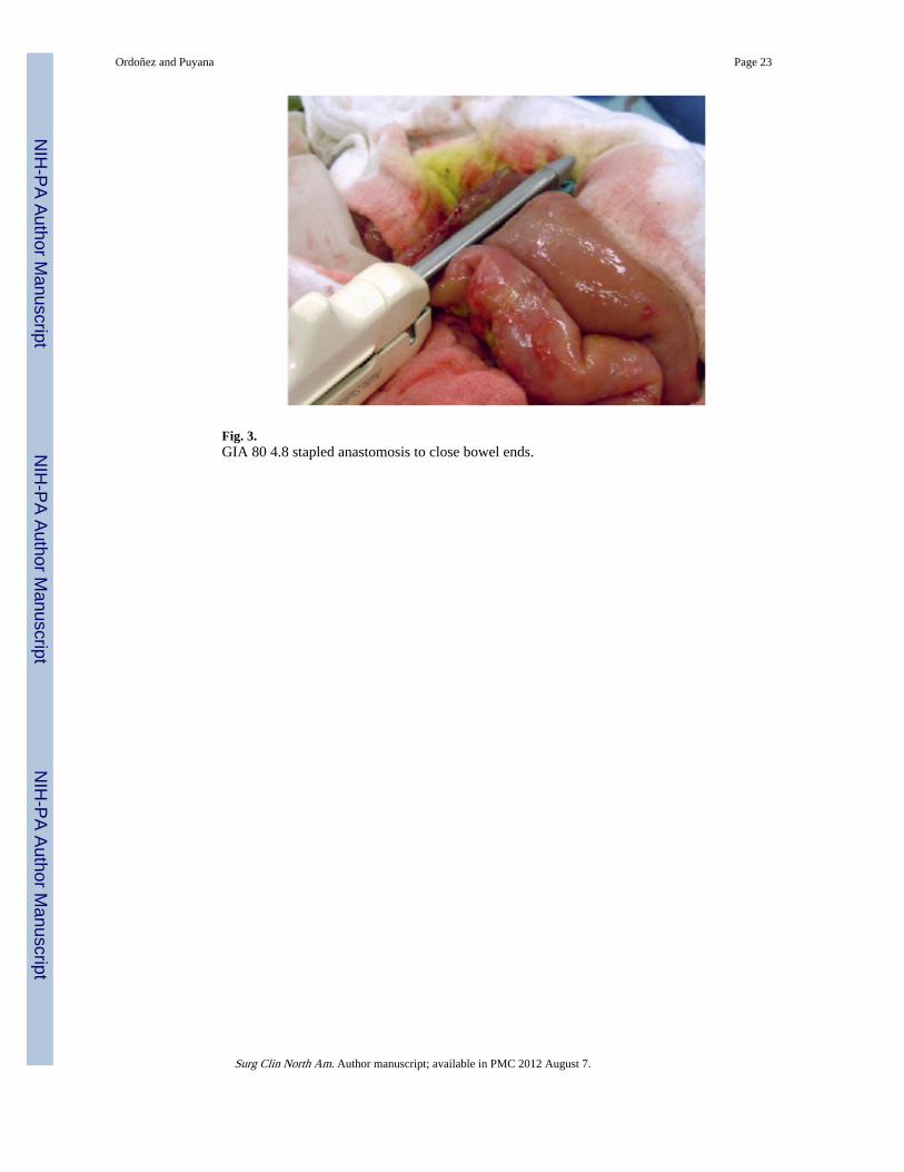
Fig. 3.GIA 80 4.8 stapled anastomosis to close bowel ends.
Ordoñez and Puyana Page 23
Surg Clin North Am. Author manuscript; available in PMC 2012 August 7.
NIH
-PA Author Manuscript
NIH
-PA Author Manuscript
NIH
-PA Author Manuscript

Fig. 4.Anastomosis completed.
Ordoñez and Puyana Page 24
Surg Clin North Am. Author manuscript; available in PMC 2012 August 7.
NIH
-PA Author Manuscript
NIH
-PA Author Manuscript
NIH
-PA Author Manuscript

NIH
-PA Author Manuscript
NIH
-PA Author Manuscript
NIH
-PA Author Manuscript
Ordoñez and Puyana Page 25
Table 1
Demographic and clinical characteristics of patients with peritonitis 1995–2004
Total number 267
Age (years) 52.2 ± 20
Male gender, % 62.5
APACHE II, %
0–10 24
11–25 70.4
26–35 5.6
Median APACHE II, % 14
Source of infection, %
Colon 33.3
Small bowel 22.5
Liver biliary tract 8.6
Stomach—duodenum 10.5
Pancreas 13.9
Others 11.3
Number of re-laparotomies
Mean ± SD 4 ± 3
Median 3
Length of hospital stay, days
Mean ± SD 25.7 ± 18
Median 20
Length of ICU stay, days
Mean ± SD 15.8 ± 13
Median 12
Fistula, % 15.3
Septic shock, % 54.3
ARDS, % 30
Days on ventilator
Mean ± SD 9.5 ± 11
Median 6
Mortality, % (95% CI) 19.9 (15.1–24.7)
Abbreviations: ARDS, acute respiratory distress syndrome; CI, confidence interval.
Surg Clin North Am. Author manuscript; available in PMC 2012 August 7.
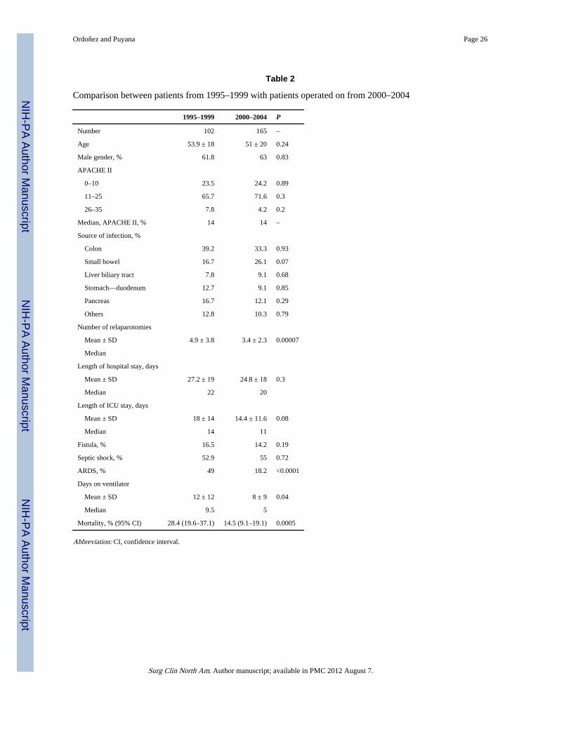
NIH
-PA Author Manuscript
NIH
-PA Author Manuscript
NIH
-PA Author Manuscript
Ordoñez and Puyana Page 26
Table 2
Comparison between patients from 1995–1999 with patients operated on from 2000–2004
1995–1999 2000–2004 P
Number 102 165 –
Age 53.9 ± 18 51 ± 20 0.24
Male gender, % 61.8 63 0.83
APACHE II
0–10 23.5 24.2 0.89
11–25 65.7 71.6 0.3
26–35 7.8 4.2 0.2
Median, APACHE II, % 14 14 –
Source of infection, %
Colon 39.2 33.3 0.93
Small bowel 16.7 26.1 0.07
Liver biliary tract 7.8 9.1 0.68
Stomach—duodenum 12.7 9.1 0.85
Pancreas 16.7 12.1 0.29
Others 12.8 10.3 0.79
Number of relaparotomies
Mean ± SD 4.9 ± 3.8 3.4 ± 2.3 0.00007
Median
Length of hospital stay, days
Mean ± SD 27.2 ± 19 24.8 ± 18 0.3
Median 22 20
Length of ICU stay, days
Mean ± SD 18 ± 14 14.4 ± 11.6 0.08
Median 14 11
Fistula, % 16.5 14.2 0.19
Septic shock, % 52.9 55 0.72
ARDS, % 49 18.2 <0.0001
Days on ventilator
Mean ± SD 12 ± 12 8 ± 9 0.04
Median 9.5 5
Mortality, % (95% CI) 28.4 (19.6–37.1) 14.5 (9.1–19.1) 0.0005
Abbreviation: CI, confidence interval.
Surg Clin North Am. Author manuscript; available in PMC 2012 August 7.

NIH
-PA Author Manuscript
NIH
-PA Author Manuscript
NIH
-PA Author Manuscript
Ordoñez and Puyana Page 27
Table 3
Predictors of death multivariate analysis
Variable OR 95% CI P
Age >50 years 1.02 1.001–1.04 0.037
APACHE II >25 1.06 0.99–1.13 <0.05
Period before year 2000 1.98 0.78–3.41 0.3
Septic shock 17.7 6.2–50.7 0.0001
Abbreviations: CI, confidence interval; OR, odds ratio.
Surg Clin North Am. Author manuscript; available in PMC 2012 August 7.

NIH
-PA Author Manuscript
NIH
-PA Author Manuscript
NIH
-PA Author Manuscript
Ordoñez and Puyana Page 28
Table 4
Characteristics of patients who underwent deferred primary anastomosis
Number 26
Age, mean, years 54.5
Male, % 61.5
Type of peritonitis, %
Secondary 30.7
Post-traumatic 6.7
Postsurgery 61.6
Median, APACHE II, % 14.5
ICU, LOS, mean, days 17.9
LOS hospital, mean, days 27
Abbreviation: LOS, length of stay.
Surg Clin North Am. Author manuscript; available in PMC 2012 August 7.

NIH
-PA Author Manuscript
NIH
-PA Author Manuscript
NIH
-PA Author Manuscript
Ordoñez and Puyana Page 29
Table 5
Overall outcome in 26 patients with deferred primary anastomosis
Outcome n %
Primary success 20 77
Fistula 3 11.5
Stoma 3 11.5
Mortality at 28 days 3 11.5
Surg Clin North Am. Author manuscript; available in PMC 2012 August 7.