INSULIN INFUSION PROTOCOLS FOR CRITICALLY ILL PATIENTS: A HIGHLIGHT OF DIFFERENCES AND SIMILARITIES

S1
Available online http://ccforum.com/supplements/9/S1
Critical Care Volume 9 Suppl 1, 200525th International Symposium on Intensive Care and EmergencyMedicineBrussels, Belgium, 21–25 March 2005
Published online: 7 March 2005These abstracts are online at http://ccforum.com/supplements/9/S1© 2005 BioMed Central Ltd
P1Severe community-acquired pneumonia in the intensive care unit
C Costa, I Gouveia, P Cunha, R Milheiro, A Bártolo, C Gonçalves,A Carvalho, T Cardoso, S Martins, J MagalhãesHSO, Guimarães, PortugalCritical Care 2005, 9(Suppl 1):P1 (DOI 10.1186/cc3064)
Introduction Community-acquired pneumonia remains a commoncondition worldwide. It is associated with significant morbidity andmortality. The aim of this study was to evaluate conditions thatcould predict a poor outcome.Design Retrospective analyse of 69 patients admitted to the ICUfrom 1996 to 2003. Demographic data included age, sex andmedical history. Etiologic agents, multiorgan dysfunction, noso-comial infections, SAPS II and PORT scores were recorded foreach patient. For statistical analysis we used a t test, chi-squaretest and Mann–Whitney U test on SPSS®. A value of P less than0.05 was considered significant.Results Forty-seven patients were male and 22 patients werefemale. Mean age was 52 years. Sixty-seven percent had seriouspre-morbid conditions including pulmonary disease (34.8%), cardiacproblems (36.2%), diabetes (13%) and chronic liver disease (5.8%);40.6% were smokers, drug abusers or alcohol dependents. Sixty-eight patients required invasive mechanical ventilation. The averagelength of ventilation was 13.5 days, median 8 days. The mean SAPSII score was 40.14 and the mean PORT score was 141. Themortality rate was 27.5% (SAPS II estimated mortality, 35%).Complications reported were ARDS (40.6%), septic shock (34.8%),acute renal failure (2.9%), cardiac arrest (8.7%) and nosocomialinfeccions (46.4%). Mortality rates were higher for previous hepatic(75%) and metabolic (33%) diseases. We found a close associationbetween crude mortality and SAPS II score (P = 0.003) anddevelopment of complications (P = 0.0028). Respiratory dysfunction(P = 0.006) and septic shock (P = 0.022) were most significantlyrelated to mortality. No significant differences were foundedregarding age, comorbidities, PORT score, etiologic agents,nosocomial infections and length of invasive mechanical ventilation.Conclusions Previous hepatic chronic disease was strictly relatedto higher mortality as well as isolation of MRSA. ARDS and septicshock predicted a poor outcome. SAPS II score was the bestseverity indicator of mortality.
P2Closed endotracheal suction system without periodic changeversus open endotracheal system
L Lorente, M Lecuona, J Villegas, M Mora, A SierraHospital Universitario de Canarias, Santa Cruz de Tenerife, SpainCritical Care 2005, 9(Suppl 1):P2 (DOI 10.1186/cc3065)
Objective It is known that the closed tracheal suction system(CTSS) produces less hemodynamic and gasometric deterioration
than an open tracheal suction system (OTSS). Use is limitedbecause no decrease in the incidence of ventilator-associatedpneumonia (VAP) was found and also because it is moreexpensive. But, is daily periodic change of the CTSS necessary?The aim of this study was to analyze the incidence of VAP using aCTSS without periodic change versus an OTSS.Methods It is a prospective study of ICU patients from 1 January2004 to 31 October 2004. Patients who required mechanicalventilation (MV) were randomized into two groups: one group wassuctioned with CTSS without periodic change and another groupwith OTSS. An aspirate tracheal swab and a throat swab onadmission and afterwards twice weekly were taken. VAP wasclassified based on throat flora in endogenous and exogenoussamples. The statistical analysis was performed by chi-square testand Student’s t test, and we took P < 0.05 as a significant difference.Results There were no significant differences between both groupsof patients (236 with CTSS without periodic change and 211 withOTSS) in age, sex, diagnosis groups, APACHE II score, number ofaspirations per day and mortality. No significant differences werefound in the percentage of patients who developed VAP (13.98%vs 14.02%), nor in the number of VAP per 1000 MV-days (14.13 vs14.67). At the same time we did not find any differences in thepercentage of patients who developed exogenous VAP (0.96% vs0.88%). In the patients who needed MV during 24 hours or more,the aspiration cost was less expensive in the CTSS without periodicchange group than in the OTSS group ($1.89 × 1.53 vs $2.45 ×0.71 per patient-day, P < 0.0001).Conclusions We conclude that in our study the CTSS withoutperiodic change decreased the aspiration cost and did not modifythe VAP incidence.
P3Ventilator-associated pneumonia caused by multidrugresistant bacteria
M Sartzi, M Charitidou, A Panagiotakopoulou, C Michas, F Tsidemiadou, P Clouva-Molyvdas‘THRIASSIO’ Hospital of Eleusis, Attica, GreeceCritical Care 2005, 9(Suppl 1):P3 (DOI 10.1186/cc3066)
Introduction The aim of the study was to investigate theepidemiology, clinical characteristics and severity of ventilator-associated pneumonia (VAP) caused by multidrug resistantbacteria (Group I) and to identify possible differences from VAPcaused by sensitive pathogens (Group II).Methods A prospective observational 2-year study of all ICUpatients who developed VAP. Age, gender, APACHE II score,underlying disease, cause of ICU admittance, length of ICU stay,duration of mechanical ventilation (MV) before the onset of VAPand total duration of MV, VAP pathogens, day of VAP onset andpatient outcome were recorded. The Multi Organ DysfunctionScore (MODS) and Clinical Pulmonary Index Score (CPIS) weremeasured at the onset of VAP. Statistical evaluation was

S2
Critical Care March 2005 Vol 9 Suppl 1 25th International Symposium on Intensive Care and Emergency Medicine
performed using the nonparametric Mann–Whitney test andPearson’s chi test.Results During this 2-year period 240 patients were admitted tothe ICU. Fifty patients (20.8%) developed VAP. All VAP was lateonset. VAP was caused by multidrug resistant pathogens in 23patients (Group I) and by sensitive bacteria in 27 patients (GroupII). ICU admittance (P = 0.27), underlying disease (P = 0.83) andtype of isolated bacteria (P = 0.53) did not differ between the twogroups. All the other results are presented in Table 1.
Table 1
Group I (n = 23) Group II (n = 27) P value
Men/women 17/6 21/6 0.75c
Age 59.8 ± 18.0a 60.7 ± 21.4a 0.6b
APACHE II score 20 (CI: 17.5–22.5) 21.1 (CI: 18.3–24) 0.71b
Outcome (deaths) 8 (34.8%) 4 (14.8%) 0.09c
CPIS 6.8 (CI: 6.6–7.0) 6.3 (CI: 6.1–6.6) 0.003b
MODS 8.9 (CI: 7.9–9.8) 4.6 (CI: 3.9–5.2) 0.0001b
Days of MV before VAP 16.1 ± 10.1a 17.8 ± 10.1a 0.11b
Total days of MV 40.1 ± 18.1a 36.7 ± 22.0a 0.37b
LOS in ICU 46 ± 21.2a 39.9 ± 23.9a 0.26b
LOS, length of stay; CPIS, Clinical Pulmonary Index Score; MODS, MultiOrgan Dysfunction Score; MV, mechanical ventilation; CI, 95% confidenceinterval. aData presented as mean ± standard deviation. bMann–Whitneytest. cPearson’s chi test.
Conclusions Patients that developed VAP due to multiresistantbacteria did not differ in their basic characteristics (age, gender,APACHE II score, underlying disease and cause of admittance)from patients that developed VAP due to sensitive strains. However,MODS and CPIS at the onset of VAP were significantly higher inpatients with VAP caused by multiresistant bacteria. The differencein outcome between the two groups, although not statisticallysignificant, may have reached significance if our sample was larger.
P4Ventilator-associated pneumonia and Clinical PulmonaryInfection Score validation in a Greek general intensive care unit
P Myrianthefs, K Ioannidis, M Mis, S Karatzas, G BaltopoulosKAT Hospital, Athens, GreeceCritical Care 2005, 9(Suppl 1):P4 (DOI 10.1186/cc3067)
Background Ventilator-associated pneumonia (VAP) is asignificant clinical problem in the ICUs and accurate diagnosisremains problematic. The purpose of the study was to examine thecharacteristics of VAP in a general Greek ICU.Methods We prospectively recorded the characteristics of VAPfor a period of 5 months in a seven-bed ICU. We collected 1032ventilator days (VD) concerning 64 patients admitted to our ICU.Data collected included demographics, VAP episodes, pathogens,resistance characteristics and outcomes. We also validated theClinical Pulmonary Infection Score (CPIS) as a guide for VAPdiagnosis [1]. We defined VAP as having CPIS ≥ 6.Results We included 64 patients admitted to our ICU (43 men) ofmean age 50.8 ± 4.6 years. Patients were admitted from theemergency department, wards, other ICUs and the operating roomsuffering from multiple trauma including head injury (25), stroke(14), postoperative respiratory failure (10), heart failure (seven),sepsis (five), and other medical conditions (three). We recorded1032 VD. Twenty-one patients (21/64, 32.8%) developed VAP.Four patients developed two separate episodes of VAP. We intotal recorded 25 episodes of VAP. The incidence of VAP was
24.2 episodes per 1000 VD. The duration of VAP until resolutionwas 11.4 ± 0.9 days. The total duration of mechanical ventilation(MV) was 29.7 ± 5.3 days and the mean duration of MV before thedevelopment of VAP was 19.9 ± 4.8 days.We identified multidrug resistant pathogens (Acinetobacter baumanii)in two patients and one of them died due to VAP. We also identifiedpathogens sensitive only to colistin (A. baumanii in eight patients andPseudomonas aeruginosa in two patients) in 10 patients, five of themdied and one of them died due to VAP (Table 1).
Table 1
Gram(–) 16/25 (65%)Acinetobacter baumanii 12Pseudomonas aeruginosa 4Mixed 9/25 (35%)A. baumanii + P. aeruginosa 5A. baumanii + Staphylococcus aureus 2A. baumanii + fungi 1Klebsiella pneumoniae + fungi 1
The VAP-attributable mortality was 23.8% (5/21). VAP wassuccessfully resolved in 20/25 episodes. Management of VAP wasguided according to culture results and in the case of multidrugresistance or single antibiotic sensitivity by the use of combinationantibiotic therapy including meropenem, high-dose ampicillin-sulbactam and collistin.CPIS sensitivity was 82.7%, specificity was 89.2%, positive predictivevalue was 73.2% and negative predictive value was 93.5%.Conclusions VAP significantly contributes to morbidity andmortality of critically ill ICU patients. However, VAP may besuccessfully treated even in the case of multidrug resistance orsingle antibiotic sensitivity by combination therapy. CPIS may be asignificant tool in the diagnosis of VAP in our ICU.Reference1. Singh N, et al.: Am J Respir Crit Care Med 2000, 162:505-511.
P5Ventilator-associated pneumonia in neuromuscular,tracheostomyzed patients
A Martini, N Menestrina, E Mantovani, B Allegranzi, G Finco, L GottinOps Poloclinico GB Rossi, Verona, ItalyCritical Care 2005, 9(Suppl 1):P5 (DOI 10.1186/cc3068)
Objective Lower-tract respiratory infections are common events intracheostomyzed mechanically ventilated patients leading to a 10-fold increase in mortality rate particularly in those patients withseverely compromized clinical conditions [1]. Our study aimed toassess epidemiology and risk factors for colonization of respiratorytract and development of ventilator-associated pneumonia (VAP) inhome-living, tracheostomyzed and mechanically ventilated patientswith neuromuscular disease.Design A retrospective study.Setting An ICU ambulatorial service for neuromuscular disease.Patients Data of 27 patients (20 male) that underwent a routine visitevery 6 months, collected from 1995 until 2003, were analyzed.Thirteen had amyotrophic lateral sclerosis (ALS) and one spinalmuscular atrophy (ALS-like group); seven had Duchenne’s dystrophy(DMP), two congenital dystrophy, three metabolic dystrophy and onesupranuclear progressive paralysis (DMP-like group).The median age was 54 years (interquartile range, 31–63). Themedian mechanical ventilation period (MVP) was 71 months(30–120).

S3
Available online http://ccforum.com/supplements/9/S1
Thirteen patients had severe dysphagy and ineffective cough reflex,12 of them were alimentated by percutaneous enteral gastrostomyand one by nasal-gastric tube, ALS-like patients were morefrequently dysphagic than DMP-like patients (P < 0.01).Low respiratory tract infections (LRTI) were defined in thepresence of cough or an abnormal increase of bronchialsecretions, with or without fever that required antibiotic therapy;pneumonia was defined as LRTI with a new pulmonary infiltrate atthorax radiography [2].Measurements and main results VAP incidence was 52episodes for 100 patients per year of MVP. The median MVP waslower in dysphagic patients (33 months [20.75–70.50]) than innon-dysphagic patients (120 months [80–156]) (P < 0.01) and inALS-like patients (33 months [24–73]) than in the DMP-like groupof patients (96 months [69.5–144]).Dysphagic patients had an higher colonization incidence (Ci) (P =0.01), LTRI incidence (LTRIi) (P = not significant), and VAPincidence (VAPi) (P = 0.02) than nondysphagic patients, permonth of mechanical ventilation.ALS-like patients had a higher Ci and VAPi than DMP-like patients(P = not significant).Bivariate analysis shown a positive correlation between Ci andLTRIi and VAPi (P < 0.01 for both). MVP was negatively correlatedwith Ci (P < 0.01) and VAPi, although the P value was 0.06.Pseudomonas aeruginosa was the most isolated bacteria incolonization (48%), LTRI (45%) and VAP (52%) episodes.Conclusion VAP is a frequent event in our patients. Dysphagicpatients, although alimentated by gastrostomy, are more frequentlycolonized and this enhances the risk to develop VAP more than innondysphagic patients. Patients ventilated for a long period seemto develop a natural defence from colonization. The longer theMVP, the less the patient is prone to be colonized and, likely, todevelop VAP. The risk of developing VAP is no different in ALS-likeand DMP-like disease although the dysphagy prevalence differs. Abigger sample is needed to definitively prove this.References1. Niedermann MS, et al.: Nutritional status and bacterial binding
in the lower respiratory tract in patients with chronic tra-cheostomy. Ann Intern Med 1972, 77:701-706.
2. Harlid R, et al.: Respiratory tract colonization and infection inpatients with chronic tracheostomy. Am J Resp Critical Care1996, 154:124-129.
P6Clinical characteristics of ventilator-associated pneumoniapatients and control patients
M Moock, LL Maia, ALC Meserami, CM Broncher, JL Oliveira,SC Oliveira, JMO OrlandoICU Hospital Geral de Pedreira, São Paulo, BrazilCritical Care 2005, 9(Suppl 1):P6 (DOI 10.1186/cc3069)
Introduction Ventilator-associated pneumonia (VAP) represents80% of all nosocomial pneumonia. The aim of this study is anepidemiological, performance and outcome evaluation of an ICU,focused in VAP.Methods The study was performed in a 13-bed ICU located in SãoPaulo, Brazil, during 2003. It was a retrospective study, where allconsecutive patients admitted were included. All data werecollected and analysed in a Brazilian system of performance andquality in the ICU, called QuaTI®. Statistical analyses were done,supported by the EpiInfo 2002 software. Clinical characteristics ofall patients were presented and then two groups were analysedand compared: VAP group (n = 28) and control group (n = 231)composed of ventilated patients who did not develop VAP.
Results From 11 January to 31 December, 483 patients wereadmitted; 55.69%, were male; median age was 59 years, 362medical and 121 surgical patients; medium length of stay was8.91 ± 13.07 days. APACHE II median score was 11.78 ± 7.13;mortality rate was 29.6% and standard mortality rate, calculated byAPACHE II, was 1.01. The rate of ventilated patients was 259/483(53.62%). The VAP incidence was 10.81% and the rate was10.84 per 1000 ventilator days. The infection agent was identifiedin 15 patients by bronchoalveolar lavage or blood cultures. Themore prevalent were Pseudomonas aeuriginosa (6/28), Klebisiellapneumoniae (2/28), Staphylococcus aureus (2/28).
Table 1
Characteristics VAP (n = 28) Controls (n = 231) P value
Age (years), median 57 66 0.10
% male 85.7 74 0.17
Medical 24 171 0.17
Surgical 4 60 0.17
ICU stay (days) 30.79 ± 18.06 12.25 ± 14.97 0.000
Ventilation days 27.61 ± 20.19 10.41 ± 13.21 0.000
APACHE II score 16.50 ± 5.77 13.96 ± 7.39 0.04
Death risk (%) 21.39 33.32 0.02
Mortality rate (%) 32.14 51.52 0.053
Conclusions VAP did not increase mortality but improvedventilated days, ICU length of stay and assistance costs. TheAPACHE II score was significantly higher in patients whodeveloped VAP. P. aeuriginosa was the predominant pathogen.ReferenceRello J: Crit Care Med 2003, 31:2544-2551.
P7Nosocomial pneumonia: impact of a very early antimicrobialtherapy
L Montesino, C Cazenave, X Jeusset, P Remi, M Thyrault, J CoudrayGeneral Hospital of Longjumeau, FranceCritical Care 2005, 9(Suppl 1):P7 (DOI 10.1186/cc3070)
Introduction Nosocomial pneumonia (NP) severely complicatesthe course of mechanical ventilation. Although the need of anappropriate antimicrobial therapy adapted from the results ofcultures from lower respiratory samples is well known, the bestmoment to start it is yet to be defined.Objective To assess the benefits of an appropriate antimicrobialtherapy started just after the direct sample examination versus afterthe culture results.Design and patients A retrospective cohort study including 50NP cases, diagnosed on clinical examination, bronchoalveolarlavage and protected specimen brush. Once the radiological andclinical characteristics of the patient have been analysed, theantimicrobial therapy modality is studied. Patients have beenseparated into two groups, depending on the delay before theantimicrobial therapy: just after the direct examination for group 1(G1) and after the culture analysis for group 2 (G2).Measurement and results For G1 the appropriate antimicrobialtherapy is started with a delay of 4.6 ± 2.5 hours after the directexamination results, while for G2 this delay reaches 34.5 ± 22.5hours. Hence, in 44 patients, initial antimicrobial therapy wasappropriate straightway and in the remaining six patients wasadapted on the results of culture. No differences have been

S4
noticed in terms of mortality (54% vs 53%, P = 0.91) and length ofstay in the intensive care unit (28.7 days vs 19.3 days, P = 0.16)between the two groups. Both were similar regarding clinical andradiological diagnosis, age and severity of the NP. However, in G2,a significantly higher number of multi-resistant bacteria have beenfound (87% vs 73%, P < 0.001) as well as a higher rate oftracheal colonization within 48 hours after the fiberopticbronchoscopy (76% vs 60%, P < 0.001).Conclusion For patients having a NP of moderate gravity, it ispossible to wait for the culture before the start of antimicrobialtherapy.
P8Antibiotics for preventing infections in open fractures areprotective against ventilator-associated pneumonia
F Cancellieri1, G Gambale1, M Baldazzi21Ospedale Maggiore, Bologna, Italy; 2Universiità degli Studi,Bologna, ItalyCritical Care 2005, 9(Suppl 1):P8 (DOI 10.1186/cc3071)
Introduction Early ventilator-associated pneumonia (VAP) remainsthe commonest infection in ICUs, in trauma in particular. Controversyexists on whether antibiotic prophylaxis for traumatic lesionsincreases the respiratory infection rate and antibiotic resistance [1].Methods Using the trauma registry and data from the infectionsurveillance program, we reviewed major trauma cases: admittedconsecutively and directly to our unit in 4 years, with a stay longerthan 3 days that received no antibiotic at all or receivedclindamycin-gentamicin from admission for open fractures.Statistical analysis consisted of: Fisher’s exact test, Student’s ttest, multiple logistic regression.Results Four hundred and eighty-two patients did not receiveantibiotics, 126 did, for 2.8 ± 1.8 days. Groups were notstatistically different in age, sex, injury severity score, GlasgowComa Score (GCS) at admission, prehospital intubation rate, daysof artificial ventilation and sedation, surgery, and transfusions.Bivariate analysis of the groups with and without VAP (272 vs 336)showed a statistically significant difference in injury severity score,GCS, days of ventilation, prehospital intubation and antibiotictreatment. Days of ventilation, anatomical severity of trauma andantibiotic treatment were independently associated with VAP in alogistic multivariate model. Antibiotics showed a protective effectagainst VAP: odds ratio, 0.386; 95% confidence interval,0.227–0.657. VAP in non-antibiotic treated patients was causedby Staphylococcus aureus in 37.7% (4% methicillin-resistant S.aureus [MRSA]) of cases, haemophilus in 25%, Gram-negative,including pseudomonas, in 25.4%. In patients treated withantibiotics S. aureus was isolated in 17.4% of cases (5.5%MRSA), haemophilus in 30.4%, Gram-negative, includingpseudomonas, in 43.1%.
Table 1
Odds 95% CI, 95% CI, Variable P value ratio lower upper
Injury severity score < 0.01 1.027 1.006 1.047Days on ventilator < 0.0001 1.241 1.188 1.296Antibiotics yes/no < 0.001 0.386 0.227 0.657
Conclusion Seventy-two hours of antibiotic therapy for open third-degree fracture is protective against VAP in trauma patients. WhenVAP occurs after or during antibiotic treatment, isolations shifttowards haemophilus, pseudomonas or other Gram-negativegerms, not MRSA.
Reference1. Hoth JJ: Prophylactic antibiotics adversely affect nosocomial
pneumonia in trauma patients. J Trauma 2003, 55:249-254.
P9A simple program to reduce the incidence of ventilator-associated pneumonia in a Brazilian intensive care unit
P Souza, M Lugarinho, A Martinez, L FelixHospital de Clinicas Niterói e Mário Lioni, Rio de Janeiro, BrazilCritical Care 2005, 9(Suppl 1):P9 (DOI 10.1186/cc3072)
Introduction Ventilator-associated pneumonia (VAP) is veryimportant in ventilated patients. There are many procedures (moreor less 23) that could reduce VAP, but many of them are difficult orexpensive to useObjectives To evaluate the efficacy of three simple measures, thesemi-recumbent position, ‘clean’ intubation and a protocol oftracheal aspiration, in the incidence of VAPMethods Our ICU has 12 adult beds. The period was the year2003 and 2004 until October. We standardized the semi-recumbent position at 45°, routine intubation and trachealaspiration in all patients under mechanical ventilation. Nothingmore changed. We compared the incidence of VAP in 2003 withthe data from 2001, 2002 and 2004.Results Patients were similar by age, sex and APACHE II score.The incidence of VAP was 28 and 22 per 1000 patient-days ofmechanical ventilation in 2001 and 2002. The incidence in 2003was 10.7, almost 50% less than 2001/2002, and 7.4 in 2004. In astudy we had done in 2002, we identified in 80% of opportunitiesthat patients under mechanical ventilation were with the trunkbelow 45°.Conclusion To keep the patient under mechanical ventilation in asemi-recumbent position is not difficult, as to standardizeintubation and tracheal aspiration. Unfortunately our doctors,nurses and respiratory therapists were not aware of thesepractices, although all of them know the rationale and advantages.With these simple and inexpensive measures, we could reducesignificantly the incidence of VAP, around 50%. We must continuethe study, with many cases, to confirm the results.
P10Risk factor analysis of pneumonias developing after openheart surgery
Y Bicer, S Simsek, N Yapici, O Aydin, F Sogut, Z AykacSiyami Ersek Thoracic and Cardiovascular Surgery Center,Istanbul, TurkeyCritical Care 2005, 9(Suppl 1):P10 (DOI 10.1186/cc3073)
Introduction Pneumonia after cardiac surgery have an importantrole among the infections that originate from hospitals as theyincrease mortality, morbidity and costs. To decrease the pneumoniaincidence after open heart surgery, we should analyse the riskfactors correctly and eliminate the preventable risk factors.Methods In our study 1000 consecutive patients who had openheart surgery since January 2002 in Siyami Ersek Thoracic andCardiovascular Surgery Center were followed about pneumoniaprospectively. Pneumonia is defined with Center of DiseaseControl–ABD criteria. In identifications of microorganisms,classical methods and BBL Chrystal Gram Positive ve BBL GramNegative (Becton Dickenson) kits were used. Data have beenanalysed statistically in the SPSS for Windows 10.0 programme.In univariant analysis a t test was used, in group variants chi squaretests were used. P < 0.05 was considered statistically significant.
Critical Care March 2005 Vol 9 Suppl 1 25th International Symposium on Intensive Care and Emergency Medicine

S5
To determine the independent risk factors multivariant logisticregression analysis was used.Results In 991 patients, data were found suitable for the study.The mean patients’ age is 57.6 ± 13 years. Demographic data ofthe patients are shown in Table 1. The pneumonia ratio is 1000device day/12.5. By univariant analysis age, emergency operation,low EF, operation time, intubation time, respiratory interventions,reintubation, enteral feeding, reoperation, resuscitation, presenceof any complication, readmission to the ICU, and usage of bloodderivatives are found to be the risk factors for pneumonia aftercardiac surgery. Independent risk factors of pneumoniadevelopment are shown in Table 2.
Table 1
Percent
Female/male 28/72
Emergency 6.4
DM 17
EF < 40% 4
CABG 67.5
Valve replacement 20
Combine procedures 3.3
Other 8.2
Table 2
Odds ratio P value (95% CI)
Age > 68 years 0.028 1.06 (1.0–1.12)
Emergency operation 0.017 4.9 (1.33–17.93)
Entubation time 0.001 2.0 (1.1–3.17)
Respiratory intervention 0.005 4.8 (1.33–22.31)
Reintubation 0.0001 14.47 (3.46–60.41)
Blood usage 0.015 1.4 (1.0–1.8)
Readmission to ICU 0.005 3.2 (1.93–11.02)
Conclusion Nosocomial pneumonia is one of the most significantinfections among cardiac surgery patients in the postoperativeperiod. In different studies the frequency of nosocomial pneumoniawas found between 0.2% and 9.7% and the mortality rate between23% and 51%. According to our study older age and emergency-operated patients are under higher risk of pneumonia after cardiacsurgery. The risk factors like intubation time, enteral feeding, usageof blood and blood derivatives must be controlled in preoperative,intraoperative and postoperative periods.
P11Perinatal risk factors for early onset neonatal infection
V Elvedji-Gasparovic, B Peter, T Klepac-PulanicKBC Zagreb, Petrova Klinika, Zagreb, CroatiaCritical Care 2005, 9(Suppl 1):P11 (DOI 10.1186/cc3074)
Introduction Neonatal infection and perinatal mortality rate are abig challenge to neonatologists and obstetricians as well. Group Bstreptococcal (GBS) infection is the most common early onset(EO) neonatal infection with an incidence of 1–4 per 1000 liveborn in the developed world.Methods We studied the newborn infection rate before and afterAmerican Academy of Pediatrics (AAP) Protocol implementation inCroatia. The protocol for beta haemolytic streptococcal B infection
includes: intrapartum antibiotic prophilaxis with ampicillin andgentamycin in preterm labour at < 37 weeks of gestation, prematurerupture of membranes at < 37 weeks of gestation, fever duringlabour, ruptures of membranes >18 hours before delivery andprevious delivery of a sibling with invasive GBS disease.Results Of 784 neonates admited to the NICU, between 1 January2003. and 31 December 2003, 60 (14 per 1000 live born)developed definite, culture confirmed, early onset (< 48 hours)GBS infection and seven (two per 1000 live born) developedprobable infection (clinical signs consistent with EOGBS in a babyunder 48 hours old, colonised with group B streptococci).Discussion Conatal infection increases the sensitivity of the foetalbrain to hypoxia and has later neuro risk sequelae to the newbornchild. Our data suggest that the incidence of GBS infection in ourcountry was considerably higher than in all current studies. Reasonsfor that can be inadequate perinatal screen in some parts of thecountry and no established policy for intrapartum antibiotic treatmentof women with antenatal risk factors. In our study more than 80% ofwomen who delivered neonates with EOGBS disease had at leastone of the risk factors. After the year 2000, when we started theAAP Protocol, the incidence of infection decreased to 5/1000 liveborn infants. The mortality from EOGBS dropped to 3%.Conclusion It is very important to establish a basis for trials ofdifferent strategies to reduce EOGBS infections in countries intransition with low social and economy status. Our resultsdocumented that the AAP Protocol for beta haemolyticstreptococcal B infection can significantly reduce perinatalmortality of neonatal infection and sepsis.
P12Urinary catheter-related infection in critically ill patients
L Lorente, C Henry, M Martín, M MoraHospital Universitario de Canarias, Santa Cruz de Tenerife, SpainCritical Care 2005, 9(Suppl 1):P12 (DOI 10.1186/cc3075)
Objective To determine the incidence and microorganismsresponsible for urinary catheter-related infection (UCRI) in criticallyill patients.Methods It is a prospective study performed during 30 months ofthe patients admitted to a 24-bed medical–surgical ICU of a 650-bed university hospital. Urinary cultures were taken on admissionand twice weekly. UCRI were diagnosed according to CDCcriteria. UCRI were classified based on the onset moment as earlyonset and late onset: early onset were those developed during thefirst 4 days of the ICU stay; and late onset were those developed 5days after ICU admission. The statistical analysis was performedusing the SPSS 11.0 program. Continuous variables are reportedas means and standard deviation, and categoric variables aspercentages.Results A total of 1582 patients were admitted, 953 males(60.24%). The mean age was 57.91 ± 18.83 years (median 63 years,interquartile range 44–73 years). The mean APACHE II score was13.95 ± 8.93 (median 14, interquartile range 10–19). Admissiondiagnoses were: 737 (46.59%) heart surgery, 189 cardiological(11.95%), 196 neurologic (12.29%), 185 trauma (11.69%), 120respiratory (7.59%), 104 digestive (6.57%) and 51 intoxication(3.22%). Mortality rate was 14.79% (234 patients). A total of 1392patients (87.99%) needed a urinary catheter, during 12,556 days.Of the 1392 patients, 72 (5.17%) developed 75 UCRI (late onsetin 54 cases and early onset in 21 cases). All UCRI were caused byonly a microorganism. The incidence density was 5.97 UCRI/1000urinary catheter-days. The microorganisms responsible for UCRIwere the following: Escherichia coli (16), Pseudomonasaeruginosa (seven), Morganella morganii (four), Klebsiella (four),
Available online http://ccforum.com/supplements/9/S1

S6
Citrobacter (five), Proteus mirabilis (three), Enterococcus faecalis(eight), coagulase-negative staphylococci (seven), MSSA (two),Candida albicans (12) and other fungi (seven).Conclusions In our series, most UCRI had a late onset, werecaused by only a microorganism and were mainly due to E. coliand C. albicans.
P13Serial changes in ICU-acquired pneumonia pathogen after theremodeling of the environment in a MICU
S Hong1, J Jung1, C Lim1, S Na1, J Ahn2, S Lee1, W Kim1, D Kim1, Y Koh1
1Asian Medical Center, University of Ulsan College of Medicine,Seoul, South Korea; 2Ulsan University Hospital, Ulsan, South KoreaCritical Care 2005, 9(Suppl 1):P13 (DOI 10.1186/cc3076)
Introduction The aim of the study was to observe effects of theimprovement of the patient’s care facility in a medical ICU (MICU)on ICU-acquired pneumonia (IAP), especially caused by drug-resistant bacteria including methicillin-resistant Staphylococcusaureus, third-generation cephalosporin-resistant Acinetobacterbaumanii (ISAB), imipenem-intermediate sensitive A. baumanii(IIAB), imipenem-resistant A. baumanii (IRAB), expanded-spectrumbeta-lactamase (ESBL), and imipenem-resistant Pseudomonasaeruginosa (IRPA).Methods All critically ill patients with clinically diagnosed IAPbetween January 2001 and June 2002 (period 1 — beforeremodeling), October 2002 and March 2003 (period 2 — afterremodeling) and October 2003 and March 2004 (period 3 — 1-year follow-up after remodeling) were observed prospectively in the28-bed MICU of a 2100-bed tertiary-care singer center. Clinicalsuspicion of IAP was defined by a new and persistent infiltrate onchest radiography associated with at least two of the following:purulent secretions, temperature ≥ 38.5°C or < 36.5°C, and aleukocyte count higher than 10,000/µl or lower than 4000/µl.Results A total 1038 patients (355, 353 and 330) were admittedto the MICU during the study periods. At each period, 48 (44cases, 11%), 38 (34 cases, 8%), and 80 (60 cases, 18%)episodes of clinically diagnosed IAP occurred (P = 0.160). Themean age (62 vs 66 vs 63, P = 0.272), percentage of males (82 vs82 vs 78, P = 0.800), and mean APACHE III score (67.3 ± 18.7 vs70.1 ± 26.5 vs 77.7 ± 28.0, P = 0.148) were not different duringthe study periods. Episodes of MRSA [13(30%) vs 12 (34%) vs29 (37%)], ISAB [6 (13%) vs 1 (3%) vs 9 (11%), P = 0.243],IRPA [2 (4%) vs 1 (3%) vs 9 (11%), P = 0.150], and ESBL [2(4%) vs 5 (13%) vs 4 (5%), P = 0.180] were not different duringthe study periods. Episodes of IIAB [4 (8%) vs 0 (0%) vs 0 (0%),P = 0.006] and IRAB [5 (10%) vs 0 (0%) vs 1 (1%), P = 0.011]were lower in periods 2 and 3 than in period 1. Duration ofmechanical ventilation (22.2 ± 13.1 days vs 24.1 ± 13.0 days vs20.0 ± 13.1 days, P = 0.340), and mortality in the ICU (54.5% vs44.1% vs 38.3%, P = 0.258) were not different during the studyperiods. Use of carbapenem [9 (19%) vs 5 (13%) vs 29 (36%),P = 0.011] and glycopeptide [13 (27%) vs 10 (26%) vs 45 (56%),P = 0.001] were higher in period 3 than in periods 1 and 2, andthird-generation cephalosporin [21 (44%) vs 20 (53%) vs 19(24%), P = 0.004] was lower in period 3 than periods 1 and 2.Conclusion The incidence of IAP caused by drug resistantbacteria was not affected by the improvement of the patient’s careenvironment in the studied ICU. However, the incidence of IAP byIRAB and IIAB has been decreased after the remodeling.
P14Duration of infusions and blood stream infections inpolytrauma patients
Y Nalapko, N Kachur, D DokashenkoState Medical University, Lugansk, UkraineCritical Care 2005, 9(Suppl 1):P14 (DOI 10.1186/cc3077)
Introduction Central venous catheters (CVCs) are generally usedin ICU patients. Nevertheless, bloodstream infections are commonand dangerous complications of CVCs. The use of CVCs in ICUpatients has contributed to the majority of nosocomial bloodstreaminfections caused by Staphylococcus aureus, Staphylococcusepidermidis and Candida species. It was shown that more than200,000 cases of CVC-related bloodstream infections occurannually in the United States, with mortality of 12–25%.Objective To evaluate the predictive value of the working time of acentral venous line as a risk factor of blood stream infections inpolytrauma patients.Materials and methods In the period of 1 February 2002 to 30November 2004, 451 patients with severe polytrauma wereadmitted to the ICU of Lugansk District Hospital. All of them hadCVCs, and the average duration of stay in the ICU was 7.12 days(6–21 days). The working time of the CVC (WT CVC) andincidence of nosocomial blood stream infections were analyzed.Patients who died on the first to third day of hospitalization wereexcluded from the investigation as inappropriate to nosocomialinfection criteria.Results WT CVC was measured as the duration ofinfusions/transfusions during the first 7 days of the post-traumaticperiod. The incidence of blood stream infections was 21 cases(4.66%). All patients were divided into two groups: Group A, withblood stream infections (21 patients); Group B, patients who haveno such complications (430 cases).WT CVC in Group A and in Group B was 128.1 ± 11.5 hours and87.5 ± 7.4 hours, respectively (P < 0.05).Infusions of crystalloids, colloids, blood components and othermedications prolonged round-a-day and depended on severity ofinjury, blood loss volume, and presence of multiple organdysfunction. Severity of injury measured by the Trauma Score was8.5 ± 1.1 and 9.7 ± 1.0, respectively (P > 0.05). Blood loss was 2.2± 0.6 l and 1.3 ± 0.2 l (P < 0.05), parameters of acute physiology(by SAPS) were 12.8 ± 0.8 and 9.4 ± 1.0, respectively (P < 0.05).Conclusion Thus, incidence of blood stream infections dependson the duration of infusions/transfusions in the ICU. The WT CVCis defined by statistically considered factors — blood loss volumeand severity of MODS. Severity of trauma is not a predictor of CVCblood infections.
P15Five years of nosocomial Gram-negative bacteremia in ageneral systems intensive care unit
W Sligl, G Taylor, P BrindleyUniversity of Alberta, Edmonton, AB, CanadaCritical Care 2005, 9(Suppl 1):P15 (DOI 10.1186/cc3078)
Objectives Nosocomial Gram-negative bacteremia in the criticallyill is associated with significant morbidity and mortality. This studyprovides epidemiological and antimicrobial susceptibility data fornosocomial Gram-negative bacteremia in a general systems ICUover a 5-year period.Methods Positive blood cultures from 1 January 1999 to 31December 2003 were reviewed for microbial etiology andsusceptibilities. Patient charts were reviewed to determine the
Critical Care March 2005 Vol 9 Suppl 1 25th International Symposium on Intensive Care and Emergency Medicine

S7
source of infection, time from admission to bacteremia, causativeorganisms, antimicrobial susceptibilities, choice of empiric anti-biotic therapy and outcome.Results There were 1632 admissions with 45 nosocomial Gram-negative bacteremias in 44 patients. Infection rates of 28.2/1000admissions and 12.1/10 000 patient-days remained stable over5 years. The mean patient age was 55.3 years (range 17–86 years);27.3% of patients were female, and 72.8% were male. Themajority (95.6%) of bloodstream infections were monomicrobial,with only one episode of polymicrobial bacteremia. Commonadmitting diagnoses included respiratory failure, solid organtransplant, post-surgery, and multi-trauma. Seven bacterial specieswere identified; Pseudomonas aeruginosa and Enterobacter spp.were most common. Sources of bacteremia included pneumonia(48.9%), followed by central venous catheterization (22.2%). Themean time from admission to hospital to development ofbacteremia was 32.9 days (95% confidence interval [CI]0–100.9), and time from admission to the ICU was slightly less at26.0 days (95% CI 0–90.1). Antimicrobial susceptibilities werehighest for imipenem, gentamicin, tobramycin, ceftazidime, andpiperacillin/tazobactam. Ciprofloxacin susceptibility was inferior toimipenem, gentamicin, and tobramycin (P < 0.05). Empiric coveragewith an agent to which the microorganism was ultimatelysusceptible was 89.2%. The mortality rate was 53.3% in the ICU,and 60.0% for overall hospitalization, compared with an overallmortality rate of 11.7% for the year 2004 in our ICU patients. Theaverage length of the ICU stay was 50.5 (95% CI 0–150.1) dayscompared with 6.13 (95% CI 4.29–7.97) days for all-comers.Conclusions Nosocomial Gram-negative bacteremia is associatedwith marked morbidity and mortality in critically ill patients.Significant resistance to ciprofloxacin was demonstrated. Empirictreatment regimens should be based on unit-specific data.
P16Epidemiology and etiology of nosocomial infections in asurgical intensive care unit
B Gelfand, T Popov, V Karabak, B BelocerkovskyDepartment of Surgery, Anaesthesiology and Intensive Care, StateMedical University of Russia, Moscow, RussiaCritical Care 2005, 9(Suppl 1):P16 (DOI 10.1186/cc3079)
Objectives To assess the epidemiology and etiology of nosocomialinfections (NI) and to identify patterns of antibiotic resistance in asurgical ICU.Setting The ICU of 1st Clinic Hospital, Clinic of Surgery, Moscow.Methods From September 2002 to December 2004 weconducted a prospective study and detected the most frequent NIin patients admitted for more than 48 hours to the ICU. Thediagnosis of NI was defined according to CDC criteria. We alsoevaluated antimicrobial resistance of isolated microorganisms. Theidentification and susceptibility to antibiotics has been performedin automated system MIC/ID panels as described by the NCCLS.Results During the study 2083 patients were admitted to the ICU,1379 patients stayed more than 48 hours in the ICU. A total of242 infection episodes were diagnosed in 149 patients withnosocomial infection (7.2%), 84 (56.4%) had a single infection, 48(32.2%) had two infections, and 17 (11.4%) had three or moreinfections. Pneumonia were the most frequently reported type ofinfection (119 episodes, 49.1% of all cases of infection), followedby intra-abdominal infections (68 episodes, 28.1%), woundinfections (28 episodes, 11.6%), laboratory confirmedbloodstream infections (14 episodes, 5.8%), urinary tractinfections (eight episodes, 3%). About 43.0% infections wereassociated with sepsis, 43.8% with severe sepsis and 4.5% with
septic shock, and 8.7% were not classified. The mortality rate forthe ICU-acquired infections after 6 weeks of follow-up was 45.6%.Age ≥ 65 years (odds ratio [OR]: 4.08; 95% confidence interval[CI]: 2.05–8.11; P < 0.001), APACHE II score > 20 (OR: 10.70;95% CI: 4.74–24.50; P < 0.001), SOFA > 5(OR: 15.6; 95% CI:6.50–37.86; P < 0.001) were independently associated withmortality. The 246 isolated pathogens were represented by 92Gram-positive cocci, 154 Gram-negative bacilli and 2% Candidaspecies. Staphylococcus aureus (20.3%) was the most frequentlyisolated bacteria, followed by Acinetobacter spp. (18.3%),Pseudomonas aeruginosa (14.6%), Klebsiella spp. (13.4%),Enterococcus spp. (13.2%), Escherichia coli (8.1%). Resistanceto oxacillin was observed for 94% of S. aureus isolates, all isolateswere susceptible to vancomycin. The most active antimicrobialagents: against Acinetobacter spp. were imipenem (91.1%),ampicillin/sulbactam (64.4%); against P. aeruginosa were imipenem(88.9%), ceftazidime (69.4%), amikacin (58.3%); against Klebsiellaspp. were imipenem (97%), amikacin (91%); against E. coli wereimipenem (90%), amikacin (95%), ciprofloxacin (50%).Conclusions The NI rate during the period was 7.2%. Themortality of the patients with a NI is 45.6%. Pneumonia were thepredominant type of infection. Staphylococci were the mostfrequently isolated pathogens, with very high levels of MR.
P17ICU stay promotes enrichment and dissemination of multi-resistant coagulase-negative staphylococcal clones
C Agvald-Ohman, C Edlund, B Lund, J Struwe, G Hedin, H HjelmqvistKarolinska Institute, Huddinge, SwedenCritical Care 2005, 9(Suppl 1):P17 (DOI 10.1186/cc3080)
Introduction Patients in the ICU are prone to be colonized andinfected by multi-resistant bacteria [1,2]. It is previously known thatnosocomial infections are often preceded by cross-transmissionevents [3].The aim of the present investigation was to study the impact of thepatient’s length of ICU stay on the resistance patterns, clonaldiversity and dissemination of coagulase-negative staphylococci(CoNS) within and between patients.Methods Two groups of patients were studied, including 20consecutive patients sampled within 2 hours from admission(shortstayers, SS), and all patients treated for at least 5 days at theICU (longstayers, LS), available for sampling every second week(n = 23). Sampling was performed from five sites: oropharynx, nares,neck, axilla and perineum. A total of 868 CoNS isolates deriving fromLS patients and 403 isolates from SS patients were analyzed forantimicrobial susceptibility, clonal diversity and disseminationwithin and between patients. All 1271 CoNS isolates were testedfor antimicrobial susceptibility and subtyped to the clonal levelaccording to their phenotype with the PhenePlate™ (PhP) systemusing PhP-CS plates (PhPlate Microplate Techniques AB,Stockholm, Sweden) designed for typing CoNS. The clonalrelationship between CoNS isolates clustered as one phenotypeisolated from at least two patients was further confirmed by pulsed-field gel electrophoresis.Results The highest resistance rates were seen for oxacillin andciprofloxacin, being 92% and 83%, respectively. LS were atsignificantly higher risk of being colonized with CoNS isolatesresistant against oxacillin, clindamycin, ciprofloxacin, gentamicin aswell as with multi-resistant strains. Genotyping revealed 16 clonesthat colonized more than one patient. One of the clones wasisolated from 10 individuals, including two SS patients, indicatingan epidemic strain.
Available online http://ccforum.com/supplements/9/S1

S8
Conclusion Prolonged ICU stay was significantly correlated todecreased clonal diversity, increased endogenous dissemination ofresistant strains and cross-transmission. The results emphasize theimportance of barrier treatment and other hygienic measures,especially in this vulnerable group of patients.References1. Maki DCC: Line sepsis in the ICU: prevention, diagnosis and
management. Seminar Resp Crit Care Med 2003, 24:23-36.2. von Eiff C, Peters G, Heilmann C: Pathogenesis of infections
due to coagulase-negative staphylococci. Lancet Infect Dis2002, 2:677-685.
3. Weist K, Pollege K, Schulz I, Ruden H, Gastmeier P: How manynosocomial infections are associated with cross-transmis-sion? A prospective cohort study in a surgical intensive careunit. Infect Contr Hosp Epidemiol 2002, 23:127-132.
P18Evaluation of outcome in critically ill patients withAcinetobacter baumannii bacteremia
M Pratikaki, C Routsi, E Platsouka, C Sotiropoulou, S Nanas, P Kaltsas, O Paniara, C RoussosEvangelismos Hospital, Athens, GreeceCritical Care 2005, 9(Suppl 1):P18 (DOI 10.1186/cc3081)
Objective Bacteremia caused by Acinetobacter baumannii has anincreasing incidence among critically ill patients. However, it is notclear whether it is associated with a higher mortality sincecontroversial results have been reported. Increased mortality ratehas been shown in some studies [1], whereas in others A.baumannii bacteremia does not alter the outcome [2]. The aim ofthis study was to evaluate the clinical impact of A. baumanniibacteremia on the outcome of patients in our ICU.Patients and methods During a 4-month period (August–December 2004), in a 30-bed multidisciplinary ICU, patients whodeveloped A. baumannii bacteremia and control patients withoutmicrobiological evidence of bacteremia were prospectively studied.Matching of control patients (1:2 ratio) was made on the basis ofthe APACHE II score (± 2 points) and diagnostic category.Results During the study period, A. baumannii bacteremia wasdiagnosed in 21 patients (59 ± 15 years, mean ± standarddeviation), admitted to the ICU (incidence 9.3 per 100admissions). The APACHE II score in case and control patientswas 19.9 ± 4.5 and 19.6 ± 4.8, respectively. The median intervalfrom admission to the A. baumannii bacteremia was 10.5 days. A.baumanii strains were resistant to all antibiotics but susceptible toimipenem/cilastin and colistin. The ICU length of stay was 37(23–51) days and 9 (1.8–16.2) days, median (95% confidenceinterval), for case and control patients, respectively, P < 0.001.Case patients had a longer duration of mechanical ventilationversus control: 21 (9–53) days versus 5 (2–18) days, respectively,P < 0.001. No differences between case and control patients werefound in gender and age. Patients with A. baumannii bacteremiahad significantly higher mortality than controls (38.1% vs 7.15%,P = 0.004). Logistic regression analysis showed that risk factorsthat were independently associated with adverse outcome werethe presence of bacteremia (odds ratio = 10.2, P = 0.013), andmultisystem organ failure (odds ratio = 20.9, P = 0.001).Conclusions In this group of critically ill patients, after adjustmentfor severity of acute illness, A. baumannii bacteremia wasassociated with a significantly increased mortality rate comparedwith matched control patients. Whether other factors contribute tothis increase has to be examined.References1. Crit Care Med 1999, 27:1794-1799.2. Intensive Care Med 2003, 29:471-475.
P19Evolution of resistance in Acinetobacter baumannii in aTunisian intensive care unit after antibiotherapy restriction
N Brahmi, Y Blel, N Kouraichi, A Mokline, N Laabidi, O Beji, H Thabet, M AmamouCAMU, Tunis, TunisiaCritical Care 2005, 9(Suppl 1):P19 (DOI 10.1186/cc3082)
Introduction The prevalence of carbapenem-resistant isolates ofAcinetobacter baumannii is increasing in Tunisian ICUs. The wideuse of antibiotherapy is implicated certainly in this mechanism ofresistance.Objective The purpose of this study is to evaluate the evolution ofA. baumannii resistance after a new strategy of prescriptionconsisting of controlling and restriction of antibiotherapyprescription.Materials and methods Evolution of resistance of all A. baumanniiisolates is compared between 2001 (before antibiotherapyrestriction) and 2003–2004 (after).Results See Table 1.
Table 1
2001 2003 2004 % of resistance (n = 134) (n = 50) P (n = 63) P′
Ticarcilline 76 63 0.05* 77 0.8
Piperacilline 97 85 0.01* 81 10–3*
Ceftazidim 95 84 0.01* 79 8 × 10⁻⁴*
Carbapenem 37 37 0.36 25 0.09
Amikacine 85 70 0.28 68 6 × 10⁻³*
Netilmycine 30 31 0.86 31 0.7
Ofloxacine 95 87 0.04* 93 0.9
Ciprofloxacine 95 87 0.16 94 0.9
P, 2003 – 2001; P′, 2004 – 2001. * P < 0.05.
Conclusion With antibiotherapy restriction we have obtained asignificant reduction of resistance for piperacillin, ceftazidim andamikacin. We have obtained also a decrease of carbapenem-resistant isolates (37% vs 25%) but not significant enough ifcompared with other countries (5% in New York and 11% in Brazil).
P20Epidemiological profile and antibiotic susceptibility ofPseudomonas areuginosa isolates within the hospitalizedburned patient
K Bousselmi1, L Thabet2, S Oueslati1, O Ben Jaber1, A Ouchtati1, S Cherif1, S Ben Redjeb2, A Messadi11Aziza Othmana Hospital, Tunis, Tunisia; 2Faculty of Medicine ofTunis, TunisiaCritical Care 2005, 9(Suppl 1):P20 (DOI 10.1186/cc3083)
Pseudomonas aeruginosa plays a predominant role as anetiological agent involved in serious infections in burned patients.Treatment of these infections is frequently complicated byantibiotic resistance, a problem that has been increasing in recentyears.The objective of this study is to analyse the epidemiological profileand antibiotic susceptibility of P. aeruginosa isolates within theburned patients admitted to our intensive care burn department.During a period of 4 years (2000–2003), 828 burn patients wereadmitted. Thermal burn (70%) was the most frequent burn,
Critical Care March 2005 Vol 9 Suppl 1 25th International Symposium on Intensive Care and Emergency Medicine
S9
followed by electric (27%) and chemical (3%) burn. The populationexamined had an average burn size of 44% of the body surfacewith the mean age of 34 ± 17 years. One hundred and seventystrains of P. aeruginosa were isolated in different samples fromburned patients. These isolates were identified using standardmicrobiological techniques and their antibiotic susceptibility wasdetermined using the disk diffusion method recommended by theFrench Society of Microbiology.Over the study period, P. aeruginosa was isolated mainly fromcutaneous superinfections (59%), urine (21.7%), blood culture(14.8%) and venous catheter culture (4.7%). P. aeruginosa wasthe most common isolate from cutaneous pus (22.9%). Bacteremiawere mainly caused by Acinetobacter baumannii (16.5%) andStaphylococcus aureus (15.5%). P. aeruginosa was only isolatedin 8.2% of bacteremia. The survey of antibiotic susceptibility of P.aeruginosa showed that 60.9% of strains were resistant topiperacillin, 53.4% to ceftazidime, 37.6% to imipenem, 70.6% tocefsulodine, 59.3% to tobramycin, 80% to gentamicin, 62.4% toamikacin and 53.4% to ciprofloxacin.During this study period, a high resistance to antibiotics of P.aeruginosa was observed. It is necessary to implement urgentmeasures to prevent the spreading of these multi-resistant strains.These measures include: sensible limitation of the use ofantimicrobial agent, strict disinfection and hygienic procedures.
P21Strategy of antimicrobal therapy in patients with severetrauma: importance of initial severity state evaluation
A Yaroshetskiy1, D Protsenko2, S Yakovlev3, B Gelfand2
1City Hospital #7, ICU, Moscow, Russia; 2State Medical Universityof Russia, Moscow, Russia; 3Moscow Medical Academy, Moscow,RussiaCritical Care 2005, 9(Suppl 1):P21 (DOI 10.1186/cc3084)
Introduction A high rate of nosocomial infections among patientswith severe multiple trauma including severe traumatic brain injuryand initial APACHE II score of 15 or more leads to increasedlength of stay (LOS), costs and high mortality. That makes theprevention of severe nosocomial infections one of the majortherapeutic interventions in these patients. Because of a high rateof mixed nosocomial infections with high resistance to antibiotics itis necessary to start antimicrobal therapy with extended-spectrumantibiotics. Taking into account pharmacodynamics, pharmaco-kinetics and microbiological features of carbapenems, they makethe best choice for these patients. In the case of MRSA infection itis necessary to use the combination of carbapenem andvancomycin.Objective The goal of the study was to assess the influence ofmaximal initial antimicrobal therapy with meropenem on mortality,rate of nosocomial infections and LOS in the ICU in patients withsevere trauma including severe traumatic brain injury and initialAPACHE II score of 15 or more.Results The study was performed in a general ICU from April2003 to September 2004. All patients with severe trauma withpredominant severe traumatic brain injury and initial APACHE IIscore of 15 or more were included in the study. We studied twogroups of patients. In the case of known or suspected nosocomialinfection, patients in the maximal antimicrobal therapy (MAT) group(n = 16, APACHE II 19.88 ± 3.6) received meropenem 3–6 g/daydepending on body weight for 14 days. In the case of provenMRSA infection, vancomycin 2 g/day was added. Patients in thecontrol group (n = 22, APACHE II 19.90 ± 3.7) with known orsuspected infection received initial antimicrobal therapy withcefalosporines 3 or fluoroquinolones with change of antimicrobal
therapy according to microbiological data. The observed data didnot follow the normal distribution. Comparisons between groupswere performed using the chi-square test. Mortality in the MATgroup was 6.25% (1/16), and in the control group was 50%(11/22), P = 0.002. Nosocomial infection rate (ventilator-associated pneumonia, meningitis) was observed in 31.25% ofpatients (5/16) in the MAT group and in 91% of patients (20/22) inthe control group, P < 0.001. LOS in the ICU did not differbetween groups (19.44 ± 3.1 in the MAT group and 18.09 ± 10.3in the control group). But comparision between LOS amongsurvivors in the two groups revealed significant reduction in LOS inthe ICU in the MAT group (19.42 ± 3.2 in the MAT group and25.54 ± 11.1 in the control group, P < 0.05).Conclusions Initial antibiotic therapy with meropenem in patientswith severe trauma including severe traumatic brain injury and initialAPACHE-II score of 15 or more leads to a reduction in mortality,nosocomial infection rate and LOS in the ICU.
P22Surveillance of ICU-acquired infections: intensive care unitcharacteristics, practices in prevention and surveillancemethodology
I Morales, C Suetens, A Versporten, C De Laet, K MertensScientific Institute of Public Health, Brussels, BelgiumCritical Care 2005, 9(Suppl 1):P22 (DOI 10.1186/cc3085)
Introduction The characteristics and practices of ICUsparticipating in the Belgian national surveillance program werereviewed within the validation study. The main objective was tobetter understand the hospital practices for nosocomial infectionprevention and the methodology used for the collection ofsurveillance data.Methods Forty-five ICUs were randomly selected through systematicrandom sampling from a list of 1997–2001 participation-quarters(3 months). The number of patients admitted to the hospital and tothe ICU was provided by the hospital administration as well as thenumber of hospitalization-days. Data were collected, through aquestionnaire, from those responsible for data collection at eachinstitution.Results Data from 44 ICUs were analyzed. The averageparticipation was six surveillance quarters (range 1–22). The ICUsbelong to hospitals admitting on average 2676 patients perquarter. The average ICUs have 10.5 beds on average, and 88.6%of them are medical/surgical ICUs. The average number of ICUadmissions was 217 patients per quarter (927 patient-days). Themean length of stay within the ICU was 4.3 days. Specificguidelines for pneumonia prevention were available in 75% ofICUs; and for catheter-related bacteremia in 85.4%. A total 86.4%of ICUs had specific guidelines for hemoculture procedures.Systematic culture of sputum (surveillance cultures) wasperformed by 77.3% ICUs, but with different periodicity. Cathetertip culture was performed by 73.8% of the ICUs (2/3systematically). Guidelines for the use of antibiotics were availableat 64% of ICUs while 15.8% of them allowed antibiotic use at thedoctor’s appreciation. Almost 70% of respondents prefer a patient-based rather than a unit-based surveillance. On average twopersons are involved in surveillance (44% ICU doctor, 26% ICUnurse, 6% infection control doctor and 18% IC nurse). It takes20 min per patient.Conclusions The study allowed identifying methodologicalproblems and areas for targeted training in order to improve thequality of the data collected in the national surveillance.
Available online http://ccforum.com/supplements/9/S1

S10
P23Monitoring of the antibiotic prescriptions in order to preventmicrobial resistance in the intensive care unit in the Russianhospital
T Lugovkina1, B Richards2, F Badaev1, A Piontek1, V Bagin1, V Shilova3
1Clinical Hospital N40, Yekaterinburg, Russia; 2UMIST,Manchester, UK; 3Center of Laboratory Diagnostics of Diseases ofMother and Child, Yekaterinburg, RussiaCritical Care 2005, 9(Suppl 1):P23 (DOI 10.1186/cc3086)
Background This presentation shows how, at the City Hospital, itwas possible to improve the quality of clinical practice in surgerydepartments and ICUs.Introduction Antibiotics play an important role in clinical practice.The expenditure on these drugs is more than 50% of the totalexpenditure for drugs at the hospital. Uncontrolled use ofantibiotics, in the environment of intolerably poor financing of thehospitals in Russia, and the absence of adequate control of theantibiotic prescriptions induce the growth of resistance in thehospital microbial flora, and as a result the expenditure on drugtreatment increases.Methodology An analysis of the antibiotics purchased by theadministration of the hospital was made in order to determine thebaseline proportions of the different classes of antibiotics in the totalstructure in the year 2003. The analysis showed a high percentageof beta-lactams in the structure of the antibiotics purchased (88%).The data from the Diagnostic Laboratory Center of the city wereimpressive: the level of Klebsiella pneumoniae producing theenlarged-spectrum beta-lactamases reached a level of 92.5% inthe ICU. An analysis of patients’ data at the ICU and surgicaldepartments (in a cohort of 300) was made in order to evaluate thequality and appropriateness of the antibiotic prescriptions. Aninappropriate choice of antibiotics, wrong dosage, or an incorrecttiming regime were revealed in 60% of cases. The protocols ofantibiotic prophylaxis and the strict rules of antibiotic prescriptionsin the ICU, prepared according to the data of evidence-basedmedicine, were implemented.Conclusion The system of governance of antibiotic prescriptions,the monitoring of the microbial resistance in the ICU, and the strictadherence to the protocols of antibiotic usage have gained approvaland enabled an improvement in the quality of treatment (from 60%to 30%) and a reduction of the expenditure for antibiotics (40%).
P24De-escalation therapy: 1-year experience in a generalintensive care unit
E Gyurov, S Milanov, M Georgieva, M MilanovUniversity Hospital, Sofia, BulgariaCritical Care 2005, 9(Suppl 1):P24 (DOI 10.1186/cc3087)
Introduction First-line choice of antibiotic treatment is animportant issue for successful outcome in surgical and traumapatients. De-escalation strategy aims to improve the first choice ofantibiotic therapy, to improve outcome and to save money.
Setting A general ICU, 17 beds, annual admission approximately800 patients.Objective To compare rates of success and failure of the empirictreatment of patients with usually prescribed antibiotics and theuse of carbapenems with subsequent de-escalation.Method Taking data from flow-charts, we determined the mostfrequently prescribed empiric antibiotics in our ICU during 2002and calculated the rates of success and failure of the treatment.We considered the failure an inadequate antibiotic therapy. Asshown in Table 1, there were two regimens used in 2002. The useof mefoxin was quite disappointing with only a 30% success rate.When other empiric antibiotic, or combination of antibiotics wereused the rate of success was slightly below 50%. This can bepartly explained with the high rate of surgical complications. In2003–2004 we accepted for use the de-escalation strategy.Starting empiric carbapenems (tienam in 106 patients, meronem in31 patients), we obtained samples for the microbiologicallaboratory from the sites of suspected infection. After receivingpositive results we de-escalated antibiotic therapy according tosusceptibility. The rate of success increased 11%, and the casesof inadequate empiric therapy fell to 4.4% of cases. Mortality ratedecreased from 31% to 13%.Conclusions Introducing de-escalation strategy into our clinicalpractice showed promising results. The percentage of cases oftreatment failure (inadequate empiric antibiotic therapy) and themortality rate decreased significantly. We need a longer period toconfirm these result, as well as to analyze cost-effectiveness.
P25The importance of antibiotics in animal models of sepsis
MO Maybauer1, DM Maybauer1, JF Fraser2, LD Traber1, K Murakami1, A Mizutani1, P Enkhbaatar1, N Morita1, M Westphal1, DL Traber1
1University of Texas Medical Branch, Galveston, TX, USA;2University of Queensland, Brisbane, AustraliaCritical Care 2005, 9(Suppl 1):P25 (DOI 10.1186/cc3088)
Objective Our group developed an ovine model of Pseudomonasaeruginosa sepsis associated with acute lung injury (ALI) [1]. Theaim of this study was to modify this model by the administration ofCeftazidime (Cef) to simulate a more clinically relevant situation. Inaddition we studied the effects of recombinant human activatedprotein C (rhAPC), as well as an combined treatment of rhAPCand Cef. This modification could be the basis for future studiesinvestigating new treatment strategies in sepsis.Methods Thirty sheep (35–40 kg) were operatively prepared forchronic study. After 7 days of recovery, sheep were randomlyallocated either to sham, control, Cef, rhAPC, or Cef + rhAPCgroups (n = 6 each). After a tracheostomy, ALI was produced andP. aeruginosa were instilled into the lungs, following an establishedprotocol [1]. The sham group received the vehicle. The sheep werestudied for 24 hours in the awake state and were ventilated with100% oxygen. The PaO2/FiO2 ratio was determined at baseline(BL) and every 3 hours. Ceftazidime (3 g) was administeredintravenously 1 and 13 hours post injury. rhAPC was given as acontinuous infusion (24 µg/kg/hour), starting 1 hour post injury.
Critical Care March 2005 Vol 9 Suppl 1 25th International Symposium on Intensive Care and Emergency Medicine
Table 1 (abstract P24)
Antibiotics Patients Success rate Escalation rate De-escalation Failure Surgical complication Death
2002, mefoxin 85 26 (31%) 41 (48%) – 24 (28%) 17 (20%) 31 (36.4%)
2002, other antibiotic 114 53 (46.5%) 30 (26.3%) – 15 (13%) 25 (22%) 30 (26.3%)
2003–2004, carbapenems 137 78 (57%) – 33 (24%) 6 (4.4%) 10 (7.3%) 18 (13%)

S11
The animals were resuscitated with Ringer’s lactate solution tomaintain filling pressures and hematocrit. Statistical analysis: two-way analysis of variance and Student–Newman–Keuls post hoccomparisons. Data are expressed as mean ± standard error of themean. Significance P < 0.05.Results The PaO2/FiO2 ratio remained stable in sham (BL:518.3 ± 12.7 vs 12 hours: 507.3 ± 18.8) animals. The controlgroup showed a significant decrease in PaO2/FiO2 ratio (BL:496.8 ± 21 vs 12 hours: 83.5 ± 9.8). The Cef group also showeda fall in PaO2/FiO2 ratio (BL: 524 ± 11.4 vs 12 hours: 173 ± 47),as well as the rhAPC (541.3 ± 12.2 vs 12 hours: 150.7 ± 28.6)group. In both groups there was a significant improvement of thePaO2/FiO2 ratio compared with the control group. The PaO2/FiO2ratio of the Cef + rhAPC group (525.4 ± 9.9 vs 12 hours:335.8 ± 25.8) was significantly higher than all other injured groups.Conclusion Since the application of Cef + rhAPC after ALIassociated with bacterial challenge improved pulmonary functionmore than Cef or rhAPC alone, it underlines the importance ofantibiotic treatments for further studies in animal models of sepsis.Acknowledgements Grant support from Shrine 8820, 8450, NIHGM066312.Reference1. Murakami, et al.: Crit Care Med 2002, 30:2083-2090.
P26Outbreak of severe Clostridium difficile-associated colitis withMOF in Quebec intensive care units: first analysis inSherbrooke ICUs
F Lamontagne, M Lalancette, O Martinet, J Pepin, O LesurCHU Sherbrooke, CanadaCritical Care 2005, 9(Suppl 1):P26 (DOI 10.1186/cc3089)
Introduction Recent reports suggest that Clostridium difficilecolitis (CDAC) may be evolving into a more severe disease [1].Over the past 2 years, an increased case fatality associated withCDAC was noted as the admission rate to our adult tertiary ICUsfor this entity increased in a context of inhospital outbreak. ThisCDAC outbreak occurred in a 682-bed regional hospital inQuebec, Canada. All cases admitted to ICUs with a diagnosis oflife-threatening CDAC were systematically reviewed over a 20-month period (between 1 January 2003 and 21 August 2004). Thefollowing is a retrospective statistical analysis.Results Until now, 41 cases of CDAC requiring intensive caremonitoring and treatment have been collected; 53.7% were of malesex and the median age was 78 years (25th–75th %: 71–82). TheCharlson morbidity index was calculated for each patient for amedian of 4 (25th–75th %: 3–6). Patients were generally exhibitinga sepsis profile with high-grade fever (median 39.20°C, 25th–75th%: 38.7–39.4°C), and high leukocyte counts (median 28.15 ×10⁹/l, 25th–75th %: 20.8–43.7) at some time during the ICU stay.Serum creatinine levels were elevated (median 204 µmol/l,25th–75th %: 139–292 µmol/l). Shock status was diagnosed in83% of the patients and catecholamines were mandatory in 53.7%of the time for a mean duration of 3.4 days (± 0.47) in addition tovolume resuscitation. Patients were resuscitated during the first 72hours following their ICU admission with a mean of 10.7 cm3
(± 986 cm3) of crystalloid solution, 586 cm3 Pentastarch solution(± 92 cm3) and 231 cm3 of 25% albumin (± 75 cm3). Upon entry tothe ICUs, the APACHE II score was calculated and the medianobtained was of 24 (predicted mortality rate 49.7%). Furthermore,the SOFA score was also obtained upon entry, 48 hours afteradmission and every subsequent week. In the first 48 hours, thetotal SOFA score increased in 26.8% and decreased in 31.7% ofthe patients. Thirty-two percent of the population reached the
maximum cardiovascular SOFA score of 4 (i.e. norepinephrine> 0.1 µg/kg/min). Twenty-two percent of patients went to theoperating room.The overall observed mortality rate was 49%. An APACHE II scoreupon entry of 22 or more and an age greater than 75 were bothassociated with higher mortality with an adjusted odds ratio (OR)of 7.6 (95% confidence interval [CI] 1.6–33.9, P = 0.01) and 4(95% CI 1.06–15,1 P = 0.07). A trend toward higher mortality wasobserved in patients with an increasing SOFA score within the first48 hours of admission (OR 4.55, 95% CI 0.91–22.6, P = 0.1) andhigh serum lactate levels (OR 18.8, 95% CI 0.97–362, P = 0.03for lactates > 6.5) but no trends were noted regarding the Charlsoncomorbidity index, the decision of surgery (colectomy for bowelperforation vs refractory shock) or catecholaminergic drug use.Discussion This case review reveals the existence of an outbreakof CDAC with increasing case fatality and worsening outcome inQuebec. Epidemiologic data will help in defining more efficientpreventive measures directed toward this nosocomial entity andallowing early recognition in order to thwart severe illness.Reference1. Pepin J, et al.: JAMC 2004.
P27Antimicrobial resistance in bloodstream infection does notaffect outcome in intensive care unit patients with acute renalfailure treated with renal replacement therapy
E Hoste, S Blot, G Verschraegen, J De Schuijmer, S Claus, J De Waele, A Dhondt, R Vanholder, N Lameire, J DecruyenaereGhent University Hospital, Gent, BelgiumCritical Care 2005, 9(Suppl 1):P27 (DOI 10.1186/cc3090)
Background Bloodstream infections (BSI) have a worse outcomewhen the microorganisms involved are antimicrobial resistant (AM-R),compared with BSI with antimicrobial susceptible (AM-S) micro-organisms. We evaluated whether this is also true in ICU patients withacute renal failure who are treated with renal replacement therapy(ARF-RRT), a particular severely ill cohort of ICU patients.Methods All ARF-RRT patients with BSI admitted in our 54-bedtertiary care ICU during the period 1995–2002 were included inthe study. Enteroccci were defined AM-R when resistant tovancomycin, staphylococci when resistant to methicillin, Gram-negative bacteria when resistant to ceftazidim, Pseudomonaswhen resistant to ceftazidim, quinolones, piperacilin, or imipenem,and Candida when resistant to fluconazole. Catheter-related BSIand BSI of unknown origin were defined as primary BSI. Data arepresented as number (percentage) or median (interquartile range).Results During the 8-year study period, 79 out of 1032 ARF-RRTpatients (7.7%) developed BSI, incurring 88 different micro-organisms. AM-R was present in 50 patients (63%). Gram-positivebacteria were more frequent in patients with AM-R BSI (72% vs24%, P < 0.001), whereas Gram-negative bacteria (24% vs 53%,P = 0.006) or Candida (2% vs 18%, P = 0.008) were less frequent.A higher proportion of patients with AM-R BSI had primary BSI(58% vs 17%, P < 0.001). Patients with AM-R BSI and AM-S BSIhad the same age (57 [42–67] years vs 62 [53–69] years,P = 0.150), and APACHE II score on admission (26 [17–32] vs 28[18–37], P = 0.338). Time to BSI after start of RRT was longer inthe AM-R group (11 [6–21] days vs 5 [1–19] days, P = 0.026).Patients with AM-R BSI had a higher need for vasopressor therapy(80% vs 52%, P = 0.008). Length of hospital stay was notdifferent (42 [26–73] days vs 31 [16–75] days, P = 0.528) as wasinhospital mortality (64% vs 72%, P = 0.443). Cox regressionanalysis identified, after adjustment for various covariates, olderage (hazard ratio = 1.03 per year, 95% confidence interval:
Available online http://ccforum.com/supplements/9/S1

S12
1.01–1.05, P = 0.02), and primary BSI (hazard ratio = 0.42, 95%confidence interval: 0.21–0.85, P = 0.02) as associated withhospital mortality. There was no significant association betweenAM-R BSI and hospital mortality.Conclusions In a subset of ICU patients with ARF-RRT, anti-microbial resistance of microorganisms that caused BSI did notaffect mortality. After adjustment for various covariates, older agewas associated with higher mortality. In addition, primary BSI wasassociated with lower inhospital mortality.
P28Evaluation of adequacy in empirical antibiotic therapy in ageneral intensive care unit
G Friedman, T Lisboa, R Moraes, E Parolo, L MallmannHospital de Clinicas, Porto Alegre, BrazilCritical Care 2005, 9(Suppl 1):P28 (DOI 10.1186/cc3091)
Introduction Infectious pathologies are among the most prevalentin the ICU and significantly influence the outcome of critically illpatients. However, the emergency of resistant pathogens todifferent antibiotics makes the choice of the initial treatment morecomplex. In addition, inadequate empirical antibiotic therapy isassociated with a poorer outcome.Objectives To evaluate the adequacy of antibiotic therapy in ageneral ICU and to collect data about the microbial flora in order tobetter adapt the empirical treatments.Materials and methods Data were prospectively collected in a 12-bed general ICU. The presence of infection was defined by the doctorin charge of the patient. Inadequate antibiotic therapy was defined asmicrobiological evidence of infection not covered by the chosen anti-biotics, or by the finding of primary resistance to the antibiotics in use.Results Data were collected in 80 consecutive patients. The mainsite of infection was the lung (50%). The most prevalentmicroorganisms were the Gram-negative (52%). The prevalence ofnosocomial infections was 60%. Empirical antibiotic therapy wasinadequate in 28 episodes (35%), and the most prevalentmicroorganisms in this group were MRSA, Pseudomonas MR,Stenotrophomonas maltophilia. The general mortality rate was 40%,but the mortality rate among patients with inadequate antibiotictherapy was 50%, but only 33% when adequate antibiotic therapy(odds ratio = 3.09, 95% confidence interval 1.06–9.16). Risk factorto an inadequate therapy was nosocomial infection (relative risk 2.07,95% confidence interval 1.01–4.26). Other risk factors that showeda non-significant trend were a delay to starting antibiotics greaterthan 24 hours, immunosupression, septic shock and higher APACHEII score. The number of empirical treatment schemes was 17.Conclusion These preliminary data showed that there is an excess inmortality rate when empirical therapy is inadequate. The inadequacyof treatment was associated with nosocomial infections. There was anexaggerated variability on the choice of empirical strategy.
P29Usefulness of quantitative analysis of beta-D-glucan in empiricaltherapy for systematic Candida infection in critically ill patients
I Kano, T Kamohara, K Ikegami, H Taguchi, K Kawamura, J Yazawa, T MouriDokkyo University School of Medicine, Koshigaya Hospital,Koshigaya, JapanCritical Care 2005, 9(Suppl 1):P29 (DOI 10.1186/cc3092)
Background The clinical importance of systemic Candida infectionin critically ill patients is a well-documented fact. However, it is notalways true that early recognition of Candida infections followed by
early pre-emptive antifungal therapy has been routinely done. Lackof an institutional written protocol for systemic Candida infectionsmay lead to delayed diagnosis and treatment of such infections. Itis expected that each institution has to establish its own clinicalpath to ensure a standardized team approach to systemic Candidainfections to improve patient outcome.Purposes One is to review indications of antifungal therapy andevaluate the relationship between risk factors and values of beta-D-glucan. Another is to test usefulness of risk factors to predict theincreased value of beta-D-glucan prospectively.Patients and methods Clinical records of 43 patients whoreceived antifungal therapy for clinical diagnosis of systemicCandida infections were reviewed. Risk factors (APACHE score,Injury Severity Score [ISS], length of ICU stay, duration ofmechanical ventilation, total blood transfusion within 48 hours, totalparenteral nutrition, and signs of systematic inflammation), bacterio-logical studies, and quantitative data of beta-D-glucan (Fungitec Gtest MK, Seikagaku-kogyo, Japan) were analyzed. In a prospectivestudy, 38 patients were followed to test the hypothesis that the riskfactors (APACHE score >15 and ISS >16, ICU stay > 7 days, andmechanical ventilation > 2 days) can predict increased levels of beta-D-glucan (> 20 pg/ml), which validate pre-emptive antifungal therapy.Results In a retrospective study, positive culture for Candidaremained low (37.2%); however, beta-D-glucan was increasedsignificantly (> 20 pg/ml) in 79.5% of patients. In patient withAPACHE score >15 and ISS >16, beta-D-glucan was increased in78.4%. Beta-D-glucan was also increased in 79.0% of patientswho stayed in the ICU longer than 7 days. In prospective study,26.3% (10/38) of patients eventually received antifungal agents.Of these, eight patients (80.0%) fulfilled APACHE score >15, ISS>16, and ICU stay > 7 days.Discussion A certain combination of risk factors of systemicCandida infections was useful to identify the patient group whoneed pre-emptive antifungal therapy. Beta-D-glucan, which is auseful serologic test for fungal infections, was useful to initiateantifungal therapy in critically ill patients who fulfilled risk factors.Conclusions APACHE score >15, ISS >16 and ICU stay> 7 days were useful to discriminate patients who are prone todevelop systemic Candida infections. Beta-D-glucan (> 20 pg/ml)is a useful clinical tool to initiate pre-emptive antifungal therapy insuch circumstances.
P30Fungal colonization and infection in critically ill patients
L Lorente, C García, M Martín, M MoraHospital Universitario de Canarias, Santa Cruz de Tenerife, SpainCritical Care 2005, 9(Suppl 1):P30 (DOI 10.1186/cc3093)
Objective To determine the incidence of fungal colonization andinfection in critically ill patients.Methods It is a prospective study during 30 months of the patientsadmitted to the ICU during 24 hours or more. A throat swab,tracheal aspirate and urine samples were taken on admission andtwice weekly. The fungal colonization and infection wereregistered. Infections were diagnosed according to CDC criteria.Infections were classified based on throat flora as: primaryendogenous (PE) when caused by germs that were alreadycolonizing the throat on ICU admission; secondary endogenous(SE) when caused by germs that were not colonizing the throat onthe ICU admission but were acquired during the stay in ICU;exogenous (EX) when caused by germs that were not colonizingthe throat. Infections were classified based on the onset momentas: early onset (EO) (developed during the first 4 days of ICU stay)and late onset (LO) (developed 5 days after ICU admission).
Critical Care March 2005 Vol 9 Suppl 1 25th International Symposium on Intensive Care and Emergency Medicine

S13
Results A total 1582 patients were admitted, 953 males(60.24%). Mean age was 57.91 ± 18.83 years. Mean APACHE IIscore was 13.95 ± 8.93. Admission diagnoses were: 737(46.59%) heart surgery, 189 cardiological (11.95%), 196 neurologic(12.29%), 185 trauma (11.69%), 120 respiratory (7.59%), 104digestive (6.57%) and 51 intoxication (3.22%). Mortality rate was14.79% (234 patients). A total of 154 patients had fungalcolonization, 40 patients at ICU admission and 114 patients duringthe ICU stay. Forty-eight fungal infections were documented (eightEO and 40 LO; four PE, 40 SE and four EX): 19 urinary tractinfections (three EO and 16 LO; three PE, 13 SE and three EX),12 pneumonias (two EO and 10 LO; one PE, 10 SE and one EX),nine primary fungemias (two EO and seven LO; nine SE), sixsurgical wounds (one EO and five LO; six SE) and two pressuresores (0 EO and two LO; two SE). The 48 fungi responsible for 48fungal infections were: 29 Candida albicans, 12 Candidatropicalis, four Candida glabrata, two Candida parapsilopsis, oneCandida famata. Death occurred in 12/48 patients (25%) withfungal infection: 3/9 (33.33%) fungemias, 5/12 (41.66%)pneumonias and 4/27 (14.81%) other infections.Conclusions Most fungal infections had a late onset, weresecondary endogenous and were due to C. albicans. The urinarytract was the more frequent origin.
P31Leptospirosis in the intensive care unit: a cohort of 57 patients
S Vieira1, J Brauner1, D Fonseca2
1Clinicas Hospital, Porto Alegre, Brazil; 2NS Conceição Hospital,Porto Alegre, BrazilCritical Care 2005, 9(Suppl 1):P31 (DOI 10.1186/cc3094)
Introduction Leptospirosis is in general a self-limited disease but itcan be associated with important complications such as multipleorganic dysfunction and high mortality [1,2].Objective The goal of this paper is to evaluate the clinicalcharacteristics and the morbimortality of severe leptospirosis ingeneral ICUs from two general hospitals.Methods All cases with the diagnosis of leptospirosis confirmedby a blood macroagglutination test and admitted from 1990 to2004 were studied. We analyzed their clinical and laboratorycharacteristics, the occurrence of multiple organ dysfunction andtheir mortality rate. We also compared survivors with nonsurvivors.The quantitative variables have been compared by unpaired t testand the qualitative variables by a chi-squared test.Results We describe 57 adult patients, 40 ± 16 years, 47 menand 10 women. The most frequent clinical manifestations werefever (n = 52), myalgias (n = 51), jaundice (n = 49) and dyspnea(n = 49). All patients showed some level of organic dysfunction:respiratory (n = 51), renal (n = 46), hepatic (n = 45), cardio-vascular (n = 35), hematologic (n = 32) and neurologic (n = 16).The mortality rate was 40% (n = 23). The comparison fromnonsurvivors with survivors showed that they have higherincidences of respiratory, cardiovascular and neurological failuresas well as higher levels of acidosis (P < 0.05).Conclusions In endemic regions leptospirosis has to beconsidered as a cause of multiple organic dysfunction with a highmortality rate mainly when respiratory, cardiovascular orneurological failures are present.References1. Ko AI, et al.: Urban epidemic of severe leptospirosis in Brazil.
Lancet 1999, 354:820-825.2. Vieira S, Brauner J: Leptospirosis as a cause of acute respira-
tory failure. Braz J Infect Dis 2002, 6:135-139.
P32Combination of quinine and artesunate may be the best toprevent ARDS in Falciparum Malaria
A AbrahamBreach Candy Hospital, Mumbai, IndiaCritical Care 2005, 9(Suppl 1):P32 (DOI 10.1186/cc3095)
The work was carried out in the intensive care unit of ParseeGeneral Hospital, Mumbai 400036, India.Introduction Falciparum Malaria is a dreaded parasitic infection,attacks all the organs in the human body cerebral, and chestdysfunction is very common in the infection followed by kidneysblood and so forth.Hypothesis To study and compare the various treatmentmodalities and the attendant complications of Falciparum Malariawith or without concomitant Vivax in the intensive care unit. Thestudy is done to find out the best treatment to offer to patientssuffering with Facliparum Malaria infection and treat ongoingmultiorgan failure and to prevent further complication of thedreaded parasitic infection.Methods Retrospective study between 1993 and 1995 andprospective study from 1996 to 2003 of patients having severemalaria infection warranting intensive care management.Results A total number of 181 patients. See Table 1.
Table 1
Number Died Year of patients (ARDS + MOF) Survived
1993–1996 39 29 (no quinine) 10 (quinine)
1996 9 5 (no quinine) 4 (quinine)
1997 19 7 (no quinine) 12 (quinine)
1998 14 7 (no quinine) 7 (quinine)
1999 15 5 (no quinine) 10 (quinine)
2000 24 10 (no quinine) 14 (quinine + artesunate)
2001 34 1 (no quinine) 33 (quinine + artesunate)
2002 19 2 (no quinine) 17 (quinine + artesunate)
2003 until March 8 Nil 8 (quinine + artesunate)
ARDS, acute respiratory distress syndrome; MOF, multiorgan failure.
Conclusions (1) Quinine has given good and consistent results.Treatment must be initiated with quinine and must be givenintravenously. (2) Parasites may have developed resistance to amultidrug regimen (mefloquine + others). (3) Quinine followed byartesunate 120 mg bolus followed by 60 mg daily intravenously for5 days (quinine + artesunate together) yielded good results. This isthe best that can presently be offered to patients with shock/multiorgan failure.
P33Zygomycosis by Rhizopus species — a very rare angioinvasivemycosis: report of three cases
J Lewejohann, M Hansen, C Zimmermann, P Kujath, H BruchUniversitätsklinikum Schleswig-Holstein, Campus Lübeck,DenmarkCritical Care 2005, 9(Suppl 1):P33 (DOI 10.1186/cc3096)
Introduction Zygomycosis caused by Rhizopus species (Rhsp) is anaggressive and rapidly progressive opportunistic fungal infection inimmunocompromised patients. It comprises mucocutaneous,rhinocerebral, pulmonary, urological and disseminated infections.Predisposing factors are immunosuppression owing to severe
Available online http://ccforum.com/supplements/9/S1

S14
diseases, immune defects or metabolic disturbances like diabeticketoacidosis. Rhizopus infections are characterized by angioinvasivegrowth, necroses of infected tissue and perineural invasion. Theinvasion of blood vessels is remarkable for a fungal infection. Themortality of zygomycosis is very high, especially for disseminateddisease and if immunosuppression cannot be corrected.Patients Patient 1 (female, 73 years): Delayed clinical courseaccording to infection of a hip-TEP and a femoropopliteal bypassof the right leg, eventual exarticulation of the right hip joint,pseudomonas pneumonia, severe sepsis caused by staphylococci,acute respiratory distress syndrome, acute renal failure andmultiple use of antibiotics. Subsequent detection of Rhsp in thebronchoalveolar lavage and treatment with amphotericin B for thisreason. Patient 2 (male, 68 years): Transplantation of kidney inpast medical history, presenting with acute renal failure and withquite a few infections before. In the sequel development ofabscessing pneumonia on the right side with a pleural empyema.Rhsp were detected by microbiological testing in the empyemafluid. These findings required surgical intervention, resection of thelower lobe of the right lung and, within the same operation, of therenal graft because of rejection. The patient was treated withCaspofungin because of Candidemia in addition to zygomycosis.The further course was delayed by several septic phases. Patient 3(male, 72 years): An insignificant past medical history withappendectomy, secondary wound-healing; hypertension,hyperlipoproteinemia. No hint for an immune defect. Operationbecause of adhesion ilueus, postoperative severe septic shockowing to peritonitis. After 1 month of intensive care therapy withmultiple lavages and antibiotics microbiologically proven abdominalinfection with Rhsp. All patients died later in spite of all efforts.Discussion The very rarely seen zygomycosis caused by Rhspdeveloped in patient 1 and patient 2 owing to immunosuppression —in the first, iatrogenic induced by immunosuppressive drugs afterorgan transplantation; in the second as a result of previousimmunosuppression and prolonged severe sepsis. Zygomycosis ofthe third patient developed because of prolonged severe septicshock without a hint of a immune deficit before.In comparison with other mycoses, treatment of Rhsp infectionsremains difficult. The affinity to blood vessels, where the fungimultiply and their feature of vascular invasion with thrombosis andinfarction are complicating therapeutic efforts, which shouldinclude surgical resection whenever possible and amphotericin B.
P34A 16-year experience with toxic epidermal necrolysis
E Dárdai, B Dede, M TelkesSt Stephen’s General Hospital, Budapest, HungaryCritical Care 2005, 9(Suppl 1):P34 (DOI 10.1186/cc3097)
Toxic epidermal necrolysis (TEN) is a devastating medication-induced extensive epidermal detachment with a reported mortalityrate of 30–60% in adults. As in severe burns, fluid losses aremassive, and superinfection, impairment of thermoregulation,excessive energy expenditure and alteration of immunologicfunctions are usual complications. Moreover, mucous membraneinvolvement increases morbidity. Previously reported data suggestthat age, total body surface area (TBSA) involvement, laterecognition and treatment of TEN are poor prognostic indicators.The purpose of the clinical study was to evaluate the effects ofspecial intensive and nutritional management on the outcome ofpatients with TEN.Between 1988 and 2004 13 patients (five men, eight women, meanage 51 years) were admitted to our department. Two patients werein very poor condition at admission and died within 24 hours. They
were excluded from the clinical study. Patients had a TBSA skinslough of 25–85% and a mean APACHE II score of 11.75.Eleven patients were placed in a bacteria-controlled nursing unit,wounds were treated with topical debridement and antimicrobialmedications. Nutritional therapy (enteral and/or parenteral) wasinstituted after fluid and electrolyte replacement. The daily caloricintake reached 146 kJ/kg body weight with a nitrogen–caloric ratio of1:150. Enteral feeding (sip feeds, nasogastric tube, PEG) was forcedas early as possible. One patient had diabetes mellitus (NIDDM).With the complex treatment of 13–26 days in the ICU, ninepatients recovered. Two patients died due to septic complications(infected central venous catheter, extended gastrointestinalmucousa involvement — mortality rate: 18%). In the last 8-yearperiod all the patients with TEN healed, only one patient (86-year-old woman) died later due to acute myocardial infarction.Our clinical experiences show that early recognition of TEN maydecrease the morbidity and mortality. The local and generalmanagement of TEN should be carried out at an isolated, asepticunit of an intensive care department. The conventional therapycombined with vigorous nutritional management may improve thepatients survival. Our results for a group of older patients with TEN,with extensive skin and/or mucosal involvement suggest that age, adelay in proper hospitalisation, steroids and early empiric antibiotictreatment are associated with a poor prognosis.
P35The removal of antibiotics during continuous venovenoushemofiltration (CVVH) in critically ill patients with acute renalfailure: measured versus estimated CVVH clearance
C Bouman, H Kan, M Schultz, M VroomAcademic Medical Center, Amsterdam, The NetherlandsCritical Care 2005, 9(Suppl 1):P35 (DOI 10.1186/cc3098)
Background The clearance of antibiotics during continuousvenovenous hemofiltration (CVVH) is calculated by multiplying thesieving coefficient (SC) (defined as the ratio of drug concentrationin the ultrafiltrate to plasma) and the ultrafiltration rate (Quf). Formost antibiotics the SC is unknown. It was suggested that the SCcan be substituted by the non-protein-bound fraction (fu). However,fu values as reported in the literature are determined in healthyvolunteers and not during critical illness. We compared measuredand estimated CVVH clearance (ClCVVH) of five antibiotics.Methods In 40 patients with ARF undergoing CVVH (predilution,cellulose-triacetate filter, Quf 36 ± 8 ml/min), prefilter, postfilterand ultrafiltrate samples were collected and drug concentrationswere determined by HPLC or by immunoassay. The prefilterconcentration (Cpre) was corrected using the dilution factor(Qb/Qb + Qinf), where Qb is the blood flow rate and Qinf is thesubstitution fluid infusion rate. The SC was calculated as follows:SC = 2 × Cuf / (Cpre + Cpost).Results Data (mean ± standard deviation) are presented inTable 1. ClCVVH (ml/min) was calculated by multiplying the SC (orthe fu) and the Quf.
Table 1
Measured Measured Non-protein- Estimated Drug (n) SC ClCVVH bound fraction ClCVVH
Ceftazidim (4) 2.14 ± 2.64 75 ± 92 0.79 28 ± 1Ciprofloxacine (16) 0.89 ± 0.33 31 ± 12 0.60 21 ± 1Flucloxacilline (5) 0.37 ± 0.38 13 ± 14 0.20 7 ± 0Fluconazol (9) 0.86 ± 0.09 30 ± 2 0.88 31 ± 1Vancomycine (6) 0.71 ± 0.19 27 ± 5 0.70 28 ± 9
SC, sieving coefficient; ClCVVH, continuous venovenous hemofiltrationclearance.
Critical Care March 2005 Vol 9 Suppl 1 25th International Symposium on Intensive Care and Emergency Medicine

S15
Conclusions Measured and estimated ClCVVH correspondedwell for fluconazol and vancomycine, but not for ceftazidim,ciprofloxacine and flucloxacilline, possibly due to changes inprotein binding induced by critical illness or drug–membraneinteractions. Therefore, for antibiotics with a narrow therapeuticrange, monitoring drug concentrations remains mandatory.
P36Ceftazidime reduces pulmonary hypertension in an ovinemodel of sepsis following smoke inhalation injury
DM Maybauer1, MO Maybauer1, JF Fraser2, LD Traber1, P Enkhbaatar1, N Morita1, DL Traber1
1University of Texas Medical Branch, Galveston, TX, USA;2University of Queensland, Brisbane, AustraliaCritical Care 2005, 9(Suppl 1):P36 (DOI 10.1186/cc3099)
Objective Our group recently developed an ovine model ofPseudomonas aeruginosa sepsis associated with acute lung injury(ALI) [1]. The aim of this study was to modify this model by theadministration of Ceftazidime (Cef) to simulate a more clinicallyrelevant situation. This modification could be the basis for futurestudies investigating new treatment strategies in sepsis.Methods Eighteen sheep (35–40 kg) were operatively prepared forchronic study. After 7 days of recovery, sheep were randomlyallocated either to sham, control, or Cef groups (n = 6 each). After atracheostomy had been performed, ALI was produced in the controland Cef group by insufflation of 48 breaths of cotton smoke underdeep halothane anesthesia. Then, live P. aeruginosa were instilled intothe lungs via a bronchoscope [1]. The sham group received thevehicle, 48 breaths of room air and 30 ml saline. Subsequently,anesthesia was discontinued. The sheep were studied for 24 hours inthe awake state and were ventilated with 100% oxygen (tidal volume15 ml/kg, 30 breaths/min). The mean pulmonary artery pressure(MPAP) and pulmonary artery occlusion pressure (PAOP = wedgepressure) were determined every 3 hours. Cef (3 g, intravenously)was administered 1 hour and 13 hours post injury. The animals wereresuscitated with Ringer’s lactate solution to maintain filling pressuresand hematocrit. Lung tissues were taken after the experiment todetermine the bloodless lung tissue wet to dry weight ratio (W/D).Statistical analysis: two-way analysis of variance, Student–Newman–Keuls post hoc comparisons, significance P < 0.05.Results MPAP and PAOP remained stable in sham animals. Thecontrol group showed a significant increase in MPAP (baseline[BL]: 19 ± 1 vs 24 hours: 31 ± 1) and PAOP (BL: 11 ± 1 vs 24 hours:17 ± 1) versus BL over time. The Cef group also showed anincrease in MPAP (BL: 20 ± 1 vs 24 hours: 26 ± 1) and PAOP(BL: 10 ± 1 vs 24 hours: 13 ± 1) vs BL, but this increase wassignificantly lower in comparison with the control group. The fluidbalance (sham: –547 ± 38, control: +1266 ± 75, Cef: +382 ± 55)was significantly higher in the control and Cef groups comparedwith sham, but also significantly reduced in the Cef groupcompared with the control group. However, the W/D ratio after24 hours (sham: 5.0 ± 0.1, control: 5.8 ± 0.2, Cef: 5.7 ± 0.2)increased significantly in the control and Cef groups comparedwith sham, but was not statistically different between those groups.Conclusion The application of Cef after ALI associated withbacterial challenge reduced pulmonary hypertension and fluidrequirement. The addition of antibiotics to the protocol will make itmore clinically relevant and is a useful new approach for furtherstudies in this modified sepsis model.Acknowledgements Grant support from Shrine 8820, 8450, NIHGM066312.Reference1. Murakami, et al.: Crit Care Med 2002, 30:2083-2090.
P37Continuous versus intermittent infusion of temocillin inintensive care unit patients
K Claeysoone, V Basma, R Hens, F Van Bambeke, R De Jongh,P TulkensZOL, Genk, BE;UCL, Brussels, BelgiumCritical Care 2005, 9(Suppl 1):P37 (DOI 10.1186/cc3100)
Background and goal β-lactams are time-dependent antibioticsand show little gain in activity once their concentration exceeds theminimum inhibitory concentration about fourfold, which suggeststheir administration by continuous infusion. We have studied thestability, the compatibility with other drugs, and the pharmaco-kinetics of continuous or intermittent administration of temocillin (aβ-lactam active against Gram-negative organisms).Methods Temocillin was assayed by HPLC. Temocillin stabilitywas measured over 24 hours after its solution in water at 37°C.Compatibility with several frequently used drugs on the ICU wasdetermined under conditions mimicking their clinical use.Temocillin was measured in plasma samples from ICU patientswith normal renal function, randomly assigned to receive acontinuous (n = 6; 2 g every 12 hours; 5 day sample observation)or intermittent (n = 6; 2 g over 30 minutes twice daily; consecutivesamples before and after dose 1 and 9) schedule.Results Recovery after storage for 24 hours at 37°C of both R andS isomers of temocillin exceeded 90%, which is better amongother studied β-lactams. Temocillin was compatible with mostfrequently used drugs on the ICU with the exception ofclarithromycin, ciprofloxacin, meropenem, imipenem, piperacillin/tazobactam, vancomycin, amoxicillin/acid clavulanic acid, propofol,midazolam, piritramide, nicardipine, milrinone, and ranitidine. Inpatients with normal renal function, plasma levels of temocillin wereabove 50 mg/l in the continuous group, and 90% of the time abovethe breakpoint of 16 mg/l with peak levels of 120 mg/l after thefirst and ninth dose in the intermittent group.Conclusion Temocillin can be used safely in the ICU, either withan intermittent or continuous schedule. The latter may be preferredbased on pharmacodynamic considerations.
P38Meropenem administration by intermittent infusion versuscontinuous infusion for the treatment of nosocomialpneumonia
L Lorente, S Huidobro, M Martín, M MoraHospital Universitario de Canarias, Santa Cruz de Tenerife, SpainCritical Care 2005, 9(Suppl 1):P38 (DOI 10.1186/cc3101)
Objective Mainly, beta-lactamic efficacy is determined by theduration of time that concentrations remain above the minimuminhibitory concentration (MIC). Some studies have found that theadministration of carbapenems by continuous infusion maintainconstantly concentrations above the MIC of susceptible organismsover the course of therapy; but limited data exist on clinical efficacy(only occasional observations have been made). The purpose ofthis study was to evaluate the clinical efficacy of meropenem bycontinuous infusion administration (CI) or by intermittent infusion(II) for the treatment of ventilator-associated pneumonia (VAP)caused by Gram-negative bacilli (GNB).Methods An historic control group (1 July 2000–20 June 2002)with VAP caused by GNB who received initial empiric antibiotictherapy with meropenem by II (n = 18) was compared with a pros-pective cohort of patients (1 July 2002–30 June 2003) treated withmeropenem by CI (n = 10), at a university hospital medical–surgical
Available online http://ccforum.com/supplements/9/S1

S16
ICU. VAP were treated during 14 days with two antibiotics:meropenem (1 g/6 hours intravenously) plus another (aminoglyco-side or quinolone). Antibiotic clinical effect was categorized as cureor failure. Differences between groups were tested by means ofStudent’s t test and exact chi-square by permutation, using StatxactSoftware 5.0. We consider values P < 0.05 as a significant difference.Results Significant differences were not found between bothgroups of patients (15 with CI and 30 with II) in sex, age, APACHEII score, diagnosis, microorganism responsible and organdysfunction severity assessed by the Sepsis-related Organ FailureAssessment score. The CI group showed significantly greaterclinical cure than the II group (CI, 14/15 [93.33%] vs II, 19/30[63.33%], P = 0.038) and smaller, but not significant, attributablemortality to VAP (1 of 15 [6.66%] vs 7 of 30 [23.33%], P = 0.236).Conclusions Our data suggest that administration of meropenemby CI may have more clinical efficacy than administration by II forthe treatment of nosocomial pneumonia, but more studies arerequired to confirm it.
P39Continuous infusion versus intermittent infusion ofceftazidime for the treatment of pneumonia caused byPseudomonas aeruginosa
L Lorente, C García, M Martín, M MoraHospital Universitario de Canarias, Santa Cruz de Tenerife, SpainCritical Care 2005, 9(Suppl 1):P39 (DOI 10.1186/cc3102)
Objective Beta-lactamics antibiotics exhibit concentration-independent bactericidal activity. Several studies have found thatceftazidime by continuous infusion appears to optimise thepharmacodynamic profile by constantly providing concentrations inexcess of the minimum inhibitory concentration of susceptibleorganisms over the course of therapy. Limited data exist on clinicalefficacy by continuous infusion of ceftazidime. The purpose of thisstudy was to evaluate the clinical efficacy associated with theadministration of continuous infusion of ceftazidime (CI) andintermittent infusion of ceftazidime (II) for the treatment of ventilator-associated pneumonia (VAP) caused by Pseudomonas aeruginosa.Methods An historic control group (1 July 2000–20 June 2002)with VAP caused by P. aeruginosa who received initial empiricantibiotic therapy with ceftazidime by II (n = 32) was comparedwith a prospective cohort of patients (1 July 2002–30 June 2003)treated with ceftazidime by CI (n = 15), at a university hospitalmedical–surgical ICU. Patients were treated during 14 days withceftazidime, by CI (4 g/day) or II (2 g/8 hours), plus tobramycin.Exclusion criteria was creatinine clearance < 60 ml/mn. Antibioticclinical effect was categorized as cure or failure. Differencesbetween groups were tested by means of Student’s t test andexact chi-square by permutation, using Statxact Software 5.0.Results Significant differences were not found between bothgroups of patients (eight with CI and 12 with II) in sex, age,APACHE II score, diagnosis and organ dysfunction severityassessed by the Sepsis-related Organ Failure Assessment score.The CI group showed greater clinical cure rate than the II group (8of 8 [100%] versus 4 of 12 [33.33%], P = 0.004) and smalleratributable mortality to VAP (0 of 8 [0%] versus 6 of 12 [50%],P = 0.024). In addition, CI patients received one-third less dailydose than those treated by II.Conclusions These data suggest that ceftazidime administrationby continuous infusion may have more clinical efficacy thatintermittent infusion, in treatment of VAP caused by P. aeruginosa.
P40Laparostomy versus laparotomy in severe abdominalinfection: microbiological assessment
V Pavoni, L Gianesello, L Paparella, G Gori, G GrittiAz. Ospedaliera Careggi Firenze, ItalyCritical Care 2005, 9(Suppl 1):P40 (DOI 10.1186/cc3103)
Introduction Mortality of severe abdominal infection, due toperitonitis after anastomotic dehiscence, visceral organ necrosis ornecrotic pancreatitis, remains high. The efficacy of laparostomy indamage control surgery and uncontrolled intra-abdominal infectionis controversial [1].Methods We retrospectively reviewed the records of all patientswith severe abdominal infection secondary to peritonitis admittedto the ICU of a university teaching hospital from 1998 to 2000. Thefollowing information was collected: age, simplified acutephysiology score (SAPS II) and sepsis-related organ failureassessment (SOFA) on admission, length of stay (LOS), ICU andhospital outcome, type of bacteria, number of isolations andhaematogenous dissemination.Results Thirty-nine critically ill patients were studied. The meanage was 66.1 ± 16.9; the overall SAPS II at admission was41.7 ± 14.6; the SOFA was 10.9 ± 4.3. A total of 13 laparostomywere performed (group I). Twenty-six patients were undergoing alaparotomy (group II). No differences regarding age (group I vsgroup II 61.1 +± 12.7 vs 68.5 ± 18.1, P = 0.08), SAPS II(41.8 ± 20.6 vs 41.5 ± 10.6, P = 0.65) and SOFA (11.8 ± 5.5 vs10.4 ± 3.5, P = 0.49) were found. The duration of ICU stay waslonger in group I than in group II (44.4 ± 52.2 vs 19.8 ± 17.3,P = 0.04). ICU mortality was equal between two groups (30.7%).Hospital mortality was not statistically different between twogroups (38.4% vs 42.3%, P = 0.77). The number of positivecultural specimens was 19 in both groups. Isolated micro-organisms were: Gram-positive cocci (21% vs 26.3%), Pseudo-monas aeruginosa (26.4% vs 31.6%), other non-fermenting Gram-negative bacilli (21% vs 10.5%), Enterobacteriaceae (16% vs10.5%), fungi (15.8% vs 15.8%). Two of five P. aeruginosa isolationsin Group I were multiresistant to available antibiotics and sensibleonly to combinations. Four out of 13 patients in group I presentedcandidemia compared with two of 26 patients in group II. Haemato-genous dissemination was greater in group I (85.7% vs 50%).Conclusions The laparostomy technique used in the managementof severe intra-abdominal sepsis appears to correlate withsignificant ICU stay and costs. Prolonged antibiotic therapy andfrequent intra-abdominal procedures determine higher incidence ofinfections caused by Gram-negative, enterobacteriaceae andmultiresistant pseudomonaceae. The incidence of fungal infectionis not influenced by surgical approach, but the ‘open abdomen’strategy seems to facilitate dissemination.Reference1. Wittmann DH, Schein M, et al.: Management of secondary peri-
tonitis. Ann Surg 1996, 224:10-18.
P41Logistic regressive analysis of potential prognostic factors forpulmonary complications after abdominal surgery in elderlypatients
XR Wang, Y Gao, YJ Zheng, J Chen, Y Deng, Y Hang, D SunDepartment of Anesthesiology, Renji Hospital, Shanghai SecondMedical University, Shanghai, ChinaCritical Care 2005, 9(Suppl 1):P41 (DOI 10.1186/cc3104)
To investigate potential prognostic factors and to predict extent ofrisks for postoperative pulmonary complications by logistic
Critical Care March 2005 Vol 9 Suppl 1 25th International Symposium on Intensive Care and Emergency Medicine

S17
regressive analysis, and to evaluate the role of non-invasiveventilation in reducing the incidence of complications in elderlypatients. A stair-climbing test was carried out with ASA score,FEV1, changes of SpO2 and HR noted at the same time. Logisticalregressive analysis based on these parameters were used toassess the relation between potential prognostic factors andpostoperative complications. Patients with limited pulmonaryreserves were selected using the equation, and the protectiveeffect of non-invasive ventilation on these patients was assessed.The incidence of postoperative pulmonary complications for high-risk patients with non-invasive ventilation was 33.2%, and theincidence of pulmonary complications for high-risk patients withoutnon-invasive ventilation was 67.7%. There was not a significantdifference between these two groups with low-risk (P > 0.05). Themathematical model of logistic regressive analysis using the stair-climbing testing combined with other parameters is a simple,reliable method to predict the cardiopulmonary reserved function inelderly patients. Non-invasive ventilation can effectively reduce theincidence of postoperative pulmonary complications for high-riskpatients, but it has no effect on patients with low risk.
P42Bariatric surgery postoperative management: complications ina series of 278 patients admitted to an intensive care unit
M Knibel, F Muller, R Hatum, C Roderjan, E Guimarães, M Lugarinho, C Vasconcelos, A Vanzan, M MattosSao Lucas Hospital, Rio De Janeiro, BrazilCritical Care 2005, 9(Suppl 1):P42 (DOI 10.1186/cc3105)
Introduction Morbid obesity is one of the most important publichealth problems these days, and bariatric surgery became anoption to patients with body mass index of 35 or higher andobesity-related complications (hypertension, diabetes, etc.).Patients and methods We observed the postoperative period ofthese patients in our ICU during 10 months (n = 278) and theirpossible postoperative complications.Results Found were athelectasy in 60%, surgical wound minorbleeding in 2.5%, pulmonary embolism in 0.35%, pneumonia in1.43%, acute renal failure in 1.07%, rhabdomyolisis in 2.8%,peritonitis in 1.43%, respiratory failure in 2.5%, acute respiratorydistress syndrome in 1.07%, cardiac arrhythmia in 1.43%, gastro-intestinal fistula in 2.5%, acute myocardial infarction in 0.72%,lower gastrointestinal hemorrhage in 0.71%, and bowelobstruction in 0.71%. The death overall rate was 1.43%.Conclusions The most common postoperative complication wasathelectasy, showing the need for a respiratory therapist at thebedside to perform non-invasive mechanical ventilation in theimmediate postoperatorium period. Peritonitis was the worstcomplication, accounting for all deaths; the intensive care team mustbe prepared to immediately recognize and manage these cases. Theresults are in accord with those found in the recent medical literature.
P43The role of the Shuttle Walking Test in predicting mortality andmorbidity post oesophagogastric surgery
P Whiting, P Murray, S Hutchinson, C Stoddard, R AckroydSheffield Teaching Hospitals, Sheffield, UKCritical Care 2005, 9(Suppl 1):P43 (DOI 10.1186/cc3106)
Introduction The Shuttle Walking Test (SWT) has been previouslyshown to correlate well with patients’ maximal oxygen uptake (VO2max) [1–3]. Older and colleagues demonstrated that an anaerobicthreshold of 911 ml/min/kg, in patients undergoing major abdominal
surgery, was an excellent predictor of mortality from cardio-respiratory causes [4]. Patients undergoing oesophagogastricsurgery currently have a 30-day mortality of approximately 9% inour institution and are all admitted to critical care postoperatively.Our aim was to assess the value of a preoperative SWT in trying toidentify high-risk patients.Method All patients listed for oesophagogastrectomy betweenApril 2002 and September 2004 undertook a SWT as a standardpart of their preoperative assessment. Routine anaesthesia,surgery and critical care was provided guided by clinical require-ments. Thirty-day mortality was compared retrospectively withshuttle test data.Results Thirty-nine patients undertook a SWT, and had surgery.The mean age of the group was 64 years (range 44–81 years).The mean SWT distance was 480 m (range 220–880 m). At the30th postoperative day, 18 patients had been discharged home(46.2%), 14 patients remained on the wards (35.9%), three stillrequired critical care (7.7%) and four patients had died (10.2%).No patient with a SWT of greater than 350 m died within 30 daysof surgery. Patients with a SWT of 350 m and below had a 50%30-day mortality.Discussion This small patient group appears to be representativeof the oesophagogastric surgical population within our institution(mortality 10.2% vs 9%). A SWT of 350 m appears to be asensitive marker of increased postoperative mortality in thisparticular patient population. This finding is consistent with Lewisand colleagues’ [2] data correlating SWT distance with VO2 maxand Older and colleagues’ [4] work on the anaerobic threshold andperioperative outcome. Further evaluation is required, but usingthis simple, cheap and reliable non-invasive preoperative test mayhelp to risk-stratify patients undergoing high-risk surgery. High-riskgroups may benefit from appropriately informed consent forsurgery but potentially also from preoperative cardiopulmonarytraining and a focused utilisation of resources.References1. Morales FJ, et al.: Am Heart J 1999, 138:291-298.2. Lewis ME, et al.: Heart 2001, 86:183-187.3. Singh SJ, et al.: Eur Resp J 1994, 7:2014-2020.4. Older P, et al.: Chest 1999, 116:355-362.
P44Prospective, randomized trial comparing fluids anddobutamine optimization of oxygen delivery in high-risksurgical patients
S Lobo, F Lobo, C Polachini, D Patini, K Andraus, A Yamamoto,P Alvarez, H Sanchez, M Queiroz, S Teixeira, P Serrano, A Christiano Jr, E Sivieiro, M Spegiorin, G Cunrath, N OliveiraFamerp — Hospital de Base, Sao José do Rio Preto, BrazilCritical Care 2005, 9(Suppl 1):P44 (DOI 10.1186/cc3107)
Introduction Preventing perioperative tissue oxygen debtcontributes to a better postoperative recovery. Whether thebeneficial effects of fluids and inotropes during optimization ofoxygen delivery (DO2) in high-risk surgical patients are due tofluids, inotropes or the combination of the two is not known. Weaimed to investigate the effect of fluid optimization, without the useof inotropes, on morbidity.Methods A randomized controlled trial with pulmonary arterycatheter (PAC)-guided haemodynamic optimization in 50 high-riskpatients (elderly with coexistent pathologies) undergoing majorelective surgery. Therapy consisted of optimization during theoperation and 24 hours postoperatively using either fluids alone(n = 25) or fluids and dobutamine (n = 25), aiming to achievesupranormal values (DO2 > 600 ml/min/m2). Clinical complicationssuch as pulmonary edema and/or heart failure, acute myocardial
Available online http://ccforum.com/supplements/9/S1
S18
infarction, bleeding, thrombosis, fistula and acute renal failure weremonitored.Results Prevalence of pulmonary edema/heart failure significantlyincreased in the fluid optimization group in comparison with thedobutamine optimization group in the postoperative period (40%vs 12%, relative risk [RR] 3.33, 95% confidence interval [CI]1.04–10.7, respectively, P < 0.05). Clinical complications weremore frequent in the fluid optimization group than in thedobutamine group (68% vs 28%, RR 2.43, 95% CI 1.22–4.81,P < 0.05). The 30-day mortality rates were 8% in dobutamineoptimization group and 20% in the fluid optimization group (RR2.5, 95% CI 0.53–11.70, not significant).Conclusion In high-risk patients undergoing major surgery, PAC-guided hemodynamic optimization using dobutamine determinesbetter outcomes, whereas fluids alone increase the incidence ofpostoperative complications.
P45Early goal directed therapy reduces morbidity and length ofhospital stay following high-risk surgery
R Pearse, D Dawson, J Fawcett, A Rhodes, M Grounds, D BennnettSt George’s Hospital, London, UKCritical Care 2005, 9(Suppl 1):P45 (DOI 10.1186/cc3108)
Introduction Several studies have described the successful use ofgoal directed therapy to guide fluid and inotropic therapies inpatients at high risk of postoperative complications. This has mainlybeen performed in either the pre-operative or intra-operativeperiod. It is unclear whether this is effective solely in thepostoperative period. The aim of this study was to evaluate theeffect of an early goal directed therapy (EGDT) protocol that doesnot utilize the pulmonary artery catheter, when commencedimmediately after general surgery in patients at high risk ofcomplications.Methods A prospective, randomized controlled trial in high-risksurgical patients on a general ITU. Patients were randomized toconventional treatment or EGDT for 8 hours immediately followingsurgery. The goals for EGDT were to optimize volume status bymaximizing stroke volume with fluid challenges and then toincrease the oxygen delivery index (DO2I) to 600 ml/min/m2 withdopexamine if required. Control group patients received fluidguided by central venous pressure. Other therapeutic targets werethe same for both groups. Stroke volume and DO2I were measuredin both groups using lithium indicator dilution and pulse contouranalysis (LiDCOplus system). Postoperative complications werepredefined and patients were followed up for 60 days. Data arepresented as medians ± standard error or percentage.Results One hundred and twenty-two patients (control = 60,EGDT = 62) were randomized. There were no significantdifferences between the two groups at baseline. Most patientsunderwent gastrointestinal, vascular or urological surgery. Patientsin the EGDT group subsequently achieved higher DO2I thancontrols (P < 0.0001) and more EGDT patients achieved a DO2Iof 600 ml/min/m2 (82% vs 42%, P < 0.0001). The DO2I targetwas achieved as a result of increases in both stroke volume(P = 0.02) and heart rate (P < 0.0001). The EGDT group receivedmore intravenous fluid (3015 ± 146 ml vs 2280 ± 164 ml,P = 0.0006) and more EGDT patients received dopexamine (89%vs 2%, P < 0.0001). The EGDT group developed fewer complica-tions (0.7 vs 1.5 per patient, P = 0.002) and had a shorter hospitalstay (11 ± 3 days vs 14 ± 5 days, P = 0.002). Infectiouscomplications in the EGDT group were half that of the controlgroup (24 episodes vs 49 episodes, P = 0.0004). The EGDT
group utilized 640 less hospital bed-days. There was no differencein 28-day (9.7% vs 10%) or 60-day (11.3% vs 11.7%) mortality.Discussion EGDT immediately following general surgery in high-risk patients reduces complications and length of hospital stay.This approach is both practical and effective.
P46Impact of a low postoperative central venous oxygensaturation on postoperative morbidity
J Divatia, V Priya, R ShingadeTata Memorial Hospital, Mumbai, IndiaCritical Care 2005, 9(Suppl 1):P46 (DOI 10.1186/cc3109)
Background Patients undergoing major surgery have an increasedoxygen debt that may affect postoperative outcomes. Currentstrategies of perioperative hemodynamic optimisation necessitateuse of a pulmonary artery catheter. In patients with severe sepsis,early goal directed therapy using a central venous catheter andtargeted to ScvO2 of 70% improves survival. This strategy isuntested in perioperative patients.Aim To investigate whether a low postoperative ScvO2 isassociated with increased postoperative morbidity.Methods Observational study from August to December 2003 in a22-bed ICU-HDU of a tertiary referral cancer centre. ScvO2 wasanalysed from blood collected from a central venous catheter at 2and 12 hours after surgery in 83 patients undergoing majorresections for cancer. Patients were divided into two groups:normal (ScvO2 ≥ 70% at both intervals) and low (ScvO2 < 70% atone or both intervals). Central venous pH and base excess werealso noted. The ICU team managing clinical care was unaware ofthe ScvO2 values. Outcomes studied were ventilator days, lengthof ICU and hospital stay, ICU and hospital mortality, incidence ofanastomotic leak and incidence of complications (any of sepsis,cardiovascular or respiratory complications, ICU stay ≥ 2 days orventilator days ≥ 2 days). Data were analysed using the unpairedt test and chi-square test. Binary logistic regression (stepwiseforward conditional) was performed to determine the parameterscontributing to morbidity.Results Fifty-one patients (62%) had a low ScvO2 at one or bothtime intervals. Patients in the low group had significantly lowerScvO2 at 2 hours (63.9 ± 8.9 vs 76.8 ± 5.8, P = 0.000) and12 hours (68.7 ± 6.1 vs 76.2 ± 4.1, P = 0.000), respectively.APACHE II scores within 24 hours of ICU admission were similar(8.9 ± 2.7 vs 9.9 ± 2.7, P = 0.1). There was no difference in ICUor hospital mortality (both 3 vs 0, P = 0.16) between the low andnormal groups, respectively. pH and base excess were notdifferent. Patients in the low group had more days on ventilator(3.5 ± 6.1 days vs 0.59 ± 1.5 days, P = 0.009), and longer ICUstay (5.6 ± 6.7 days vs 1.75 ± 2.6 days, P = 0.009) and hospitalstay (17.8 ± 10.5 days vs 13.7 ± 5.06 days, P = 0.002). Theyrequired more colloid infusion postoperatively (682 ± 398 ml vs228.4 ± 279 ml). There was no difference in the number ofpatients requiring inotropes (1 vs 0, P = 0.1). Significantly morepatients in the low group developed postoperative complications(56.8% vs 18.7%, P = 0.009) and anastomotic dehiscence (26%vs 9%, P = 0.03). On logistic regression, only ScvO2 at 12 hours(P = 0.012) affected morbidity.Conclusions A low ScvO2 was associated with major post-operative morbidity. The results of this observational, hypothesis-generating study justify a prospective trial of perioperative goaldirected therapy.
Critical Care March 2005 Vol 9 Suppl 1 25th International Symposium on Intensive Care and Emergency Medicine

S19
P47Can central venous oxygen saturation intermittently measuredwithin the first 24 postoperative hours of cardiac surgerypredict death?
P Matos Nogueira1, R Vieira Gomes1, A Rouge1, M Fernandes1,B Tura1, J Rocco2, L Almeida Campos1, H Rocha Dohmann1, A Melo1, F Graça Aranha1, J Sabino1
1Pró-Cardíaco, Rio de Janeiro, Brazil; 2Hospital Clementino FragaFilho, Rio de Janeiro, BrazilCritical Care 2005, 9(Suppl 1):P47 (DOI 10.1186/cc3110)
Background Central venous oxygen saturation (ScVO2) has beenconsidered an important parameter for follow-up, prognostic estimate,and therapeutic target in the management of critically ill patients.Objective To analyze the impact of ScVO2 on the postoperativeperiod of cardiac surgery, and to correlate it with inhospital death.Three ScVO2 measurements were taken within the first 24postoperative hours, and the overall mean was calculated.Case series and methods A classic cohort of 132 consecutivepatients selected from January 2004 to August 2004 and dividedinto the following two groups: Group I, death (n = 11, 8.3%); andGroup II, survivors. Blood samples were collected through acentral venous catheter properly positioned in the right atriumaccording to a previously validated method. The ScVO2 measure-ments were taken in the postoperative period as follows:immediately (SV0), after 6 hours (SV1), and after 24 hours (SV2).A mean of the three measurements was calculated (mSV).Inhospital mortality was the occurrence of death duringhospitalization. The Student t test was used for statistical analysis.Results The mean ScVO2 values of Group I compared with thoseof Group II were as follows, respectively: SV0, 54.8% ± 12.6 vs65.4% ± 8.9 (P < 0.0001); SV1, 56.6% ± 7.3 vs 68.5% ± 5.9(P < 0.001); SV2, 61.1% ± 7 vs 69.3% ± 5.3 (P < 0.001); andmSV, 57.3% ± 7.8 vs 67.7% ± 4.9 (P < 0.001). The distribution ofvariation of ScVO2 was normal. The EuroScore was as follows: inthe total sample, 5.3 ± 3.6; in Group I, 8.7 ± 6.1; and in Group II,5 ± 3.1 (P = 0.001). The predicted mortality was around 11%, andthe inhospital mortality was 8.3%.Conclusions In the population studied, a lowest ScVO2 measuredwithin the first 24 postoperative hours and the EuroScore wererelated to inhospital death.
P48Central venous saturation as a weaning success predictor
C Teixeira, S Vieira, N Silva, L Savi, L Nasi, J Horer, R Oliveira, T Tonietto, R Wickert, R Cremonese, S Brodt, E Oliveira, F Alves,J Barth, C Trevisan, F Callefe, K Pinto, K Hartmann, L BorgesMoinhos De Vento Hospital, Porto Alegre, BrazilCritical Care 2005, 9(Suppl 1):P48 (DOI 10.1186/cc3111)
Introduction The frequently used parameters to predict weaningfailure (WF) from mechanical ventilation (MV) weaning parametershave low predictive capacity. Although being the most commoncause of WF, early detection of respiratory muscle fatigue isdifficult. Central venous saturation (ScvO2) is a marker of oxygenconsumption and could be applied for detection of WF.Objective To evaluate the predictive capacity of ScvO2 indetecting WF or success.Design A prospective observational clinical multicentric study inthree ICUs of Porto Alegre.Methods Between August 2003 and December 2004, all patientswith more than 48 hours of MV, in the weaning process, weresubmitted, after informed consent, to a spontaneous breathing trial(SBT) of 30 min and followed during the next 48 hours. All patientsin the trial had arterial and venous gas analysis, and hemodynamic(cardiac rate, systolic and diastolic pressure) and ventilatoryparameters (respiratory rate, tidal volume, f/VT index and maximalinspiratory pressure) during the MV time and in the 30th minute ofSBT. The outcomes were reintubation and mortality rates.Results Sixty-three MV patients were included, 55.5% male, meanage 55.8 ± 18.6 years, APACHE II score 18.2 ± 6.3. Septic shockwas the most frequent diagnosis with 50.8% of cases; mortalityICU rate in the period 22.2%; weaning failure with reintubation rate31.7% and mortality higher in WF patients (75% vs 14%,P < 0.001). ScvO2 at the 30th minute of SBT was lower in WFpatients (58.2 ± 7.1 vs 66.3 ± 5.4, P = 0.003). Hemodynamic andmechanic ventilatory parameters were not able to predict WF ormortality. The PaO2, SaO2 and ScvO2 values dropped comparingthe MV time and the 30th minute of SBT (110.1 ± 39.8 vs95.8 ± 28.8, P < 0.01; 97 ± 2.4 vs 95.3 ± 3.7, P < 0.001; and68.3 ± 7.3 vs 64.6 ± 8.6, P < 0.001, respectively). WF patientshad the most accentuated fall in these parameters (102.1 ± 38.5vs 85.8 ± 20.1, P = 0.008; 96.9 ± 2.7 vs 94.2 ± 3.9, P = 0.001;and 68.2 ± 7.4 vs 59.5 ± 7.3, P < 0.001, respectively).Conclusion The reduction of ScvO2 values during SBT iscorrelated with the outcome reintubation. This is probably due toan increase in oxygen muscle consumption and not only correlatedto changes in oxygen muscle offer.
P49Effects of optimizing cardiac output by fluid loading on theindocyanine green plasma disappearance rate and splanchnicmicrocirculation
S Sakka, D Hofmann, O Thuemer, C Schelenz, K ReinhartFriedrich-Schiller-University of Jena, GermanyCritical Care 2005, 9(Suppl 1):P49 (DOI 10.1186/cc3112)
Introduction Hypovolemia may be associated with splanchnichypoperfusion and reduced in liver blood flow and function. Ingeneral, optimizing the cardiac preload to increase the cardiacoutput — a major determinant of systemic oxygen delivery — is aprimary clinical goal. In this study, we tested the hypothesis thatincreasing the cardiac output by optimizing the intravascular fluidstatus leads to an improved regional (i.e. hepato-splanchnic) bloodflow and function as assessed by the indocyanine green plasma
Available online http://ccforum.com/supplements/9/S1
Figure 1 (abstract P47)

S20
disappearance rate (ICG-PDR), which has been shown to be ofmajor prognostic relevance [1].Methods With approval by our ethics committee and written patientconsent we prospectively studied 12 postoperative cardiac surgicalpatients (mean age 66 ± 13 years) who underwent electivecoronary artery bypass grafting. All patients underwent extendedhemodynamic monitoring by a pulmonary artery and left atrialcatheter for clinical indication. Microcirculation within the splanchnicarea was assessed by gastric tonometry, and liver blood flow andfunction were determined non-invasively by transcutaneousmeasurement of ICG-PDR. All these patients who were consideredhypovolemic underwent hemodynamic optimization by infusion ofhydroxyethylstarch (130 kDa). Global and regional parameters weremeasured at baseline and 1 hour after fluid challenge. All patientsreceived pressure-controlled mechanical ventilation and respiratorsettings remained unchanged throughout the study. Data areexpressed as mean ± standard deviation. For statistical analysis, apaired t test was used and P < 0.05 was considered significant.Results Overall, 630 ± 130 ml hydroxyethylstarch were admini-stered. In all patients, the cardiac index significantly increasedfollowing fluid administration, on average from 2.8 ± 0.7 to3.6 ± 0.6 l/min/m2 and the stroke volume index from 30 ± 7 to38 ± 8 ml/m2, respectively. With respect to cardiac preload, thecentral venous pressure significantly increased from 6 ± 2 to12 ± 2 mmHg and the left atrial pressure from 5 ± 3 to11 ± 3 mmHg, respectively. However, the ICG-PDR and PCO2gap (difference between gastric mucosal and end-tidal CO2tension) did not change significantly (i.e. from 21.2 ± 6.5 to21.6 ± 6.5%/min and from 0.9 ± 0.5 to 1.0 ± 0.7 kPa).Conclusion Optimizing cardiac output by fluid loading per se isnot associated with a significant change in ICG-PDR or gastricmucosal PCO2. However, since ICG-PDR in all patients with avalue <18%min increased, we hypothesize that particularlypatients with an a priori low ICG-PDR may benefit from optimizingcardiac index by fluid loading. Further studies are needed to testthis hypothesis.Reference1. Sakka SG, Reinhart K, Meier-Hellmann A: Prognostic value of
the indocyanine green plasma disappearance rate in criticallyill patients. Chest 2002, 122:1715-1720.
P50Carbon dioxide output in septic shock
V Pilas, N Vucic, K CalaGH Sveti Duh, Zagreb, CroatiaCritical Care 2005, 9(Suppl 1):P50 (DOI 10.1186/cc3113)
Introduction Tissue hypoperfusion with concomitant hypoxia is aleading sign of septic shock. Despite increased DO2 after volumeadministration, VO2 remains low. Our hypothesis was that in thissetting, lower CO2 output would also occur. We calculatedanaerobic output of CO2 and found its small proportion tocomplete CO2 output.Methods We measured the output of CO2 as well as thevenous–arterial difference in CO2 concentration (v–aDCO2) in 25patients in septic shock. All patients were mechanically ventilated,and the circulatory volume and pressure variables were monitoredand changed according to pulmonary catheter findings.CO2 output was measured by use of our own technical innovation;a 5 l balloon was placed on the expiratory outlet and the CO2concentration was measured by standard capnograph. Minuteventilation was measured by a volume monitor over an ascendingand a descending line.Results In 10 survivors VCO2 was higher than in nonsurvivors atthe baseline (189 ± 10 vs 173 ± 14, P = 0.0005). The cardiac
index was also higher (3.1 ± 0.27 vs 2.7 ± 0.6, P = 0.03). Therewas no significant difference in v–aDCO2 (7.3 ± 1.6 vs 7.3 ± 1.4,P = 0.95).During resuscitation VCO2 increased markedly in survivors(P = 0.0004), whereas in nonsurvivors only an increasing trendwas noted, but it did not reach statistical significance (P = 0.12).Conclusion We conclude that this simple and noninvasive test ofmeasurement of VCO2 in patients in septic shock may be useful inearly detection of metabolic disarrangements caused by hypoxia,as well as outcome of disease. It may also be used as a measure oftreatment efficiency.
P51What effect of vasoactive drugs on passive leg raisingresponse in critically ill patients?
S Alaya, S Abdellatif, H Ksouri, R Nasri, H Ghadhoun, O Béji, H Neffati, Y Sellami, S Ben LakhalRabta Hospital, Tunis, TunisiaCritical Care 2005, 9(Suppl 1):P51 (DOI 10.1186/cc3114)
Introduction Passive leg raising (PLR) mimics the hemodynamiceffects of rapid fluid loading (FL) by shifting venous blood from thelegs towards the intrathoracic compartment and by increasing rightand left ventricular preloads.Aim To test the effect of vasoactive drugs on PLR response.Patients and methods During a period of 2 months (September–November 2004) were enrolled all patients with sepsis or septicshock under mechanical ventilation who needed FL. The hemo-dynamic measures include heart rate, invasive arterial pressure,pulse pressure (PP), venous central pressure in a supine positionand after leg raising (45°); before and immediately after FL of300 ml saline solution.Results Twenty-eight hemodynamics measures were made. Allpatients were mechanically ventilated and deeply sedated. Age55 ± 18 years, SAPS II 37 ± 17. The patients were divided intotwo groups.Group 1 (with vasoactive drug): nine measures, age 58 ± 15 years,SAPS II 48 ± 26. The increase of PP after PLR is 4 ± 2.7 mmHg,while the increase of PP after FL is 5 ± 3 mmHg.Group 2 (without vasoactive drug): 19 measures, age 54 ± 19 years,SAPS II 33 ± 10. The increase of arterial PP after PLR is10 ± 8 mmHg while the increase of PP after FL is 8.3 ± 7 mmHg.Discussion PLR predicts the effect of FL in all patients. It dependsin part on the volume of blood contained in the leg vessels. The PPincrease after PLR is more important in patients without vasoactivedrugs (10 ± 8 vs 4 ± 2.7 mmHg).Conclusion PLR is a very simple technique to predict the effect ofFL. Its effects seem to be less important in patients with vasoactivedrugs.
P52Respiratory variation of plethysmography signal with a pulseoxymeter: new predictive parameters of fluid responsiveness?
M Feissel1, J Badie1, K Bendjelid2, I Mangin1, G Robles1, O Ruyer1, J Faller1, J Teboul31Centre Hopital Général, Belfort, France; 2Geneva UniversityHospital, Geneva, Switzerland; 3CHU Bicètre, Kremlin Bicètre,FranceCritical Care 2005, 9(Suppl 1):P52 (DOI 10.1186/cc3115)
Introduction Respiratory variation of pulse pressure (∆PP) and oftime of pre-ejection (∆PEP) obtained from an arterial catheter havebeen shown as reliable predictors of fluid responsiveness in
Critical Care March 2005 Vol 9 Suppl 1 25th International Symposium on Intensive Care and Emergency Medicine

S21
mechanically ventilated patients on sinus rhythm. The curve ofplethysmography obtained from a pulse oxymeter allows one tocalculate respiratory variation of ‘pulsed plethysmography’(∆PlethP) and of pre-ejection time (∆PEPPleth).Aim To examine whether ∆PlethP and ∆PEPPleth could predictvolume responsiveness as well as ∆PP and ∆PEP calculated froman arterial catheter.Patients Nineteen patients with septic shock, fully adapted to theventilator and on sinus rhythm, were studied before and after a fluidchallenge.Methods Pulsed plethysmography was defined by the amplitude ofthe pulse oxymeter wave. The pre-ejection period was defined bythe time between the onset of the QRS and the onset of the bloodpressure curve for PEPKT and the onset of the plethysmographycurve for PEPPleth. The ECG, blood pressures and plethysmo-graphy tracings were recorded simultaneously on a computer. Therespiratory variation of the parameters were calculated andaveraged from five respiratory cycles according to the followingformula: ∆P = [Pmax – min / Pmax + min / 2] × 100. Responderswere defined as patients who increased their cardiac output —measured by Doppler transthoracic echocardiography — by morethan 15% after fluid challenge.Results A strong correlation between the baseline value of thestudied indices and the changes in cardiac output was observed(r = 0.86 and 0.85 for ∆PPKT and ∆PEPKT and r = 0.82 for both∆PPPleth and ∆PEPPleth). Table 1 confirms the reliability of∆PPKT and ∆PEPKT to discriminate responders from non-responders. It also shows that the plethysmography indices givesimilar information with similar threshold values.
Table 1
Sensibility and specificity: patients are fluid responsive if theirvalues are over the threshold value with a Se and Spe value shown
∆PPKT ∆PEPKT ∆PlethP ∆PEPPleth Threshold% 12% 4% 12% 6%
Se 92% 86% 88% 92%
Spe 100% 100% 100% 77%
Conclusion The use of respiratory variation of a digitalplethysmography signal (∆PlethP and ∆PEPPleth) is helpful toassess fluid responsiveness in mechanically ventilated patients.
P53CVP does not reflect changes in preload when optimal PEEPis determined
I Tóth, T Leiner, A Mikor, T Szakmány, Z MolnárUniversity of Pécs, HungaryCritical Care 2005, 9(Suppl 1):P53 (DOI 10.1186/cc3116)
Objective High intrathoracic pressures during lung recruitmentand defining optimal PEEP may cause haemodynamic instabilitydue decreased venous return [1]. The aim of this study was toevaluate volumetric (ITBV) and pressure (CVP) indicators ofpreload during the manoeuvre.Materials and methods Eighteen patients suffering from ARDSwere recruited. All patients were ventilated in pressure controlmode (FiO2 = 1.0, respiratory rate = 20, I:E = 1:1). Following basichaemodynamic measurements and blood gas analysis (Tep)alveolar recruitment was done: PEEP was set at 26 cmH2O, then40 cmH2O of pressure amplitude was applied for 40 s (T26).Optimal PEEP was then determined as follows: VT was reduced to4 ml/kg, then the PEEP was reduced from 26 cmH2O by 2 cmH2O
every 4 min and the optimal PEEP was defined as 2 cmH2O abovethe level of PEEP, where the PaO2 suddenly dropped by >10%.After setting the PEEP at the optimal level, the ‘40/40’ manoeuvrewas applied again and the tidal volume was set as 6 ml/kg, endpoint (Tep). Haemodynamic parameters were determined byarterial thermodilution (PiCCO) during lung recruitment (T26), thenevery 8 min until the end point was reached (Tep). Data arepresented as the mean ± standard deviation. For statisticalanalysis a paired t test and Pearson’s correlation was performed.Results The PaO2 improved significantly from T0 to Tep:203 ± 108 vs 322 ± 101 mmHg, P < 0.001. While CI, SVI andITBVI increased as PEEP was reduced from 26 cmH2O (T0) to anaverage of 15 ± 4 cmH2O (Tep), CVP decreased (Table 1). Therewas a significant positive correlation between CI and ITBVI(r = 0.699, P < 0.01), and a significant negative correlationbetween CI and CVP (r = –0.294, P < 0.05).
Table 1
T26 Tep P
CI (l/min/m2) 3.6 ± 0.9 4.2 ± 1.1 0.003
SVI (ml/min/m2) 32 ± 8 36 ± 9 0.013
ITBVI (ml/m2) 794 ± 188 870 ± 423 0.279
CVP (mmHg) 24 ± 6 19 ± 5 0.001
Discussion As was expected, as PEEP dropped the CI improveddue to increased venous return. This increase in preload wasreflected by ITBV but not by CVP, which is in accord with findingsof a recent animal experiment [2]. These results suggest that CVPcannot be relied on as a measure of preload during lungrecruitment or when defining optimal PEEP in ARDS.References1. Lichtwark-Aschoff M, Zeravik J, Pfeiffer UJ: Intensive Care Med
1992, 18:142-147.2. Luecke T, Roth H, Herrmann P, et al.: Intensive Care Med 2004,
30:119-126.
P54Respiratory variability of aortic blood velocity: predictor ofpreload responsiveness in healthy spontaneously breathingvolunteers
R Skulec1, J Belohlavek1, A Linhart1, O Cermak2, T Kovarnik1, H Skalicka3, J Kolar1, M Aschermann1
12nd Internal Clinic of Cardiology and Angiology, General FacultyHospital, Prague, Czech Republic; 2Nemocnice Slany, CzechRepublic; 3Charless University, Prague, Czech RepublicCritical Care 2005, 9(Suppl 1):P54 (DOI 10.1186/cc3117)
Introduction One of the most important problems in therapy ofcritically ill patients is the assessment of preload responsiveness.Echocardiographic measuring of respiratory variations of aorticblood velocity in ventilated septic shock patients can accuratelypredict the effect of volume expansion. On the other hand, itremains unclear whether such respiratory variability is a commonphysiological reaction to hypovolemia and whether its measurementis applicable also in spontaneously breathing patients.Aim To assess whether the respiratory variability of aortic peakblood velocity (∆Vpeak) and the respiratory variability of the aorticvelocity time integral measured by continual Doppler (∆VTI) predictpreload responsiveness determined by a more than 15% increaseof cardiac index (CI) after volume expansion in spontaneouslybreathing healthy volunteers.Method ∆Vpeak, ∆VTI and CI were measured by transthoracicechocardiography in 20 volunteers at baseline and after
Available online http://ccforum.com/supplements/9/S1
S22
intravenous administration of furosemide (0.5 mg/kg). After diureticresponse, volunteers were randomized to rapid intravenous volumeexpansion (group A) or to a group without volume expansion(group B). With that, the final measurement was performed.Results Hypovolemia induction was associated with a 30%decrease of CI (P < 0.001). Correlation between CI decline and astrong increase of ∆Vpeak (r = –0.490, P = 0.028) and ∆VTI(r = –0.554, P = 0.011) was identified in both groups. In group A,subsequent volume expansion was followed by a drop of ∆Vpeak(from 16.04 ± 1.99 to 2.97 ± 1.65, P < 0.001) and ∆VTI (from20.43 ± 5.13 to 3.43 ± 1.68, P < 0.001) and an increase of CI inthe last measurement strongly correlated with the value of ∆Vpeak(r = 0.782, P = 0.008) and ∆VTI (r = 0.770, P = 0.009) beforevolume expansion. Conversely, there was no statistically significantchange of ∆Vpeak, ∆VTI and CI in group B between the secondand the last measurement.Conclusions ∆Vpeak and ∆VTI reflect changes of intravascularvolume status in healthy spontaneously breathing volunteers, andpredict preload responsiveness.
P55Respiratory changes in arterial pulse pressure and fluidresponsiveness in spontaneously breathing patients
S Soubrier1,2, F Saulnier1,2, H Hubert2, P Delour1, H Lenci1, T Onimus1,2, S Nseir1,2, A Durocher1,2
1CHRU, Lille, France; 2Lille 2 University, Lille, FranceCritical Care 2005, 9(Suppl 1):P55 (DOI 10.1186/cc3118)
Introduction We investigated whether the respiratory changes inarterial pulse pressure (∆PP), an accurate indicator duringmechanical ventilation (MV), and in arterial systolic pressure (∆PS),easy to calculate, could predict fluid responsiveness inspontaneously breathing patients. The respiratory changes inintrathoracic pressure during spontaneous breathing, reversedcompared with MV, could be insufficient to modify loadingconditions of ventricles. We thus also tested those indicatorsduring a forced respiratory cycle.Methods We prospectively studied 32 spontaneously breathingICU patients (mean age: 61 ± 13 years) with clinical signs justify-ing volume expansion (VE). Hemodynamic measurements wereperformed during quiet respiration and during a forced respiratoryeffort (f) at baseline and immediately after a 500 ml 6% hydroxy-ethylstarch VE. Patients were then separated into responders (R)(increase in stroke volume [SV] assessed by transthoracicechocardiography = 15% after VE) and nonresponders (NR).Results Before VE, ∆PP (13 ± 5% vs 7 ± 3%, P = 0.003) and∆PS (10 ± 4% vs 6 ± 3%, P = 0.002) were higher in R (n = 19)than in NR (n = 13). Receiver operating characteristic (ROC)curve analysis showed that the forced respiratory effort did notsensitize the method (∆PP f = 0.72 ± 0.09 vs ∆PP = 0.82 ± 0.08,P = 0.048 and ∆PS f = 0.69 ± 0.10 vs ∆PS = 0.82 ± 0.08,P = 0.045). No statistical difference was observed between ∆PPand ∆PS ROC curves areas (0.81 ± 0.08 vs 0.82 ± 0.08,P = 0.888). Using a threshold ∆PP baseline value of 12%, R andNR were discriminated with 93% of specificity and 63% ofsensitivity. No relation was observed between ∆PP and ∆PSbaseline value and the increase of stoke volume during VE (r = 0.5,P = 0.058 for ∆PP).Conclusion In spontaneous breathing patients, ∆PP = 12%predicts an increase in SV with high probability (92%) withoutprejudging the importance of this increase. On the contrary, ∆PP<12% does not allow one to predict fluid responsiveness.
P56A follow-up study of severe sepsis/septic shock patientsusing echocardiography
P Marcelino1,2, N Germano1, S Heitor1, L Costa1, A Fernandes1,S Marum1, J Ribeiro1, M Lopes2
1Hospital Curry Cabral, Lisbon, Portugal; 2Faculty of Medicine,University of Lisbon, PortugalCritical Care 2005, 9(Suppl 1):P56 (DOI 10.1186/cc3119)
Objective To follow-up severe sepsis/septic shock patients usingechocardiographic parameters.Setting and design A prospective study in a 14-bed medical/surgical ICU.Materials and methods In septic patients the followingparameters were obtained: left ventricular (LV) telediastolicdimensions, LV shortening fraction (LVSF), cardiac output (CO) byanalysis of pulsed-wave Doppler at the LV outflow tract and inferiorvena cava (IVC) evaluation (maximum and minimum dimensionsand IVC index). Other parameters were also obtained: age, sex,severity scores (APACHE II, SAPS II, SOFA, and MODS), serumlactate levels, arterial pressure, heart rate, central venous pressure,and need for renal replacement techniques. All data were obtainedon the first day of ICU admittance, and at days 2, 3, 5, and theneach 5 days. Only patients with three or more evaluations wereincluded. The data analysis was performed by dividing the patientsinto two groups: survivors (group 1) and nonsurvivors (group 2).Results Twenty-three patients were enrolled. Thirteen patientsdied (group 2, 56.5%). In group 2 we observed a progressive, butstatistically nonsignificant, increase in CO, severity scores, andserum lactate. The IVC index decreased in group 2 over the studytime with statistical significance (P < 0.05). The main feature ofpatients in group 1 was a decrease in CO, severity scores and astatistically significant increase in the IVC index. LV dimensionschanged in both groups, always not significantly and not linked to aparticular outcome or in a consistent manner. Several patientspresented with a decrease in LVSF at any time during the study,not linked with any particular outcome, or to changes in CO.Conclusion The fatal course of severe sepsis/septic shock wascharacterized by a progressive increase in CO, severity scores andserum lactate, although lacking statistical significance. A progres-sive decrease in the IVC index was statistically significant in thenonsurvivors group. Changes in LV dimension (enlargement) orLVSF were not a constant feature, and we were not able to linkthese changes to any particular clinical outcome.
P57Assessment of left ventricular diastolic pressure in theemergency room and intensive care unit: a new Doppler index
M Feissel1, G Robles1, O Ruyer1, J Badie1, I Mangin1, J Faller1,M Slama2
1Centre Hopital Général, Belfort, France; 2CHU Amiens, FranceCritical Care 2005, 9(Suppl 1):P57 (DOI 10.1186/cc3120)
Introduction Assessment of left ventricular diastolic pressure(LVDP) by transthoracic echocardiography (TTE) contributes to thediagnosis of dyspnea in the emergency room and ICU. Classicalparameters are derived from mitral blood flow recorded usingpulsed Doppler (maximal velocity of early and late mitral wave: Eand A), mitral annulus movement using Doppler tissue imaging(early lateral mitral annulus movement: Ea) and early left ventricularinflow propagation velocity (Vp) using M-mode color Doppler.Unfortunately, Doppler tissue imaging and M-mode color Doppler
Critical Care March 2005 Vol 9 Suppl 1 25th International Symposium on Intensive Care and Emergency Medicine

S23
are not always available on echocardiography machines and thesemeasurements are also time-consuming.Objective In this study, we propose a new simple index obtainedexclusively from mitral flow recorded by pulsed Doppler: E/DTE(DTE: deceleration time of E).Method We analysed the sensitivity and specificity of classicalindices of LVDP (E/A, E/Ea, E/Vp) and this new index in order todetermine their ability to discriminate cardiogenic andnoncardiogenic dyspnea.One hundred and twenty-five patients with acute dyspnea werestudied by echocardiography (80 in the emergency room, 45 in theICU). Cardiogenic dyspnea was defined at hospital discharge onthe basis of clinical examination, classical echocardiographicparameters, and clinical course during hospital stay.Results Forty-four percent of patients were considered to havecardiogenic dyspnea and 72% were in sinus rhythm allowinginterpretation of the E/A ratio.Sensitivity and specificity of the parameters analyzed are presentedin Table 1 for all patients and for patients in sinus rhythm.
Table 1
Sensitivity and specificity: patients have cardiogenic dyspnea if thevalues of the different parameters are over the threshold value
E/a E/Vp E/Ea E/DTE threshold threshold threshold threshold
1.5a 2b 10b 0.5b
Se 27% 72% 75% 96%
Spe 89% 90% 87% 93%
aFor patients in sinus rhythm. bFor all patients.
Conclusion In conclusion, we demonstrated that E/DTE is a verysimple, rapidly recorded and highly sensitive and specific index todiscriminate cardiogenic and noncardiogenic dyspnea in patientsadmitted to the emergency room or ICU with normal or altered leftventricular systolic function, whether or not they are in sinusrhythm.
P58Does the central venous pressure still have the authority toevaluate a patient’s volume status in clinical routine?
C Schmidt, W Huber, A Umgelter, R Hollweck, M Mayr, F Geisler, M Retzer-Lidl, M Bajbouj, J Reichenberger, W Reindl,K Stock, K Wagner, S von Delius, R SchmidSecond Medical Department, Klinikum Rechts der Isar, Munich,GermanyCritical Care 2005, 9(Suppl 1):P58 (DOI 10.1186/cc3121)
Background Not rarely in clinical routine it is highly difficult toevaluate the volume status of a patient. Therefore, the attendingphysician can revert to the following parameters in ascending orderconcerning their invasive character: clinical signs (e.g. inspectionof mucosa, skin turgor, the filling of jugular veins, edemas of thelower extremity, auscultation of the lungs), echocardiography,various laboratory findings (e.g. packed cell volume, electrolytes,fractional excretion of sodium), central venous pressure (CVP),thermodilution and pulse contour analysis, respectively, as well aspulmonary arterial wedge pressure.Recent data suggest that the intrathoracic blood volume index(ITBI) determined by thermodilution can be characterized as thegold standard for the evaluation of a patient’s volume status.However, in clinical routine the CVP, which has been used fordecades, is still the most frequently applied nonclinical parameterfor volume assessment. However, this is astonishing, because CVP
has hardly been tested on modern hemodynamic measurementmethods.Aim It was therefore the aim of our prospective study to obtainpredictive values for the CVP — for ITBI values in a normal range,as well as for ITBI values falling short of or exceeding the normalrange. In other words: Does the CVP still have entitlement or is itmore appropriate not to identify any CVP value as to identify onethat might be misleading concerning the clinical situation?Methods In 42 patients of an internal ICU, 837 hemodynamicmeasurements (respectively the average of three singlemeasurements) including CVP as well as ITBI in combination withother thermodilution parameters (such as cardiac output index [CI],stroke volume index [SVI], extravascular lung water index [EVLWI],stroke volume variation) were determined using the PiCCO®
system (Pulsion, Munich). Twenty-four of the patients were male,18 were female; age 60.6 years (± 13.5), APACHE II score of 22.6on average. Statistics were analysed using SAS version 6.12.Results CVP, 10.9 mmHg (± 5.4); ITBI, 967 ml/m2 (± 170); CI,4.14 l/m2 (± 1.16); SVI, 1505 dyn s/cm5/m2 (± 514); EVLWI,9.2 ml/kg (± 4.03).A total 24.25% of the patients had ITBI < 850, resulting in apositive predictive value (PPV) of CVP of 24.49% and a negativepredictive value (NPV) of 75.76% with regard to volume deficit. Atotal 39.07% of the patients had ITBI > 1000, resulting in a PPV ofCVP of 37.74% and a NPV of 59.24% with regard to overfilling.The PPV and NPV for normal ITBI values is 34.80% and 62.16%.Two hundred and fifteen of 837 (25.70%) measurements wereperformed under catecholamine therapy; 64 (29.77%) of themwere correctly classified with CVP, significantly less than formeasurements performed without catecholamine therapy (37.94%accuracy; P = 0.031, chi-square test).Conclusions CVP has low PPV and NPV with regard to superiormethods of preload assessment such as the ITBI. This lowpredictive effect is even more pronounced in patients withcatecholamine therapy. In summary, volume assessment basedonly on CVP should be a thing of the past.
P59Are venous pressures obtained from femoral central linescomparable with pressure readings from neck lines?
V Poulose, S Chuin, A Tee, C TanChangi General Hospital, SingaporeCritical Care 2005, 9(Suppl 1):P59 (DOI 10.1186/cc3122)
Objective To evaluate whether femoral (femoro-iliac) venouspressures are comparable with the central venous pressures(CVP) obtained from neck lines. We hypothesised that there wouldnot be a clinically significant difference in the readings, in theabsence of intra-abdominal hypertensionMethods Simultaneous measurements were taken from ICUpatients who had both femoral and neck (superior veno-caval)central catheters in situ. Only 20 cm femoral lines were consideredfor the study. Hemodialysis catheters were included in the study.On each occasion, measurements were taken by two independentobservers and recorded separately. Intra-abdominal pressureswere also measured by the intra-vesical method. We estimated adifference > 2 cmH2O to be clinically significantResults Thirty sets of readings were taken from 25 patients. Onlytwo sets had a difference > 2 cm. There was no statisticallysignificant difference between the two groups (P = 0.054, pairedt test). The femoro-iliac venous pressures correlated well with thesuperior vena-caval CVP (r2 = 0.721, P < 0.01).Conclusions Venous pressures from femoro-iliac central linescorrelated well with CVP taken from neck lines.
Available online http://ccforum.com/supplements/9/S1

S24
P60The reliability of the central venous pressure measured viacatheter inserted in the abdominal vena cava inferior
M Cengiz, L Dosemeci, M Yilmaz, F Purnek Akin, A RamazanogluUniversity of Akdeniz, Antalya, TurkeyCritical Care 2005, 9(Suppl 1):P60 (DOI 10.1186/cc3123)
Objective In our study, we compared the central venous pressure(CVP) measured via catheters inserted in the abdominal vena cavainferior (aVCI) and vena cava superior (VCS) in intensive carepatients. The effects of mechanical ventilation, sedation and intra-abdominal pressure (IAP) on CVP at both localisations wereevaluated.Methods During a 1-year period, 49 critically ill patients in the ICUwere included in the study. In the patients in whom it was decidedto change the pre-existing catheter due to prolonged stay, recentcatheters were inserted in other localisations from the pre-existinglocalisations (the recent CVC was inserted in the aVCI if the pre-existing CVC was at the VCS, or the opposite). The pre-existingCVC was kept in place for 24 hours and CVP measurements wereperformed simultaneously via both of the catheters. IAP wasmeasured via urinary bladder and the sedation scores, ventilationstatus, sedative drug use, PEEP values, peak and mean airwaypressures (PAP and MAP) were recorded during the CVPmeasurements.Results We performed 148 simultaneous measurements in 49patients. The mean CVP values obtained from the aVCI and VCSwere 7.3 ± 2.9 mmHg and 6.4 ± 3.1 mmHg, respectively(P < 0.01). CVP values obtained via the aVCI and VCI were similarin 32.7% of the 123 measurements performed in mechanicallyventilated patients. Eight percent of the 25 CVP measurementsperformed in spontaneously breathing patients were similar(P < 0.05). In the CVP measurements performed during IAP< 8 mmHg (n = 72) and IAP ≥ 8 mmHg (n = 76), the mean differencesbetween aVCI and VCS pressures were 1.04 ± 1.06 mmHg and1.71 ± 1.4 mmHg, respectively (P < 0.01). Mean CVP valuesobtained from the aVCI were higher when PAP ≥ 25 mmHg orMAP ≥ 12 mmHg (P < 0.01). The CVP differences were ≥ 3 mmHgat 26 (17%) of 148 simultaneous measurements.Conclusions We found that mechanical ventilation, PEEP, PAP,MAP and IAP were effective on the difference between CVP valuesobtained simultaneously via two different routes. Although themean difference between the pressures obtained via the cathetersinserted in the aVCI and VCS (0.9 mmHg) were statisticallysignificant, the clinical importance of this difference may not beimportant.
P61Continuous pulse contour analysis after cardiopulmonarybypass in cardiac surgery
M Sander, C von Heymann, A Foer, A Döpke, W Konertz, C SpiesUniversity Hospital Charité, Berlin, GermanyCritical Care 2005, 9(Suppl 1):P61 (DOI 10.1186/cc3124)
Introduction Continous pulse contour analysis is an alternative tothe pulmonary artery catheter for continous cardiac index (CI)measurement after cardiac surgery. However, it remains to bedetermined whether pulse contour analysis has to be re-calibratedafter cardiopulmonary bypass (CPB). The aim of the study was tocompare continous pulse contour CI analysis after CPB and aperiod of initial stabilisation with a simultaneous transpulmonarythermodilution measurement of CI.
Method After ethical approval and written informed consent 50patients were included into this prospective study. Duringaortocoronary bypass surgery, CI was determined by a PiCCOmonitor (Pulsion Medical AG, Munich, Germany) in all patients.Prior to surgery the continous pulse contour analysis wascalibrated by triple transpulmonary thermodilution measurement ofCI. In case of a deviation >10% of a measurement, five measure-ments were performed and the highest and lowest were rejected.After termination of CPB and initial stabilisation of the patient(20 ± 2 min) continuous pulse contour CI was documented.Simultaneously, the PiCCO monitor was re-calibrated by trans-pulmonary thermodilution measurement and this CI wasdocumented as well. Statistical analysis was performed by themethod described by Bland and Altman.Results After termination of CPB and initial stabilisation of thepatient, continous pulse contour analysis determined a CI of3.1 ± 0.9 l/min/m2. After re-calibration, a CI of 3.6 ± 0.7 l/min/m2
was measured (P < 0.01). The mean bias between continouspulse contour analysis CI and transpulmonary thermodilutionmeasurement of CI after CPB was –0.5 l/min/m2. Precision (2SD)was 1.5 l/min/m2.Conclusion Due to the broad distribution and the underestimationof the CI after CPB, a re-calibration of the continous pulse contouranalysis is essential after weaning from CPB to prevent falsetherapeutic consequences.
P62Validation of a continuous cardiac output measurement usingarterial pressure waveforms
W McGee, J Horswell, G JanvierBaystate Medical Center, Springfield, MA, USA; Medical CityDallas Hospital, Dallas, TX, USA; CHU Bordeaux GroupHospitallier Sud, Pessac, FranceCritical Care 2005, 9(Suppl 1):P62 (DOI 10.1186/cc3125)
Introduction Intermittent bolus thermodilution cardiac output(ICO) and continuous cardiac output (CCO) measurements withpulmonary artery catheters (PAC) are the most commonlypreferred methods of cardiac output (CO) measurement forguiding hemodynamic therapy in the clinical setting. A novel arterialpressure cardiac output (APCO) measurement method requiresonly access to a radial or femoral artery via a standard arterialcatheter and does not require use of venous access or injection ofa dilution medium for calibration.Hypothesis/methods We hypothesize that APCO, whichmeasures CO via arterial pressure analysis, is reliable whencompared with ICO. We compared a prototype APCO system(FloTrac™; Edwards Lifesciences, Irvine, CA, USA) with PAC(Swan–Ganz®; Edwards Lifesciences, Irvine, CA, USA) routinelyused in clinical practice. APCO, ICO, and CCO data werecollected from a total of 36 patients (29 cardiac surgery, sevenICU) from three centers (two USA, one France). Average age was64.1 (± 14.4) years, and 72.2% of the patients were male.Grouped measurements (252 data points) for APCO, ICO andCCO were analyzed for bias, precision and correlation via Bland–Altman analysis.Results Bland–Altman analysis of the differences between CCOand ICO and between APCO and ICO (Fig. 1) were conducted.The analysis yielded a mean bias and precision (± one standarddeviation) of 0.8 ± 1.1 and 0.2 ± 1.3 l/min, respectively. APCOtrends correlated with ICO in 98% of all data points collected.Conclusions Data show that the APCO method of CCOmeasurement is reliable and correlates well with ICOmeasurements when used in critically ill patients. The development
Critical Care March 2005 Vol 9 Suppl 1 25th International Symposium on Intensive Care and Emergency Medicine

S25
of an accurate, less invasive, simple method of measuring CO maycontribute to the expansion of CO monitoring to critically illpatients who currently are not monitored with a PAC.
P63The PiCCO system: feasibility and complications of its use inpaediatric intensive care
F Valla, S LansiauxHôpital Pédiatrique Debrousse, Lyon, FranceCritical Care 2005, 9(Suppl 1):P63 (DOI 10.1186/cc3126)
Background and objectives The PiCCO system provideshaemodynamic evaluation and monitoring, using two differenttechniques: transpulmonar peripheric arterial thermodilution andpulse contour analysis. Paediatric literature is still limited. Weretrospectively analysed the use of the PiCCO system in ourpaediatric intensive care unit, focusing on its feasibility andcomplications.Results During 16 months, we have used 26 Pulsiocath® (meanpatient weight 24 kg and age 80 months) versus 126 arterialcatheters (mean patient weight 15 kg and age 40 months).Complications are presented in Table 1 (major complicationsinclude thrombosis, peripheral necrosis and general infection;minor complications include obstruction, temporary ischaemia oroedema and local infection).
Table 1
Complications Pulsiocath® (n = 26) Arterial catheter (n = 126)
Major 2 2
Minor 2 31
Feasibility The first values could be measured on average 1 hourafter the decision of haemodynamic monitoring had been taken.The use of the PiCCO system failed twice: once the patient diedbefore the first measures, and once because of an arterial stenosisdue to a previous catheterization. We encountered technicaldifficulties with the 3 F Pulsiocath®, because of its weakness.
Conclusions The rates of complications are acceptable, in ourpopulation characterized by haemodynamic dysfunction and smallartery diameters (two parameters known as risk factors for arterialcatheterization complication). Its feasibility is good, and technicalfailure is rare. This technique therefore appears interesting inpaediatrics and moderately invasive, compared with othertechniques allowing haemodynamic monitoring. It encourages usto extend its use in the paediatric population.
P64Femoral central venous catheter (CVC) versus internal jugularCVC for assessment of haemodynamic parameters bytranspulmonary thermodilution using pulse contour cardiacoutput
S Grundler, L MacchiavelloUniversity Hospital of Wales, Cardiff, UKCritical Care 2005, 9(Suppl 1):P64 (DOI 10.1186/cc3127)
Objective To compare the intrathoracic blood volume index (ITBVI),cardiac index (CI) and extravascular lung water index (ELWI) valuesin critically ill patients as measured by transpulmonarythermodilution (TPD) with the pulse contour cardiac output(PiCCO) system, using a femoral central venous catheter (CVC)versus conventional measurement using an internal jugular CVC.Introduction There is no evidence that the use of femoral venousaccess, instead of internal jugular venous access, for TPDmeasurements significantly alters the haemodynamic parametersobtained via PiCCO. We postulate that the ITBVI and ELWI differsignificantly depending on the CVC site used.Design A prospective, controlled clinical study in an adult criticalcare unit in a large tertiary-care teaching hospital.Patients Five mechanically ventilated patients with sepsis/septicshock (n = 4) and acute liver failure (n = 1). Participants had aninternal jugular CVC and femoral CVC or triple-lumen vascath inplace as part of routine management, as well as a femoral arterialcatheter connected to the Pulsion PiCCO system.Materials and methods Haemodynamic parameters wereobtained by the TPD technique using bolus injections of 15 ml icedsaline solution via the internal jugular and femoral CVCs (orderrandomly allocated). Bolus injections were performed in < 8 s andall measurements were completed in less than 10 min. Whereapplicable, haemofiltration was discontinued for the duration of themeasurements.Results Ten separate sets of paired measurements were obtainedfrom five patients. The paired t test (SPSS 11) indicated that theITBVI was significantly overestimated by an average of 27%(P < 0.001) when the femoral CVC was used for the injectate. TheCI was not affected by the site of the injectate and, in thesepreliminary results, there was only marginal overestimation of theELWI (average 12%, P = 0.049) using the femoral CVC.
Table 1
Internal jugular Femoral central central
Haemodynamic venous venous % parameter catheter catheter difference Significance
Intrathoracic blood 1059 1345 27 < 0.001volume index (ml/m2)
Extravascular lung 14.2 15.8 12 0.049water index (ml/kg)
Cardiac index (ml/m2) 4.05 4.06 <1 0.92
Available online http://ccforum.com/supplements/9/S1
Figure 1 (abstract P62)
Bland–Altman analysis of the differences between arterial pressurecardiac output (APCO) and intermittent bolus thermodilution cardiacoutput (ICO).

S26
Conclusions In critically ill patients the TPD technique withPiCCO using a femoral CVC may not provide as accurate orreliable measures of the ITBVI and possibly the ELWI asconventional measurement with an internal jugular CVC.
P65The effect of continuous veno-venous haemofiltration onPiCCO® haemodynamic parameters
N Mason, A Froude, B Holst, A Saayman, G FindlayUniversity Hospital of Wales, Cardiff, UKCritical Care 2005, 9(Suppl 1):P65 (DOI 10.1186/cc3128)
Introduction It is known that measurement of the intrathoracicblood volume (ITBV) is a more accurate estimate of cardiacpreload than pressure measurements using central venous orpulmonary artery occlusion pressures. There is evidence tosuggest that therapy to reduce extravascular lung water mayimprove outcome in the critically ill patient. The PiCCO®
monitoring system (Pulsion Medical Systems, Munich, Germany)provides an estimate of the ITBV, extravascular lung water (EVLW)and cardiac output (CO) via a thermodilution system. Wehypothesised that the use of continuous veno-venous haemo-filtration (CVVH) during these measurements could affect thePiCCO® system and give inaccurate results.Methods Twenty-six patients (aged 20–81 years) were studied.Patients were hypotensive, required haemodynamic monitoring andwere receiving CVVH for either renal failure or sepsis. Threemeasurements of CO, ITBV and EVLW were recorded on and offCVVH. The measurements were indexed for body surface area andthe mean of each set of three values recorded. Data were normallydistributed and analysed by paired t tests.Results Results are presented in Table 1 and expressed as themean and 95% confidence intervals. There was no correlationbetween CVVH pump speed, fluid exchange rate or use ofinotropes and pressors and the changes in cardiovascularparameters on and off CVVH.Conclusions Performing PiCCO® measurements with CVVHrunning underestimates CO, underestimates ITBV and over-estimates EVLW. These measurement errors could result in wrongtherapeutic decisions in the management of shocked patients.PiCCO® measurements should be made with CVVH temporarilyswitched
P66Effects of vasodilation cardiac output measured byPulseCOTM
K Yamashita1, T Nishiyama2, T Yokoyama1, H Abe1, M Manabe1
1Kochi Medical School, Kochi, Japan; 2The University of Tokyo, JapanCritical Care 2005, 9(Suppl 1):P66 (DOI 10.1186/cc3129)
Introduction PulseCOTM (LiDCO Ltd, London, UK) is a continuouscardiac output (CO) monitor using the pulse-contour method. Thepulse-contour method was defined as the method to determine COfrom characteristics of the arterial pressure waveform. However, thearterial pressure waveform often changes during surgery becauseof the arterial compliance changes using vasoactive drugs. Thesefactors may induce miscalculation of the CO by PulseCOTM. In thepresent study, we investigated the effects of vasodilation inducedby prostaglandin E1 (PGE1) on CO measured by PulseCOTM incomparison with CO measured by the bolus thermodilution method.Patients and methods Twelve patients who underwent off-pumpcoronary artery bypass grafting were enrolled in this study. Patientswho suffered from aortic valve stenosis and regurgitation wereexcluded from the study. After premedication with oral diazepam10 mg, anesthesia was induced and maintained with midazolam,fentanyl and vecuronium. After induction, radial artery andpulmonary artery catheters (Edwards Lifescience LLC, Irvine, CA,USA) were inserted. The CO and systemic vascular resistance(SVR) were measured after induction to calibrate the PulseCOTMand, subsequently, 10 min after PGE1: (1) 0.01, (2) 0.02 and (3)0.04 mg/kg/min infusion. CO by the standard thermodilutionmethod was measured using the VigilanceTM system. PulseCOTMwas initially calibrated with the value of CO measured by thethermodilution method and no recalibration was performed duringthe study. For the thermodilution method, the CO was measuredthree times by injection of 0.2 ml/kg saline of less than 5 degreesand the mean value was calculated.Results Patients were eight males and four females, 68 ± 7 yearsold, 156 ± 10 cm in height and 61 ± 7 kg in weight. SVR atconcentrations (2) and (3) were significantly lower than the controlvalue (Table 1). The correlation coefficient between the twotechniques at each point was: (1) R2 = 0.71, (2) R2 = 0.18, (3)R2 = 0.41. The limits of agreement (bias ± two standard deviationsof bias) were: (1) 0.04 ± 0.91 l/min, (2) –0.31 ± 1.82 l/min, (3)–0.49 ± 1.33 l/min.Conclusions PulseCOTM might underestimate the CO when theSVR is decreased significantly by infusion of PGE1 0.02 and 0.04mg/kg/min in comparison with the CO measured by the bolusthermodilution method.
Critical Care March 2005 Vol 9 Suppl 1 25th International Symposium on Intensive Care and Emergency Medicine
Table 1 (abstract P65)
On CVVH Off CVVH 95% CI Diff Means P
Cardiac index (l/min/m2) 3.42 (3.10–3.74) 4.49 (4.08–4.91) –1.38 to –0.76 < 0.001
Intrathoracic blood volume index (ml/m2) 943 (873–1013) 1324 (1181–1467) –500 to –262 < 0.001
Extravascular lung water index (ml/kg) 11 (9–13) 9 (7–11) 1.3–2.8 < 0.001
CVVH, continuous veno-venous haemofiltration; 95% CI Diff Means, 95% confidence interval of difference of means.
Table 1 (abstract P66)
Prostaglandin E1, Prostaglandin E1, Prostaglandin E1, Control 0.01 mg/kg/min 0.02 mg/kg/min 0.04 mg/kg/min
Cardiac output by thermodilution (l/min) 3.2 ± 0.7 3.5 ± 0.9 3.9 ± 1.0 4.0 ± 0.9
Cardiac output by PulseCOTM (l/min) 3.2 ± 0.7 3.7 ± 0.8 3.6 ± 0.7 3.5 ± 0.6*
Systemic vascular resistance (dyne/s/cm5) 2099 ± 518 1900 ± 528 1743 ± 505** 1604 ± 355**
*P < 0.05 vs thermodilution, **P < 0.05 vs control.

S27
P67USCOM: an accurate system for cardiac outputmeasurement?
P Lichtenthal1, R Phillips2, J Sloniger1, J Copeland1
1University of Arizona, Tucson, AZ, USA; 2University ofQueensland, Brisbane, AustraliaCritical Care 2005, 9(Suppl 1):P67 (DOI 10.1186/cc3130)
Introduction The CardioWest device (Syncardia, Tucson, AZ,USA) is an FDA-approved, total artificial heart system designed tosustain optimized hemodynamics in intractable heart failurepatients until suitable organ replacement can be effected. TheCardioWest consists of a pneumatic blood pump that deliversaccurately measured pulsatile flow to an implanted artificial heartand the native circulation, and displays the delivered hemodynamicparameters. The USCOM device (USCOM Ltd, Sydney, Australia)is a novel non-invasive two-dimensional independent CW Dopplerdevice for assessment of right-sided and left-sided cardiachemodynamics. This study was to compare and validate thehemodynamic values measured by the USCOM device with thoseof the controlled CardioWest circulatory model and determine theaccuracy of the USCOM device in the clinical setting.Methods Four hundred and sixty serial measurements on fourpatients were made of right-sided and left-sided cardiac output(CO), stroke volume (SV), and heart rate (HR) using the USCOMdevice and compared with contemporaneous averaged valuesrecorded on the CardioWest. Flow cross-sectional areas weredetermined from the CardioWest engineers and used to calculateUSCOM flow volumes.Results Mean CO, SV and HR values by USCOM andCardioWest were 7.26 ± 0.66 and 7.23 ± 0.57 l/min, 55.0 ± 4.6and 54.6 ± 3.7 ml, and 132 ± 5 and 132 ± 4 bpm, respectively.The mean differences between methods for CO, SV, and HR were0.03 ± 0.49 l/min, 0.42 ± 3.66 ml, and –0.38 ± 3.7 bpm,respectively, with mean errors between measures of 0.34%,0.64% and –0.38%. See Table 1. There was a good correlation ofCO, SV, and HR measures by both methods without significantdifference (all P < 0.005).
Table 1
Mean values USCOM CardioWest
Cardiac output (l/min) 7.26 ± 0.66 7.23 ± 0.57
Stroke volume (ml) 55.0 ± 4.6 54.6 ± 3.7
Heart rate (bpm) 132 ± 5 132 ± 4
Conclusions This study confirms both the accuracy of flowmeasurements using the USCOM device, and the feasibility ofobtaining reliable non-invasive cardiac output and other parameterson patients in a clinical setting
P68Survey of cardiac output monitoring in intensive care units inEngland and Wales
B Esdaile1, R Raobaikady2
1Chelsea & Westminster Hospital, London, UK; 2St George’sHospital, London, UKCritical Care 2005, 9(Suppl 1):P68 (DOI 10.1186/cc3131)
Background Haemodynamic monitoring is essential for themanagement of critically ill patients. There are many methods ofmonitoring the haemodynamic status of patients, and cardiac
output (CO) is one of the major determinants of organ perfusion.Currently there are various techniques available in clinical practiceto measure CO in ICUs including pulmonary artery catheter (PAC),oesophageal Doppler, lithium dilution cardiac output (LiDCO) andpulse-induced contour cardiac output (PiCCO) studies. Wesurveyed current CO monitoring practices in adult ICUs in Englandand Wales.Methods Adult ICUs in England and Wales were surveyed viatelephone. A senior member of the on-call ICU team was consultedto ascertain their unit’s preferred CO monitoring techniques.Results Two hundred and twenty-six adult ICUs were surveyedand all the replies were recorded on paper (100% response). Inthe majority of ICUs, PAC (76%) and oesophageal Doppler (53%)devices are available. Among the other techniques 33% of theICUs use PiCCO and a further 19% use LiDCO systems for COmonitoring (Table 1). Most of the ICUs (69%) have two or moreCO monitoring techniques available and oesophageal Doppler(41%) was preferred as a first choice for CO monitoring. Only20% of the ICUs regularly measure central venous saturation(SvO2).
Table 1
Cardiac output monitoring technique availability in the ICUs inEngland and Wales
Pulmonary artery catheters 76% (171/226)
Oesophageal Doppler 53% (120/226)
Lithium dilution cardiac output 19% (43/226)
Pulse-induced contour cardiac output 33% (74/226)
Others 8% (18/226)
Conclusion The majority of the ICUs have two or more COmonitoring techniques available. The most popular technique forCO monitoring in England and Wales is oesophageal Doppler.This least invasive technique is now the preferred choice of theintensive care physicians in England and Wales. Although PAC isavailable in most ICUs, only 20% prefer to use them in critically illpatients as the less invasive CO monitoring devices are becomingmore accessible.
P69The use of pulmonary artery catheterization has declined
S Appavu, J Cowen, M BunyerUniversity of Illinois College of Medicine, Rockford, IL, USACritical Care 2005, 9(Suppl 1):P69 (DOI 10.1186/cc3132)
Introduction Even without the proof of benefit by randomizedcontrolled trials, pulmonary artery catheterization (PAC) has beenin wide clinical use in critical care and cardiology since itsintroduction in 1970. However, during the past decade, severalstudies and editorials have questioned its safety and havesuggested increased risk of morbidity and mortality from its use.During the same period, there were also publications supporting itscontinued wide use. We hypothesized that these publications hadno effect on the frequency of use of the PAC. We performed thisstudy to determine whether or not there was a change in the use ofPAC, and if so in which direction and under what circumstances.Methods We studied all the PACs performed during the years2000 and 2001 in all the hospitals within the state of Illinois, USA.We used the Illinois Health Care Cost Containment Councildatabase for the years of this study. We used the InternationalClassification of Diseases, ninth revision, Clinical Modification(ICD9M) code for PAC to identify all the PACs performed in the
Available online http://ccforum.com/supplements/9/S1

S28
Illinois hospitals during 2000 and 2001. We then analyzed thedata based upon age, gender, hospital size, and regions of thestate using SPSS statistical software.Results There were 1,636,046 hospital discharges in 2000 and1,684,089 discharges in 2001 with the PAC rates of 3.65 and2.98 per 1000 discharges, respectively. In spite of the increase inthe number of patients treated in 2001, there was an overalldecrease in the use of PAC by 943 (15.8%), from 5965 in 2000 to5022 in 2001 throughout the state. Among patients older than 75,there was a reduction of 297 (15.5%), from 1917 in 2000 to 1620in 2001. Among patients 65–74 years of age, there was areduction of 364 (21%), from 1739 in 2000 to 1375 in 2001. Thelargest reduction of 57% was seen in the age group of 0–17, from21 in 2000 to nine in 2001. Among males there was a PAC usagereduction of 522 (15%), from 3492 in 2000 to 2970 in 2001.Among females there was a reduction of 421 (17%), from 2473 in2000 to 2052 in 2001. Across the state, 10 large medical centershad a 177 (20%) decline in PAC use, from 873 in 2000 to 696 in2001. All the other hospitals had a 766 (15%) decrease from5092 to 4326. Among the regions of the state, the city of Chicago,the Rockford area, and the southern region of the state, close to StLouis, had the largest reductions of 431 (39.4%), 199 (40%), and94 (33.6%), respectively. The central region of the state had areduction of 15%.Conclusion The use of PAC has decreased on the average of15% in 1 year. The larger cities and metropolitan areas had agreater decline of 40% in the use of PAC during the same periodof time, indicating rapid adaptation of new knowledge.
P70Measurement of rate of pressure development (dP/dtmax)based on femoral artery pressure waveforms: comparisonwith left ventricular dP/dtmax
D Robert, S Cromheecke, I Rodrigus, S De HertUniversity Hospital Antwerp, Edegem, BelgiumCritical Care 2005, 9(Suppl 1):P70 (DOI 10.1186/cc3133)
Background Recently, calculation of the rate of pressuredevelopment (dP/dtmax) has been incorporated in the pulsecontour cardiac output (PCCO) software technology (PulsionMedical Systems, Munich, Germany).Until now its measurement necessitated the presence of anintraventricular pressure catheter. Since dP/dtmax represents arelative load-independent measure of left ventricular function, itsavailability using standard invasive monitoring technology mayprovide additional information on the patient’s cardiac function. Thepresent study assessed the reliability of PCCO dP/dtmax underdifferent experimental conditions.Methods In 40 coronary surgery patients a high-fidelity pressurecatheter was positioned in the left ventricular cavity and a 5-Fthermistor-tipped catheter (Pulsiocath PV2015L13) in the femoralartery. Studies were performed before initiation of cardiopulmonarybypass. PCCO dP/dtmax was compared with dP/dtmax measuredin the left ventricle under the following experimental conditions:increase in cardiac load obtained by leg elevation, and increase inblood pressure obtained by injection of calcium chloride.Results A weak correlation was observed between absolutedP/dtmax values measured by both systems (leg elevation:r = 0.36, P = 0.001; calcium: r = 0.52, P < 0.001) (Bland–Altmanbias ± standard deviation: leg elevation: 81 ± 213; calcium:93 ± 243). However, when changes in dP/dtmax with leg elevationand with calcium administration were analyzed, a close correlationwas observed between both measurements (leg elevation: r = 0.9,
P < 0.001; calcium: r = 0.58, P < 0.001) (Bland–Altman bias +standard deviation: leg elevation: 8 ± 30; calcium: 19± 99).Conclusion The changes in PCCO dP/dtmax with a physiologicalor pharmacological intervention closely correlated with thechanges in left ventricular dP/dtmax. These findings suggest thatPCCO dP/dtmax analysis may provide a valuable tool to guidecardiac treatment. Further studies will have to elucidate whetherthese findings also hold for other interventions.
P71Comparison of non-invasive blood pressure versus radialarterial catheter measurement in an intensive care setting
R Jackson, M JonasSouthampton University, Southampton, UKCritical Care 2005, 9(Suppl 1):P71 (DOI 10.1186/cc3134)
Introduction Many critical care patients are haemodynamicallyunstable and have clinical therapy directed by measurements, suchas blood pressure. Surprisingly, there appears a failure toappreciate that indirect devices (non-invasive blood pressure[NIBP]) may be inaccurate in critically ill patients. This issubstantiated by several manufacturer’s legal disclaimers advisingagainst the use of NIBP devices except in normotensive patientswith normal heart rates and rhythmsObjectives To evaluate the accuracy of NIBP measurements in theICU and to investigate sources of error in arterial blood pressuremeasurementsMethods In 47 ICU patients blood pressure was auscultated usinga mercury sphygmomanometer and then measured using two NIBPdevices (GE Marquette) and Dinamap (Critikon) The arterial linefidelity was validated using a return to flow technique and itsharmonic/damping characteristics analysed using a recorded flushtest. The non-invasive methods were then compared using standardstatistical analysis with the arterial catheter measurements.Results The mercury sphygmomanometer was the most accuratewhen compared with the arterial line. Both automated devices hadunacceptable ranges in the intensive care setting with bothalgorithms markedly overestimating at low blood pressures. Of the29 waveforms analysed, 62% of the arterial waveforms hadinadequate dynamic responses.
Critical Care March 2005 Vol 9 Suppl 1 25th International Symposium on Intensive Care and Emergency Medicine
Figure 1 (abstract P71)

S29
Conclusions The sphygmomanometer gave acceptable readingsversus the arterial catheter. Both NIBP algorithms were inaccurateand unreliable in the intensive care setting, with a tendency to over-read at low arterial pressures and under-read at high arterialpressures. Damping characteristics of the arterial catheters arefrequently unfavourable.
P72Sidestream dark field imaging: an improved technique toobserve sublingual microcirculation
C InceAcademic Medical Center, Amsterdam, The NetherlandsCritical Care 2005, 9(Suppl 1):P72 (DOI 10.1186/cc3135)
Sublingual orthogonal polarization spectral (OPS) imaging hasrevealed the central role of the microcirculation in thepathophysiology, outcome and treatment of sepsis by its ability tovisualize the microcirculation in great detail under clinicalconditions. Of particular importance in this context has been theresponse of the smallest microvessels, the capillaries. OPSimaging illuminates the tissues with polarized green light andmeasures the reflected light from the tissue surface after filteringout the polarized portion of the reflected light. This filters out thesurface reflection of the tissues and allows the visualization of theunderlying microcirculation. Due to the reflected and emitted lightpassing down the same light guide (mainstream), however, OPSimaging is highly sensitive to internal scatter of light. This results inlimited visualization of the capillaries due to blurring. The techniquealso requires high-powered bulky light sources, limiting its utility indifficult conditions such as emergency medicine. In thiscommunication we introduce sidestream dark field (SDF) imagingas a new way of clinical observation of the microcirculation. In thismodality a light guide imaging the microcirculation is surroundedby light-emitting diodes (Fig. 1A, 1) of a wavelength (530 nm)absorbed by the hemoglobin of erythrocytes so that they can beclearly observed as flowing cells. Covered by a disposable cap(not shown) the probe is placed on tissue surfaces. Theconcentrically placed light-emitting diodes at the tip of the probedirectly penetrate deep into the tissue illuminating themicrocirculation. By not being in direct optical contact with thesensing central core of the probe, no direct surface reflectionsinterfere with the image of the microcirculation. A five or 10 times
magnifying lens (Fig. 1A, 3) projects the image onto a videocamera (Fig. 1A, 2). This way of observing the microcirculationprovides clear images of the capillaries without blurring. Thedeeper sublingual arterioles can also be clearly observed (Fig. 1B,a). Improved image quality allows better computer automaticanalysis of the images and the low energy requirement of SDFimaging further enhances its utility by allowing battery and/orportable computer operation. It is expected that SDF imaging willprovide an improved imaging modality of the microcirculation invarious clinical scenarios.Competing interest CI is CSO of Micro Vision Medical.
P73Sublingual NIRS and reflectance spectrophotometry: newmethods to monitor sublingual oxygen availability
B Atasever, A van der Veen, P Goedhart, B de Mol, C InceAcademic Medical Center, University of Amsterdam, TheNetherlandsCritical Care 2005, 9(Suppl 1):P73 (DOI 10.1186/cc3136)
Introduction Sublingual hemodynamic monitoring (with orthogonalpolarization spectral imaging) [1] and metabolic monitoring (withcapnography) [2] has revealed detailed information about themicrocirculatory and cellular dysfunction in sepsis and shock.These techniques, however, give no information about oxygenavailability. To this end we hypothesized that NIRS and reflectancespectrophotometry can be used to monitor sublingual oxygenavailability in a non-invasive and continuous manner. We tested thishypothesis in patients during cardiac surgery with cardiopulmonarybypass (CPB) as a clinical environment, where large changes inblood flow and oxygen availability occur.Methods NIRS and reflectance spectrophotometry were used tomeasure the microcirculatory haemoglobin oxygen saturation andhaemoglobin concentration in deeper layers (0–23 mm;InSpectra®; Hutchinson Technology, Arnhem, The Netherlands)and superficial layers (0–8 mm; O2C®; Lea Medizintechnik,Giessen, Germany), respectively. Measurements were madebefore and just after (<15 min) CPB was initiated.Results Group A (deeper layers, n = 9): six males, three females,72 ± 6 years, BSA 1.83 ± 0.19 m2. Group B (superficial layers,n = 11): five males, six females, 70 ± 8 years, BSA 1.81 ± 0.21 m2.The microcirculatory haemoglobin oxygen saturation in group Adecreased significantly from 91.9 ± 5.0 to 86.8 ± 6.7% (P < 0.01),whereas the microcirculatory haemoglobin oxygen saturation in
Available online http://ccforum.com/supplements/9/S1
Figure 2 (abstract P71) Figure 1 (abstract P72)
(A) 1, green light-emitting diodes; 2, ccd camera; 3, magnifying lens.(B) Sublingual sidestream dark field image showing arterioles (a) in themicrocirculation.
S30
group B showed a significant increase from 51.3 ± 4.8 to62.6 ± 6.8% (P < 0.01). In both groups the systemic haemoglobinconcentrations decreased significantly after a switch to CPB (from7.5 ± 1.0 to 5.3 ± 1.1 mmol/l, P < 0.01, respectively, from 7.4 ± 1.3to 4.6 ± 0.9 mmol/l, P < 0.01). The sublingual haemoglobin concen-trations measured by the different techniques in the different layersshowed, however, both a decrease in a similar manner.Conclusion The deeper layers of sublingual tissue monitored byNIRS showed a significant decrease of oxygen availability, whereasthe superficial layers showed a significant increase of oxygenavailability. It is suggested that redistribution of oxygen availabilityduring CPB can be monitored in this way. It is concluded thatmonitoring sublingual oxygen availability by these techniques incombination with orthogonal polarization spectral imaging andsublingual capnography will provide comprehensive and integrativeinformation about the functional state of the sublingualmicrocirculation.Acknowledgement This study is supported by The NetherlandsHeart Foundation grant 2001B142.References1. Spronk PE, Ince C, Gardien MJ, Mathura KR, Oudemans-van
Straaten HM, Zandstra DF: Nitroglycerin in septic shock afterintravascular volume resuscitation. Lancet 2002, 360:1395-1396.
2. Nakagawa Y, Weil MH, Tang W, Sun S, Yamaguchi H, Jin X,Bisera J: Sublingual capnometry for diagnosis and quantita-tion of circulatory shock. Am J Respir Crit Care Med 1998, 157:1838-1843.
P74Dynamic near-infrared spectroscopy measurements inpatients with severe sepsis correlate with invasivehemodynamic measurements
D Skarda1, J Taylor1,2, K Mulier1, D Myers3, G Beilman1
1University of Minnesota, Minneapolis, MN, USA; 2HennepinCounty Medical Center, Minneapolis, MN, USA; 3HutchinsonTechnology Inc., Hutchinson, MN USACritical Care 2005, 9(Suppl 1):P74 (DOI 10.1186/cc3137)
Introduction Near-infrared spectroscopy (NIRS) has recently beenutilized to monitor tissue perfusion in hemorrhagic shock. NIRS hasalso demonstrated changes in tissue perfusion in severe sepsis.NIRS combined with arterial and venous occlusion cuffing can beused to noninvasively measure thenar and systemic oxygen delivery(DO2I), and oxygen consumption (VO2I). We hypothesized thatDO2I and VO2I measured by NIRS in patients with severe sepsiswill be associated with severity of illness and will correlate withDO2I and VO2I measured invasively and by metabolic cart.Methods Septic patients (n = 10) were selected for the study whomet criteria for severe sepsis, had a pulmonary artery catheter, andhad family able to give written consent. Normal age-matchedcontrol patients (n = 9) were enrolled for comparison. Wemeasured the mean arterial pressure (MAP), serum lactate, plasmahemoglobin, mixed venous oxygen saturation (SVO2), and systemicoxygen consumption via metabolic cart (metVO2I), and calculatedsystemic oxygen consumption and delivery via pulmonary arterycatheter (paVO2I, paDO2I), thenar oxygen delivery (nir.thenarDO2I),thenar oxygen consumption (nir.thenarVO2I), systemic oxygendelivery (nir.sysDO2I), systemic oxygen consumption (nir.sysVO2I),and mixed venous oxygen saturation (nirSVO2) in patients withsevere sepsis in a surgical ICU. The same data, without invasivehemodynamic measurements, were measured once in volunteers.NIRS data were collected from the thenar eminence using theInspectra system (Hutchinson Technology, Hutchinson, MN, USA).A poor outcome was defined by multiorgan dysfunction 7 days
after the development of severe sepsis, death within 28 days ofsevere sepsis, or both.Results Healthy volunteers and septic patients were similar withrespect to age and sex. MAP and hemoglobin were decreased andserum lactate increased in septic patients compared with healthyvolunteers. In septic patients, nirSVO2 correlated with SVO2(P < 0.001), nir.sysVO2I correlated with metVO2I (P = 0.017), andnir.sysDO2I correlated with paDO2I (P < 0.001). Interestingly,paVO2I did not correlate well with metVO2I (P = 0.435). Of thesepatients, 4/10 developed poor outcome. Patients with pooroutcomes had significant elevations of paDO2I (P < 0.001),nir.thenarDO2I (P = 0.005), nir.sysDO2I (P < 0.001), and SVO2(P < 0.001) compared with patients who did not develop pooroutcomes.Conclusions NIRS measurements of SVO2 and DO2I correlatedwith invasively measured values in patients with severe sepsis.Interestingly, nir.sysVO2I correlated more closely to metVO2I thandid paVO2I. Dynamic NIRS values were predictive of outcomes.This suggests a role for NIRS measurements of SVO2, DO2I, andVO2I in patients in severe sepsis.
P75Impact of early sepsis on oxygen delivery in themicrovasculature
G Fraser1, D Goldman2, M Sharpe1, C Ellis1
1University of Western Ontario, London, ON, Canada; 2New JerseyInstitute of Technology, Newark, NJ, USACritical Care 2005, 9(Suppl 1):P75 (DOI 10.1186/cc3138)
A defining characteristic of sepsis is progressive blood flowdysfunction in the microvasculature of organs remote to the originalsite of injury. Previous work has established that microvascularoxygen transport is compromised in sepsis due to a loss ofperfused capillaries. In a companion study to this project, whichinvestigated the role of leukocyte traffic in sepsis, it was observedthat increased transit times of leukocytes passing through thecapillary bed not only resulted in the occlusion of some capillaries,but also served to cause some vessels to experience prolongedperiods of low flow. The objective of the present study was toexamine how the progressive loss of functional capillary density(FCD) impacts oxygen transport and consumption in skeletalmuscle during sepsis. Hemodynamic and oxygen saturation datafrom video recordings are incorporated into an experiment-basedmathematical model of oxygen transport in a three-dimensionalvolume. This modeling helps to further our understanding of theimpact that capillary loss has on tissue oxygenation andconsumption. Sepsis was induced in rats by cecal ligation andperforation (CLP). Rats received crystalloid fluid resuscitation tomaintain blood pressure and hematocrit at baseline levels.Microvascular flow was observed in the extensor digitorum muscleusing a dual wavelength intravital video microscopy set-up. Thesame field of view was recorded at 30 min intervals between 2 and5 hours post CLP to follow the progression of capillary dys-function. Individual capillaries were analyzed for oxygen saturationsand hemodynamics. As sepsis progresses we observed thatcapillaries’ hemodynamic profiles transiently change betweennormal, stopped and fast flow states. Although it has previouslybeen assumed that once a capillary becomes occluded flow is noteasily re-established, we observed that capillaries could suddenlybecome reperfused after being occluded for up to 1 hour or more.The occlusion of capillaries was found to decrease oxygensaturations in nearby vessels by as much as 20%. Subsequentrecruitment of previously unperfused capillaries was shown to
Critical Care March 2005 Vol 9 Suppl 1 25th International Symposium on Intensive Care and Emergency Medicine

S31
increase saturations in adjacent vessels by as much as 50%.Changes in perfusion of adjacent vessels had a greater impact onoxygen saturation than changes in hematocrit or velocity in thevessel itself. At 2, 3 and 4 hours post CLP the percentage ofstopped flow vessels was 17%, 28% and 48% in CLP comparedwith 13%, 13% and 20% in sham. The extent of FCD loss isprogressive over the course of the injury. However, it is importantto note that while the percentage of occluded capillaries increases,the individual vessels constituting this percentage were variable.Our study suggests that in this early stage where the FCD is intransition, affected tissue experiences dynamic changes in tissueoxygenation. This highlights the importance of understanding themechanisms responsible for this dynamic change in FCD.
P76Increased leukocyte transit times through capillariescontributes to maldistribution of flow
T Jeganathan1, G Fraser1, D Goldman2, M Sharpe1, CG Ellis1
1University of Western Ontario, London, ON, Canada; 2New JerseyInstitute of Technology, Newark, NJ, USACritical Care 2005, 9(Suppl 1):P76 (DOI 10.1186/cc3139)
Sepsis has been shown to result in a decrease in functionalcapillary density (FCD) in animal models and in septic patients. Wehave previously shown that increased numbers of stopped flowcapillaries result in a fall in capillary oxygen saturation [1,2]. Thegoal of this study was to determine whether leukocytes played arole in capillary occlusion. Our objective was to examine leukocytetraffic within the microcirculation in the extensor digitorum longus(EDL) muscle using fluorescently (rhodamine 6G) labeledleukocytes and to correlate this with the loss of FCD seen in a5 hour rat cecal ligation and perforation (CLP) model of sepsis. Tenrats were randomized to sham (n = 5) or CLP (n = 5). The capillarybed in the EDL muscle was observed using intravital videomicroscopy. Functional capillary density was determined prior tothe loss of FCD at 1.5 hours after induction of sepsis or the shamprocedure. The location of all capillaries within a single capillarynetwork from arteriole to venule was mapped. At 4 hoursrhodamine 6G was injected and the same capillary network wasobserved using fluorescence microscopy to determine whichcapillaries contained flowing or stopped leukocytes and tomeasure leukocyte transit times. Leukocytes were found topreferentially traverse a subpopulation of capillaries in both sham(40% of total capillary paths) and CLP animals (60%). In the shamanimals, 80% of the leukocytes traversed the capillary bed in <1 sand only 7±4% of all capillaries had stopped flowing by 5 hours. InCLP animals, leukocyte transit times increased significantly withonly 15% traversing in <1 s and 40% taking longer than 10 s (ofwhich half remained stopped for > 30 s). In CLP animals 45 ± 5%of capillaries had stopped flowing by 5 hours; 63 ± 2% of thesecapillaries were preferential flow paths for leukocytes. The slowingof leukocytes through the capillary bed suggested the possible roleof selectins. Six CLP animals were treated with fucoidin, anonselective general inhibitor of selectins. Fucoidin prevented theincrease in leukocyte transit times and the loss of FCD at 5 hours,indicating that selectins were involved in the slowing of leukocytesin sepsis. Leukocytes play a significant role in the maldistribution ofcapillary blood flow through the loss of perfused capillaries andincreased leukocyte transit times. Both effects probably cause aconcomitant fall in oxygen saturation in neighboring capillaries,leading to periods of tissue hypoxia.Acknowledgement This research was funded by a grant from theCanadian Institutes for Health Research to CGE.
References1. Ellis CG, Bateman RM, Sharpe MD, Sibbald WJ, Gill R: Effect of
a maldistribution of microvascular blood flow on capillaryO(2) extraction in sepsis. Am J Physiol Heart Circ Physiol2002, 282:H156-H164.
2. Goldman D, Bateman RM, Ellis CG: Effect of sepsis on skeletalmuscle oxygen consumption and tissue oxygenation: interpret-ing capillary oxygen transport data using a mathematicalmodel. Am J Physiol Heart Circ Physiol 2004, 287:H2535-H2544.
P77Activated protein C improves intestinal microcirculation inexperimental endotoxemia in the rat
A Knöck, S Diedrich, D Pavlovic, M Wendt, C LehmannGreifswald University, Greifswald, GermanyCritical Care 2005, 9(Suppl 1):P77 (DOI 10.1186/cc3140)
Objective We studied the effects of activated protein C (APC) onthe intestinal microcirculation during experimental endotoxemia inrats using intravital microscopy.Design A prospective, randomized, controlled animal study.Setting Experimental laboratory of a university.Animals Forty-four male Lewis rats.Interventions The animals were divided into four groups. Group 1served as control. Group 2 (lipopolysaccharide Escherichia coli[LPS]) and group 3 (LPS + APC) received endotoxin infusion (15mg/kg). In groups 3 and 4 (APC) 2 mg/kg Drotrecogin alfa(activated) was administered. All animals underwent studies ofintestinal functional capillary density (FCD) and leukocyteadherence on venular endothelium in the microcirculation of theterminal ileum by intravital fluorescence microscopy (IVM).Measurements and results APC reduced significantly thenumber of firmly adhering leukocytes in V3 venules (416.5 ± 54.7)and V1 venules (235.4 ± 59.8) (Fig. 1) as compared with LPS(678.9 ± 144.4 in V3 venules; 374.5 ± 90.4 in V1 venules; Fig. 2).The FCD (cm/cm2) in the mucosa of LPS rats receiving APC wasincreased (172.2 ± 14.1) as compared with the LPS rats (143.1 ±9.6, P < 0.05). The FCD of longitudinal and circular muscularlayers showed a better perfusion in rats treated with APC (172.2 ±14.1 vs 167.4 ± 16.7) in comparison with untreated animals(143.1 ± 9.6 vs 141.1 ± 15.2, P < 0.05).
Available online http://ccforum.com/supplements/9/S1
Figure 1 (abstract P77)
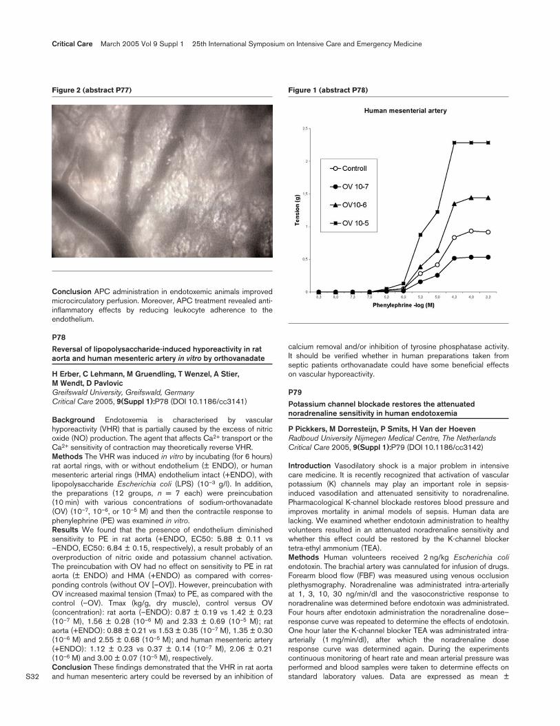
S32
Conclusion APC administration in endotoxemic animals improvedmicrocirculatory perfusion. Moreover, APC treatment revealed anti-inflammatory effects by reducing leukocyte adherence to theendothelium.
P78Reversal of lipopolysaccharide-induced hyporeactivity in rataorta and human mesenteric artery in vitro by orthovanadate
H Erber, C Lehmann, M Gruendling, T Wenzel, A Stier, M Wendt, D PavlovicGreifswald University, Greifswald, GermanyCritical Care 2005, 9(Suppl 1):P78 (DOI 10.1186/cc3141)
Background Endotoxemia is characterised by vascularhyporeactivity (VHR) that is partially caused by the excess of nitricoxide (NO) production. The agent that affects Ca2+ transport or theCa2+ sensitivity of contraction may theoretically reverse VHR.Methods The VHR was induced in vitro by incubating (for 6 hours)rat aortal rings, with or without endothelium (± ENDO), or humanmesenteric arterial rings (HMA) endothelium intact (+ENDO), withlipopolysaccharide Escherichia coli (LPS) (10–3 g/l). In addition,the preparations (12 groups, n = 7 each) were preincubation(10 min) with various concentrations of sodium-orthovanadate(OV) (10–7, 10–6, or 10–5 M) and then the contractile response tophenylephrine (PE) was examined in vitro.Results We found that the presence of endothelium diminishedsensitivity to PE in rat aorta (+ENDO, EC50: 5.88 ± 0.11 vs–ENDO, EC50: 6.84 ± 0.15, respectively), a result probably of anoverproduction of nitric oxide and potassium channel activation.The preincubation with OV had no effect on sensitivity to PE in rataorta (± ENDO) and HMA (+ENDO) as compared with corres-ponding controls (without OV [–OV]). However, preincubation withOV increased maximal tension (Tmax) to PE, as compared with thecontrol (–OV). Tmax (kg/g, dry muscle), control versus OV(concentration): rat aorta (–ENDO): 0.87 ± 0.19 vs 1.42 ± 0.23(10–7 M), 1.56 ± 0.28 (10–6 M) and 2.33 ± 0.69 (10–5 M); rataorta (+ENDO): 0.88 ± 0.21 vs 1.53 ± 0.35 (10–7 M), 1.35 ± 0.30(10–6 M) and 2.55 ± 0.68 (10–5 M); and human mesenteric artery(+ENDO): 1.12 ± 0.23 vs 0.37 ± 0.14 (10–7 M), 2.06 ± 0.21(10–6 M) and 3.00 ± 0.07 (10–5 M), respectively.Conclusion These findings demonstrated that the VHR in rat aortaand human mesenteric artery could be reversed by an inhibition of
calcium removal and/or inhibition of tyrosine phosphatase activity.It should be verified whether in human preparations taken fromseptic patients orthovanadate could have some beneficial effectson vascular hyporeactivity.
P79Potassium channel blockade restores the attenuatednoradrenaline sensitivity in human endotoxemia
P Pickkers, M Dorresteijn, P Smits, H Van der HoevenRadboud University Nijmegen Medical Centre, The NetherlandsCritical Care 2005, 9(Suppl 1):P79 (DOI 10.1186/cc3142)
Introduction Vasodilatory shock is a major problem in intensivecare medicine. It is recently recognized that activation of vascularpotassium (K) channels may play an important role in sepsis-induced vasodilation and attenuated sensitivity to noradrenaline.Pharmacological K-channel blockade restores blood pressure andimproves mortality in animal models of sepsis. Human data arelacking. We examined whether endotoxin administration to healthyvolunteers resulted in an attenuated noradrenaline sensitivity andwhether this effect could be restored by the K-channel blockertetra-ethyl ammonium (TEA).Methods Human volunteers received 2 ng/kg Escherichia coliendotoxin. The brachial artery was cannulated for infusion of drugs.Forearm blood flow (FBF) was measured using venous occlusionplethysmography. Noradrenaline was administrated intra-arteriallyat 1, 3, 10, 30 ng/min/dl and the vasoconstrictive response tonoradrenaline was determined before endotoxin was administrated.Four hours after endotoxin administration the noradrenaline dose–response curve was repeated to determine the effects of endotoxin.One hour later the K-channel blocker TEA was administrated intra-arterially (1 mg/min/dl), after which the noradrenaline doseresponse curve was determined again. During the experimentscontinuous monitoring of heart rate and mean arterial pressure wasperformed and blood samples were taken to determine effects onstandard laboratory values. Data are expressed as mean ±
Critical Care March 2005 Vol 9 Suppl 1 25th International Symposium on Intensive Care and Emergency Medicine
Figure 2 (abstract P77) Figure 1 (abstract P78)

S33
standard error of the mean. Differences were tested by analysis ofvariance repeated measures or Student t test, as appropriate.P < 0.05 was considered to indicate significance.Results Endotoxin administration induced the expected flu-likesymptoms and fever (maximum temperature 38.3 ± 0.1°C,P < 0.001). Mean arterial pressure decreased from 93 ± 2 to79 ± 2 mmHg (P < 0.001) and the heart rate increased from60 ± 2 to 95 ± 2 bpm (P < 0.001). After the administration ofendotoxin, leucocytes increased to 14.2 ± 0.6 × 10⁹/l (P < 0.001)and C-reactive protein increased to 36.0 ± 2.7 mg/l (P < 0.001).Intra-arterial noradrenaline infusion decreased forearm blood flow:percentage of baseline ratio (infused/noninfused arm), 100 ± 0%,84 ± 4%, 70 ± 4%, 55 ± 4%, 38 ± 4%. Following endotoxinadministration, the noradrenaline-induced vasoconstriction wasattenuated: 100 ± 0%, 101 ± 4%, 92 ± 4%, 83 ± 6%, 56 ± 7%(P < 0.001, pooled data, n = 25). Time control experiments (n = 6)demonstrated excellent repeatability of the attenuatednoradrenaline response after the administration of endotoxin (seeFig. 1, top). Intra-arterial infusion of the K-channel blocker TEAalmost completely restored the vasoconstrictive effect ofnoradrenaline (see Fig. 1, bottom; n = 6, P = 0.045).Conclusions In experimental human endotoxemia, noradrenalinesensitivity is decreased. K-channel blocker TEA almost completelyrestores the vasoconstrictive effects of noradrenaline. Our studydemonstrates that endotoxin-induced vascular K-channel activationplays a major role in the observed attenuated sensitivity tonoradrenaline during human endotoxemia.
P80Dose-dependent and temporal effects of pentoxifylline in a ratmodel of acute hepatic failure
T Rahman1, H Hodgson2
1St George’s Hospital, London, UK; 2Royal Free Campus,University College, London, UKCritical Care 2005, 9(Suppl 1):P80 (DOI 10.1186/cc3143)
Introduction The methylxanthine pentoxifylline (PTX) is known toincrease intracellular cAMP concentrations by inhibitingphospodiesterases. Despite beneficial in vitro effects its role inacute hepatic failure (AHF) is controversial. Several studies havedemonstrated no benefit in the use of PTX in AHF, sepsis or shock.Aims To investigate three hypotheses: (1) the use of PTX leads toimproved outcome in AHF; (2) this effect is dose dependent; and(3) improved outcome is limited by temporal constraints. We willcompare PTX administration pre and post hepatic injury.Methods We have previously described a robust and reproduciblemodel of AHF in the Wistar rat. The animals were randomly allocatedinto five groups (each n = 10): Group I received 2 × intra-peritonealinjections of thioacetamide (TAA) 8 hours apart (500 mg/kg).Groups II and III were pretreatment groups: Group II was pretreatedwith low-dose PTX (25 mg/kg body weight) and Group III waspretreated with high-dose PTX (300 mg/kg) and received the TAAprotocol as in Group I. Groups IV and V were post-treatment groups.All followed the protocol for Group I; however, 30 min prior toreceiving the second dose of TAA they received low-dose PTX(Group IV) and high-dose PTX (Group V). Clinical, biochemical andpathological analysis occurred at 24-hour time points.Results Encephalopathy, blood ammonia levels and transaminitisis significantly reduced by pretreatment with low-dose PTX (GroupII) (P < 0.05). A significant reduction in mortality is seen in Group IIwhere mortality at 96 hours is 30% compared with greater than90% in the other pretreatment group (P < 0.005) and 70% in theTAA-treated animals (P < 0.005). This reduction in clinical andbiochemical parameters is not seen in the post-treated groups.Conclusion Low-dose pretreatment with PTX significantly improvesencehalopathy, biochemical markers of AHF and mortalitycompared with controls (TAA only). Beneficial effects are, however,negated if PTX is administered at high dose pretreatment or anydose post-treatment with TAA, exacerbating hepatic injury,increasing mortality and therefore demonstrating dose-dependentand temporal constraints.
P81Tumor necrosis factor increases the sensitivity of thecontractile apparatus in isolated mouse resistance arteries viaa calcium-dependent mechanism
Y OuelletteMayo Clinic College of Medicine, Rochester, MN, USACritical Care 2005, 9(Suppl 1):P81 (DOI 10.1186/cc3144)
Introduction The myogenic response is the modulation ofresistance artery diameter in response to changes in luminalpressure. It is thought to be an important determinant of peripheralvascular resistance and therefore of arterial blood pressure. Sincealtered peripheral resistance is known to occur during the systemicinflammatory response syndrome (such as in sepsis), we suspectthat an abnormal myogenic response might be responsible. Ourmain objective is to assess the effects of tumor necrosis factoralpha (TNF-α) on the myogenic response of mouse mesentericresistance arteries and to determine the role of Ca2+ in thisresponse.
Available online http://ccforum.com/supplements/9/S1
Figure 1 (abstract P79)
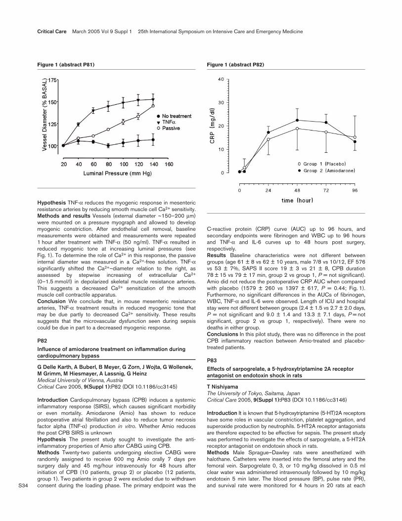
S34
Hypothesis TNF-α reduces the myogenic response in mesentericresistance arteries by reducing smooth muscle cell Ca2+ sensitivity.Methods and results Vessels (external diameter ~150–200 µm)were mounted on a pressure myograph and allowed to developmyogenic constriction. After endothelial cell removal, baselinemeasurements were obtained and measurements were repeated1 hour after treatment with TNF-α (50 ng/ml). TNF-α resulted inreduced myogenic tone at increasing luminal pressures (seeFig. 1). To determine the role of Ca2+ in this response, the passiveinternal diameter was measured in a Ca2+-free solution. TNF-αsignificantly shifted the Ca2+–diameter relation to the right, asassessed by stepwise increasing of extracellular Ca2+
(0–1.5 mmol/l) in depolarized skeletal muscle resistance arteries.This suggests a decreased Ca2+ sensitization of the smoothmuscle cell contractile apparatus.Conclusion We conclude that, in mouse mesenteric resistancearteries, TNF-α treatment results in reduced myogenic tone thatmay be due partly to decreased Ca2+ sensitivity. These resultssuggests that the microvascular dysfunction seen during sepsiscould be due in part to a decreased myogenic response.
P82Influence of amiodarone treatment on inflammation duringcardiopulmonary bypass
G Delle Karth, A Buberl, B Meyer, G Zorn, J Wojta, G Wollenek,M Grimm, M Hiesmayer, A Lassnig, G HeinzMedical University of Vienna, AustriaCritical Care 2005, 9(Suppl 1):P82 (DOI 10.1186/cc3145)
Introduction Cardiopulmonary bypass (CPB) induces a systemicinflammatory response (SIRS), which causes significant morbidityor even mortality. Amiodarone (Amio) has shown to reducepostoperative atrial fibrillation and also to reduce tumor necrosisfactor alpha (TNF-α) production in vitro. Whether Amio reducesthe post CPB SIRS is unknownHypothesis The present study sought to investigate the anti-inflammatory properties of Amio after CABG using CPB.Methods Twenty-two patients undergoing elective CABG wererandomly assigned to receive 600 mg Amio orally 7 days presurgery daily and 45 mg/hour intravenously for 48 hours afterinitiation of CPB (10 patients, group 2) or placebo (12 patients,group 1). Two patients in group 2 were excluded due to withdrawnconsent during the loading phase. The primary endpoint was the
C-reactive protein (CRP) curve (AUC) up to 96 hours, andsecondary endpoints were fibrinogen and WBC up to 96 hoursand TNF-α and IL-6 curves up to 48 hours post surgery,respectively.Results Baseline characteristics were not different betweengroups (age 61 ± 8 vs 62 ± 10 years, male 7/8 vs 10/12, EF 576vs 53 ± 7%, SAPS II score 19 ± 3 vs 21 ± 8, CPB duration78 ± 15 vs 79 ± 17 min, group 2 vs group 1, P = not significant).Amio did not reduce the postoperative CRP AUC when comparedwith placebo (1579 ± 260 vs 1397 ± 617, P = 0.44; Fig. 1).Furthermore, no significant differences in the AUCs of fibrinogen,WBC, TNF-α and IL-6 were observed. Length of ICU and hospitalstay were not different between groups (2.4 ± 1.5 vs 2.7 ± 2.0 days,P = not significant and 9.0 ± 1.4 and 13.3 ± 7.1 days, P = notsignificant, group 2 vs group 1, respectively). There were nodeaths in either group.Conclusions In this pilot study, there was no difference in the postCPB inflammatory reaction between Amio-treated and placebo-treated patients.
P83Effects of sarpogrelate, a 5-hydroxytriptamine 2A receptorantagonist on endotoxin shock in rats
T NishiyamaThe University of Tokyo, Saitama, JapanCritical Care 2005, 9(Suppl 1):P83 (DOI 10.1186/cc3146)
Introduction It is known that 5-hydroxytriptamine (5-HT)2A receptorshave some roles in vascular constriction, platelet aggregation, andsuperoxide production by neutrophils. 5-HT2A receptor antagonistsare therefore expected to be effective for sepsis. The present studywas performed to investigate the effects of sarpogrelate, a 5-HT2Areceptor antagonist on endotoxin shock in rats.Methods Male Sprague–Dawley rats were anesthetized withhalothane. Catheters were inserted into the femoral artery and thefemoral vein. Sarpogrelate 0, 3, or 10 mg/kg dissolved in 0.5 mlclear water was administered intravenously followed by 10 mg/kgendotoxin 5 min later. The blood pressure (BP), pulse rate (PR),and survival rate were monitored for 4 hours in 20 rats at each
Critical Care March 2005 Vol 9 Suppl 1 25th International Symposium on Intensive Care and Emergency Medicine
Figure 1 (abstract P81) Figure 1 (abstract P82)
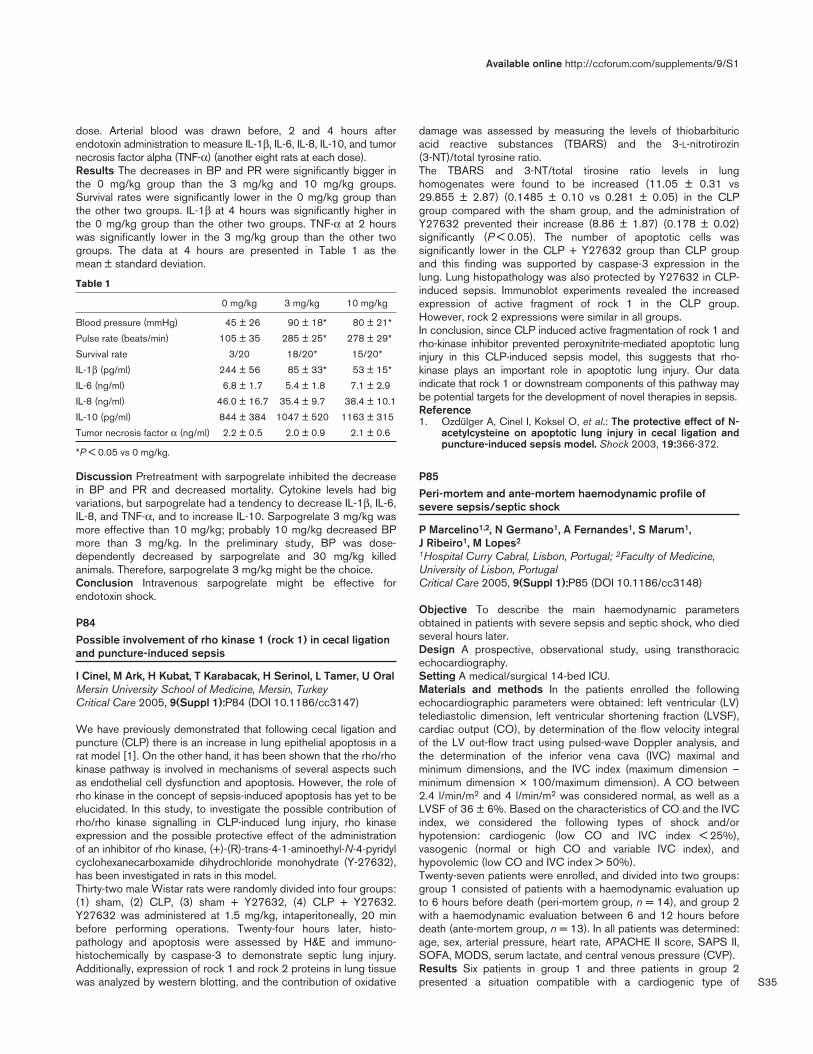
S35
dose. Arterial blood was drawn before, 2 and 4 hours afterendotoxin administration to measure IL-1β, IL-6, IL-8, IL-10, and tumornecrosis factor alpha (TNF-α) (another eight rats at each dose).Results The decreases in BP and PR were significantly bigger inthe 0 mg/kg group than the 3 mg/kg and 10 mg/kg groups.Survival rates were significantly lower in the 0 mg/kg group thanthe other two groups. IL-1β at 4 hours was significantly higher inthe 0 mg/kg group than the other two groups. TNF-α at 2 hourswas significantly lower in the 3 mg/kg group than the other twogroups. The data at 4 hours are presented in Table 1 as themean ± standard deviation.
Table 1
0 mg/kg 3 mg/kg 10 mg/kg
Blood pressure (mmHg) 45 ± 26 90 ± 18* 80 ± 21*
Pulse rate (beats/min) 105 ± 35 285 ± 25* 278 ± 29*
Survival rate 3/20 18/20* 15/20*
IL-1β (pg/ml) 244 ± 56 85 ± 33* 53 ± 15*
IL-6 (ng/ml) 6.8 ± 1.7 5.4 ± 1.8 7.1 ± 2.9
IL-8 (ng/ml) 46.0 ± 16.7 35.4 ± 9.7 38.4 ± 10.1
IL-10 (pg/ml) 844 ± 384 1047 ± 520 1163 ± 315
Tumor necrosis factor α (ng/ml) 2.2 ± 0.5 2.0 ± 0.9 2.1 ± 0.6
*P < 0.05 vs 0 mg/kg.
Discussion Pretreatment with sarpogrelate inhibited the decreasein BP and PR and decreased mortality. Cytokine levels had bigvariations, but sarpogrelate had a tendency to decrease IL-1β, IL-6,IL-8, and TNF-α, and to increase IL-10. Sarpogrelate 3 mg/kg wasmore effective than 10 mg/kg; probably 10 mg/kg decreased BPmore than 3 mg/kg. In the preliminary study, BP was dose-dependently decreased by sarpogrelate and 30 mg/kg killedanimals. Therefore, sarpogrelate 3 mg/kg might be the choice.Conclusion Intravenous sarpogrelate might be effective forendotoxin shock.
P84Possible involvement of rho kinase 1 (rock 1) in cecal ligationand puncture-induced sepsis
I Cinel, M Ark, H Kubat, T Karabacak, H Serinol, L Tamer, U OralMersin University School of Medicine, Mersin, TurkeyCritical Care 2005, 9(Suppl 1):P84 (DOI 10.1186/cc3147)
We have previously demonstrated that following cecal ligation andpuncture (CLP) there is an increase in lung epithelial apoptosis in arat model [1]. On the other hand, it has been shown that the rho/rhokinase pathway is involved in mechanisms of several aspects suchas endothelial cell dysfunction and apoptosis. However, the role ofrho kinase in the concept of sepsis-induced apoptosis has yet to beelucidated. In this study, to investigate the possible contribution ofrho/rho kinase signalling in CLP-induced lung injury, rho kinaseexpression and the possible protective effect of the administrationof an inhibitor of rho kinase, (+)-(R)-trans-4-1-aminoethyl-N-4-pyridylcyclohexanecarboxamide dihydrochloride monohydrate (Y-27632),has been investigated in rats in this model.Thirty-two male Wistar rats were randomly divided into four groups:(1) sham, (2) CLP, (3) sham + Y27632, (4) CLP + Y27632.Y27632 was administered at 1.5 mg/kg, intaperitoneally, 20 minbefore performing operations. Twenty-four hours later, histo-pathology and apoptosis were assessed by H&E and immuno-histochemically by caspase-3 to demonstrate septic lung injury.Additionally, expression of rock 1 and rock 2 proteins in lung tissuewas analyzed by western blotting, and the contribution of oxidative
damage was assessed by measuring the levels of thiobarbituricacid reactive substances (TBARS) and the 3-L-nitrotirozin(3-NT)/total tyrosine ratio.The TBARS and 3-NT/total tirosine ratio levels in lunghomogenates were found to be increased (11.05 ± 0.31 vs29.855 ± 2.87) (0.1485 ± 0.10 vs 0.281 ± 0.05) in the CLPgroup compared with the sham group, and the administration ofY27632 prevented their increase (8.86 ± 1.87) (0.178 ± 0.02)significantly (P < 0.05). The number of apoptotic cells wassignificantly lower in the CLP + Y27632 group than CLP groupand this finding was supported by caspase-3 expression in thelung. Lung histopathology was also protected by Y27632 in CLP-induced sepsis. Immunoblot experiments revealed the increasedexpression of active fragment of rock 1 in the CLP group.However, rock 2 expressions were similar in all groups.In conclusion, since CLP induced active fragmentation of rock 1 andrho-kinase inhibitor prevented peroxynitrite-mediated apoptotic lunginjury in this CLP-induced sepsis model, this suggests that rho-kinase plays an important role in apoptotic lung injury. Our dataindicate that rock 1 or downstream components of this pathway maybe potential targets for the development of novel therapies in sepsis.Reference1. Ozdülger A, Cinel I, Koksel O, et al.: The protective effect of N-
acetylcysteine on apoptotic lung injury in cecal ligation andpuncture-induced sepsis model. Shock 2003, 19:366-372.
P85Peri-mortem and ante-mortem haemodynamic profile ofsevere sepsis/septic shock
P Marcelino1,2, N Germano1, A Fernandes1, S Marum1, J Ribeiro1, M Lopes2
1Hospital Curry Cabral, Lisbon, Portugal; 2Faculty of Medicine,University of Lisbon, PortugalCritical Care 2005, 9(Suppl 1):P85 (DOI 10.1186/cc3148)
Objective To describe the main haemodynamic parametersobtained in patients with severe sepsis and septic shock, who diedseveral hours later.Design A prospective, observational study, using transthoracicechocardiography.Setting A medical/surgical 14-bed ICU.Materials and methods In the patients enrolled the followingechocardiographic parameters were obtained: left ventricular (LV)telediastolic dimension, left ventricular shortening fraction (LVSF),cardiac output (CO), by determination of the flow velocity integralof the LV out-flow tract using pulsed-wave Doppler analysis, andthe determination of the inferior vena cava (IVC) maximal andminimum dimensions, and the IVC index (maximum dimension –minimum dimension × 100/maximum dimension). A CO between2.4 l/min/m2 and 4 l/min/m2 was considered normal, as well as aLVSF of 36 ± 6%. Based on the characteristics of CO and the IVCindex, we considered the following types of shock and/orhypotension: cardiogenic (low CO and IVC index < 25%),vasogenic (normal or high CO and variable IVC index), andhypovolemic (low CO and IVC index > 50%).Twenty-seven patients were enrolled, and divided into two groups:group 1 consisted of patients with a haemodynamic evaluation upto 6 hours before death (peri-mortem group, n = 14), and group 2with a haemodynamic evaluation between 6 and 12 hours beforedeath (ante-mortem group, n = 13). In all patients was determined:age, sex, arterial pressure, heart rate, APACHE II score, SAPS II,SOFA, MODS, serum lactate, and central venous pressure (CVP).Results Six patients in group 1 and three patients in group 2presented a situation compatible with a cardiogenic type of
Available online http://ccforum.com/supplements/9/S1

S36
hypotension. The remainder presented as a vasogenic type ofhypotension. Eight patients in group 1 presented LVSF < 30%, aswell as four patients in group 2 All severity scores of group 1 wereslightly higher, as well as serum lactate levels. The main results arepresented in Table 1, with means and standard deviation for eachvariable.
Table 1
Parameter Group 1 Group 2
APACHE II score 40 ± 4 39 ± 9
SAPS II 95 ± 8 88 ± 9
SOFA 18.7 ± 1.7 16 ± 4
MODS 17.3 ± 2 15 ± 3.6
Serum lactate 11.7 ± 5.7 9.8 ± 5.6
Cardiac output (l/min/m2) 3692 ± 1254 2829 ± 958
Left ventricular shortening fraction (%) 32 ± 5.6 28.4 ± 7.3
Conclusions Septic shock presents as a vasogenic type until thelate disease course. A LV failure with a cardiogenic type ofsituation characterizes better the later stages, up to 6 hours beforedeath occurs.
P86Effect of reduced bronchial circulation on the lung fluid flux inthe case of combined burn and smoke inhalation injury insheep
N Morita, P Enkhbaatar, F Schmalstieg, M Westphal, M Maybauer, D Maybauer, L Traber, D TraberUTMB, Galveston, TX, USACritical Care 2005, 9(Suppl 1):P86 (DOI 10.1186/cc3149)
Introduction We reported that the bronchial circulation contributesto pulmonary function after acute lung injury with smoke inhalation.But the observation period was 24 hours. We hypothesize that thebronchial circulation plays a major role in the lung injury seen withthe combination of burn and smoke inhalation injury.Method Merino ewes (n = 13) were surgically prepared for chronicstudy. After a recovery (5–7 days) they were randomly divided intothree groups: (1) the bronchial artery was ligated (n = 5); (2) shamgroup (n = 5), the bronchial artery was surgically exposed but leftintact without ligation; (3) the bronchial artery was exposed, notligated and the animals were not injured. One day after theseoperations, under deep halothane anesthesia, two groups receiveda tracheotomy and were subjected to a 40% third-degree flameburn and insufflated with 48 breaths of cotton smoke inhalation orwere sham injured. All sheep were mechanically ventilated andresuscitated by Ringer’s lactate for 48 hours. We confirmed theligation of the bronchial artery with microspheres. Statistics wereperformed using repeated-measures analysis of variance forrepeated measurements and Scheffe’s post hoc comparisons wasused and P < 0.05 was considered significant. Data are expressedas mean ± standard error of the mean.Result After ligation (24 hours), blood flow of the bronchidecreased to 24.48% of the baseline value. Pulmonary dysfunctionafter the combined injury in the ligation group was significantlyreduced. Lung lymph flow, an index of pulmonary transvascularfluid flux, was markedly increased in in the ligated and intact groups.Ligation reduced this response (24 hours: sham 65 ± 5.0; ligation15.5 ± 7.51; sham–sham 9.4 ± 5.6; 48 hours: sham 64.5 ± 16.5;ligation 12.58 ± 5.743; sham–sham 7.25 ± 3.95 ml/hour). Ligationalso improved the PaO2/FiO2 ratio (24 hours: sham 270 ± 99.347;ligation 390.381 ± 53.781; sham–sham 595 ± 13.856; 48 hours:
sham 132.5 ± 33.15; ligation 375 ± 62.253; sham–sham592.33 ± 8.95). PMN production also decreased (24 hours: sham32.5 ± 31.8; ligation 6.2 ± 2.2; 48 hours: sham 38.3 ± 46.2;ligation 2.7 ± 2.6 × 10⁶).Conclusions Bronchial circulation plays a significant role in thelung inflammation after combined burn and smoke inhalation injury.Acknowledgements Supported by National Institutes of HealthGM066312, Shriners of N America 8450 and 8954.
P87A pilot study of left main bronchus pulse oximetry
XR Wang, YJ Zheng, J Tian, ZH Wang, ZY PanDepartment of Anesthesiology, Renji Hospital, Shanghai SecondMedical University, Shanghai, ChinaCritical Care 2005, 9(Suppl 1):P87 (DOI 10.1186/cc3150)
Objective To assess the feasibility and accuracy of measuringmixed venous oxygen saturation (SvO2) through the left mainbronchus (SpO2trathea). The hypothesis that tracheal oximetryreadings are not primarily derived from the tracheal mucosa wasalso tested. The pulmonary artery lies in close proximity to thebronchus, with nothing but some connective tissues in the interval,raising the possibility that an appropriately located and directedbronchial oximetry probe might be able to derive oximetry readingsfrom the mixed venous system. The present study was undertakento test the feasibility of measuring SvO2 through the left mainbronchus (SpO2trachea), and to compare SpO2trachea with theSwan–Ganz catheter SpO2 (SvO2catheter) and oxygen saturationfrom pulmonary artery samples (SvO2blood) in hemodynamicallystable, well-oxygenated, anesthetized white swines. We alsotested the hypothesis that bronchial oximetry readings are primarilyderived from the pulmonary artery, not the tracheal mucosa.Furthermore, the stability and accuracy of SpO2trachea was testedby correlating the oximetry readings with altered SvO2 or withinternal environment instability.Methods Twenty hemodynamically stable, well-oxygenated,anesthetized white swine were studied. A Robertshaw double-lumen tracheal tube was directed toward the left main bronchususing a fibrobronchoscope. A single-use pediatric pulse oximeterwas attached to the left lateral surface of the tube. SpO2trathea,Swan–Ganz catheter SpO2 (SvO2catheter) and oxygen saturationfrom pulmonary artery samples (SvO2blood) were taken with theintracuff pressure at 0–60 cmH2O. The intracuff pressure was thenset at 60 cmH2O, and changes of SvO2 were induced using threedifferent concentrations of inspiratory oxygen. The influence of thechanges on SpO2trathea, SvO2catheter and SvO2blood wasmeasured respectively at the same time.Results SpO2trathea was the same as SvO2catheter andSvO2blood at an intracuff pressure of 10–60 cmH2O, but was lesswhen the intracuff pressure was zero (P < 0.001 compared withSvO2catheter or SvO2blood) in hemodynamically stable states.The descending of SvO2 followed with decreased inspiratoryoxygen concentration. SpO2trachea agreed with SvO2catheter andSvO2blood at each concentration with significant correlationamong them.Conclusion Left main bronchus SpO2 is feasible and providessimilar readings to SvO2catheter and SvO2blood in hemo-dynamically stable or low saturation states. Tracheal oximetryreadings are not primarily derived from the tracheal mucosa. Thetechnique merits further evaluation.
Critical Care March 2005 Vol 9 Suppl 1 25th International Symposium on Intensive Care and Emergency Medicine

S37
P88Relationship between extravascular lung water andoxygenation indices in patients with acute respiratory failure
J Graf, V Tomicic, P Aguilera, M Espinoza, C CanalsClinica Alemana de Santiago, ChileCritical Care 2005, 9(Suppl 1):P88 (DOI 10.1186/cc3151)
Introduction The main strategy to restore organ perfusion incritically ill patients is fluid administration, which increases thelikelihood of acquiring pulmonary edema with detrimental effectson gas exchange. The thermal-dye dilution technique has beenused to measure extravascular lung water (EVLW). Recently, thistechnique has been simplified using only the thermal indicator [1].The relationship between EVLW and the PaO2/FiO2 ratio has beensparsely studied and described as weak [2]. Oxygenation index(OI) relates PaO2, FiO2, and mean airway pressure (Paw) by thefollowing formula: OI = Paw × FiO2 × 100 / PaO2 [3]. We are notaware of publications correlating EVLW with OI.Methods Simultaneous measurements of EVLW, arterial bloodgases and Paw were performed in mechanically ventilated patientsat different moments of their illness course. EVLW was measuredby a single transpulmonary thermodilution technique using aPiCCO® monitor. The EVLW value was indexed (EVLWi) by thepredicted body weight [4]. PaO2/FiO2 and OI were calculated andcorrelated with EVLWi using linear regression analysis with a 95%confidence interval (CI).Results We studied 21 patients, 20 with ALI/ARDS (age 64 ± 19years, APACHE II score 24.4 ± 6.8, SOFA 10.3 ± 3.2, LIS2.55 ± 0.57). Mean admission EVLWi, Paw, PaO2/FiO2 and OIvalues were 14 ± 6.5 ml/kg, 17.8 ± 3.9 cmH2O, 186 ± 80 and12.1 ±7.5, respectively. We obtained 152 EVLWi measurementswith simultaneous PaO2/FiO2 and OI values. A significant negativecorrelation was found between EVLWi and PaO2/FiO2 (r = –0.53,CI –0.63 to –0.40, P < 0.01) (Fig. 1). A significant positivecorrelation was found between EVLWi and OI (r = 0.70, CI 0.60–0.77, P < 0.01) (Fig. 2).Conclusions There is a stronger correlation between EVLWi andOI than with PaO2/FiO2. This finding emphasizes the importance ofincorporating Paw into the evaluation of patient oxygenation duringmechanical ventilation.
References1. Intensive Care Med 2000, 26:180-187.2. Chest 2004, 125:1166.3. J Pediatr 1985, 106:963-969.4. N Engl J Med 2000, 342:1301-1308.
P89Diagnostic test of the desaturation index and the calculateddesaturation index to identify acute lung injury and/or acuterespiratory distress syndrome at the bedside
G Vazquez de Anda1,2, J Arzate Villafaña1,2, J Talavera Piña2, S Laraza1, J Gutierrez1, D Rodriguez Cadena2
1ISSEMYM Medical Center, Toluca, Mexico; 2UniversidadAutonoma del Estado de Mexico, Toluca, MexicoCritical Care 2005, 9(Suppl 1):P89 (DOI 10.1186/cc3152)
Introduction We have previously shown that the desaturationindex (DI) and the calculated desaturation index (Dic) have astatistical correlation with several indices of gas exchange and withlung infiltrates on the thorax X-ray. However, the utility of the DI andDic as a diagnostic tool has never been tested. The aim of thisstudy is therefore to determine the capability of the DI and the DIc:to identify acute lung injury (ALI) and/or acute respiratory distresssyndrome (ARDS) at the bedside.Patients and methods Patients with and without ALI and/or acuterespiratory failure (ARDS) were followed during the period ofmechanical ventilation. Every morning, blood gases analysis andthorax X-rays were taken and definitions for ALI/ARDS werecompleted according to the American–European ConsensusConference. We registered the positive end expiratory pressure,inspired fraction of oxygen (FiO2) and arterial saturation by pulseoximetry (SpO2) to calculate the Dic. Then, in order to determinethe DI, all patients were subjected to breathe 100% oxygen, with agradual reduction of the FiO2 to 21%, in steps of 20% lasting8 min each, and at each step the SpO2 was registered (we stopthe maneover after reaching 85% of the SpO2). Finally, wecalculated the DI according our equation that has been describedpreviously. Two or more quadrants with infiltrates on the thorax X-ray and PaO2/FiO2 ratio (adjusted to high altitude) lower than 221
Available online http://ccforum.com/supplements/9/S1
Figure 1 (abstract P88)
Figure 2 (abstract P88)

S38
mmHg were considered positive for ALI/ARDS. The sensitivity andspecificity were calculated according to conventional equations.Results During the study period, 198 ALI/ARDS definitions werecompleted. Table 1 presents the sensitivity and specificity tests forthe DI and the Dic. Results are shown as the percentage andconfidence interval
Table 1
Pao2/FiO2 Thorax X-ray ALI/ARDS
DI sensitivity 0.86 (0.78–0.91) 0.77 (0.69–0.84) 0.87 (0.78–0.92)
DI specificity 0.84 (0.75–0.89) 0.85 (0.76–0.91) 0.83 (0.74–0.87)
DIc sensitivity 0.93 (0.87–0.97) 0.85 (0.77–0.90) 0.99 (0.95–1)
DIc specificity 0.82 (0.74–0.89) 0.85 (0.76–0.91) 0.93 (0.86–0.97)
ALI, acute lung injury; ARDS, acute respiratory distress syndrome; DI,desaturation index; Dic, calculated desaturation index.
Conclusion We conclude that the DI and the DIC, which are non-invasive indices, have an acceptable sensitivity and specificity toidentify ALI/ARDS patients at the bedside.
P90Effect of pharmacological inhibition of chloride transport onlung fluid balance in acute Escherichia coli pneumonia in mice
L Robriquet1, Y Song2, H Folkesson3, J Frank2, A Verkman4, M Matthay2
1CHRU, Lille, France; 2Cardiovascular Research Institute, SanFrancisco, CA, USA; 3Department of Physiology, Rootstown, OH,USA; 4Department of Medicine and Department of Physiology,Cardiovascular Research Institute, San Francisco, CA, USACritical Care 2005, 9(Suppl 1):P90 (DOI 10.1186/cc3153)
Introduction Clearance of fluid from the alveolar spaces primarydepends on the active sodium transport across distal lungepithelium. Some experimental studies supported a potential rolefor the cystic fibrosis transmembrane conductance regulator(CFTR), especially in the cAMP-mediated upregulation of fluidclearance. Hypothetically, pharmacological inhibition of Cl–channels could worsen lung edema by decreasing the clearance ofalveolar edema. In this study, we tested the affect of chloridetransport inhibition on lung edema formation in acute pneumonia.Methods A mouse model of acute pneumonia was establishedusing IT instillation of Escherichia coli (107 cfu). Basal andisoproterenol (0.1 mM)-stimulated alveolar fluid clearance werestudied using the in situ mice model. Glibenclamide (0.1 mM) or aspecific CFTR-inhibitor (100 mM CFTR-172) was instilled into thelung to study the effects of CFTR blockade.Results In response to cAMP stimulation by isoproterenol,clearance was inhibited by glibenclamide or the CFTR-172inhibitor (respectively, 12.2 ± 0.39% and 9.7 ± 3.5% versus 20.7± 2.9% in the control group, P < 0.05). Four hours after bacterialinstillation, the lung wet-to-dry ratio and lung vascular permeabilityas measured by the extravascular accumulation of 125I-albuminwere increased compared with the control group (respectively,4.8 ± 0.18 g/g versus 3.8 ± 0.18 g/g, P < 0.05 and 61.8 ± 10.4 µlversus 20 ± 5.9 µl, P < 0.05). No significant statistical change wasfound among lung water and endothelial permeability in theglibenclamide or CFTR-172 inhibitor group.Conclusion These experiments indicated that the c-AMP-dependent fluid clearance from the distal airspaces of the lunginvolves chloride transport by CFTR. In vivo, we found no changein pulmonary edema formation following E. coli intratrachealinstillation. These data suggest that chloride transport is not a
major mechanism in lung edema formation in a mouse model ofacute pneumonia.
P91Effect of beta-adrenergic agonist on alveolar fluid clearance inrats with acute lung injury
H Qiu, H Sun, Y YangNanjing Zhong-Da Hospital, Southeast University, Nanjing, ChinaCritical Care 2005, 9(Suppl 1):P91 (DOI 10.1186/cc3154)
Background Although it is well known that beta-adrenergic agonistimproves alveolar fluid clearance (AFC) in several animal species, itis not known whether beta-adrenergic agonist improves AFC ininfectious acute lung injury (ALI). This study is aimed at investigatingthe effect of beta-adrenergic agonist (dobutamine) on AFC in ratswith infectious ALI and exploring the associated mechanism.Methods Thirty-two male Sprague–Dawley rats were randomlydivided into four groups (normal control group, ALI group,dobutamine control group, dobutamine treatment group). A rat withALI was induced by infusion of endotoxin intravenously. Alveolarfluid clearance was measured by the single-nuclide tracertechnique. α-Rat epithelial sodium channel (α-rENaC), β-rENaCand γ-rENaC mRNA expressions were measured by RT-PCR.Results AFC of the ALI group was 14.0 ± 1.2%, which wassignificantly lower than the normal control group (21.0 ± 3.9%),the dobutamine treatment group (20.0 ± 3.8%) and thedobutamine control group (26.6 ± 1.6%) (P < 0.05). AFC of thedobutamine control group was significantly higher than the normalcontrol group, dobutamine treatment group and ALI group(P < 0.05). α-rENaC and β-rENaC mRNA expressions of the ALIgroup (1.4 ± 0.4 and 0.7 ± 0.8, respectively) and the dobutaminetreatment group (1.38 ± 0.13 and 0.71 ± 0.17, respectively) weresignificantly higher than the dobutamine control group(1.01 ± 0.14 and 0.58 ± 0.12, respectively) and the normal controlgroup (1.00 ± 0.28 and 0.44 ± 0.11, respectively) (P < 0.05).There were no significant differences of α-rENaC and β-rENaCmRNA expressions between the normal control group anddobutamine control group, and no significant difference betweenthe ALI group and dobutamine treatment group (P > 0.05). γ-rENaC mRNA expressions of the dobutamine control group anddobutamine treatment group were 0.90 ± 0.19 and 0.97 ± 0.15,respectively, which were significantly higher than normal controlgroup (0.69 ± 0.10) and the ALI group (0.7 ± 0.3) (P < 0.05).There was no significant difference of γ-rENaC mRNA expressionsbetween the normal control group and the ALI group, and nosignificant difference between the dobutamine control group andthe dobutamine treatment group (P > 0.05).Conclusion Beta-adrenergic agonist may upregulate γ-rENaCexpression and improve AFC in rats with endotoxin-induced ALI. Itmay be beneficial to increase AFC and reduce lung edema usingbeta-adrenergic agonist in ALI patients.
P92Effect of beta-adrenergic agonist on alveolar fluid clearance inrats
H Qiu, Y Yang, H SunNanjing Zhong-Da Hospital, Southeast University, Nanjing, ChinaCritical Care 2005, 9(Suppl 1):P92 (DOI 10.1186/cc3155)
Objective To investigate the effect and the associated mechanismof beta-adrenergic agonist on alveolar fluid clearance (AFC) in rats.Methods Forty-eight Sprague–Dawley male rats were randomlydivided into six groups (normal control group, dobutamine group,
Critical Care March 2005 Vol 9 Suppl 1 25th International Symposium on Intensive Care and Emergency Medicine

S39
amiloride [specific inhibitor of epithelial sodium channel] group,dobutamine + amiloride group, dobutamine + atenolol [specificbeta-1 antagonist] group, dobutamine + ICI118,551 [specificbeta-2 antagonist] group). AFC was measured by the single-nuclide tracer technique. Alpha-rat epithelial sodium channel(alpha-rENaC), beta-rENaC and gamma-rENaC mRNA expressionswere measured by RT-PCR.Results (1) AFC of the dobutamine group and dobutamine +atenolol group were 26.6 ± 1.6% and 25.0 ± 5.3%, respectively,which were significantly higher than the normal control group(21.0 ± 3.9%) and dobutamine + ICI 118,551 group (21.0 ± 4.8%)(P < 0.05). There was no significant difference of AFC betweenthe dobutamine group and dobutamine + atenolol group(P > 0.05); and no significant difference of AFC between thenormal control group and dobutamine + ICI 118,551 group(P > 0.05). (2) AFC of the amiloride group was 6.0 ± 2.8%, whichwas significantly lower than the normal control group (P < 0.05).(3) AFC of the dobutamine + amiloride group was 10.0 ± 2.3%,significantly higher than the amiloride group (P < 0.05). (4)Gamma-rENaC mRNA expression of the dobutamine group anddobutamine + atenolol group was 0.90 ± 0.19 and 0.90 ± 0.09,respectively, significantly higher than the normal control group(0.69 ± 0.09) and dobutamine + ICI 118,551 group (0.68 ± 0.06)(P < 0.05). There was no significant difference of gamma-rENaCmRNA expression between the dobutamine group and dobutamine +atenolol group; and no significant difference of gamma-rENaCmRNA expression between the normal control group anddobutamine + ICI 118,551 group (P < 0.05). There was nosignificant difference of alpha-rENaC and beta-rENaC mRNAexpressions between each group (P > 0.05). (5) Gamma-rENaCmRNA expression of the dobutamine + amiloride group was0.70 ± 0.14, significantly lower than the dobutamine group anddobutamine + atenolol group (P < 0.05).Conclusion The amiloride-sensitive NaC pathway was importantfor lung epithelium to actively transport alveolar fluid. The effect ofbeta-2 adrenergic stimulation may upregulate gamma-rENaCexpression and also stimulate the amiloride-insensitive NaCpathway to improve AFC in rats.
P93Change of extravascular lung water in sheep with early acuterespiratory distress syndrome
H Qiu, H SunNanjing Zhong-Da Hospital, Southeast University, Nanjing, ChinaCritical Care 2005, 9(Suppl 1):P93 (DOI 10.1186/cc3156)
Objective To determine the change of extravascular lung water(EVLW) and the effect of positive end-expiratory pressure (PEEP)and cardiac output (CO) on EVLW in sheep with early acuterespiratory distress syndrome (ARDS).Methods The sheep ARDS model was induced by infusion ofendotoxin intravenously, then divided into the ARDS model group(n = 12), PEEP group (n = 21) and dobutamine group (n = 13).The PEEP group was divided into three subgroups: PEEP5 cmH2O group (n = 7), PEEP 10 cmH2O group (n = 8) andPEEP 15 cmH2O group (n = 6). EVLW was measured by thesingle indicator thermodilution technique. The EVLW, hemo-dynamics and lung mechanics parameters were observed at 6 hoursafter the sheep ARDS model was induced, 2 hours after theyreceived mechanical ventilation with PEEP (5, 10, 15 cmH2O),respectively, and 2 hours after CO increase > 50% of the basevalue (induced by dobutamine infusion).Results (1) When ARDS was induced in sheep, EVLW increasedfrom 12.8 ± 4.7 ml/kg to 18.1 ± 7.1 ml/kg (P < 0.01), and EVLW
had no significant change (P > 0.05) during the early phase ofARDS (6 hours). The PaO2/FiO2 was 136.8 ± 34.9 mmHg atARDS 0 hours, which was higher than that before infusion ofendotoxin (444.3 ± 127.7 mmHg, P < 0.01). EVLW had norelationship with PaO2/FiO2, but EVLW after the ARDS model wasinduced had good correlation with PaO2/FiO2 (r = –0.501,P < 0.001). When ARDS was induced, static lung compliancedecreased from 26.9 ± 8.4 to 16.4 ± 5.0 (P < 0.01). (2) Aftermechanical ventilation with PEEP for 2 hours, EVLW values in thePEEP 10 cmH2O group and PEEP 15 cmH2O group were14.7 ± 4.5 ml/kg and 15.3 ± 3.7 ml/kg, respectively, which wassignificantly less than that before applied PEEP (16.5 ± 4.7 ml/kgand 18.4 ± 6.0 ml/kg, respectively; all P < 0.05). But EVLW in the5 cmH2O group was no different during 2 hours PEEP application.(3) After CO increase > 50% of the base value at 1 hour and2 hours, EVLW values were 16.3 ± 4.9 ml/kg and 16.9 ± 6.9 ml/kg,respectively, which did not differ from that of the base value(15.1 ± 4.6 ml/kg; all P > 0.05).Conclusions EVLW in ARDS sheep increased significantly andremained at the same level during the early phase of ARDS. PEEPhad a marked effect on reducing EVLW and the increase of COinduced by dobutamine did not significantly raise the EVLW.
P94Pulmonary surfactant function in patients with acute lunginjury/acute respiratory distress syndrome treated bymechanical ventilation
E Miller1, M Pirozynski1, J Jastrzebski1, M Pawelec2, T Sosnowski21Medical Centre of Postgraduate Medicine, Warsaw, Poland;2Warsaw University of Technology, Warsaw, PolandCritical Care 2005, 9(Suppl 1):P94 (DOI 10.1186/cc3157)
Aim The evaluation of the quality and surface active properties ofsurfactant carried out in vitro in setting the bubble, that is pulsating,with the use of a surfactometer (Pulsating Bubble Surfactometer).Materials and methods Patients requiring mechanical ventilationwere enrolled to the study. All patients met the criteria for acutelung injury (ALI) or acute respiratory distress syndrome, accordingto the American–European Consensus Conference. Additionally,the Lung Injury Score criteria to determine the severity and thecourse of lung injury were used. Sixty-five patients were included inthe study — 45 with respiratory insufficiency, of the latter 30 withindirect ALI and 15 with direct ALI. Twenty patients were includedin the reference group. All patients underwent bronchoscopy andbronchoalveolar lavage. The procedures were carried out duringthe first 24 hours of mechanical ventilation. Analysis of pulmonarysurfactant activity was carried out using Pulsating BubbleSurfactometer (Electronetics Corp., USA). Changes of value of thesurface tension in the cycle compression–expansion, minimumvalue of the surface tension, maximum value surface tension andthe hysteresis loop changes of the surface tension measured astheir area value changes were measured. The normalizedhysteresis area, the stability index and the rate of effectiveness ofreducing the surface tension were calculated.Results Differences were seen between groups with direct ALIand indirect ALI and the control group, with reference to separateanalyses parameters. Minimum value of the surface tension(P < 0.001) and maximum value of the surface tension (P < 0.002)were significantly lower in the control group, whereas thenormalized hysteresis area (P < 0.001), the stability index (P < 0.001)and the rate of effectiveness of reducing the surface tension(P < 0.02) were significantly higher.Comparison of separate groups revealed significantly lower valuesof the minimum value of the surface tension (P < 0.003), the
Available online http://ccforum.com/supplements/9/S1

S40
normalized hysteresis area (P < 0.001), the stability index(P < 0.007) and the rate of effectiveness of reducing the surfacetension (P < 0.003) in the group with direct ALI, compared with thegroup with indirect ALI. There were no differences in relation tomaximum values of the surface tension (P = 0.135).Conclusions (1) Pulmonary surfactant dysfunction was observedin the early phase ALI. (2) Direct lung injury was associated withhigher impairment of surfactant function than indirect lung injury.(3) Correlation exists between the level of the lung injury anddynamic surface active properties of pulmonary surfactant.
P95Alveolar fibrinolysis is not altered by mechanical ventilation inhumans with healthy lungs
G Choi, E Wolthuis, P Bresser, M Levi, T van der Poll, M Dzoljic,M Vroom, M SchultzAcademic Medical Center, Amsterdam, The NetherlandsCritical Care 2005, 9(Suppl 1):P95 (DOI 10.1186/cc3158)
Introduction Mechanical ventilation (MV) aggravates pulmonaryinflammation in patients with acute lung injury. Pulmonaryinflammation is characterized by alveolar fibrin depositions, whichis the net result of increased thrombin-mediated generation of fibrinand upregulation of plasminogen activator inhibitor (PAI), inhibitingfibrin degradation. We recently showed that injurious MV (usinghigh pressures) induces PAI-1 upregulation in rats, thereby limitingpulmonary fibrinolytic activity. In the present study we determinethe influence of MV on alveolar PAI-1 expression in humans withhealthy lungs.Methods Patients with healthy lungs expected to be mechanicallyventilated for ≥ 5 hours (elective surgery) were randomized to a MVstrategy using either a tidal volume (VT) of 12 ml/kg and zeropositive end expiratory pressure (HVT/ZEEP) or VT 6 ml/kg and10 cmH2O PEEP (LVT/PEEP). Bronchoalveolar lavage (BAL) wasperformed directly after initiation of MV, and repeated in the contra-lateral lung after 5 hours of MV. Pulmonary thrombin generationwas quantified by measuring thrombin–antithrombin complexes(TATc) in the BAL fluid; fibrinolytic activity by determining plas-minogen activator activity (PAA), PAI-1, and tissue-type plasmino-gen activator (tPA).Statistics Within groups, Wilcoxon signed-rank test; betweengroups, Mann–Whitney U test. Data are presented as medianswith interquartile ranges.Results Nine patients were randomized to HVT/ZEEP, 11 patientsto LVT/PEEP. There were no differences in baseline characteristicsor outcome. After 5 hours, patients ventilated with HVT/ZEEPshowed an increase in alveolar thrombin generation (TATc: 0.91[0.82–0.98] ng/ml vs 0.75 [0.67–0.81] ng/ml at baseline,P < 0.01), in contrast to those with LVT/PEEP (0.84 [0.74–0.91]ng/ml vs 0.81 [0.72–0.87] ng/ml, not significant [NS]). PulmonaryPAI-1 levels did not change (LVT/PEEP: 2.0 [1.6–2.2] ng/ml vs 1.9[1.8–2.1] ng/ml, NS; HVT/ZEEP: 2.1 [1.8–2.1] ng/ml vs 2.0[1.7–2.4] ng/ml, NS). Moreover, alveolar PAA was unaffected inboth groups (LVT/PEEP: 106 [102–113]% vs 100 [97–105]%,NS; HVT/ZEEP: 102 [97–110]% vs 102 [100–104]%, NS),although levels of tPA increased more with HVT/ZEEP MV(LVT/PEEP: 0.84 [0.65–0.98] ng/ml vs 0.61 [0.55–0.63] ng/ml,P < 0.001; HVT/ZEEP: 0.70 [0.70–0.90] ng/ml vs 0.50[0.40–0.55] ng/ml, P < 0.01; between groups, NS).Conclusion Despite upregulation of tPA, alveolar PAA is notaltered during 5 hours of MV in patients with healthy lungs.Disturbed alveolar fibrin turnover during MV in healthy lungs seemssolely the result of activation of coagulation.
P96Effect of lung protective ventilation strategy onextrapulmonary organs inflammatory response in an acuterespiratory distress syndrome rabbit model
H Qiu, Y Yang, Y YanNanjing Zhong-Da Hospital, Southeast University, Nanjing, ChinaCritical Care 2005, 9(Suppl 1):P96 (DOI 10.1186/cc3159)
Objective To evaluate the influence of a lung protective ventilationstrategy on extrapulmonary organ inflammatory response in anacute respiratory distress syndrome (ARDS) rabbit model.Methods The ARDS rabbit models were duplicated by alveoli-lavaged saline. The rabbit were divided into six groups: (1) normal,(2) ARDS group, (3) low volume (VT) with best end-expiratorypressure (PEEP) (LVBP), (4) normal volume with best PEEP(NVBP), (5) low-volume with high PEEP (LVHP), and (6) high-volume zero PEEP (HVZP). Tumor necrosis factor alpha (TNF-α)and IL-10 levels in liver and intestine homogenates were measuredby ELISA and their mRNA expression was measured by RT-PCR.Results In the LVBP group, the mRNA expression of TNF-α andIL-10 in liver was lower than the NVBP, LVHP, and HVZP groups.But there was no difference between LVBP and ARDS groups.TNF-α mRNA expression in the LVHP group was higher than theLVBP group in liver tissue, but significantly lower than the HVZPgroup. Compared with the LVBP group, the TNF-α and IL-10concentrations in liver were increased markedly in NVBP, LVHP,and HVZP. But there was no difference between the LVBP andARDS groups. Live TNF-α and IL-10 concentrations were highestin the HVZP group, which was higher than the LVBP and LVHPgroups. In the LVBP group, the mRNA expression of TNF-α andIL-10 in intestine was lower than the NVBP, LVHP, and HVZPgroups. But there was no difference between the LVBP and ARDSgroups. As compared with the LVBP group, the TNF-α and IL-10concentrations of intestine in NVBP, LVHP, and HVZP wereincreased markedly, but there was no difference between theLVBP and ARDS groups.Conclusions A lung protective ventilation strategy candownregulate inflammatory mediator expression in the liver andintestine of ARDS rabbits and may prevent the occurrence ofmultiple organ dysfunction syndrome.
P97Persistent use of lower tidal volumes after a simpleintervention consisting of feedback and education
E Wolthuis1, R Determann1, M Vroom1, J Kesecioglu2, M Schultz1
1Academic Medical Centre, Amsterdam, The Netherlands; 2UMC,Utrecht, The NetherlandsCritical Care 2005, 9(Suppl 1):P97 (DOI 10.1186/cc3160)
Introduction Use of lower tidal volumes (Vt) is recommended forpatients suffering from ALI/ARDS. We previously reported on asimple ‘intervention’ aimed at lowering Vt (goal: Vt 6–8 ml/kg): thisintervention consisted of feedback and education on the use oflower Vt, during which special attention was paid to the importanceof closely adjusting Vt to predicted body weight (PBW) instead ofactual body weight. This intervention resulted in a significantdecline of Vt in our IC [1].Methods To determine the longstanding effects of theaforementioned intervention: (a) we compared data on Vt beforefeedback and education (June 2003, n = 50, period I) with Vt15 months later (September 2004, n = 103, period III); (b) inaddition, we collected data on Vt of patients recruited in twoconsecutive randomized controlled ALI/ARDS trials: the first trial
Critical Care March 2005 Vol 9 Suppl 1 25th International Symposium on Intensive Care and Emergency Medicine

S41
was performed in the 10-month period before the intervention(March 2002–December 2002, n = 12), and the second wasperformed in a 10-month period after the intervention (July 2003–May 2004, n = 8).Statistical analysis Mann–Whitney test. P < 0.05 was consideredsignificant.Results (a) Before intervention, Vt was 9.4 (IQR: 8.2–10.8) ml/kgPBW; Vt declined shortly after the intervention and remained low(7.7 [6.9–8.5] ml/kg PBW) 15 months after the intervention(Fig. 1a); (b) Vt in the second randomized controlled trial wassignificantly lower as compared with Vt in the first study onALI/ARDS patients (Fig. 1b).Conclusions Feedback and education caused a persistent declinein Vt in our IC. These results possibly underscore the use of PBW,instead of actual body weight, to adjust Vt.Reference1. Wolthuis, et al.: Intensive Care Med, in press.
P98How did randomized trials impact mechanical ventilatorsetting in our unit?
D Chiumello, E Noè, L Ghisoni, B Lavelli, M Cressoni, M Racagni, L Landi, P TacconePoliclinico IRCCS, Milan, ItalyCritical Care 2005, 9(Suppl 1):P98 (DOI 10.1186/cc3161)
Mechanical ventilation (MV) is the principle supportive care inALI/ARDS patients. MV can be associated with several negativeside effects and lung injury (VILI). In recent years severalrandomized trials tried to focus the optimal ventilatory strategy inALI/ARDS patients aimed to avoid or minimize the VILI [1–3]. Inthis study we evaluated how MV has been employed in recentyears in ALI/ARDS patients in our intensive care unit (eight beds).We retrospectively collected data of all ALI/ARDS patients, from2001 to August 2004. To be included in the study the patient mustto be ventilated for at least 48 hours without an unfavorable short-term prognosis. Sixty-two patients were enrolled; the mean ageand the body mass index were not different between the years(54 ± 17, 62 ± 12, 56 ± 16 and 55 ± 20 years and 24 ± 3, 24 ± 2,25 ± 6 and 25 ± 4 kg/m2, respectively). The variables in Table 1were not different at day 3 and day 7 between the four years. Wedid not find any difference in our ‘local’ lung ventilatory setting
through the years regarding level of PEEP or tidal volume. Instead,to set the tidal volume based on body weight we prefer to settaking into account the airway plateau pressure.
Table 1
Year 2001 2002 2003 2004
Patients (n) 14 16 21 11
PaO2/FiO2 189 ± 74 134 ± 77 173 ± 64 146 ± 63
Tidal volume (ml/kg) 9.9 ± 1.9 8.5 ± 2.1 9.9 ± 1.8 10.1 ± 2.5
Airway plateau 26 ± 6 27 ± 6 27 ± 6 27 ± 4pressure (cmH2O)
PEEP (cmH2O) 8 ± 3 10 ± 4 9 ± 4 9 ± 4
Primary ARDS 11 (79%) 12 (75%) 12 (57%) 3 (27%)
ICU stay (days) 35 ± 21 37 ± 28 36 ± 35 32 ± 25
Mortality 3 (21%) 8 (50%) 8 (38%) 3 (27%)
References1. Acute Respiratory Distress Syndrome Network: N Engl J Med
2000, 342:1301-1308.2. Amato M, et al.: N Engl J Med 1998, 338:347-354.3. Ranieri VM, et al.: JAMA 1999, 282:54-61.
P99Ventilation according the open lung concept attenuatespulmonary dysfunction after cardiac surgery
D Reis Miranda, P Koetsier, A Struys, R van Thiel, A Bogers, B Lachmann, D GommersErasmus MC, Rotterdam, The NetherlandsCritical Care 2005, 9(Suppl 1):P99 (DOI 10.1186/cc3162)
Introduction After cardiac surgery, pulmonary dysfunction maydevelop as shown by a significant reduction of functional residualcapacity (FRC) after extubation [1]. We hypothesized thatventilation according to the open lung concept (OLC) attenuatesFRC reduction after extubation.Methods Sixty-three patients undergoing cardiopulmonary bypasswere randomly assigned into three groups: conventional mechanicalventilation (CMV); OLC before, during and after cardiothoracicsurgery (early open lung [EOL]); and CMV during surgery, OLC onthe intensive care unit (late open lung [LOL]). The CMV group wasventilated with low tidal volume (6–8 ml/kg) with 5 cmH2O PEEP.During OLC ventilation, recruitment maneuvers were applied untilPaO2/FiO2 was above 50 kPa. This was maintained by the use ofsufficient levels of PEEP. FRC was measured preoperatively and 1,3 and 5 days after extubation. Peripheral oxygen saturation (SpO2)was measured after extubation, breathing room air. SpO2 < 91%was defined as hypoxia. Serum IL-8 was measured after aorticcross-clamp release. The variables were compared using analysis ofvariance for repeated measurements.Results Compared with the CMV group, FRC was significantly higherin the EOL group. In the LOL group, FRC was only significantly higherthan the CMV group on the first day after extubation. In the CMVgroup, 37% of the patients were hypoxic on the third day afterextubation, whereas none of the patients of both OLC groups. Afterrelease of the aortic cross clamp, IL-8 decreased significantly faster inthe early OLC group compared with the CMV group.Conclusion Especially early application of the OLC groupattenuated postoperative pulmonary dysfunction, measured byFRC and the occurrence of postoperative hypoxemia. This isprobably founded on an attenuated pulmonary inflammation, asshown by a faster decrease of serum IL-8 concentration duringearly application of OLC.
Available online http://ccforum.com/supplements/9/S1
Figure 1 (abstract P97)

S42
Reference1. Ng CS, Wan S, Yim AP, Arifi AA: Pulmonary dysfunction after
cardiac surgery. Chest 2002, 121:1269-1277.
P100Effects of positive end-expiratory pressure on liver functionand splanchnic microcirculation
A Holland, O Thuemer, C Schelenz, S SakkaFriedrich-Schiller-University, Jena, GermanyCritical Care 2005, 9(Suppl 1):P100 (DOI 10.1186/cc3163)
Introduction The effects of positive end-expiratory pressure(PEEP) on liver function and blood flow in experimental and clinicalstudies revealed non-uniform results. In this clinical study, weinvestigated the effects of PEEP on liver function (indocyaninegreen plasma disappearance rate [ICG-PDR]) and splanchnicmicrocirculation as estimated by regional PCO2 (PRCO2) usinggastric tonometry.Methods With approval by our local ethics committee and writtenconsent, we studied 14 patients after elective coronary bypasssurgery using extracorporal circulation (13 males, one female; age48–74, mean 63 ± 7 years). All patients underwent extendedhemodynamic monitoring by a pulmonary artery catheter for clinicalindication. ICG-PDR and gastric mucosal PRCO2 were assessedon ICU admission (PEEP 5 mbar), 2 hours after increasing PEEPto 10 mbar and again after 2 hours at PEEP 5 mbar. In addition,the cardiac index, central venous pressure (CVP) and left atrialpressure (LAP) were measured. All patients were on pressure-controlled ventilation and the inspiratory peak pressure wasadapted to maintain PaCO2 constant. Vasoactive drugs, bloodpressure, minute volume and sedation were kept constant.Results The cardiac index significantly increased during the study.However, there was a trend for ICG-PDR to change between thedifferent time points (P = 0.05). However, the difference betweenregional and arterial PCO2 (PCO2-gap) significantly increasedfollowing PEEP5 (1) and remained higher at PEEP5 (2) than atPEEP5 (1) (Table 1).
Table 1
PEEP5 (1) PEEP10 PEEP5 (2)
CI (l/min/m2) 2.7 ± 0.5 3.0 ± 0.6* 3.1 ± 0.4*
CVP (mmHg) 8 ± 4 9 ± 3 8 ± 3
LAP (mmHg) 7 ± 3 9 ± 3 8 ± 3
PDR (%/min) 24.0 ± 6.9 22.0 ± 7.9 25.3 ± 7.8
PCO2-gap (kPa) 0.2 ± 0.9 1.1 ± 1.0* 0.9 ± 1.0*
*P < 0.05 vs PEEP5 (1) (analysis of variance).
Conclusion Increasing PEEP from 5 to 10 mbar was accompaniedwith a trend towards a decrease in liver function and blood flow(ICG-PDR). The changes in PCO2-gap were within thephysiological range and of no clinical relevance. An increase inPEEP from 5 to 10 mbar can be applied without compromisingliver blood flow and function and splanchnic microcirculation.
P101Linear and nonlinear analysis of respiratory resistance inARDS patients
F Frantzeskaki1, A Betrosian1, A Amygdalou2, M Vassileiou3, P Behrakis4
1Ippokration General Hospital of Athens, Greece; 2Red CrossHospital, Athens, Greece; 3University of Ioannina, Medical School,Ioannina, Greece; 4University of Athens, Medical School, Athens,GreeceCritical Care 2005, 9(Suppl 1):P101 (DOI 10.1186/cc3164)
Purpose We analyzed during the whole respiratory cycle theresistance, elastance, and flow dependence of resistance in ARDSpatients, at three levels of PEEP, using two different models: linearmodeling, and nonlinear modeling for flow dependence of resistance.Methods Airway opening pressure (P), flow (V′) and volume (V)signals were recorded in 18 patients with ARDS and in 15 patientswithout known respiratory disorder (control group), all artificiallyventilated, at three levels of PEEP (0, 5, and 10 hPa). Data wereanalyzed using multiple linear regression analysis according to theequations: P = Ers*V + Rrs*V′ + Po, and (b) P = Ers*V + (k1 +k2*Vα′)*V′ + Po, where Ers and Rrs represent the respiratorysystem elastance and resistance, k1 the linear coefficient ofresistance, k2 the flow-dependent coefficient of resistance, and Pothe end expiratory pressure.Results The respiratory resistance (controls: P = 0.01, ARDS:P = 0.001), the linear coefficient of resistance (controls: P = 0.05,ARDS P = 0.02) and the flow-dependent coefficient of resistance(controls: P = 0.04, ARDS: P = 0.04) were diminished significantlywith the application of PEEP in both groups of patients, especiallyat a high level of PEEP (10 hPa). All the measured mechanicalparameters were significantly higher in the ARDS group comparedwith the controls.Conclusion Mechanical ventilation with PEEP application in ARDSpatients is related to a decline of linear and nonlinear coefficients ofrespiratory resistance, during the respiratory cycle. More complexmodels accounting for the flow dependence of resistance mayimprove the accuracy of measurements of respiratory mechanicsand offer more efficient mechanical ventilation in ARDS patients.
P102Temporal effect of applied pressures on observed alveolarrecruitment
J DiRocco1, D Carney1, L Gatto2, S Landas1, G Nieman1
1Upstate Medical University, Syracuse, NY, USA; 2SUNY —Cortland, NY, USACritical Care 2005, 9(Suppl 1):P102 (DOI 10.1186/cc3165)
Introduction Lung recruitment has been hypothesized to be animportant component of protective ventilatory strategy in acuterespiratory distress syndrome. Determination of optimal recruitmentstrategies is limited by uncertainty regarding the relationshipbetween time, pressure, and recruitment at the alveolar level. In thisstudy, we assessed the temporal characteristics of alveolarrecruitment.Methods Rats were subjected to saline lavage lung injury. In vivomicroscopy identified five separate alveolar sacs for whichindividual recruitment pressures (RP) were determined byincreasing the continuous positive airway pressure (CPAP). Thetime to recruitment was recorded upon application of threepressures for a maximum of 120 s: RP, RP – 1 cmH2O, and RP –2 cmH2O. Volume history was normalized before each pressureapplication.
Critical Care March 2005 Vol 9 Suppl 1 25th International Symposium on Intensive Care and Emergency Medicine
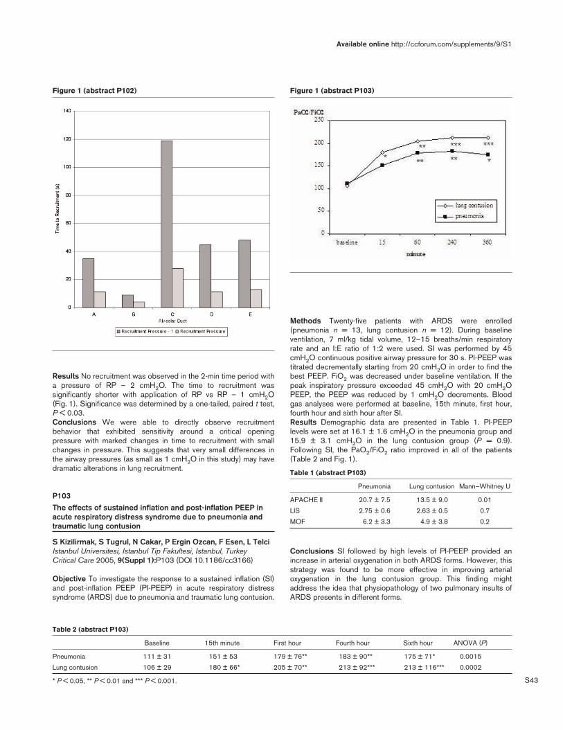
S43
Results No recruitment was observed in the 2-min time period witha pressure of RP – 2 cmH2O. The time to recruitment wassignificantly shorter with application of RP vs RP – 1 cmH2O(Fig. 1). Significance was determined by a one-tailed, paired t test,P < 0.03.Conclusions We were able to directly observe recruitmentbehavior that exhibited sensitivity around a critical openingpressure with marked changes in time to recruitment with smallchanges in pressure. This suggests that very small differences inthe airway pressures (as small as 1 cmH2O in this study) may havedramatic alterations in lung recruitment.
P103The effects of sustained inflation and post-inflation PEEP inacute respiratory distress syndrome due to pneumonia andtraumatic lung contusion
S Kizilirmak, S Tugrul, N Cakar, P Ergin Ozcan, F Esen, L TelciIstanbul Universitesi, Istanbul Tip Fakultesi, Istanbul, TurkeyCritical Care 2005, 9(Suppl 1):P103 (DOI 10.1186/cc3166)
Objective To investigate the response to a sustained inflation (SI)and post-inflation PEEP (PI-PEEP) in acute respiratory distresssyndrome (ARDS) due to pneumonia and traumatic lung contusion.
Methods Twenty-five patients with ARDS were enrolled(pneumonia n = 13, lung contusion n = 12). During baselineventilation, 7 ml/kg tidal volume, 12–15 breaths/min respiratoryrate and an I:E ratio of 1:2 were used. SI was performed by 45cmH2O continuous positive airway pressure for 30 s. PI-PEEP wastitrated decrementally starting from 20 cmH2O in order to find thebest PEEP. FiO2 was decreased under baseline ventilation. If thepeak inspiratory pressure exceeded 45 cmH2O with 20 cmH2OPEEP, the PEEP was reduced by 1 cmH2O decrements. Bloodgas analyses were performed at baseline, 15th minute, first hour,fourth hour and sixth hour after SI.Results Demographic data are presented in Table 1. PI-PEEPlevels were set at 16.1 ± 1.6 cmH2O in the pneumonia group and15.9 ± 3.1 cmH2O in the lung contusion group (P = 0.9).Following SI, the PaO2/FiO2 ratio improved in all of the patients(Table 2 and Fig. 1).
Table 1 (abstract P103)
Pneumonia Lung contusion Mann–Whitney U
APACHE II 20.7 ± 7.5 13.5 ± 9.0 0.01
LIS 2.75 ± 0.6 2.63 ± 0.5 0.7
MOF 6.2 ± 3.3 4.9 ± 3.8 0.2
Conclusions SI followed by high levels of PI-PEEP provided anincrease in arterial oxygenation in both ARDS forms. However, thisstrategy was found to be more effective in improving arterialoxygenation in the lung contusion group. This finding mightaddress the idea that physiopathology of two pulmonary insults ofARDS presents in different forms.
Available online http://ccforum.com/supplements/9/S1
Figure 1 (abstract P102) Figure 1 (abstract P103)
Table 2 (abstract P103)
Baseline 15th minute First hour Fourth hour Sixth hour ANOVA (P)
Pneumonia 111 ± 31 151 ± 53 179 ± 76** 183 ± 90** 175 ± 71* 0.0015
Lung contusion 106 ± 29 180 ± 66* 205 ± 70** 213 ± 92*** 213 ± 116*** 0.0002
* P < 0.05, ** P < 0.01 and *** P < 0.001.

S44
P104Use of a sigmoidal equation to analyze the pressure–volumecurve obtained by the low flow method
V Tomicic, L Fidanza, M Espinoza, J Graf, C CanalsClínica Alemana de Santiago, Santiago de Chile, ChileCritical Care 2005, 9(Suppl 1):P104 (DOI 10.1186/cc3167)
Introduction The adjustment of PEEP and tidal volume to thelower inflection point and upper inflection point of the P–V curve,respectively, has been proposed to optimize mechanical ventilationin ARDS. Usually, the P–V curve is analyzed by eye from a graph,with high variability. Venegas and colleagues [1] developed asigmoidal equation that fits remarkably well to P–V curves obtainedby the supersyringe method. Our objective was to determinewhether this equation is able to model P–V curves obtained by thelow flow method.Methods The P–V curve was obtained by the low flow methodusing a Servo Siemens 300 ventilator [2]. Pressure and flowsignals were obtained from the analog port (N81) and converted todigital format through a PCMCIA card (Measurement Computing,PC-CARD DAS 16/12). A program was developed to acquire,display, save, and analyze pressure and volume signals from theventilator. The sigmoidal equation has four fitting parameters: (a)lower asymptote (volume); (b) distance from lower asymptote toupper asymptote (inspiratory capacity); (c) true inflection point(pressure); and (d) range of pressure from (c) to the point of highcompliance. The equation is fitted to the P–V data by the leastmean square (LMS) algorithm to minimize the sum of squaredresiduals. After iteration, the best fit correlation (R2) between realcurves and those obtained from the equation was calculated. Fromthe fitted equation, three points of interest were defined: theinflection point (c), where the first derivative has a maximum andthe second derivative has a zero; the point of maximal complianceincrease (Pmci), where the rate of change of upward slope ismaximal or where the second derivative of the function has amaximum (calculated as (c) – 1.317 (d)); and the point of maximalcompliance decrease (Pmcd), where rate of change of downwardslope is maximal or where the second derivative of the function hasa minimum (calculated as (c) + 1.317 (d)).Results The inspiratory limb of low flow P–V curves were obtainedfrom six mechanically ventilated ARDS patients. The mean R2 valuebetween the ventilator volume signal and the volume predicted bythe fitted curves was 0.996 ± 0.002 (standard deviation).Conclusions The sigmoidal equation described by Venegas andcolleagues fits well to P–V curves obtained by the low flowmethod. Our system may contribute to improve the evaluation ofrespiratory mechanics and to adjust mechanical ventilation settingsat the bedside in ARDS patients.References1. Venegas, et al.: J Appl Physiol 1998, 84:389-395.2. Am J Respir Crit Care Med 1999, 159:275-282.
P105Modeling time effects of recruitment and P–V curvecharacteristics: a simulation study
K Möller1, C Stahl2, J Guttmann2
1University of Applied Sciences, Villingen-Schwenningen,Germany; 2University of Freiburg, GermanyCritical Care 2005, 9(Suppl 1):P105 (DOI 10.1186/cc3168)
Introduction In a multicenter study on respiratory mechanics in ALIand ARDS [1] we observed phenomena most probably related totime effects: change in compliance after a recruitment maneuver,
transient end-expiratory volume (Fig. 1) after changing PEEP, and atransient change in shape of the P–V curve after a change in tidalvolume (Fig. 2). To gain a better understanding of temporal effectswe developed a mathematical model of the respiratory system toinvestigate these phenomena.Methods We extended the model of recruitment proposed byHickling [2] to include chest wall characteristics, FRC, and aninitial opening volume of alveolar units once recruited. Alveolarunits had a nonlinear compliance and were organized in a bronchialtree structure. Depending on the position in the tree each lung unitwas exposed to a gravitational superimposed pressure from 0 inthe ventral compartment to a maximal value in the dorsalcompartment. Opening and closing pressures of lung units werenormally distributed with a modifiable mean and standard deviation.Time dependency was modeled by including timing parameters forrecruitment and derecruitment of alveolar units similar to theapproach of Bates and colleagues [3].Results In contrast to Hickling’s model, simulations with theextended model produced realistic transients in volume.Furthermore, transient changes in compliance were predicted bythe model. Modeling of recruitment and derecruitment as time-
Critical Care March 2005 Vol 9 Suppl 1 25th International Symposium on Intensive Care and Emergency Medicine
Figure 1 (abstract P105)
Observed transient volume after switching from ZEEP to PEEP 18 inan ARDS patient. End expiratory volume at ZEEP was defined as 0.
Figure 2 (abstract P105)
Transient in the shape of the P–V curve after an increase in tidalvolume.

S45
dependent phenomena allowed one to mimic the transients asobserved in ARDS patients.Conclusion The shape of simulated P–V loops is influenced bytime effects only related to closing and opening of alveolar units.Up to now, transient effects have been explained by viscoelasticityand by mechanical inhomogeneity. From the present study weconclude that timing effects during recruitment may be a thirdmechanism of transient mechanical behaviour of the respiratorysystem.References1. Stahl, et al.: Critical Care 2004, 8(suppl 1):P28.2. Hickling: Am J Respir Crit Care Med 1998, 158:194-202.3. Bates, et al.: J Appl Physiol 2002, 93:705-713.
P106How to choose the duration of prone-position ventilationbetween patients with acute respiratory distress syndromecaused by pulmonary and extrapulmonary disease?
Y Huang, H Qiu, L LiuDepartment of Critical Care Medicine, Zhong-da Hospital andSchool of Clinical Medicine, Southeast University, Nanjing, ChinaCritical Care 2005, 9(Suppl 1):P106 (DOI 10.1186/cc3169)
Objectives To determine the different effect of prone-positionventilation (PPV) in patients with acute respiratory distresssyndrome (ARDS) resulting from a pulmonary cause (ARDSp) andthat from an extrapulmonary cause (ARDSexp) on oxygenation,respiratory mechanics and hemodynamics. To choose the durationof ventilation in the prone position in the two groups.Methods Nine ARDSp patients and seven ARDSexp patientswithin 3 days of onset of ARDS were included in this study, whichwere classified as two groups. Patients were placed in proneposition for 2 hours. The effect of different times (pre-PPV, PPV0.5 hours, PPV 2 hours) on oxygenation, respiratory mechanicsand hemodynamics were observed. Lung computerizedtomography (CT) was obtained in both the supine position and10 min after turning to the prone position.Results Compared with pre-PPV, in ARDSp the PaO2/FiO2 wasnot increased after 0.5 hours, and increased only after 2 hours inthe prone position (130.6 ± 36.2 mmHg to 165.1 ± 72.3 mmHg,P < 0.05). But in ARDSexp, PaO2/FiO2 was significantly increasedafter 0.5 hours and 2 hours in the prone position (116.5 ± 55.0 mmHgto 163.2 ± 46.4 mmHg and 182.7 ± 87.7 mmHg, P < 0.05). After0.5 hours in the prone position the responding ratio of ARDSexpwas higher than ARDSp (100% vs 11.1%, P = 0.0007). After2 hours, no significant difference of responding ratio was foundbetween the two groups (85.7% vs 66.7%, P = 0.392). Thechanges of the PO2 were similar to the PaO2/FiO2. The PCO2 andthe Cst,rs did not differ significantly between the prone positionand the supine position in the two groups. In ARDSp, the Raw was10.8 ± 1.4 cmH2O/s/l in the supine position, and it was signifi-cantly decreased after 2 hours in the prone position (8.4 ± 1.8cmH2O/s/l) (P < 0.05). Chest CT scans of ARDSp and ARDSexppatients were markedly different.Conclusions PPV could be used to improve severe hypoxemia ofARDS. It improved the PaO2/FiO2 ratio rapidly in ARDSexp, butshould prolong the period in ARDSp.
P107PEEP and prone ventilation in liver transplantation:consideration or contraindication?
E Sykes, C O’Suilleabhain, I Nesbitt, J CosgroveFreeman Hospital, Newcastle Upon Tyne, UKCritical Care 2005, 9(Suppl 1):P107 (DOI 10.1186/cc3170)
Introduction Acute respiratory distress syndrome (ARDS) cancomplicate liver transplantation (LT). Positive end expiratory pressure(PEEP) and prone ventilation (PV) have theoretical adverse hepaticeffects. An adult paracetamol overdose required super-urgent LT.Immediate postoperative hypoxaemia due to ARDS developed(Table 1, i), as supported by FiO2/PaO2 ratio, bilateral pulmonaryinfiltrates, and normal pulmonary artery occlusion pressures. PEEP(15cm H2O) and PV improved gas exchange immediately (Table 1,ii) and at 18 hours (Table 1, iii). Doppler examination of hepaticvessels was normal. Discharge home was on day 41.
Table 1
Cardiac PaCO2 PaO2 Lactate index
FiO2 pH (kPa) (kPa) (mmol/l) (l/min/m2)
i 1.0 7.15 6.4 6.0 12.3 4.30
ii 1.0 7.279 5.5 21.6 6.4 4.27
iii 0.5 7.408 5.8 11.3 3.2 3.06
Methods A telephone questionnaire to British LT intensive careunits about using PEEP and PV postoperatively after emergency LT.Results Completed questionnaires were obtained from the sixother British LT intensive care units. All responders wereconsultant intensivists. Four routinely set PEEP on the ventilator.Three stated levels depended on oxygen requirements. Oneroutinely used PEEP irrespectively. Two used PEEP depending onoxygen requirements. Two felt PV was ‘absolutely contraindicated’,quoting an evidence base, alternative available measures, andconcerns regarding hepatic perfusion. No responder could recallPV being used in this context.Discussion Opinions regarding the use of PEEP post emergencyLT are mixed. Opinions of PV appear polarised. No units appear tohave used it in this context. Potential detrimental effects of PEEPand PV in LT include reduced cardiac output and hepatic bloodflow. Increased intra-abdominal pressure with PV may compoundthis. However, restoring cardiac output maintains normal liverfunction and blood flow [1,2]. Positioning to allow abdominalexcursion minimises risk [2]. Concerns raised in the questionnairemay be overstated. We suggest both modalities can be usedeffectively, without detriment to donor liver or patient outcome.Further research is required.References1. Matuschak GM: J Appl Physiol 1987, 62:1377-1383.2. Hering JR: Intensive Care Med 2002, 28:53-58.
P108Effects of prone position ventilation and kinetic therapy inacute respiratory failure
J Lewejohann, E Rieh, B Börner, E Muhl, H BruchUniversitätsklinikum Schleswig-Holstein — Campus Lübeck,GermanyCritical Care 2005, 9(Suppl 1):P108 (DOI 10.1186/cc3171)
Introduction In acute respiratory failure (ARF), in particular acutelung injury (ALI) and respiratory distress syndrome (ARDS), change
Available online http://ccforum.com/supplements/9/S1

S46
from the supine position (SP) to the prone position (PP) or the useof kinetic therapy can improve oxygenation by recruiting alveolisituated in dorsal-dependent regions of the lung and by alterationof the ventilation/perfusion ratio. The efficacy of this interventioncan be demonstrated by the course of oxygenation index. The aimof our study is to compare prone position ventilation (PP) andkinetic therapy (KT) in the first 5 days after intervention in patientswith acute respiratory failure.Methods We studied n = 149 patients with ARF at a surgical ICUin a university hospital using the American–European consensusdefinition in a clinical follow-up design. The physicians on duty hadthe freedom of choice to use PP or KT guided by their clinicalexperience. One hundred and eleven patients (ALI: n = 18/ARDS:n = 93; mean age 66 ± 13 [standard error] years) were ventilatedintermittently in SP and PP (135° left/right-side position) for atleast 6 hours per day for supportive treatment of respiratory failure.Thirty-eight patients (ALI: n = 16/ARDS: n = 22; mean age60 ± 16 [standard error] years) were treated with kinetic therapyusing the Rotorest®-bed (60° left/right side; KCI).Data collection included, apart from baseline characteristics, theindividual oxygenation index of the patients in the course of the first5 days after intervention. The individual oxygenation index beforeand after intervention was compared with linear regression analysisfor each group (linear regression procedure and t test; SPSS®).Results During conventional ventilation in the supine position therewas no significant improvement in oxygenation in both groups. Themean oxygenation index (PaO2/FiO2) decreased until interventionto 157.6 ± 0.48 mmHg in the PP group and to 165.8 ± 1.47 mmHgin the KT group (mean ± standard error of the mean). Both forms ofpositional changes lead to a distinct improvement of oxygenation:after 120 hours the oxygenation index in the PP group was219.67 ± 0.66 and in the KT group was 197.89 ± 2.07 mmHg.Subsequent to intervention the PP group showed a more rapid andsignificant increase of oxygenation index in comparison with the KTgroup as the equations of linear regression show: y[PPV] =4.4618 × X + 188.72 and y[KT] = 3.9349 × X + 160.47. Mortalitywas 62.2% in the PP group and 63.2% in the KT.Discussion In ARF the change of body position in the form of PPand KT leads to an improvement of oxygenation. PP seems to havea more rapid and marked effect than KT. It is remarkable thatmortality is nearly the same in both groups, despite the unequalgroup size. PP is a comparatively simple strategy to treat theheterogenity of ventilation and perfusion in ARF and needs nospecial technical equipment, while KT is an advantageousalternative in patients with contraindications for PP.
P109Is the quantitative analysis of lung computed tomographyaccurate in ALI/ARDS patients?
D Chiumello, M Cressoni, M Racagni, A D’Adda, S Azzari, S TerragniPoliclinico IRCCS, Milan, ItalyCritical Care 2005, 9(Suppl 1):P109 (DOI 10.1186/cc3172)
The CT scan allows an accurate morphologic analysis of an‘ARDS’ lung. In addition, with the spiral CT it is also possible toperform a quantitative analysis using dedicated software. Thequantitative analysis is based on the strict correlation between theX-ray attenuation in a given volume of tissue and the physicaldensity of that volume. The CT is determined by the percentage ofradiation that is absorbed by that volume.However, due to the necessity of a manually drawing of the lung (i.e.to include the lung and to exclude big vessels, trachea, pleural effusion,etc.), it would be possible to increase the inaccuracy of the analysis.
The aim of this study was to evaluate the accuracy of analysis bet-ween trained physicians (four medical doctor) and two radiologists.We enrolled 12 intubated sedated paralyzed patients ALI/ARDSpatients (mean age 63.5 ± 16.9 years, BMI 24.1 ± 4.7 m2, tidalvolume 532 ± 195 ml, PEEP 11.2 ± 1.9 cmH2O, PaO2/FiO2205 ± 57). The CT scan was done for clinical purpose, in staticconditions, at end expiration 5 or 15 cmH2O PEEP and at endinspiration 45 cmH2O airway pressure. The exposures were takenat 120 KV and 250 mA. The lung volume, weight and thedistribution of lung weight between the different parts was measuredusing the ‘Maluna’ software (University of Mannheim, Germany).The results show that the quantitative analysis, although requiring aphysician to manually draw the region of interest of the lung,presents a high degree of accuracy.
Table 1
Physicians Radiologists
Total volume (ml) 3956 ± 1608 3598 ± 1654
Total weight (mg) 1419 ± 471 1391 ± 449
Weight hyperinflated 41 ± 79 41 ± 82
Weight inflated 442 ± 160 440 ± 164
Weight poorly 439 ± 269 419 ± 270
Weight not inflated 496 ± 350 489 ± 362
P110A validation study of a new software for computedtomography lung measurement
D Chiumello, M Cressoni, M Racagni, L Landi, A D’Adda, S Azzari, S TerragniPoliclinico IRCCS, Milan, ItalyCritical Care 2005, 9(Suppl 1):P110 (DOI 10.1186/cc3173)
The use of computed tomography (CT) in the management ofALI/ARDS patients is quite common, due to the possible additionalclinical information and the influence on the patient’s treatment.With the new spiral CT scan it is possible to measure the total lungvolume, the percentage of lung tissue and gas, there being a linearcorrelation between the physical density and the CT coefficient ofattenuation.
Critical Care March 2005 Vol 9 Suppl 1 25th International Symposium on Intensive Care and Emergency Medicine
Figure 1 (abstract P110)

S47
The aim of this study was to evaluate in vitro the new computerprogram ‘Maluna’ (University of Mannheim, Germany) dedicated tomeasuring the lung volume, weight and gas/tissue ratio.A series of different known volumes of water (100, 500, 1500 and2000 ml) were studied. In addition, the contrast material wasdiluted with water to obtain solutions of increasing concentrations(0, 0.5, 1, 1.5, 2.5 and 5%). The spiral CT was performed at 120KV and 240 mA. Each CT section was manually delineated by aphysician. The total volume was computed as the total number ofvoxels present in a given region times the volume of the voxels,while the CT number was directly related to the physical density.Figure 1 shows the Bland–Altman analysis between the knownvolumes of water and the measured volumes by ‘Maluna’; Fig. 2shows the linear regression between the CT attenuation coefficientwith increasing concentrations of contrast material in the solution.These data show that ‘Maluna’ is able to correctly compute thevolumes and CT number.
P111Are three computed tomography lung sections comparablewith the whole lung?
D Chiumello, N Bottino, M Cressoni, M Racagni, L Landi, A D’Adda, F Polli, S Terragni, L GattinoniPoliclinico IRCCS, Milan, ItalyCritical Care 2005, 9(Suppl 1):P111 (DOI 10.1186/cc3174)
Computed tomography (CT) has been shown to provide accuratemeasurements of lung tissue mass and volume. However, besidesthe risk of moving the patient from intensive care to CT scan andthe high costs, the high radiological exposure to X-rays may limitthe possibility for relatively frequent CT acquisition. One chest CTscan corresponds to 400 chest X-rays [1]. A possible solution toreduce the X-ray exposure could to be perform only three lungsections compared with the whole lung.The aim of this study was to investigate the difference between thethree CT slices technique versus the whole lung in acquiringclinical relevant data (i.e. hyperinflated, normally, poorly and notinflated lung zones).Thirty-four intubated sedated paralyzed patients with ALI/ARDS(mean age 56.2 ± 18.2 years, BMI 23.7 ± 3.6 kg/m2, PEEP11.2 ± 2.8 cmH2O, PaO2/FiO2 195 ± 81) were enrolled. Spiral CTscan of the lung was acquired at 120 kV and 250 mA during anend expiratory pause at 5 and 15 cmH2O PEEP. The lung CT
regions of interest were manually delineated on each image bydedicated software (Maluna; University of Maanheim, Germany). Thethree CT lung sections were selected at the apex, hilum and base.The Bland–Altman analysis showed a bias of 0.001% with aninterval limits for agreement of 7.2% at 5 cmH2O PEEP and of0.00001% with 98% at 15 cmH2O PEEP.These data showed that the three CT scan technique provides anadequate measure of the distribution of lung compartments whilestrongly reducing X-ray exposure.
Table 1
PEEP 5 PEEP 15
Inflation Whole lung Three sections Whole lung Three sections
Hyper (%) 0.04 ± 0.1 0.05 ± 0.1 1.1 ± 0.1 1.2 ± 0.1
Normal (%) 26.5 ± 0.1 26.3 ± 0.1 38.7 ± 0.1 37.6 ± 0.1
Poor (%) 33.2 ± 0.1 33.6 ± 0.2 31.3 ± 0.1 31.6 ± 0.1
Not (%) 39.7 ± 0.2 40.0 ± 0.1 28.5 ± 0.1 29.6 ± 0.1
Reference1. Picano E: Br Med J 2004, 329:849-851.
P112Beneficial effects of CT scan guided protective ventilation:multislice thoracic CT analysis
G De Matos1, J Borges2, F Stanzani1, V Okamoto2, M Bueno1,M Rodrigues1, C Hoelz1, R Albaladejo1, M Fontana1, R Caserta1,D Santos1, M Amato2, C Barbas1
1Hospital Israelita Albert Einstein, Sao Paulo, Brazil; 2University ofSão Paulo Medical School, São Paulo, BrazilCritical Care 2005, 9(Suppl 1):P112 (DOI 10.1186/cc3175)
Introduction Recruitment maneuvers (RM) to open the lungs andPEEP titration to keep the lungs opened are believed to be animportant component of a lung protective strategy in ARDS. Thereare scarce data assessing the occurrence of tidal recruitment (TR)and the gas distribution throughout the lung parenchyma bythoracic CT scan before and after maximal recruitment.Objectives To analyze the occurrence of TR before and afterstepwise recruitment maneuver (SRM) and PEEP titration bythoracic CT scan. To analyze the effects of the proposed maneuveron gas distribution throughout the lung parenchyma.Methods Ten ARDS patients under mechanical ventilation weretransported to the CT room and sequences of CT scans at expiratoryand inspiratory pauses were performed before and after a SRM. TheSRM consisted of 2-min steps of tidal ventilation with a fixed deltapressure PCV = 15 cmH2O and progressive PEEP levels (10, 20, 25,35, 45, 25, 20, 15, 10 cmH2O), RR = 10, I:E 1:1, and FiO2 1.0. Atequivalent PEEP levels, the amount of collapsed tissue as well as thedifference of collapsed tissue between inspiration and expiration (TR)were compared (before versus after maximal SRM). The amount of aircontent (ml) at end expiration — functional residual capacity (FRC) —was calculated and compared before maximal recruitment at PEEP of20 cmH2O and after maximal SRM at PEEP of 25 cmH2O.Results See Figs 1 and 2. The TR difference is statisticallysignificant at PEEP 25 cmH2O (Wilcoxon P < 0.015). There was asignificant decrease of gas at FRC at non-dependent regions(Wilcoxon P < 0.008) and a significant increment of gas at themost dependent region with PEEP 25 cmH2O after maximalrecruitment (Wilcoxon P < 0.008).Conclusion The TR decreased significantly only under PEEP of25 cmH2O after the SRM (P < 0.0003), suggesting that theapplication of SRM might be effective when accompanied by
Available online http://ccforum.com/supplements/9/S1
Figure 2 (abstract P110)

S48
sufficient PEEP levels. Aeration of dependent portions of the lungafter maximal SRM with maintenance of sufficient PEEP levels tokeep the lungs opened (Wilcoxon P < 0.008) was associated withdeflation of non-dependent regions, rendering the gas distributionmore homogeneous.
P113Gas exchange impairment induced by open suctioning inARDS: impact of permissive hypercapnia
M Caramez, E Miyoshi, R Scott Harris, A Malhotra, R KacmarekMassachusetts General Hospital, Boston, MA, USACritical Care 2005, 9(Suppl 1):P113 (DOI 10.1186/cc3176)
Background and aims There is still controversy over the impact ofopen suctioning (OS) on gas exchange and the duration ofsuctioning. The goal of this physiologic study was to determinewhether hypercarbia occurs following the use of OS in a lunglavage injured sheep and whether the level of baseline PaCO2 andduration of suctioning impacts gas exchange.Methods Six female sheep were lung injured by isotonic salinelavage until their PaO2 dropped to 100 mmHg on an FIO2 of 1.0,
PEEP 5 cmH2O and VT 10 ml/kg. Then the VT was decreased to6 ml/kg. RR was set to establish a PaCO2 of 40 mmHg or 80mmHg. OS was performed on each animal. PaCO2 was randomlyadjusted to 40 mmHg and 80 mmHg and the suction timerandomly set at 10 s and 30 s. Before each of the fourexperimental conditions animals received recruitment maneuvers of40 cmH2O CPAP for 40 s to establish a volume history, followingby baseline for 15 min where FiO2 and PEEP were set based onthe ARDSnet FiO2/PEEP [1]. OS was performed using a 14 Fr,TrachCare catheter at a pressure of –100 mmHg. Mean arterialblood pressure, heart rate, pulmonary and arterial wedge pressure,cardiac output, and arterial blood gases, were measured before,1 min and then every 2 min after OS for 30 min.Results During suctioning at 80 mmHg PaCO2 at both 10 s and30 s suctioning times, the PaCO2 increased at 1, 15 and 31 minpost suctioning (P < 0.05). PaCO2 also increased at 1 min whenthe baseline PaCO2 was 40 mmHg and the suction time was 30 s(P < 0.05) (Fig. 1). PaO2 decreased 1 min post suctioning in allconditions but recovered by 15 min. Hemodynamics did notdemonstrate any significance differences regardless of theexperimental condition.Conclusions OS causes in a significant increase in baselinePaCO2 during baseline hypercapnia and when suctioning is appliedfor 30 s. OS regardless of conditions results in a decrease in PaO2that recovers rapidly post suctioning.Reference1. ARDS Network: N Engl J Med 2000, 342:1301-1308.
P114Rational airway control with high-frequency jet ventilation forpercutaneous tracheostomy in patients with short fat neck
R Badaev1, M Croitoru2, F Badaev3, S Krimerman2, U Teitelman4, E Altman4
1Carmel Hospital, Haifa, Israel; 2Bnai Zion Medical Center, Haifa,Israel; 3City Hospital No 40, Ekaterinburg, Russia; 4RambamHospital, Haifa, IsraelCritical Care 2005, 9(Suppl 1):P114 (DOI 10.1186/cc3177)
Background and goal Percutaneous tracheostomy (PCT) is awell-established bedside procedure for critically ill patients (CIP).Several techniques of airway control and ventilation during PCThave been suggested. Standard mechanical ventilation (SMV)
Critical Care March 2005 Vol 9 Suppl 1 25th International Symposium on Intensive Care and Emergency Medicine
Figure 2 (abstract P112)
Figure 1 (abstract P113)
Comparison of PaCO2 between groups.
Figure 1 (abstract P112)

S49
through an endotracheal tube (ETT) is widely used. Short fat neck(SFN) is considered a relative contraindication for PCT, mainlybecause of the possibility for airway complications during theprocedure: loss of airway control, massive air leak, and suddendesaturation. We decided to examine our experience of airwaycontrol and ventilation by SMV and by high-frequency jet ventilation(HFJV) during PCT in the CIP with SFN and to determine which ismore effective and reliable.Materials and methods From January 1998 to June 2000, agroup of 23 CIP with SFN underwent PCT by the Griggstechnique and SMV, and from July 2000 to November 2001 agroup of 25 patients underwent PCT by the same technique butwith HFJV. One trained team performed all the bedside PCT undergeneral anesthesia (GA). In the HFJV group, a Cook catheter wasinserted through the existing ETT into the upper part of trachea andHFJV was initiated by the special ventilator AMS-1000ACUTRONIC with the following parameters: FiO2 = 0.8–1.0, Vt =160–200 cm3, I/E ratio = 0.5, respiratory rate = 100 bpm, drivingpressure = 1.6–2.6 bar. Then the ETT was withdrawn from thetrachea and larynx into the mouth along the ventilation catheter andPCT was performed. When PCT was finished the Cook catheterwas removed together with the ETT. During PCT continuousroutine monitoring and repeated blood gases were performed.Results There was no significant statistically difference betweenthe two groups in the sizes of the neck (mean 47.1 vs 47.3 cm,median 47 vs 47 cm, range 45–51 vs 47–53 cm, P = 0.9) anddistance between the cricoid and sternal notch (mean 1.35 vs 1.37cm, median 1.35 vs 1.5 cm, range 0.5–1.9 vs 0.5–1.9 cm, P = 0.8).Duration of the procedure for the two groups was identical: mean =10.7 min, median = 11 min, range = 10–12 min. When SMV wasused, only in 11 from 23 patients was it possible to create goodconditions for performance of PCT. Impaling of the ETT and cuffrupture happened in one case, and displacement of the ETT into thepharynx took place in three cases (due to a short larynx). Moderateair leak from the larynx around the ETT accompanied almost everyprocedure. Additional egress of air from the tracheotomy site duringthe time of the dilation was the reason for pronounced desaturationin seven patients. On the contrary, PCT with HFJV passedsmoothly: a small air leak from tracheotomy site during its dilationdid not influence the level of oxygenation, only an increase inPaCO2 values within acceptable limits was found. The differencebetween SMV and HFJV was statistically significant (P < 0.001).Conclusion SMV cannot always supply the necessary conditionsfor PCT in patients with SFN and there are possibilities fordevelopment of serious complications. HFJV through a Cookcatheter provides optimal conditions for manipulation on SFN,secures success of the procedure and prevents potential airwaycomplications
P115Comparison of two different percutaneous tracheostomytechniques with early and late complications in critically illpatients
F Ugun, R Iscimen, N Kelebek Girgin, F Kasapoglu, H Coskun,F KahveciUludag University Faculty of Medicine, Bursa, TurkeyCritical Care 2005, 9(Suppl 1):P115 (DOI 10.1186/cc3178)
Background Percutaneous tracheostomy (PT) is used in patientswho receive long-term invasive mechanical ventilation. There aremany different techniques in performing PT.Aim The aim of this prospective study was to compare twodifferent PT techniques with early and late complications incritically ill patients.
Methods After obtaining ethical committee approval, 60 adultpatients were assigned randomly into two groups. Forcepsdilatational PT and single-step dilatational PT were used in group Fand group P, respectively. Heart rate, blood pressure andperipheral oxygen saturation, the level of tracheal entrance andnumber of punctions, major and minor complications and durationof the procedure were recorded during the procedure. Arterialblood samples were taken before the incision and aftertracheostomy cannula was inserted, then pH, pCO2, pO2, HCO3,BE, and SaO2 were recorded. The endoscopic examinations of thecases were done during the procedure, at the decannulation timeand 1 month after decannulation. The t test, Mann–Whitney U test,Wilcoxon signed rank test, chi-squared test and Kruskal–Wallistest were used for statistical analysis.Results The demographic data, duration of intubation time andprocedure time were similar in both groups. There were nosignificant differences in hemodynamic data between the groups. Itwas noticed that in both groups there was a decrease in pH(P < 0.001) and an increase in pCO2 (P < 0.01). Tracheal ringdamage that was one of the minor complications in Group P wassignificantly gretaer (P < 0.05). There were no significantdifferences in major complications between the groups. In Group Ftracheal stenosis in one case and laryngotracheal separation inanother case were detected with endoscopic examination.Conclusion The present study demonstrated that single-stepdilatational PT may be an alternative to forceps dilatational PT, butwe conclude that to maintain a decrease in complicationsendoscopic visualisation could be used.
P116Biomechanical experimental evaluation of percutaneoustracheostomy to compare the Ciaglia and Griggs techniques
I Rozin1, M Croitoru2, E Radinsky3, N Altman3, U Teitelman3, E Altman3
1Puria Hospital, Tveria, Israel; 2BZMC, Haifa, Israel; 3RambamHospital, Haifa, IsraelCritical Care 2005, 9(Suppl 1):P116 (DOI 10.1186/cc3179)
Introduction The aim of our study was to create a method forbiomechanical experimental evaluation of the percutaneoustracheostomy (PCT) technique and perform its assessment inorder to minimize possible trauma of cervical trachea inherent inthis procedure. Nowadays two techniques of PCT, Ciaglia andGriggs, are employed.Every technique is based on gradual tearing of the hole in thecervical trachea by specially designed dilators. We suggested thatbiomechanical study of the different stages of PCT will help tounderstand all these processes and to clarify the reasons fortraumatic perioperative complications.Materials and methods PCT by the Ciaglia technique (n = 10)and by the Griggs technique (n = 10) were performed on freshdead pigs with body mass of 115 ± 3 kg. A Portex set ofinstruments was used for the Griggs technique and a Cook set ofinstruments for the Ciaglia technique. During PCT specialmeasurements were performed with the help of an electronicdynamometer (MRC): piercing force applied on needle cannula,penetrating force applied on 14 F dilator, pulling force applied onbranches of Griggs dilating forceps, pushing force applied onCiaglia Blue Rhino dilator, pushing force applied on distal end oftracheostomy tube no. 8 with introducer (Portex set) and pushingforce applied on distal end of tracheostomy tube no. 8 loaded ondilator (Cook set). On the base of these measurements calculationof energy spent for these stages of PCT was made. At the end ofeach experiment evaluation of cervical trachea was performed to
Available online http://ccforum.com/supplements/9/S1

S50
measure the size of tracheotomy performed by the Ciaglia andGriggs dilators.Results Application of the Ciaglia dilator requested 1.54 timesmore energy than the Griggs dilator forceps (P < 0.05). Formationof tracheotomy by the Ciaglia dilator was more exact than byGriggs forceps due to special markings on the dilator. The workwith Griggs dilating forceps requested more experience due to theabsence of any markings on the dilator. The most dangerousmoments of PCT for laceration of the cervical trachea were:dilation of hole in the cervical trachea in the Griggs technique andinsertion of the tracheostomy tube loaded on the dilator in theCiaglia technique. The macroscopic appearance and transversediameter of tracheotomy performed by the Ciaglia and Griggstechniques were the same despite the different instruments usedfor dilation (P < 0.05)Conclusions PCT by the Ciaglia and Griggs techniques hasalmost similar biomechanical characteristics. In spite of thedifference in the design of dilators the final result of the twotechniques is the same. Both of them have dangerous moments. Inorder to prevent laceration of the cervical trachea during PCTsome improvements must be made in instrumentation.
P117Does tracheostomy improve respiratory functions inmechanically ventilated patients
F Pürnek Akin, L Dösemeci, M Cengiz, M Yilmaz, A RamazanogluAkdeniz University Faculty of Medicine, Antalya, TurkeyCritical Care 2005, 9(Suppl 1):P117 (DOI 10.1186/cc3180)
Objectives Tracheostomy has been widely performed in theintensive care unit and has many advantages when compared withtranslaryngeal intubation. Our hypothesis was that tracheostomywould also result in improved pulmonary function through changesin respiratory mechanics and tracheabronchial touilette.Study design A prospective, observational, clinical study. Abefore-and-after trial of 65 patients undergoing tracheostomy.Patients and methods A consecutive group of critically ill patientsrequiring prolonged mechanical ventilation was included in thestudy (15 July 2004–1 December 2004). Percutaneoustracheostomy was performed using the dilatational forcepstechnique described by Griggs. Respiratory mechanics (ventilatorsettings, airway pressures) and arterial blood gases (PaO2,PaCO2, pH) were measured before and after tracheostomy (at 24,48 and 72 hours) in 65 patients. Calculated variables includedminute ventilation, dynamic and static compliances and PaO2:FiO2ratio (Harowitz).Results A total of 65 patients (with a mean age of 50.6 ± 20 years)underwent tracheostomy a mean of 5 ± 2.6 days after admission.The Glasgow Coma Scale score and Acute Physiology andChronic Health Evaluation II score were 8.3 ± 3.7 and 20.61 ± 5.3,respectively. We could not find statistically significant differencesin respiratory mechanics, blood gases and pH after tracheostomywhen we considered all of the patients. Then, we evaluated theresults by dividing the patients into two groups according to theHarowitz ratio. Forty-three patients had Harowitz ratio greater than250 (396.5 ± 105.5) and 22 patients less than 250 (201.1 ± 36.5).Both groups were analyzed with repeated-measures analysis ofvariance and we found that the Harowitz ratio at 24 hours(265.3 ± 71), 48 hours (269 ± 26) and 72 hours (274 ± 93)improved significantly after tracheostomy individually (P = 0.002,P = 0.018, P = 0.004, respectively) in the patients with a Harowitzratio less than 250.Conclusion Our study showed that percutaneous tracheostomyimproves the PaO2:FiO2 (Harowitz) ratio in the mechanically
ventilated patients who had Harowitz ratio less than 250 beforetracheostomy, but does not improve it in the patients with Harowitzratio greater than 250.
P118Routine chest radiography following percutaneous dilatationaltracheostomy
M Hughes, L Clark, G DempseyUniversity Hospital Aintree, Liverpool, UKCritical Care 2005, 9(Suppl 1):P118 (DOI 10.1186/cc3181)
Background and goal The role of routine chest radiography(CXR) following percutaneous dilatational tracheostomy (PDT) hasrecently been questioned [1].Materials and methods We have performed a prospectiveobservational study, on a mixed medical–surgical critical care unit,on 110 patients undergoing PDT under bronchoscopic guidanceto assess the utility of routine postoperative chest radiography.Data were collected on all patients undergoing PDT from 1November 2003–14 December 2004. Two post-procedure CXRswere reviewed and compared with those taken prior to PDT.Significant findings were barotrauma (pneumothorax,pneumomediastinum) and consolidation not noted on the pre-procedure film. Post-procedural films reviewed were those takenimmediately after PDT and, to exclude the possibility of overlookingevidence of minor barotrauma, one further film taken between 24and 96 hours.Results and observations One hundred and ten patientsunderwent PDT; 83 (75%) were uncomplicated. Complicationswere recorded in 27 (25%) patients. These included multipleattempts at needle insertion (> 2), bleeding (> 3 soaked gauzeswabs), tracheal ring fracture, posterior tracheal wall injury, andmalplacement. Ninety-five (86%) patients had two post-proceduralCXRs reviewed. Fourteen (13%) patients had at least one CXRreviewed after PDT. One patient had no CXR after PDT. Newabnormalities were noted on three (3%) post-procedure CXRs. Nonew pneumothoraces were seen. Patients having uncomplicatedPDTs had no new CXR changes (Table 1).
Table 1
Number of New problem PDT complicated? CXRs reviewed on CXR
NO, n = 71 (65%) 2 0 (0%)
YES, n = 24 (22%) 2 3 (13%)
NO, n = 11 (10%) 1 0 (0%)
YES, n = 3 (3%) 1 0 (0%)
PDT, percutaneous dilatational tracheostomy; CXR, routine chestradiography.
Conclusions Routine CXR following uncomplicated PDTperformed under bronchoscopic guidance is not warranted.Concern that evidence of minor barotrauma may be missed on theearly post-procedural film is not bourne out by review of the filmtaken 24–96 hours after the event. The role of CXR following PDTappears to be restricted to those patients undergoing complicatedprocedures. This will lead to reductions in both medical costs [2]and exposure of staff and patients to ionising radiation.References1. Datta D, Onyirimba F, McNamee MJ: Chest 2003, 123:1603-
1606.2. Tarnoff M, Moncure M, Jones F, et al.: Chest 1998, 113:1647-
1649.
Critical Care March 2005 Vol 9 Suppl 1 25th International Symposium on Intensive Care and Emergency Medicine

S51
P119Consequences of adverse events with handling of theorotracheal cannula in intensive care unit
S Silva1, K Padilha2
1Sírio Libanês Hospital, São Paulo, Brazil; 2University of São Paulo,BrazilCritical Care 2005, 9(Suppl 1):P119 (DOI 10.1186/cc3182)
Objectives To identify the adverse events regarding handling ofthe orotracheal cannula in the ICU and to evaluate its impact onthe patient’s severity and on the nursing workload.Method Data were prospectively collected during a 3-monthperiod, in two general ICUs of a hospital in the city of São Paulo,Brazil, using a file card to record the occurrences. Patient’sseverity and nursing workload were evaluated 24 hours before and24 hours after the occurrence of the event, respectively, by meansof the Simplified Acute Physiology Score (SAPS II) and theTherapeutic Intervention Scoring System-28 (TISS-28).Results In the period of the study 212 patients were admitted, 47(22%) of which were victims of 80 adverse events during the theirstay at the ICU. There was a total of 19 (24.0%) adverse eventsregarding handling of the orotracheal cannula. Of these wereobserved 14 (73.0%) not programmed withdrawals of theorotracheal cannula, three (16%) blockages of the cannula forsecretion corks and two (11%) cuffs emptied. A statisticallysignificant difference was found both in the SAPS II (P < 0.042)and TISS-28 (P < 0.001), evidenced before and after the event.One verified reduction of the patient’s severity, but an increase onthe nursing workload as a consequence of these events.Conclusions The results of this investigation reinforce the need forinvestments to qualify professionals to work with critical patients asa major measurement for safety nursing assistance and quality inthe ICU.
P120A prospective study comparing standard laryngoscopy withthe trachview videoscope system for orotracheal intubation
L Roppolo, P Pepe, J MartinezUniversity of Texas Southwestern Medical Center and the ParklandHealth and Hospital System, Dallas, TX, USACritical Care 2005, 9(Suppl 1):P120 (DOI 10.1186/cc3183)
Background As a technology to facilitate a difficult endotrachealintubation (ETI), the TrachView Videoscope (TrVV) consists of anarrow high-resolution fiberoptic cable whose tip is positioned atthe distal end of the endotracheal tube. The image is displayed ona small portable bedside monitor.Objective To determine the reported ease of use of the TrVV andthe accompanying percent improvement in assessment of theglottic opening (POGO) score when operated by individuals ofvaried levels of ETI experience.Methods The study was conducted in two phases using amannequin model during an airway workshop for first-year, second-year and third-year (R1, R2, R3) emergency medicine (EM)residents with ETI experience and first-year and second-yearmedical students with no ETI experience. In phase 1, after a 10-mindemonstration of the TrVV, EM residents assessed the POGOscore using direct laryngoscopy (DL) and sequentially compared itwith their observed POGO score using the TrVV. Phase 2consisted of a crossover study with the medical students whowere randomized into two groups: a group first instructed in DLand a second first instructed in TrVV. The students were given a10-min demonstration of each technique and had two
opportunities to return the demonstration. The POGO scoresnoted by the students were then recorded for each technique. Thegroups were then crossed and the process was repeated.Additional information collected from study subjects includedreported ease of use of the TrVV and improvement in ETI success.Results In Phase 1, the overall median POGO score for DL was50%, and the median POGO score for TrVV was 100%(P < 0.001). The median differences in POGO scores (TV – DL)were: 50% for R1s (n = 4), 50% for R2s (n = 10), and 25% forR3s (n = 11). Of these, 86% (n = 24) reported that the TrVV was‘easy’ to use and 11% (n = 3) were undecided. Only one reportedit to be ‘difficult’ to use. Most (82%; n = 23) reported that the TrVVimproved their ETI attempt, but 14% (n = 4) reported nodifference. In Phase 2, the overall median TrVV and DL POGOscores (n = 34) were 75% and 25%, respectively (P = 0.004). Themedian difference in POGO scores (TV – DL) for the two groupswere: 75% for Group 1 (DL first) and 50% for Group 2 (TrVV). Ofall participants, 68% (n = 25) ranked the TrVV as ‘easy’, 22%(n = 8) were undecided, and 11% (n = 4) ranked the TrVV as‘difficult’ to use. Meanwhile, 57% (n = 21) reported that it improvedtheir ETI attempts, 27% (n = 10) reported no difference, and 11%(n = 4) reported ‘more difficulty’.Conclusion The Trachview Videoscope can significantly improvethe POGO score assessment over direct laryngoscopy and, in thelaboratory setting, most operators report it to be an easy to usetechnique that improves their intubation success, regardless oflevel of experience.
P121Treatment of post-intubation tracheal stenosis with theNd-YAG laser at the NRITLD
S Arami1, H Jabbardarjani1, M Masjedi21NRITLD, London, UK; 2NRITLD, Tehran, IranCritical Care 2005, 9(Suppl 1):P121 (DOI 10.1186/cc3184)
Background and objective Tracheal intubation is a commonprocedure for critically ill patients and may lead to localcomplications such as tracheal destruction and post-intubationstenosis. Utilization of the Nd-YAG laser can correct intraluminalairway lesions including webs, granulation tissues and fibrousbands. It decreases the hospitalization period, complications andexpenses and also saves the medical staff’s time and energy.Materials and methods An uncontrolled clinical trial wasperformed in post-intubation tracheal stenosis cases admitted tothe NRITLD between 1994 and 1999. Based on bronchoscopicfindings, patients with the following inclusion criteria underwentlaser therapy: length of stenosis less than 2 cm, tracheal lumendiameter more than 5 mm and lesions with granulation tissue type.Others candidated for surgery. Laser therapy was carried out byfiberoptic bronchoscopy under local anesthesia. For those patientswith tracheal lumen diameter between 5 and 10 mm rigid broncho-scopy was performed under general anesthesia.Results Of the total 32 patients, 25 met the primary criteria forlaser therapy, from which 22 (88%) were completely cured.Because of poor general condition, five of seven other patientswho had been referred for surgery also underwent laser therapy;therefore, a total of 30 patients (93%) took advantage of thistechnique. Overall, 23 patients were treated only with a laser, twopatients only with surgery and seven patients with a combination ofthese two approaches.Conclusions This trial revealed that if patients with post-intubationtracheal stenosis are selected correctly, laser therapy could beused as a beneficial and safe method with satisfactory results.
Available online http://ccforum.com/supplements/9/S1

S52
P122Weaning from mechanical ventilation using assistedspontaneous breathing plus CPAP versus assistedspontaneous ventilation: our experience
M Di Nardo, A Tomagnini, P Cosimini, F Forfori, L Roventini, F GiuntaUniversity of Pisa, IV ICU, Department of Anesthesiology andIntensive Care, Pisa, ItalyCritical Care 2005, 9(Suppl 1):P122 (DOI 10.1186/cc3185)
Introduction In this retrospective study, we evaluated two differentmodes of weaning: assisted spontaneous breathing (ASB) plusCPAP with automatic tube compensation (Evita 4, DraegerMedical, Germany) versus assisted spontaneous ventilation (ASV)(Galileo Gold, Hamilton Medical, Switzerland) in 80 patients whohad undergone abdominal surgery. Our target was to assessdifferences in weaning times using different modes of assistance.Methods Forty patients were enrolled in two groups of weaning:group A was treated with ASB and CPAP, and group B wastreated with ASV. Both groups after mechanical supportunderwent a 2-hour T-tube ventilation before extubation and weresedated with remifentanil beginning from 6 µg/kg/hour and titratedto reach the Ramsay sedation scale 3–4 without the help of otherdrugs; supplemental midazolam bolus was administered if thepatients were still agitated. The time of weaning and other eventsduring ASB or ASV (discomfort during ventilation, bad-adaptation)were recorded in each patient.Results We did not find a great statistical difference among thetwo groups (20 ± 10.75 vs 21.6 ± 8.46, P = 0.3). We had threecases of weaning failure with ASB and CPAP and five with ASVafter 30–36 hours from extubation. If the ventilator setting wasdone following the patient requests, few signs of discomfort werefound (agitation, tachycardia, hypertension, etc.) and a goodpatient–ventilator interaction can be granted using both methodsof ventilatory support.Conclusion We did not find any statistical difference in the twogroups: times for weaning were almost the same. Our data cannotsuggest the use of a specific modality of weaning, because thenumber of patients in the study is too small. But both ways ofassistance were well tolerated by the patients.
P123Intraoperative monitoring of pHi as a predictor of successfulextubation at the end of major abdominal operations
P Drimousis, K Stamou, A Koutras, C Cristoloveanou, F Archodovassilis, A Kapralou, D Theodorou, S KatsaragakisHippocratio Hospital, Athens, GreeceCritical Care 2005, 9(Suppl 1):P123 (DOI 10.1186/cc3186)
Introduction Reintubation following failed extubation is anindependent predictor of prolonged ICU stay, morbidity and mortality.Extubation criteria at the end of surgical procedures are similar tothose applied to critically ill patients in mechanical ventilation.Patients and methods This prospective, observational, non-interventional study was conducted in a teaching UniversitySurgical Clinic. Data from all consecutive patients that underwentelective major abdominal operations by a single surgeon in a 6-month period were recorded and examined. In all patients, 30 minprior to the induction of anesthesia, a nasogastric tonometrycatheter was inserted and measurements were obtained by atonometer. At the end of the operation, the optimal goal wassuccessful extubation unless prevented by the patient’s generalcondition. Patients were extubated when they complied with the
following criteria: patient alert and rested, arterial pH > 7.25, PO2> 60 mmHg with FiO2 < 50% and positive end expiratory pressure(PEEP) < 5 cmH2O, PCO2 < 50 mmHg, tidal volume > 5 ml/kg,respiratory rate < 24/min, negative inspiratory force more than–20 cmH2O, minute ventilation <10 l/min, stable hemodynamicstatus, hemoglobin >10 mg/dl, presence of cough and noexcessive or thick secretions. Extubation failure was defined as theneed for reintubation within 24 hours.Results Twenty-four consecutive patients (11 males, 13 females)of mean age of 66.79 years (range: 27–85), who underwentelective major abdominal operations, were included in the study.Mean operative time was 3.16 hours (range: 1.8–4.5). Patientswere divided into two groups based on extubation outcome.Twenty-two of the patients met the extubation criteria at the end ofthe procedure and were therefore extubated. Three of thesepatients required reintubation within the next hour due to acuterespiratory failure. One patient was not extubated due to acidosis(arterial pH < 7.25, hypothermia: 35.5°C) and one other patient wasnot extubated as his hemoglobin level had dropped to 8 mg/dl.Consequently, 19 patients that were successfully extubated wereincluded in group I and five patients were included in group II.Mean values of baseline measurements for pHi in the two groupswere similar (P = 0.773). In all patients, the mean pHi value atinduction of anesthesia compared with the means of 30-min and60-min measurements differed significantly (P < 0.001). pHi valuesdecreased for the first 2 hours of operating time in a linear pattern.Thereafter, pHi values appeared stable. No further decrease wasobserved until the end of the operation. The mean pHi values at theend of the operation between the two groups differed significantly(P = 0.027, confidence interval: –6.175 to –0.408).Conclusion Early detection of extubation failure in the operativeroom will save time and will orient resuscitation efforts towardsmore beneficial actions for the patient (hypothermia prevention,volume depletion, hemodynamic stability).
P124Low f/Vt ratio is associated with successful extubation in ourpulmonary patients
T Subic, I Drinovec, F SifrerClinical Hospital Golnik, SloveniaCritical Care 2005, 9(Suppl 1):P124 (DOI 10.1186/cc3187)
Background Weaning from mechanical ventilation is usuallyconducted in an empirical manner and a standardized approachhas not been developed. A variety of criteria have been proposedas predictors of the success or failure of weaning attempts, amongthem rapid shallow breathing — the ratio of respiratory frequency totidal volume (f/Vt).Aims The aim of the study was to evaluate whether low f/Vt ratio(< 105) is associated with successful extubation in our pulmonarypatients. We also wanted to find out whether there is a correlationbetween low f/Vt ratio and our previous criteria: stable clinicalstate, maximal inspiratory pressure > 25 cmH2O, vital capacity>1000 ml, respiratory frequency < 35/min and also arterial bloodgas analysis.Methods A prospective trial was conducted in 37 patientsreceiving mechanical ventilation for more than 24 hours because ofacute respiratory failure in our intensive care unit because ofpulmonary disease: pneumonia (15 patients), chronic obstructivepulmonary disease (nine patients), asthma (four patients),bronchiectasis (four patients), fibrothorax (four patients),pyopneumothorax (one patient). When patients were able tobreathe spontaneously we measured the respiratory frequency (f),tidal volume (Vt), maximal inspiratory pressure (MIP), vital capacity
Critical Care March 2005 Vol 9 Suppl 1 25th International Symposium on Intensive Care and Emergency Medicine

S53
(VC), minute ventilation (MV), PaCO2, PaO2 and calculated thef/Vt. We measured criteria many times in each patient untilextubation.Criteria for extubation were a stable clinical state of patient and allthree or at least two of three criteria: MIP > 25 cmH2O, VC >1000ml and/or f < 35/min. Successful extubation was determined asspontaneous breathing for at least 72 hours after extubation.Results Data from 37 patients were obtained (51% women, age30–84 years). Median duration of mechanical ventilation was13 days, range 2–30 days. Thirty patients (30/37, 82%) wereextubated, all had ratio f/Vt less than 105. Among them 28 patients(28/30, 93%) underwent successful extubation, two patients(2/30, 7%) were reintubated because of laryngeal edema. Sevenpatients (7/37, 18%) were not extubated, all of them had high f/Vtratio (>105). There was positive correlation between f/Vt andrespiratory frequency, and negative correlation between f/Vt andvital capacity and between f/Vt and maximal inspiratory pressure.No correlation was found between f/Vt ratio and PaCO2 (P = 0.7),PaO2 (P = 0.7), FiO2 (P = 0.4).Conclusions Low f/Vt ratio (<105) was associated withsuccessful extubation in our pulmonary patients. There was goodcorrelation between low f/Vt ratio and maximal inspiratory pressure,vital capacity and respiratory frequency. There was no correlationbetween f/Vt ratio and PaCO2 and PaO2 — blood gas analysis onlyshould not be a criterion for extubation.
P125Weaning patients with high cardiac output state: influence oncentral venous pressure and right ventricular stroke work index
P Marcelino1, D Sargento1, N Germano1, S Marum1, A Fernandes1, J Ribeiro1, M Lopes2
1Hospital Curry Cabral, Lisbon, Portugal; 2Faculty of Medicine,University of Lisbon, PortugalCritical Care 2005, 9(Suppl 1):P125 (DOI 10.1186/cc3188)
Objective The authors studied the changes in several haemo-dynamic parameters during a weaning trial of a cohort of post-livertransplant patients with a high cardiac output (CO).Design and setting An observational, prospective study at a 14-bed medical/surgical ICU.Materials and methods Twenty-five patients were enrolled; allpresented cardiac index > 4 l/min/m2. The following parameterswere evaluated: heart rate (HR), mean arterial pressure, centralvenous pressure (CVP), pulmonary capillary wedge pressure (Pw),CO, systolic pulmonary artery pressure (SPAP) and diastolicpulmonary artery pressure (DPAP), systemic vascular resistance
Table 1
30 min 4–6 hours Parameter Ventilated post-discharge post-discharge
MBP (mmHg) 96.8 ± 10.2 97.1 ± 9.8 97.3 ± 9.9
MPAP (mmHg) 24.3 ± 3 23.6 ± 2.8 26 ± 3.2
HR (bpm) 97.6 ± 8.1 100.4 ± 9.2 75.5 ± 10.1
Pw (mmHg) 12.4 ± 3.9 13.4 ± 4 13.6 ± 3.8
CVP (mmHg) 12.6 ± 4.4 9 ± 3.1 9.9 ± 3.3
CI (l/min/m2) 4.9 ± 0.71 5.3 ± 0.82 5 ± 0.78
LVSWI (g m/m2) 57.8 ± 5.5 60.5 ± 8.2 57.9 ± 6.1
RVSWI (g m/m2) 8.1 ± 1.7 10.6 ± 1.5 12.3 ± 1.6
SVRI (dynes s) 1378 ± 190 1334 ± 202 1396 ± 197
PVRI (dynes s) 195 ± 21 158 ± 20 193 ± 17
MBP, mean blood pressure; CI, cardiac index.
index (SVRI), pulmonary vascular resistance index (PVRI), leftventricular stroke work index (LVSWI) and right ventricular strokework index (RVSWI). The determinations were performed beforedisconnection from mechanical ventilation, 30 min after dis-connection and 4–6 hours after disconnection.Results CVP decreased significantly after disconnection (–24% at30 min and –20% at 4–6 hours), the CO remained unchanged,inducing a marked and progressive increase in RVSWI. Theremaining parameters changed insignificantly. All patients weresuccessfully weaned from mechanical ventilation. The variation ofthe parameters evaluated is presented in Table 1, with mean valuesand standard deviation.Conclusions The weaning from mechanical ventilation in patientswith high CO, particularly post-liver transplant patients, induces asignificant increase in RVSWI, and a decrease in CVP. From thedata presented, the clinical impact of this observation is not clear.
P126Predicting success in weaning from mechanical ventilation:preliminary results from a multicentric study
S Vieira1, C Teixeira2, L Nasi1, R Oliveira3, C Trevisan1, A Savi2,R Wickert2, R Cremonesi21Clinicas Hospital, Porto Alegre, Brazil; 2Moinhos de Vento Hospital,Porto Alegre, Brazil; 3Santa Casa Hospital, Porto Alegre, BrazilCritical Care 2005, 9(Suppl 1):P126 (DOI 10.1186/cc3189)
Introduction Failure in weaning from mechanical ventilation (MV)occurs in up to 25–30% of patients, being associated with highmortality. Indexes predicting success can be very helpful clinically.However, their predictive capacity is sometimes low and there arecontroversies concerning which ones have to be used.Objective To evaluate weaning predictor indexes in a group ofpatients during weaning from MV.Methods Patients under MV for at least 48 hours, submitted to aspontaneous breathing trial (SBT) during 30 min, extubatedaccording to the clinical assistant physician’s decision andfollowed for 48 hours, were included. They were evaluatedconcerning age, sex, APACHE score, Glasgow score, causes ofICU admission and mechanical ventilation, length of hospital andICU stay, time of mechanical ventilation, drugs used and clinicalcharacteristics. At the first and 30th minutes from SBT wereanalyzed: arterial blood gases, hemodynamic parameters as arterialblood pressure and cardiac rate, respiratory parameters asrespiratory rate (RR), tidal volume, rapid shallow breathing index(f/VT), maximal inspiratory (PImax) and expiratory (PEmax)pressures. Comparisons were made between two groups ofpatients: success versus failure, defining failure as return tomechanical ventilation in the first 48 hours.Results Two hundred and one patients were studied. Overallmortality rate was 16%. Return to mechanical ventilation occurredin 32%. The most important differences comparing success withfailure groups were: lower mortality rate (12% versus 27%,P < 0.01), shorter length of hospital and ICU stay (27 ± 21 versus35 ± 21 days, P < 0.001 and 13 ± 12 versus 19 ± 14 days,P < 0.001); less incidence of dyspnea (37% versus 58%, P < 0.001),higher PaO2 at 30 min (100 ± 30 versus 88 ± 25 mmHg, P < 0.001),lower RR at the first and 30th minutes (24 ± 6 versus 28 ± 7 bpm,P < 0.001, and 24 ± 6 versus 30 ± 8 bpm, P < 0.001), lower f/VTat first and 30th minutes (58 ± 31 versus 78 ± 45, P < 0.01 and56 ± 38 versus 98 ± 74, P < 0.001), and higher PImax at 30 min(42 ± 15 versus 36 ± 14 cmH2O, P < 0.05).Conclusions In this group of patients a great number failed in theweaning process showing, as expected, a higher mortality rate.Parameters related to failure were longer length of hospital and
Available online http://ccforum.com/supplements/9/S1

S54
ICU stay, higher incidence of dyspnea, higher respiratory rate andf/VT index both at the beginning and at the end of the trial, andlower level of oxygenation and lower PImax at the end of the trial.Acknowledgements Other members from the Weaning ResearchGroup: CE Hahn, ES Oliveira, FC Alves, F Callefe, JB Hervé, KBPinto, K Hartmann, L Cassel, LG Borges, MB Blom, P Pinheiro, RZancanaro, SFM Brodt, TF Tonietto, J Horer, NB Silva.
P127Weaning of mechanical ventilation in the intensive care unitsof Brazil
M Damasceno1, C David1, P Souza2, P Mello3, J Valiatti4, A Gut5, J Amaral6, S Group of Mechanical Ventilation7
1UFRJ, Rio de Janeiro, Brazil; 2Hospital de Clínicas de Niterói,Brazil; 3Hospital São Marcos, Teresina, Brazil; 4Hospital PadreAlbino, Catanduva, Brazil; 5HC FMB, Botucatu, Brazil; 6HospitalSão Paulo, Brazil; 7AMIB, São Paulo, BrazilCritical Care 2005, 9(Suppl 1):P127 (DOI 10.1186/cc3190)
Introduction The discontinuation of mechanical ventilationrepresents an important stage for patients in mechanical ventilationin the ICU, and knowing how this procedure is being managedmake us better intensivist physicians. Our purpose in this studywas to determine some aspects of the patients that were beingweaned from mechanical ventilation in Brazil.Methods A study of 1 day prevalence was done in 34 ICUs ofBrazil. In the questionaire answered by the ICUs, we present herethe modes of ventilation used in the weaning stage, the diseasesthat were the reason for the indication of mechanical ventilation,and some aspects about the patients with ventilator-associatedpneumonia (VAP).Results The study evaluated 390 patients, with 215 patients beingin some kind of ventilatory support. Sixty-nine patients (69/215)were in discontinuation of mechanical ventilation. The patientswere distributed as: PSV, 48 (48/69); SIMV + PSV, nine (9/69);spontaneous, four (4/69); SIMV, three (3/69); PCV, three (3/69);and VCV, two (2/69). The causes of admission in mechanicalventilation of this group were COPD (1/69), neuromusculardisease (2/69), coma (9/69), and acute respiratory failure (57/69).On the day of the study, 38 patients were with pneumonia (38/69):20 were with VAP, 11 were with nosocomial pneumonia and sevenwere with community pneumonia. The medium times of mechanicalventilation were, respectively, 39.2 days, 23.7 days and 8.3 days.Conclusion About 32% of the patients in mechanical ventilationwere being discontinued. PSV was the principal mode used, whileacute respiratory failure was the main cause of mechanicalventilation’s use. The incidence of VAP was 29%, and it wasrelated with the long median time of mechanical ventilation.
P128Chest physiotherapy is effective in the management ofintensive care unit patients immediately after extubation
ES Papadopoulos, TH Kyprianou, S NanasCritical Care Department, Medical School, National andKapodistrian University, Evangelismos Hospital, Athens, GreeceCritical Care 2005, 9(Suppl 1):P128 (DOI 10.1186/cc3191)
Introduction Physiotherapists are routinely involved in the manage-ment of patients in ICUs. Despite the necessity of physiotherapistsin the ICU, little scientific evidence exists as to the therapeuticefficacy of their intervention. Physiotherapists are often involved inthe weaning process, in order to assist patients to maintain a goodrespiratory function and prevent reintubation. Preliminary results
showed that the respiratory function was significantly improvedwith chest physiotherapy immediately after extubation [1]. No otherstudies have been carried out so far to support or reject the role ofphysiotherapists in the weaning process.The aim of this study is to investigate the short-term effect of chestphysiotherapy versus no physiotherapy following extubation, inorder to provide further scientific insight of its role in this area.Patients and methods Twenty-four ICU patients with variousdiagnoses were entered into the study as soon as they wereextubated. Profusely confused patients were excluded. Patientseligible for the study were randomly allocated to two groups. Thefirst group (n = 12) was the experimental group where patientsreceived chest physiotherapy (upper and lower limb activeexercises, deep breathing exercises, chest percussion andvibrations, huffing and assisted cough), and the second group(control, n = 12) received instructions but no physiotherapy.Outcome measures, which are described in Table 1, took placeimmediately after extubation in both groups followed by chestphysiotherapy only in the experimental group. Following treatment,patients in both groups were allowed to rest for 30 min and allmeasurements were repeated in order to detect differences.Results Wilkoxon rank tests revealed significant differencesbetween the two groups (Table 1). Specifically, patients in theexperimental group had significantly improved vital capacity(P < 0.05) and Pimax (P < 0.05) following chest physiotherapy. Nosignificant differences were noted in any of the other outcomemeasures. Furthermore, within-group comparisons revealed thatpatients in the experimental group had significantly increased VC(P < 0.01) and Pimax (P < 0.01) immediately after physiotherapywhereas no significant improvement was noted within the controlgroup.
Table 1
Physiotherapy Control Measurement group group t test
pH 7.4 ± 0.03 7.3 ± 0.01 Not significant
PO2 96.4 ± 33 100.13 ± 40 Not significant
PCO2 41.6 ± 9.4 48.9 ± 14 Not significant
SaO2 96.2 ± 3 91.9 ± 0.1 Not significant
Pimax –66 ± 33 –41 ± 23 P < 0.05
VC 1501 ± 536 997 ± 459 P < 0.05
Conclusion and discussion The results of the present studyrevealed that standard chest physiotherapy resulted in significantimprovement of both the vital capacity and the maximum negativeinspiratory pressure in ICU patients immediately after extubation.Despite the small number of patients, the presence of a controlgroup to count for time and learning effect indicate that chestphysiotherapy may have an important role in the success ofweaning. Future controlled studies with more patients andadditional outcome measures and categorization according todiagnosis are necessary in order to further establish an evidence-based physiotherapy practice in this area.Reference1. Papadopoulos M, Kyprianou T, Nanas SN, Routsi C, Roussos C:
Chest physiotherapy after extubation affects vital capacityand inspiratory muscles strength. Intensive Care Med 2002, 28(suppl 1):S198.
Critical Care March 2005 Vol 9 Suppl 1 25th International Symposium on Intensive Care and Emergency Medicine

S55
P129Can high efficiency aerosol delivery continue after extubation?
J Fink1, P Dunne2, R MacLoughlin2, G O’Sullivan2
1Aerogen, Inc, Mountain View, CA, USA; 2Aerogen, Ireland, Ltd,Galway, IrelandCritical Care 2005, 9(Suppl 1):P129 (DOI 10.1186/cc3192)
Background Aerogen is currently developing drug/devicecombinations for delivery of drug to the lungs of mechanicallyventilated patients with efficiencies above 60%. When therapyneeds to continue post extubation, an aerosol delivery device ofsimilar efficiency is required to deliver the same drug dose.Commercially available nebulizers with inhaled mass ranging from6 to 30% would require several doses to achieve the same inhaledmass. We modified the pulmonary drug delivery system (PDDS;Aerogen) to provide similar drug delivery efficiencies to patients onand off the ventilator.Methods Three PDDS units were used to aerosolize albuterolsulfate (2.5 mg) to simulated patients in both on vent and off ventconfigurations (n = 3). On vent was simulated with a PuritanBennett 740 Ventilator with tidal volume of 500 ml, peak flow40 l/min, ramp flow pattern, I:E ratio 1:2, and rate of 15 breaths/minthrough an 8-mm endotracheal tube attached to a passive lungmodel (Ingmar). The off vent configuration consisted of an adultlung simulator (Hans Rudolph) with a tidal volume of 500 ml, peakflow 28 l/min, and I:E ratio 1:2. The amount of drug deposited onan absolute filter distal to the endotracheal tube and mouthpiecewas eluted and determined by reverse-phase HPLC with isocraticelution and UV detection at 275 nm. The volume median diameter(VMD) and geometric standard deviation of the aerosol weredetermined by laser diffraction (Spraytech™ Malvern).Results The percentage of total dose ± standard deviationdelivered to the test lung was 72 ± 11% on vent and 70 ± 0.3%off vent. The mean VMD was 3.5 µm with a GSD of 2.1.Summary Our in vitro results demonstrate that it is feasible toprovide comparable high-efficiency aerosol delivery to adultpatients whether on or off the ventilator. Additional studies with thePDDS in vivo will be required to establish the in vitro/in vivocorrelations of this model.Conclusion The Aerogen PDDS delivered > 60% of the total doseto the airway of simulated adult patients during mechanicalventilation and spontaneous breathing.
P130High-dose versus low-dose nebulized albuterol in acuteasthmatic attack: a randomized double-blind clinical trial
A Hosseinnezhad, M AbbasiRasoul Akram Medical Center, Iran University of Medical Sciences,Tehran, IranCritical Care 2005, 9(Suppl 1):P130 (DOI 10.1186/cc3193)
Introduction Asthma is a common condition, and seems to beincreasing in prevalence in most countries around the world.Current guidelines for the treatment of patients with acute asthmahave recommended the use of inhaled beta-agonists to reverseairflow obstruction. The optimal dose of albuterol for treatment ofacute asthma has yet to be established. Most American referencesrecommend a starting dose of 2.5 mg aerosolized albuterol every20 min, while European authobities recommend higher doses.Objectives The purpose of this study was to compare 2.5 mg with7.5 mg nebulized albuterol for the treatment of acute asthma, usingspirometric data and arterial blood gas sampling.Study design A double-blind randomized clinical trial.
Subjects We studied 106 patients older than 15 years whopresented to the emergency department with moderate to severeacute asthmatic attacks.Methods On enrolment, patients underwent baseline testing,including initial spirometory/peak flowmetry and arterial blood gassampling. A standardized treatment algorithm was used for allpatients so they received oxygen and corticosteroid and underwentcardiopulmonary monitoring. Patients then received in arandomized, double-blinded fashion, nebulized albuterol at a doseof either 2.5 or 7.5 mg over 20 min. Spirometry or peak flowetryand ABG sampling were repeated after treatment. Measurementswere taken of FEV1, PEFR, FVC, PH, PCO2, PO2.Results While there was no statistically significant difference inpretreatment and post-treatment values between the two groups,there was a both clinically and statistically significant improvementin post-treatment values in each group.Conclusion Overall, we may reach the conclusion that there is noadvantage in the routine administration of albuterol at doses higherthan 2.5 mg for adult patients with acute asthmatic attacks. It ispossible that there may be an advantage in the most severelyobstructed patients, although this study did not enrol enoughpatients with very severe asthma to evaluate this.
P131Should noninvasive positive pressure ventilation be used toprevent post-extubation respiratory failure?
P Nery, A Vasconcellos, L Pastore, G SchettinoSírio Libanês Hospital, São Paulo, BrazilCritical Care 2005, 9(Suppl 1):P131 (DOI 10.1186/cc3194)
Rationale The length of mechanical ventilation and the need forreintubation is associated with increased mortality. Weaningprotocols are used to reduce the length of invasive ventilation.Noninvasive positive pressure ventilation (NPPV) has beensuggested to shorten the length of intubation or to treat respiratoryfailure after extubation, but the study results are conflicting. Webelieve that NPPV can be useful to reduce the reintubation ratiowhen applied systematically and immediately after extubation inpatients at risk for developing post-extubation respiratory failure.Objective To evaluate the use of NPPV immediately afterextubation as part of a weaning protocol.Methods The data of consecutive patients mechanically ventilatedfor > 2 days, and who have been extubated, were collected before(pre-protocol group, 100 cases) and after the implementation of aweaning protocol (protocol group, 100 cases) in a high-complexitymedical/surgical ICU. We compared the data of patients who usedNPPV to treat respiratory failure in the first 48 hours afterextubation (pre-protocol group) with the patients at risk for post-extubation respiratory failure (mechanical ventilation > 4 days,former T-trial failure, COPD and heart failure) who systematicallyused NPPV to prevent post-extubation respiratory failure (protocolgroup).Results The population that used NPPV was similar in gender, daysof mechanical ventilation before extubation (8.17 ± 6.8 vs 7.72 ±3.78 days, P = 0.26) and proportion of COPD patients (28% vs14%, P = 0.14) in the pre-protocol and protocol groups. NPPV wasused in 70% of patients in the protocol group versus 28% of thepre-protocol (P < 0.001), with a lower NPPV failure ratio(reintubation < 48 hours) in proportion (10.7% vs 5.7%) but withoutdifferences in statistical analysis (P = 0.18). The ICU mortality forthose patients who used NPPV was significantly lower in theprotocol group 3.4% versus 34.8% pre-protocol (P < 0.001).Conclusion NPPV when used immediately after extubation, as partof a weaning protocol for patients at risk for post-extubation
Available online http://ccforum.com/supplements/9/S1

S56
respiratory failure, is a safe intervention. Moreover, we report betterresults when comparing this technique with NPPV used to treatpost-extubation respiratory failure.
P132Evaluation of noninvasive mechanical ventilation with positivepressure in the management of patients with difficult weaningfrom invasive mechanical ventilation
C Trevisan1, S Vieira1, M Blom2, R Zancanaro2, L Cassel2, C Hahn2, P Pinheiro2
1Clinicas Hospital, Porto Alegre, Brazil; 2ULBRA, Canoas, BrazilCritical Care 2005, 9(Suppl 1):P132 (DOI 10.1186/cc3195)
Introduction NPPV has been intensely investigated and used inpatients with acute respiratory failure of several etiologies. However,recent studies have left several open questions with respect to thebenefits of its use in invasive mechanical ventilation (IMV) weaning.A randomized controlled study has shown that NPPV has reducedthe IMV use time in patients with persistent failure in weaning.However, some studies did not confirm these results.Objective To evaluate the application of NPPV, using the Bilevelmode, in patients with IMV weaning difficulties, characterized byspontaneous ventilation failure during a spontaneous breathing trial(SBT).Methods All patients under IMV for more than 48 hours from June2003 to July 2004 were submitted to a SBT. Those that failedduring the first 30 min of the T-piece trial, and withoutcontraindications to NPPV, were randomized to be back to IMV(conventional treatment) or to be changed to NPPV. Contra-indications to NPPV included patients with facial trauma or cranialsurgery, recent gastric or esophagic surgery, tracheostomy,respiratory secretion excess, agitation and noncooperative behaviour,and were excluded from the experiment. Inclusion in the experimenthas been authorized by signed informed consent. Prior tosubjecting the patient to the SBT we collected a sample of arterialblood gases and a measure of maximal inspiratory pressure(PImax) was taken. During spontaneous ventilation in the T-piecetrial, in the first and 30th minutes measurements of tidal volume(VT), minute ventilation (Ve), respiratory rate (f), rapid shallowbreathing index (f/VT), heart rate and peripheral oxygen saturationwere taken. After randomizing to IMV or NPPV, patients werefollowed clinically and evaluated concerning time of ventilation,complications and mortality rate.Results A total of 158 patients were submitted to a SBT. Amongpatients that failed in the T-piece trial 43 patients were eligible forthis study, 21 being studied in NPPV and 22 in IMV. The mean ageof the NPPV group was 68 ± 15 years, and of the IMV group was59 ± 17 years. The average of mechanical ventilation previous toexposition to SBT was 7 days for the NPPV group and 8 days forthe IMV group. The values of Ve, VT, and PImax were similar inboth groups, in the first and 30th minute of ventilation in the T-piece. The average ventilation support use time, after failure in theT-piece, was 2 days for the NPPV group and 9 days for the IMVgroup, with statistical significance (P < 0.05). Total mortality was26% (four patients in IMV and seven patients in NPPV).Considering the total number of complications, NPPV had aprotective effect (RR 0.205).Conclusions From these preliminary data, we believe that NPPVcould be an effective treatment for patients presenting difficultiesin weaning from mechanical ventilation
P133Is the success of positive pressure ventilation dependent onthe level of care?
A Zito, E Meleo, F Chiappini, G Corbo, S Valente, G CiappiUniversità Cattolica del Sacro Cuore, Rome, ItalyCritical Care 2005, 9(Suppl 1):P133 (DOI 10.1186/cc3196)
Objective To report our experience with positive pressure ventilation(PPV), pressure support ventilation or assisted-controlled pressuremechanical ventilation mode, delivered by face mask (ninepatients) or tracheostomy (one patient) in acute hypercapnicrespiratory failure (AHRF).Design An observational study.Setting A respiratory care unit.Patients and intervention Ten patients with AHRF, related tobronchial superinfection or pneumonia, were treated with PPV. Themean (± standard deviation) PaCO2 and pH at entrance were,respectively, 95.96 ± 12.86 mmHg and 7.29 ± 0.05.Results PPV resulted in a significant improvement in PaCO2 (from95.96 ± 12.86 mmHg to 73.72 ± 10.95 mmHg) and pH (from7.29 ± 0.05 to 7.39 ± 0.05) within 2 hours. However, NIMV wasdiscontinued in three patients (33%) after more than 5 days andendotracheal intubation was required because of inability to wean(one patient) or to manage bronchial secretions (two patients).In the other patients NIMV was effective in improving PaCO2 (from95.96 ± 12.86 mmHg to 51.16 ± 11.75 mmHg) and pH (from7.29 ± 0.05 to 7.40 ± 0.06). Five patients continued mechanicalventilation at home.Conclusion Application of PPV is feasible outside the ICU, alsowhen baseline pH should indicate admission to the ICU (pH< 7.25 in three patients) or the respiratory intermediate care unit(pH < 7.3 in one patient). Since all these patients but one avoidedendotracheal intubation, whereas two patients with pH > 7.3 wereintubated, arterial blood pH at entrance probably does not predictsuccess or failure and it is not suitable to decide the level of care.
P134Effects of the domiciliary use of benzodiazepines on themanagement of patients presenting to the emergencydepartment with exacerbations of chronic obstructivepulmonary disease
C Antro, F Merico, L Lovera, E Migliore, E Scalabrino, V GaiOsp. Molinette, Torino, ItalyCritical Care 2005, 9(Suppl 1):P134 (DOI 10.1186/cc3197)
Introduction The use of hypnotics for sleep disturbances has beendiscouraged for patients with severe chronic obstructive pulmonarydisease (COPD), because of potential adverse effects onventilation. We evaluated the clinical impaction of benzodiazepineuse in patients presenting to the Emergency Department (ED) withacute exacerbation of COPD.Methods Of 317 consecutive patients presenting to the ED withacute dyspnoea from March to September 2004, 47 were foundaffected by exacerbations of COPD complicated by acuterespiratory failure. Diagnosis was based on the 2004 COPDGuidelines of American Thoracic Society/European RespiratorySociety. A thorough investigation (through the pharmacologichistory collected from patient and/or caregivers and any availablemedical record) was made on each patient about the habitual useof hypnotics, particularly benzodiazepines, up until the day ofadmission. We evaluated mortality at discharge, need for andduration of invasive and noninvasive mechanical ventilation and
Critical Care March 2005 Vol 9 Suppl 1 25th International Symposium on Intensive Care and Emergency Medicine
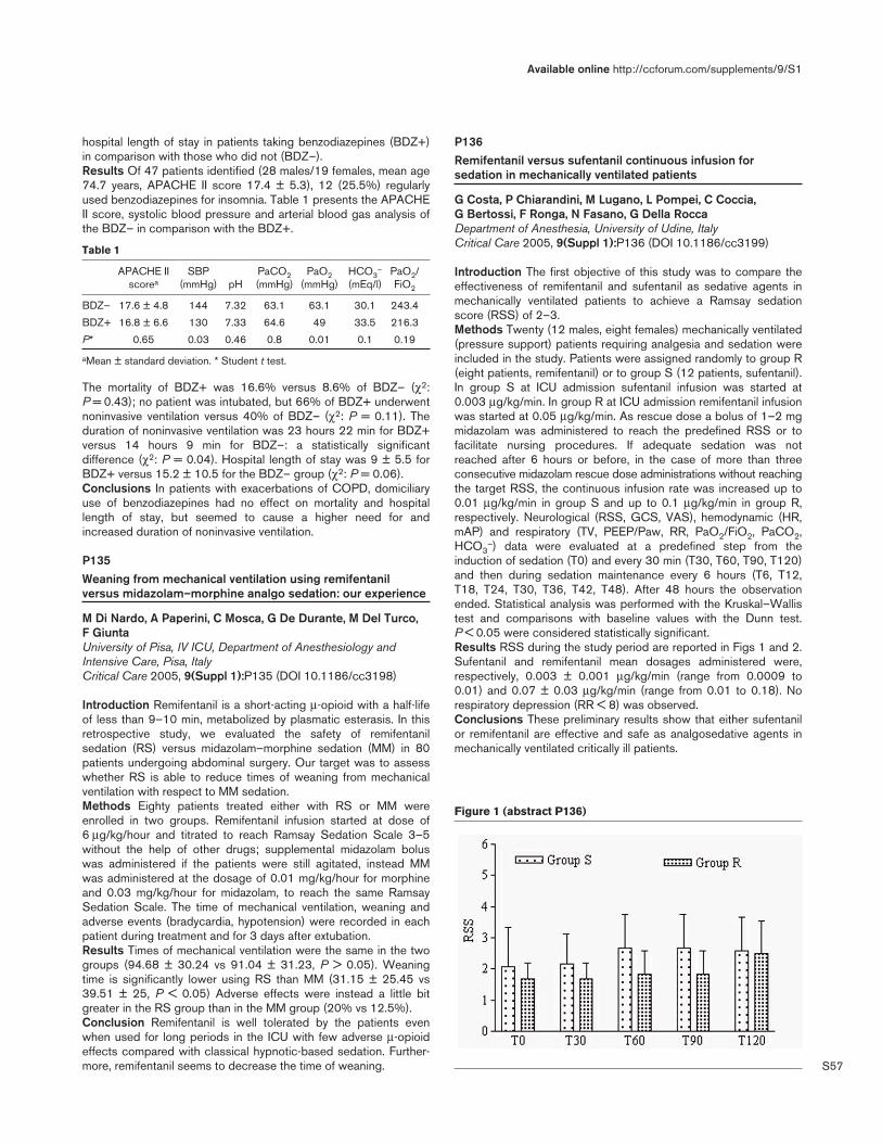
S57
hospital length of stay in patients taking benzodiazepines (BDZ+)in comparison with those who did not (BDZ–).Results Of 47 patients identified (28 males/19 females, mean age74.7 years, APACHE II score 17.4 ± 5.3), 12 (25.5%) regularlyused benzodiazepines for insomnia. Table 1 presents the APACHEII score, systolic blood pressure and arterial blood gas analysis ofthe BDZ– in comparison with the BDZ+.
Table 1
APACHE II SBP PaCO2 PaO2 HCO3– PaO2/
scorea (mmHg) pH (mmHg) (mmHg) (mEq/l) FiO2
BDZ– 17.6 ± 4.8 144 7.32 63.1 63.1 30.1 243.4
BDZ+ 16.8 ± 6.6 130 7.33 64.6 49 33.5 216.3
P* 0.65 0.03 0.46 0.8 0.01 0.1 0.19
aMean ± standard deviation. * Student t test.
The mortality of BDZ+ was 16.6% versus 8.6% of BDZ– (χ2:P = 0.43); no patient was intubated, but 66% of BDZ+ underwentnoninvasive ventilation versus 40% of BDZ– (χ2: P = 0.11). Theduration of noninvasive ventilation was 23 hours 22 min for BDZ+versus 14 hours 9 min for BDZ–: a statistically significantdifference (χ2: P = 0.04). Hospital length of stay was 9 ± 5.5 forBDZ+ versus 15.2 ± 10.5 for the BDZ– group (χ2: P = 0.06).Conclusions In patients with exacerbations of COPD, domiciliaryuse of benzodiazepines had no effect on mortality and hospitallength of stay, but seemed to cause a higher need for andincreased duration of noninvasive ventilation.
P135Weaning from mechanical ventilation using remifentanilversus midazolam–morphine analgo sedation: our experience
M Di Nardo, A Paperini, C Mosca, G De Durante, M Del Turco, F GiuntaUniversity of Pisa, IV ICU, Department of Anesthesiology andIntensive Care, Pisa, ItalyCritical Care 2005, 9(Suppl 1):P135 (DOI 10.1186/cc3198)
Introduction Remifentanil is a short-acting µ-opioid with a half-lifeof less than 9–10 min, metabolized by plasmatic esterasis. In thisretrospective study, we evaluated the safety of remifentanilsedation (RS) versus midazolam–morphine sedation (MM) in 80patients undergoing abdominal surgery. Our target was to assesswhether RS is able to reduce times of weaning from mechanicalventilation with respect to MM sedation.Methods Eighty patients treated either with RS or MM wereenrolled in two groups. Remifentanil infusion started at dose of6 µg/kg/hour and titrated to reach Ramsay Sedation Scale 3–5without the help of other drugs; supplemental midazolam boluswas administered if the patients were still agitated, instead MMwas administered at the dosage of 0.01 mg/kg/hour for morphineand 0.03 mg/kg/hour for midazolam, to reach the same RamsaySedation Scale. The time of mechanical ventilation, weaning andadverse events (bradycardia, hypotension) were recorded in eachpatient during treatment and for 3 days after extubation.Results Times of mechanical ventilation were the same in the twogroups (94.68 ± 30.24 vs 91.04 ± 31.23, P > 0.05). Weaningtime is significantly lower using RS than MM (31.15 ± 25.45 vs39.51 ± 25, P < 0.05) Adverse effects were instead a little bitgreater in the RS group than in the MM group (20% vs 12.5%).Conclusion Remifentanil is well tolerated by the patients evenwhen used for long periods in the ICU with few adverse µ-opioideffects compared with classical hypnotic-based sedation. Further-more, remifentanil seems to decrease the time of weaning.
P136Remifentanil versus sufentanil continuous infusion forsedation in mechanically ventilated patients
G Costa, P Chiarandini, M Lugano, L Pompei, C Coccia, G Bertossi, F Ronga, N Fasano, G Della RoccaDepartment of Anesthesia, University of Udine, ItalyCritical Care 2005, 9(Suppl 1):P136 (DOI 10.1186/cc3199)
Introduction The first objective of this study was to compare theeffectiveness of remifentanil and sufentanil as sedative agents inmechanically ventilated patients to achieve a Ramsay sedationscore (RSS) of 2–3.Methods Twenty (12 males, eight females) mechanically ventilated(pressure support) patients requiring analgesia and sedation wereincluded in the study. Patients were assigned randomly to group R(eight patients, remifentanil) or to group S (12 patients, sufentanil).In group S at ICU admission sufentanil infusion was started at0.003 µg/kg/min. In group R at ICU admission remifentanil infusionwas started at 0.05 µg/kg/min. As rescue dose a bolus of 1–2 mgmidazolam was administered to reach the predefined RSS or tofacilitate nursing procedures. If adequate sedation was notreached after 6 hours or before, in the case of more than threeconsecutive midazolam rescue dose administrations without reachingthe target RSS, the continuous infusion rate was increased up to0.01 µg/kg/min in group S and up to 0.1 µg/kg/min in group R,respectively. Neurological (RSS, GCS, VAS), hemodynamic (HR,mAP) and respiratory (TV, PEEP/Paw, RR, PaO2/FiO2, PaCO2,HCO3
–) data were evaluated at a predefined step from theinduction of sedation (T0) and every 30 min (T30, T60, T90, T120)and then during sedation maintenance every 6 hours (T6, T12,T18, T24, T30, T36, T42, T48). After 48 hours the observationended. Statistical analysis was performed with the Kruskal–Wallistest and comparisons with baseline values with the Dunn test.P < 0.05 were considered statistically significant.Results RSS during the study period are reported in Figs 1 and 2.Sufentanil and remifentanil mean dosages administered were,respectively, 0.003 ± 0.001 µg/kg/min (range from 0.0009 to0.01) and 0.07 ± 0.03 µg/kg/min (range from 0.01 to 0.18). Norespiratory depression (RR < 8) was observed.Conclusions These preliminary results show that either sufentanilor remifentanil are effective and safe as analgosedative agents inmechanically ventilated critically ill patients.
Available online http://ccforum.com/supplements/9/S1
Figure 1 (abstract P136)

S58
ReferencesSoltész, et al.: Br J Anaesth 2001, 86:763-768.Jacobi J, Fraser GL, Coursin DB, et al.: Crit Care Med 2002, 30:119-141.
P137Remifentanil in neurosurgical patients in the intensive careunit
G Koukoulitsios, C Mandila, E Paramythiotou, E Agelopoulou,G Stathopoulos, N Noulas, A KarabinisGeneral Hospital of Athens ‘G Gennimatas’, Athens, GreeceCritical Care 2005, 9(Suppl 1):P137 (DOI 10.1186/cc3200)
Introduction Remifentanil is a rapidly metabolized opioid analgesicthat is being increasingly used in critically ill patients [1,2]. Ourpurpose was to evaluate the hemodynamic changes of sedatedneurosurgery patients in the ICU caused by remifentanil as well asthe time elapsed until the wakening in comparison with fentanyland morphine. Patients received remifentanil for more than 5 daysin continuous infusion.Methods The study was conducted in the 12-bed ICU of a generalhospital. One hundred and fifty patients of both sexes wereincluded in the study. Their mean body weight was 77 kg. Meanduration of the surgical intervention was 180 min. After theiradmission to the ICU, patients were randomly assigned to threegroups. Patients in group A received remifentanil 9–10 µg/kg/hour,in group B fentanyl 0.5–6 µg/kg/hour and in group C they receivedmorphine 0.03–0.15 mg/kg/hour. Every 24 hours, sedation andanalgesia were stopped for a neurological examination (GlascowComa Scale). All patients were sedated with propofol and theyremained sedated for 5 days at least. Parameters recorded werecardiac rhythm, mean arterial pressure and time to recovery(judged by spontaneous respiration).Results See Table 1 for comparison of hemodynamic parametersbetween the three groups. Recovery time was much shorter whenwe compared the remifentanil group with the two other groups(P < 0.001). A statistically significant difference was also observedwhen we compared group B with group C (P < 0.001).Conclusion We conclude that remifentanil is safe enough forneurosurgerical patients under sedation in the ICU, and that theweakening time is much shorter for patients receiving thisanalgesic.
Table 1 (abstract P137)
Remifentanil Fentanyl Morphine
Bradycardia 3 (6%) 2 (4%) 2 (4%)
Hypotension 8 (16%) 6 (12%) 2 (4%)
Mean arterial pressure (mmHg) 76 74 75
Mean heart rate 68 70 71
References1. Tipps LB, Coplin WM, Murry KR, Rhoney DH: Safety and feasi-
bility of continuous of remifentanil in the neurosurgical inten-sive care unit. Neurosurgery 2000, 46:596-602.
2. Maze M, Scarfini C, Cavaliere F: New agents for sedation in theintensive care unit. Crit Care Clin 2001, 17:881-897.
P138Risk factors, coma, and consequences of intensive care unitdelirium
Y Skrobik, M Dumont, S OuimetHopital Maisonneuve Rosemont, Montreal, CanadaCritical Care 2005, 9(Suppl 1):P138 (DOI 10.1186/cc3201)
Objectives To establish risk factors for the development ofdelirium in an ICU, to determine the effect of ICU delirium screenedwith the Intensive Care Delirium Checklist on mortality and ICU andhospital lengths of stay, and to clarify the comorbidity and clinicaleffect of coma on the incidence of delirium and on outcome.Design and setting Prospective study in a 16-bed universityhospital medical/surgical ICU.Patients A total of 820 consecutive patients admitted to the ICUfor more than 24 hours.Interventions Fifteen covariates, including medical history, severityof illness on admission (APACHE II), laboratory values, andmedications were prospectively recorded for a total of 4707patient-days (mean = 5.7 ± 7 days). All patients were screened aminimum of three times daily. Follow-up to hospital discharge wasprovided in all patients. Hospital length of stay, and hospitalmortality, were compared between delirium and non-deliriumpatients. Comatose patients were arbitrarily separated into thosewhose coma was transient (< 5 days) or lasting (5 days or more).Results Hypertensive patients, smokers, and alcoholics were morelikely to develop delirium (odds ratios of 1.68, 1.56, and 1.77,respectively). Delirium was independently associated with severityof illness on admission; each additional point on the APACHEscore was associated with a 4% increase in risk of delirium. Coma,regardless of its duration, increased the incidence of delirium (51%and 54% for shorter and longer duration of coma, vs 20% inpatients with no coma, P < 0.05). The development of delirium wasnot associated with benzodiazepine or opiate use. Patients whodeveloped ICU delirium were more likely to die in hospital (47%compared with 37% in patients without delirium, P < 0.005), andhad a significantly longer length of stay (18 days compared with13 days, P < 0.005). Mortality was proportional to severity ofillness on admission (APACHE), presence of diabetes, total lengthof hospital stay and delirium occurrence.Conclusion Delirium risk factors include tobacco and alcohol use,a history of hypertension, and preceding ICU coma but not ICUbenzodiazepine or opiate use. Significant mortality and longerhospital stay are attributable to this morbid condition. Awarenessof patients at risk may lead to better recognition and earlierintervention.Acknowledgement Partial funding ($18,000) for this project wasobtained from the Fonds de Recherche en Santé du Quebec.
Critical Care March 2005 Vol 9 Suppl 1 25th International Symposium on Intensive Care and Emergency Medicine
Figure 2 (abstract P136)

S59
P139Evaluation of EM and orthopedic residents’ pain estimationabout patients with limb trauma who were visited in theEmergency Department of Hazrat Rasoul Hospital
S Farahmand, M AhmadiImam Khomeini Hospital, Tehran, IranCritical Care 2005, 9(Suppl 1):P139 (DOI 10.1186/cc3202)
Objective To evaluate pain estimation in the patients with limb(s)trauma by EM and orthopedic resident in the EmergencyDepartment (ED) of Hazrat-e-Rasoul Hospital.Patients and methods In a prospective study, 160 patients thatmerely had limb(s) trauma and were admitted to the EM ward ofHazrat Rasoul Hospital were included. Patients with GSC <15 —due to every reason, like head trauma, drug overdose, and so on —at the time of physical examination, and positive history ofanalgesic use before ED arrival or history of drug or alcohol abuse,were excluded from this study. At the time of the presentation tothe ED, first of all the patients marked the level of their pain on avisual analogue scale (VAS) scale by supervision of theresearchers and then the EM and orthopedic residents marked thepain level of every patient on a VAS, separately. The EM andorthopedic residents select randomly from on-call residents.Results Of 160 enrolled cases (65 females and 95 males), themean age was 32.05 ± 17.87 years. The mean of pain scoresdetermined by the patients was 6.41 ± 2.52. The mean of therecorded pain scores by EM and orthopedic residents was 4.41 ±1.83 and 4.05 ± 1.92, respectively. There was a significantdifference between the mean of patients’ scores and the mean ofpain scores determined by both the EM and orthopedic residents,separately (P < 0.001); but there is no significant differencebetween EM and orthopedic residents. Pain estimation of EM andorthopedic residents in 20 cases were the same as the patientpain score.Conclusion According to previous studies, the only accuratebarometer in treating pain is the patient’s report, but in our centerpain treatment was done according to physicians’ estimation; ourstudy indicates that they underestimate the pain of their patients.To properly sedate patients, the VAS should be included as aroutine part of vital signs and the pain score should be recorded,especially for such patients who have pain and might need painfulprocedures.
P140Sedation in haemofiltered patients: a retrospective study ofcompliance with the sedation policy
S Al-Saffar, R Shulman, A WebbUCL Hospital NHS Foundation Trust, London, UKCritical Care 2005, 9(Suppl 1):P140 (DOI 10.1186/cc3203)
Aim This study was designed to review sedation of critically illpatients requiring haemofiltration and to assess the compliancewith our sedation policy.Introduction Such patients are of particular concern as there is adanger of accumulation of sedatives. The UCLH sedation scaleinvolves hourly assessment, –3 correlates with unrousable, –2(roused by painful stimuli), –1 (roused by movement), 0 (roused byvoice), 1 (aware but calm), 2 (aware and comfortable), 3 (agitatedand restless). The sedation policy requires the followinginterventions: at +3 or +2 the dose is increased; at +1, 0 or –1 (i.e.the ‘target range’) no dose adjustment is attempted until threeconsecutive scores are achieved within the target range whendose reduction is attempted; at –2 or –3, sedation is stopped.
Method This retrospective study was conducted in a 22-bed,mixed medical/surgical ICU in a teaching hospital. A total of 427patients received haemofiltration between 10 September 2003and 18 January 2004 for a total of 56,024 hours. The data forthese patients who were managed with continuous sedationinfusions were extracted using the UCLH Clinical InformationManagement System (GE Medical-healthcare QS 5.6). The level ofcompliance to our sedation policy was analysed by comparisonwith the aforementioned rules. A chi-squared test was used forstatistical significance.Results In 42% of oversedation hours (–2, –3) the sedativeinfusion was not reduced (see Fig. 1). Where sedation scores fellwithin the target range on three consecutive occasions sedationinfusions were decreased or stopped appropriately in 78% ofepisodes. In undersedated hours (+2, +3), sedation infusions wereincreased in 29% of episodes. Sedation scores within the targetrange were achieved for 77.7% of the time, regardless of whetheror not sedatives were given. Furthermore, sedation scores shareda similar distribution pattern in both sedated and unsedated hoursexcept that the +1 sedation score was more prevalent inunsedated hours compared with sedated hours (1818 hours[10.9%] vs 15,451 hours [39.2%]). Episodes of undersedationwere three times (3151 hours vs 1312 hours) more likely to betreated incorrectly than correctly (P < 0.05). In oversedatedepisodes, correct adjustment occurred > 3 times more often thanincorrect changes (27,810 hours vs 7900 hours) (P < 0.05).Conclusion Our data suggest that when patients wereundersedated a reluctance to follow the sedation protocol byincreasing the sedative dose was seen. In contrast, when patientswere oversedated nurses were more inclined to follow theprotocol.
Available online http://ccforum.com/supplements/9/S1
Figure (abstract P140)
Percentage of appropriate adjustments in sedation infusions.

S60
P141Does implementation of a goal sedation score improvemanagement of mechanically ventilated adults?
K Feeley1, A Gardner2, I Mitchell1, A Leditschke1
1The Canberra Hospital, Canberra, Australia; 2Cabrini DeakinCentre for Nursing Research, Melbourne, AustraliaCritical Care 2005, 9(Suppl 1):P141 (DOI 10.1186/cc3204)
Background Sedation management of mechanically ventilatedpatients has been a source of concern for intensive care nursingand medical staff worldwide in recent years. An earlier prospectiveaudit of 48 ventilated patients showed that our sedationmanagement was suboptimal. Patients were oversedated 45% ofthe time, with the most frequent management strategy being use ofmorphine and midazolam infusions. Despite this, 23% of patientswere physically restrained. A literature review suggested that amove away from sedative infusions would reduce the length ofventilation, length of ICU stay and tracheostomy incidence. Use ofsedation scales, setting of goal sedation scores and implemen-tation of a nurse-initiated sedation protocol were also deemed bestpractice.Objective To determine whether introduction of a goal sedationscore would be associated with improved management of sedationas indicated by a reduction in incidence of oversedation and in useof physical restraints.Method The study design was prospective and exploratory.Following multidisciplinary staff training, an optimal sedation goalscore was set daily by senior medical staff and changed whenclinically indicated. Nurses titrated intravenous infusions ofanalgesia and sedation with the aim of achieving the desired goalsedation score. Sedation patterns were collected on 52 ventilatedpatients. Institutional ethics approval was obtained.Results The median APACHE II score was 19 (range 8–33);median age was 65.9 years (range 18–88); sex ratio, male/female,was 7:3. For patients ventilated for more than 5 days only sedationpatterns for the first 5 days are presented. The goal sedation scorewas achieved in 45% of patient-hours, with only 7% of patient-hours recorded as agitated. Incidence of oversedation wasunchanged from the previous audit (45% of time). Patients deemedat risk of oversedation had a goal score compliance of 48.6%.Patients at risk of undersedation had a goal score compliance of34.4%. When compared with prior audit results, there was anincrease in the use of physical restraints from 11/48 (23%) to
21/52 (40%) (Fishers exact P = 0.086). Morphine and midazolaminfusion use was unchanged with use in 63% and 65% of patients,respectively, and propofol was used in 73% of patients. Incidenceof deliberate self-extubation was 0.9/100 ventilated days, medianlength of ventilation was 3.9 days and incidence of tracheostomywas 7.6%.Conclusion Despite a sedation goal score being set daily, patientsreached their goal score for less than one-half of ventilated hours.Oversedation continued to be a problem and may be associatedwith the frequent use of morphine and midazolam infusions.Considering agitation appeared to be well managed, the increaseduse of physical restraints is concerning. Due to the limited successof use of goal sedation scores alone to improve sedationmanagement, we are now prospectively evaluating the use of asedation management algorithm in conjunction with goal scores.
P142The Richmond agitation–sedation scale and bispectral indexduring dexmedetomidine sedation
A Turkmen, A Altan, N Turgut, S Vatansever, S GökkayaSSK Okmeydani Training Hospital, Istanbul, TurkeyCritical Care 2005, 9(Suppl 1):P142 (DOI 10.1186/cc3205)
Introduction Patients require continuous reassessment of theirpain and need for sedation. The routine use of standardized andvalidated sedation scales and monitors are needed. The bispectralindex (BIS) uses a monitor of cortical suppression to maintain theoptimal level of sedation and hypnosis. The Richmond agitationsedation scale (RASS) has high reliability and validity in ICUpatients. We aimed to assess the correlation of the BIS with theRASS during dexmedetomidine sedation.Methods Eleven ventilated critically ill patients, aged 17–82(50.09 ± 17.76) years, APACHE II score of 12.63 ± 3.90, SOFAscore of 3.27 ± 1.73, were enrolled in the study. Patients receiveda loading dexmedetomidine infusion of 1 µg/kg over 10 minfollowed by a maintenance infusion of 0.5 µg/kg/hour for 8 hours.The efficacy of sedation was assessed using the RASS and BISmonitoring. The Wilcoxon test and Spearman’s correlation analysiswere used for statistical analysis.Results Significant correlations between RASS and BIS valueswere found in this study (Table 1).Conclusion RASS levels significantly correlated with BIS valuesduring dexmedetomidine sedation.
Critical Care March 2005 Vol 9 Suppl 1 25th International Symposium on Intensive Care and Emergency Medicine
Table 1 (abstract P142)
RASS 1 RASS 2 RASS 3 RASS 4 RASS 5 RASS 6 RASS 7 RASS 8
BIS 1 r = 0.77, P = 0.005
BIS 2 r = 0.87, P = 0.0001
BIS 3 r = 0.84, P = 0.001
BIS 4 r = 0.84, P = 0.001
BIS 5 r = 0.94p, P = 0.0001
BIS 6 r = 0.85, P = 0.001
BIS 7 r = 0.92, P = 0.0001
BIS 8 r = 0.90, P = 0.0001
BIS, bispectral index; RASS, Richmond agitation sedation scale.
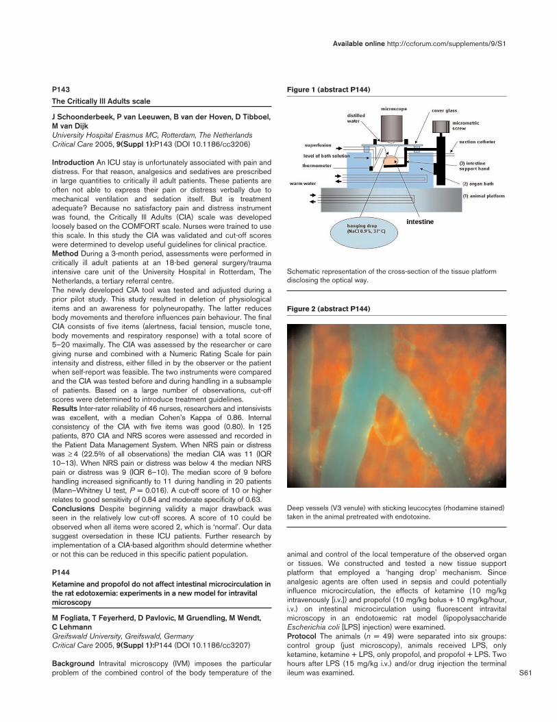
S61
P143The Critically Ill Adults scale
J Schoonderbeek, P van Leeuwen, B van der Hoven, D Tibboel,M van DijkUniversity Hospital Erasmus MC, Rotterdam, The NetherlandsCritical Care 2005, 9(Suppl 1):P143 (DOI 10.1186/cc3206)
Introduction An ICU stay is unfortunately associated with pain anddistress. For that reason, analgesics and sedatives are prescribedin large quantities to critically ill adult patients. These patients areoften not able to express their pain or distress verbally due tomechanical ventilation and sedation itself. But is treatmentadequate? Because no satisfactory pain and distress instrumentwas found, the Critically Ill Adults (CIA) scale was developedloosely based on the COMFORT scale. Nurses were trained to usethis scale. In this study the CIA was validated and cut-off scoreswere determined to develop useful guidelines for clinical practice.Method During a 3-month period, assessments were performed incritically ill adult patients at an 18-bed general surgery/traumaintensive care unit of the University Hospital in Rotterdam, TheNetherlands, a tertiary referral centre.The newly developed CIA tool was tested and adjusted during aprior pilot study. This study resulted in deletion of physiologicalitems and an awareness for polyneuropathy. The latter reducesbody movements and therefore influences pain behaviour. The finalCIA consists of five items (alertness, facial tension, muscle tone,body movements and respiratory response) with a total score of5–20 maximally. The CIA was assessed by the researcher or caregiving nurse and combined with a Numeric Rating Scale for painintensity and distress, either filled in by the observer or the patientwhen self-report was feasible. The two instruments were comparedand the CIA was tested before and during handling in a subsampleof patients. Based on a large number of observations, cut-offscores were determined to introduce treatment guidelines.Results Inter-rater reliability of 46 nurses, researchers and intensivistswas excellent, with a median Cohen’s Kappa of 0.86. Internalconsistency of the CIA with five items was good (0.80). In 125patients, 870 CIA and NRS scores were assessed and recorded inthe Patient Data Management System. When NRS pain or distresswas ≥ 4 (22.5% of all observations) the median CIA was 11 (IQR10–13). When NRS pain or distress was below 4 the median NRSpain or distress was 9 (IQR 6–10). The median score of 9 beforehandling increased significantly to 11 during handling in 20 patients(Mann–Whitney U test, P = 0.016). A cut-off score of 10 or higherrelates to good sensitivity of 0.84 and moderate specificity of 0.63.Conclusions Despite beginning validity a major drawback wasseen in the relatively low cut-off scores. A score of 10 could beobserved when all items were scored 2, which is ‘normal’. Our datasuggest oversedation in these ICU patients. Further research byimplementation of a CIA-based algorithm should determine whetheror not this can be reduced in this specific patient population.
P144Ketamine and propofol do not affect intestinal microcirculation inthe rat edotoxemia: experiments in a new model for intravitalmicroscopy
M Fogliata, T Feyerherd, D Pavlovic, M Gruendling, M Wendt, C LehmannGreifswald University, Greifswald, GermanyCritical Care 2005, 9(Suppl 1):P144 (DOI 10.1186/cc3207)
Background Intravital microscopy (IVM) imposes the particularproblem of the combined control of the body temperature of the
animal and control of the local temperature of the observed organor tissues. We constructed and tested a new tissue supportplatform that employed a ‘hanging drop’ mechanism. Sinceanalgesic agents are often used in sepsis and could potentiallyinfluence microcirculation, the effects of ketamine (10 mg/kgintravenously [i.v.]) and propofol (10 mg/kg bolus + 10 mg/kg/hour,i.v.) on intestinal microcirculation using fluorescent intravitalmicroscopy in an endotoxemic rat model (lipopolysaccharideEscherichia coli [LPS] injection) were examined.Protocol The animals (n = 49) were separated into six groups:control group (just microscopy), animals received LPS, onlyketamine, ketamine + LPS, only propofol, and propofol + LPS. Twohours after LPS (15 mg/kg i.v.) and/or drug injection the terminalileum was examined.
Available online http://ccforum.com/supplements/9/S1
Figure 1 (abstract P144)
Schematic representation of the cross-section of the tissue platformdisclosing the optical way.
Figure 2 (abstract P144)
Deep vessels (V3 venule) with sticking leucocytes (rhodamine stained)taken in the animal pretreated with endotoxine.

S62
Results LPS injection did not affect the functional capillary densityin the longitudinal and circular muscular and the mucosal layers butincreased the number of firmly adhering leukocytes (stickers) in V3venules (LPS vs controls; 491.2 ± 100.5 vs 187.3 ± 123.0 n/mm2;mean ± standard deviation, P < 0.05) and in V1 venules (LPS vscontrols: 256.7 ± 49.7 vs 97.9 ± 27.5 n/mm2; P < 0.05). Thenumber of rolling leucocytes in V3 venules was also reduced inLPS rats as compared with the control group (3.73 ± 2.7 vs33.3 ± 28.2 n/min; P < 0.05). The levels of IL-1β, IL-6, IL-10 andtumour necrosis factor alpha were significantly higher in the LPSanimals. The LPS induced changes were not affected by ketamineor propofol administration.Conclusion The ‘hanging drop’ technique prevented tissuequenching, assured undisturbed microcirculation, provided stabletemperature and humidity, and assured a clear visual field. It wasfound that LPS produced important microcirculatory changes.However, neither ketamine nor propofol affected microcirculation inendotoxemia in the rat and had no effect on the leukocyteadherence, and therefore could be used for microcirculatorystudies.
P145Ketamine and the coagulation response during and aftercardiopulmonary bypass
I Husedzinovic, S Barisin, N BradicClinical Hospital Drubrava, Zagreb, CroatiaCritical Care 2005, 9(Suppl 1):P145 (DOI 10.1186/cc3208)
Introduction The inflammatory response and the coagulation-fibrinolytic cascades are closely interconnected. Vascular injuryfollowing cardiopulmonary bypass (CPB) may result in uncontrolledplatelet activation, thrombin generation, and disseminatedintravascular coagulation. Many of the anesthetic agents possesimmunomodulatory effects. The clinical implications of such effectsin the context of CPB remain unknown. Ketamine attenuates IL-6response and production of the superoxide anion by neutrophilsafter CPB, and reduces coronary uptake of neutrophils followingmyocardial ishemia and reperfusion. We studied the effect of small-dose ketamine on the coagulation response (antithrombin III,fibrinogen, D-dimers, platelets) during and after CPB.Methods In the preliminary prospective study report, werandomized 30 patients who underwent open heart surgery intotwo groups: K-group (n = 15), which received 0.5 mg/kg ketamine;and P-group (n = 15), which received placebo. Serum sampleswere collected before starting the operation (T0), after aorta clampreleasing (T1), 30 min (T2), 2 hours (T3), 6 hours (T4), 12 hours(T5), 24 hours (T6), and 48 hours (T7) after weaning from CPB.Data (mean ± standard deviation) were tested by variance analysis.Results Both groups were comparable for age, sex, and bodysurface area. Differences of operative data (CPB and aorta cross-clamping time) and postoperative data (ventilatory support, ICUstay, blood transfusion) between groups were not significant.Furthermore, there was no statistical significance between all othercollected data in this study.Conclusion In this preliminary study, findings suggest that smalldoses of ketamine do not possess any effect on the coagulationresponse during and after CPB.
P146Combined general and epidural anaesthesia improvesperioperative immune function
O Ahlers, I Kuerer, J Lenze, K Schulmeister, C Höhne, G Fritz,K Falke, D KehCharite, Campus Virchow-Klinikum, Berlin, GermanyCritical Care 2005, 9(Suppl 1):P146 (DOI 10.1186/cc3209)
Background Intraoperative stress may impair perioperativeimmune function. The hormones epinephrine, cortisol, and thestress-associated anti-inflammatory IL-10 are known to inhibit Th1helper cells. Interferon-gamma (IFN-γ), released mainly by Th1cells, seems to be crucial for host resistance; abolished Th1function (anergy) is associated with increased risk of infection andmortality. We hypothesized that combined general and thoracicepidural anaesthesia (TEA) reduces intraoperative stress duringmajor abdominal surgery preventing perioperative impairment ofTh1 cells.Methods Fifty-four patients undergoing major abdominal surgerywere enrolled in a controlled, prospective, randomized, and single-blinded study. The protocol was approved by the local ethicscommittee. All patients were supported with an epidural catheterbefore induction of general anaesthesia. In one group (n = 27), thecatheter was used for intraoperative analgesia (TEA-I); the othergroup (n = 27) received systemic opioids during surgery (TEA-P).Subsequently, epidural analgesia was used in both groups forpostoperative pain management. Blood samples were taken beforeplacing the epidural catheter, 2 hours after the beginning ofsurgery, and at days 1 and 4 after surgery. IFN-γ and IL-10 weremeasured in plasma and concanavalin-A (ConA)-stimulated bloodsamples. Stress hormone levels of epinephrine and cortisol wereexamined. Differences between the groups were analysed usingnon-parametric tests, significance was defined by P < 0.05.Results The plasma IFN-γ/IL-10 ratio in TEA-P was lower 2 hoursafter the beginning of surgery (not significant). In ConA-stimulatedlymphocytes, the IFN-γ/IL-10 ratio was significantly decreased inTEA-P from 2 hours after the beginning of surgery until day 1(Fig. 1) (P = 0.005). This effect was mainly due to a significant
Critical Care March 2005 Vol 9 Suppl 1 25th International Symposium on Intensive Care and Emergency Medicine
Figure 1 (abstract P146)

S63
decrease of IFN-γ in TEA-P, but not due to differences in IL-10increase. Plasma levels of epinephrine (P = 0.022) and cortisol(P = 0.015) were significantly higher in TEA-P during surgery.Conclusion The intraoperative use of a thoracic epidural catheterprevents stress-induced perioperative impairment of IFN-γ release.Since IFN-γ plays a key role in host defence, the results may be ofclinical relevance for the postoperative course
P147Influence of thiopentone and midazolam on monocytedifferentiation into dendritic cells
C Karcher, H Raidt, H Haeberle, H Dieterich, K Unertl, T SchroederUniversity of Tuebingen, GermanyCritical Care 2005, 9(Suppl 1):P147 (DOI 10.1186/cc3210)
Background Dendritic cells (DC) play a major role in the innateimmune response by directing B lymphocytes and T lymphocytes.We studied the contribution of DC to the immune suppressionduring thiopentone and midazolam treatment. Previous in vitrostudies have shown negative effects of thiopentone on immunefunction [1,2]. Clinical studies suggest that long-term sedation withthiopentone contributes to an increased incidence of nosocomialinfections [3]. However, effects of thiopentone on DC have not yetbeen elaborated.Materials and methods We investigated the effects of clinicaldoses of thiopentone and midazolam on the differentiation ofmonocytes to dendritic cells in vitro. Monocytes were isolated frombuffy coats of healthy donors and cultured in the presence of GM-CSF and IL-4 over 8 days. The cells were incubated with differentconcentrations of thiopentone and midazolam, respectively. DCwere characterized through surface expression of CD1a, CD14,CD16, CD40, CD80, CD83, CD86 and HLA-DR. Cell functionwas quantified through FITC-dextrane phagocytosis by cytometry.Results In contrast to midazolam, high concentrations of thio-pentone (50–100 µg/ml) led to notable alteration in the morphologyand differentiation of DC. Typical morphologic characteristics likespikes and cell clustering were absent. The surface expression ofCD1a was reduced by 83% (± 41%), CD40 by 93% (± 8%) andHLA-DR by 68% (± 35%). Phagocytosis capacity of these cellswas decreased by 50% (± 33%).Conclusion We conclude that differentiation from monocytes intoDC is affected by high concentrations of thiopentone but not bymidazolam. This may contribute to an immune suppression duringlong-term thiopentone sedation.References1. Larsen B, Hoff G, Wilhelm W, et al.: Anesthesiology 1998,
89:1218-1227.2. Heller A, Heller S, Blecken S, et al.: Acta Anaesthesiol Scand
1998, 42:518-526.3. Bronchard R, Albaladejo P, Brezac G, et al.: Anesthesiology
2004, 100:234-239.
P148Propofol and methohexital comparably affectpolymorphonuclear leukocyte function in patients after cardiacsurgery
E Hüttemann, M Büttner, K Vogelsang, S SakkaFriedrich-Schiller-University Jena, GermanyCritical Care 2005, 9(Suppl 1):P148 (DOI 10.1186/cc3211)
Background Experimental and clinical studies suggest thatanesthetics have a variety of negative side effects onpolymorphonuclear (PMN) leukocytes, which play a vital role in thedefense against invading bacteria. In this study, we analyzed the
effects of propofol and methohexital on PMN leukocyte function inpatients after cardiac surgery.Patients and methods In this observational clinical study, westudied 46 patients undergoing elective cardiac surgery whopostoperatively either received propofol (n = 29) or methohexital(n = 17). Patients in the propofol group (P) (19 males, 10 females)had a mean age of 65 ± 11 (range 36–80) years. In themethohexital group (M) (12 males, five females) the mean age was65 ± 8 (range 43–77) years. Both sedatives were administeredaccording to clinical requirements. PMN leukocyte function wasassessed as respiratory oxidative burst and expressed as thepercent of oxidating cells (normal 70–100%). Furthermore, themean fluorescence intensity (MFI) was determined as a measure ofintracellular uptake of bacteria. Both variables were assessedbefore induction of anesthesia (MP1), on ICU admission aftercardiac surgery (beginning of sedative administration) (MP2),6 hours after ICU admission (MP3) and 24 hours after beginning ofanesthesia (MP4). Patients’ demographics and measurementresults were compared by t test and analysis of variance for repeatedmeasures.Results Both groups were well matched in terms of patientdemographics, age, body weight and duration of sedativeadministration (P: 9.4 ± 5.4 hours vs M: 8.4 ± 5.6 hours). Withinboth groups, respiratory oxidative burst significantly decreasedover time (MP1 and MP2 vs MP3 and MP4, respectively). Forcomparison, respiratory oxidative burst at MP4 was significantlyhigher for methohexital (82%) than propofol (72%). Whileoxidation decreased between MP3 and MP4 for propofol, nosignificant change was observed between these two time pointsfor methohexital. For MFI, a time-dependent decrease was foundfor both drugs, without any difference between both groups.Conclusions PMN leukocyte function is comparably depressed bypropofol rather than methohexital in patients after cardiac surgery.
P149Effects of propofol versus methohexital on neutrophil functionand immune status in critically ill patients
S Sakka, A Jung, H Vogelsang, E HuettemannFriedrich-Schiller-University Jena, GermanyCritical Care 2005, 9(Suppl 1):P149 (DOI 10.1186/cc3212)
Introduction Sedatives (i.e. propofol and methohexital) have avariety of negative side effects on neutrophil leukocytes,lymphocytes and monocytes, which all play a vital role in thedefense against invading microorganisms [1]. In this study, weinvestigated these effects in critically ill patients with long-termsedation.Patients and methods In this observative clinical study, weanalyzed 21 critically ill patients with long-term sedation who eitherreceived propofol (n = 12, APACHE II score 26 ± 4) ormethohexital (n = 9, APACHE II score 28 ± 6) after ICU admission.Patients in the propofol group (P) (nine males, three females) had amean age of 55 ± 15 years. In the methohexital group (M) (eightmales, one female) the mean age was 48 ± 18 years. Bothsedatives were administered according to clinical requirements.Neutrophil leukocyte function was assessed as phagocytosis andrespiratory oxidative burst activity. Furthermore, cellular markers ofmonocytes and lymphocytes (CD3, CD4, CD8, CD19, CD57,CD122) were assessed. Measurements were made on ICUadmission, day 3, day 7 and day 14 of drug administration.Patients’ demographics and results were compared byMann–Whitney U test and one-way repeated-measures analysis ofvariance with an all pair-wise multiple comparison procedure.
Available online http://ccforum.com/supplements/9/S1

S64
Results Both groups were well matched in terms of age, heightand body weight. ICU length of stay was comparable (22 ± 7 daysvs 20 ± 9 days). Mortality was 0/12 and 2/9, respectively. Absolutenumbers of leukocytes and subpopulations were comparablebetween both groups at each time point. We found no differencein neutrophil oxidative burst and phagocytosis within and betweenboth groups at the different time points. With respect tolymphocyte and monocyte CD marker expression, no differenceswithin each group and between the time points were found.Conclusion Effects of methohexital and propofol on neutrophilleukocyte function, expression of lymphocyte and monocytemarkers were not different in critically ill patients with long-termsedation.Reference1. Kelbel I, Weiss M: Anaesthetics and immune function. Curr
Opin Anaesthesiol 2001, 14:685-691.
P150HLA-DR expression on monocytes and the T-cell subset inseptic patients
N Menestrina, A Martini, A Milan, G Soldati, C Parolini, G Finco,L GottinOps Poloclinico GB Rossi, Verona, ItalyCritical Care 2005, 9(Suppl 1):P150 (DOI 10.1186/cc3213)
Introduction Sepsis is one major cause of mortality in the ICU,despite increasing knowledge about its physiopathology, newgeneration of antibiotics and an advance in supportive therapy [1].Interaction between microorganisms and toxins produced anactivation of the immune system, resulting in release ofproinflammatory and anti-inflammatory cytokines. Recent studiesdemonstrated an imbalance in the immune system during sepsisdue to a high level of antinflmmatory cytokines, with a decrease ofmajor histocompatibility leukocyte antigen (HLA-DR) on monocytesand alteration on total lymphocyte phenotype frequency.Aim To determine the time of HLA-DR expression in peripheralblood monocytes, variation in the lymphocyte subset, and theirrelationship with markers of inflammation, gravity score anddevelopment of sepsis.Design A prospective, longitudinal study.Setting A university hospital ICU.Patients Twenty consecutive medical patients admitted to the ICU,13 with sepsis and seven noninfected, without haemopoietccancer, immunodepression and chronic therapy with steroids.Interventions Daily SOFA score, IPS score and clinical sepsiscriteria as defined by the ACCP/SCCM were checked. Serumprocalcitonin, C-reactive protein, leukocyte antigens andlymphocyte subset features were serially recorded every day afterarrival during all periods of stay in the ICU.Measurements and results The percentage of HLA-DR onmonocytes was significantly depressed in septic patients, particularlyrelated to worsening of disease. In patients with septic shock thelowest value of HLA-DR on monocytes was recorded; that valueincreases with resolution of sepsis-correlated organ failure. Thearea under the ROC curve for HLA-DR to discriminate sepsis was0.86 (sensibility 94% and specificity 61% with cutoff 0.7). TheCD4/CD8 T-cell ratio was reduced in septic patients comparedwith nonseptic patients; the nadir value was in patients with septicshock as the HLA-DR value. Procalcitonin, C-reactive protein andgravity scores were correlated with monocytic HLA-DR expressionand CD4/CD8 T-cell ratio.Conclusion A decrease of monocyte expression of HLA-DR andthe CD4/CD8 T-cell ratio occurred during infection disease. Inseptic shock signs of uncontrolled hyperinflammation are
contemporary with a severely depressed cellular response. HLA-DR has a good sensibility and specificity to differentiate sepsisfrom other noninfectious conditions in critically ill patients.Reference1. Dellinger RP, Carlet JM, Masur H, et al.: Surviving Sepsis Cam-
paign guidelines for management of severe sepsis and septicshock. Crit Care Med 2004, 32:858-873.
P151Serum cholesterol level: is it a marker of sepsis?
B Abraham, J Raja, N RamakrishnanDepartment of Critical Care Medicine, Apollo Hospitals, Chennai,IndiaCritical Care 2005, 9(Suppl 1):P151 (DOI 10.1186/cc3214)
Background There is no ideal biomarker for sepsis. Recentstudies have linked cholesterol levels with critical illness [1]. Thisstudy has been conducted to document the baseline cholesterollevel in critically ill patents and to correlate its trend with thedevelopment, progression and outcome of sepsis.Method Prospective analysis of laboratory and clinical data of 184consecutive patients admitted to a 64-bed critical care unit (CCU)of a tertiary care hospital in South India. Data collected were thedemographics, and the cholesterol level at the time of admission,at the onset of sepsis (with or without multiorgan failure) and ondischarge from the unit. Sepsis was defined as per the 1992ACCP consensus conference criteria. MOD scoring was donedaily. Patients who were admitted with sepsis and those whodeveloped sepsis during their stay in the unit were considered asthe study group, while those who did not develop sepsis acted asthe control group. Data were expressed as mean ± standarddeviation. The paired Student’s t test was used to compare thetotal cholesterol levels between the two groups. Correlationcoefficient and logistic multiple regression analysis was performedwith the MOD score, cholesterol levels and mortality. P < 0.05 wasconsidered significant.Results A total of 183 patients were enrolled into the study (studygroup – 73 and control – 110). The mean age was 56.4 ± 18.7 yearsand the male/female ratio was 1.8:1. The mean admission chol-esterol was 212.5 ± 56 mg/dl, while the mean cholesterol level onday 5 was 112.3 ± 34.5 mg/dl in the sepsis group and179.4 ± 47.6 mg/dl in the control group. The lowest cholesterollevels recorded were in patients who died in the study group:101.3 ± 18.1 (n = 54). The cholesterol level at the time of dischargefrom the CCU in the study group (n = 19) was 109.74 ± 20.7 andin the control group (n = 102) was 201 ± 51.3Discussion The drop in cholesterol levels during the CCU courseshows a correlation with the onset of a new infection anddevelopment of sepsis. Lower cholesterol levels were associatedwith increasing mortality. Convalescence/discharge from CCU wasassociated with improvement in cholesterol levels.Conclusion This study reveals the negative prognostic value ofhypocholesterolemia in critical illness. It is a sensitive marker foronset of infection and sepsis.Reference1. van Leeuwen HJ, Heezius EC, Dallinga GM, van Strijp JA, Verhoef
J, van Kessel KP: Lipoprotein metabolism in patients withsevere sepsis. Crit Care Med 2003, 31:1359-1366.
Critical Care March 2005 Vol 9 Suppl 1 25th International Symposium on Intensive Care and Emergency Medicine
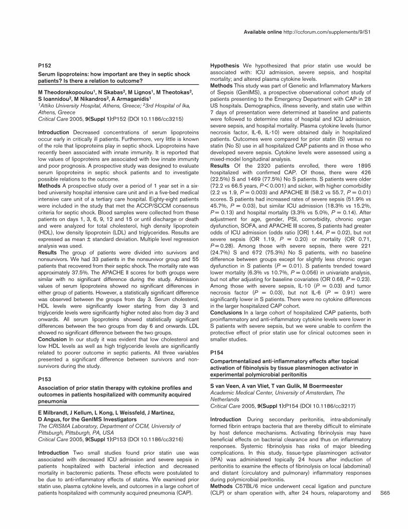
S65
P152Serum lipoproteins: how important are they in septic shockpatients? Is there a relation to outcome?
M Theodorakopoulou1, N Skabas2, M Lignos1, M Theotokas2, S Ioannidou2, M Nikandros2, A Armaganidis1
1Attiko University Hospital, Athens, Greece; 23rd Hospital of Ika,Athens, GreeceCritical Care 2005, 9(Suppl 1):P152 (DOI 10.1186/cc3215)
Introduction Decreased concentrations of serum lipoproteinsoccur early in critically ill patients. Furthermore, very little is knownof the role that lipoproteins play in septic shock. Lipoproteins haverecently been associated with innate immunity. It is reported thatlow values of lipoproteins are associated with low innate immunityand poor prognosis. A prospective study was designed to evaluateserum lipoproteins in septic shock patients and to investigatepossible relations to the outcome.Methods A prospective study over a period of 1 year set in a six-bed university hospital intensive care unit and in a five-bed medicalintensive care unit of a tertiary care hospital. Eighty-eight patientswere included in the study that met the ACCP/SCCM consensuscriteria for septic shock. Blood samples were collected from thesepatients on days 1, 3, 6, 9, 12 and 15 or until discharge or deathand were analyzed for total cholesterol, high density lipoprotein(HDL), low density lipoprotein (LDL) and triglycerides. Results areexpressed as mean ± standard deviation. Multiple level regressionanalysis was used.Results The group of patients were divided into survivors andnonsurvivors. We had 33 patients in the nonsurvivor group and 55patients that recovered from their septic shock. The mortality rate wasapproximately 37.5%. The APACHE II scores for both groups weresimilar with no significant difference during the study. Admissionvalues of serum lipoproteins showed no significant differences ineither group of patients. However, a statistically significant differencewas observed between the groups from day 3. Serum cholesterol,HDL levels were significantly lower starting from day 3 andtriglyceride levels were significantly higher noted also from day 3 andonwards. All serum lipoproteins showed statistically significantdifferences between the two groups from day 6 and onwards. LDLshowed no significant difference between the two groups.Conclusion In our study it was evident that low cholesterol andlow HDL levels as well as high triglyceride levels are significantlyrelated to poorer outcome in septic patients. All three variablespresented a significant difference between survivors and non-survivors during the study.
P153Association of prior statin therapy with cytokine profiles andoutcomes in patients hospitalized with community acquiredpneumonia
E Milbrandt, J Kellum, L Kong, L Weissfeld, J Martinez, D Angus, for the GenIMS InvestigatorsThe CRISMA Laboratory, Department of CCM, University ofPittsburgh, Pittsburgh, PA, USACritical Care 2005, 9(Suppl 1):P153 (DOI 10.1186/cc3216)
Introduction Two small studies found prior statin use wasassociated with decreased ICU admission and severe sepsis inpatients hospitalized with bacterial infection and decreasedmortality in bacteremic patients. These effects were postulated tobe due to anti-inflammatory effects of statins. We examined priorstatin use, plasma cytokine levels, and outcomes in a large cohort ofpatients hospitalized with community acquired pneumonia (CAP).
Hypothesis We hypothesized that prior statin use would beassociated with: ICU admission, severe sepsis, and hospitalmortality; and altered plasma cytokine levels.Methods This study was part of Genetic and Inflammatory Markersof Sepsis (GenIMS), a prospective observational cohort study ofpatients presenting to the Emergency Department with CAP in 28US hospitals. Demographics, illness severity, and statin use within7 days of presentation were determined at baseline and patientswere followed to determine rates of hospital and ICU admission,severe sepsis, and hospital mortality. Plasma cytokine levels (tumornecrosis factor, IL-6, IL-10) were obtained daily in hospitalizedpatients. Outcomes were compared for prior statin (S) versus nostatin (No S) use in all hospitalized CAP patients and in those whodeveloped severe sepsis. Cytokine levels were assessed using amixed-model longitudinal analysis.Results Of the 2320 patients enrolled, there were 1895hospitalized with confirmed CAP. Of those, there were 426(22.5%) S and 1469 (77.5%) No S patients. S patients were older(72.2 vs 66.5 years, P < 0.001) and sicker, with higher comorbidity(2.2 vs 1.9, P = 0.003) and APACHE III (58.2 vs 55.7, P = 0.01)scores. S patients had increased rates of severe sepsis (51.9% vs45.7%, P = 0.03), but similar ICU admission (18.3% vs 15.2%,P = 0.13) and hospital mortality (3.3% vs 5.0%, P = 0.14). Afteradjustment for age, gender, PSI, comorbidity, chronic organdysfunction, SOFA, and APACHE III scores, S patients had greaterodds of ICU admission (odds ratio [OR] 1.44, P = 0.02), but notsevere sepsis (OR 1.19, P = 0.20) or mortality (OR 0.71,P = 0.28). Among those with severe sepsis, there were 221(24.7%) S and 672 (75.3%) No S patients, with no baselinedifference between groups except for slightly less chronic organdysfunction in S patients (P = 0.01). S patients trended towardlower mortality (6.3% vs 10.7%, P = 0.056) in univariate analysis,but not after adjusting for baseline covariates (OR 0.68, P = 0.23).Among those with severe sepsis, IL-10 (P = 0.03) and tumornecrosis factor (P = 0.03), but not IL-6 (P = 0.91) weresignificantly lower in S patients. There were no cytokine differencesin the larger hospitalized CAP cohort.Conclusions In a large cohort of hospitalized CAP patients, bothproinflammatory and anti-inflammatory cytokine levels were lower inS patients with severe sepsis, but we were unable to confirm theprotective effect of prior statin use for clinical outcomes seen insmaller studies.
P154Compartmentalized anti-inflammatory effects after topicalactivation of fibinolysis by tissue plasminogen activator inexperimental polymicrobial peritonitis
S van Veen, A van Vliet, T van Gulik, M BoermeesterAcademic Medical Center, University of Amsterdam, TheNetherlandsCritical Care 2005, 9(Suppl 1):P154 (DOI 10.1186/cc3217)
Introduction During secondary peritonitis, intra-abdominallyformed fibrin entraps bacteria that are thereby difficult to eliminateby host defence mechanisms. Activating fibrinolysis may havebeneficial effects on bacterial clearance and thus on inflammatoryresponses. Systemic fibrinolysis has risks of major bleedingcomplications. In this study, tissue-type plasminogen activator(tPA) was administered topically 24 hours after induction ofperitonitis to examine the effects of fibrinolysis on local (abdominal)and distant (circulatory and pulmonary) inflammatory responsesduring polymicrobial peritonitis.Methods C57BL/6 mice underwent cecal ligation and puncture(CLP) or sham operation with, after 24 hours, relaparotomy and
Available online http://ccforum.com/supplements/9/S1
S66
therapeutic peritoneal lavage with either saline or tPA (0.5 mg/ml).Inflammatory response, coagulation and fibrinolysis were assessedin peritoneal fluid, systemic circulation and lung compartments.Results CLP increased bacterial load, neutrophil influx and activity,coagulation and fibrinolysis compared with sham (all parametersP < 0.01). Compared with saline lavage, tPA lavage reduced intra-abdominal bacterial load, KC concentration and thrombin–anti-thrombin complexes (all P < 0.05), and also reduced intra-abdominal as well as pulmonary neutrophil influx and activity, andclotting times (all P < 0.05). Mediator levels of fibrinolysis were notsignificantly altered by tPA; histology showed less fibrindeposititions concomitant with reduced peritoneal, liver and lungdamage after tPA lavage (P < 0.05). Mortality decreased from 50%to 35% (P = 0.3). Different kinetics were seen when comparingabdominal and pulmonary compartments.Conclusions In this model of polymicrobial peritonitis, abdominalactivation of fibrinolysis by tPA lavage effectively reduced intra-abdominal bacterial load, thereby reducing inflammatory responsesin abdominal and extra-abdominal compartments and remote fibrindeposits and tissue damage. Different kinetics between peritoneal,systemic and pulmonary compartments suggested compartment-alised responses.
P155Endotoxin enhances the fibrinolytic activity of monocytesincorporated in a fibrin clot
A Grosjean1, K Zouaoui Boudjeltia1, M Piagnerelli2, M Vanhaeverbeek1
1ULB, Montigny-le-Tilleul, Belgium; 2ULB — Erasme Hospital,Brussels, BelgiumCritical Care 2005, 9(Suppl 1):P155 (DOI 10.1186/cc3218)
Background In sepsis, one of the most important problems is themodification of the hemostasis balance with activation of thecoagulation. This is due, in part, to increased expression of tissuefactor at the monocyte surface [1]. In these patients, we alsoobserve a systemic hypofibrinolysis [2]. However, it is known thatleukocytes have a profibrinolytic potential [3].Aim To explore the role of monocytes in the fibrinolytic balance.Materials and methods THP-1, a monocytoïd cell line, waschallenged or not with 10 µg/ml lipopolysaccharide during 4 hours,before being introduced in an euglobulin clot lysis time assay,measured by a new semi-automatic method [4]. Resting cells wereintroduced in the assay at concentrations ranging from 0.25 × 10⁶
cells/ml to 106 cells/ml and stimulated cells at the concentration of106 cells/ml. Control without cells is the baseline. Statistics usedanalysis of variance.Results First, we observed a gradual diminution of lysis time withresting cells, from 12% to 25% of the baseline, according to theconcentration of cells (n = 6; P < 0.001). Then, when we addedstimulated THP-1, we observed a greater diminution of lysis time:31% of the baseline (n = 8; P < 0.001 from the baseline andP < 0.05 from resting cells).Conclusion Monocytes play a role in the fibrinolytic balance andthis role is enhanced when they are activated by lipopoly-saccharide, like in sepsis. In the future, we will have to investigatewhy monoyctes are responsible for such an antagonism, stimu-lating coagulation and fibrinolytic processes, at the same time.References1. Gregory SA, Morrissey JH, Edgington TS: Regulation of tissue
factor gene expression in the monocyte procoagulantresponse to endotoxin. Mol. Cell Biol 1989, 9:2752-2755.
2. van Deventer SJH, Büller HR, Ten Cate JW, Aarden LA, Hack CE,Sturk A: Experimental endotoxemia in humans: analysis ofcytokine release and coagulation, fibrinolytic and complement
pathways. Blood 1990, 76:2520-2526.3. Rulot H: Intervention des leucocytes dans l’autolyse de la
fibrine. Arch Int Physiol 1904, 1:152-158.4. Zouaoui Boudjeltia K, Cauchie P, Remacle C, Guillaume M,
Brohee D, Hubert JL, Vanhaeverbeek M: A new device for mea-surement of fibrin clot lysis: application to the euglobulin clotlysis time. BMC Biotechnol 2002, 2:8.
P156Is fibrinolysis a predictive marker of multiple organ failure incritically ill patients?
Y Bouckaert1, K Zouaoui Boudjeltia2, M Piagnerelli1, J Ducobu3, M Vanhaeverbeek2
1Hôpital Erasme, Bruxelles, Belgium; 2A Vésale Hospital, Montigny-le-Tilleul, Belgium; 3CHU Tivoli, La Louvière, BelgiumCritical Care 2005, 9(Suppl 1):P156 (DOI 10.1186/cc3219)
Background C-reactive protein (CRP) is correlated with the risk ofmultiple organ failure (MOF) and death in intensive care patients[1]. CRP inhibits fibrinolysis by inducing plasminogen activatortype 1 release by endothelial cells [2]. We observed a link betweenCRP and fibrinolysis at ICU admission [3].Objective We aimed to evaluate the relationship between CRPand fibrinolysis and the potential predictive value of fibrinolysis atadmission in the ICU on the development of MOF.Methods Thirty-eight patients were enrolled. The plasmafibrinolytic capacity was assessed by the euglobulin clot lysis time(ECLT) determined by a semi-automatic method [4]. The ECLT,CRP and the SOFA score were measured on admission (time 1)and between the third and the fifth day (time 2) of the ICU stay.Results The ECLT was higher in septic than nonseptic patients(970 ± 928 min versus 503 ± 304 min [P = 0.042]). Platelet countremained stable between the two times. We found a correlationbetween ECLT on admission and SOFA score at time 2 (r = 0.67;P < 0.001). There was no link between ECLT and the mortality rate(P = 0.68). CRP was correlated with ECLT during the ICU stay(time 1: r = 0.74, P < 0.001; time 2: r = 0.60, P < 0.001).Conclusion The ECLT at admission could be a good predictivemarker for the development of MOF between the third and the fifthday of the ICU stay.A correlation exists between CRP and the ECLT during all the ICUstay. This finding is compatible with a link between inflammationand fibrinolysis and may explain the negative prognostic value ofhigh concentrations of CRP in critically ill patients.References1. Lobo SMA, et al.: Chest 2003, 123:2043-2049.2. Devaraj S, et al.: Circulation 2003, 107:398-404.3. Zouaoui Boudjeltia K, et al.: Thromb J 2004, 2:7.4. Zouaoui Boudjeltia K, et al.: BMC Biotechnol 2002, 2:8.
P157A proteomic study of sepsis
A Soares, M Santos, M Junqueira, C David, G DomontHFAG, Rio de Janeiro, BrazilCritical Care 2005, 9(Suppl 1):P157 (DOI 10.1186/cc3220)
Introduction Sepsis, characterized by intense reaction of theorganism, due to infection, particularly of the inflammatory andcoagulation systems, is a common illness, of high cost treatmentand deaths. Thus, new technologies for the detection of sepsisearly-stages are urgently needed. Our objective was to identifyserum proteomic patterns that would distinguish sepsis patientsfrom healthy controls.Materials and methods Six ICU patients that matched the criteria(ACCP/SCCM) for sepsis diagnosis were selected: male/female,
Critical Care March 2005 Vol 9 Suppl 1 25th International Symposium on Intensive Care and Emergency Medicine

S67
3/3; mean/range age of 81.1/68–89 years; mean LOS_ICU of15 ± 25 days; 28-day mortality (dead/total/%) = 4/6/66%; meanAPACHE II score of 22 (20–26); and mean SOFA = 6 (5–8); VAP(Pseudomonas aeruginosa, three patients) and CRBSI Staphy-lococcus aureus, Staphylococcus epidermidis, Candida albicans(one each). Serum samples were submitted to isoelectric focusingof proteins in the first dimension with a nonlinear strip of pH 3–10and in the second to 12.5% 2D-SDS-PAGE. Gels were stainedwith Coomassie Blue and the final image analyzed using the ImageMaster Platinum software (Amersham). Selected spots were cut,washed, dried, swollen with trypsin solution and incubated for16 hours at 37°C. The peptides were extracted, concentrated andanalyzed by mass spectrometry in a MALDI-TOF Voyageur DE-PROinstrument (ABI). Peptide maps were processed (MS-Fit, ProteinProspector) for protein identification in the NCBI Data Bank.Results Image analyses of the gels showed the presence of spotsonly in patients with sepsis (Fig. 1, arrows) and the presence ofother spots with different intensities in sepsis and controls. Peptidemass fingerprinting of some of these spots identified actin, zinc-finger protein, matrin 3, haptoglobin-2 and apolipoprotein A1.Discussion Actin, a major component of the cytoskeleton, is foundin cellular injury as in apoptosis; zinc-finger protein is expressed incellular injury as a cytoprotective protein with anti-apoptosisactivity; matrin 3 functions in transcription and interacts with otherproteins of the cellular matrix; apolipoprotein A1 is capable ofdirectly inactivating endotoxin (protein–endotoxin interaction);haptoglobin-2 is present during the acute phase reaction. Thesefindings justify the use of proteomic techniques as a screening toolfor the study of all stages of sepsis in high-risk patients.
P158Circulating levels of the long pentraxin PTX3, an acute phasemediator of innate immunity, in acute lung injury/acute respiratorydistress syndrome: functional and prognostic correlations
T Mauri1, M Bombino1, E Bertoli1, A Doni2, A Mantovani2, A Pesenti11University of Milan-Bicocca, Hospital St Gerardo, Monza,Giussano, Italy; 2University of Milan, Istituto Mario Negri, Milan, ItalyCritical Care 2005, 9(Suppl 1):P158 (DOI 10.1186/cc3221)
Acute lung injury/acute respiratory distress syndrome (ALI/ARDS)is characterized by an important inflammatory reaction involving
both lungs, often spreading systemically. Pentraxin 3 (PTX3) hasrecently been described as a mediator of the acute phase of innateimmunity. It belongs, together with C-reactive protein (CRP), to thepentraxin superfamily. PTX3 is produced by endothelial cells,fibroblasts, mononuclear phagocytes and dendritic cells inresponse to primary inflammatory signals (IL-1, tumor necrosisfactor, LPS). Its involvement in the pathogenesis of ALI/ARDSseems probable and potentially interesting for the pathophysiology,the diagnosis and possibly the therapy of the syndrome.We measured PTX3 levels, by an ELISA test, in the sera of 25patients affected by ALI/ARDS. We also collected data related toetiology, pulmonary mechanics, gas exchange, systemicinflammatory involvement, microbiology, blood cells and theoutcome of these patients.PTX3 was markedly elevated in all 25 patients (average111.07 ± 192.01 ng/ml; range 4.94–922.91 ng/ml). In no patientwere PTX3 levels lower or equal to the physiological threshold of2 ng/ml.Levels of logPTX3 were significantly different between survivor andnonsurvivor patients (1.465 ± 0.529 log ng/ml vs 1.950 ± 0.643log ng/ml; P = 0.05).Patients with two or more organ failures (besides the lung) alsohad logPTX3 levels significantly higher than patients with just oneadditional organ failure (1.936 ± 0.597 log ng/ml vs1.423 ± 0.542 log ng/ml; P < 0.04). The same was true whenPTX3 levels in patients with septic shock were compared withPTX3 levels in patients without shock (262.465 ± 350.727 ng/mlvs 63.257 ± 70.261 ng/ml; P < 0.03).PTX3 and logPTX3 were correlated with body temperature(r = 0.56 P < 0.01; r = 0.51 P = 0.01) and with arterial blood pH(r = –0.53 P < 0.01; r = –0.53 P < 0.01).We have underlined a relationship between circulating levels ofPTX3 and some markers of the severity of systemic involvement.PTX3 could be an indicator of the gravity of the systemicinflammatory reaction and of unfavourable outcome in ALI/ARDSpatients.
P159Soluble haemoglobin scavenger receptor (sCD163): a markerassociated with bacteraemia
S Gaïni1, S Pedersen1, S Moestrup2, C Pedersen1, H Møller3
1Odense University Hospital, Odense, Denmark; 2University ofAarhus, Denmark; 3Aarhus University Hospital, Aarhus, DenmarkCritical Care 2005, 9(Suppl 1):P159 (DOI 10.1186/cc3222)
Objective Soluble haemoglobin scavenger receptor (sCD163) is amacrophage-specific marker, and elevated levels have beenobserved in patients with pneumonia and sepsis. The objective ofour study was to evaluate sCD163 as a diagnostic and prognosticmarker in patients with community acquired sepsis.Design A prospective observational study.Methods One hundred and ninety-four adult patients admitted to aDepartment of Internal Medicine at a tertiary hospital withsuspected community acquired infections. Daily blood sampling forsCD163 analysis was performed for up to 5 days. Laboratoryanalyses were performed with a sandwich ELISA using polyclonalrabbit anti-CD163 as the catching antibody and monoclonal anti-CD163 as the secondary antibody. The patients were classifiedaccording to SIRS criteria. The patients were divided in thefollowing groups: patients with no proven infection (n = 67, controlgroup), patients with possible infection (n = 21, not included inanalysis), patients with proven infection without SIRS (n = 25),patients with sepsis (n = 52), patients with severe sepsis (n = 29).Only one patient had septic shock. Twelve patients had
Available online http://ccforum.com/supplements/9/S1
Figure 1 (abstract P157)
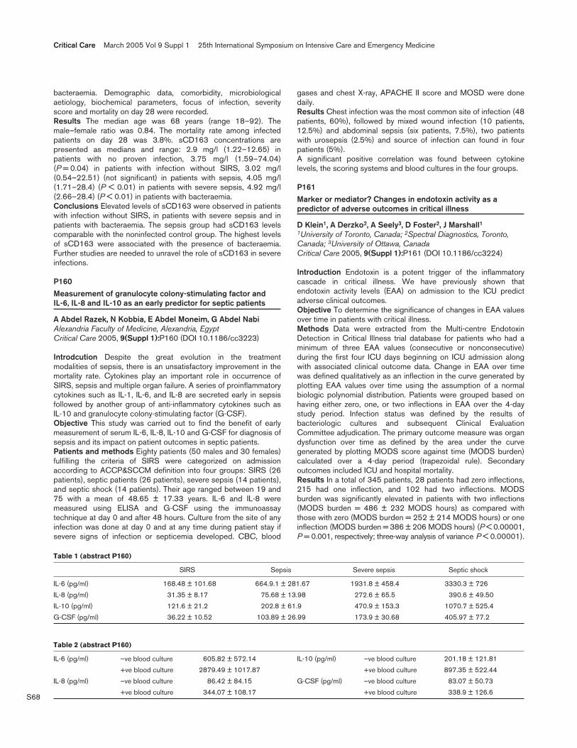
S68
bacteraemia. Demographic data, comorbidity, microbiologicalaetiology, biochemical parameters, focus of infection, severityscore and mortality on day 28 were recorded.Results The median age was 68 years (range 18–92). Themale–female ratio was 0.84. The mortality rate among infectedpatients on day 28 was 3.8%. sCD163 concentrations arepresented as medians and range: 2.9 mg/l (1.22–12.65) inpatients with no proven infection, 3.75 mg/l (1.59–74.04)(P = 0.04) in patients with infection without SIRS, 3.02 mg/l(0.54–22.51) (not significant) in patients with sepsis, 4.05 mg/l(1.71–28.4) (P < 0.01) in patients with severe sepsis, 4.92 mg/l(2.66–28.4) (P < 0.01) in patients with bacteraemia.Conclusions Elevated levels of sCD163 were observed in patientswith infection without SIRS, in patients with severe sepsis and inpatients with bacteraemia. The sepsis group had sCD163 levelscomparable with the noninfected control group. The highest levelsof sCD163 were associated with the presence of bacteraemia.Further studies are needed to unravel the role of sCD163 in severeinfections.
P160Measurement of granulocyte colony-stimulating factor andIL-6, IL-8 and IL-10 as an early predictor for septic patients
A Abdel Razek, N Kobbia, E Abdel Moneim, G Abdel NabiAlexandria Faculty of Medicine, Alexandria, EgyptCritical Care 2005, 9(Suppl 1):P160 (DOI 10.1186/cc3223)
Introdcution Despite the great evolution in the treatmentmodalities of sepsis, there is an unsatisfactory improvement in themortality rate. Cytokines play an important role in occurrence ofSIRS, sepsis and multiple organ failure. A series of proinflammatorycytokines such as IL-1, IL-6, and IL-8 are secreted early in sepsisfollowed by another group of anti-inflammatory cytokines such asIL-10 and granulocyte colony-stimulating factor (G-CSF).Objective This study was carried out to find the benefit of earlymeasurement of serum IL-6, IL-8, IL-10 and G-CSF for diagnosis ofsepsis and its impact on patient outcomes in septic patients.Patients and methods Eighty patients (50 males and 30 females)fulfilling the criteria of SIRS were categorized on admissionaccording to ACCP&SCCM definition into four groups: SIRS (26patients), septic patients (26 patients), severe sepsis (14 patients),and septic shock (14 patients). Their age ranged between 19 and75 with a mean of 48.65 ± 17.33 years. IL-6 and IL-8 weremeasured using ELISA and G-CSF using the immunoassaytechnique at day 0 and after 48 hours. Culture from the site of anyinfection was done at day 0 and at any time during patient stay ifsevere signs of infection or septicemia developed. CBC, blood
gases and chest X-ray, APACHE II score and MOSD were donedaily.Results Chest infection was the most common site of infection (48patients, 60%), followed by mixed wound infection (10 patients,12.5%) and abdominal sepsis (six patients, 7.5%), two patientswith urosepsis (2.5%) and source of infection can found in fourpatients (5%).A significant positive correlation was found between cytokinelevels, the scoring systems and blood cultures in the four groups.
P161Marker or mediator? Changes in endotoxin activity as apredictor of adverse outcomes in critical illness
D Klein1, A Derzko2, A Seely3, D Foster2, J Marshall11University of Toronto, Canada; 2Spectral Diagnostics, Toronto,Canada; 3University of Ottawa, CanadaCritical Care 2005, 9(Suppl 1):P161 (DOI 10.1186/cc3224)
Introduction Endotoxin is a potent trigger of the inflammatorycascade in critical illness. We have previously shown thatendotoxin activity levels (EAA) on admission to the ICU predictadverse clinical outcomes.Objective To determine the significance of changes in EAA valuesover time in patients with critical illness.Methods Data were extracted from the Multi-centre EndotoxinDetection in Critical Illness trial database for patients who had aminimum of three EAA values (consecutive or nonconsecutive)during the first four ICU days beginning on ICU admission alongwith associated clinical outcome data. Change in EAA over timewas defined qualitatively as an inflection in the curve generated byplotting EAA values over time using the assumption of a normalbiologic polynomial distribution. Patients were grouped based onhaving either zero, one, or two inflections in EAA over the 4-daystudy period. Infection status was defined by the results ofbacteriologic cultures and subsequent Clinical EvaluationCommittee adjudication. The primary outcome measure was organdysfunction over time as defined by the area under the curvegenerated by plotting MODS score against time (MODS burden)calculated over a 4-day period (trapezoidal rule). Secondaryoutcomes included ICU and hospital mortality.Results In a total of 345 patients, 28 patients had zero inflections,215 had one inflection, and 102 had two inflections. MODSburden was significantly elevated in patients with two inflections(MODS burden = 486 ± 232 MODS hours) as compared withthose with zero (MODS burden = 252 ± 214 MODS hours) or oneinflection (MODS burden = 386 ± 206 MODS hours) (P < 0.00001,P = 0.001, respectively; three-way analysis of variance P < 0.00001).
Critical Care March 2005 Vol 9 Suppl 1 25th International Symposium on Intensive Care and Emergency Medicine
Table 1 (abstract P160)
SIRS Sepsis Severe sepsis Septic shock
IL-6 (pg/ml) 168.48 ± 101.68 664.9.1 ± 281.67 1931.8 ± 458.4 3330.3 ± 726
IL-8 (pg/ml) 31.35 ± 8.17 75.68 ± 13.98 272.6 ± 65.5 390.6 ± 49.50
IL-10 (pg/ml) 121.6 ± 21.2 202.8 ± 61.9 470.9 ± 153.3 1070.7 ± 525.4
G-CSF (pg/ml) 36.22 ± 10.52 103.89 ± 26.99 173.9 ± 30.68 405.97 ± 77.2
Table 2 (abstract P160)
IL-6 (pg/ml) –ve blood culture 605.82 ± 572.14 IL-10 (pg/ml) –ve blood culture 201.18 ± 121.81
+ve blood culture 2879.49 ± 1017.87 +ve blood culture 897.35 ± 522.44
IL-8 (pg/ml) –ve blood culture 86.42 ± 84.15 G-CSF (pg/ml) –ve blood culture 83.07 ± 50.73
+ve blood culture 344.07 ± 108.17 +ve blood culture 338.9 ± 126.6

S69
EAA variability was predictive of organ dysfunction (MODSburden) over time (P < 0.00264). MODS burden correlated withboth ICU mortality (P < 0.00001) and hospital mortality(P < 0.0001). EAA variability was found to be independent ofinfection status (P = 0.7915). The number of EAA inflections weremore predictive of outcome than the absolute EAA valuecalculated as the total endotoxin activity load over the 4-day period(AUC, trapezoidal rule) (univariate regressions, P < 0.00001,P = 0.5440, respectively). In a separate study of 15 normal healthyvolunteers, EAA variability was shown to contain zero inflections inall subjects studied.Conclusions These data suggest that increased variability inendotoxin activity is associated with worsening outcomes in thecritically ill. Our assumption of a normal biologic polynomialdistribution of endotoxin activity requires validation with morefrequent EEA sampling. Further studies are warranted to assesswhether the dynamic alteration of endotoxin activity may play animportant role in the ongoing inflammatory response and organdysfunction in the critically ill.
P162Rates of agreement of endotoxin, procalcitonin and lactate inseptic and non-septic critically ill patients
D Foster1, A Romaschin2, A Derzko1, P Walker1
1Spectral Diagnostics, Toronto, Canada; 2University HealthNetwork, Toronto, CanadaCritical Care 2005, 9(Suppl 1):P162 (DOI 10.1186/cc3225)
Introduction In contrast with many acute and severe diseasessuch as myocardial infarction, liver failure and renal failure, severesepsis lacks a specific biomarker. A useful marker of severe sepsiswill guide therapy by identifying which patients have thepathological process of interest.Objective To compare the rates of agreement between PCT andlactate (LAC) assays with endotoxin levels in severely septic andnon-septic critically ill patients.Methods Data were collected from stored frozen plasma samplesfrom a multicenter study evaluating the Endotoxin Activity (EAA)Assay [1]. We evaluated test samples based on the presence ofclinical determination of severe sepsis where n = 17 had elevatedEAA levels and n = 7 had low EAA levels. There were n = 18 fromnon-septic critically ill patients with low EAA values. We thenperformed PCT and lactate assays on the same samples bytechnicians who were blinded to EAA results and clinicaldiagnosis. Overall percent agreement (weighted kappa statistic):EAA vs PCT = 73.81% (0.462), EAA vs lactate = 40.5% (–0.247),lactate vs PCT = 42.9% (–0.183) The EAA correctly identified 17of 24 severely septic patients, thus showing a positive agreementof 70.8%. The PCT and lactate assays correctly identified 15 andsix patients, respectively. The positive agreement with theconsensus definition of severe sepsis was 62.5% for PCT and25% for lactate.
Table 1
PCT < 2.0 PCT > 2.0 LAC < 2.0 LAC > 2.0
EAA < 0.6 19 6 13 13
EAA > 0.6 5 12 12 4
LAC < 2.0 13 13
LAC > 2.0 11 5
Conclusions Both PCT and endotoxin are elevated in patients withsevere sepsis. The reported positive agreement with a clinical
diagnosis of severe sepsis provides evidence of their value as adiagnostic marker. The poor positive agreement between lactatelevels and the presence of severe sepsis suggests this marker mayhave other value such as for prognosis. Further study is required toevaluate each of these markers as a measure of response to therapy.Reference1. Marshall J, et al.: J Infect Dis 2004, 190:527-534.
P163Prevention of postoperative infectious complications withprocalcitonin-controlled early postoperative administration ofIgM-enriched immunoglobulin preparation in high-riskchildren with congenital heart diseases
N Beloborodova, D Popov, K Schatalov, M Zelenikin, G LobatchovaBakoulev Scientific Center for Cardiovascular Surgery, Moscow,RussiaCritical Care 2005, 9(Suppl 1):P163 (DOI 10.1186/cc3226)
Objectives To evaluate the effectiveness of procalcitonin (PCT)-controlled use of IgM-enriched immunoglobulin preparation in high-risk children with congenital heart diseases after cardiac surgerywith cardiopulmonary bypass (CPB).Methods During January–July 2004, 31 consecutive high-riskpediatric patients were enrolled into the study. The age was 23(19–30) months. Severe concomitant extracardiac pathologieswere registered in 22 (71%) of cases; 21 (68%) patients hadpreceding cardiovascular operations. Patients who had PCT bloodplasma levels above 2 ng/ml on the first day after surgery (n = 28)were randomized into two groups. These groups were comparableby severity of initial condition, age and CPB time. IgM-enrichedimmunoglobulin preparation (Pentaglobin; Biotest Pharma GmbH,Germany) was administrated to the patients in the study group(n = 15) in addition to standard treatment (first 3 days aftersurgery, 5 ml/kg each day). Patients in the control group (n = 13)received only standard treatment. PCT blood plasmaconcentrations were measured by immunoluminometric method(PCT LIA; BRAHMS Aktiengesellschaft GmbH, Germany). Thedata are shown as median and 25th and 75th percentiles. The datawere compared by Mann–Whitney U test, P < 0.05 consideredstatistically significant. Postoperative infections rates wereanalyzed in studied groups.Results None of the patients had exhibited any signs of infectionbefore surgery. Patients with PCT blood plasma levels less than2 ng/ml on the first day after surgery (n = 3) had a smoothpostoperative period. PCT levels on the first day after surgery weresignificantly higher in the control group in comparison with thestudy group (7.77 [5.95–10.72] ng/ml and 3.60 [2.98–6.54]ng/ml, respectively; P = 0.009). Postoperatively 1/15 (6.7%) ofpatients in the study group and 5/13 (38.5%) of patients in thecontrol group suffered from infectious complications (study group:urinary tract infection — one patient; control group: pneumonia —two patients, pneumonia and sepsis — one patient, peritonitis withmultiorgan failure — one patient, urinary tract infection — onepatient). The rate of postoperative infectious complications wassignificantly lower in the study group (P = 0.03). Two deaths in thecontrol group occurred due to sepsis (n = 1) and peritonitis withmultiorgan failure (n = 1).Conclusions High PCT levels on the first day after surgery areassociated with high rates of infectious complications. PCTmonitoring allows one to select patients with systemic bacterialinflammation after CPB. Early (on the first day after surgery)administration of IgM-enriched immunoglobulin preparationeffectively prevents infectious complications in these patients.
Available online http://ccforum.com/supplements/9/S1

S70
P164Evaluation of procalcitonin as a diagnostic and prognosticmarker in patients with sepsis, severe sepsis and septic shockassociated with ventilator-associated pneumonia
S Virtzili1, D Zervakis1, A Koronaios1, P Alevizopoulou1, A Flevari1,A Kotanidou1, V Kousoulas2, H Giamarellou2, C Roussos1
1ICU, Medical School, Athens University, Athens, Greece; 24th Department of Internal Medicine, Medical School, AthensUniversity, Athens, GreeceCritical Care 2005, 9(Suppl 1):P164 (DOI 10.1186/cc3227)
Objectives Although the significance of procalcitonin (PCT) hasbeen evaluated for general populations of patients with sepsisattributed to various infections, application as a marker of severitywith both diagnostic and prognostic implications in patients withventilator-associated pneumonia (VAP) has not been specificallystudied.Methods Serum samples were collected on the first, third, fifth andseventh days of symptoms from 68 patients with sepsis, severesepsis/shock (ASCP/SCCM 1992) and VAP. APACHE II andSOFA scores were measured on the first day of the symptoms.Patients were followed-up for 28 days for overall survival. PCT wasestimated in sera of patients by immunochemiluminometric assay.Results Among 68 patients, 34 presented with sepsis, five withsevere sepsis and 29 with septic shock. Mean age (years,± standard deviation) was 57.86 ± 18.85; 48 patients were maleand 20 female. APACHE II and SOFA (mean ± standard deviation)were 18.28 ± 6.11 and 7.64 ± 3.24, respectively. After 28 days offollow-up, 38 patients were alive and 21 have died. Mean PCTconcentrations (ng/ml) on days 1, 3, 5 and 7 in patients withsepsis, severe sepsis and septic shock are presented in Table 1.The mean ± standard error serum concentrations of PCT ofsurvivors at days 1, 3, 5 and 7 were 0.71 ± 0.19, 0.45 ± 0.09,0.35 ± 0.05 and 1.42 ± 1.04, respectively. Respective values innon-survivors were 1.83 ± 0.86 (P = not significant compared withsurvivors), 2.63 ± 1.08 (P = 0.05), 3.61 ± 1.33 (P = notsignificant) and 2.89 ± 0.99 (P = 0.02).
Table 1
Mean procalcitonin concentrations (ng/ml)
Day 1 Day3 Day 5 Day 7
Sepsis 0.73 0.53 0.92 0.50
Severe sepsis 0.61 0.55 0.83 0.99
Septic shock 1.45 1.88 1.93 3.54
Conclusions PCT is a crucial marker of overall survival in patientswith septic syndrome and VAP. This suggestion is furthersupported by the fact that PCT correlates to the severity of diseaseconstituting an objective marker of sepsis.
P165Changes in procalcitonin in septic shock patients treated withendotoxin adsorption therapy
T Ikeda, K Ikeda, M Nagura, H Taniuchi, Y Kuroki, M MatsushitaHachiouji Medical Center, Tokyo Medical University, Tokyo, JapanCritical Care 2005, 9(Suppl 1):P165 (DOI 10.1186/cc3228)
Introduction The endotoxin adsorption method (PMX-DHP: TorayIndustries, Inc., Tokyo, Japan) is used for treatment of patients withsepsis and septic shock primarily caused by Gram-negativeinfections in Japan. Procalcitonin (PCT) levels may be a good
marker of infection and levels exceeding 10 ng/ml occur almostexclusively in severe sepsis. The aim of this study was to evaluatePMX-DHP for severe sepsis or septic shock patients according toPCT values. Patients were classified as a group in which PCT ishigher than 10 ng/ml (H group) or a group in which PCT is lowerthan 10 ng/ml (L group).Patients and methods This mode of blood purification isprincipally applied on direct hemoperfusion therapy. Sixty-sevenpatients (40 in H group, 27 in L group) were treated with PMX-DHP. Sepsis was diagnosed according to the criteria of theAmerican College of Chest Physicians/Society of Critical CareMedicine Consensus Conference Committee. The followingparameters (tumor necrosis factor alpha, IL-6, IL-8, IL-1ra, ICAM-1,PAI-1 and PCT) were measured just before and immediately afterPMX-DHP. PCT was measured by immunoluminometric assay(LUMI test PCT; BRAHMS Diagnostica, Berlin, Germany).Results APACHE II score in the H group was 26.7 ± 7.6 and inthe L group was 24.7 ± 9.2, respectively. SOFA score in the Hgroup was 11.2 ± 3.8 and in the L group was 11.3 ± 4.2. The 28-day all-cause mortality rate in the H group was 30% and was 33%in the L group. These were no significant differences between thegroups. IL-6, IL-8, and IL-1ra in H group were remarkably higherthan in the L group. (Median IL-6, IL-8, and IL-1ra in H group were7580 pg/ml, 646 pg/ml, 55,000 pg/ml, respectively, and in Lgroup were 462 pg/ml, 40 pg/ml, 3650 pg/ml, respectively.) PCTin the H group showed a tendency to decline from 108 ± 120 ng/mlto 94 ± 98 ng/ml before and after PMX-DHP. Especially, PCT ofthe survival group in H group showed a significant decrease from119 ± 138 ng/ml to 93 ± 102 ng/ml (P < 0.02). On the otherhand, PCT of the nonsurvival group in H group increased from83 ± 58 ng/ml to 96 ± 91 ng/ml.Discussion Serum PCT values were less than 0.1 ng/ml in healthyindividuals, but markedly increased, mostly due to extra-thyroidproduction in cases of severe infection. Recent findings suggestthat sources of PCT may include liver cells and monocytes/macrophages. PCT is consistently increased after endotoxininjection, suggesting an association of endotoxin with septic shockand high PCT serum concentration.Conclusion Our results may suggest that PMX-DHP can reducevarious cytokines and serum PCT in the survival group.
P166Procalcitonin guidance significantly reduces antibiotic durationin community-acquired pneumonia: the ‘ProCAP’ study
M Christ-Crain, D Stolz, R Bingisser, C Mueller, J Leuppi, M Battegay, P Huber, M Tamm, B MuellerUniversity Hospital, Basel, SwitzerlandCritical Care 2005, 9(Suppl 1):P166 (DOI 10.1186/cc3229)
Background Community-acquired pneumonia (CAP) is routinelytreated with antibiotic courses of 10–14 days. Procalcitonin(ProCT) is elevated in systemic bacterial infections, but remainslow in viral infections. Its potential as a tool to guide ABprescription in lower respiratory tract infections has been shown[1]. This study aims to evaluate ProCT stewardship to reduce ABduration in patients with CAP.Methods From a prospective, single-blinded, randomized study,data of 200 patients with CAP were analyzed. The standard group(n = 101) received antibiotics as generally recommended. In theProCT group (n = 99), ProCT levels were measured at days 4, 6and 8 and antibiotic therapy was discontinued if levels were< 0.25 µg/l, measured with a sensitive assay (Kryptor PCT,BRAHMS, Germany). At baseline and at follow-up at 6 weeks, astandardized work-up was performed.
Critical Care March 2005 Vol 9 Suppl 1 25th International Symposium on Intensive Care and Emergency Medicine
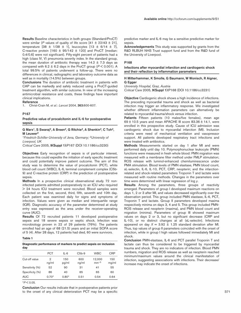
S71
Results Baseline characteristics in both groups (Standard/ProCT)were similar (P values of quality of life score [41 ± 22/43 ± 21],temperature [38 ± 1/38 ± 1], leucocytes [13 ± 6/14 ± 7],C-reactive protein [160 ± 95/142 ± 120] and ProCT [median:0.4/0.6] were not significant). Fifty-eight percent of patients had ahigh (class IV, V) pneumonia severity index. In the standard group,the mean duration of antibiotic therapy was 14.2 ± 7.3 days ascompared with 6.2 ± 6.2 days in the ProCT group (P < 0.001). Atotal 99.5% of patients underwent a follow-up. There were nodifferences in clinical, radiographic and laboratory outcome data aswell as in mortality (14.5%) between groups.Conclusions The duration of antibiotic treatment in patients withCAP can be markedly and safely reduced using a ProCT-guidedtreatment algorithm, with similar outcome. In view of the increasingantimicrobial resistance and costs, these findings have importantclinical implications.Reference1. Christ-Crain M, et al.: Lancet 2004, 363:600-607.
P167Predictive value of procalcitonin and IL-6 for postoperativesepsis
G Marx1, S Swaraj2, A Breen2, G Ritchie2, A Shenkin2, C Toh2,M Leuwer2
1Friedrich-Schiller-University of Jena, Germany; 2University ofLiverpool, UKCritical Care 2005, 9(Suppl 1):P167 (DOI 10.1186/cc3230)
Objectives Early recognition of sepsis is of particular interest,because this could expedite the initiation of early specific treatmentand could potentially improve patient outcome. The aim of thisstudy was to determine the value of procalcitonin (PCT), whiteblood cell count (WBC), IL-6, terminal complement complex (C5b-9) and C-reactive protein (CRP) in the prediction of postoperativesepsis.Methods In a prospective clinical observational study 72 non-infected patients admitted postoperatively to an ICU who required> 24 hours ICU treatment were recruited. Blood samples werecollected on the first, second, third, fifth, seventh and 10th day.Each patient was examined daily for signs and symptoms ofinfection. Values were given as median and interquartile range(IQR). Diagnostic accuracy of the parameter determined at studyentry was expressed as the area under the receiver-operatingcurve (AUC).Results Of 72 recruited patients 11 developed postoperativesepsis and 18 severe sepsis or septic shock. Infection wasmicrobiology proven in 22 of 29 patients (76%). The patientsenrolled had an age of 68 (21.5) years and an initial SOFA scoreof 5 (4). After 28 days, 12 patients had died, 60 were survivors.
Table 1
Diagnostic performance of markers to predict sepsis on inclusionday
PCT IL-6 C5b-9 WBC CRP
Cut-off value 2 150 600 12,000 150 ng/ml pg/ml ng/ml mm–3 mg/ml
Sensitivity (%) 52 90 31 41 55
Specificity (%) 86 40 65 65 60
AUC 0.70* 0.80* 0.51 0.54 0.64
*P < 0.05.
Conclusion Our results indicate that in postoperative patients priorto the onset of any clinical deterioration PCT may be a specific
predictive marker and IL-6 may be a sensitive predictive marker forsepsis.Acknowledgements This study was supported by grants from theR&D RLBUH NHS Trust support fund and from the R&D fund ofthe University of Liverpool.
P168Infections after myocardial infarction and cardiogenic shockand their reflection by inflammation parameters
H Mitterhammer, K Smolle, G Baumann, W Wonisch, R Aigner,G EggerUniversity Hospital, Graz, AustriaCritical Care 2005, 9(Suppl 1):P168 (DOI 10.1186/cc3231)
Objective Cardiogenic shock shows a high incidence of infections.The preceding myocardial trauma and shock as well as bacterialinfection may trigger an inflammatory response. We investigatedwhether different inflammation parameters can alternatively berelated to myocardial trauma/shock versus infection.Patients Fifteen patients (10 males/five females), mean age69 ± 10.5 years and mean APACHE III score 83.36 ± 14.1, wereenrolled in this prospective study. Cause of ICU admission wascardiogenic shock due to myocardial infarction (MI). Inclusioncriteria were need of mechanical ventilation and vasopressorsupport. All patients developed respiratory tract infections andwere treated with antibiotics.Methods Measurements started on day 1 after MI and wereperformed daily until day 10. Polymorphonuclear leukocyte (PMN)functions were measured in fresh whole blood: PMN migration wasmeasured with a membrane filter method under FMLP stimulation;ROS release with luminol-enhanced chemiluminescence underPMA stimulation. Blood levels of PMN elastase, PMN blood count,sL-selectin, IL-6, IL-10, PCT, CRP, neopterin; and the infarction-related and shock-related parameters Troponin T and lactate weremeasured with routine methods. Changes in the parameters overtime were determined with linear regression of log y.Results Among the parameters, three groups of reactivityemerged. Parameters of group I developed maximum reactions ondays 1, 2 or 3 after MI, and values decreased significantly over theobservation period. This group included PMN elastase, IL-6, PCT,Troponin T and lactate. Group II parameters developed maximarespectively minima on days 3, 4 and 5. This group included PMN-ROS release and neopterin (maxima), and PMN blood count andmigration (minima). Parameters of group III showed maximumvalues on days 2 or 3, but no significant decrease (CRP andIL-10), or no distinct changes at all (sL-selectin). Infectionsappeared on day X = 3.92 ± 1.25 standard deviation after MI.Thus, top values of group II parameters coincided with the onset ofinfection, while in group I high values followed immediately MI andshock.Conclusion PMN-elastase, IL-6 and PCT parallel Troponin T andlactate can thus be considered to be triggered by myocardialtrauma and shock. They are no indicators of infection. Blood PMNnumbers, migration and ROS release as well as neopterin reachedminimum/maximum values around the clinical manifestation ofinfection, suggesting associations with infections. Their decrease/increase may indicate the onset of infections.
Available online http://ccforum.com/supplements/9/S1

S72
P169Does cardiopulmonary bypass affect the procalcitonin level?
F Kalay, Y Bicer, S Simsek, N Yapici, Z Alicikus, G Golcuklu, Z AykacSiyami Ersek Thoracic and Cardiovascular Surgery Center,Istanbul, TurkeyCritical Care 2005, 9(Suppl 1):P169 (DOI 10.1186/cc3232)
Introduction Cardiopulmonary bypass (CPB) causes aninflammatory response with clinical and biological changes. Thissystemic inflammatory response syndrome (SIRS) is a result ofseveral stimuli such as exposure of blood to non-physiologicalsurfaces, surgical trauma, myocardial ischaemia-reperfusion andendotoxin release. Because of this response, conventional clinicaland biological signs may be misled in the diagnosis ofpostoperative complications, particularly infection. Procalcitonin(PCT) is useful as a marker of infection. Further studies suggestedthat PCT is an early, sensitive and specific indicator of infection butrecent investigations showed increases in other conditionsassociated with systemic inflammation, such as severe trauma,burns and heat stroke. The aim of this study was to determine thenormal profile of PCT during preoperative, peroperative andpostoperative (PO) periods in patients undergoing cardiac surgerywith cardiopulmonary bypass (CPB).Methods We have measured serum PCT levels in 91 patientsundergoing elective cardiac surgery with CPB. PCT levels weremeasured after induction of anaesthesia (baseline), after CPB andPO days 1, 2, 3 and 4. The demographic data of all patients, cross-clamping and bypass times, inotropic medication or intra-aorticballoon pump (IABP) needs were recorded. Repeated-measuresanalysis of variance was used for statistical analyses.Results Baseline PCT levels were measured before the operation(< 0.05 ng/dl) and all increased at the end of the CPB. Theincrease in serum PCT levels had a peak on the first postoperativeday (P < 0.001) (Fig. 1). PCT levels were higher in some patientswhose cross-clamping and total bypass times were longer thanothers or they needed inotropic agents or IABP. Repeated variantanalyses showed that there was a closed relationship between the
PCT levels and cross-clamp and total bypass times, need ofinotropic medications and IABP (Fig. 2).Conclusion Although PCT is a useful marker of infection, it can beaffected from many factors (CPB itself, prolonged operation, cross-clamping and bypass time, inotropic agent and IABP need, etc.)during CPB. So PCT alone is not enough to decide for infection inthe early postoperative period in cardiac surgery patients.
P170The prognostic value of procalcitonin in cardiac surgicalpatients
C Iasonidou, G Vasiliadou, F Ampatzidou, N Manolakoglou, V Soulountzi, N Kapravelos2nd Intensive Care Unit, Papanikolaou General Hospital,Thessaloniki, GreeceCritical Care 2005, 9(Suppl 1):P170 (DOI 10.1186/cc3233)
Introduction The aim of this prospective study was to determinethe prognostic value of serum levels of procalcitonin (PCT) inpatients early after cardiac surgery using cardiopulmonary bypass(CPB). We studied whether PCT is useful as an early prognosticmarker in cardiosurgical patients with respect to complications andinfections and whether PCT is a specific marker for the occurrenceof infections.Methods We studied 74 patients (mean ± standard deviation age,65 ± 9) after elective cardiac surgery and daily until the secondpostoperative day. Demographic data, operative data and clinicalendpoints (SIRS, infection and severe complication) weredocumented. Complications were defined as circulatory failure,respiratory failure, renal dysfunction, hepatic dysfunction, andcoagulation dysfunction.Results SIRS occurred in 46 patients (62%), infection in fourpatients (5.4%) and non-infectious complication in 35 patients(47.2%). Circulatory failure occurred in 29 patients, respiratoryfailure in 19 patients, renal failure in two patients, hepaticdysfunction in six patients, coagulation dysfunction in eightpatients. Serum PCT concentrations increased in all patients with
Critical Care March 2005 Vol 9 Suppl 1 25th International Symposium on Intensive Care and Emergency Medicine
Figure 1 (abstract P169)
The procalcitonin (PCT) levels and distribution of measurement times:A, baseline; B, after cardiopulmonary bypass; C, postoperative (PO)day 1; D, PO day 2; E, PO day 3; and F, PO day 4.
Figure 2 (abstract P169)
Procalcitonin (PCT) levels in patients with and without intra-aorticballoon pump (IABP).

S73
peak levels occurring on the first postoperative day (PCT1: 0.495standard error of the mean [SEM] 0.068, PCT2: 2.62 SEM 0.42,PCT3: 1.65 SEM 0.23). The serum PCT levels were increased inpatients with SIRS compared with patients with no-SIRS. Usingthe Mann–Whitney U test we found a statistically significantincrease on PCT levels in patients with infection (P = 0.001) and inpatients with non-infectious complications (P = 0.005). PCT wasnot significantly different in patients with infectious complicationsin comparison with patients with non-infectious complications.There was good correlation (Pearson correlation) between PCTlevel and CPB duration, duration of aortic clamping and EUROscore.Conclusion PCT is a valuable prognostic marker early aftercardiac surgery using CPB with respect to infection and severecomplications. However, PCT does not differentiate infectious andnon-infectious complications.
P171The procalcitonin test in the evaluation of clinical severity inpatients with acute pancreatitis
B Gelfand, M Filimonov, S Burnevich, T Brazhnik, N SergeyevaThe State Medical University of Russia, Moscow, RussiaCritical Care 2005, 9(Suppl 1):P171 (DOI 10.1186/cc3234)
Purpose To determine the role of the procalcitonin test in theclinical and laboratory evaluation of the clinical severity and theintensity of the systemic inflammatory reaction in patients withdifferent forms of acute pancreatitis.Design A prospective study.Patients The study covered 50 patients with acute pancreatitis ofvarious aetiologies. Patients were subdivided into five groupsdepending on morphological and microbiological criteria: (1) 10patients with acute interstitial pancreatitis (AIP); (2) 16 non-operated patients with limited sterile pancreatic necrosis (LSN-N);(3) six operated patients with limited necrosis (LSN-O); (4) sixpatients with extensive sterile necrosis (ESN); (5) 12 patients withinfected forms of pancreatic necrosis (IPN).Methods Plasma procalcitonin (PCT) concentrations were deter-mined using an immunoluminometric method. The clinical severityof the patients’ condition was evaluated with the help of theintegral APACHE II scale. Laboratory and instrumental techniquesof investigation were supplemented by calculations of theleucocyte intoxication index.Results The mean PCT concentration in infected forms ofdestructive pancreatitis was markedly higher than in acuteinterstitial pancreatitis or sterile pancreatic necrosis, at a level of2.95 ± 0.84 ng/ml. The PCT concentration rose as the patient’scondition became more severe, and as it evolved from an interstitialform to infected pancreatic necrosis. A direct correlation wasestablished between the PCT concentration and the APACHE IIscore (r = 0.56). A PCT concentration > 2 ng/ml is a thresholdlevel indicating infection of pancreatic tissue.Conclusions The procalcitonin test is an objective criterion in thecomplex evaluation of clinical severity in patients with acutepancreatitis. Monitoring of the PCT concentration assists in theearly diagnosis of infective complications of acute pancreatitis, andallows surgical and intensive-care treatment strategies to beoptimised in these patients.
P172Procalcitonin in the critically ill
A Kazda1, J Valenta1, H Brodska1, M Stritesky1, Z Stach1, J Hendl21University Hospital, Prague 2, Czech Republic; 2CharlesUniversity, Prague, Czech RepublicCritical Care 2005, 9(Suppl 1):P172 (DOI 10.1186/cc3235)
Aim To evaluate the diagnostic significance of procalcitonin (PCT)in critically ill patients and to compare its changes with changes ofother acute phase reactants.Methods In 45 patients a total of 188 investigations of orosomucoid,alpha-1-antitrypsin, C-reactive protein (CRP), PCT, albumin, pre-albumin, cholinesterase, number of leukocytes, platelets, fibrinogenand indexed Quick’s test (INR) were carried out. The investigationswere repeated as requested by the clinical state. Also followedwere the results of haemoculture, bacteriological investigations,the presence of the systemic inflammatory response syndrome(SIRS), frequency of sepsis, severe sepsis, septic shock and thenumber of organ failures. PCT was investigated by LUMItest-PCT-kit (BRAHMS Berlin, Germany), and other parameters by routinelaboratory methods.Results From all investigated acute phase reactants only PCT andCRP offered the highest diagnostic and differentially diagnosticvalue. Therefore only these two parameters are presented in thefollowing survey together with the number of leukocytes andtemperature. Based on the criterion of the presence of apparentinfection (no:yes [134:50 findings]) and positivity of bacteriologicalcultivation (no:yes [42:143]), there was a significant difference inPCT and temperature, but not in CRP or number of leukocytes. Asfor haemoculture positivity (no:yes [169:16]), a significantdifference was only found for the temperature. For the presence ofSIRS (no:yes [52:142]), the differences were significant for PCT,CRP, as well as for temperature and leukocytes.MODS was divided according to the number of affected organsinto categories 1–4 (number of findings 47:56:63:12).The foundvalues: PCT (ng/ml) 0.9 ± 1.9, 1.4 ± 2, 3.2 ± 4.4 and 7.5 ± 5.9,and CRP (mg/l) 108 ± 65, 195 ± 129, 191 ± 165 and 130 ± 62.Out of all findings, 36 were classified as sepsis, 50 as severesepsis and 48 as septic shock. In following the order parameter, itsvalue in sepsis, severe sepsis and septic shock is presented. Thesignificance of differences on the 1% level is marked; it is forsepsis:severe sepsis +, for sepsis:septic shock ++ and for severesepsis:septic shock +++; on the 5% level the same symbols are inparentheses. Leukocytes (109/l): 11.8 ± 4.9, 16.0 ± 7.6,13.9 ± 8.8 (+, ++). CRP (mg/l): 128 ± 86, 191 ± 148, 204 ± 155([+], ++). PCT (ng/ml): 1.26 ± 2.31, 4.06 ± 6.46, 6.35 ± 11.91(+, ++). Temperature (°C): 38.3 ± 0.7, 38.5 ± 0.8, 39.0 ± 1.2 (+,++, +++).Conclusion PCT helps to distinguish the gravity of the patient’sstate as shown by different values in sepsis and severe sepsis orby its relations to MODS. The investigation of PCT is demandedfor more accurate diagnosis of infected patients and in case ofdiscrepancy between the clinical state and CRP.
P173Serum C-reactive protein levels in patients with sepsis variesaccording to the site of infection
S Lobo, E Higashiama, D Rezende Jr, E Sivieiro, L SanchesFamerp — Hospital de Base, Sao José do Rio Preto, BrazilCritical Care 2005, 9(Suppl 1):P173 (DOI 10.1186/cc3236)
Introduction C-reactive protein (CRP) is a marker of inflammationused to monitor the course of infection and inflammatory diseases.
Available online http://ccforum.com/supplements/9/S1

S74
In critically ill patients the presence of several confounding factorsvery frequently covers up the site of infection. We aimed toinvestigate whether serum CRP levels vary according to the site ofinfection.Methods Patients with sepsis, severe sepsis or septic shock wereincluded. Serum C-reactive protein was measured within the firsthours of ICU admission and day 1. Patients were classifiedaccording to the site of infection.Results One hundred and fifty-eight patients with sepsis, severesepsis and septic shock were included. Main sites of infectionwere abdominal in 44.3%, the lungs in 39.8%, the urinary tract in8.2% and the bloodstream in 3.8%. Median levels of serum CRPon day 1 were 11.2 mg/dl, 14.3 mg/dl, 15.9 mg/dl and 20.3 mg/dl,for the lungs, bloodstream, urinary tract and abdominal, respectively.On day 1, CRP levels in patients with abdominal sepsis weresignificantly higher than in those with pneumonia-induced sepsis(21.6 ± 10.7 mg/dl vs 13.6 ± 10.3 mg/dl, P < 0.05).Conclusion Serum CRP concentrations are higher in patients withabdominal sepsis than in patients with pneumonia induced-sepsis.
P174C-reactive protein as a marker of septic shock and outcome inthe intensive care unit
M Theodorakopoulou1, N Skabas2, M Lignos1, A Kandili2, S Ioannidou2, E Kavadia2, A Armaganidis1
1Attiko University Hospital, Athens, Greece; 23rd Hospital of Ika,Athens, GreeceCritical Care 2005, 9(Suppl 1):P174 (DOI 10.1186/cc3237)
Introduction As an acute phase protein C-reactive protein (CRP)is directly related to the inflammatory response and it has beenused to monitor the course of infection. The association of CRPlevels and septic shock are not known. In this study we willexamine the relationship of CRP levels to septic shock and to theoutcome.Methods A prospective study on a six-bed ICU of a universityhospital and on a five-bed medical ICU of a tertiary care hospital.Data were collected prospectively over a period of 1.5 years. Sixty-eight patients were included in the study. Patient that stayed in theICU for less than 5 days were excluded from the study, as well aspatients that had infection at the time of admission. CRP, APACHEII score, SOFA, WBC, platelets and use of inotropes wererecorded on days 1, 3, 6, 9, 12 and 15. Patients were followed upfor a septic episode up to day 15 and were followed up to theirdischarge for outcome. All patients that were included in the study
met the ACCP/SCCM consensus criteria for sepsis and septicshock. Serum CRP levels are expressed as mean ± standarddeviation.Results The group of patients were divided into septic (n = 28)and nonseptic (n = 40). Septic shock showed a strong relationshipto CRP levels. Twenty-eight patients (41%) developed septicshock and all these patients had significantly higher CRP levels notonly upon admission (13.3 ± 8.9 mg/dl vs 5.2 ± 7.1 mg/dl), but thehigh values persisted the following days as well. The overallmortality rate was (27.8%). Nonsurvivors had significantly higherCRP levels than survivors. Patients that showed further increase ofCRP on day 3 showed a higher mortality rate (64%) whencompared with patients that showed a reduction of CRP on day 3(22%) and remained low. CRP values that remained high(>15 mg/dl) after day 9 were associated with an even higherincidence of death (72%). APACHE II and SOFA scores,leucocyte count and platelets showed no significant relationship toCRP on either groups. Furthermore, APACHE II and SOFA scoreswere similar for both groups upon admission.Conclusions In our ICU elevated CRP values upon admissioncorrelate strongly with increased risk of death. Persistently highCRP values correlate with even poorer outcome. Moreover, CRP isa good marker of septic shock. Overall, persistently high CRPvalues are associated with longer stay in the ICU, with septicshock, and with higher mortality. Since CRP seems to be a markerof an ongoing inflammatory process, then following the trends ofsuch a marker might help in decision-making for further inter-ventions on septic patients.
P175Serial changes in soluble triggering receptor expressed onmyeloid cells in the lung during ventilator-associatedpneumonia
R Determann1, J Millo2, S Gibot3, M Vroom1, C Garrard2, M Schultz1
1Academic Medical Center, Amsterdam, The Netherlands; 2JohnRadcliffe Hospital, Oxford, UK; 3Réanimaton Medicale, Nancy,FranceCritical Care 2005, 9(Suppl 1):P175 (DOI 10.1186/cc3238)
Introduction The diagnosis of ventilator-associated pneumonia(VAP) remains a clinical challenge. Biological markers may facilitatethe diagnosis of VAP. The triggering receptor expressed onmyeloid cells (TREM-1) is a member of the immunoglobulin super-family and is upregulated on phagocytes in the presence ofbacterial products. Soluble (s) TREM-1 in bronchoalveolar-lavagefluid has been shown to be a promising biological marker forpneumonia. In the present study we performed serial measure-ments of sTREM-1 in lungs and plasma of patients who were atrisk of developing VAP.Methods In a single-centre prospective study, a non-directedbronchial lavage (NBL) was performed on alternate days inpatients expected to require mechanical ventilation for > 5 days. Atotal of 28 patients were studied, nine of whom developed VAP.Diagnosis of pneumonia required a combination of clinical featuresplus microbiological confirmation. Levels of sTREM-1 weremeasured in NBL fluid (NBLF) and matching plasma samples byELISA. Data are presented as medians (± IQR). Changes over timewere tested within each group separately using analysis ofrepeated measures (linear mixed models). The P value in Fig. 1denotes statistical significance.Results Levels of sTREM-1 in NBLF (upper graphs) and plasma(lower graphs) of patients without VAP (left graphs) and of patientsdeveloping VAP (right graphs). In the first patient group ‘day 0’
Critical Care March 2005 Vol 9 Suppl 1 25th International Symposium on Intensive Care and Emergency Medicine
Figure 1 (abstract P173)

S75
represents the day at which mechanical ventilation was initiated, inthe latter group ‘day 0’ denotes the day at which the diagnosis ofVAP was made clinically. The P value indicates statisticalsignificance of rise of local sTREM-1 levels.Conclusion NBLF sTREM-1 levels significantly rise in ventilatedpatients developing VAP. These results further confirm theusefulness of sTREM-1 as a biological marker for VAP.
P176High mobility group box 1: as a ‘testament’ mediator
I Maruyama1, K Abeyama1, T Ito1, KK Biswas1, K Kawahara1, T Hashiguchi1, K Yakabe2, S Yamada2, H Wada3
1Department of Laboratory and Molecular Medicine, Faculty ofMedicine, Kagoshima University, Kagoshima, Japan; 2CentralInstitute, Shino-Test Corporation, Kanagawa, Japan; 3MieUniversity, Mie, JapanCritical Care 2005, 9(Suppl 1):P176 (DOI 10.1186/cc3239)
Introduction In general, cells responded to stresses by thefollowing hierarchy: (1) stored mediator release reaction phase(e.g. serotonin, histamine); (2) lipid mediator generation/releasephase (e.g. PGs, LTs, endocannabinoids); (3) genomic reactionphase (e.g. self-defensive protein generation phase through NF-κB, AP-1 activation). As the last response, we propose (4) highmobility group box 1 (HMGB1) releases from necrotic cells.In this study we present intracellular and intercellular dynamics ofHMGB1, and their roles in the pathogenesis in inflammation andorgan failures.Methods and results We developed a specific and sensitiveHMGB1 assay by the ELISA method as presented elsewhere in
this congress (Yakabe K, et al., P177). Using this assay method,we evaluated serum HMGB1 levels in patients with variousdiseases. As a whole, the HMGB1 concentrations were elevated invarious diseases that were correlated to the severity of organiccellular damages. These include infections, trauma, surgicaloperations, malignancies, and so on. The marked increase ofHMGB1 (>10 ng/ml) was suggestive of presence of severe organdamages. However, the increased level of HMGB1 was not alwayssustained at higher levels and resultant lethality — because in somecases we observed that the elevated levels of HMGB1 rapidlydecreased to subnormal or normal ranges with resulting recoveryof the illness. This result suggested the presence of degrading orclearance mechanism of the protein. Thus, we examined the fate ofthe increased level of HMGB1 in the circulation.At present, we have identified at least three pathways to decreaseas well as produce the clearance of the circulatory HMGB1. Thefirst is the degradation of HMGB1 by plasmin. We found thatplasmin efficiently degraded HMGB1. The second way is theendothelial-dependent HMGB1 degradation by protein ‘X’.HMGB1 binds to endothelium and be degraded by ‘X’. The thirdway is the binding of HMGB1 by specific binding sites on diversecells. These may include RAGE and proteoglycans. By these threeroutes, HMGB1 may be efficiently sequestered from the circulationand localized and enriched to the damaged tissue. The HMGB1 insitu acted as a stem cell inducer resulting in repair of damagedtissue/organ.Discussion HMGB1 is released from most necrotic cells andbehaves as a ‘testament’ mediator because this protein may bereleased in necrotic tissues including infection, injury and so on.The localized HMGB1 may act as an immune adjuvant and stemcell chemoatractant as recently described, and may play animportant role for self-defense and repair of damaged tissue.However, if the HMGB1 released in cellular necrotic lesionsentered into the circulation, this may diffuse the inflammation, andresult in ‘metastasis’ of inflammation and organ failures. Therefore,HMGB1 should be enriched in injury sites, inhibiting its diffusion.We propose that this HMGB1 sequestering system is composedof plasmin, syndecan and endothelium. When this HMGB1-sequestering system is impaired, HMGB1 will enter into thecirculation and act as a mediator of organ failure.
P177High mobility group box 1 (HMGB1) quantified by ELISA thatdose not cross-react with HMGB2
K Yakabe1, S Yamada1, M Kobayashi2, I Maruyama3
1Shino-Test Corporation, Kanagawa, Japan; 2Hyogo EmergencyMedical Center, Hyogo, Japan; 3Department of Laboratory andMolecular Medicine, Faculty of Medicine, Kagoshima University,Kagoshima, JapanCritical Care 2005, 9(Suppl 1):P177 (DOI 10.1186/cc3240)
Introduction Recently, high mobility group box 1 (HMGB1) wasidentified as a late mediator of endotoxin lethality. Patients withsepsis who succumbed to infection had increased serum HMGB1.However, it revealed that HMGB1 and HMGB2, with extremelyhigh homology (81%) to HMGB1, coexist in the serum. We reportan ELISA method we have developed that measured only HMGB1without simultaneous determination of HMGB2.Methods We developed anti-HMGB1 polyclonal and anti-HMGB1, 2 monoclonal antibody. We checked that developedantibodies both cross-reacted with porcine, bovine, rabbit, rat andmouse HMGB1 by western blotting because of more than 98%identity in amino acid sequence between these kinds and human.Using these antibodies, we developed a sandwich ELISA method
Available online http://ccforum.com/supplements/9/S1
Figure 1 (abstract P175)

S76
to measure HMGB1 specifically but be insensitive to HMGB2 inhuman serum. Using this method, we measured HMGB1 insamples obtained from 44 patients with septic shock andexamined the following: (1) the participation of HMGB1 in septicshock, (2) the correlation of cytokines and HMGB1 in septicshock, and (3) the correlation of HMGB1 level and the therapeuticeffect of endotoxin adsorption therapy (PMX-DHP).Results With this method, the detection limit was 1 and 0.3 ng/ml,respectively, using the chromogenic substrate TMBZ and thechemoluminescent substrate PS-atto. This assay system showed85–108% recovery by external addition recovery test. (1) TheHMGB1 concentration in serum was significantly higher in theseptic shock death group than the survival group. In addition, thereappeared to be a positive correlation between serum HMGB1concentration and SOFA score. (2) A negative correlation wasobserved between serum HMGB1 and cytokine concentration. (3)The HMGB1 concentration in serum decreased after PMX-DHP inall cases. In particular, the serum concentration decreasedsignificantly after PMX-DHP in the survival group.Discussion Using our assay method, HMGB1 was detected in theblood of patients with septic shock. The results suggested thatHMGB1 plays an important role in aggravation of the condition inseptic multiple organ failure.
P178High-mobility group box 1: role in ARDS
A Ishizaka1, S Hashimoto2
1Keio University School of Medicine, Tokyo, Japan; 2KyotoPrefectural University of Medicine, Kyoto, JapanCritical Care 2005, 9(Suppl 1):P178 (DOI 10.1186/cc3241)
Extracellular high-mobility group box 1 (HMGB-1), first isolatedapproximately 30 years ago from the thymus as a chromosomalprotein and recognized as a transcriptional factor, has recentlybeen proposed as one of the late mediators of sepsis andlipopolysaccharide (LPS) endotoxin acute lung injury (ALI) [1,2]. Inrecent studies, HMGB-1 has been implicated in the developmentof arthritis, activation of human monocytes, smooth muscle cellchemotaxis, and induction of adhesion molecules in endothelialcells. These observations suggest that HMGB-1 is a key mediatorof cell injury, and that its inhibition may be key in improving clinicaloutcomes. We conducted a translational study to examine theparticipation of HMGB-1 in the pathogenesis of ALI caused bysepsis. To test the hypothesis that HMGB-1 plays a key role as alate mediator, we first measured its concentrations and those of itsrelated compound, HMGB-2, in the blood and lungs of patientspresenting with septic ALI/ARDS, and in a murine model of LPS-induced lung injury. We then studied the localization of theextracellular HMGB-1 protein and examined the anti-inflammatoryeffects of anti-HMGB-1 antibodies in our LPS-induced lung injurymurine model. The direct effects of HMGB-1 and HMGB-2 on thelung were also examined. This study was performed to examine theputative role of HMGB protein in the pathogenesis of ALI.Observations were made (1) in 21 septic patients with ALI and 15patients with normal lung function, and (2) in a mouse model, 24hours after intratracheal instillation of lipopolysaccharide. Theconcentrations of HMGB-1 were increased in plasma and lungepithelial lining fluid of patients with ALI and mice instilled withLPS. LPS-induced ALI was mitigated by anti-HMGB-1 antibody.Although this protein was not detected in the plasma of controlhumans or mice, the concentrations of HMGB-1 in lung epitheliallining fluid or in bronchoalveolar lavage fluid were unexpectedlyhigh. The nuclear expression of HMGB-1 was apparent in epithelialcells surrounding terminal bronchioles in normal mice, while its
nuclear and cytoplasmic expression was observed in alveolar macro-phages in LPS-instilled mice. Lung instillation of HMGB-2 did notcause as much inflammation as HMGB-1. Extracellular HMGB-1may play a key role in the pathogenesis of clinical and experimentalALI. However, its expression in normal airways is noteworthy, andsuggests that it also plays a physiologic role in the lung.References1. Wang H, Bloom O, Zhang M, Vishnubhakat JM, Ombrellino M,
Che J, Frazier A, Yang H, Ivanova S, Borovikova L, et al.: HMG-1as a late mediator of endotoxin lethality in mice. Science1999, 285:248-251.
2. Abraham E, Arcaroli J, Carmody A, Wang H, Tracey KJ: HMG-1as a mediator of acute lung inflammation. J Immunol 2000,165:2950-2954.
P179Effect of dexamethasone on the myocardial expression ofcytokines and heat shock proteins in human neonates
E Wehage1, M Qing1, P Tassani2, F Haas2, J Hess2, M Seghaye1
1Pediatric Cardiology, Aachen University of Technology, Aachen,Germany; 2Department of Pediatic Cardiology, German HeartCentre at the Technical University Munich, GermanyCritical Care 2005, 9(Suppl 1):P179 (DOI 10.1186/cc3242)
Background and aim Cardiac surgery with cardiopulmonary bypass(CPB) stimulates the synthesis of the proinflammatory cytokinessuch as tumor necrosis factor alpha (TNF-α), IL-6, IL-8, and anti-inflammatory cytokines such as IL-10. This study was intended toanalyze the effect of preoperative administration of dexamethasoneon gene expression of proinflammatory and antiinflammatorycytokines and heat shock protein in the myocardium.Methods Neonates (n = 22) scheduled for arterial switch operationwere double-blinded randomized to receive dexamethasone(n = 11) (1 mg/kg) or saline (n = 11) given intravenously 2 hoursbefore induction of anesthesia. Biopsies were taken from the rightatrial appendage immediately before initiation of CPB. Myocardialgene expression and synthesis of TNF-α, IL-1β, IL-6, IL-8, IL-10,and of heat shock proteins HSP32, HSP90, and HSP70 weredetected by quantitative real-time RT-PCR and by western blot.Results Proinflammatory and anti-inflammatory cytokines as well asHSPs were expressed at the mRNA level. IL-6 and HSP70 were alsoexpressed at the protein level. Expression of mRNA encoding forTNF-α, IL-1β, IL-6, IL-8, IL-10, and HSP32 were lower in neonatestreated with dexamethasone than in the others. In contrast,expression of HSP70 mRNA and for HSP90 mRNA and proteinlevels of IL-6 and HSP70 were not different between both groups.Conclusion Our study shows for the first time expression ofproinflammatory and antiinflammatory cytokines and of HSPs in themyocardium of human neonates undergoing cardiac surgery.Dexamethasone given before cardiac surgery is associated with areduction of cytokine expression at induction of CPB. This mightprovide myocardial protection.
P180Pretreatment of dexamethasone before cardiopulmonary bypassupregulates intrahepatic synthesis of IL-10 via the transcriptionfactors Sp1 and Sp3 and CCAAT/enhancer-binding protein
Q Ma, M Wöltje, K Schumacher, M Sokalska, J Vazquez-Jimenez, M SeghayeKlinik fur Kinderkardiologie, Aachen, GermanyCritical Care 2005, 9(Suppl 1):P180 (DOI 10.1186/cc3243)
Background and aim IL-10 is an anti-inflammatory cytokineproduced by a variety of cell types including Kuffper cells and
Critical Care March 2005 Vol 9 Suppl 1 25th International Symposium on Intensive Care and Emergency Medicine

S77
hepatocytes. A recent study showed that IL-10 gene expressioninduced by endotoxin (LPS) in mouse macrophages is controlledby the cooperation of the promoter-selective transcription factors(Sp)1 and Sp-3 as well as by CCAAT/enhancer-binding protein β(C/EBPβ). Dexamethasone (Dex) has been recommended duringcardiac surgery since it increases blood IL-10 levels during cardio-pulmonary bypass (CPB) and reduces the release of proinflam-matory mediators including tumor necrosis factor alpha. In anexperimental model of cardiac surgery, it reduces the production ofiNOS and COX-2.This study was designed to identify the signaling pathwaysinvolved in intrahepatic overexpression of IL-10 induced bypretreatement by Dex before CPB.Methods Animals were treated with Dex (1 mg/kg) (n = 9) or withsaline (n = 6) given intervenously before normothermic CPB.Samples of liver tissue were taken before and 6 hours after CPB.IL-10 mRNA was assessed by competitive RT-PCR. Protein levelsof IL-10, iNOS and COX-2 were assessed by western blot.Phosphorylation of extracellular regulating kinase (ERK1/2) and ofthe inhibitory protein of NF-κB (IkB) as well as nuclear proteinlevels of C/EBPβ were also detected by western blot. Activation ofSp1 and Sp3, C/EBPβ, NF-κB, and AP-1 were assessed byelectrophoretic mobility shift assay with supershift. Liver tissuedamage score was assessed by standard histology.Results Pigs treated with Dex showed significantly higherintrahepatic concentration of IL-10 and lower concentrations ofiNOS and COX-2 than the others. The former also showed lowertissue damage score. This upregulation of IL-10 and down-regulation of iNOS and COX-2 observed in the treated group wasassociated with a higher activation of Sp1 and Sp3 as well asC/EBPβ. Moreover, levels of phospho-ERK1/2 and of C/EBPβ innuclear extract were significantly higher 6 hours after CPB in pigstreated with Dex than in the others. Activation of NF-κB and ofactivator protein-1 was not significantly different between bothgroups.Conclusion Our results show for the first time that Dexadministration prior to CPB upregulates IL-10 via Sp1 and Sp3 aswell as throughout C/EBPβ activation. This leads to attenuation ofexpression of iNOS and COX-2 and to hepatic protection.
P181Moderate hypothermia during cardiopulmonary bypassreduces intramyocardial expression of tumor necrosis factoralpha via inhibition of AP-1
Q Ma1, M Wöltje1, K Schumacher1, M Sokalska1, J Vazquez-Jimenez2, E Wehage1, M Seghaye1
1klinik fur Kinderkardiologie, Aachen, Germany; 2PediatricCardiology, Aachen University of Technology, Aachen, GermanyCritical Care 2005, 9(Suppl 1):P181 (DOI 10.1186/cc3244)
Background and aim Moderate hypothermia during CPB inhibitsintramyocardial tumor necrosis factor alpha (TNF-α) synthesis.Since the expression of TNF-α and other inflammatory mediatorssuch as cyclooxygenase-2 (COX-2) and inducible nitric oxidesynthase (iNOS) is regulated by the NF-κB and activator protein(AP)-1 pathways, this study was intended to analyze the effect ofmoderate hypothermia during CPB on these signaling pathways inthe myocardium.Methods Twelve young pigs were randomly mounted onstandardized CPB in moderate hypothermia or normothermia(temperature 28°C or 37°C, respectively, n = 6 each group).Myocardial probes were sampled from the right ventricle before,during and 6 hours after CPB. Messenger RNA encoding forTNF-α was assessed by competitive RT-PCR. Protein levels of
TNF-α, iNOS, and COX-2 were assessed by western blot.Activation of NF-κB and phosphorylation of its inhibitory proteinIkBa, activation of AP-1 and phosphorylation of its complex c-Junas well as of mitogen-activated protein kinase (MAPK) p38 wereassessed by electrophoretic mobility shift assay with supershiftand/or western blot, respectively.Results Both TNF-α mRNA and TNF-α protein were detected assoon as 30 min after initiation of bypass in both groups. At that time,expression of TNF-α mRNA and protein levels tended to be lower inpigs operated on under moderate hypothermia than those operatedon under normothermia (P < 0.1, respectively). Moreover, pigsoperated on under moderate hypothermia showed lower expressionof TNF-α mRNA and protein levels that the others 6 hours afterCPB (P < 0.05, respectively). The course of the expression ofCOX-2 but not that of iNOS during and after CPB paralleled that ofTNF-α. The activation of p38 MAPK and of its downstream effectorAP-1 was lower in animals operated on in hypothermia than in theothers (P < 0.05, respectively). In contrast, phosphorylation of IkBand NF-κB activity were similar in both groups.Conclusion This study shows that the inhibition of theintramyocardial expression of TNF-α and of its secondary mediatorCOX-2 related to moderate hypothermia during CPB is associatedwith the inhibition of p38 MAPK–AP-1, but not of the NF-κBpathway.
P182Prototype reagents for an automated serum inducible nitricoxide synthase assay
S Roberts1, D Binger1, C Knott2, C Evans2, H Linton2, K Marker2, S Mikolajczyk2
1Beckman Coulter Inc., Chaska, MN, USA; 2Beckman Coulter, Inc.,San Diego, CA, USACritical Care 2005, 9(Suppl 1):P182 (DOI 10.1186/cc3245)
Introduction Recent reports indicate that inducible nitric oxidesynthase (iNOS) can be detected in the serum of sepsis patients.In order to study the potential clinical utility of iNOS, both ananalytical mass standard and an automated, specific immunoassayhave been developed. A subcloned cell-line derived from DLD-1cells has been developed that can be induced to express highlevels of human iNOS. A prototype Access®-based immunoassayhas also been developed.Methods DLD-1-5B2 mammalian cells were grown in tissueculture using DMEM + 10% FCIII media. Cells were grown toconfluency and changed to induction media containing DMEM plusinterferon gamma, IL-1β and tumor necrosis factor alpha. Followinginduction, cells were incubated in 5% CO2 at 37°C for18–30 hours. INOS expression was evaluated with the prototypeimmunoassay consisting of dual iNOS-specific monoclonalantibodies and by production of nitric oxide (NO) as determined bynitrite detection using the Griess assay. iNOS was extracted frominduced DLD-1 cells by freeze–thaw disruption.Results Production of NO was not detected by the Griess assayuntil 10 hours after induction. The production of NO increasedproportionally from 2.6 µM NO at 10 hours to 18.7 µM at30 hours. Using the prototype immunoassay, iNOS was detectedin cell extracts at 2 hours after induction at a concentration of2.4 ng per 106 cells. Levels increased steadily until reaching aplateau of approximately 150 ng per 106 cells from 18 to30 hours. While the iNOS immunoassay was much more sensitivein detecting the early stages of iNOS induction than themeasurement of NO production, the increase in iNOS expressionwas proportional to NO production over the first 18 hours postinduction, once NO became measurable.
Available online http://ccforum.com/supplements/9/S1

S78
Summary DLD-1-5B2 cells can provide a stable source of humaniNOS. NO production is proportional to mass of iNOS producedup to 18 hours in confluent, induced cells. Nominal values of iNOSdetected by immunoassay were based upon calibration withcommercial murine iNOS standards. Additional work is required tooptimize the prototype for clinical use. Purification, characterization,and analytical mass assignment of iNOS from DLD-1-5B2 cells arein progress to provide mass standardization for the iNOS immuno-assay.
P183Oxygen metabolism as an indicator in criteria forhemoperfusion using a polymyxin-B immobilized columnintroduction
H Kushi1, H Shoji2, T Miki1, J Nakahara1, K Okamoto1, T Saito1,K Tanjo1
1Department of Emergency and Critical Care Medicine, NihonUniversity School of Medicine, Tokyo, Japan; 2Toray Medical Co.Ltd, Tokyo, JapanCritical Care 2005, 9(Suppl 1):P183 (DOI 10.1186/cc3246)
Introduction It has been reported that hemoperfusion with acolumn of polymyxin B immobilized on fibers (PMX-DHP)ameliorates hyperdynamic circulation in septic shock and improvessurvival rate. However, a variety of criteria for the introduction ofPMX-DHP in sepsis patients are used by a number of facilities,standard criteria have yet to be established. We introduced PMX-DHP using oxygen metabolism as an indicator, and evaluated theeffectiveness.Subjects and methods Subjects consisted of 24 sepsis patients(19 men and five women; mean age 60 ± 15.4 years) who hadundergone PMX-DHP between January 2003 and October 2004.A thermodilution catheter was inserted into all patients. Mixedvenous oxygen saturation (SvO2), oxygen delivery index (DO2I),oxygen consumption index (VO2I), and oxygen extraction ratio(O2ER) were used as indicators of systemic oxygen metabolism,and the PCO2 gap (gastric submucosal carbon dioxide partialpressure minus the partial pressure of carbon dioxide in arterialblood) measured by gastric air tonometry was used as theindicator of tissue oxygen metabolism. These parameters weremeasured before and 24, 48, 72, and 120 hours after PMX-DHPintroduction. The severity of infection was evaluated by the AcutePhysiology and Chronic Health Evaluation (APACHE) score.Results Life expectancy: 18 patients survived; six died. APACHEscores of survivors and non-survivors were 22 ± 4.0 and 24 ± 3.5,respectively. There was no significant difference. In the survivorsSvO2, DO2I, VO2I, and O2ER had shifted to the normal range priorto treatment. Pretreatment, the PCO2 gap was 17 ± 3.6 mmHg inthese patients, who presented markedly dysfunctional tissueoxygen metabolism. However, this decreased over time, and at120 hours after treatment the PCO2 gap had decreasedsignificantly, improving to values in the normal range. On the otherhand, the non-survivors also presented high PCO2 gap levelsbefore treatment, similar to the survivors. Although DO2I was withinthe normal range, VO2I and O2ER were lower than normal.Conclusion Although dysfunctional tissue oxygen metabolismoccurred in both the survivors and non-survivors, non-survivors alsopresented dysfunctional systemic oxygen intake. These resultssuggest that early-stage introduction of PMX-DHP is desirable,when systemic oxygen metabolism is stable.
P184Early introduction of hemoperfusion with an immobilizedpolymyxin B fiber column eliminates humoral mediators andimproves pulmonary oxygenation
H Kushi1, H Shoji2, T Miki1, J Nakahara1, K Okamoto1, T Saito1,K Tanjo1
1Department of Emergency and Critical Care Medicine, NihonUniversity School of Medicine, Tokyo, Japan; 2Toray Medical Co.Ltd, Tokyo, JapanCritical Care 2005, 9(Suppl 1):P184 (DOI 10.1186/cc3247)
Introduction We previously reported that early introduction ofhemoperfusion with an immobilized polymyxin B fiber column(PMX-DHP) improved life expectancy. This time, we report thatearly introduction of PMX-DHP eliminates humoral mediators andimproves pulmonary oxygenation.Subjects and methods Thirty-six sepsis patients were thesubjects, consisting of 21 men and 15 women aged 62 ± 18.5 years.A thermodilution catheter was inserted in each patient, and themixed venous oxygen saturation, oxygen delivery index, oxygenconsumption index, and oxygen extraction ratio were measured.After it was confirmed that systemic oxygen metabolism was notimpaired, PMX-DHP was started. Patients with uncontrolledinfection were excluded from the study. The Acute Physiology andChronic Health Evaluation (APACHE II) score was used as theindex of severity, and survival of the patients was assessed after 1month. The humoral mediators measured were IL-8 as achemokine, plasminogen activator inhibitor-I (PAI-1) as an index ofvascular endothelial cell activation, and polymorphonuclearneutrophil elastase (PMN-E) as an index of neutrophil activation.These mediators were measured before the start of PMX-DHP, andat 24, 48, and 78 hours after the start. The PaO2/FiO2 (P/F) ratiowas used as an index of pulmonary oxygenation; it was measuredbefore the start of PMX-DHP, and at 24, 48, 72, 92, and 120 hoursafter the start.Results Due to the early introduction of PMX-DHP with oxygenmetabolism as the index, all the patients remained alive after1 month. Before the start, the APACHE II score was 24 ± 2.0.Before treatment, the IL-8 level was 54 ± 15.8 pg/ml, but itdecreased significantly from 48 hours onwards. PAI-1 was133 ± 28.1 before treatment, but decreased significantly from48 hours onwards. Similarly, PMN-E was also a high 418 ± 72.1before treatment, but improved significantly from 48 hoursonwards. The P/F ratio was 228 ± 68 before treatment, butimproved significantly from 96 hours onwards.Conclusion The mechanism of action of PMX-DHP is still not fullyunderstood, but the following findings were clarified from ourinvestigation. (1) Early introduction of PMX-DHP improves lifeexpectancy. (2) It is probable that changes of humoral mediatorsinhibit vascular endothelial cell activation, neutrophil activation, andchemokine activation. (3) It is probable that pulmonary oxygenationis improved due to elimination of humoral mediators.
P185Prehydration shifts the cytokine response towards a moreanti-inflammatory balance during human endotoxemia
P Pickkers, M Dorresteijn, L Van Eijk, M Netea, P Smits, H Van der HoevenUniversity Hospital Nijmegen, The NetherlandsCritical Care 2005, 9(Suppl 1):P185 (DOI 10.1186/cc3248)
Introduction Animal experiments have shown that dehydrationsignificantly alters the effects of endotoxin administration. Clinical
Critical Care March 2005 Vol 9 Suppl 1 25th International Symposium on Intensive Care and Emergency Medicine

S79
experience suggests that the administration of fluids in humanendotoxemia causes an attenuation of symptoms. In the presentstudy the effects of a standardised fluid protocol on symptoms,inflammatory and hemodynamic parameters in human endotoxemiaare determined.Methods With approval of the local ethics committee, 16 healthyvolunteers received 2 ng/kg Escherichia coli endotoxin (O:113).After an overnight fast, seven subjects only received a continuousinfusion of 75 ml/hour glc2.5%/NaCl 0.45% solution during theexperiment (‘non-prehydrated group’), while nine subjects received1.5 l the hour prior to the endotoxin administration and 150 ml/hourduring the course of the experiment (‘prehydrated group’). In orderto determine the inflammatory effects of the endotoxin CRP, whiteblood cell count and cytokine concentrations were determined atdifferent time points. The course of body temperature wasdetermined and subjects were asked to score the severity of theirsymptoms. Mean arterial pressure and heart rate were measured.Results Prehydration reduced the inflammatory response afterendotoxin administration. The induction of the proinflammatorycytokines (tumour necrosis factor alpha, IL-1β, IL-6, IL-8) wasattenuated by the prehydration. The prehydrated group tumournecrosis factor alpha reached a peak value of 522 ± 63 pg/ml(mean ± standard error) compared with 927 ± 187 pg/ml in thenon-prehydrated group (peak P = 0.04, analysis of variance [ANOVA]repeated measures P = 0.046). In contrast, the anti-inflammatorycytokine IL-10 demonstrated a trend towards higher values in theprehydrated group (prehydrated 117 ± 18 pg/ml and non-prehydrated 99 ± 18, P = not significant). The prehydrated groupdeveloped a significantly lower fever (38.1 versus 38.8°C in thenon-prehydrated group, P < 0.05), scored significantly lower ontheir symptom score (ANOVA repeated measures, P = 0.036) andrecovered sooner. Endotoxin administration induced a 15 ± 3%and 15 ± 1% fall in MAP and an increase in heart rate (54 ± 2%and 47 ± 3%) in the prehydrated and non-prehydrated group,respectively. Both revealed no significant differences betweengroups.Conclusions We demonstrate that prehydration significantly shiftsthe cytokine balance towards a more anti-inflammatory pattern inhuman endotoxemia. This effect is associated with a reduction insymptoms, whereas the changes in hemodynamic parameters arenot influenced by prehydration. To correctly interpret theinflammatory effects of endotoxin in humans, the use ofprehydration should be standardized.
P186Repairing of acute lung injury by transvenous injection of bonemarrow-derived stem cells
K Sugahara, J Tokumine, K TeruyaUniversity of the Ryukyus, Okinawa, JapanCritical Care 2005, 9(Suppl 1):P186 (DOI 10.1186/cc3249)
Introduction Bone marrow stromal cells (BMSC) are progenitorsfor bone, cartilage, hematopoietic stroma, adipocyte, brain andneural cells. Recently some reports suggested that BMSC had apotency of differentiating lung epithelial cells. In this study, we triedto implant BMSC transvenously to a bleomycin-induced lung injurymodel, and evaluate the differentiation of BMSC.Materials and methods The study was approved by theInstitutional Review Boad. We used green fluorescent proteintransgenic rats (green rat, TgN[act-EGFP] OsbCZ-004; JapanSLC Co.) as the BMSC donors, and SD rats as the bleomycin-induced lung injury model for BMSC recipients (Japan SLC Co.).Two days before BMSC implantation, bleomycin was injected
transtracheally by transient tracheostomy in recipient SD rats. 5-FU(fluorouracil, antineoplastic agent) had been injected intra-peritoneally into the green rats to increase the yield of BMSC. Onthe experiment day, the green rats were anesthetized, and thenfemurs were removed immediately, and a part of the bone cut toobtain BMSC. At the same time, the surgery of recipient SD ratswas performed to identify and obtain the femoral veins, which werecannulated and filled with heparinized saline. The collected BMSCin a syringe were injected to recipient SD rats via the cannulatedfemoral vein for implantation. For the control sham operation, salinewas injected instead of BMSC. Ten days, and 4 weeks afterimplantation, recipient rats were sacrificed and the lungs werefixed and examined.Results We obtained well-proliferated BMSC. Then SMSC alsohad microspheres with good proliferation. We showed the presenceof BMSC alive in the implanted lung tissue, and these BMSCspread widely. In a short survival, we did not find the alveolar cellsexpressing both SP-A and green fluorescent. In a long survival afterimplantation, we found that some small population of implantedBMSC expressed SP-A. The BMSC with histological SP-A-positivewere present out of the vessels.Discussion We showed excellent viability of BMSC and presencealive after transvenous implantation. We thought that the implantedBMSC remain in the vessel or migrate to the alveolus, and thensome BMSC remaining in the vessel will migrate later anddifferentiate to the alveolar cells, which will contribute toconstructing the injured lungs. Further studies are in progress toclarify differentiation of BMSC to alveolar cells.Acknowledgements This study was supported in part by grants toKS and JT from the Japan Society for the Promotion of Science,and The Ministry of Education, Culture, Sports, Science andTechnology, Japan.
P187Cyclosporin A and a non-immunosuppressive derivative (NIM 811) improve survival and mitochondrial myocardialdysfunction in a murine model of severe sepsis by cecalligation and puncture
J Larché1, S Lancel2, A Gérard1, R Nevière2
1CHU Brabois, Vandoeuvre-les-Nancy, France; 2Lille 2 University,Lille, FranceCritical Care 2005, 9(Suppl 1):P187 (DOI 10.1186/cc3250)
Introduction We have previously shown that apoptotic andmyocardial mitochondrial respiratory abnormalities, occurringduring severe sepsis in a murine model, can be prevented byoverexpression of Bcl-2, a regulatory protein of mitochondrialfunction. So, we sought to evaluate, in this model, a pharmaco-logical modulation by cyclosporin A (CsA), a mitochondrialpermeability transition pore (MPTP) inhibitor, and by its non-immuno-suppressive derivative N-methyl-4-isoleucin CsA (NIM 811).Materials and methods Using a model of severe sepsis cecalligation and puncture (CLP) induced (21-gauge needle) in femalemice aged from 6 to 10 weeks, we studied the effects on 48-hoursurvival, echocardiographic data, myocardial apoptosis (nuclearapoptosis and caspase-3 and caspase-9 activation), andmyocardial mitochondrial respiration, of MPTP inhibitors: CsA andNIM 811. A dose–response study was carried out with CsA in thefollowing groups: CLP mice (n = 15), CLP + CsA 2 mg/kg (n = 8),CLP + CsA 10 mg/kg (n = 20), and CLP + CsA 100 mg/kg(n = 5). CsA was administered postoperatively, during the 1 mlsubcutaneous injection for vascular filling. NIM 811 (dose of2 mg/kg) was administered identically.
Available online http://ccforum.com/supplements/9/S1

S80
Results Compared with sham, CLP mice present a low 48-hoursurvival (32% vs 90%; P < 0.05), an increased shortening fractionat H24, and a greater myocardial apoptotic activity. Forty-eight-hour survival in septic mice is significantly improved with use ofCsA or NIM 811 (100% vs 32%; P < 0.05). Low (2 mg/kg) ormoderate (10 mg/kg) doses of CsA improve 48-hour survival.Shortening fraction increases early at H6 in the CLP + CsA group,and is normalized at H24. Myocardial nuclear apoptosis andcaspase-3 and caspase-9 activation are prevented by CsA andNIM 811. We also observed, in the CLP group, an early increase ofoxygen consumption, at the complex IV level of the mitochondrialrespiratory chain. This was also prevented by MPTP inhibitors inthe CLP + CsA and CLP + NIM 811 groups.Conclusion These results suggest that mitochondrial respirationdisruption could be responsible for myocardial dysfunctionoccurring during severe sepsis. To our knowledge, this is the firststudy addressing effects of NIM 811 in a severely septic animalmodel. Thus, the mitochondrial permeability transition poreconstitutes a major target, and NIM 811 should be considered atherapeutic agent of potential great interest.
P188Poly (ADP-ribose) polymerase activity during hemorrhagicshock and normotensive versus hypotensive resuscitation
G Beilman1, D Skarda1, P Hergenrother2, K Mulier1
1University of Minnesota, Minneapolis, MN, USA; 2University ofIllinois, Urbana, IL, USACritical Care 2005, 9(Suppl 1):P188 (DOI 10.1186/cc3251)
Background Poly (ADP-ribose) polymerase (PARP-1) over-activation may lead to depletion of NAD+ and ATP within the celland proceed to necrotic cell death. Recently, published reports demonstrate high levels of PARP-1 activity in a model of porcine
hemorrhagic shock. There is also increased interest in hypotensiveresuscitation used for battlefield-wounded soldiers and traumapatients. We wished to evaluate PARP-1 activity during a model ofporcine hemorrhagic shock with the hypothesis that PARP-1activity will be increased using a hypotensive resuscitation strategy.Methods Hemorrhagic shock was induced in splenectomized pigswith a 35% controlled hemorrhage via IVC cannula. Pigs wereresuscitated to either normotensive (towards baseline) orhypotensive (80 mmHg) pressures using hextend and LR. Muscleand liver biopsies were taken prior to hemorrhage, after 90 min ofshock, and after resuscitation. PARP-1 activity in biopsies wasmeasured using chemical quantization of NAD+. Physiologic andlaboratory data collected during the experiment included bloodpressure, heart rate, mean arterial pressure, cardiac output, oxygendelivery, oxygen consumption, and serum lactate.Results Physiologic and laboratory parameters measured duringthe experiment were consistent with hemorrhagic shock,normotensive resuscitation and hypotensive resuscitation. PARP-1activity increased from baseline with hemorrhage and returnedtowards baseline with resuscitation (Fig. 1). PARP-1 activity duringnormotensive resuscitation versus hypotensive resuscitation wasnot statistically different (Fig. 2).Conclusion PARP-1 activity in our porcine model of hemorrhagicshock increases with the physiologic changes associated withshock. PARP-1 activity decreases with resuscitation in both normo-tensive and hypotensive resuscitation strategies. This providesevidence in support of short-term hypotensive resuscitation forhemorrhagic shock.
Critical Care March 2005 Vol 9 Suppl 1 25th International Symposium on Intensive Care and Emergency Medicine
Figure 1 (abstract P188)
Poly (ADP-ribose) polymerase (PARP-1) activity increased withhemorrhage. With both normotensive and hypotensive resuscitationPARP activity returned towards baseline.
Figure 2 (abstract P188)
Muscle poly (ADP-ribose) polymerase (PARP-1) activity duringnormotensive resuscitation was not statistically different from thatduring hypotensive resuscitation.

S81
P189Alpha-1-acid glycoprotein reduces hepatic leukocyterecruitment in early sepsis
A Fox-Robichaud, H Kumar Ondiveeran, T McCurdy, W SheffieldMcMaster University, Hamilton, CanadaCritical Care 2005, 9(Suppl 1):P189 (DOI 10.1186/cc3252)
Background Alpha-1-acid glycoprotein (AGP) is a positive acute-phase circulating colloid reported to have anti-inflammatoryproperties. We have previously shown that hepatic leukocyterecruitment at 6 hours of sepsis varies with the choice of fluid usedfor resuscitation. Our objective in this study was to compare theability of AGP versus normal saline (SAL) or albumin (ALB) toreduce the systemic inflammation associated with early sepsis.Methods Human AGP and ALB were isolated and purified underpyrogen-free conditions from plasma. Sepsis was induced in maleC57Bl/6 mice by cecal ligation and perforation (CLP) with an18-gauge needle. Sham-operated (SHAM) and CLP mice received2 ml subcutaneous saline before surgery. Mice received either20 ml/kg SAL or 5 ml/kg of 3% AGP or 5% ALB in saline(matched for molar weight) as an intravenous bolus after inducingsepsis. The hepatic microcirculation was examined by intravitalmicroscopy 4 hours later. Pulmonary inflammation was assessedby myeloperoxidase assay.Results We have previously published that bolus SAL induces anincrease in leukocyte rolling within the hepatic microcirculation. Asimilar finding was noted in the animals receiving saline in thisstudy. Surprisingly, in the colloid-resuscitated CLP mice this rollingwas reduced by 50%. In the central venules SHAM surgery did notinduce significant leukocyte adhesion. CLP mice receiving SALhad a 30-fold increase in leukocyte adhesion, which was reducedby 30% when mice were treated with ALB and by 60% in thosemice receiving AGP. Both colloids maintained sinusoidal perfusionduring sepsis but there was a 20% reduction in perfusion in theSAL-treated CLP mice. Only treatment with AGP, however, wasable to reduce the sepsis-induced increase in sinusoidal leukocyteadhesion to SHAM levels. At this early time point there was noevidence of neutrophil sequestration within the lungs as measuredby myeloperoxidase assay.Conclusions At 4 hours of sepsis there is evidence of hepatic butnot pulmonary inflammation. In this model of early sepsis, treatmentwith a colloid significantly reduced the hepatic leukocyte–endothelial cell interactions compared with saline resuscitation.The presence of a glycosylated protein in the circulation appears tobe more beneficial than albumin in limiting the systemicinflammatory response.
P190Protein C in murine neonatal sepsis
E Muchitsch1, H Schwarz1, K Varadi1, C Esmon2, G Mancuso3,G Teti31Baxter BioScience, Vienna, Austria; 2Oklahoma Medical ResearchFoundation, Oklahoma City, OK, USA; 3University of Messina, ItalyCritical Care 2005, 9(Suppl 1):P190 (DOI 10.1186/cc3253)
Neonatal sepsis is frequently associated with activation of thecoagulation system. Generally, protein C levels are markedlyreduced in the majority of septic patients, being associated with anincreased risk of death. There is an activated protein C concentratelicensed for the treatment of severe sepsis in adults. However, therisk of severe bleeding may limit its use in neonates. Since thelikelihood to induce bleeding with the zymogen form of protein Cmay be reduced, we therefore assessed both human and
recombinant murine protein C zymogen in a murine neonatal sepsismodel. In this model neonatal mice were challenged with viablegroup B streptococci (GBS). The impact of this septic conditionon endogenous protein C levels was evaluated and the effect oftreatment with either recombinant murine protein C or humanplasma-derived (non-activated) protein C (Ceprotin®) investigated.During severe GBS sepsis murine endogenous protein C levelsdecreased over time in neonatal mice, resulting in a maximumdecrease of 30% at 16 hours after GBS challenge and returnedtowards baseline at 30 hours. Concomitantly, there was anincrease in endogenous protein C activation up to 59% at 6 hoursafter GBS challenge, returning to baseline levels at 16 hours.Blocking endogenous murine protein C with an anti-mousemonoclonal antibody increased the mortality rate significantly from62% to 91%. Treatment of neonatal septic mice (n = 36) with300 U/kg murine protein C subcutaneously 4 hours before GBSchallenge decreases the mortality rate significantly in severe sepsis(LD90) to 64% (P = 0.002). Similarly, pretreatment with humanplasma-derived protein C (200 U/kg) 4 hours before GBSchallenge increased the survival rate significantly in severe septicmice. Human plasma derived protein C at the dose of 200 IU/kgwas even effective given 18 hours after GBS challenge, leading toa decrease of the mortality rate in severe sepsis from 87.5% to48%. Despite the species differences human protein C zymogenwas activated to activated protein C and was detectable in thecirculation of mice, showing a slow and low in vivo recovery,possibly due to the subcutaneous route of administration. This isthe first preclinical study where a beneficial effect of a non-activated protein C could be shown in an animal model of severeneonatal sepsis.
P191The effect of activated protein C on experimental acutenecrotizing pancreatitis
L Yamanel1, M Mas1, B Comert1, A Isik1, S Aydin2, N Mas3, S Deveci1, M Ozyurt4, I Tasci1, T Unal11GATA, Ankara, Turkey; 2Numune Training and Research Hospital,Ankara, Turkey; 3Hacettepe School of Medicine, Ankara, Turkey;4GATA, Istanbul, TurkeyCritical Care 2005, 9(Suppl 1):P191 (DOI 10.1186/cc3254)
Introduction Bacterial contamination has been estimated to occurin 30–40% of patients with necrotizing pancreatitis. Developmentof pancreatic necrosis mainly depends on the degree ofinflammation and microvascular circulation of the pancreatic tissue.Translocation of bacteria from the gut is one of the most importantfactors in the development of septic complications and mortality inacute pancreatitis. We aimed to investigate the effect of activatedprotein C (APC) on histopathology and bacterial translocation inexperimental acute necrotizing pancreatitis.Materials and methods Forty-five male Sprague–Dawley ratswere studied. Rats were randomly allocated into three groups.Acute pancreatitis was induced in Group II (positive control,n = 15), and Group III (treatment, n = 15) by retrograde injection oftaurocholate into the common biliopancreatic duct. Group I rats(Sham, n = 15) received normal saline injection into the commonbiliopancreatic duct for mimicking the pressure effect. Group IIIrats were treated with intravenous activated protein C 6 hours afterinduction of pancreatitis. Pancreatic tissue samples were obtainedfrom all animals for histopathological examination when they weresacrificed. Bacterial translocations to the pancreas, andmesenteric lymph nodes were examined.Results Acute pancreatitis developed in all groups, but not inGroup I (Sham), as indicated by microscopic parenchymal
Available online http://ccforum.com/supplements/9/S1

S82
necrosis, fat necrosis and abundant turbid peritoneal fluid. Thepathologic score of the pancreatitis in the APC group(10.31 ± 0.47) was lower when compared with the positive controlgroup (14.00 ± 0.52) (P < 0.001). Bacterial translocations tomesenteric lymph nodes and to the pancreas in the group treatedwith APC were significantly lower when compared with the controlgroup (P < 0.02, and P < 0.007, respectively).Conclusion We suggest that beneficial effect of APC onhistopathology and bacterial translocation may be the results ofimproving pancreatic microcirculation and anti-inflammatory effectof APC.
P192First experiences with protein C concentrate in adult patientswith severe sepsis and septic shock associated to low proteinC levels: a dose-finding study
F Baratto1, F Michielan2, G Gagliardi2, M Meroni21University of Padua, Padova, Italy; 2University, Padua, ItalyCritical Care 2005, 9(Suppl 1):P192 (DOI 10.1186/cc3255)
Aim To describe the first experiences on the use of protein Cconcentrate (PC) in adult patients with severe sepsis and septicshock and clinical contraindications to activated protein C (APC).On the basis of the effectiveness demonstrated by the activatedform in sepsis and of the encouraging results expressed in theliterature of PC mainly about meningococcus fulminatinginfections, we carried out an observational study on PC with 28-day follow-up and a daily analysis of the hematochemical andclinical parameters. Particular attention was paid to the variations inthe PC plasma levels, to the modifications of the coagulationsystem, to the SOFA score as well as to the safety under bleedingrisk conditions. The study included 10 patients (seven females andthree males) either with severe sepsis (four patients) or septicshock (six patients); one of them had DIC, with PC plasma levelsless than 50%. APC could not be administered because of clinicalreasons. Patients’ mean age was 63.5 years (43–78), the averageSAPS II was 51.8 (36–72), and the pathologies leading to sepsiswere lung infections (three patients), peritonitis (five patients), andone patient with cutaneous and one patient with haematologicalorigin. The average time elapsed between the onset of the organfailure and the beginning of treatment with PC was 27.7 hours(12–42).Results Mortality on day 28 was 30% (three deaths), in all patientsthe PC plasma levels were brought again to the physiologicalvalues. The bolus-dose delivered was the following: UI of protein C =(100 – plasmatic basal level of PC) × body weight. It was followedby a continuos infusion of 3 IU/kg/hour for 72 hours. Among theparameters recorded during the PC infusion was observed inparticular a significant decrease of PDFs, a general rise of theplatelet count, and a reduction of the lactic acid levels. No adversereaction or bleeding complication were seen, even if most of thepatients’ coagulation was altered or at risk due to neurologicalproblems or repeated surgery.Conclusions In our small number of patients, protein Cconcentrate has proven to be safe and particularly useful in thecontrol of the coagulopathy triggered and sustained by sepsis.References1. Pescatore SL: Clinical management of protein C deficiency.
Expert Opin Pharmacother 2001, 2:431-439.2. Fisher CJ Jr, Yan SB: Protein C levels as a prognostic indicator
of outcome in sepsis and related diseases. Crit Care Med2000, 28(9 suppl):S49-S56.
3. Clarke RCN, Johnston JR, Mayne EE: Meningococcal septi-caemia: treatment with protein C concentrate. Intensive CareMed 2000, 26:471-473.
4. De Kleijn ED, De Groot R, Hack CE, H Mulder PG, Engl W, MoritzB, Joosten KF, Hazelzet JA: Activation of protein C followinginfusion of protein C concentrate in children with severemeningococcal sepsis and purpura fulminans: a randomized,double-blinded, placebo-controlled, dose-finding study. CritCare Med 2003, 31:1839-1847.
5. Fourrier F, Leclerc F, Aidan K, Sadik A, Jourdan M, Tournois A, etal.: Combined antithrombin and protein C supplementation inmeningococceal purpura fulminans: a pharmacokinetic study.Intensive Care Med 2003, 28:1081-1087.
6. Makris PE, Girtovitis F, Papadopulos A, Tamioulaki A, KosmidouM, Pithara E: Treatment of DIC the role of PC. J ThrombHaemost 2003, 1:P0600.
7. Schellongowski P, Bauer E, Locker G, Frass M, Staudinger T,Knobl P: Treatment of adults with sepsis-induced coagulopa-thy and purpura fulminans with a plasma-derived protein Cconcentrate (Ceprotin). Crit Care Med 2003, (suppl 2):P021.
P193Eligibility for drotrecogin alfa therapy on the basis of APACHEII score and organ failure
R Shulman1, S Bagga2
1University College London Hospitals, London, UK; 2School ofPharmacy, London, UKCritical Care 2005, 9(Suppl 1):P193 (DOI 10.1186/cc3256)
Introduction Drotrecogin alfa was licensed for severe sepsis inEurope in patients with >1 organ failure (OF); in contrast to theUSA where it is licensed on the basis of APACHE II score > 24. Atour institution, the drug and therapeutics committee (despite ICUrepresentation) insisted that the drug should be reserved forpatients with an APACHE II score > 24 and >1 OF. Following1 year of use, there was a concern that only one patient receivedthe drug. To investigate the reason for this, we conducted thisstudy to establish what proportion of patients fitted our criteria andwhether suitable patients were missed.Aims To assess patients with APACHE II score > 24 for theirsuitability for drotrecogin alfa therapy. To establish whether patientsmet the inclusion criteria and exclusion criteria. Additionally, tofollow the outcome of those who were suitable for the drug.Method Using the GE Medical Systems QS Clinical InformationSystem, the last 50 patients with APACHE II score > 24 wereidentified from 23 March 2004 to 1 June 2004 (70 days). Theywere retrospectively assessed to identify whether they met theorgan failure criteria from the Prowess study [1] and the SIRScriteria [1]. Patients were deemed unsuitable if they did not have arealistic chance of recovery on admission. This was reviewed bythe ICU pharmacist and clinical director. Infection was regarded asthose being treated with an antibiotic within the first 48 hours(microbiologists advised on all antibiotic prescribing).Results During the monitoring period, 222 ICU patients wereadmitted. Of the 50 patients with APACHE II score > 24, 46% metthe SIRS criteria, 54% had >1 OF and 58% had signs of aninfection. Seventy per cent of patients had contraindications todrotrecogin alfa use, the most common being active internalbleeding, recent major surgery and GI bleeding within 6 weeks(12% each). Only one patient (0.45% of admissions) met all thesuitability criteria. This same patient was the only case where thedrug was used; the patient initially rallied but died 7 days later.Conclusion The single patient who fitted the criteria received thedrug, but did not survive. For severely septic patients, using thecriteria of APACHE II score > 24 and >1 OF for suitability oftreatment with drotrecogin alfa resulted in an extremely lowproportion of patients who were eligible for therapy. These criteriashould be reviewed so a wider population may benefit from this drug.Reference1. Bernard G, et al.: N Engl J Med 2001, 344:699-709.
Critical Care March 2005 Vol 9 Suppl 1 25th International Symposium on Intensive Care and Emergency Medicine
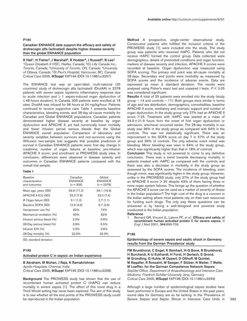
S83
P194Canadian ENHANCE data support the efficacy and safety ofdrotrecogin alfa (activated) despite higher disease severitythan the global ENHANCE population
R Hall1, H Fisher2, J Marshall3, R Hodder4, J Russell5, B Lee2
1Queen Elizabeth II HSC, Halifax, Canada; 2Eli Lilly Canada Inc.,Toronto, Canada; 3University of Toronto, ON, Canada; 4Universityof Ottawa, Canada; 5St Paul’s Hospital, Vancouver, BC, CanadaCritical Care 2005, 9(Suppl 1):P194 (DOI 10.1186/cc3257)
The ENHANCE trial was an open-label, multi-national (25countries) study of drotrecogin alfa (activated) (DrotAA) in 2378patients with severe sepsis (systemic inflammatory response dueto acute infection and ≥ 1 sepsis-induced organ dysfunction of≤ 48 hours duration). In Canada, 305 patients were enrolled at 18sites. DrotAA was infused for 96 hours at 24 mg/kg/hour. Patientscontinued to receive supportive care. Table 1 presents baselinecharacteristics, bleeding events, and 28-day all-cause mortality forCanadian and Global ENHANCE populations. Canadian patientsdemonstrated higher disease severity at baseline by organdysfunction and APACHE II, yet had numerically lower mortalityand fewer infusion period serious bleeds than the GlobalENHANCE overall population. Comparison of laboratory andseverity variables between survivors and non-survivors using theWilcoxon two-sample test suggests that possible predictors ofsurvival in Canadian ENHANCE patients were: first day change increatinine; number of organ failures at baseline; pre-infusionAPACHE II score; and enrollment at PROWESS study sites. Inconclusion, differences were observed in disease severity andoutcomes in Canadian ENHANCE patients compared with theoverall trial sample.
Table 1
Baseline Canadian Global characteristics ENHANCE ENHANCE and outcomes (n = 305) (n = 2378)
Mean age, years (SD) 56.9 (17.2) 59.1 (16.9)
APACHE II ICU (SD) 25.3 (7.8) 22.0 (7.4)
# Organ failure (SD) 3.1 (1.2) 2.7 (1.1)
Baseline SOFA (SD) 10.9 (3.6) 9.7 (3.5)
Vasopressor use (%) 82% 74%
Mechanical ventilation (%) 90% 82%
Infusion serious bleed (%) 2.0% 3.6%
28-Day serious bleed (%) 3.9% 6.5%
Infusion ICH (%) 0.3% 0.6%
28-Day mortality (%) 22.6% 25.3%
SD, standard deviation.
P195Activated protein C in sepsis: an Indian experience
B Abraham, M Mohan, J Raja, N RamakrishnanApollo Hospitals, Chennai, IndiaCritical Care 2005, 9(Suppl 1):P195 (DOI 10.1186/cc3258)
Background The PROWESS study has shown that the use ofrecombinant human activated protein C (rhAPC) can reducemortality in severe sepsis [1]. The effect of this novel drug in aThird World setting has never been explored. The aim of this studyis to see whether all the end points of the PROWESS study couldbe reproduced in the Indian population.
Method A prospective, single-center observational study.Consecutive patients who fulfilled the inclusion criteria of thePROWESS study [1] were included into the study. The studygroup was patients who received rhAPC. Patients who did notreceive rhAPC formed the control group. Data collected weredemographics, details of premorbid conditions and organ function,markers of disease severity and infection. APACHE II scores wererecorded at baseline. Organ dysfunction was measured usingSOFA scoring. The primary end point was all-cause mortality at28 days. Secondary end points were morbidity as measured bySOFA scores and the incidence of adverse events. Data areexpressed as mean ± standard deviation. The results wereanalysed using Fisher’s exact test and unpaired t tests. P < 0.05was considered significant.Results A total of 25 patients were enrolled into the study (studygroup — 14 and controls — 11). Both groups were similar in termsof age and sex distribution, demographics, comorbidities, baselineAPACHE II score, ventilatory and inotropic support and number oforgan dysfunction. In the study group only 27% had an APACHE IIscore > 25. Treatment with rhAPC was started at a mean of34.9 ± 21.9 hours from the onset of first organ dysfunction oradmission, whichever occurred earlier. The 28-day mortality in ourstudy was 36% in the study group as compared with 64% in thecontrols. This was not statistically significant. There was animprovement in the SOFA score on day 7 in 57% of the studygroup and 36% of controls. The only adverse event noted wasbleeding. Minor bleeding was seen in 64% of the study group,which was significantly higher than that in 18% of controls.Conclusion This study is not powered to come to any definitiveconclusion. There was a trend towards decreasing mortality inpatients treated with rhAPC as compared with the controls andthere was also a decrease in morbidity in the study group asassessed by the SOFA scores. The incidence of bleeding, eventhough minor, was significantly higher in the study group. However,unlike in the PROWESS study, only 27% of the study group hadan APACHE II score > 25 despite 43% of them having three ormore organ system failures. This brings up the question of whetherthe APACHE II score can be used as a marker of severity of illnessin the Indian population? The high cost of the drug is a concern inthe Indian setting where most patients rely on their own resourcesfor funding such drugs. The only way these questions can beanswered is by having a well-designed and powered studyconducted in the Indian population.Reference1. Bernard GR, Vincent JL, Laterre PF, et al.: Efficacy and safety of
recombinant human activated protein C for severe sepsis. NEngl J Med 2001, 344:699-709.
P196Epidemiology of severe sepsis and septic shock in Germany:results from the German ‘Prevalence’ study
FM Brunkhorst, C Engel, K Reinhart, H-G Bone, R Brunkhorst,H Burchardi, K-U Eckhardt, H Forst, H Gerlach, S Grond, M Gründling, G Huhle, M Oppert, D Olthoff, M Quintel, M Ragaller, R Rossaint, W Seeger, F Stüber, N Weiler, T Welte,M Loeffler, for the German Competence Network SepsisSepNet Office, Department of Anaesthesiology and Intensive CareMedicine, Friedrich-Schiller-University Jena, GermanyCritical Care 2005, 9(Suppl 1):P196 (DOI 10.1186/cc3259)
Although a large number of epidemiological sepsis studies havebeen performed in Europe and the United States in the past years,sound data for Germany are so far lacking. In the ‘Prevalence ofSevere Sepsis and Septic Shock in Intensive Care Units in
Available online http://ccforum.com/supplements/9/S1

S84
Germany’ study, a prospective observational cross-sectional study,the network gathered data from 454 randomly selected ICUs in310 hospitals in Germany and screened 3877 patients —according to the ACCP/SCCM Consensus Conference criteria —by local 1-day visits of trained physicians from the 17 GermanCompetence Network Sepsis (SepNet) regional study centers.Visits were randomly distributed over a 1-year period (2003) toallow assessment of seasonal variations of sepsis prevalence. TheICU sample was taken from a registry of all German hospitals withICUs (1380 hospitals with 2075 ICUs). Pediatric ICUs were notconsidered. The study was completed in January 2004 and thedatabase was closed on 31 May. Seven percent of ICUs weresituated in university, 34% in university-affiliated and 53% ingeneral hospitals. Fifty-six percent of ICU directors wereanesthesiologists and 26% internists. An infection wasmicrobiologically documented in 22% and diagnosed by clinicalcriteria alone in 12% of screened patients. Respiratory tractinfections were most common (52%), followed by intra-abdominal(15%) and urogenital infections (7%). Gram-negative and Gram-positive infections were nearly equally distributed (33% vs 35%);in 16% a fungal infection was suspected. The prevalence of sepsiswas 12%, infection without SIRS was 7%, and severesepsis/septic shock was 11%. There were significant differencesin the prevalence of severe sepsis/septic shock over 1 year withthe highest prevalence in May/June 2003 (18%). The infection wasICU acquired in 37%, hospital acquired in 20% and communityacquired in 35.5%. ICU mortality in patients with severesepsis/septic shock was 47% and hospital mortality was 54%.Based on these findings the incidence of severe sepsis/septicshock in German ICUs can be estimated as 75,000 cases per year(110 per 100,000 inhabitants), comparable with the incidence ofacute myocardial infarction (143 per 100,000 inhabitants). With anestimated 40,000 deaths per year, severe sepsis/septic shock isthe third most frequent cause of death in Germany after coronaryartery disease and acute myocardial infarction.Incidence and mortality rate of severe sepsis and septic shock inGerman ICUs are higher than reported in recent studies. This maybe due to the representative sample size, the more standardizeddiagnostic criteria and a lower interobserver variability.Acknowledgements Supported by the German Federal Ministry ofEducation and Research (BMBF), Grant No: 01 KI 0106 and LillyDeutschland GmbH.
P197Sepsis Brazil: an epidemiological study in intensive care units
J Sales1, C David1, P Souza2, M Knibel3, R Hatum3, F Dias4, A Neto5, C Pinheiro6, S Lima7, A Japiassú8, S Lobo9, A Estrada10, J Anjos11, Sepsis Study Group12
1HUCFF-UFRJ, Rio de Janeiro, Brazil; 2Hospital Clínicas Niterói,Brazil; 3H São Lucas, Rio de Janeiro, Brazil; 4H São Lucas, PortoAlegre, Brazil; 5H do Trabalhador, Curitiba, Brazil; 6H Clínicas,Porto Alegre, Brazil; 7H VOT Penitência, Rio de Janeiro, Brazil; 8H Quinta Dor, Rio de Janeiro, Brazil; 9H de Base, S J Rio Preto,Brazil; 10H Doenças Tropicais, Goiânia, Brazil; 11H Madre Tereza,Belo Horizonte, Brazil; 12Fundo AMIB, São Paulo, BrazilCritical Care 2005, 9(Suppl 1):P197 (DOI 10.1186/cc3260)
Introduction Sepsis represents the major cause of death in theICUs all over the world. Many studies have shown an increasingincidence over time and only a slight reduce in mortality. Many newtreatment strategies are arising and we should know our incidenceof sepsis and features.Methods A prospective cohort study in 50 hospitals of all regionsof Brazil. The patients who were admitted or who developed sepsis
during the month of September 2003 were enrolled. They werefollowed until the 28th day. The diagnoses were made inaccordance with the criteria proposed by ACCP/SCCM.Demographic features, APACHE II score, Sepsis-related OrganFailure Assessment (SOFA) score, mortality, sources of infectionsand lengh of stay (LOS) were evaluated.Results A total of 2419 patients were identified and 409 (16.9%)filled the criteria of sepsis, severe sepsis or septic shock. Theaverage age was 61.9 years, 225 (55%) were male, and the overall28-day mortality rate was 46.2%. The average APACHE II scorewas 21 and the SOFA score on the first day was 7. The SOFAscore in the mortality group was higher on day 1 (8), and hadincreased on day 3. We observed 326 episodes of sepsis, 118(28.9%) patients with severe sepsis, and 210 (51.4%) patientswith septic shock. The mortality rate for sepsis, severe sepsis andseptic shock was 15%, 35.6% and 63.8%, respectively. Theaverage LOS was 16 days. The main source of infection was therespiratory tract (71.6%). Gram-negative bacilli were moreprevalent (55.6%). Gram-positive cocci accounted for 32% andfungi infections for 10.6%.Conclusion A high incidence and mortality of sepsis in ICUs in ourcountry was observed. The high frequency of severe sepsis andseptic shock demonstrated a group at high risk of death. In orderto have a better use of the resources and to promote a reduction inmortality in the next 5 years it is very important to identify ourspecific features. After the final analysis of the database we intendto give our contribution to a better understanding of sepsis inBrazil.
P198Costs of severe sepsis according to the guidelines of the‘Surviving Sepsis Campaign’
G Vazquez de Anda, T Mondragon Rangel, J Arzate Villafaña, A Mondragon, A Jimenez, M GomezISSEMYM Medical Center, Toluca, MexicoCritical Care 2005, 9(Suppl 1):P198 (DOI 10.1186/cc3261)
Introduction Despite advanced organ support, patients sufferingfrom severe sepsis still have a high mortality rate [1]. Recently, ithas been advocated to follow the ‘Surviving Sepsis Campaign’(SSC) [2] to treat patients suffering from severe sepsis. Eventhough it is unpredictable which patient will survive and for whatreason, an important amount of the ICU budget has to be investedon drugs and supplies (DS). Therefore, the present study aimed todetermine the amount of money that our ICU (which isadministrated by public funds) has to invest on DS to treat apatient suffering from severe sepsis.Methods Data on costs for DS were recorded daily for everypatient admitted to the ICU from June to October 2004. Patientswith severe sepsis were separated from the main database. Costsfor each item of the SSC (including activated protein C) weretaken as independent variables, and the survey as a dependentvariable. We used the cost of each DS exactly as it was sold to ourinstitution. Costs are presented in Euros according to the currencyrate at 30 October 2004.Results During the 5-month period, 211 patients were admitted tothe ICU. The treatment for severe sepsis according to the SSCwas affordable for all patients admitted to the ICU. The cost for DSduring this period was €327,026.36. Of all ICU patients, 37(17.53%) suffered from severe sepsis and the DS cost for thisgroup was €146,513.10. Postoperative sepsis was observed in18 patients (48.6%) while 19 (51.3%) were non-postoperativesepsis; the cost for DS in these two groups was €58,294.61 and€88,218.49, respectively. The total costs for DS to produce a
Critical Care March 2005 Vol 9 Suppl 1 25th International Symposium on Intensive Care and Emergency Medicine

S85
43% survival on severe sepsis was €55,121.38; that is,postoperative sepsis €30,253.91 and non-postoperative sepsis€24,867.48. The cost for patients who died was €91,391.72.Patients who died with non-postoperative sepsis had the highestDS costs (i.e. €63,351.01). The investment to save 16 patientswas €63,357.02.Conclusions In our ICU, and following the SSC guidelines, wehave to invest in every patient suffering from severe sepsis at least€9157.07 to produce a 43% survival. That means, in the future,any increase in survival with the same investment on DS mayreflect our best approach to treat severe sepsis.References1. Moerer O, Burchardi H: The costs of sepsis. ICU management
2004, 4:25-29.2. Dellinger PR, Carlet JM, Masur H, Gerlach H, Calandra T, et al.:
Surviving Sepsis Campaign guidelines for management ofsevere sepsis and septic shock. Crit Care Med 2004, 32:858-873.
P199Selective poly(ADP-ribose)-polymerase inhibition affects neitherDNA repair nor cell senescence after ischemia-reperfusion
B Hauser1, U Ehrmann2, M Albicini2, M Gröger2, P Radermacher2,C Szabó3, J Kick2
1Semmelweis University, Budapest, Hungary; 2University of Ulm,Germany; 3Inotek Pharmaceuticals, Beverly, MA, USACritical Care 2005, 9(Suppl 1):P199 (DOI 10.1186/cc3262)
Background Poly(ADP-ribose)-polymerase (PARP) inhibition hasbeen beneficial in different models (sepsis, ischemia-reperfusion[I/R], hemorrhage) where oxidative stress induced DNA damageand subsequent PARP activation plays an important role. Theeffect of PARP inhibition on DNA repair in this context, however, isunknown. We therefore tested the effect of the selective PARP-1blocker INO-1001 on DNA repair and cell senescence in a porcinemodel of thoracic aortic cross-clamping.Materials and methods After instrumentation, anesthetized andmechanically ventilated pigs received vehicle (n = 9) or INO-1001(n = 9, 2 mg/kg each before clamping and during reperfusion,respectively). Norepinephrine (NE) was infused after declampingas required to maintain mean arterial pressure ≥ 80% of thepreclamping level. Hemodynamic and metabolic data wererecorded before clamping as well as prior to and 2 and 4 hoursafter declamping. The effect of INO-1001 on DNA damage andrepair (single-cell gel electrophoresis, ‘comet assay’) wasevaluated ex vivo in isolated lymphocytes (Ficoll gradient) thatwere sampled immediately before clamping, subsequently exposedto 4 bar of 100% O2 in a hyperbaric chamber (HBO) for 2 hoursand analyzed before as well as immediately 1 and 2 hours afterHBO. Surgery and I/R-related DNA damage in vivo was assessedin whole blood samples taken before surgery, before clamping, andbefore and 2 hours after declamping. At the end of the experimentthe spinal cord, gut, liver, and kidney tissue specimens weresampled for immonuhistochemistry of cyclin-dependent kinaseinhibitor (p21 and p27) gene expression as a measure of cellsenescence. Data are median (minimum–maximum).Results Significantly shorter NE infusion time (53 [23–93] vs 70[41–83] min, P = 0.042) with reduced total NE doses (12 [6–19]vs 22 [9–38] µg/kg, P = 0.034) were needed for hemodynamicmanagement in the INO-1001-treated animals. The time course ofboth the HBO-induced and surgery and I/R-related oxidative DNAdamage was identical in the two groups. There were no differencesin p21 and p27 expression, either.Conclusion The markedly reduced NE infusion time and doseneeded for post-I/R hemodynamic stabilization confirm the well-
known positive inotropic effect of selective PARP-1 blockers [1].INO-1001 proved to be safe with respect to DNA repair and cellsenescence.Acknowledgements Supported by the Alexander-von-Humboldt-Stiftung (BH) and Land Baden-Württemberg, Germany (MG).Reference1. Szabó G, et al.: Shock 2004, 21:426-432.
P200Rosiglitazone, a peroxisome proliferator-activated receptor-gamma agonist, attenuates intercellular adhesion molecule-1and cytokine-induced neutrophil chemoattractant-1expression in lungs of rats with acute lung injury
D Liu1, B Zeng1, S Zhang1,2, Z Geng2
1Union Hospital, Tongji Medical College, Huazhong University ofScience and Technology, WuHan, China; 2Lanzhou GeneralHospital, Lanzhou Command of PLA, LanZhou, ChinaCritical Care 2005, 9(Suppl 1):P200 (DOI 10.1186/cc3263)
Aim To investigate the effects of rosiglitazone (ROSI), a peroxisomeproliferator-activated receptor-gamma (PPAR-γ) agonist, on thelung expression of intercellular adhesion molecule-1 (ICAM-1) andcytokine-induced neutrophil chemoattractant-1 (CINC-1) in ratswith acute lung injury.Methods Thirty-six anesthetized male Wistar rats were randomlydivided into six groups: DMSO control group, ROSI group,GW9662 group, lipopolysaccharide (LPS) group, ROSI + LPSgroup and GW9662 + ROSI group. Rats received either LPS(6 mg/kg intravenously) or vehicle (saline, 2 ml/kg intravenously).ROSI (0.3 mg/kg intravenously) or vehicle (10% DMSO) wasadministered 30 min before treatment with LPS. The selectivePPAR-γ antagonist, GW9662 (0.3 mg/kg intravenously), was given20 min before ROSI. Four hours after LPS injection, the wet/drylung weight (W/D) ratio, myeloperoxidase (MPO) activity,malondialdehyde (MDA) and CINC-1 concentrations were assayedin the lung tissues. Immunohistochemical analysis of ICAM-1expression was also studied.Results Pretreatment with ROSI attenuated LPS-induced increasesof the W/D ratio, MPO activity, MDA and CINC-1 concentrationsas well as the expression of ICAM-1 in the lung tissues (P < 0.01).The specific PPAR-γ antagonist GW9662 significantly antagonizedthe effects of ROSI.Conclusion Pretreatment with ROSI significantly reducesendotoxin-induced acute lung injury in rats; the mechanism may bethrough activation of PPAR-γ accompanied by inhibition of ICAM-1and CINC-1 expression.
P201Protective effects of rosiglitazone, a peroxisome proliferator-activated receptor-gamma agonist, on endotoxin-inducedacute lung injury in rats
D Liu1, B Zeng1, S Zhang1, Y Wang1, L Zeng1, Z Geng2
1Union Hospital, Tongji Medical College, Huazhong University ofScience and Technology, WuHan, China; 2Lanzhou GeneralHospital, Lanzhou Command of PLA, LanZhou, ChinaCritical Care 2005, 9(Suppl 1):P201 (DOI 10.1186/cc3264)
Objective To determine whether rosiglitazone, a peroxisomeproliferator-activated receptor-gamma (PPAR-γ) agonist, decreasesthe severity of endotoxin-induced acute lung injury and toinvestigate cellular mechanisms contributing to these effects.Setting A university research laboratory.
Available online http://ccforum.com/supplements/9/S1
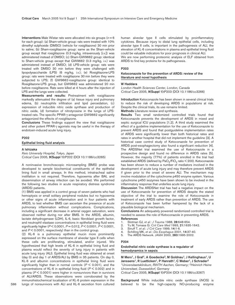
S86
Interventions Male Wistar rats were allocated into six groups (n = 6for each group): (a) Sham-vehicle group: rats were treated with 10%dimethyl sulphoxide (DMSO) (vehicle for rosiglitazone) 30 min priorto saline; (b) Sham-rosiglitazone group: same as the Sham-vehiclegroup except that rosiglitazone (0.3 mg/kg, intravenously [i.v.]) wasadministered instead of DMSO; (c) Sham-GW9662 group: identicalto Sham-vehicle group except that GW9662 (0.3 mg/kg, i.v.) wasadministered instead of DMSO; (d) LPS-vehicle group: rats weretreated with DMSO 30 min before they were challenged withlipopolysaccharide (LPS) (6 mg/kg, i.v.); (e) Rosiglitazone-LPSgroup: rats were treated with rosiglitazone 30 min before they weresubjected to LPS; (f) GW9662-rosiglitazone group: identical toRosiglitazone-LPS group, but GW9662 was administered 20 minbefore rosiglitazone. Rats were killed at 4 hours after the injection ofLPS and the lungs were collected.Measurements and results: Pretreatment with rosiglitazonemarkedly attenuated the degree of (a) tissue injury and pulmonaryedema, (b) neutrophils infiltration and lipid peroxidation, (c)expression of inducible nitric oxide synthase and production ofnitric oxide, (d) formation of nitrotyrosine in the lungs of LPS-treated rats. The specific PPAR-γ antagonist GW9662 significantlyantagonized the effects of rosiglitazone.Conclusions These findings support the view that rosiglitazoneand other potent PPAR-γ agonists may be useful in the therapy ofendotoxin-induced acute lung injury.
P202Epithelial lining fluid analysis
A IshizakaKeio University Hospital, Tokyo, JapanCritical Care 2005, 9(Suppl 1):P202 (DOI 10.1186/cc3265)
A noninvasive bronchoscopic microsampling (BMS) probe wasdeveloped to sample biochemical constituents of the epitheliallining fluid in small airways. In this method, intratracheal salineinstillation is not required. Therefore, hypoxemia after BAL anddissemination of airway infection can be avoided. We performedthe following two studies in acute respiratory distress syndrome(ARDS) patients.(1) BMS was applied in a control group of seven patients who hadhemoptysis or small solitary peripheral nodules but no hypoxemiaor other signs of acute inflammation and in four patients withARDS, to test whether BMS can ascertain the presence of acutepulmonary inflammation without complications. Complications,including a significant decrease in arterial oxygen saturation, wereobserved neither during nor after BMS. In the ARDS, albumin,lactate dehydrogenase (LDH), IL-6, basic fibroblast growth factor,and neutrophil elastase concentrations in epithelial lining fluid weresignificantly higher (P < 0.0001, P = 0.012, P < 0.0001, P < 0.0001,and P < 0.0001, respectively) than in the control group.(2) KL-6 is a pulmonary epithelial mucin more prominentlyexpressed on the surface membrane of alveolar type II cells whenthese cells are proliferating, stimulated, and/or injured. Wehypothesized that high levels of KL-6 in epithelial lining fluid andplasma would reflect the severity of lung injury in patients withacute lung injury (ALI). Epithelial lining fluid was obtained at onset(day 0) and day 1 of ARDS/ALI by BMS in 35 patients. On day 0,KL-6 and albumin concentrations in epithelial lining fluid weresignificantly higher than in normal controls (P < 0.001), and theconcentrations of KL-6 in epithelial lining fluid (P < 0.002) and inplasma (P < 0.0001) were higher in nonsurvivors than in survivorsof ALI/ARDS. These observations were corroborated by theimmunohistochemical localization of KL-6 protein expression in thelungs of nonsurvivors with ALI and KL-6 secretion from cultured
human alveolar type II cells stimulated by proinflammatorycytokines. Because injury to distal lung epithelial cells, includingalveolar type II cells, is important in the pathogenesis of ALI, theelevation of KL-6 concentrations in plasma and epithelial lining fluidcould be valuable indicators for poor prognosis in clinical ALI.We are now performing proteomic analysis of ELF obtained fromARDS to find key proteins for its pathogenesis.
P203Ketoconazole for the prevention of ARDS: review of theliterature and novel hypothesis
W HaddaraLondon Health Sciences Center, London, CanadaCritical Care 2005, 9(Suppl 1):P203 (DOI 10.1186/cc3266)
Introduction Ketoconazole has been shown in several clinical trialsto reduce the risk of developing ARDS in populations at risk.Despite the clinical trials, its use remains limited.Methods Literature review and synthesis.Results Two small randomized controlled trials found thatKetoconazole prevents the development of ARDS in mixed andseptic surgical ICU populations [1,2]. A third study examined theeffect of a guideline implementation for the use of Ketoconazole toprevent ARDS and found that postguideline implementation ratesof ARDS were significantly lower than both historical rates andrates in another hospital that did not implement the guideline [3]. Ahistorical case control study of Ketoconazole for prevention ofARDS post-esophagectomy also found a significant reduction [4].The ARDSNet trial examined the use of Ketoconazole in aprospective design and found no difference in ARDS rates [5].However, the majority (77%) of patients enrolled in the trial hadestablished ARDS (defined by PaO2/FiO2 ratio < 200). Ketoconazolehas been shown to reduce a number of mediators involved in thedevelopment of acute lung injury and may therefore prevent ARDSif given prior to the onset of severe ALI. The mechanism mayinvolve modulation of the cytochrome p450 enzyme system. Variouscytochrome p450 isozymes have been shown to be involved in theinflammatory response that underlies acute lung injury.Discussion The ARDSNet trial has had a negative impact on theuse of Ketoconazole for prevention of ARDS despite the statedobjective of the trial to examine the role of Ketoconazole intreatment of early ARDS rather than prevention of ARDS. The useof Ketoconazole has been further hampered by the lack of aplausible biological mechanism.Conclusions An adequately powered randomized controlled trial isneeded to assess the role of Ketoconazole in preventing ARDS.References1. Slotman GJ, et al.: J Trauma 1988, 28:648-654.2. Yu M, Tomasa G: Crit Care Med 1993, 21:1635-1642.3. Sinuff T, et al.: J Crit Care 1999, 14:1-6.4. Schilling MK, et al.: Dis Esophagus 2001, 14:37-40.5. The ARDS Network. JAMA 2000, 283:1995-2002.
P204Endothelial nitric oxide synthase is a regulator ofhemodynamics in sepsis
M Merx1, J Graf1, A Goedecke2, M Goldnau1, J Kellinghaus1, UJanssens1, R Luetticken1, P Hanrath1, C Weber1, J Schrader2
1Universitaetsklinikum, RWTH Aachen, Germany; 2Heinrich HeineUniversitaet, Duesseldorf, GermanyCritical Care 2005, 9(Suppl 1):P204 (DOI 10.1186/cc3267)
Background While inducible nitric oxide synthase (iNOS) isbelieved to be the high-capacity NO-producing enzyme
Critical Care March 2005 Vol 9 Suppl 1 25th International Symposium on Intensive Care and Emergency Medicine

S87
responsible for the detrimental hemodynamic alterations observedduring sepsis, the role of endothelial nitric oxide synthase (eNOS)remains more obscure. Thus, it was the present study’s aim toelucidate the influence of eNOS on hemodynamics in septic shockusing eNOS–/– mice in the clinically relevant polimicrobial cecumligation and puncture (CLP) model of sepsis.Methods and results Prolonged survival was observed ineNOS–/– mice (68 ± 25 hours vs 25 ± 6 hours in WT mice,n = 12, P < 0.001). Chronic unselective NOS inhibition withethylthiourea (ETU) in eNOS–/– mice after sepsis inductiondiminished this survival benefit, while ETU application in WT miceafter CLP led to slight improvement in survival time. Cardiacoutput, studied by serial echocardiography, increased to amaximum of 60% over baseline at 10 hours after sepsis inductionin WT mice but not in septic eNOS–/– mice and septic mice (WTand eNOS–/–) treated by unselective NOS inhibition. WT CLPmice were refractory to β-stimulation, while eNOS–/– CLP miceresponded comparably with sham-operated WT mice. Meanarterial blood pressure was decreased by 25 ± 8 mmHg(P < 0.001; n = 8) in WT CLP mice, with eNOS–/– CLP miceshowing no sign of hypotension. Contractility of isolated WT CLPhearts was impaired by 35% (P < 0.001, n = 8), but hearts fromCLP eNOS–/– mice remained unaltered. While eNOS expressionremained stable, coronary flow studies indicated a high level ofeNOS activation secondary to sepsis.Conclusion eNOS is a key player in the hemodynamics of sepsis.Selective eNOS inhibition appears beneficial in this model ofsepsis with global nitric oxide synthase inhibition diminishing mostof the observed benefit.
P205Application of the bovine hemoglobin solution HBOC-301 withand without isovolemic hemodilution reduces thehistopathologic damage in acute severe pancreatitis in swine
M Freitag, O Mann, T Strate, A Gottschalk, S Petri, C Rempf, J Izbicki, T StandlUniversity Hospital Hamburg-Eppendorf (UKE), Hamburg, GermanyCritical Care 2005, 9(Suppl 1):P205 (DOI 10.1186/cc3268)
Introduction Severe acute pancreatitis (AP) remains associated witha high mortality. In experimental AP the application of the bovinehemoglobin solution HBOC-301 with and without IHD was able tonormalize pancreatic tissue oxygen tension [1,2]. The aim of thisstudy was to evaluate the therapeutic approach of volume therapywith HES plus HBOC-301 with and without IHD with regard to thehistologic damage and survival time in pigs suffering from AP.Methods After approval of the local animal ethics committee 33pigs were anesthetized (FIO2: 0.3). AP was induced bycombination of cerulein and glucodeoxycholic acid. After inductionof AP animals were randomized and either isovolemiclyhemodiluted with 10% HES 200,000/0.5 plus HBOC-301 (+0.6 g/dlplasmatic Hb; Oxyglobin, Biopure, USA) (IHD + HBOC group) orreceived volume replacement without IHD with HES plus HBOC-301 (+0.6 g/dl plasmatic Hb) (HBOC group), or were hemodilutedwith Ringer`s solution (IHD RINGER group) to a hematocrit (Hct)of 15%. Six hours after therapy all animals were extubated, andkilled after 6 days at the latest. Two 5 µm slices from thespecimens of the pancreas were examined in a blinded mannerusing a light microscope. The histopathologic findings were scoredas previously described quantifying acinar necrosis, fat necrosis,inflammation and edema on a scale from 0 to 3 points [3]. The totalscore was the sum of the median of two tissue areas (maximumvalue 24). Statistical analyses: Kruskal–Wallis and Mann–WhitneyU test (P < 0.05).
Results The histopathologic tissue damage (Table 1) was higher inthe IHD RINGER group in comparison with the IHD + HBOCgroup and the HBOC group. The survival rate at the end of theobservation period was higher in the IHD + HBOC group (10/11)and the HBOC group (8/11) in comparison with the IHD RINGERgroup (2/11) (P < 0.001).
Table 1
Histopathologic score (range 0–24) of two areas from thepancreas,
Acinar Fat Inflam- Total Group necrosis necrosis mation Edema score
IHD + HBOC 3 (0–5)† 1 (0–5) 3 (1–4)*† 4 (2–5) 11 (4–17)†
HBOC 3 (0–4)† 1 (0–3)† 4 (3–4) 4 (2–4) 12 (7–15)†
IHD RINGER 5.5 (4–6) 3 (1–5) 4 (3–5) 4 (3–5) 16 (14–20)
† P < 0.05 versus Ringer. * P < 0.05, IHD + HBOC group versus HBOCgroup.
Implications In a former experiment application of HBOC-301with and without IHD normalized pancreatic tissue oxygen tensions[1]. The higher tpO2 is now reflected by less tissue damage and abetter outcome. The application of HBOC-301 deserves furtheranalysis to assess its potential in the clinical treatment of AP.References1. Freitag, et al.: Anesthesiology 2004.2. Strate, et al.: Ann Surg 1993.3. Schmidt, et al.: Ann Surg 1992.
P206Volume substitution therapy with HES 130/0.4 (Voluven®)versus HES 450/0.7 (hetastarch) during major orthopedicsurgery
S Gandhi1, D Warltier1, R Weiskopf2, F Bepperling3, D Baus3, C Jungheinrich3
1Medical College of Wisconsin, Milwaukee, WI, USA; 2Universityof California, San Francisco, CA, USA; 3Fresenius Kabi, BadHomburg, GermanyCritical Care 2005, 9(Suppl 1):P206 (DOI 10.1186/cc3269)
Background The first-generation hetastarch remains the onlyhydroxyethyl starch (HES) available for volume substitution in theUnited States despite its controversial safety profile. The safetyand efficacy of a new HES, Voluven®, was assessed in thisinvestigation.Patients and methods After local IRB approval and signed,written informed consent, 100 adult patients scheduled for majororthopedic surgery were randomly allocated to receive 6% HES130/0.4 (Voluven®, n = 49) or 6% HES 450/0.7 (hetastarch,n = 51) for intraoperative volume replacement in a double-blindfashion guided by a predefined algorithm that included CVP-guided therapy. HES volume served as the primary efficacyvariable. Safety variables were: calculated total red blood cell(RBC) loss, minimum factor VIII activity and vW factorconcentration between the end of surgery and 2 hours later,laboratory parameters, and adverse events through POD 28. Thetreatment groups were compared for equivalence in efficacy andsuperiority of HES 130/0.4 in safety.Results Demographic data and other baseline characteristics ofthe treatment groups were similar. Mean infused HES volumeswere equivalent between groups (1615 ± 778 ml for HES 130/0.4and 1584 ± 958 ml for HES 450/0.7). Primary safety results arepresented in Table 1 (mean ± standard deviation; *P < 0.05).Three coagulopathies were reported as serious adverse events in
Available online http://ccforum.com/supplements/9/S1

S88
Table 1
RBC Factor VIII vW factor RBC loss (%) with (%) with use (l) >1 l HES >1 l HES (ml/kg)
HES 130/0.4 1.17 ± 0.63 88.7 ± 55.7 89.2 ± 40.4 8.0 ± 6.4
HES 450/0.7 1.31 ± 0.84 65.9 ± 39.4* 71.5 ± 36.9* 13.8 ± 12.9*
the HES 450/0.7 group (> 3 l HES 450/0.7 administered in allcases). No coagulopathies were reported for HES 130/0.4.Conclusion HES 130/0.4 (Voluven®) and HES 450/0.7(hetastarch) are equally effective volume expanders in majororthopedic surgery. HES 130/0.4, however, demonstrated a morefavorable safety profile including preservation of critical coagulationfactors and reduced requirement for transfusion of blood products.
P207Sepsis Brazil — early results: albumin and mortality rate inseptic shock
C David, R Hatum, J Andrade, M Knibel, M Gomes, J Duarte, M Damasceno, P PereiraFederal University of Rio de Janeiro, BrazilCritical Care 2005, 9(Suppl 1):P207 (DOI 10.1186/cc3270)
Introduction The fluid replacement therapy in septic shock is apolemic issue, regarding the use of crystalloid versus colloidsolutions. Albumin is the more often used colloid solution. A recentlypublished study (SAFE study) showed no benefits in albumin versuscrystalloid use, for fluid replacement in shock patients.Objective To analyse the septic shock subgroup in our study,regarding the use of albumin as a fluid replacement solution and itsinfluence on mortality.Patients and methods We conducted a prospective cohort studyin 50 hospitals of all regions of Brazil. The patients who wereadmitted or who developed sepsis during the month of September2003 were enrolled. They were followed until the 28th day or lessaccording to their discharge. The diagnoses were made inaccordance with the criteria proposed by ACCP/SCCM. Weevaluated demographic features, APACHE II, Sepsis-relatedOrgan Failure Assessment (SOFA) score, mortality, sources ofinfections, microbiology and interventions. We also recordedunderlying diseases and length of stay.Results A total of 2419 patients were identified and 409 (16.9%)filled the criteria of sepsis, severe sepsis or septic shock. Twohundred and ten patients (51.4%) formed the septic shocksubgroup, with a mean APACHE II score of 22 and a overallmortality rate of 63.8%. Eighty-five patients (40.5%) in thissubgroup used albumin; 125 patients (59.5%) did not. Fifty-onepatients (60%) in the albumin group died; 34 (40%) were alive onthe 28th day. In the non-albumin septic shock patients subgroup,one patient (0.8%) was transferred from the hospital and excludedfrom the study; 83 patients (66.4%) died and 41 (32.8%) were stillalive after 28 days.When we apply the binomial probability test, the mortality rate inthe albumin group is 66.93% versus 60% in the non-albumingroup. Using 60% as the expected mortality rate for bothsubgroups (because it is the little one), P = 0.05. In the albuminsubgroup we had 44 females and 41 males; in the non-albuminsubgroup, 50 females and 75 males. The mean age and APACHEII score were the same in both subgroups (61.98 years old and20.88, respectively).Conclusions We can see a trend towards not using albumin as afluid replacement solution in septic shock patients, a standard
therapy previously. Albumin really seems not to be a protectivetherapy for these patients, since the mortality rate in both groups(albumin and non-albumin users) are too close (actually, when thestatistical test is applied, we see a trend towards a greatermortality rate in the albumin subgroup).
P208Case series of dermatomyositis complicated by acuterespiratory failure requiring intensive care
Y Teo, B Thong, D Tai, S GohTan Tock Seng Hospital, SingaporeCritical Care 2005, 9(Suppl 1):P208 (DOI 10.1186/cc3271)
Introduction Dermatomyositis (DM) is a rare disorder of variableseverity, ranging from mild weakness that is highly responsive tocorticosteroid therapy to a relentless downhill course with pulmonaryinvolvement that is unresponsive to all immunosuppressive modalities.Method In this retrospective case series, we reviewed the clinicalpresentation and outcomes of all DM patients who were admittedto the medical intensive care unit (MICU) of a university-affiliatedgeneral hospital from 1 January 2000 to 31 December 2004. Allhad acute respiratory failure requiring mechanical ventilation (MV).Results The nine patients had a mean age of 47.6 ± 12.4 (range:27–66) years. The female to male ratio was 3.5:1. All werediagnosed to have DM before MICU admission with medianduration of illness of 8 (range: 3–28) weeks. DM was classified asclassical (Peter and Bohan’s criteria) in six patients and amyopathic(Sontheimer’s criteria) in three. Initial high-resolution computedtomography thorax in eight patients showed pulmonary fibrosis (fourpatients), interstitial/alveolar changes (two patients), and ground-glass changes (two patients). All patients were negative for anti-Jo1antibody, and none had an associated malignancy.The mean PaO2/FiO2 was 109.2 ± 40.9 (range: 61.8–181.7)mmHg. The median duration of hospital and MICU stay were 16(range: 2–38) days and 13 (range: <1 to 37) days, respectively.The median duration of MV was 10 (range: <1 to 37) days.All chest radiographs showed bilateral lung infiltrates. Broncho-alveolar lavage was performed on six patients, with findings ofpneumocystis cariini in one patient and mycobacteriumtuberculosis in another. All patients were treated empirically withbroad-spectrum antibiotics for pneumonia including cotrimoxazolefor Pneumocystis carinii on admission to the MICU. Immuno-suppressive therapies in the MICU included high-dose systemiccorticosteroids in all, pulse methylprednisolone (five patients),pulse cyclophosphamide (three patients), high-dose intravenousimmunoglobulin (one patient) and cyclosporine (two patients). Allpatients died of respiratory failure with four having hypotension andacute renal failure as contributing causes.Conclusion DM with pulmonary involvement that required MVcarries an extremely high mortality. Early diagnosis and effectiveimmunosuppressive therapies for lung disease may help to improveoutcome in this high-risk group.
P209Outcome following burns from 1985 to 2004 in the Centre forSeverely Burned Patients, Ghent University Hospital, Belgium
N Brusselaers, E Hoste, K Vandewoude, J De Waele, K Colpaert, S Monstrey, S BlotUniversity of Ghent, BelgiumCritical Care 2005, 9(Suppl 1):P209 (DOI 10.1186/cc3272)
Background Mortality in burn patients can be estimated by threemajor risk factors for death: age > 60 years, total burned surface
Critical Care March 2005 Vol 9 Suppl 1 25th International Symposium on Intensive Care and Emergency Medicine
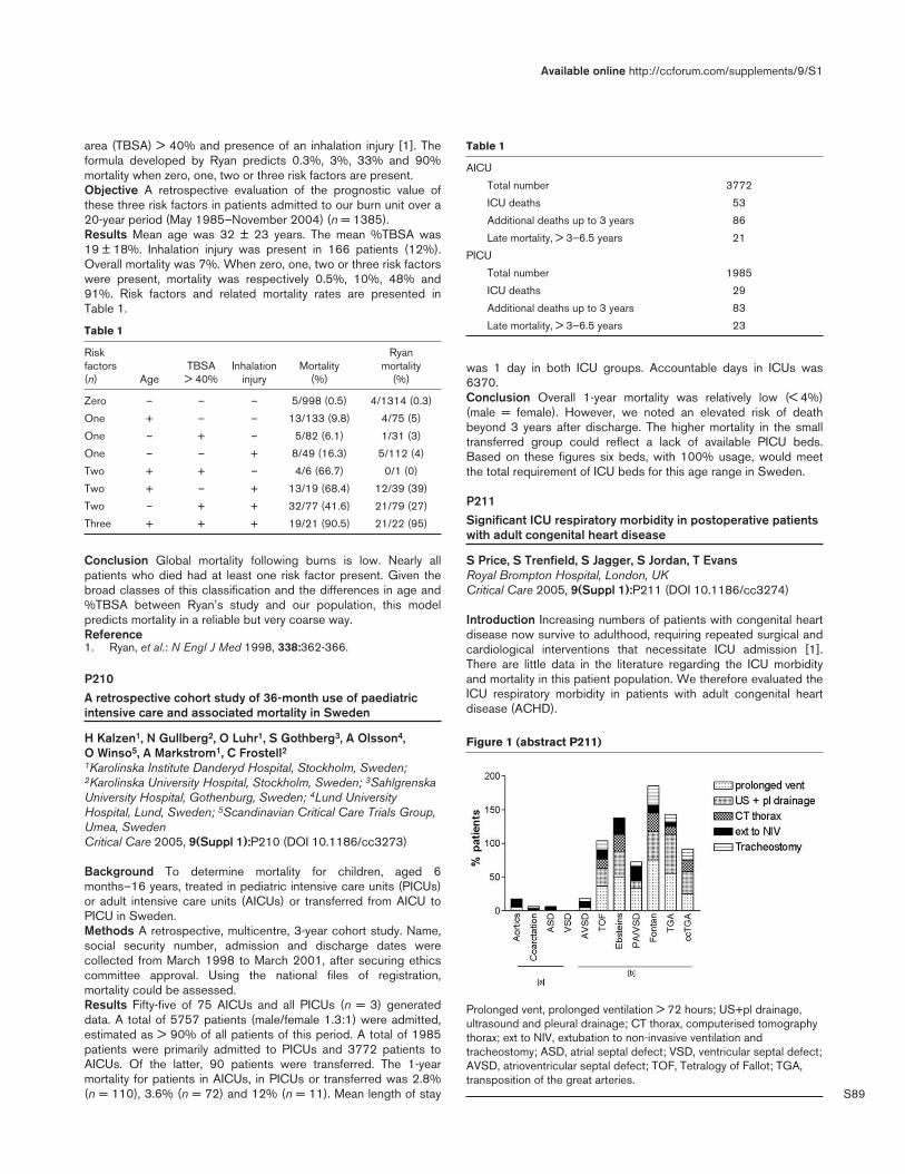
S89
area (TBSA) > 40% and presence of an inhalation injury [1]. Theformula developed by Ryan predicts 0.3%, 3%, 33% and 90%mortality when zero, one, two or three risk factors are present.Objective A retrospective evaluation of the prognostic value ofthese three risk factors in patients admitted to our burn unit over a20-year period (May 1985–November 2004) (n = 1385).Results Mean age was 32 ± 23 years. The mean %TBSA was19 ± 18%. Inhalation injury was present in 166 patients (12%).Overall mortality was 7%. When zero, one, two or three risk factorswere present, mortality was respectively 0.5%, 10%, 48% and91%. Risk factors and related mortality rates are presented inTable 1.
Table 1
Risk Ryan factors TBSA Inhalation Mortality mortality (n) Age > 40% injury (%) (%)
Zero – – – 5/998 (0.5) 4/1314 (0.3)
One + – – 13/133 (9.8) 4/75 (5)
One – + – 5/82 (6.1) 1/31 (3)
One – – + 8/49 (16.3) 5/112 (4)
Two + + – 4/6 (66.7) 0/1 (0)
Two + – + 13/19 (68.4) 12/39 (39)
Two – + + 32/77 (41.6) 21/79 (27)
Three + + + 19/21 (90.5) 21/22 (95)
Conclusion Global mortality following burns is low. Nearly allpatients who died had at least one risk factor present. Given thebroad classes of this classification and the differences in age and%TBSA between Ryan’s study and our population, this modelpredicts mortality in a reliable but very coarse way.Reference1. Ryan, et al.: N Engl J Med 1998, 338:362-366.
P210A retrospective cohort study of 36-month use of paediatricintensive care and associated mortality in Sweden
H Kalzen1, N Gullberg2, O Luhr1, S Gothberg3, A Olsson4, O Winso5, A Markstrom1, C Frostell21Karolinska Institute Danderyd Hospital, Stockholm, Sweden;2Karolinska University Hospital, Stockholm, Sweden; 3SahlgrenskaUniversity Hospital, Gothenburg, Sweden; 4Lund UniversityHospital, Lund, Sweden; 5Scandinavian Critical Care Trials Group,Umea, SwedenCritical Care 2005, 9(Suppl 1):P210 (DOI 10.1186/cc3273)
Background To determine mortality for children, aged 6months–16 years, treated in pediatric intensive care units (PICUs)or adult intensive care units (AICUs) or transferred from AICU toPICU in Sweden.Methods A retrospective, multicentre, 3-year cohort study. Name,social security number, admission and discharge dates werecollected from March 1998 to March 2001, after securing ethicscommittee approval. Using the national files of registration,mortality could be assessed.Results Fifty-five of 75 AICUs and all PICUs (n = 3) generateddata. A total of 5757 patients (male/female 1.3:1) were admitted,estimated as > 90% of all patients of this period. A total of 1985patients were primarily admitted to PICUs and 3772 patients toAICUs. Of the latter, 90 patients were transferred. The 1-yearmortality for patients in AICUs, in PICUs or transferred was 2.8%(n = 110), 3.6% (n = 72) and 12% (n = 11). Mean length of stay
Table 1
AICU
Total number 3772
ICU deaths 53
Additional deaths up to 3 years 86
Late mortality, > 3–6.5 years 21
PICU
Total number 1985
ICU deaths 29
Additional deaths up to 3 years 83
Late mortality, > 3–6.5 years 23
was 1 day in both ICU groups. Accountable days in ICUs was6370.Conclusion Overall 1-year mortality was relatively low (< 4%)(male = female). However, we noted an elevated risk of deathbeyond 3 years after discharge. The higher mortality in the smalltransferred group could reflect a lack of available PICU beds.Based on these figures six beds, with 100% usage, would meetthe total requirement of ICU beds for this age range in Sweden.
P211Significant ICU respiratory morbidity in postoperative patientswith adult congenital heart disease
S Price, S Trenfield, S Jagger, S Jordan, T EvansRoyal Brompton Hospital, London, UKCritical Care 2005, 9(Suppl 1):P211 (DOI 10.1186/cc3274)
Introduction Increasing numbers of patients with congenital heartdisease now survive to adulthood, requiring repeated surgical andcardiological interventions that necessitate ICU admission [1].There are little data in the literature regarding the ICU morbidityand mortality in this patient population. We therefore evaluated theICU respiratory morbidity in patients with adult congenital heartdisease (ACHD).
Available online http://ccforum.com/supplements/9/S1
Figure 1 (abstract P211)
Prolonged vent, prolonged ventilation > 72 hours; US+pl drainage,ultrasound and pleural drainage; CT thorax, computerised tomographythorax; ext to NIV, extubation to non-invasive ventilation andtracheostomy; ASD, atrial septal defect; VSD, ventricular septal defect;AVSD, atrioventricular septal defect; TOF, Tetralogy of Fallot; TGA,transposition of the great arteries.

S90
Methods A database (Medicus, High Wycombe, UK) wasinterrogated for all patients admitted to the ICU of a cardiothoracictertiary referral centre during the period January 1997–2002 withACHD. Classification of ACHD complexity was according to themodified Canadian Consensus Conference criteria. Patientdemographics, details of any surgical procedure and mortality wereextracted. Analysis of respiratory investigations and interventions(Fig. 1) was performed.Results Over the accounting period 342 patients with ACHD wereadmitted (6.4% of total ICU admissions), of which 305 (90%) werefollowing cardiac surgery. Perioperative mortality was 3.3%.Patients with simple ACHD had zero mortality, a low requirementfor respiratory investigations and interventions (Fig. 1a). Patientswith moderately complex and complex ACHD had a highermortality (10.6%) and significantly higher requirement forrespiratory investigations and interventions (Fig. 1b).Conclusion Patients undergoing surgery for complex ACHD havea significant requirement for respiratory investigations andinterventions. As the case mix is changing, with more complexpatients requiring intervention in adulthood, this has implicationsfor future planning of ICU ACHD care.Acknowledgement SP is the BHF Jill Dando Adult CongenitalCardiology Fellow.Reference1. Somerville J: Management of adults with congenital heart
disease: an increasing problem. Annu Rev Med 1997, 48:283-293.
P212Intensive care in adults with congenital heart disease: cost implications of a changing patient population
S Price, S Trenfield, S Jagger, S Jordan, T EvansRoyal Brompton Hospital, London, UKCritical Care 2005, 9(Suppl 1):P212 (DOI 10.1186/cc3275)
Introduction Current estimations suggest there are around800,000 patients in the USA with congenital cardiac lesionsrequiring specialist care [1]. Improved surgical and cardiologicalinterventions mean that numbers of patients with moderate-severecongenital lesions will increase [2], resulting in increasedrequirement for repeat surgery in adulthood. This is reflected by anincreasingly complex case mix requiring ICU admission. Thepotential impact of this changing patient population upon ICUresources has not been addressed.Methods A database (Medicus, High Wycombe, UK) wasinterrogated for all patients admitted to the ICU during the periodJanuary 1997–2002 with adult congenital heart disease (ACHD),which was classified as simple or complex according to themodified Canadian Consensus Conference criteria. Patientdemographics, the length of ICU stay (LOS) and mortality wereextracted, and the severity of illness and intensity of interventioncalculated (APACHE II, TISS-28).Results Of 5312 patients admitted during the accounting period,342 had ACHD (6.4%). ACHD ICU mortality was 4.4%. Patientswith simple ACHD (32%) had APACHE II scores of 18.4 ± 4.8,zero mortality, short LOS (1.8 ± 2 days), and low therapeuticintervention (TISS-28 42.3 ± 10). Patients with complex ACHD(68%) had similar APACHE II scores (18.5 ± 6.4, not significant)but significantly higher mortality (10.6%), increased LOS(3.9 ± 2 days, P < 0.05) and requirement for intervention (TISS-2859.8 ± 13, P < 0.05). Overall mean cost per admission (TISSscore point 46.31, €67.0) was therefore significantly higher in themore complex patients (simple €5101 ± 1340 vs complex€15,623 ± 1742, P < 0.05).
Conclusion Improved survival of patients with complex congenitalheart disease to adulthood will have significant implications(clinical and financial) in any ICU undertaking this work.Acknowledgement SP is the British Heart Foundation AdultCongenital Cardiology Fellow.References1. Warnes CA, Liberthson R, Danielson GK, Dore A, Harris L,
Hoffman JI, Somerville J, Williams RG, Webb GD: Task force 1:the changing profile of congenital heart disease in adult life. J Am Coll Cardiol 2001, 37:1170-1175.
2. Wren C, O’Sullivan JJ: Survival with congenital heart diseaseand need for follow up in adult life. Heart 2001, 85:438-443.
P213HELLP syndrome: analysis of 23 cases
C Kaddour, M Laamourou, R Souissi, Z Haddad, T Chaaoua, H Ouragini, W GdouraNational Institute of Neurology, Tunis, TunisiaCritical Care 2005, 9(Suppl 1):P213 (DOI 10.1186/cc3276)
Context HELLP syndrome (hemolysis, elevated liver enzymes, lowplatelets) is a severe complication in late pregnancy. This disordercomplicates between 2% and 7% of gestations and is associatedwith a high maternal morbidity ranging between 1% and 4%.Objective We undertook this study to evaluate the repercussionson maternal outcome in patients with HELLP syndrome and todetermine its clinical and biological characteristics.Design A retrospective and analytical study.Setting The intensive care unit of the National Hospital ofNeurology, Tunisia.Sample Pregnant or postdelivery women who had HELLPsyndrome between January 1996 and December 2003.Main measurements Analysis was made of maternal age, parity,hypertension classification, gestational age at HELLP syndromediagnosis, alterations in laboratory test for HELLP syndrome, timeelapsed to discharge from hospital, maternal complications andmode of delivery.Results Twenty-three patients with HELLP syndrome wereselected. Mean age was 33 (26–40) years. The mean gestationwas 32 weeks, and 85% were delivered by caesarean section.One patient had postpartum HELLP syndrome. Hypertension wasobserved in all cases. Twenty-two women needed blood producttransfusions. A nadir platelet count of 50,000/µl was not anindependent risk factor for adverse outcome. The maincomplication was acute renal failure. Five patients died; three ofthem had a cerebral hemorrhage.Conclusion The high maternal morbid–mortality of HELLPsyndrome requires management in a center where intensivematernal care is available.
P214Critically ill severe pre-eclamptic patients admitted to anobstetric intensive care unit
W Teoh1, H Neo1, I Tan2
1KK Women’s & Children’s Hospital, Singapore; 2SingaporeGeneral Hospital, SingaporeCritical Care 2005, 9(Suppl 1):P214 (DOI 10.1186/cc3277)
Objective To profile severe pre-eclamptic (SPE) patients admittedto an obstetric ICU, with regards to manifestations, interventionsand clinical outcome.Methods Medical records of all SPE patients admitted from 2002to 2004 to our ICU were retrospectively analysed.
Critical Care March 2005 Vol 9 Suppl 1 25th International Symposium on Intensive Care and Emergency Medicine

S91
Results One hundred and four patients with SPE were admitted,89% postpartum. This represents 38.5% of all obstetricadmissions to the ICU, and 0.23% of all deliveries in our hospital,during the same period.All except five patients (4.8%) received antenatal care, of which40.4% were known pre-eclamptics on medication. The meangestational age at detection of SPE was 32.5 (0.4) weeks, with amean arterial pressure of 129.2 (1.7) mmHg.Reasons for ICU admission in addition to SPE included pulmonaryedema (14.4%), oliguria (14.4%), hemolysis elevated liver enzymeslow platelet (HELLP) syndrome (18.3%), eclampsia (7.7%), andintracerebral hemorrhage (1%). The incidence of eclampsia was1.7 per 10,000 births.Arterial lines were inserted in 64.4% and central venous cathetersin 18% of patients. Magnesium sulphate therapy was instituted in69.2% of patients for a mean duration of 28.7 (2.6) hours. Forty-seven percent of patients received intravenous and oralantihypertensives, while 45% required oral antihypertensives only.Nine patients (8.6%) received invasive mechanical ventilation for29.6 (13.4) hours, and three patients (2.9%) required renalreplacement therapy.The mean ICU length of stay was 46.5 (2.0) hours. Indications fordelivery in addition to SPE were worsening maternal biochemicalindices (42.3%), fetal distress (18.3%), pulmonary edema(11.5%), impending eclampsia (16.3%), and eclampsia (3.8%).Delivery was by emergency Caesarean section in 90.4%, andvaginal delivery in 9.6%. Mean gestational age at delivery was 33.2(0.4) weeks, with birthweight 1894 (81) g, and mean Apgar scoreof 7 at 1 min, and 8 at 5 min. A total 14.4% of babies were smallfor gestational age, and four perinatal mortalities occurred (0.09per 1000 births). There were only two maternal deaths (0.04 per1000 deliveries).Conclusion SPE is a prevalent cause of maternal–fetal morbidity.With improved obstetric and intensive care, the current incidenceof eclampsia (1.7 per 10,000 births) is one of the lowest inpublished literature for a tertiary referral maternity hospital.
P215Nursing workload and prognosis in the critically ill obstetricpatient
Z Haddad, C KaddourNational Institute of Neurology, Tunis, TunisiaCritical Care 2005, 9(Suppl 1):P215 (DOI 10.1186/cc3278)
Introduction A critically ill obstetric patient usually recovers rapidlyafter delivery, resulting in a short length of stay (LOS), with areduced number of specific interventions in the ICU and a goodoverall prognosis. Some authors therefore stated a possiblemanagement of these patients on the delivery/labor floor or in highdependency units.Objective To measure nursing workload with the NEMS score [1],evaluate whether management in the ICU was required (NEMS> 20) and correlation with mortality.Design A retrospective chart review study.Setting An independent multidisciplinary ICU in a tertiary universityhospital.Study period January 1996–June 2004.Patients All obstetric patients (n = 590) admitted to the ICU.Measurements and results Forty-nine patients were excludeddue to insufficient data. Mean NEMS was 27.1 ± 6.75 with valuesranging from 18 to 50. Mean NEMS for survivors was 25.8 ± 5.19,mean NEMS for nonsurvivors was 37.75 ± 8.78 (P < 0.001). Atotal 21.6% (n = 117) of our patients got a NEMS score < 20(mean 18), among them 18 patients (15.4%) either increased their
NEMS the next day and then died, or lasted with the same NEMSfor a few days.NEMS_H24 discriminated well with an AUROC of 0.855 ± 0.072,but calibrated badly (Pr < 0.001). Also NEMS_H48 (afterexcluding patients with LOS < 2 days) discriminated well(AUROC = 0.977), and calibrated better than NEMS_H24 but stillinsufficiently (Pr = 0.02).Conclusion According to the NEMS score 81.7% of our patientsdeserved to be hospitalized in the ICU. NEMS_H24 andNEMS_H48 correlated with mortality. Fifteen percent of patientswith initial NEMS < 20 needed intensive monitoring for more than1 day, and five patients died. We suggest a large admission policyto a multidisciplinary ICU be encouraged for obstetric patients,because monitoring is better and treatment of possible latecomplications occurs earlier, a sole guarantee to reduce mortality.References1. R Moreno, DR Miranda: Nursing staff in intensive care in
Europe. The mismatch between planning and practice. Chest1998, 113:752-758.
P216Pre-eclampsia in the intensive care unit: indicators of severityand hospital outcome
I Gouveia, C Costa, P Cunha, A Bártolo, R Milheiro, C Gonçalves, A Carvalho, T Cardoso, S Martins, J MagalhãesHSO, Guimarães, PortugalCritical Care 2005, 9(Suppl 1):P216 (DOI 10.1186/cc3279)
Introduction Pre-eclampsia is a disease unique to pregnancy that isoften associated with significant maternal morbidity and mortality,especially when it is severe. The aim of this study was to evaluatewhich indicators of severe pre-eclampsia were among the reasonsfor ICU admission and their impact on the length of hospital stay.Materials and methods We used a computerized database toidentify 444 women with pre-eclampsia–eclampsia among 32,382deliveries from 1996 to 2003 in our institution. Of these, 24women were admitted to the ICU because of severe pre-eclampsiaand were retrospectively included in our study. Maternaldemographic data included age, parity and medical history.Gestational age, mode of delivery, days staying in the ICU and inthe ward, outcome of ICU admission and SAPS II score wererecord for each patient. Indicators of severe pre-eclampsia onadmission to the ICU included a sustained BP of 160/110 mmHgor more, proteinuria 4+ on qualitative assessment, oliguria(< 20–30 ml/hour), renal insufficiency (creatinine level >1.3 mg/dl),elevated liver enzymes, low platelet count (<100,000/µl), positivetesting for hemolysis, epigastric pain, headache, visualdisturbances and seizures. In the statistical analysis we used the ttest on SPSS®. Data were considered significant when P < 0.05.Results The mean maternal age was 29 years. Twelve womenwere nulliparous (50%). No women had history of chronichypertension. All 24 women underwent a cesarian deliverybecause of severe pre-eclampsia at > 20 weeks of gestation,except for one woman with pregnancy of 16 weeks. All 24 womenhad at least one indicator of severity on admission to ICU thatincluded BP ≥ 160/110 mmHg (n = 13), elevated liver enzymes(n = 13), low platelet count (n = 12), oliguria (n = 11), headaches(n = 8), epigastric pain (n = 7), seizures (n = 6), renal insufficiency(n = 2) and hemolysis (n = 2). The mean SAPS II score was 15.Complications occurred in eight women. None of the women died.The average length of stay was 3 days in the ICU and 10 days inthe ward. Indicators of severity related to longer hospital stay wereseizures (P = 0.002), epigastric pain (P = 0.004) and oliguria(P = 0.004). No significant difference was found regarding otherseverity indicators, SAPS II score and development of complications.
Available online http://ccforum.com/supplements/9/S1

S92
Conclusions Our results have limited significance mainly becauseof the sample size (n = 24). However, they may potentially guideintensive care of these patients in order to shorten hospital stayand reduce inherent morbidity and costs. Intensivists as well asobstetricians should work closely toward this purpose.
P217Development and validation of a new scoring system for thecritically ill obstetric patient
Z Haddad1, C Kaddour1, T Chaaoua1, M Laamourou1, O Kaabachi1, R Souissi1, S Nagi1, L Skandrani1, J Golmard2
1National Institute of Neurology, Tunis, Tunisia; 2University ofMedecine Pitié Salpêtrière, Paris, FranceCritical Care 2005, 9(Suppl 1):P217 (DOI 10.1186/cc3280)
Introduction Measuring physiological status prior to ICUadmission could help refine prognosis after ICU discharge. Theliterature lacks such studies.Objective To determine the correlation between a New SimplifiedAcute Physiology Score designed for obstetric patients beforetheir admission to the ICU and ICU mortality.Design A prospective study, part of the Assessment of Prognosisand Risk of Mortality in Obstetrics (APRiMO) study. Seventeenphysiologic and biologic parameters were measured, andpathological intervals shifted to fit physiologic changes related topregnancy as described by Margaria [1] with a slight modification.An aggregate score included the worst value for each parameterduring a maximum of 24 hours before admission to the ICU.Patients were devided into two datasets: development (n = 350)and validation (n = 191). Discrimination was assessed by the areaunder the receiver operator characteristic curve (AUROC).Calibration was assessed by Hosmer–Lemeshow (Pr) goodness-of-fit C statistics. After validation of the overall score, we tried tosimplify it without altering its statistical power. Statistical analysiswas computed on SPSS 11.5 XP-Windows compatible. P < 0.05was considered significant. Results are expressed by mean ±standard deviation.Setting Patients were first managed in a tertiary care obstetrichospital, a referral center for high-risk pregnancies, with a level 3neonatal ICU. Then they were transferred to our independantmultidisciplinary ICU.Study period January 1996–December 2003.Patients All obstetric patients (n = 541) transferred to the ICU.Inclusion criteria: severe pre-eclampsia/eclampsia, HELLPsyndrome, stroke, and so on.Measurements and results Obstetric complications accountedfor 70% of admissions. Mean age was 31.2 ± 5.9 years, Meanterm was 34.7 ± 4.5 weeks. The majority of our patients were
admitted after delivery. The mortality rate was 10.4% (n = 57). Inthe development dataset, the SAPS-O discriminated (AUROC =0.902) and calibrated (Pr = 0.524) well. The validation dataset alsodiscriminated (AUROC = 0.904) and calibrated (Pr = 0.895) well(Fig. 1). A simplified model made with 7/17 parameters alsodiscriminated and validated well with, respectively, AUROC and Prin the same development and validation data of: AUROC 0.947and 0.793; Pr 0.968 and 0.716.Conclusion SAPS-O is correlated to ICU prognosis. This couldhelp adjust for lead-time bias, comparison between centers andmanagement policies.Reference1. Margaria E, Collo E, Castelli I: Which patients must be admitted
to the intensive care unit. Anesthésie-Réanimation enobstétrique. Editions Arnette JEPU; 1994.
P218Critically ill obstetric patients: outcome and predictability
Z Haddad1, J Golmard2, C Kaddour3, M Laamourou3, R Souissi3, T Chaaoua3, O Kaabachi3, S Nagi3, L Skandrani31Neurochirurgie, Paris, France; 2Pitié-Salpêtrière University ofMedecine, Paris, France; 3National Institute of Neurology, Tunis,TunisiaCritical Care 2005, 9(Suppl 1):P218 (DOI 10.1186/cc3281)
Introduction Outcome prediction in the critically ill obstetricpatient is controversial.Objective To determine the applicability of SAPS II, APACHE IIand APACHE III-J scores calculated at day 1 of admission inoutcome prediction.Design An open prospective data collection, as part of theAssessment of Prognosis and Risk of Mortality in Obstetrics(APRiMO) study. Discrimination was assessed by area under thereceiver operator characteristic curve (AUROC). Calibration wasassessed by Hosmer–Lemeshow (HL) C statistic. P < 0.05 wasconsidered significant.Setting A multidisciplinary ICU.Study period January 1996–December 2003.Patients Obstetric patients (n = 541) admitted for at least 4 hoursin the ICU.Measurements and results Mean age was 31.2 ± 5.9, mean termwas 34.7 ± 4.5 weeks. Seventy percent of admissions were
Critical Care March 2005 Vol 9 Suppl 1 25th International Symposium on Intensive Care and Emergency Medicine
Figure 1 (abstract P217)
Figure 1 (abstract P218)
Receiver operator characteristic curves of the severity scoringsystems’ admissions.
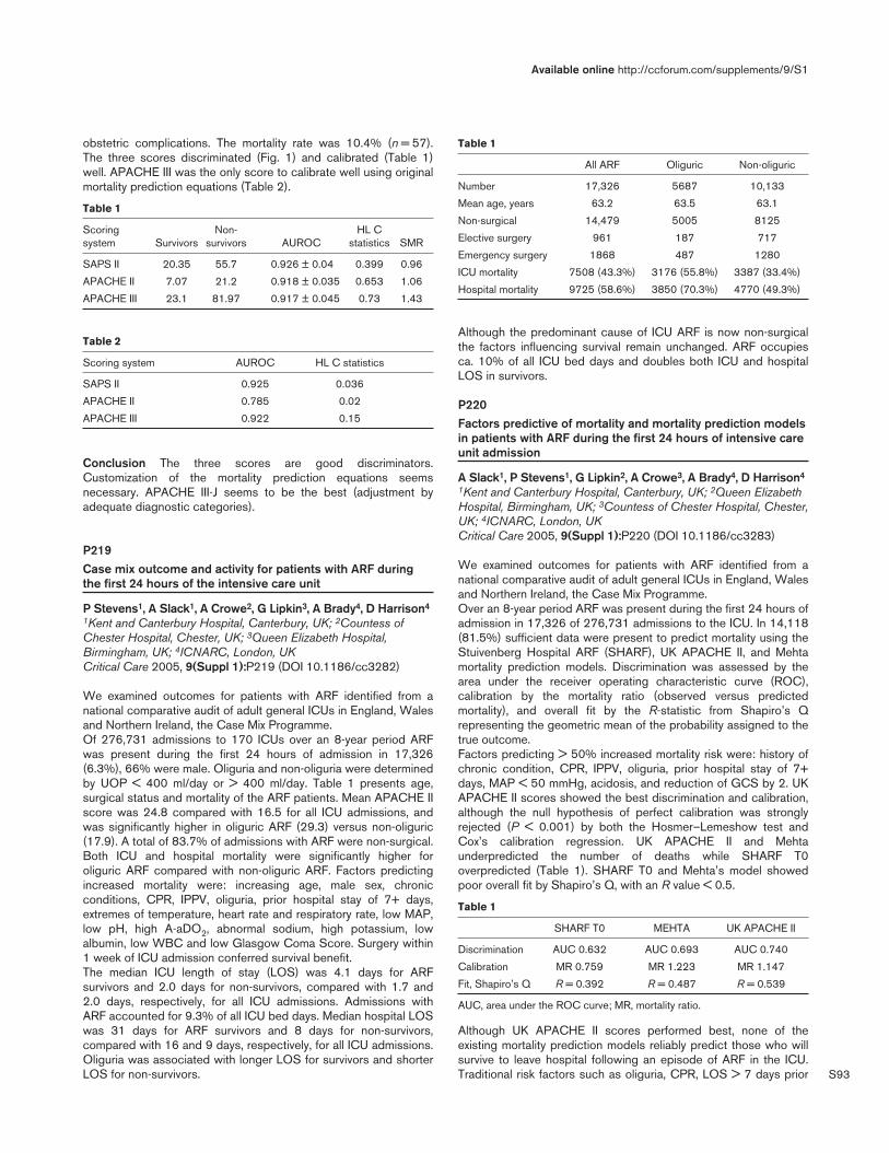
S93
obstetric complications. The mortality rate was 10.4% (n = 57).The three scores discriminated (Fig. 1) and calibrated (Table 1)well. APACHE III was the only score to calibrate well using originalmortality prediction equations (Table 2).
Table 1
Scoring Non- HL C system Survivors survivors AUROC statistics SMR
SAPS II 20.35 55.7 0.926 ± 0.04 0.399 0.96
APACHE II 7.07 21.2 0.918 ± 0.035 0.653 1.06
APACHE III 23.1 81.97 0.917 ± 0.045 0.73 1.43
Table 2
Scoring system AUROC HL C statistics
SAPS II 0.925 0.036
APACHE II 0.785 0.02
APACHE III 0.922 0.15
Conclusion The three scores are good discriminators.Customization of the mortality prediction equations seemsnecessary. APACHE III-J seems to be the best (adjustment byadequate diagnostic categories).
P219Case mix outcome and activity for patients with ARF duringthe first 24 hours of the intensive care unit
P Stevens1, A Slack1, A Crowe2, G Lipkin3, A Brady4, D Harrison4
1Kent and Canterbury Hospital, Canterbury, UK; 2Countess ofChester Hospital, Chester, UK; 3Queen Elizabeth Hospital,Birmingham, UK; 4ICNARC, London, UKCritical Care 2005, 9(Suppl 1):P219 (DOI 10.1186/cc3282)
We examined outcomes for patients with ARF identified from anational comparative audit of adult general ICUs in England, Walesand Northern Ireland, the Case Mix Programme.Of 276,731 admissions to 170 ICUs over an 8-year period ARFwas present during the first 24 hours of admission in 17,326(6.3%), 66% were male. Oliguria and non-oliguria were determinedby UOP < 400 ml/day or > 400 ml/day. Table 1 presents age,surgical status and mortality of the ARF patients. Mean APACHE IIscore was 24.8 compared with 16.5 for all ICU admissions, andwas significantly higher in oliguric ARF (29.3) versus non-oliguric(17.9). A total of 83.7% of admissions with ARF were non-surgical.Both ICU and hospital mortality were significantly higher foroliguric ARF compared with non-oliguric ARF. Factors predictingincreased mortality were: increasing age, male sex, chronicconditions, CPR, IPPV, oliguria, prior hospital stay of 7+ days,extremes of temperature, heart rate and respiratory rate, low MAP,low pH, high A-aDO2, abnormal sodium, high potassium, lowalbumin, low WBC and low Glasgow Coma Score. Surgery within1 week of ICU admission conferred survival benefit.The median ICU length of stay (LOS) was 4.1 days for ARFsurvivors and 2.0 days for non-survivors, compared with 1.7 and2.0 days, respectively, for all ICU admissions. Admissions withARF accounted for 9.3% of all ICU bed days. Median hospital LOSwas 31 days for ARF survivors and 8 days for non-survivors,compared with 16 and 9 days, respectively, for all ICU admissions.Oliguria was associated with longer LOS for survivors and shorterLOS for non-survivors.
Table 1
All ARF Oliguric Non-oliguric
Number 17,326 5687 10,133
Mean age, years 63.2 63.5 63.1
Non-surgical 14,479 5005 8125
Elective surgery 961 187 717
Emergency surgery 1868 487 1280
ICU mortality 7508 (43.3%) 3176 (55.8%) 3387 (33.4%)
Hospital mortality 9725 (58.6%) 3850 (70.3%) 4770 (49.3%)
Although the predominant cause of ICU ARF is now non-surgicalthe factors influencing survival remain unchanged. ARF occupiesca. 10% of all ICU bed days and doubles both ICU and hospitalLOS in survivors.
P220Factors predictive of mortality and mortality prediction modelsin patients with ARF during the first 24 hours of intensive careunit admission
A Slack1, P Stevens1, G Lipkin2, A Crowe3, A Brady4, D Harrison4
1Kent and Canterbury Hospital, Canterbury, UK; 2Queen ElizabethHospital, Birmingham, UK; 3Countess of Chester Hospital, Chester,UK; 4ICNARC, London, UKCritical Care 2005, 9(Suppl 1):P220 (DOI 10.1186/cc3283)
We examined outcomes for patients with ARF identified from anational comparative audit of adult general ICUs in England, Walesand Northern Ireland, the Case Mix Programme.Over an 8-year period ARF was present during the first 24 hours ofadmission in 17,326 of 276,731 admissions to the ICU. In 14,118(81.5%) sufficient data were present to predict mortality using theStuivenberg Hospital ARF (SHARF), UK APACHE II, and Mehtamortality prediction models. Discrimination was assessed by thearea under the receiver operating characteristic curve (ROC),calibration by the mortality ratio (observed versus predictedmortality), and overall fit by the R-statistic from Shapiro’s Qrepresenting the geometric mean of the probability assigned to thetrue outcome.Factors predicting > 50% increased mortality risk were: history ofchronic condition, CPR, IPPV, oliguria, prior hospital stay of 7+days, MAP < 50 mmHg, acidosis, and reduction of GCS by 2. UKAPACHE II scores showed the best discrimination and calibration,although the null hypothesis of perfect calibration was stronglyrejected (P < 0.001) by both the Hosmer–Lemeshow test andCox’s calibration regression. UK APACHE II and Mehtaunderpredicted the number of deaths while SHARF T0overpredicted (Table 1). SHARF T0 and Mehta’s model showedpoor overall fit by Shapiro’s Q, with an R value < 0.5.
Table 1
SHARF T0 MEHTA UK APACHE II
Discrimination AUC 0.632 AUC 0.693 AUC 0.740
Calibration MR 0.759 MR 1.223 MR 1.147
Fit, Shapiro’s Q R = 0.392 R = 0.487 R = 0.539
AUC, area under the ROC curve; MR, mortality ratio.
Although UK APACHE II scores performed best, none of theexisting mortality prediction models reliably predict those who willsurvive to leave hospital following an episode of ARF in the ICU.Traditional risk factors such as oliguria, CPR, LOS > 7 days prior
Available online http://ccforum.com/supplements/9/S1

S94
to admission to ICU and extremes of physiology continue to beassociated with the greatest increased risks of mortality.
P221Mortality and predictors of death in patients with pneumoniaadmitted to the intensive care unit
J Lee, Y Ryu, E Cheon, J ChangEwha Womans University Hospital, Seoul, South KoreaCritical Care 2005, 9(Suppl 1):P221 (DOI 10.1186/cc3284)
Background Community-acquired pneumonia remains a commonand serious condition.Methods We retrospectively evaluated patients with pneumoniaadmitted to the medical intensive care unit (MICU) of EwhaWomans University Hospital from 1 January 2000 through 31December 2003. We excluded patients who died within 48 hoursafter admission, were pretreated before admission or hadpulmonary tuberculosis or suspected nosocomial pneumonia.Finally, 118 patients were included.Results Mortality was 41.5% (49/118). At admission, survivorshad significantly higher white blood cell counts (P < 0.05), lowerserum potassium levels (P < 0.05), larger first 24-hour urine output(P < 0.01), higher Glasgow coma scales (P < 0.001), and lowerAcute Physiology and Chronic Health Evaluation Score (APACHEII) (P < 0.01) than non-survivors. During hospitalization, there wassignificantly lower incidence of gastrointestinal (GI) bleeding insurvivors compared with non-survivors (P < 0.001). MICU stay(P < 0.05) and days requiring mechanical ventilation (P < 0.001)and intubation were significantly shorter in survivors compared withthose in non-survivors (P < 0.01). Tracheostomy was lessfrequently performed in survivors compared with that in non-survivors (P < 0.01). In survival analysis using Cox regression, GIbleeding (relative risk [RR], 2.35; 95% confidence interval [CI],1.24–4.48) and an APACHE II score of 20 or more (RR, 2.19;95% CI, 1.21–3.96) were independently associated with death.Conclusion In patients with severe pneumonia, mortality was stillhigh. GI bleeding during hospitalization as well as high APACHE IIscore was related to mortality.
P222Survival and prognosis after assisted ventilation for acuterespiratory failure in cystic fibrosis
S van Gestel1, M Slieker1, G Tramper1, F Teding van Berkhout2, H Heijerman3, K van der Ent1, K Jansen1
1UMC Utrecht, Wilhelmina Children’s Hospital, Utrecht, TheNetherlands; 2UMC Utrecht, The Netherlands; 3LeyenburgHospital, The Hague, The NetherlandsCritical Care 2005, 9(Suppl 1):P222 (DOI 10.1186/cc3285)
Introduction The use of mechanical ventilation for acuterespiratory failure (ARF) in patients with cystic fibrosis (CF) haslong been discouraged because of a poor outcome in this group ofpatients. However, new insights into ventilatory strategies and abetter life expectancy per se in CF patients necessitate regular re-evaluation of this attitude. The most recent studies report amortality in adult CF patients requiring mechanical ventilation thatvaries widely between 45% and 80%. Results in children arereported as more favourable, but little is known about the long-termpulmonary outcome of children with CF who required mechanicalventilation for ARF already at a young age.Objectives The goals of this study were threefold: to assessoutcome of assisted ventilation in CF patients with ARF; to identify
risk factors associated with a poor outcome; and to analysewhether children with CF who required mechanical ventilation forARF at a young age had more pulmonary and CF-relatedcomplications later in life compared with unventilated CF controls.Design A retrospective cohort study.Setting The study was performed at the University Medical CentreUtrecht (Utrecht, The Netherlands) and at the Leyenburg Hospital(The Hague, The Netherlands). These centres care forapproximately one-half of the Dutch CF population.Methods Medical charts were reviewed of all CF patients who hadbeen admitted to the intensive care unit for ARF between January1990 and August 2003. ARF was defined as a respiratorydeterioration requiring assisted ventilation, due to an acute illnessin a previously stable patient. Assisted ventilation could be eithernon-invasive or invasive. Patients with chronic respiratory failurewith a slow progressive decline in lung function were not included.To identify risk factors for mortality, the following data wererecorded: demographic data, body mass index, spirometric data,mode of ventilation, sputum microbiology, history of hemoptysis orpneumothorax, the presence of CF-related diabetes and thepresence of CF-related liver disease.To analyse long-term outcome in children who survived assistedventilation for ARF, all unventilated age-matched and gender-matched CF controls were identified. Lung function and thepresence of CF-related complications 5 years after admission tothe ICU were compared.Results Thirty-two CF patients were included, five children (aged2–18 months) and 27 adults (aged 15 years and older). Allchildren and 30% of the adults survived.In the total population, age was a statistically significant risk factorfor poor outcome (P = 0.03). In the adult population, none of thedemographic and clinical data could predict outcome significantly.In patients who had an episode of ARF in childhood, clinicalcourse and lung function 5 years after assisted ventilation was notsignificantly different compared with their controls.Conclusions CF patients aged 2 years or younger who areventilated because of ARF have a good prognosis and assistedventilation does not seem to affect lung function or development ofCF-related complications later in their lives. ARF in adult CFpatients still is associated with a high mortality. Risk factors forpoor outcome could not be identified.
P223Outcome of septic shock in patients with malignancies andneutropenia
D FurmanchukBelorussian Medical Academy for Postgraduate Education, Minsk,BelarusCritical Care 2005, 9(Suppl 1):P223 (DOI 10.1186/cc3286)
Background Septic complication remains an important influenceon survival rate in patients with malignancies. Bone marrowsuppression and neutropenia induced by chemotherapy determinecertain difficulties in early diagnose of sepsis, and lead to rapidhemodynamics and respiratory disorders. The aim of this study wasto standardize most important issues in treatment of sepsis-induced arterial hypotension and tissue hypoperfusion and toestimate its influence on survival rate in patients with severe sepsisand septic shock.Materials and methods Sixty-two consecutive patients aged from1 to 17 years old, with malignancies, neutropenia and sepsisassociated with hypotension, were followed in the ICU of theRepublican Center for Pediatric Oncology and Hematology. Thirty-five patients treated from January 2000 to December 2001
Critical Care March 2005 Vol 9 Suppl 1 25th International Symposium on Intensive Care and Emergency Medicine

S95
composed the retrospective group; the prospective groupconsisted of 27 patients, treated from March 2003 to September2004. Treatment protocol for septic shock therapy used EBMprinciples and the design for the prospective group strictlydetermined the following treatment options: hydroxyethyl starch(HES) as a first choice for volume resuscitation; combination ofdopamine and norepinephrine for correction of arterialhypotension; double-lumen catheter (or two central lines) fordividing infusions of dopamine and noradrenalin from others;mandatory use of arterial lines for invasive blood pressuremonitoring; and start-up of mandatory ventilation in all patients withseptic shock, even in patients with compensated blood gas status,We compared the survival rate and survival probability during thefirst 75 days after ICU admission. For the statistical analysis, thechi-square test and the Caplan–Meyer method were used.Results Survival probability measured by log-rank test during 75days after ICU admission showed a significant difference. Usingour protocol, we also found a difference in maximal lactate level onthe day of admission, duration of arterial hypotension, length of ICUstay and length of ventilation between two groups.The overall ICU mortality rate was significantly different in theretrospective and prospective groups — 81.3% and 53%,respectively (P < 0.05).
Table 1
Retrospective Prospective Mortality rate group group P value
At first 24 hours 28% 7.6% 0.04
At day 7 80% 26.9% 0.001
At day 30 82.8% 53.8% 0.01
Conclusion Using the presented treatment protocol for septicshock positively influenced mortality rate in the prospective group.
P224A validation of different prognostic scoring systems in theprediction of outcome in peritonitis
D Borisov, M Kirov, V Kuzkov, D Uvarov, E NedashkovskyNorthern State Medical University, Arkhangelsk, RussiaCritical Care 2005, 9(Suppl 1):P224 (DOI 10.1186/cc3287)
Background and goal Early classification of patients withperitonitis by means of scoring systems provides adequateselection for surgical and therapeutic procedures and comparisonof different therapies [1]. We compared the prognostic value ofAPACHE II and SAPS II scoring systems as well as the MannheimPeritonitis Index (MPI) for assessment of outcome in the patientswith diffuse bacterial peritonitis.Materials and methods We enrolled 194 adult patients (meanage 49.6 ± 19.5 years) with secondary diffuse bacterial peritonitisin a prospective observational study during the period from 1999to 2003. All patients were hospitalized in the ICU of the universityhospital. The scores according to APACHE II, SAPS II, and MPIwere assessed during 24 hours after admission to the ICU. Thepredictive values of the scores were estimated using regressionanalysis (coefficient of determination r2). The discrimination wasassessed using areas under the receiver operating characteristiccurves (AUCs). Standardized mortality ratios (SMRs) werecalculated. Variables were expressed as mean ± standarddeviation or 95% confidence interval and as relative frequencies.Results and discussions The mean APACHE II, SAPS II and MPIscores were 9.6 ± 8.4, 28.8 ± 16.7, and 23.9 ± 6.9, respectively.
The hospital mortality rate was 19.1% (37 patients). The predictedmortality risk was 18.9%, 9.6%, and 33.6% for APACHE II(diagnostic category weight for gastrointestinal perforation/obstruction), SAPS II and MPI, respectively. The values ofcalibration (P < 0.05) and discrimination and SMR are summarizedin Table 1.
Table 1
r2/residual AUC/95% standard deviation confidence interval SMR
APACHE II 0.87/12.1 0.87/0.82–0.92 1.01
SAPS II 0.89/11.7 0.82/0.76–0.87 1.99
MPI 0.38/24.3 0.85/0.79–0.90 0.57
The calibration was adequate for APACHE II and SAPS II scores.The discrimination was good for all systems. The APACHE II scorehad the most accurate overall mortality prediction, as reflected bythe SMR.Conclusion In patients with diffuse bacterial peritonitis, APACHE IIis the most accurate prognostic scoring system as compared withSAPS II and MPI.Reference1. Bosscha K, van Vroonhoven ThJMV, van der Werken Ch: Br J
Surg 1999, 86:1371-1377.
P225APACHE III outcome prediction after esophagectomy
M Keegan, A Jacob, S Cassivi, F Whalen, D Brown, T Roy, B AfessaMayo Clinic College of Medicine, Rochester, MN, USACritical Care 2005, 9(Suppl 1):P225 (DOI 10.1186/cc3288)
Introduction The Acute Physiology and Chronic Health Evaluation(APACHE) III prognostic system has not been validated in patientsadmitted to the ICU after esophagectomy.Hypothesis APACHE III predicts hospital mortality afteresophagectomy.Methods A retrospective review of all adult patients admitted to asingle thoracic ICU after esophagectomy between October 1994and December 2003. Patient demographics, ICU admission dayAPACHE III score, actual and predicted hospital mortality andlength of hospital and ICU stays were collected for the first ICUadmission only. Performance of the APACHE III prognostic systemwas assessed by the Hosmer–Lemeshow statistic for calibrationand the area under the receiver operating characteristic curve(AUC) for discrimination.Results There were 924 esophagectomies performed during thestudy period. Data are presented for the 483 patients that wereadmitted to the ICU. Mean age was 63.9 years. Mean APACHE IIIscore on the day of ICU admission was 41.5 (standard deviation18.1). Mean predicted (standard deviation) ICU and hospitalmortality rates were 3.01% (6.31) and 7.90% (11.0), respectively.Median (interquartile range) lengths of ICU and hospital stay were1.68 (0.79–3.79) and 13.52 (12.0–21.0) days, respectively.Observed ICU and hospital mortality rates were 2.7% (13 of 483patients) and 5.4% (26 of 483), respectively.There were differences (P < 0.001) between survivors to hospitaldischarge and non-survivors in age, acute physiology score andAPACHE III score. Predicted ICU and hospital survival rates on theday of ICU admission were also different when survivors werecompared with non-survivors (P < 0.001). Although most patientswere male (82.6%), gender did not predict survival. The mean
Available online http://ccforum.com/supplements/9/S1

S96
(95% confidence interval [CI]) ICU and hospital length of stayratios (observed/predicted) were 0.88 (0.76–1.00) and 1.01(0.92–1.10), respectively.The standardized ICU and hospital mortality ratios (95% CI), basedon APACHE III prediction, were 0.89 (0.41–1.38) and 0.68(0.42–0.94), respectively. In predicting mortality, the AUC ofAPACHE III prediction was 0.860 (95% CI 0.791–0.928) and theHosmer–Lemeshow statistic was 8.581 with a P value of 0.379.Conclusions The APACHE III prognostic scoring system has gooddiscrimination and calibration in predicting hospital mortality ofpatients admitted to the ICU following esophagectomy. The lownumber of deaths may have influenced the statistical analyses.
P226Multimodal strategies to improve APACHE II scoredocumentation
L Donahoe1, M Kho1, E McDonald1, M Maclennan2, S McIntyre2,P Varga2, D Cook1
1McMaster University, Hamilton, Canada; 2St Joseph’s Healthcare,Hamilton, CanadaCritical Care 2005, 9(Suppl 1):P226 (DOI 10.1186/cc3289)
Background The APACHE II score is used widely in the ICUsetting. In our phase I study [1], APACHE II scores collected by anexpert research coordinator and two research clerks were reliable(intraclass correlation coefficient = 0.90, lower 95% confidenceinterval [L-95% CI] = 0.85). However, we found substantialvariability in the Chronic Health Index (CHI) (0.67, L-95% CI =0.53) and Glasgow Coma Scale, verbal component (GCS-V)(0.42, L-95% CI = 0.25). To improve the reliability of the APACHEII score, we conducted phase II, aimed at changing the behaviourof ICU clinicians who are involved in documenting the APACHE IIscore in practice.Objective To educate ICU clinicians regarding two specificcomponents of the APACHE II score with suboptimal reliability:CHI and GCS-V.Design An educational quality improvement interventional project.Population ICU clinicians, primarily bedside nurses.Methods We convened a combined clinical and administrativeworking group. In-person meetings, conference calls, andelectronic communication were used to generate ideas forstrategies that would efficiently serve our objective.Results We implemented multimodal strategies to try to improveICU clinicians’ knowledge of, and compliance with, documentationof CHI and GCS-V. Strategies were: re-structuring the ClinicalInformation System Carevue (Philips, Andover, MA, USA), in-service education, use of local opinion leaders, reminders, auditand feedback, and support from the ICU Working Group (MDs,Allied Health professionals, bedside RNs, RN manager, RNeducator, RN informatician [RNI]) who approved an improved CHIpolicy. We reconfigured the Carevue workstation screens toimprove the visibility of CHI. In-services were conducted by an ICUeducator and RNI. Education focused on the CHI components,GCS-V domain in intubated patients, and expectations fordocumentation of these items. As local opinion leaders, RNs(manager and charge nurses) prompted bedside nurses tocomplete chronic health documentation for all new admissions.We provided written information sheets, electronic resources, andcomputer-generated electronic reminders at RN workstations andon Carevue. The RNI provided regular, informal audit and feedbackto bedside RNs to reinforce timely documentation.Conclusions These diverse methods were delivered over 1 year,were multiply redundant to maximize the chance of behaviourchange, were led by an integrated team of clinical and
administrative managers, and were well accepted. We will nextproceed to phase III, a formal re-evaluation of the reliability of theAPACHE II score in our institution.Reference1. Crit Care Med 2004, 31(suppl):A68.
P227Comparison of Acute Physiology and Chronic HealthEvaluation (APACHE II) and Simplified Acute PhysiologyScore (SAPS II) in a Greek general intensive care unit
K Gkolfinopoulou, G Kouvatseas, A Armaganidis, U DafniNational and Kapodistrian University of Athens, GreeceCritical Care 2005, 9(Suppl 1):P227 (DOI 10.1186/cc3290)
Objectives To determine the applicability of the Acute Physiologyand Chronic Health Evaluation (APACHE II) scoring system, aswell as that of the Simplified Acute Physiology Score (SAPS II) inan interdisciplinary intensive care unit in Greece and to evaluatethe scores’ ability to predict hospital mortality.Design A prospective cohort study.Setting A mixed medical and surgical intensive care unit at a majorgeneral hospital in the Athens Metropolitan area, Greece.Patients A total of 309 adult patients, admitted to the ICU fromJanuary 2001 to January 2002, were enrolled in the study.Methods and results Data enabling the calculation of APACHE IIand SAPS II were collected for every patient on the day ofadmission. Patients’ vital status at hospital’s discharge wasrecorded. Probabilities of hospital death for patients wereestimated by applying APACHE II and SAPS II, and predicted risksof hospital death were compared with observed outcomes. Thepatients’ median age was 57 years old. The median APACHE IIscore was 15, higher in the 105 female patients (16) than in the204 male patients (15). The median APACHE II estimatedprobability of death was 0.16 (0.2 for female patients and 0.14 formale patients). The median APACHE II was lower in the 168operative cases (14) than in the 141 medical ones (16). Themedian APACHE II estimated probability of death was respectively0.13 and 0.20 for the two groups. The median SAPS II score was37 (40 for females and 36 for males, 35 for operative patients and41 for medical patients). The median SAPS II estimated probabilityof death was 0.2 (0.25 for females and 0.18 for males, 0.17 foroperative cases and 0.27 for medical cases). The overall goodnessof fit and the areas under the receiver operating characteristiccurve were assessed for the two scores.
P228The impact of missing components of the Acute PhysiologyScore on the standardized mortality ratio calculated by theAPACHE III prognostic model
B Afessa, M Keegan, O Gajic, R Hubmayr, S PetersMayo Clinic College of Medicine, Rochester, MN, USACritical Care 2005, 9(Suppl 1):P228 (DOI 10.1186/cc3291)
Introduction In the APACHE III prognostic model, a weight of zerois given to missing and normal Acute Physiology Score (APS)values. The standardized mortality ratio (SMR) is used to evaluatethe performance of an ICU. The objective of this study was todetermine the impact of missing APS values on SMR.Methods The study involved a retrospective analysis of theprospectively collected APACHE III database. The studypopulation included adults treated in four intensive care units of atertiary medical center with the APACHE III database. We obtainedthe APACHE III predicted mortality rate and hospital outcome. We
Critical Care March 2005 Vol 9 Suppl 1 25th International Symposium on Intensive Care and Emergency Medicine

S97
noted whether the first day values of the variables used to calculatethe APS were missing. We created eight categories based on thenumber of missing values. We summarized descriptive data asmean (standard deviation [SD]), median (interquartile range [IQR])and percentage. We created a logistic regression model todetermine the association of the number of missing values withhospital mortality by entering the number of missing APS variablesand the APACHE III predicted mortality rate as predictor variables.P < 0.05 was considered significant.Results We had 49,682 admissions during the study period.Excluding patients who did not authorize their medical records tobe reviewed for research and those with four or less availablelaboratory values, 42,607 patients were included in the study.Patients’ mean (SD) age was 62.7 (17.3) years; 40,417 (94.9%)were white and 24,294 (57.0%) were male. The overall predictedand observed hospital mortality rates were 9.7% (4151/42607)and 12.0%, respectively, with SMR (95% confidence interval [CI])of 0.813 (0.789–0.838). The median (IQR) predicted hospitalmortality rate was 4.36 (1.65–13.20)%. Complete data wereavailable in 934 (2.2%) admissions. The predicted mean andobserved mortality rates as well as the SMR with the 95% CI foreach of the missing categories are presented in Table 1. Four ormore missing APS values were independently associated withhigher observed hospital mortality.
Table 1
Missing Observed dead Predicted dead SMR (95%CI)
Zero 183/934 (19.6%) 25.8% 0.76 (0.65–0.88)
One 565/3607 (15.7%) 19.6% 0.80 (0.73–0.87)
Two 952/9486 (10.0%) 13.7% 0.73 (0.69–0.78)
Three 978/11641 (8.4%) 10.4% 0.81 (0.76–0.86)
Four 753/8717 (8,6%) 9.8% 0.88 (0.82–0.95)
Five 579/6799 (8.5%) 9.5% 0.90 (0.82–0.97)
Six 127/1338 (9.5%) 10.1% 0.94 (0.79–1.12)
Seven or eight 14/85 (16.5%) 8.9% 1.85 (1.01–3.11)
Conclusions The majority of patients admitted to the ICU have oneor more missing laboratory values needed to calculate the APSscores of the APACHE III prognostic system. When using suchprognostic systems to evaluate the performance of ICUs, thenumber of missing values should be taken into consideration.
P229Preliminary update of the Mortality Probability Model (MPM0)
T Higgins1, D Teres2, W Copes3, B Nathanson4, M Stark3, A Kramer3
1Baystate Medical Center, Springfield, MA, USA; 2Tufts University,Boston, MA, USA; 3Cerner Corporation, Kansas City, MO, USA;4OptiStatim, Longmeadow, MA, USACritical Care 2005, 9(Suppl 1):P229 (DOI 10.1186/cc3292)
Introduction The Mortality Probability Model (MPM II), developedon an international sample of 12,610 patients in 1989–1990, isused by Project IMPACT as a benchmarking tool. We updated themodel based on more recent (2001–2004) data.Hypothesis and methods Project IMPACT data on 125,610patients age >18 and eligible for MPM scoring were analyzed.Multivariate analysis defined the relationship between hospitalmortality and standard MPM physiologic variables plus patienttype, location and lead time prior to ICU admission. The samplewas randomly split into development and validation sets.
Discrimination was assessed by ROC C statistic and calibration bygraphic display and Hosmer–Lemeshow goodness of fit.Results Overall mortality was 13.8%. The logistic model for allpatients is presented in Table 1, and goodness of fit in Fig. 1. Thearea under the ROC curve was 0.82. Lead time and location didnot influence outcome. Addition of a ‘zero-factor’ term for patientswith no risk factors other than age improved model performance.Subgroup models (medical, coronary, trauma, neurosurgical,elective and emergent non-neuro, non-cardiac and non-traumasurgery) exhibit improved discrimination and calibration comparedwith the main model, which is superior in calibration to the existingMPM model.
Table 1
Variable Odds ratio Coefficients P value
Coma-stupor 5.37 1.680172 0.000
HR ≥ 150 1.77 0.570394 0.000
SBP < 90 2.49 0.9111615 0.000
Chronic renal 1.68 0.5179099 0.000
Cirrhosis 2.18 0.7804761 0.000
Metastasis 2.69 0.9889827 0.000
Acute renal 2.17 0.7752536 0.000
Arrythmia 1.08 0.0782759 0.000
Cerebrovascular 1.31 0.2679498 0.000
GI bleed 0.84 –0.1712258 0.003
IC mass 2.16 0.768795 0.000
Age 1.03 0.0302588 0.000
CPR w/in 24 hours 2.20 0.7888974 0.000
Mechanical ventilation 2.25 0.8123237 0.000
Med/unsched S 2.40 0.8760618 0.000
Zero factors 0.79 –0.2368908 0.007
Full code 0.46 –0.7693753 0.000
Constant NA –4.778739 0.000
Conclusions Severity-adjusted mortality has decreased over time.Use of the updated model will allow more accurate assessment ofquality of care. Subgroup models further improve discriminationand calibration and offer additional information in ICUs where thecase mix is unusual.
Available online http://ccforum.com/supplements/9/S1
Figure 1 (abstract P229)

S98
P230Mortality prediction by hormonal profile versus APACHE scoreassessment in critically ill patients
K Osman Hassan, S El-Maraghy, S El-Gengehy, D Ragab, K Abd El-Aziz, S Aref, H Mowafi, H NagiCairo University, EgyptCritical Care 2005, 9(Suppl 1):P230 (DOI 10.1186/cc3293)
Objectives To investigate hormonal changes during the acutephase of critical illness, to analyze their relationship to severity, andto evaluate their discriminating ability on critically ill patients’outcome.Design A prospective cohort study of patients requiring intensivecare.Setting Adult medical, surgical and coronary care units inMansoura University Emergency Hospital.Patients A total of 40 consecutive patients requiring intensive careover a 5-month period and 10 healthy subjects of matched ageand sex were selected as references control subjects.Interventions For each patient included in our study the followingwas done. First, APACHE III score. Second, blood samples weretaken in three occasions, on admission, third and seventh days ofadmission to the ICU. The plasma was separated and kept at–80°C until time of analysis. The following hormones (cortisol,growth hormone, thyroxin, and triiodothyronine hormones) weredetermined by ELISA technique.Measurements and main results The best discriminators ofpatient outcome in descending order were the serum basalcortisol, then APACHE III score, then thyroxine hormone, thentriiodothyronine on day 1 of admission with odd ratios of 11, 8.5,3.2 and 2.85, respectively.Conclusion Certain endocrinal parameters, mainly basal serumcortisol level obtained during ICU admission, might be simple,cheap and superior discriminators of patient outcome than theAPACHE III score.
P231Serial evaluation of the Sequential Organ Failure Assessmentscore in non-coronary medical patients
M Wehler, U Reulbach, E Hahn, R StraussUniversity of Erlangen–Nuremberg, Erlangen, GermanyCritical Care 2005, 9(Suppl 1):P231 (DOI 10.1186/cc3294)
Introduction To determine the usefulness of daily SequentialOrgan Failure Assessment (SOFA) score [1] for prediction ofmortality in non-coronary medical ICU patients.Patients and methods A prospective, observational study wasconducted from May 1997 to April 2001. Nine hundred andnineteen consecutive adult patients admitted to the ICU for morethan 24 hours were included (10,164 patient days). The SOFAscore was calculated on admission and every 24 hours untildischarge.Results Study patients had a mean age of 59 ± 17 (± standarddeviation) years; 70% were male, mean ICU length of stay was11 ± 15 days, mean APACHE II score after 24 hours was 19 ± 10.Initial, maximum and mean (sum of daily SOFA scores dividedthrough days of ICU stay) SOFA scores correlated well withmortality in the ICU (25.7%). The ability of the SOFA todiscriminate between ICU survivors and non-survivors wasassessed using the area under the receiver operatingcharacteristic curve (AUC). The AUC was largest for the meanSOFA score (0.90, 95% confidence interval: 0.88–0.93) and themaximum SOFA score (0.88, 95% confidence interval:
0.86–0.91). A mean SOFA score of more than 7 corresponded toa mortality of more than 83% versus 10% for a mean score of 7 orless. When analyzing trends in the first 48 hours, regardless of theinitial score, the mortality was 56% when the score increased,24% when it remained unchanged, and less than 20% when itdecreased.Conclusion Sequential assessment of organ dysfunction duringthe first few days of ICU admission is a good indicator ofprognosis. Both the mean and the maximum SOFA scores areparticularly useful predictors of ICU outcome. Independent of theinitial score, an increase in SOFA score during the first 48 hourspredicted in our sample a mortality rate of 56%.Reference1. Vincent JL, et al.: Intensive Care Med 1996, 22:707-710.
P232Association of the SOFA score and mortality in elderlypatients with severe sepsis and septic shock
R Machado1, C David1, G Oliveira2, P Godoy2, R Nagato2
1Universidade Federale, Rio De Janeiro, Brazil; 2Prontocor, Rio DeJaneiro, BrazilCritical Care 2005, 9(Suppl 1):P232 (DOI 10.1186/cc3295)
Introduction The SOFA score is an excellent predictive marker ofoutcome in sepsis; however, there are very few studies that relate itto mortality in elderly patients with sepsis and septic shock.Objective To assess the association of the SOFA score and otherfactors related to mortality in elderly patients with severe sepsisand septic shock.Methods A 3-month prospective cohort study of 30 patients aged≥ 65 showing severe sepsis or septic shock (ConsensusConference of 1991). Arterial hypotension (systolic arterialpressure < 90 mmHg) corresponded to the ‘time-zero’ of the study.The variables used were: SOFA score on days 1, 3, 5, 7, 14, and28, ∆SOFA (variation of the SOFA score on days 1 and 3),APACHE II, Troponin I dosage, BNP and RCP, plasma glucoselevels, organ failures [1], the presence of previous cardiovasculardisease, assessment of dependence and cognitive deficit [2,3],length of ICU stay, and need for mechanical ventilation. We usedStudent’s t test and the Fischer Exact test for a statistical analysis.We considered the significance level of 5%.Results The mean age of patients, of whom 60% were females,was 82 ± 9 years (minimum = 65, maximum = 99). The predominantdiagnosis was septic shock in 67% of the cases, while 33% of thepatients developed severe sepsis. On days 1, 3, 5, 7, 14, and 28,the SOFA score presented mean values of 7, 6, 4, 3, 2, and 2,respectively (minimum = 2 and maximum = 15), thus evidencing asignificant relationship between the SOFA score on day 1(P = 0.0001) and day 3 (P = 0.001), including ∆SOFA score(P = 0.043), and mortality. The number of failures was alsoassociated with mortality when two or more organic failures(P = 0.001) were present. Age, gender, APACHE II, length of ICUstay, dependence level, presence of cognitive deficit and/orprevious cardiovascular diseases, plasma glucose levels, troponinI, BNP and RCP were not associated with the mortality.Conclusion Mean SOFA average above 5 as well as SOFAvariation within the first 72 hours proved to be good predictivemarkers in elderly patients with septic shock and severe sepsis.The same occurred in the presence of two or more organic failuresduring the course of sepsis.References1. Le Gall JR, Brun-Buisson C, Trunet P, Laturnerie J, Chantereau S,
Rapin M: Influence of age, previous health status, and severityof acute illness on outcome from intensive care. Crit Care
Critical Care March 2005 Vol 9 Suppl 1 25th International Symposium on Intensive Care and Emergency Medicine
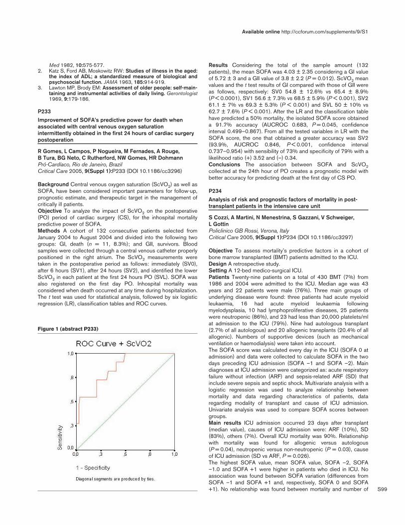
S99
Med 1982, 10:575-577.2. Katz S, Ford AB, Moskowitz RW: Studies of illness in the aged:
the index of ADL; a standardized measure of biological andpsychosocial function. JAMA 1963, 185:914-919.
3. Lawton MP, Brody EM: Assessment of older people: self-main-taining and instrumental activities of daily living. Gerontologist1969, 9:179-186.
P233Improvement of SOFA’s predictive power for death whenassociated with central venous oxygen saturationintermittently obtained in the first 24 hours of cardiac surgerypostoperation
R Gomes, L Campos, P Nogueira, M Fernades, A Rouge, B Tura, BG Neto, C Rutherford, NW Gomes, HR DohmannPró-Cardíaco, Rio de Janeiro, BrazilCritical Care 2005, 9(Suppl 1):P233 (DOI 10.1186/cc3296)
Background Central venous oxygen saturation (ScVO2) as well asSOFA, have been considered important parameters for follow-up,prognostic estimate, and therapeutic target in the management ofcritically ill patients.Objective To analyze the impact of ScVO2 on the postoperative(PO) period of cardiac surgery (CS), for the inhospital mortalitypredictive power of SOFA.Methods A cohort of 132 consecutive patients selected fromJanuary 2004 to August 2004 and divided into the following twogroups: GI, death (n = 11, 8.3%); and GII, survivors. Bloodsamples were collected through a central venous catheter properlypositioned in the right atrium. The ScVO2 measurements weretaken in the postoperative period as follows: immediately (SV0),after 6 hours (SV1), after 24 hours (SV2), and identified the lowerScVO2 in each patient at the first 24 hours PO (SVL). SOFA wasalso registered on the first day PO. Inhospital mortality wasconsidered when death occurred at any time during hospitalization.The t test was used for statistical analysis, followed by six logisticregression (LR), classification tables and ROC curves.
Results Considering the total of the sample amount (132patients), the mean SOFA was 4.03 ± 2.35 considering a GI valueof 5.72 ± 3 and a GII value of 3.8 ± 2.2 (P = 0.012). ScVO2 meanvalues and the t test results of GI compared with those of GII wereas follows, respectively: SV0 54.8 ± 12.6% vs 65.4 ± 8.9%(P < 0.0001), SV1 56.6 ± 7.3% vs 68.5 ± 5.9% (P < 0.001), SV261.1 ± 7% vs 69.3 ± 5.3% (P < 0.001) and SVL 50 ± 10% vs62.7 ± 7.6% (P < 0.001). After the LR and the classification tablehave predicted a 50% mortality, the isolated SOFA score obtaineda 91.7% accuracy (AUCROC 0.683, P = 0.045, confidenceinterval 0.499–0.867). From all the tested variables in LR with theSOFA score, the one that obtained a greater accuracy was SV2(93.9%, AUCROC 0.846, P < 0.001, confidence interval0.737–0.954) with sensibility of 73% and specificity of 79% with alikelihood ratio (+) 3.52 and (–) 0.34.Conclusions The association between SOFA and ScVO2collected at the 24th hour of PO creates a prognostic model withbetter accuracy for predicting death at the first day of CS PO.
P234Analysis of risk and prognostic factors of mortality in post-transplant patients in the intensive care unit
S Cozzi, A Martini, N Menestrina, S Gazzani, V Schweiger, L GottinPoliclinico GB Rossi, Verona, ItalyCritical Care 2005, 9(Suppl 1):P234 (DOI 10.1186/cc3297)
Objective To assess mortality’s predictive factors in a cohort ofbone marrow transplanted (BMT) patients admitted to the ICU.Design A retrospective study.Setting A 12-bed medico-surgical ICU.Patients Twenty-nine patients on a total of 430 BMT (7%) from1986 and 2004 were admitted to the ICU. Median age was 43years and 22 patients were male (76%). Three main groups ofunderlying disease were found: three patients had acute myeloidleukaemia, 16 had acute myeloid leukaemia followingmyelodysplasia, 10 had lymphoproliferative diseases, 25 patientswere neutropenic (86%), and 23 had less than 20,000 platelets/mlat admission to the ICU (79%). Nine had autologous transplant(2.7% of all autologous) and 20 allogenic transplants (20.4% of allallogenic). Numbers of supportive devices (such as mechanicalventilation or haemodialysis) were taken into account.The SOFA score was calculated every day in the ICU (SOFA 0 atadmission) and data were collected to calculate SOFA in the twodays preceding ICU admission (SOFA –1 and SOFA –2). Maindiagnoses at ICU admission were categorized as: acute respiratoryfailure without infection (ARF) and sepsis-related ARF (SD) thatinclude severe sepsis and septic shock. Multivariate analysis with alogistic regression was used to analyze relationship betweenmortality and data regarding characteristics of patients, dataregarding modality of transplant and cause of ICU admission.Univariate analysis was used to compare SOFA scores betweengroups.Main results ICU admission occurred 23 days after transplant(median value), causes of ICU admission were: ARF (10%), SD(83%), others (7%). Overall ICU mortality was 90%. Relationshipwith mortality was found for allogenic versus autologous(P = 0.04), neutropenic versus non-neutropenic (P = 0.03), causeof ICU admission (SD vs ARF, P = 0.026).The highest SOFA value, mean SOFA value, SOFA –2, SOFA–1.0 and SOFA +1 were higher in patients who died in ICU. Noassociation was found between SOFA variation (differences fromSOFA –1 and SOFA +1 and, respectively, SOFA 0 and SOFA+1). No relationship was found between mortality and number of
Available online http://ccforum.com/supplements/9/S1
Figure 1 (abstract P233)

S100
supportive devices, presence of severe thrombocytopenia, andunderlying disease.Conclusions Our data support previously known risk factors forICU mortality in BMT recipients. SOFA was confirmed to welldescribe organ failure and predict mortality. No cutoff value wasfound to be highly suggestive for bad prognosis. Non-neutropenicautologous BMT recipients admitted to the ICU for non-sepsis-related acute pathologic event has a relatively good prognosis.
P235Outcome of patients with systematic lupus erythematosus inthe intensive care unit
A Alzeer, A Al-ArfajKing Khalid University Hospital, Riyadh, Saudi ArabiaCritical Care 2005, 9(Suppl 1):P235 (DOI 10.1186/cc3298)
The objective of the study was to identify the causes, outcome andprognosis of severe illness in patients with systematic lupuserythematosus (SLE) requiring ICU care in a university hospitalover a 5-year period. The design was a cohort study. Forty-eightSLE patients requiring ICU management over a 5-year period(January 1997–December 2001) were studied prospectively. Of48 patients, 14 (29.2%) died, predominantly with multiorgandysfunction syndrome (MODS). Patients whose APACHE II scorewas ≥ 20 had higher mortality than those with APACHE score< 20 (60% vs 7.1%; P < 0.01). All 18 patients whose health statuswas rated as ‘good’ survived, while 46.7% of 30 patients whosehealth was rated as ‘poor’ died (P < 0.01). Patients who hadthrombocytopenia associated with sepsis and/or disseminatedintravascular coagulopathy had the highest mortality (75%, 5-yearsurvival). In conclusion, SLE patients admitted to the ICU had alower mortality rate than some of the previous reports. Patientswith SLE with high APACHE score > 20, poor health status,thrombocythopenia and MODS had poor prognosis in the ICU.
P236The outcome and follow-up of trauma in elderly intensive careunit patients
A Katsaros, V Gougoutas, S Karatzas, G Bartzis, P Athanasiou,V Zidianakis, P Myrianthefs, I Pastos, G Baltopoulos‘KAT’ Hospital, Athens, GreeceCritical Care 2005, 9(Suppl 1):P236 (DOI 10.1186/cc3299)
Introduction Geriatric trauma is steadily expanded, mainly as aresult of increased vehicle accidents. The outcome of geriatrictrauma is considered significantly worse in comparison with that ofyounger people. Although the cutoff of age above which a patientis grouped as a geriatric one on a pathophysiological basis is notclear, patients above 65 years old are generally accepted asbelonging to the geriatric group. In Greece, trauma is a verycommon disease and elderly patients are affected in a continuouslyincreased manner. In our retrospective study we have recorded theincidence, ICU, hospital and home mortality as well as an approachto the quality of life of geriatric trauma critically ill patients after aperiod up to 5 years after ICU discharge.Methods All multi-trauma patients over 65 years of age admitted ina seven-bed university ICU during the period 2000–2004 wererecorded retrospectively. The collected data include: ICU mortality,hospital mortality, home mortality, and gross estimation of quality oflife, 1–58 months after ICU discharge. For this estimation we useda simplified questionnaire on the basis of that published byFernandez and colleagues [1], and the modified criteria arepresented in Table 1. Each of them had the answer ‘yes = 1 or no
= 0’ and the range can be between 0 and 10. The information forthe outcome of patients outside the hospital was taken bytelephone communication of ICU physicians with the patients ortheir relatives.
Table 1
Dressing
Psychology
Total
Patient
At home?
Oral communication
Urination control
Defecation control
Food adequacy
Personal cleaning
Home movement
Outside movement
Results During a nearly 5-year period (lasting from 2 March 2000to 14 November 2004) 229 multi-trauma patients were admitted toour seven-bed university ICU. Seventy of them (31%) were above65 years old (mean ± standard error, 76.5 ± 8). Thirty-seven(53%) (77 ± 9 years old) died inside the ICU. The remaining 33(76.5 ± 6.6 years, range 66–89) were discharged from the ICUand treated on different medical departments (surgical,neurosurgical, etc.). Seven of them died during the hospital stayoutside the ICU. The outcome 1–58 months after ICU dischargewas recorded after telephone communication in 15 from theremaining 26 patients. From those 15 patients, three died at homeand 12 (17% of the total 70 patients) are alive 32.6 ± 9.6 monthsafter discharge. Nearly all the alive stay at home and they have anexcellent quality of life on the basis of our simplified scale (with theexception of one patient the mean value is 9.3 ± 1).Conclusions Geriatric trauma has a poor prognosis with high ICUmortality. In our study the ICU mortality of geriatric multi-traumapatients was in accordance with the literature [2]. The follow-up ofthese patients is difficult, at least on a retrospective basis, and inour study we have obtained data from about two-thirds of thewhole number of hospital discharged patients. Surprisingly, thequality of life in this restricted part of geriatric patients can becharacterized as very good or excellent.Reference1. Fernandez RR, et al.: Validation of a quality of life questionnaire
for critically ill patients. Intensive Care Med 1996, 22:1034-1042.
2. Jacobs D: Special considerations in geriatric injury. Curr OpinCrit Care 2003, 9:535-539.
P237Trends of age utilization in intensive care unit resources arematched by the population aging process in Brazil
M Rocha, D Schout, A Santoro, S Oliveira, M KnibelComite QuaTI — AMIB/DIXTAL, Sao Paulo, BrazilCritical Care 2005, 9(Suppl 1):P237 (DOI 10.1186/cc3300)
Introduction The QuaTI System, sponsored by AMIB and DIXTAL,is developing an important database for intensive care in Brazil.The QuaTI System offers significant information for theparticipating ICUs for their continuous improvement, and givesdata, although partial, of demographic characteristics of patientsseen at participant ICUs. In our country, there is a steady decline in
Critical Care March 2005 Vol 9 Suppl 1 25th International Symposium on Intensive Care and Emergency Medicine

S101
natality and mortality in the past 30 years, showing an irreversibleand accelerated aging population process. Life expectancy at birthis 68 years and expectation at 60 years is more 18 years onaverage. In 1970, 5.07% of the population was older than 60; in2000, 8.56%; and in 2002, 9.3%. In the South and Southeast,10.2% of population are more than 60 years old.Materials and methods The QuaTI System is formed by 54 ICUs.Descriptive analysis of the database for the period January2003–June 2004.Results and discussion The database contains 17,620 records.We observed a linear growing tendency for all age groups. In the60–74 years old age group there is a more marked increase inrelation to those younger than 60 years old. Patients older than75 years show an even more accentuated tendency, as shown inFig. 1. The conditions responsible for this increase are acutemyocardial infarction (Fig. 2) and femur fracture. In this database,58.8% of all records are of patients older than 60 years, 28.7%between 60 and 74 years old, and 30.1% older than 75 years.These findings confirm the trends shown by populational studies inBrazil and call the attention of ICU care providers to develop plansto support this older group of patients with proper care.
P238Hospital mortality associated with day and time of dischargefrom intensive care units in the United Kingdom
S Harvey, H Wunsch, C Welch, D Harrison, K RowanICNARC, London, UKCritical Care 2005, 9(Suppl 1):P238 (DOI 10.1186/cc3301)
Background The day and time of admission to the ICU does notaffect subsequent hospital mortality in the United Kingdom afteradjustment for severity of illness [1]. An independent editorialpublished alongside this study [2], commented that “time and dayof discharge from the ICU may have an even greater impact onpatient outcomes” because of reduced staffing levels and serviceson the wards at weekends and at night.Aim To investigate whether day or time of discharge is associatedwith subsequent hospital mortality.Design A retrospective cohort study.Setting One hundred and seventy-two adult general (medical/surgical) ICUs in England, Wales and Northern Irelandparticipating in the Case Mix Programme.Population A total of 174,361 consecutive admissions betweenDecember 1995 and June 2004 discharged alive from the ICU to award in the same hospital (not for palliative care).Analysis The day of the week, defined a priori, commenced at08:00 hours on one day to 07:59 hours the following day. Eachweekday was spilt into three time periods: day 08:00–17:59hours; evening 18:00–23:59 hours; and night 00:00–07:59 hours.Crude and case mix adjusted hospital mortality were calculated forthe day of the week and the time of day of discharge from the ICU.Results After exclusions for APACHE II, 144,210 admissions to172 ICUs were included in the analysis. Most patients weredischarged during the day (n = 113,595, 79%) compared withevening (n = 26,345, 18%) and night (n = 4270, 3%). Dischargeswere lowest on Sunday (n = 13,638) and highest on Friday(n = 25,069). Selecting Wednesday (day of week) and day (time ofday) as reference, discharge on Sunday or Monday wasassociated with higher crude hospital mortality compared withWednesday (Sunday odds ratio [OR] 1.12, 95% confidenceinterval [CI] 1.04–1.20; Monday OR 1.19, 95% CI 1.11–1.26).These were no longer significant after adjusting for case mix(Sunday OR 1.04, 95% CI 0.96–1.13; Monday OR 1.02, 95% CI0.96–1.09). Discharge in the evening and at night was associatedwith higher crude hospital mortality compared with day (eveningOR 1.37, 95% CI 1.32–1.43; night OR 1.47 95% CI 1.34–1.61),which was still significant after adjusting for case mix (evening OR1.22, 95% CI 1.16–1.28; night OR 1.34, 95% CI 1.20–1.50).Conclusion Hospital mortality is not associated with day of theweek of discharge from the ICU but is associated with time of dayof discharge. As previously reported, this is partly due to prematuredischarges from the ICU occurring more frequently in the eveningand/or at night [3], but may also be due to discharged patients notreceiving the appropriate level/amount of care on the wards atthese times, possibly due to reduced staffing levels. Furtherinvestigation is warranted.References1. Wunsch, et al.: Hospital mortality associated with day and
time of admission to intensive care units. Intensive Care Med2004, 30:895-901.
2. Bell CM, Scales DC: Variation in intensive care unit outcomesby day of week: no weak-end. Intensive Care Med 2004,30:739-734.
3. Goldfrad C, Rowan K: Consequences of discharges fromintensive care at night. Lancet 2000, 355:1138-1142.
Available online http://ccforum.com/supplements/9/S1
Figure 1 (abstract P237)
Monthly distribution of hospital discharges by age group: QuaTISystem, January 2003–June 2004.
Figure 2 (abstract P237)
Distribution discharge diagnosis: acute myocardial infarction by agegroup, QuaTI System, January 2003–June 2004.

S102
P239Health-related quality of life and functional performancepredictors in secondary peritonitis patients
K Boer, C Mahler, B Opmeer, O van Till, J Reitsma, M Boermeester, C De BorgieAcademic Medical Center, Amsterdam, The NetherlandsCritical Care 2005, 9(Suppl 1):P239 (DOI 10.1186/cc3302)
Introduction Health-related quality of life (HR-QoL) is an importantpatient outcome, playing a complementary role to both mortalityand long-term morbidity outcomes in the assessment andmeasurement of treatment effects and hospital process. Our aim isto determine predictive factors for the HR-QoL in patients withsecondary peritonitis. We evaluated patient, surgery andpostoperative characteristics to determine their roles in HR-QoL.Methods Patients participating in an ongoing trial evaluatingsurgical strategies for secondary peritonitis were consecutivelysurveyed by questionnaire 6 months after initial laparotomy. Eighty-three patients were asked to fill in the Euroqol-5 Dimensions (EQ-5D) and Euroqol Visual analogue scale (VAS) measuring HR-QoL,while their attending doctors assessed the Karnofsky performancescale, measuring functional impairment, at the outpatients clinic.Statistical analysis Univariate analysis was carried out using theMann–Whitney U test, the Kruskal–Wallis analysis of ranks and theWilcoxon rank test. Relationship significance was determined bythe Spearman’s rank correlation coefficient. Multivariate analyseswere done by general linear model main effects model, usingpatient characteristics, disease characteristics and postoperativecharacteristics.Results The following factors were determined predictive in theunivariate analysis and were therefore added to the multivariateanalyses: age, gender, enterostomy, comorbidity (assessed by theCharlson index), type of contamination, and finally length ofhospital stay (LOH) (LOH was used as a summary value torepresent, length of ICU stay, ventilation duration, and the numberof relaparotomies; as these were all highly correlated). Predictivefactors for worse HR-QoL measured by the VAS, determiningoverall patient well-being, are: being female (β = –11.1,P = 0.009); having an enterostomy (β = –8.8 P = 0.049); purulentcontamination (β = –11.0, P = 0.028); LOH stay (β = –14.5,P = 0.042); and severity of comorbidity (β = –3.6, P = 0.063).Predictive factors for poor mobility are: increasing age (P = 0.030);being female (P = 0.026); having an enterostomy (P = 0.014); andLOH stay (P = 0.019). Predictive factors for reduced ability to carefor oneself are: having an enterostomy (P = 0.031); and LOH stay(P = 0.003). Predictive factors for limited ability to perform dailyactivities are: having an enterostomy (P = 0.011); LOH stay(P = 0.037); and severity of comorbidity (P = 0.018). Only beingfemale (P = 0.060) was predictive for reporting more pain.Predictive factors for increased depression and anxiety are: beingyounger (P = 0.005); purulent contamination (P = 0.010); andfaecal contamination (P = 0.003). LOH stay was the onlypredictive factor for the Karnofsky performance scale (β = –25.2,P < 0.001).Conclusion Six months following initial surgery, it appears thatLOH stay and enterostomy are overall the most predictive factorsfor HR-Qol and functional impairment in patients with secondaryperitonitis.
P240Quality of life 6 months after discharge from an intensive careunit
D Markantonaki, C Nikolaou, M Lascou, N MaginaAgia Olga, Athens, GreeceCritical Care 2005, 9(Suppl 1):P240 (DOI 10.1186/cc3303)
Objective The objective of this study is to determine the quality oflife of patients 6 months after their discharge from the ICU and itsrelationship to age, the APACHE II score during admission,admitting diagnosis, and length of stay.Design One hundred and forty-five consecutive patients wereadmitted to the ICU from August 2001 to June 2003 and thatfulfilled the following criteria: age >18 years, an ICU stay longerthan 4 days, and place of residence Greece.Of the 145 patients, 47 died within 6 months of their dischargefrom the ICU, and from the rest 38 (20 men, 18 women)completed the questionnaire and were included in the study.Methods After telephone contact and briefing, a special question-naire was sent by post. The questionnaire was completed by thepatient, or if he/she was unable to by his/her relative or the closestperson that was living with him/her. The questionnaire that wasused was the Short Form Survey 36. This questionnaire has a verygood reliability and validity and has been weighted for the Greeklanguage. It consists of 36 questions in eight health dimensionsthat yield summary measures in two higher order clusters of mentaland physical health. Data analysis was performed using the non-parametric Kruskal–Wallis test.Results The average age of the participating patients was56.71 years, the average APACHE II score at admission was14.84 ± 7.23 and the average length of stay was 15.84 days.Every health dimension was scored separately and received avalue from 0% to 100%. The lowest average score that wasrecorded was 31% and concerned the physical role scale, whilethe highest average score was 76% and concerned the bodily painscale. In the rest of the scales the scores ranged from 44% to59%. In a model that compared quality of life scores for eachdimension separately, with age, with APACHE II scores atadmission, with length of stay and with admitting diagnosis to theICU, statistical significance was observed between age andphysical functioning (P = 0.015), vitality (P = 0.024) and socialfunctioning (P = 0.014).Conclusions Limitations were noted in the physical role, that is theparameter that evaluates social well-being, return to work andparticipation in daily activities. Age was the only variable thatseemed to be related to physical functioning, that is the ability toperform physical activities, social functioning, that is participating insocial activities, and vitality, the subjective feeling of tiredness andexhaustion.
P241Changes in health-related quality of life in severe sepsis
J Hofhuis1, J Bakker2, H van Stel3, A Schrijvers3, J Rommes1, P Spronk1
1Gelre Ziekenhuizen Location Lukas, Apeldoorn, The Netherlands;2Erasmus MC, Rotterdam, The Netherlands; 3Julius CentreUniversity Medical Centre, Utrecht, The NetherlandsCritical Care 2005, 9(Suppl 1):P241 (DOI 10.1186/cc3304)
Introduction Mortality from severe sepsis remains high despiteongoing searches to improve treatment modalities. Patients maysurvive sepsis at the cost of an impaired health-related quality oflife (HRQOL). We hypothesized that HRQOL decreases during
Critical Care March 2005 Vol 9 Suppl 1 25th International Symposium on Intensive Care and Emergency Medicine

S103
severe sepsis and intensive care (IC) treatment and remainsimpaired several months after IC survival and discharge.Methods We performed a long-term prospective study in patientswith severe sepsis admitted to a 10-bed mixed IC unit in a 654-bed university-affiliated hospital. Patients were included if they metpredefined criteria of severe sepsis and were admitted to the ICunit for > 48 hours. A population of sex-matched and age-matchedhealthy Dutch individuals served as control. The HRQOL wasassessed using the Short-form 36 (SF-36), a generic instrumentfor measuring health status in different domains (i.e. physicalfunctioning, role-physical, bodily pain, general health, vitality, socialfunctioning, role-emotional, and mental health). Patients or proxiescompleted this questionnaire in the first 48 hours of admission [1].Patients with established sepsis completed the SF-36 at ICdischarge, hospital discharge, and 3 and 6 months after ICdischarge. Demographic data and admission severity of illness(APACHE II score) were obtained. Statistical comparisons atadmission and at 6 months were done with t tests, changes overtime were assessed with both univariate and multivariate analysisof variance.Results Of the 170 patients with severe sepsis 95 could beevaluated 6 months after IC discharge (eight patients were lost tofollow-up, 67 patients had died). In all dimensions of the SF-36,HRQOL changed significantly over time in survivors of severesepsis (P < 0.05). These changes over time were not influenced byage or admission APACHE II score. A distinct pattern of a sharpdecline during IC treatment and gradual improvement approachingnormal functioning at 6 months after IC discharge was found inalmost all HRQOL dimensions (i.e. physical functioning, role-limitation due to physical, vitality, general health, emotional role,social functioning, and mental health). Nevertheless, the averageSF-36 scores of survivors of severe sepsis were still lower in six ofthe eight dimensions 6 months after IC discharge with theexception of social functioning and bodily pain compared with anormal population (all P < 0.05). Interestingly, the pre-admissionHRQOL of severe sepsis survivors was already lower in three ofthe eight dimensions (role-physical, mental health and vitality) whencompared with HRQOL in the normal population (all P < 0.01).Conclusion In survivors of severe sepsis, HRQOL showed a sharpmultidimensional decline during IC treatment and gradual improve-ment approaching normal values 6 months after IC discharge. Pre-admission HRQOL was already lower in severe sepsis survivorsthan in the normal population. This interesting finding in view of therisk of surviving sepsis warrants further research.Reference1. Hofhuis J, Hautvast JLA, Schrijvers AJP, Bakker J: Quality of life
on admission to the intensive care: can we query the rela-tives? Intensive Care Med 2003, 29:974-979.
P242Stress, anxiety and depression in cardial infarct and bypasssurgery patients
J Grailet1, S Deckers2, V Fraipont1, A Etienne2, F Damas1
1CHR Citadelle, Liège, Belgium; 2ULg, Liège, BelgiumCritical Care 2005, 9(Suppl 1):P242 (DOI 10.1186/cc3305)
Introduction Our study aimed at studying stress, anxiety anddepression symptoms of ICU patients while focusing on arestricted pathology (i.e. patients with cardiac disease), and whilecomparing the impact of stress between patients exposed todifferent levels of medical information (i.e. informed bypass surgerypatients versus uninformed inaugural infarct patients) and to adifferent perceived control of their medical treatment (i.e.controlled uncomplicated stay vs uncontrolled complicated stay).
Materials and methods Thirty-two ICU patients not older than 75were assigned to either one of four subgroups according to theirmedical history: complicated bypass (CB), uncomplicated bypass(UB), complicated infarct (CI) and uncomplicated infarct (UI). Allpatients were examined twice. On Time 1 (2 days after dischargefrom ICU) they answered stress (Perceived Stress Scale [PSS])and post-traumatic stress questionnaires (the Impact of EventsScale [IES]), the Hospital Anxiety and Depression Scale (HADS)and the State subscale of the State-Trait Anxiety Inventory (STAI-T,testing constitutive anxiety). On Time 2 (6 weeks later), theyanswered the same scales minus the STAI-T. Assuming thatbypass patients are better informed than infarct patients, and thefewer the complications the larger the sense of control, we testedthe hypothesis that CI patients would performed badly on ourscales whereas UB would do better.Results On Time 1, overall results indicated poor levels of stress,anxiety or depression. Yet UI patients steadily produced the lowestscores of the sample, whereas bypass patients produced thehighest. UI patients thus presented less depressive symptoms onthe HADS (H = 8.335, P = 0.039). The STAI-T results alsoshowed a dissociation between those patients who experiencedcomplications (CI and CB) and those who did not (UI and UB):self-rated ‘anxious’ patients were more numerous in the‘complicated’ groups CI and CB (F = 4.614 and P = 0.005).On Time 2, measures of stress remained low on the PSS. Yetsigns of post-traumatic stress (IES) emerged in bypass patients(CB and UB) and to a lesser extent in CI patients, with significantgroup effects in the avoidance scale (F = 2.286 and P = 0.036)and in the total scale (F = 3.13 and P = 0.042) of the IES.Conclusion Our results show mild signs of stress, anxiety ordepression shortly after release of ICU. Yet some consistent post-traumatic stress affects bypass patients several weeks later. UIpatients steadily display the weakest symptoms of all. In contrast toour initial hypothesis, ‘informed’ bypass patients presented thestrongest signs of stress, anxiety and depression, while the‘control’ factor primarily impacts on infarct patients.
P243Prevalence of symptoms related to post-traumatic stressdisorder during long-term follow-up in patients after surgicaltreatment for secondary peritonitis
C Mahler, K Boer, C Unlu, M Vroom, B Lamme, D Gouma, C de Borgie, M BoermeesterAcademic Medical Center, Amsterdam, The NetherlandsCritical Care 2005, 9(Suppl 1):P243 (DOI 10.1186/cc3306)
Background Symptoms related to post-traumatic stress disorder(PTSD) may occur after exposure to a traumatic event and areclustered; symptoms of re-experiencing, symptoms of avoidance,and hyperarousal symptoms. It is unknown to what extentrecovered peritonitis patients experience PTSD-related symptoms.The aim of this study was to screen patients for persisting PTSD-related symptoms 4–10 years after surgical treatment forsecondary peritonitis and to detect whether a difference isobserved in prevalence of PTSD-related symptoms in patients withICU admission and those without.Methods An existing database of 278 patients surgically treatedfor secondary peritonitis between 1994 and 2000 revealed that byOctober 2003, 131 patients were alive of which 118 patientscould be tracked. A standardized validated questionnaire wasmailed. PTSD-related symptoms were measured by the Post-Traumatic Stress Syndrome 10-Questions Inventory (PTSS-10)and the Impact of Events Scale Revised questionnaire (IES-R).PTSS-10 scores > 35 or IES-R scores > 39 were regarded as
Available online http://ccforum.com/supplements/9/S1

S104
fitting with PTSD-related symptoms. In addition, informationregarding the presence of traumatic memories was obtained.Results The response rate was 88%; 101 questionnaires weresuitable for analysis (86%). The mean age of the responders was58.3 (± 14.5) years and 59% was male. The mean follow-upperiod after peritonitis was 7.2 (4.0–10.4) years. The meanAPACHE II score on admission and Mannheim Peritonitis Indexscore at operation were 9.5 (± 5.2) and 22.0 (± 7.4), respectively.Sixty percent of the patients had been admitted to the ICU for amean of 19 (± 16) days. Overall PTSS-10 scores and IES-Rscores were 25.4 ± 13.6 and 28.5 ± 31.7, respectively. Accordingto the PTSS-10 questionnaire, the overall prevalence of PTSD-related symptoms was 23%. The symptoms were observed in 28%of former ICU patients and in 15% of non-ICU patients (P = 0.21).The IES-R revealed an overall prevalence of PTSD-relatedsymptoms in 29% of patients, 31% of former ICU patients versus25% of non-ICU patients (P = 0.78). The concordance ratebetween the questionnaires was 80%. Furthermore, the presenceof a traumatic memory was significantly related to the scores onthe PTSS-10 (P = 0.03) and on the IES-R (P = 0.04).Conclusion Long-term follow-up of patients after admission forperitonitis showed a prevalence of symptoms fitting with PTSD in23–29%. In peritonitis patients, attention should be given to earlyrecognition of this presumably underestimated problem. Given thesize and duration of the problem, research is needed intopreventative measures.
P244Initial assessment of a guesthouse for relatives of patientsadmitted to a general intensive care unit
Y Goren, J Cohen, O Ben Shimon, S Raz, P SingerRabin Medical Center, Petah Tikva, IsraelCritical Care 2005, 9(Suppl 1):P244 (DOI 10.1186/cc3307)
Introduction The families of critically ill patients constantly worryabout the welfare of their loved ones. To this end, we recentlyestablished a guesthouse in close proximity to our ICU exclusivelyserving the families of patients hospitalized in the unit. The purposeof this study was to evaluate their response to this new service.Methods The ICU comprises a 10-bed acute care unit and a four-bed mechanical ventilation weaning unit. The guesthousecomprises six single rooms, a communal kitchen and bathingfacilities, situated on the floor below the ICU. The guesthouse isrestricted to use by relatives of ICU patients. All families areinformed of this option at the first meeting with the ICU staff androoms are allocated when available (i.e. no specific criteria forallocating a room are applied). Once a patient is no longer beingtreated in the ICU, the room must be vacated. Questionnaires weredistributed to the first 80 families using the facility, after theoccupant had vacated the guesthouse. Where appropriate,responses were graded on a scale of 1 (negative response) to 5(very positive response).Results The rate of return was 43/82 (52.4%). All the guests werefirst-degree relatives of the patients. Mean traveling time fromhome to the hospital was 1.26 hours (range, 0.25–3.5 hours). Themean duration of stay was 19.69 (range, 2–120) days, and 31/40(77%) respondents to this question stayed in the guesthouse forthe whole period. Two respondents used this service only onSaturdays when religious Jews do not drive, whereas theremainder used it intermittently. Forty guests (93.02%) gave as themain reason for requesting a room a strong need to remain at thehospital at all times. Thirty-eight (88.37%) felt that the continuouspresence of one family member at the hospital enabled other familymembers to continue with their daily routine. The majority (55.81%)
graded the physical conditions of the guesthouse as 5/5, 37.2%as 4/5 and 6.9% as 3/5. Regarding close contact with otherfamilies, 23.2% felt this to have a very positive impact, 18.6% amoderate impact, 34.1% felt it had no impact and 4.65% claimedthis had a negative effect. There was a very positive response bothto the way the ICU staff interacted with the guests (mean score4.69) and to fulfilled expectations (mean score 4.41). Finally, 37%felt a similar service should be provided to all families in thehospital, 30% only in selected cases, and 30% only to families ofICU patients.Conclusion The positive feedback received is encouraging andsuggests that a definite need has been fulfilled. Efforts should bemade to open such facilities in other units in the hospital, especiallyICUs.
P245Re-humanization in the intensive care unit: perspective of thestaff
P Souza, M Lugarinho, A Santana, M Guianze, S Silva, L FélixHospital de Clinicas Niterói e Mário Lioni, Rio de Janeiro, BrazilCritical Care 2005, 9(Suppl 1):P245 (DOI 10.1186/cc3308)
Objectives Humanization was introduced in our ICU in 1996. Weperformed research with patients and relatives, but never auditedthe staff’s opinion. Through research for the ICU staff (doctors,nurses, respiratory therapists), we tried to create a profile of theirpoint of view and then to improve the re-humanization program.Materials and methods Between January and March 2004, wecarried out research with 15 questions about our re-humanizationprogram, the profile of the interviewer and comments. The datawere analyzed and communicated by posters inside the ICU. Oursample was 52 questionnaires.Results The answer rate was 96.5%. The mean age was 28.7 yearsold, 64.3% were female, 51.8% Catholics, and between10 months and 5 years of labour time in the ICU. The idea and theprogram of re-humanization is important for 95.4%, but around50% did not know it in detail. Noise was a problem in 67.3%.Relatives disturbed nurse’s work for 82.6%. For patients, stressfactors were fear (57.7%), anxiety (53.9%) and solitude (55.8%)Conclusion Listening to the staff, we could improve re-humanization and approximate who is involved. The staff has tounderstand the importance of the process and be aware ofbenefits from taking care with dignity.
P246Protecting healthcare workers from SARS
C Gomersall, G Joynt, P Lam, T Li, F YapThe Chinese University of Hong Kong, Shatin, Hong KongCritical Care 2005, 9(Suppl 1):P246 (DOI 10.1186/cc3309)
Introduction There are only limited data from which to estimate therisk to ICU healthcare workers of acquiring SARS. We describeour experience of infection of healthcare workers in our SARS ICU.Methods The study was a retrospective audit of the outcome ofmeasures to prevent SARS infection among ICU staff. During thestudy period (13 March–31 May) only patients with SARS orsuspected to have SARS were admitted to our ICU. Dataextraction was carried out by a trained research nurse. Thefollowing data were collected: patient demographic data, durationof invasive mechanical ventilation, place of intubation, ICU length ofstay, number of days with diarrhea and days from symptom onsetto ICU admission. Duration of exposure to SARS patients wasobtained from nursing and medical rosters. Infection control
Critical Care March 2005 Vol 9 Suppl 1 25th International Symposium on Intensive Care and Emergency Medicine

S105
procedures evolved during the first few weeks of the epidemic. Inbrief, the final protocol consisted of the following. All staff enteringthe ICU were required to clean their hands and don a gown,gloves, hat, face shield and fit-tested N95 or N100 mask. Initial fit-testing involved a qualitative fit-test, with a subsequent quantitativetest for those who failed the qualitative test. Those staff for whoman adequate fit could not be achieved with a N95 or N100 maskwere issued with powered air purifying respirators.Results Sixty-seven patients with SARS were admitted, with eightpatients being admitted twice. The median (interquartile range[IQR]) length of ICU stay was 13 (6–24) days. The mean (standarddeviation) number of days between symptom onset and ICUadmission was 9.5 (4.7) days. The median (IQR) age was 47(36–59) years and median (IQR) APACHE II score 10 (8–13).Thirty-two patients were ventilated for a median (IQR) of 14.5(7–25) days. Total number of ventilated patient-days was 525days. No patients received non-invasive ventilation. All patientswho were ventilated were intubated by ICU doctors, 28 in the ICUand four in other areas of the hospital. Diarrhoea occurred in 55%of patients. Thirty-five doctors worked in the ICU for a median(IQR) of 284 (97–376) hours. One hundred and fifty-two nursesand healthcare assistants worked for a median (IQR) of 119(57–166) hours. Five ICU healthcare workers developed SARS(2.67%, 95% confidence intervals 1.15–6.11). Four of these werenurses and the fourth was a healthcare assistant. Three becamesymptomatic in the first 10 days of exposure to patients withSARS, strongly suggesting that they were infected in the first fewdays of exposure. Three of the five were subsequently underwentquantitative fit-testing of the N95 mask that they were using at thetime of infection. In all cases the fit was adequate.Conclusion The incidence of SARS among healthcare workers inour ICU was low despite a prolonged period of exposure topatients with SARS. Three of five infections occurred when theprotective strategies were being developed and vigilance in thecorrect use of protective equipment was probably lowest.
P247Burnout syndrome among intensive care staff
M Cubrilo-TurekSveti Duh General Hospital, Zagreb, CroatiaCritical Care 2005, 9(Suppl 1):P247 (DOI 10.1186/cc3310)
Background Burnout is a syndrome of emotional exhaustion (EE),depersonalization (DEP) and lack of personal accomplishment(PA). The Maslach Burnout Inventory (MBI) is a widely usedpsychometric instrument for measuring excessive job-relatedstress in different working areas.Objective To determine the existence of the burnout syndromeamong medical (MICU) and surgical (SICU) intensive care staff.Methods A sample group of 41 nurses and emergency physiciansfrom the MICU and 30 from the SICU was tested anonymously byMBI using a 22-item questionnaire. Statistical analyses werecarried out by X ± standard deviation, and Mann–Whitney ranksum t test (P < 0.05).Results Total scores (X ± standard deviation) of the MBI werehigher for the MICU (65.53 ± 6.78) than for the SICU(55.7 ± 3.89) staff (P < 0.05). MICU staff showed a moderatedegree of EE (24.97 ± 11.25), DEP (6.06 ± 5.64), and PA(34.44 ± 8.85). The same parameters showed better resultsamong the SICU staff; a low degree of EE (17.10 ± 5.27), as wellas a low level of MBI DEP (5.27 ± 5.08), and a moderate degree ofPA (33.70 ± 9.85). Between the groups statistically significantdifferences were found for total MBI and EE (P < 0.05). Therewere no significant differences between MICU and SICU staff for
DEP or PA parameters. Overall job burnout was represented in amoderate degree.Conclusion Early recognition of burnout as a result of prolongedstress and frustration among intensive care staff contributes tobetter professional behaviour, organizational structure changes inthe work environment and better health care quality for critically illpatients.
P248The use of a checklist to guide multidisciplinary roundsimproves the utilization of prophylactic and safety actions in aclosed intensive care unit
S Silva, G Schettino, R ConishSirio Libanes, São Paulo, BrazilCritical Care 2005, 9(Suppl 1):P248 (DOI 10.1186/cc3311)
Background Checklist forms have been successfully used duringmultidisciplinary rounds in an ‘open’ ICU to increase the use ofprophylactic and risk reduction interventions.Objective To evaluate the benefits of using a checklist form in a‘closed’ ICU to increase the utilization of prophylactic and safetyinterventions.Methods The data of the head of bed position, peak inspiratorypressure during mechanical ventilation, deep vein thrombosis anddigestive bleeding prophylaxis were recorded for 2 weeks before(n = 93 cases) and after (n = 72) the implementation of a checklist(modified from [1]) to guide the multidisciplinary visit in themorning, noon, and night in a high-complexity (22 medical/surgicalbeds) ‘closed’ ICU.Results The percentage of patients with head bed position > 30°(49% vs 77%, P < 0.001) and the use of deep vein thrombosisprophylaxis (61% vs 82%, P < 0.001) increased after the use ofthe checklist during the rounds. Digestive bleeding prophylaxis(93%) and limiting the peak inspiratory pressure < 35 cmH2O(90%) were observed in almost all patients in the pre-checklistperiod and this intervention did not increase their utilization (94%vs 90%, respectively, P = 0.6).Conclusion As previously demonstrated in ‘open’ ICUs, using achecklist to guide the multidisciplinary rounds is helpful to increasethe utilization of prophylactic and safety interventions in a ‘closed’ICU.Reference1. Pronovost P, Berenholtz S, Dorman T, Lipsett P, Simmonds T,
Haraden C: Improving communication in the ICU using dailygoals. J Crit Care 2003, 18:71-75.
P249The monitoring of vital signs on adult medical wards
C Brown, K Daly, R BealeGuy’s & St Thomas’ NHS Foundation Trust, London, UKCritical Care 2005, 9(Suppl 1):P249 (DOI 10.1186/cc3312)
Aims Ward patients have been found to have abnormal vital signsin the hours prior to ICU admission and preceding cardio-pulmonary arrest. This study aimed to assess nursing staffs’knowledge of vital signs, and whether they increase the frequencyof observations in response to abnormal values.Method The study was performed over a 24-hour period on themedical wards of a London hospital. Observations charts of 233patients were audited; 634 sets of observations were documented.Accepted normal values for this study were; temperature36.0–37.4°C; heart rate 50–99 beats/min; systolic BP100–179 mmHg; respiratory rate 10–19 breaths/min; SaO2
Available online http://ccforum.com/supplements/9/S1

S106
≥ 95%. Comparisons were made of the time interval betweenobservations when values were both within and outside thedefined range. To assess the knowledge of nursing staff,questionnaires were given to 101 staff; 73 were returned (72%response). The questionnaire had questions about normal valuesfor vital signs and the significance of deviation from these norms.Results Comparisons of the intervals between observations arepresented in Table 1. Data are given as the mean interval in hourswith 95% confidence intervals. A Mann–Whitney U test showed asignificant difference in frequency of observations for increasedheart rate, increased temperature and decreased SaO2. Nosignificant difference was detected for abnormal respiratory rate orBP. Performance on the questionnaire was poor; 56% ofrespondents obtained less than 50%. Areas of concern wereoxygen therapy (12%), hypotension (15%), respiratory assessment(23%), recognition of oliguria (31%) and normal respiratory rate(49%).Conclusion Ward staff failed to increase the frequency ofobservation for abnormal respiratory rate and BP. Performance inthe questionnaire suggests that staff may not understand theclinical significance of these vital signs.
P250Prehospital emergency medicine and non-heart-beatingdonors: is there a future together?
A Lufinha1,2, C Ferreira2,3
1Hospital Militar Principal, Lisbon, Portugal; 2Hospital SãoFrancisco Xavier SA, Lisbon, Portugal; 3Hospital Curry Cabral,Lisbon, PortugalCritical Care 2005, 9(Suppl 1):P250 (DOI 10.1186/cc3313)
Objectives One of the major questions for transplant coordinationteams in Portugal is how to expand the donors’ pool, in order tosatisfy the population needs. The use of non-heart-beating donors(NHBD) could help to deal with this problem of organ shortage. Inaddition to difficulties with legal and ethical acceptability, there areconcerns regarding medical safety, which prevent the widespreaduse of these donors [1].Methods Lisbon has an emergency medical service with aresponse time under 10 min and all four Prehospital EmergencyMedical Teams (VMER) are able to perform all kinds of advancedlife support manoeuvres in situ and during transport to hospital.We perform a retrospective study of all cardiopulmonary arrestemergency calls (112 calls) assisted by our VMER, at Hospital SFrancisco Xavier, during 2003. Then, we analyse how many ofthese cases could have ‘matched’ a protocol for uncontrolledNHBD [2]; that is, patients who die outside of hospital and aretransported to hospital for organ donation.Results In 2003, our VMER assisted 2266 emergency calls;13.8% (312) were cardiopulmonary arrest (CPA) cases andadvanced cardiac life support was always performed. All majortrauma and drowning events, as well as patients older than55 years of age, were excluded (total of 217) from this study. Only
16.9% (16) of the remaining CPA (95) were resuscitated (lessthan our global average rate of 27%) and transported to thehospital with return of spontaneous circulation. A total of 79 casesresulted in patients who died outside of hospital and could havebeen transported to the hospital for organ donation if there were aprotocol for NHBD in Portugal.Conclusions An average rate of 25% of all CPA events assistedby our VMER could have resulted in NHBD A specific protocolmust be developed in the near future because non-heart beatingprogrammes are a good option to increase the donors’ pool.Current data suggest that organs and tissues obtained are of thesame or better quality than those obtained from encephalic deathdonors [3].References1. Arias-Diaz J: Non-heart-beating donation: current state of the
art. Transplant Proc 2004, 36:1891-1893.2. Protocol of NHBD. Madrid: Transplantation Care Unit, Hospital
Clinic San Carlos.3. Rio Gallegos F: Non heart beating donors. Successfully
expanding the donor’s pool. Ann Transplant 2004, 9:19-20.
P251Organ donation: a 10-year experience
M Sharpe, R Butler, C HarrisLondon Health Sciences Centre — University Campus, London,CanadaCritical Care 2005, 9(Suppl 1):P251 (DOI 10.1186/cc3314)
Introduction There is a growing disparity between patients placedon a waiting list and those who are transplanted, and the numberof patients who die while on a waiting list is also increasing. A 10-year retrospective chart review was performed on all organ donorsto define our current practice in order to propose initiatives toincrease our organ donor rate.Results Seventy-seven charts were reviewed; mean age of donorswas 47 ± 16 years (range 16–82). A vascular accident (77%) wasthe most common etiology of brain death. An organ donormanagement protocol had not been developed. The majority ofpatients (87%) had central venous pressure monitoring; only 21%patients had a pulmonary artery catheter. Complications includeddiabetes insipidus (DI) (74%), hypotension requiring inotropic/vasoconstrictor support (88%) and hypothermia (100%) requiringactive warming measures; 9/10 patients who had no invasivepressure monitoring were receiving inotropic support. The majorityof patients (47%) had an abnormal serum sodium withhypernatremia as the most common abnormality. The mean numberof organs donated per donor was 3.29 ± 1.21; the kidney was themost utilized (87%), followed by the liver (73%), heart (44%), lung(18%), pancreas (8%) and intestine (1%). As the number ofinotropic support medications increased, there was a tendency forthe mean number of organs donated per donor to decrease(P = 0.1; Table 1).Conclusions Without an organ donor management protocol, therewas a considerable variation in the management of our organ
Critical Care March 2005 Vol 9 Suppl 1 25th International Symposium on Intensive Care and Emergency Medicine
Table 1 (abstract P249)
Normal range (A) > normal range (B) < normal range (C) A vs B A vs C
Temperature 6.43 (5.89–6.97) 3.47 (2.64–4.31) 5.20 (4.05–6.35) 0.000 0.238
Heart rate 6.28 (5.78–6.78) 3.62 (2.70–4.55) 3.06 (0.61–6.74) 0.000 0.056
Blood pressure 5.79 (5.33–6.26) 5.37 (3.09–7.66) 3.63 (2.57–4.69) 0.710 0.126
Respiratory rate 6.29 (5.14–7.45) 5.76 (5.08–6.44) None recorded 0.380
Oxygen saturation 6.15 (5.61–6.71) Invalid category 4.79 (3.97–5.61) 0.000
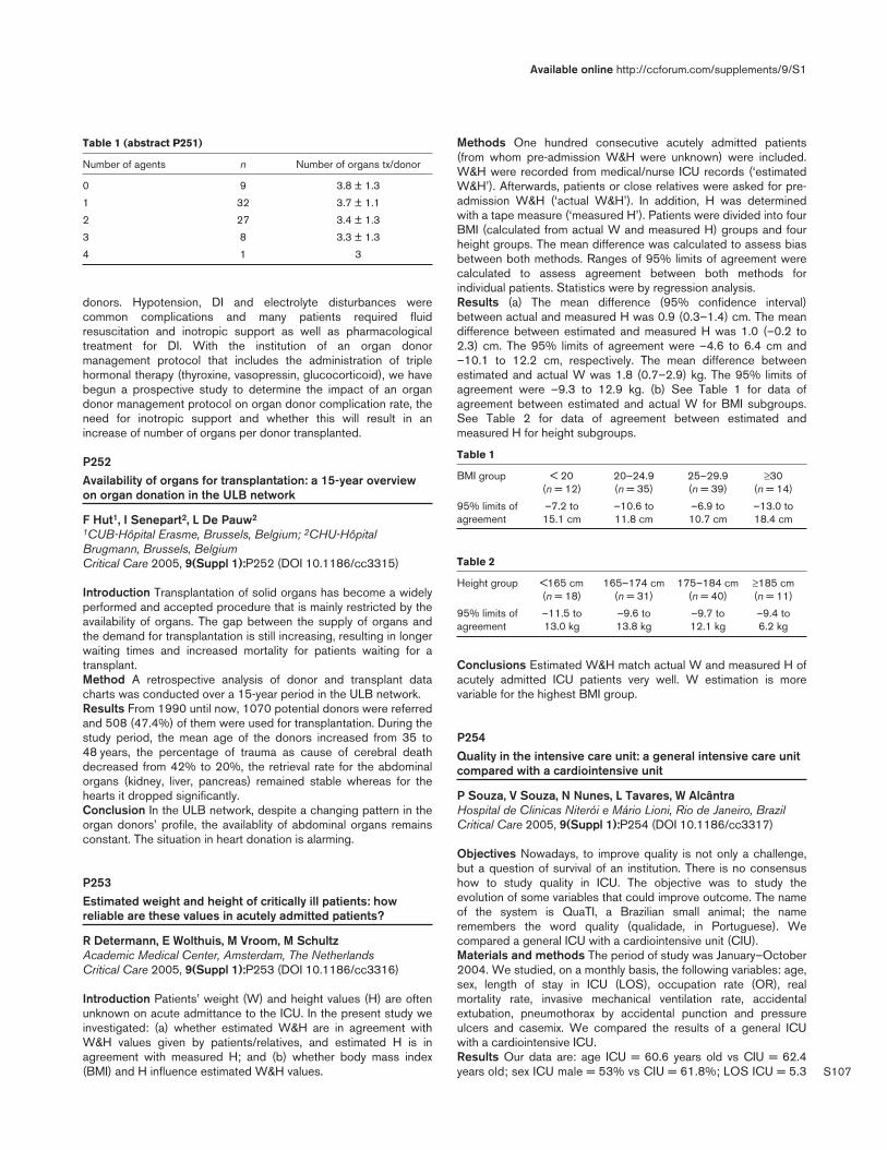
S107
Table 1 (abstract P251)
Number of agents n Number of organs tx/donor
0 9 3.8 ± 1.3
1 32 3.7 ± 1.1
2 27 3.4 ± 1.3
3 8 3.3 ± 1.3
4 1 3
donors. Hypotension, DI and electrolyte disturbances werecommon complications and many patients required fluidresuscitation and inotropic support as well as pharmacologicaltreatment for DI. With the institution of an organ donormanagement protocol that includes the administration of triplehormonal therapy (thyroxine, vasopressin, glucocorticoid), we havebegun a prospective study to determine the impact of an organdonor management protocol on organ donor complication rate, theneed for inotropic support and whether this will result in anincrease of number of organs per donor transplanted.
P252Availability of organs for transplantation: a 15-year overviewon organ donation in the ULB network
F Hut1, I Senepart2, L De Pauw2
1CUB-Hôpital Erasme, Brussels, Belgium; 2CHU-HôpitalBrugmann, Brussels, BelgiumCritical Care 2005, 9(Suppl 1):P252 (DOI 10.1186/cc3315)
Introduction Transplantation of solid organs has become a widelyperformed and accepted procedure that is mainly restricted by theavailability of organs. The gap between the supply of organs andthe demand for transplantation is still increasing, resulting in longerwaiting times and increased mortality for patients waiting for atransplant.Method A retrospective analysis of donor and transplant datacharts was conducted over a 15-year period in the ULB network.Results From 1990 until now, 1070 potential donors were referredand 508 (47.4%) of them were used for transplantation. During thestudy period, the mean age of the donors increased from 35 to48 years, the percentage of trauma as cause of cerebral deathdecreased from 42% to 20%, the retrieval rate for the abdominalorgans (kidney, liver, pancreas) remained stable whereas for thehearts it dropped significantly.Conclusion In the ULB network, despite a changing pattern in theorgan donors’ profile, the availablity of abdominal organs remainsconstant. The situation in heart donation is alarming.
P253Estimated weight and height of critically ill patients: howreliable are these values in acutely admitted patients?
R Determann, E Wolthuis, M Vroom, M SchultzAcademic Medical Center, Amsterdam, The NetherlandsCritical Care 2005, 9(Suppl 1):P253 (DOI 10.1186/cc3316)
Introduction Patients’ weight (W) and height values (H) are oftenunknown on acute admittance to the ICU. In the present study weinvestigated: (a) whether estimated W&H are in agreement withW&H values given by patients/relatives, and estimated H is inagreement with measured H; and (b) whether body mass index(BMI) and H influence estimated W&H values.
Methods One hundred consecutive acutely admitted patients(from whom pre-admission W&H were unknown) were included.W&H were recorded from medical/nurse ICU records (‘estimatedW&H’). Afterwards, patients or close relatives were asked for pre-admission W&H (‘actual W&H’). In addition, H was determinedwith a tape measure (‘measured H’). Patients were divided into fourBMI (calculated from actual W and measured H) groups and fourheight groups. The mean difference was calculated to assess biasbetween both methods. Ranges of 95% limits of agreement werecalculated to assess agreement between both methods forindividual patients. Statistics were by regression analysis.Results (a) The mean difference (95% confidence interval)between actual and measured H was 0.9 (0.3–1.4) cm. The meandifference between estimated and measured H was 1.0 (–0.2 to2.3) cm. The 95% limits of agreement were –4.6 to 6.4 cm and–10.1 to 12.2 cm, respectively. The mean difference betweenestimated and actual W was 1.8 (0.7–2.9) kg. The 95% limits ofagreement were –9.3 to 12.9 kg. (b) See Table 1 for data ofagreement between estimated and actual W for BMI subgroups.See Table 2 for data of agreement between estimated andmeasured H for height subgroups.
Table 1
BMI group < 20 20–24.9 25–29.9 ≥30 (n = 12) (n = 35) (n = 39) (n = 14)
95% limits of –7.2 to –10.6 to –6.9 to –13.0 to agreement 15.1 cm 11.8 cm 10.7 cm 18.4 cm
Table 2
Height group <165 cm 165–174 cm 175–184 cm ≥185 cm (n = 18) (n = 31) (n = 40) (n = 11)
95% limits of –11.5 to –9.6 to –9.7 to –9.4 to agreement 13.0 kg 13.8 kg 12.1 kg 6.2 kg
Conclusions Estimated W&H match actual W and measured H ofacutely admitted ICU patients very well. W estimation is morevariable for the highest BMI group.
P254Quality in the intensive care unit: a general intensive care unitcompared with a cardiointensive unit
P Souza, V Souza, N Nunes, L Tavares, W AlcântraHospital de Clinicas Niterói e Mário Lioni, Rio de Janeiro, BrazilCritical Care 2005, 9(Suppl 1):P254 (DOI 10.1186/cc3317)
Objectives Nowadays, to improve quality is not only a challenge,but a question of survival of an institution. There is no consensushow to study quality in ICU. The objective was to study theevolution of some variables that could improve outcome. The nameof the system is QuaTI, a Brazilian small animal; the nameremembers the word quality (qualidade, in Portuguese). Wecompared a general ICU with a cardiointensive unit (CIU).Materials and methods The period of study was January–October2004. We studied, on a monthly basis, the following variables: age,sex, length of stay in ICU (LOS), occupation rate (OR), realmortality rate, invasive mechanical ventilation rate, accidentalextubation, pneumothorax by accidental punction and pressureulcers and casemix. We compared the results of a general ICUwith a cardiointensive ICU.Results Our data are: age ICU = 60.6 years old vs CIU = 62.4years old; sex ICU male = 53% vs CIU = 61.8%; LOS ICU = 5.3
Available online http://ccforum.com/supplements/9/S1

S108
vs CIU = 3.8; OR: ICU = 93% vs CIU = 96%; real mortality rateICU = 14.5% vs CIU = 3,6%; invasive mechanical ventilation ICU =25.6% vs CIU = 17.3%; accidental extubation ICU = 1.4% vs B(CIU) = 0.8%; pneumothorax by accidental punction HCN (ICU) =2.8 vs CIU = 0; and pressure ulcers HCN (ICU) = 3.7% vs CIU =0.5; the casemix in the ICU was 33.3% neurologic, 19.4%respiratory and 15.6% cardiovascular, and in the CIU the casemixwas 77.2% cardiovascular, 8.5% neurologic and 5.1% respiratory.Conclusion The study of the performance of one ICU is difficult,complex and expensive. The best way to know one specificperformance is to compare it with similar ICUs that have similarresources and casemix. To compare a Brazilian ICU with aEuropean or American ICU is not ideal and could give equivocalinformation that generates erroneous decisions. With QuaTI, webegin a very interesting project that will create a Brazilian databankon ICU performance.In our study, the main difference is the casemix, with much moreneurologic cases in the ICU versus the CIU where predominant iscardiovascular, 77.2%. The outcome of mortality is completelydifferent — 14.5% versus 3.6%. Our worse rates as pressureulcers could be explained by the casemix (more neurologicpatients), and the difference among the pneumothorax is explainedbecause in a CIU we usually do not make profound venous access.The main importance is to have data and establish strategies forimproved quality and safety. The QuaTI works in this direction.
P255Quality of care on an intensive care unit: using subjectiveindicators as an analysis tool
M Knibel1, C David2, R Hatum1
1CardioTrauma Ipanema Hospital, Rio de Janeiro, Brazil; 2FederalUniversity of Rio de Janeiro, BrazilCritical Care 2005, 9(Suppl 1):P255 (DOI 10.1186/cc3318)
Introduction The routine follow-up of objective quality indicators(product analysis) and subjective quality indicators (service analysis)has essential importance to adequate management of an ICU. Itallows continuous PDCA cycles (to plan, to do, to check and to act)and the adoption of preventive and corrective measures, which is asimple, dynamic and efficient management system.Patients and methods We applied satisfaction questionnaries topatients admitted from February to November 2003 and theirrelatives. These questionaries generated the Patient SatisfactionIndex (PSI) and the Relative Satisfaction Index (RSI), by means ofthe 17 patients’ answers and 10 relatives’ answers. The statisticalanalysis used: Mann–Whitney test (to compare two groups inwhich variables did not present a normal distribution),Kruskal–Wallis analysis of variance (to compare three or moregroups), chi-squared test (to compare qualitative data betweeneach 5-month period), and Pearson coefficient (to measure thecorrelation strength between PSI and RSI). We acceptedP < 0.005 as significance level. The software pack used was theSAS® system.Results Indexes general descriptive analysis: PSI 2.856 (meanvalue), RSI 2.882 (mean value). PSI descriptive and statisticalanalysis by month: no difference (P = 0.10). RSI descriptive andstatistical analysis by month: no difference (P = 0.005). PSIdescriptive and statistical analysis by 5-month period: nodifference (P = 0.31). RSI descriptive and statistical analysis by 5-month period: no difference (P = 0.65). Correlation coefficientbetween PSI and RSI: positive correlation (P = 0.0001 r = 0.516).
Conclusions Our management system was good in services issuebecause: (1) the PSI mean was 2.856 and the RSI was 2.882 —both above 2.5, our initial goal; (2) there was no significativevariance in the PSI between the months, which indicates a uniformbehavior of the service; (3) there was no significative difference inthe PSI and RSI measures between both 5-month periods, whichshows uniformity in the offered services; and (4) there was strongassociation between PSI and RSI, which confirms that the ICUteam really sees patients’ relatives like clients, which means a goodquality of service.
P256Managing performance in the intensive care unit: a newsystem in Brazil
P Souza1, J Lain1, J Andrade1, M Lugarinho1, M Amaral2, R Turon1
1Hospital de Clinicas Niterói e Mário Lioni, Rio de Janeiro, Brazil;2Hospital Evangélico, Rio de Janeiro, BrazilCritical Care 2005, 9(Suppl 1):P256 (DOI 10.1186/cc3319)
Objective The study of the performance of one ICU is still settingup. There is no consensus about criteria to guarantee quality. Toanalyse the performance of an ICU and to benchmark it with otherICUs could be a valuable experience. The objective was to studythe performance of one ICU and, for benchmarking proposes, tocompare it with itself and against a standard composed of sevenother ICUs. The name of this system is QuaTI, a Brazilian smallanimal; the name remembers the word quality (qualidade, inPortuguese).Materials and methods A group of intensivists supported by aBrazilian producer of medical equipment (Dixtal) have created acomputer program that collects many variables that compose theidea of performance.We register, on a daily basis, variation of the data studied, themean value, the mean value of benchmarking (the same subject inother ICUs), and control graphics (with upper limit and lower limitof control covering a confidence interval of 95%). This informationis in one ICU computer covering a period of 16 weeks; every3 months we receive a report with more complete analyses, andafter 12 months there is a special report with consolidatedinformation. The data are divided into management, clinical and riskindicators. Each group of indicators are composed of some data;for example, risk indicators are composed of the rate of patientswith some infection, rate of accidental extubation, rate of pneumo-thorax by barotraumas, rate of pneumothorax by venous puncture,and rate of pressure ulcers. The system has confidentiality, so youonly know your data and the benchmarking, but not the data fromone specific ICU. The databank is the domain of AMIB (BrazilianSociety of Intensive Medicine). We start using QuaTI at the end of2001. There were 30 ICUs in the system at the end of 2002 and56 at the end of 2003.Results We could compare the performance of our ICU withourselves along time, every 3 months, each year and with data frombenchmark ICUs. With this information we know our comparativeperformance and we can decide whether it is necessary to changesome processes on a real and comparative basis.Conclusion The study of the performance of one ICU is difficult,complex and expensive. The better way to know one specificperformance is to compare it with similar ICUs that have similarresources and casemix. To compare a Brazilian ICU with aEuropean or American ICU is not ideal and could give equivocalinformation that generates erroneous decisions. With QuaTI, webegin a very interesting project that will create a Brazilian databankon ICU performance.
Critical Care March 2005 Vol 9 Suppl 1 25th International Symposium on Intensive Care and Emergency Medicine

S109
P257The value of daily routine chest radiographs in intensive carepatients
M Graat, E Wolthuis, G Choi, J Stoker, M Vroom, M SchultzAcademic Medical Center Amsterdam, The NetherlandsCritical Care 2005, 9(Suppl 1):P257 (DOI 10.1186/cc3320)
Introduction It is uncertain whether daily routine chest radiographs(CXRs) truly affect the daily management of critically ill patients.The consensus opinion of the American College of Radiology,however, is that routine CXRs are indicated in all mechanicallyventilated patients (http://www.acr.org).Methods The study was performed in a mixed surgical/medicalICU. During a period of 6 months, routine CXRs were performednext to CXRs on clinical indication (on-demand CXRs). During asecond phase of 3.5 months, the routine CXR was abandoned andonly on-demand CXRs were made. Questionnaires (on the backside of each CXR request form) were completed for all CXRs,addressing the indication and expected findings. The presence ofthe following abnormalities was scored by the radiologist:pulmonary abnormalities (atelectasis, major infiltrates, anypneumothorax, pulmonary congestion or significant pleural fluid),and abnormal position of any invasive device (tube, central venouslines). Statistical analysis was by chi-squared test.Results During the first 6 months, 5180 CXRs were made in 888patients (3400 routine and 1780 on-demand CXRs). Of all routineCXRs, only 221 revealed an unexpected abnormality (6.5%)compared with 249 of the on-demand CXRs (14.0%) (χ2 = 79.42,df = 1, P < 0.0001). These findings included (on the routineCXRs) 1.2% malposition of the tube, 1.1% malposition of centralvenous lines and 1.2% atelectasis, versus 1.8% malposition of thetube, 1.8% malposition of central venous lines and 1.2%atelectasis in the on-demand CXRs. In the second period, 1093(all ‘by definition’ on-demand) CXRs were made in 425 patients. Ofthese CXRs, 153 revealed an ‘unexpected’ abnormality (14.3%)(χ2 < 0.0001, df = 1, P = not significant); these findings included2.2% malposition of the tube, 2.7% malposition in central venouslines and 2.8% pleural fluid.Conclusion Compared with the first period, the number of on-demand CXRs did not change in the second period. In addition,the incidence and type of unexpected findings of on-demandCXRs did not change. From this we can conclude that daily routineCXRs may not be necessary for all ICU patients.
P258Accidental catheter removal in the intensive care unit: aprospective observational study
S Myatra, J Divatia, P Mehta, S Kalaniya, R SareenTata Memorial Hospital, Mumbai, IndiaCritical Care 2005, 9(Suppl 1):P258 (DOI 10.1186/cc3321)
Introduction Accidental catheter removal (ACR) is associated withcomplications caused by the removal itself and by catheterreinsertion. Hence determining the incidence is important toimplement preventive measures. The objective of the present studywas to analyse the incidence of ACR for all types of catheters inthe ICU.Methods A prospective observational study in a 22-bedmedical/surgical ICU-HDU in a 550-bed tertiary cancer care centeron all consecutive adult ICU patients between 1 October 2004and 30 November 2004. ACR was considered to be accidentalremoval of a catheter either by the patient or by the ICU staff. Theincidences of ACR for all types of catheters per 100 catheters (%)
and per 100 catheter-days (%cds) were determined. Complica-tions arising due to the ACR were noted. The nurse–patient ratioduring the ACR and daily was noted.Results A total of 913 catheters were inserted in 147 patients fora total of 3614 catheter-days. ACR occured in 43 catheters (4.7%,1.19%cds) in 25 patients. The incidence of ACR was higher forarterial than for central venous catheters (2.4 vs 0.7%cds). Theincidence of ACR for nonvascular catheters (%cds) was:endotracheal tube 1.65; nasogastric tube 2.9; urinary catheter0.17; thoracic drain 0.56; abdominal drain 0.53; epidural catheters1.1; and neck drains 0. Four patients (9.3%) with accidental ETTremoval had transient arterial hypoxemia necessitating reintubation.Twenty-six catheters (60%) – six ETT, seven arterial, 10nasogastric, one thoracic and two others — needed reinsertion.The daily median nurse–patient ratio was 0.36 (range 0.27–0.57),and during ACR it was 0.38 (0–1).Conclusion Our incidence of ACR was high for nasogastric tubes,arterial lines and ETTs. Development and implementation ofimproved protocols are essential to reduce the incidence of ACR inthese catheters. There was no obvious correlation between ACRand nurse–patient ratio. To minimize ACR, it is necessary to monitorits incidence carefully and to implement preventive measures.
P259Complications of care in a neonatal–paediatric intensive care:first results of a prospective observational survey
V Payen, A Maury, K Norbert, E Saliba, S CantagrelHopital Gatien de Clocheville, Tours, FranceCritical Care 2005, 9(Suppl 1):P259 (DOI 10.1186/cc3322)
Background Adverse events can increase the mortality ormorbidity of patients hospitalized in ICUs. Their frequency inpaediatric or neonatal ICUs is between 8% and 45%. A survey ofthese complications is an indicator to improve quality of care.Objectives To evaluate by a prospective observational survey overa 1-year period the frequency of iatrogenic complications in amultidisciplinary paediatric and neonatal intensive care unit; toexamine their type, severity, associate factors that favour theiroccurrence, and the role of medical staff and parents inprecipitation or detection of iatrogenic complications.Patients All consecutive admissions of patients over a 1-yearperiod from October 2004 to October 2005 in a paediatric andneonatal intensive care unitPreliminary results After 1 month, 71 children were admitted,53% in neonatal and 47% in paediatric ICUs. Seventy-five per centof all admissions were for medical reasons, 20% after surgery and5% following trauma. There were 42 iatrogenic complications in 31patients (44% of admissions). Peripheral venous cathetercomplications represented the major adverse event (50%) with81% of diffusion followed by skin oedema; respiratory eventsinvolved 24% of complications and included accidentalextubations (69%) and laryngeal dyspnea (16%); bedsore werefound in 18% of adverse events; 7% of them are secondary to thearterial or central venous catheter. Forty-six per cent were major,7% were moderate and 47% were minor. No death was directlydue to complications. Human error was implicated in 46% ofcases; 46% of iatrogenic complications did not have anyexplanation. They were related mostly to insufficient surveillance.Precipitating staff members were nurses in 73% of times,especially for minor events. They detected the majority of thecomplications (81%).Conclusion As in adult ICUs, iatrogenic complications arefrequent in paediatric and neonatal ICUs. Knowledge of adverseevents frequency is a precious source for quality improvement.
Available online http://ccforum.com/supplements/9/S1

S110
P260Adverse events and death in patients with community-acquired pneumonia in the emergency department of a majoruniversity hospital in Brazil
A Machado, R Gallotti, H Novaes, M Lorenzi, J Neto, I VelascoHospital das Clínicas, São Paulo, BrazilCritical Care 2005, 9(Suppl 1):P260 (DOI 10.1186/cc3323)
Introduction Adverse events (AEs), defined as unintended injuriesdue to medical care, have been related to patient death. Urgentcare is an important AE risk factor.Objectives This paired case–control study aimed to identify theoccurrence of AEs in patients with community-acquired pneumonia(CAP) attending a medical Emergency Department (ED) of aBrazilian major university hospital, disclosing the AEs categoriesassociated with death.Methods We investigated 202 patients admitted for CAP fromMarch 1996 to September 1999. The cases comprised 101consecutive deaths and the controls 101 discharged patientsmatched for primary diagnosis and admission period. AEs,detected by chart review, were classified regarding: severitydegree (major and minor), immediate cause, and professionalcategory. The association with death was analyzed by multivariateconditional regression including variables related to demographicfeatures, severity on admission and care characteristics.Results A total of 603 AEs were identified in 202 patients: 456events (75.6%) in 85 cases and 147 (24.4%) in 55 controls. Twohundred and twenty-four major AEs were detected: 201 events in65 cases and 23 in 10 controls. Diagnostic procedures andnursing activities accounted for 43.1% of all events. Nursing(43.8%) and medical (36.5%) AEs were the main professionalcategories of AEs detected in both groups. The occurrence of atleast one AE was associated with a higher risk of death, withadjusted odds ratio (OR) estimates of 2.99 (95% confidenceinterval [CI] = 1.09–8.17; P = 0.033). A stronger association withdeath was found regarding major AEs (OR 6.05; 95% CI =2.19–16.68; P = 0.001). Medical AEs and administrative AEswere also associated with death, with adjusted OR estimates of3.74 (95% CI = 1.40–10.00; P = 0.009) and 2.97 (95% CI =1.12–7.90; P = 0.029), respectively.Conclusions In summary, AEs predominated among the deceasedpatients admitted with CAP. Diagnostic and therapeuticprocedures and nursing activities corresponded to the main AEimmediate causes. Regarding the professional involved, AEsrelated to nurses and physicians predominated. The occurrence ofAEs was associated with a higher risk of death in CAP patientsadmitted to a tertiary ED.
P261Adverse medication events in the intensive care unit:avoidable or inevitable?
J Mitchell, F Cox, J Wagg, J FarrimondRoyal Brompton & Harefield NHS Trust, Harefield, UKCritical Care 2005, 9(Suppl 1):P261 (DOI 10.1186/cc3324)
Introduction Recent publications have focused on patient safetyand managing medications effectively [1]. Adverse reactions tomedicines and medication errors cost the National Health Service(NHS) approximately £0.5 billion per annum [1]. To inform practiceand facilitate policy revision we explored medication errors in twospecialist cardiothoracic ICUs that utilise identical reportingmechanisms.
Methods A retrospective analysis of the DATIX risk managementdatabase was performed for the 3-year period January2002–November 2004. These events were categorised using theNational Patient Safety Agency impact and consequenceclassifications [2]. Events categorised as moderate or severe wereindividually investigated and an individual risk reduction strategyinitiated.Results During this 3-year period 114 medication-related adverseevents were reported. There were 9.5 events per 1000 ICU bed-days. A toal 14.9% of events were categorised as moderate orsevere, while 85.1% were insignificant or mild.
Table 1
Event type (all) Number (% of total) Major events (%)
Prescribing 20 (18) 3 (3)
Supply 11 (9) 2 (2)
Mislabelling 18 (16) 1 (1)
Route/drug/dose 23 (20) 6 (5)
Freq/documentation 32 (28) 2 (2)
Controlled/K+ 4 (3) 3 (3)
Other 7 (6) 0 (0)
Conclusion The rate of adverse events is comparable withpublished work (9.5 vs 10.4) [3] but there may be significant under-reporting. However, it was considered that all major events wereavoidable. The current UK Medication Safety Initiative focuses onstrengthening the prescription, supply and administration ofmedications in the hospital setting. This study suggests thatmedication safety should be a priority in the ICU in 2005.References1. Audit Commission: A Spoonful of Sugar: Medicines Management
in NHS Hospitals. Audit Commission; 2001.2. NPSA: 7 Steps to Patient Safety — Your Guide to Safer Patient
Care. National Patient Safety Agency; 2004.3. Leape LL, et al.: Pharmacist participation on physician rounds
and ADEs in the ICU. JAMA 1999, 282:267-270.
P262Budget savings following a rationalisation of practice: acomparison of drug-prescribing costs in two differentintensive care units
P Campbell1, J Murphy1, R Forrest2, M Garrioch2, A Davidson1
1Victoria Infirmary, Glasgow, UK; 2Southern General Hospital,Glasgow, UKCritical Care 2005, 9(Suppl 1):P262 (DOI 10.1186/cc3325)
Aim To ascertain whether a comparison of drug budget spending,in comparable ICUs, could lead to a rationalisation of clinicalpractice and budget savings.Method Two similar ICUs were chosen, both with the same bednumbers, similar patient groups, severity of admission (usingAPACHE II scores) and length of stay. The pharmacy departmentscross-referenced the financial costs of all medications andsundries used in both ICUs. Where any major financial differencesbecame apparent, the clinical practice related to these costs wasinvestigated.Results This budget analysis highlighted several variations inclinical practice. The main differences found were in sedationpractices, the use of neuromuscular blocking drugs, antimicrobialprescribing practices and the use of differing haemofiltrationsundries. The standardisation of practice in these areas led to afinancial saving of over £30,000 in a year between the two units,from a combined drug budget of approximately £300,000.
Critical Care March 2005 Vol 9 Suppl 1 25th International Symposium on Intensive Care and Emergency Medicine

S111
Discussion This analysis facilitated discussion among themultidisciplinary intensive care team in an effort to improve patientcare and save money at the same time. By analysing drug-prescribing costs and comparing clinical practices, we have shownthat significant savings can be made. We were able to rationaliseclinical practice without any significant changes in currentmanagement strategies.Further differences in practice were also demonstrated by thisstudy. It is hoped that, following discussions with all concernedindividuals, further standardisations in practice and subsequentcost savings can be made.
P263Significance and outcome of critical care system management
S Kiatboonsri, P Charoenpan, C Kiatboonsri, P Pornsuriyasak,V BoonsrangsukRamathibodi Hospital, Mahidol University, Bangkok, ThailandCritical Care 2005, 9(Suppl 1):P263 (DOI 10.1186/cc3326)
Objective To set up a holistic critical care system.Background Ramathibodi Hospital is a 1000-bed medical school,tertiary care and referral center. The ICU of the Department ofMedicine was founded in 1980 and comprised six beds thatserved the total 132 medical inpatient beds. The ratio of ICU:totalbeds was thus 0.4:10. In 1990, the number of ICU beds wasextended to eight, which raised the new ICU:total bed ratio to1:10. Such a proportion of ICU beds was apparently too small.This resulted in a number of critical patients being treated in thegeneral medical wards with difficulties in raising and maintainingthe standards of critical and respiratory care. The unclear jobdescription also deteriorated working morale and led to a rapidturnover rate among the nursing personnel.Steps of development (1) In 1989–1990, attempts was made tovalidate the precision of the APACHE II scoring system whenapplied to our group of ICU patients. Among the 334 patientstested, the APACHE II scoring system was proved to be valid andthe ICU performance as assessed by the Actual Death/Predicteddeath ratio was 1.17. (2) To estimate the actual critical care bedsneeded, the number of patients who were mechanically ventilatedin the general wards were recorded over years (1995–1996). Thedata showed at least 12–20 patients were mechanically ventilatedoutside the ICU each day — a number that indicated the minimumadditional critical care beds required. (3) In 1998, one of the 30-bed general wards was renovated to be a 20-bed intermediatecare unit (IMU). The total inpatient beds was thus reduced by 10and the new critical:total beds ratio was 2.5:10. Criteria of criticalpatients’ admission pathways to the ICU or IMU were set. A criticalcare team was formed to raise and maintain standards of patientand equipment care, and to provide continued medical educationin critical care medicine. A number of indices of quality controlwere also monitored and evaluated continuously.Results Since October 1998, none of the patients who werehemodynamically unstable and/or needed ventilatory support wastreated in the general ward. Despite the reduction of total inpatientbeds by 10, the total number of inpatient admission has increasedfrom 3742 in 1997 to 4657 in 2003 (24% increment). Theaverage hospital length of stay (LOS) of the Department ofMedicine has declined from 10.84 days in 1997 to 8.81 days in2003 (18.7% reduction). The overall inpatient hospital mortalityhas reduced from 17.37% in 1997 to 9.90% in 2003 (43%reduction). The critical care unit performance as measured by theActual Death/Predicted Death ratio has reduced from 1.17 in1990 to 0.76 and 0.72 in 2002 and 2003, respectively.
Conclusion Our holistic critical care system management not onlyresulted in a better care of critical patients, but also led to anoverall improvement in department performance.
P264The early efficacy of CABG care delivery in a low procedure-volume community hospital: operative and midterm results
T PapadimosMedical College of Ohio, Toledo, OH, USACritical Care 2005, 9(Suppl 1):P264 (DOI 10.1186/cc3327)
The Leapfrog Group recommended that CABG surgery should bedone at high volume hospitals (> 450 per year) withoutcorresponding surgeon-volume criteria. Recent studies haveindicated that the effects of hospital volume are substantiallyconfounded by surgeon volume, and that high surgeon volume(>125 per year) may be a more appropriate indicator of CABGquality. We thus compared operative morbidity and mortalityoutcomes for a low-volume hospital (LVH) (504 CABG,2001–2003) served by five high-volume surgeons (161–285 peryear) with the corresponding Society of Thoracic Surgeons (STS)national data over the same period. The LVH program implemented‘best practice’ care including effective practice guidelines,protocols, data acquisition capabilities, case review process,dedicated facilities and support personnel (anesthesiology,intensivists and pharmacists). Operative mortality was similar forLVH and STS (OM: 2.38% vs 2.53%), and the corresponding LVHobserved-to-expected mortality (O/E = 0.81) indicated goodquality relative to the STS risk model (O/E <1). Also, these resultswere consistent irrespective risk category: O/E was 0, 0.9 and1.03 for very-low risk (<1%), low risk (1–3%) and moderate-to-high risk (> 3%) categories, respectively. While postoperative legwound infections, ventilator hours, renal failure, and atrial fibrillationwere increased in the LVH, the length of stay was not. Theunadjusted Kaplan–Meier survival for the LVH cohort was 96%,94%, and 92% at 1, 2, and 3 years, respectively. Our resultsdemonstrated that high-quality CABG care can be achieved atLVH programs when served by high-volume cardiac surgeons. Thisapproach may prove a useful paradigm to ensure high-qualityCABG care and early efficacy at low-volume institutions (Table 1shows predictors of 0-year to 3-year mortality; Cox regression).
Table 1
Variable B SE Wald P value Risk ratio 95% CI
CPB time 0.02 0.00 16.26 0.0001 1.15 1.08 1.23(10 min)
All vein grafts 1.75 0.55 10.15 0.0014 5.74 1.96 16.82
Cerebrovascular 1.33 0.43 9.82 0.0017 3.79 1.65 8.72disease
Age (per 0.06 0.02 8.28 0.0040 1.63 1.20 1.9110 years)
Redo surgery 1.24 0.55 5.00 0.0254 3.46 1.17 10.26
Congestive 0.91 0.45 4.07 0.0435 2.48 1.03 6.00heart failure
Preoperative 1.34 0.76 3.11 0.0779 3.81 0.86 16.86renal failure
Available online http://ccforum.com/supplements/9/S1

S112
P265Rising use of intensive care unit services in Medicare
E Milbrandt1, A Kersten1, S Watson1, M Rahim1, G Clermont1, D Angus1, W Linde-Zwirble2
1The CRISMA Laboratory, Department of Critical Care Medicine,University of Pittsburgh, PA, USA; 2ZD Associates, Perkasie, PA, USACritical Care 2005, 9(Suppl 1):P265 (DOI 10.1186/cc3328)
Introduction In recent years, rising cost, new technology,demographic shifts, and demands for higher quality have placedpressure on Medicare, the largest part of US healthcare. Theconsequence for the use of ICU services is unknown.Hypothesis Use of ICU services is growing in the Medicarepopulation.Methods We selected all Medicare Prospective Payment System(PPS) hospitalizations from 1994 to 2001 and generated ratesusing corresponding beneficiary files. We calculated total ICUadmission rates, rates per 1000 beneficiaries, and rates stratifiedby age and medical versus surgical admission.Results In 1994, there were 10.7 million Medicare PPShospitalizations, 2.2 million (20.5%) of which incurred ICU care. By2001, hospitalizations increased 5.3% to 11.3 million, while ICUadmissions increased 14.5% to 2.5 million. After adjusting forchanges in the number of beneficiaries, hospitalizations fell 2.1%,while ICU admissions increased 6.4%. Those ≥ 80 years saw thegreatest increase in ICU admission rates (41%), accounting for33.4% of ICU admissions in 2001. Growth in ICU use was due toincreased medical admissions, which rose 18.9% while surgicalICU admissions fell 4.7%.
Conclusions ICU use has been rising dramatically in Medicare,especially in the elderly. This rise is occurring despite a reductionin the number of hospitalizations per beneficiary.
P266A comparative cost analysis before and after opening of anintermediate care unit
B Solberg1, G Ramsay2, C Dirksen1, F Nieman1
1University Hospital Maastricht, The Netherlands; 2Atrium MedicalCentre, Heerlen, The NetherlandsCritical Care 2005, 9(Suppl 1):P266 (DOI 10.1186/cc3329)
Objective To compare the hospital cost of intensive care patientsbefore and after the opening of an intermediate care unit.
Design A before–after prospective observational study.Setting University-affiliated mixed ICU, intermediate care unit(IMC) and standard wards.Patients and participants A total of 1539 ICU stays wereadmitted from January 2001 to December 2002; 786 ICU patientswere included in the study by taken a random sample. During theperiod before opening the IMC 329 ICU patients were included,and 457 ICU patients after opening the IMC.Interventions Opening of a six-bed mixed IMC.Measurements and results For each ICU patient, the followingcost data were collected: total hospital cost; divided into costsduring ICU stay, IMC stay and general ward stay. The total hospitalcost was computed from the first ICU admission to hospitaldischarge. The cost of readmissions to the ICU, IMC and wardsduring the same hospital stay were included. Comparisonsconcerning costs were performed after logarithmic transformation,to improve the symmetry of distributions. The mean total hospitalcost before opening the IMC was €12.961 (± 14.530) and afteropening the IMC was €16.513 (± 19.324) with a significantincrease of €3552 (P value = 0.01) (Table 1).
Table 1
Before After Before IMC After IMC PIMC mean IMC mean value
Costs (€) (n) (SD) (n) (SD) (logtrans.)
ICU stay 329 10,017 457 13,020 0.08(13,317) (17,659)
IMC stay 0 0 138 2612 –(4060)
Ward stay 236 4105 304 4065 0.12(5924) (5095)
Total hospital 329 12,961 457 16,513 0.01stay (14,530) (19,324)
Statistical relations between individual hospital cost and patientcharacteristics were analysed (age, gender, mortality, surgical/medical, kind of organ failure and nursing workload [TISS-28] ofadmission day). After correction of these variables, the increases ofthe hospital cost were explained by kind of organ failure (P = 0.08),surgical/medical (P = 0.26), and nursing workload (TISS-28) ofadmission day (P = 0.43).Conclusion This study showed an increase of total hospital costafter opening the IMC. A different patient population was admittedto the ICU after opening the IMC. More surgical patients, higherTISS score and the kind of organ failure varied between thepopulations before and after opening the IMC. The mean costduring the ICU stay per patient increased most (€3003).
P267ICU research coordinator activities: a time-in-motionmulticenter study
E McDonald1, F Clarke1, C Dale2, C Davidson3, R Farrell3, L Hand1, T McArdle3, O Smith2, M Steinberg2, I Watpool3, R Ward3, D Heels-Ansdell1, D Cook1
1McMaster, Hamilton, Canada; 2University of Toronto, Canada;3University of Ottawa, CanadaCritical Care 2005, 9(Suppl 1):P267 (DOI 10.1186/cc3330)
Background ICU Research Coordinators (ICURCs) represent agrowing membership in the Canadian Critical Care Trials Group(CCCTG). The professional contribution of ICURCs to clinical criticalcare research is profound, but has not been well characterized.
Critical Care March 2005 Vol 9 Suppl 1 25th International Symposium on Intensive Care and Emergency Medicine
Figure 1 (abstract P265)

S113
Objective To describe the settings, studies, resources, tools, andactivities of Canadian ICURCs.Methods We conducted a prospective observational time-in-motion study of CCCTG ICURCs from 20 September to 15October 2004. We generated items and domains in focus groups.The instrument was formatted using both open and closed endedresponses. We piloted for comprehensiveness and clarity. Weobtained data on the setting, studies, tools and resources availablein each center, then used a weekly management log to documentactivities under seven domains. Completed forms were faxed to theMethods Center at St Joseph’s Healthcare and entered into anExcel Spreadsheet for descriptive analyses.Results Twenty ICURCs participated from 13 mixed ICUs (100%university-affiliated hospitals with 440 ± 225 hospital beds and 23± 11 ICU beds). Over 4 weeks in 13 ICUs, 104 ± 64 patientswere admitted, 74 ± 60 were screened and 9 ± 9 were enrolled instudies. On average, there were 4.6 ± 2.1 peer reviewed, and3.8 ± 3.5 industry studies; 5.8 ± 3.4 were RCTs, 2.8 ± 2.3 wereobservational and 0.8 ± 1.2 were other designs. On average,7.1 ± 5.8 studies enrolled adults and 1.5 ± 2.4 enrolled children.These resources were accessed electronically: laboratory data(100%), diagnostic images (53.8%), radiology reports (69.2%),ECGs (15.4%) and clinical information systems (15.4%). Datacollection tools include: paper (100%), NCR paper (38.5%),datafax (38.5%), laptop (7.7%), PDAs (0%), and Web-basedapproaches (84.6%). Time spent in each of seven activities was:study preparation (literature review, inhouse protocol development,REB submissions and amendments, budget preparation, CRFdevelopment and piloting, regulatory document assembly, in-services), 16.5%; research conduct (screening, consent and familymeetings, pharmacy and other study supplies, data collection andqueries, conducting and preparing tests, AE and SAE reporting,bedside protocol compliance), 44.1%; communication (email/telephone calls/mail, staff education), 14.3%; management (teammeetings, multicentered management, screening and finance logs,initiation audit and close-out visits), 13.5%; data management(database creation and data entry, analysis), 3.9%; dissemination(interim and final reports, conference preparation and presentation),3.0%; and miscellaneous, 4.7%.Conclusions CCCTG ICURCs work primarily in mixed university-affiliated ICUs, conducting randomized trials in adults. The availabilityof electronic resources is modest, and variable data collectionmethods are employed. ICURCs engage in many activities to prepare,implement, and manage investigations for critically ill patients.Acknowledgements This study was funded by Father SeanO’Sullivan Research Center and the Canadian Institutes for HealthResearch.
P268Impact of critical care outreach on cardiac arrest rates andreadmission rates in an adult cardiothoracic centre
P Wilton, C Carbery, J Farrimond, J Mitchell, J Hall, T AppsRoyal Brompton & Harefield NHS Trust, Harefield, UKCritical Care 2005, 9(Suppl 1):P268 (DOI 10.1186/cc3331)
Introduction Following the recommendations of ComprehensiveCritical Care [1], outreach services have been implementednationally with the remit of avoiding admission to intensive care;facilitating discharge of critically ill patients from Level 3 areas andeducating ward staff in critical care skills. This initiative consumessignificant resources, and to date there has been disappointingevidence that this investment has been worthwhile in terms ofimproving outcomes, reduction in cardiac arrests, and unplannedadmissions to intensive care.
Objective This audit aims to identify whether the implementation ofan outreach team has had an impact on inhospital cardiac arrestrates and readmission rates to ICU.Methods A retrospective analysis of the ICU and outreachdatabase was performed to quantify the readmission rate within 48hours due to organ failure and the inhospital cardiac arrest rate. Thisanalysis was performed 1 year pre and post the implementation of adedicated nurse-led outreach team.Results Cardiac arrest rate has reduced by 40% since theintroduction of the outreach service. The readmission rate within48 hours with organ failure was less than 2%.Conclusion We report a reduction in the cardiac arrest rate postimplementation of outreach, although this may be due to otherexternal factors. The readmission rate within 48 hours of ICUdischarge is low, reflecting an appropriate and effectively facilitateddischarge practice.Reference1. DoH: Comprehensive Critical Care: A Review of Adult Critical
Care Services. Department of Health; 2000.
P269Critical care outreach: implementation in a cardiothoracic centre
C Carbery, P Wilton, J Farrimond, J Mitchell, J Hall, T AppsRoyal Brompton & Harefield NHS, Harefield, UKCritical Care 2005, 9(Suppl 1):P269 (DOI 10.1186/cc3332)
Introduction Outreach teams have recently been implementedacross the United Kingdom with a remit to deliver a specialist level ofassessment and intervention with the ward setting. These goalsinclude early identification of deteriorating patients to avert admissionto ICU; facilitating discharge from ICU and education of ward staff.Objective An evaluation of a nurse-led outreach team to facilitatethe threefold remit of outreach in a specialist cardiothoracic unit.Methods Establishment of a unique referral criteria to reflect thespecialist cardiothoracic casemix. One hundred per cent of Level 3discharges are followed up and actively managed to facilitate earlydischarge (including patient on intermittent haemofiltration, intermittentCPAP, tracheostomies in situ and low levels of inotropic support).Ward-based education programme. Electronic data collection.Results Over 800 patients were reviewed over a 12-month periodencompassing more than 2000 patient assessments. Level 3discharge follow-up increased to 100% from 45% prior toestablishment of a dedicated team. The overall readmission ratehas remained unchanged; however, the readmission rate within48 hours with organ failure is below 2%, reflecting appropriatedischarge from Level 3. Forty-five per cent of patients showedbiochemical evidence of renal impairment post ICU dischargerequiring a longer stay on outreach.Conclusion This evaluation has highlighted the increasingdependency of patients managed outside of ICU. Facilitating ICUdischarge and follow-up of patients with renal and respiratoryimpairment impacts significantly on ICU length of stay.
P270A ‘Count Outreach Warning Score’ forecasts need for ‘step-up’care
M Kuper, J Adams, S Singh, A Blay, N SoniChelsea Hospital, London, UKCritical Care 2005, 9(Suppl 1):P270 (DOI 10.1186/cc3333)
Objective The number of physiological abnormalities predictsmortality [1], so we defined a positive ‘Count Outreach WarningScore’ (COWS) as three or more physiological abnormalities
Available online http://ccforum.com/supplements/9/S1

S114
(temperature < 35°C or > 39°C; saturation < 95%; urine outputbelow 30 ml/hour; heart rate > 99 or < 51, respiratory rate >19,mental state V/P/U [i.e. not alert]). Could COWS forecast the needfor ‘step-up’ from normal ward care?Method All observations on the 79 patients present on onemedical and one surgical ward over 7 days at the Chelsea andWestminster Hospital, London were scored using COWS and alsothe published scores EWS, MEWS and PART. A patient waspredicted ‘sick’ by a score given a positive result on any one set ofobservations. Outcome was followed for 28 days. ‘Stepped-up’care was prospectively defined as any of intensive care, highdependency or outreach referral or admission, cardiac arrest,death, or placement of a ‘do not resuscitate’ order. Confidenceintervals were calculated by constant chi-squared boundaries andsensitivities compared with McNemar’s test.Results Of 79 patients studied, 17 required ‘step-up’ from normalward care (Table 1). COWS appeared more sensitive at predicting‘step-up’ (P = 0.06), with similar specificity to the other scores. Infour patients correctly identified by more than one score, COWSbecame positive 4–24 hours earlier whereas only one patient wasidentified by another score before identification by COWS.Conclusion COWS forecast the requirement for ‘step-up’ careearlier and with more sensitivity than previous scores.Reference1. Goldhill D, McNarry A: BJA 2004. 92:882.
P271First national survey on the use of the Internet and otherelectronic resources among Spanish intensivists
V Gómez Tello1, N de Lucas2, J Añón3, E Palencia4, J Latour5, R Díaz Alersi61Clínica Moncloa, Madrid, Spain; 2Hospital San Rafael, Madrid,Spain; 3Hospital Virgen de la Luz, Cuenca, Spain; 4HospitalGregorio Marañón, Madrid, Spain; 5Hospital General Universitario,Elche, Spain; 6Hospital de Puerto Real, Cádiz, SpainCritical Care 2005, 9(Suppl 1):P271 (DOI 10.1186/cc3334)
Objective To assess Spanish intensivists’ knowledge and use ofdifferent electronic resources; the Internet, electronic mail, mailinglists and portable electronic devices (PDAs).Design A cross-sectional survey.Participants Eight hundred and twenty Spanish intensivists werecontacted by hospital commercial delegates of a pharmaceuticalcompany. The survey was simultaneously published in electronicforums.Methods and results A total of 682 responses were obtained(response rate: 73.9%). The Internet was used autonomously by95.7% (n = 653) of responding physicians and 78.8% (n = 513)were autodidactic in this field. Learning gaps were reported by66.7% (n = 451) of the respondents and up to 58.3% (n = 398)acknowledged at least two deficit areas. The responses fromfemale intensivists showed lower Internet use and greater learninggaps in at least one area (odds ratio [OR] 2.44; confidence interval[CI] 1.5–3.95).
The Internet was the second most frequent source for clinicalconsultation (60.9%; n = 409), only preceded by peer consultationwith ICU colleagues (65.2%; n = 438). The most frequently visitedwebpages were bibliographical databases (64.5%; n = 412) andelectronic journals (63.4%; n = 405). EBM pages were rarelyvisited (18.9%; n = 421). In fact, the number of different types ofwebsites was the main factor associated with solving clinicalquestions by the Internet (OR 1.37; CI 1.25–1.5).Email correspondence was used by 92.4% (n = 630) of thosesurveyed and 89.6% (n = 611) used it for professional purposes,but 25.3% (n = 159) admitted to ignoring its full potential. Emailcorrespondence as a resource correlated inversely with age (OR0.95; CI 0.92–0.99; P = 0.015).Electronic mailing lists were used by 62.3% (n = 425) of theintensivists. Of the remainder, 41.7% (n = 113) were not familiarwith them and 31.7% (n = 86) reported insufficient expertise tomanage them correctly. Only 20.1% (n = 136) own portableelectronic devices. However, 63.5% (n = 416) considered thesedevices would be a useful tool for immediate consultations at thebedside. Female respondents were less predisposed towards theiruse (OR 2.44; CI 1.5–3.95; P = 0.0002).An index of five punctuated items was elaborated to summarize in asingle variable the use of all resources: preferable access to theInternet to solve clinical questions (one point); Internet consultationtwo or more times per week (one point); electronic mailing listconsultation two or more times per week (one point); intensivecare mailing list consultation (one point); and use of at least onemedical software program in the PDA (one point). The variablesassociated with a low score in this index were: female gender,increase of age, educational character of the hospital and being aresident physician.Conclusions A majority of Spanish intensivists uses the Internetand electronic mail. Important educational deficits exist and a clearunderuse of fundamental webpage sources has been observed.Mailing lists and portable devices are not prevalent electronicresources. Training courses and educational programs are neededto encourage and facilitate the use of these resources and topromote email lists. Special attention needs to be directed atfemale intensivists who seem to be more reluctant to access theseresources.
P272Critical illness polyneuromyopathy: a major problem in ageneral intensive care unit
S Nanas, K Kritikos, E Angelopoulos, C Routsi, S Tsikriki, S Dimopoulos, M Pratikaki, T Loukas, E Papadopoulos, I Floros, C RoussosCritical Care Department, Medical School, National andKapodistrian University, Evangelismos Hospital, Athens, GreeceCritical Care 2005, 9(Suppl 1):P272 (DOI 10.1186/cc3335)
Introduction Critical illness polyneuromyopathy (CIPM) constitutesa significant cause of difficulty in weaning patients from theventilator, prolongation of their stay in the ICU and increased
Critical Care March 2005 Vol 9 Suppl 1 25th International Symposium on Intensive Care and Emergency Medicine
Table 1 (abstract P270)
Test PART PART EWS EWS MEWS MEWS COWS COWS
Test says … ‘Sick’ ‘Well’ ‘Sick’ ‘Well’ ‘Sick’ ‘Well’ ‘Sick’ ‘Well’‘Stepped-up’ 2 15 6 11 6 11 10 7Normal care 0 62 2 60 2 60 2 60Sensitivity 2/17 = 12% 95% CR 21–40 6/17 = 35% 95% CR 20–44 6/17 = 35% 95% CR 20–44 10/17 = 59% 95% CR 41–70Specificity 62/62 = 100% 95% CR 98–100 60/62 = 97% 95% CR 92–99 60/62 = 97% 95% CR 92–99 60/62 = 97% 95% CR 92–99
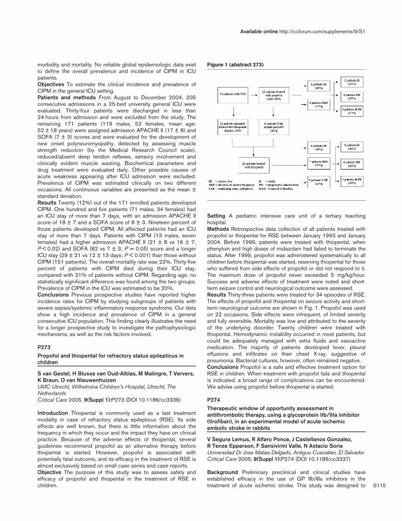
S115
morbidity and mortality. No reliable global epidemiologic data existto define the overall prevalence and incidence of CIPM in ICUpatients.Objectives To estimate the clinical incidence and prevalence ofCIPM in the general ICU setting.Patients and methods From August to December 2004, 205consecutive admissions in a 25-bed university general ICU wereevaluated. Thirty-four patients were discharged in less than24 hours from admission and were excluded from the study. Theremaining 171 patients (119 males, 52 females, mean age:52 ± 18 years) were assigned admission APACHE II (17 ± 8) andSOFA (7 ± 3) scores and were evaluated for the development ofnew onset polyneuromyopathy, detected by assessing musclestrength reduction (by the Medical Research Council scale),reduced/absent deep tendon reflexes, sensory involvement andclinically evident muscle wasting. Biochemical parameters anddrug treatment were evaluated daily. Other possible causes ofacute weakness appearing after ICU admission were excluded.Prevalence of CIPM was estimated clinically on two differentoccasions. All continuous variables are presented as the mean ±standard deviation.Results Twenty (12%) out of the 171 enrolled patients developedCIPM. One hundred and five patients (71 males, 34 females) hadan ICU stay of more than 7 days, with an admission APACHE IIscore of 18 ± 7 and a SOFA score of 8 ± 3. Nineteen percent ofthose patients developed CIPM. All affected patients had an ICUstay of more than 7 days. Patients with CIPM (13 males, sevenfemales) had a higher admission APACHE II (21 ± 8 vs 16 ± 7;P < 0.02) and SOFA (82 vs 7 ± 3; P = 0.05) score and a longerICU stay (29 ± 21 vs 12 ± 13 days; P < 0.001) than those withoutCIPM (151 patients). The overall mortality rate was 22%. Thirty-fivepercent of patients with CIPM died during their ICU stay,compared with 21% of patients without CIPM. Regarding age, nostatistically significant difference was found among the two groups.Prevalence of CIPM in the ICU was estimated to be 20%.Conclusions Previous prospective studies have reported higherincidence rates for CIPM by studying subgroups of patients withsevere sepsis/systemic inflammatory response syndrome. Our datashow a high incidence and prevalence of CIPM in a generalconsecutive ICU population. This finding clearly illustrates the needfor a longer prospective study to investigate the pathophysiologicmechanisms, as well as the risk factors involved.
P273Propofol and thiopental for refractory status epilepticus inchildren
S van Gestel, H Blusse van Oud-Alblas, M Malingre, T Ververs,K Braun, O van NieuwenhuizenUMC Utrecht, Wilhelmina Children’s Hospital, Utrecht, TheNetherlandsCritical Care 2005, 9(Suppl 1):P273 (DOI 10.1186/cc3336)
Introduction Thiopental is commonly used as a last treatmentmodality in case of refractory status epilepticus (RSE). Its sideeffects are well known, but there is little information about thefrequency in which they occur and the impact they have on clinicalpractice. Because of the adverse effects of thiopental, severalguidelines recommend propofol as an alternative therapy beforethiopental is started. However, propofol is associated withpotentially fatal outcome, and its efficacy in the treatment of RSE isalmost exclusively based on small case series and case reports.Objective The purpose of this study was to assess safety andefficacy of propofol and thiopental in the treatment of RSE inchildren.
Setting A pediatric intensive care unit of a tertiary teachinghospital.Methods Retrospective data collection of all patients treated withpropofol or thiopental for RSE between January 1993 and January2004. Before 1999, patients were treated with thiopental, whenphenytoin and high doses of midazolam had failed to terminate thestatus. After 1999, propofol was administered systematically to allchildren before thiopental was started, reserving thiopental for thosewho suffered from side effects of propofol or did not respond to it.The maximum dose of propofol never exceeded 5 mg/kg/hour.Success and adverse effects of treatment were noted and short-term seizure control and neurological outcome were assessed.Results Thirty-three patients were treated for 34 episodes of RSE.The effects of propofol and thiopental on seizure activity and short-term neurological outcome are shown in Fig. 1. Propofol was usedon 22 occasions. Side effects were infrequent, of limited severityand fully reversible. Mortality was low and attributed to the severityof the underlying disorder. Twenty children were treated withthiopental. Hemodynamic instability occurred in most patients, butcould be adequately managed with extra fluids and vasoactivemedication. The majority of patients developed fever, pleuraleffusions and infiltrates on their chest X-ray, suggestive ofpneumonia. Bacterial cultures, however, often remained negative.Conclusions Propofol is a safe and effective treatment option forRSE in children. When treatment with propofol fails and thiopentalis indicated, a broad range of complications can be encountered.We advise using propofol before thiopental is started.
P274Therapeutic window of opportunity assessment inantithrombotic therapy, using a glycoprotein IIb/IIIa inhibitor(tirofiban), in an experimental model of acute ischemicembolic stroke in rabbits
V Segura Lemus, R Alfaro Ponce, J Castellanos Gonzalez, R Tenze Epperson, F Sansivirini Valle, N Astacio SoriaUniversidad Dr Jose Matias Delgado, Antiguo Cuscatlan, El SalvadorCritical Care 2005, 9(Suppl 1):P274 (DOI 10.1186/cc3337)
Background Preliminary preclinical and clinical studies haveestablished efficacy in the use of GP IIb/IIIa inhibitors in thetreatment of acute ischemic stroke. This study was designed to
Available online http://ccforum.com/supplements/9/S1
Figure 1 (abstract 273)

S116
assess the therapeutic window of opportunity of tirofiban in strokeand the risk of intracerebral hemorrhage (ICH).Methods We conducted a randomized, double-blind, placebo-controlled, experimental preclinical trial. Seventy-one male NewZealand white rabbits were included in the study. Rabbits wereembolized by injecting an autologous fibrin-rich blood clotincubated for 22 hours into the common right carotid artery. Rabbitswere divided into three groups (3, 6, and 9 hours of ischemia). Eachone was randomly divided into two subgroups, placebo andtirofiban (0.4 µg/kg/hour) parenterally infused for 30 min. Aneuroscore was performed 24 and 48 hours postembolization.Postmortem pathologic analysis measured infarct volume and ICH.Results Analysis of variance analysis showed a significantdifference between the volumes of the 3 and 9 hours groupscompared with the 6 hours group (P = 0.005, power = 85.9%);there was no significant difference between the placebo andtirofiban subgroups (P = 0.552, power = 9.1%). No significantdifference was found among the three groups between placeboand tirofiban (P = 0.109, power = 45.1). Incidence of ICH was1.4%. Median analysis showed a rise of the infarcted volume in thesubgroups between 3 and 6 hours and a decline between 6 and9 hours was more evident in the tirofiban subgroup. There was nosignificant difference in neuroscores among subgroups.Conclusions There is no statistical significant difference betweentirofiban and placebo in reducing the infarct volume, but the resultsobtained give the impression that tirofiban used in longer periodsof ischemia (> 6 hours) starts to show an effect in reducing thesize of the infarct. This result points out that tirofiban administeredafter 6 hours might reduce the infarct by blocking microthrombosis,and protecting the penumbra area to convert in necrosis. Nosignificative difference was found in the clinical outcome. Tirofibandoes not increase the incidence of ICH. Further studies need to bemade to establish significant differences in order to obtainconclusive results.
P275Hemodynamic instability following bilateral carotidendarterectomy
R Govindarajan, N Kodali, J Aronsohn, N Elshamma, A Samaan, J Thomas, M FamBrookdale University Hospital & Medical Center, Brooklyn, NY, USACritical Care 2005, 9(Suppl 1):P275 (DOI 10.1186/cc3338)
Background Blood pressure (BP) and heart rate (HR) instabilityfollowing carotid endarterectomy (CEA) have been extensivelydocumented in the literature [1,2]. CEA has been associated withdisturbance of the baroreceptor control mechanism caused bysurgery in the carotid sinus region [1]. Smit and colleaguesreported bilateral carotid sinus denervation producing long-termblood pressure variability [3]. In our study, we report findingprofound hemodynamic instability during the 24-hour periodfollowing CEA on four patients who had undergone a previouscontralateral CEA (Group 1) and compare management of thesepatients with five controls who underwent CEA without previouscontralateral CEA surgery (Group 2).Methods All patients underwent general endotracheal anesthesiawith the same induction and maintenance regimen. BP and HRwere recorded continuously perioperatively and for 24 hourspostoperatively. Incidence of postoperative hypertension (HTN),hypotension, bradycardia, maximal increase in HR and theassociated use of corrective medications were recorded.Results The incidence of refractory hypotension and bradycardiawas higher in the bilateral CEA group (Group 1, n = 3/4), while inthe unilateral CEA group (Group 2) hypotensive and bradycardicresponse was transient and readily reversible. All patients exhibitingrefractory hemodynamic instability in Group 1 required aggressivemanagement with sympathomimetic and parasympatholyticmedications for a period of 24–36 hours postprocedure in thePACU and SICU. Hypertension was a prominent feature in threepatients in Group 2 (n = 3/5) while one patient in Group 2developed cardiac dysrhythmia that required cardioversion. Onlyone patient in both groups needed hospitalization longer than 72hours on account of recurrent cardiac dysrhythmia.Conclusion Not withstanding the small sample size, our resultsindicate that bilateral CEA is associated with significant hemo-dynamic instability and often requires aggressive pharmacotherapy.Awareness, early diagnosis and prompt treatment improve out-come and often single out patients who need closer follow-upduring the posthospitalization period. Our data showed that hemo-dynamic instability does not significantly affect early discharge [4]while intraoperative injection of bupivicaine at the carotid sinuslevel does not reduce the level of postoperative hypotension.References1. J Vasc Surg 2002, 36:793-798.2. Stroke 1996, 27:1167-1172.3. Circulation 2002, 19:1329-1335.4. Cardiovasc Surg 1998, 6:470-474.
P276New presentations about risk factors of ischemic stroke inyoung persons
I Ustyantseva, T Vizilo, O Khokhlova, E Belogortseva, I Pisareva, G AgadzhanyanFederal State Medical Prophylactic Institution Scientific ClinicalCenter, Leninsk-Kuznetsky, RussiaCritical Care 2005, 9(Suppl 1):P276 (DOI 10.1186/cc3339)
The aim of the study was the demonstration of risk factors in youngpersons who have an acute disorder of the cerebral circulation.
Critical Care March 2005 Vol 9 Suppl 1 25th International Symposium on Intensive Care and Emergency Medicine
Figure 1 (abstract P274)
Figure 2 (abstract P274)
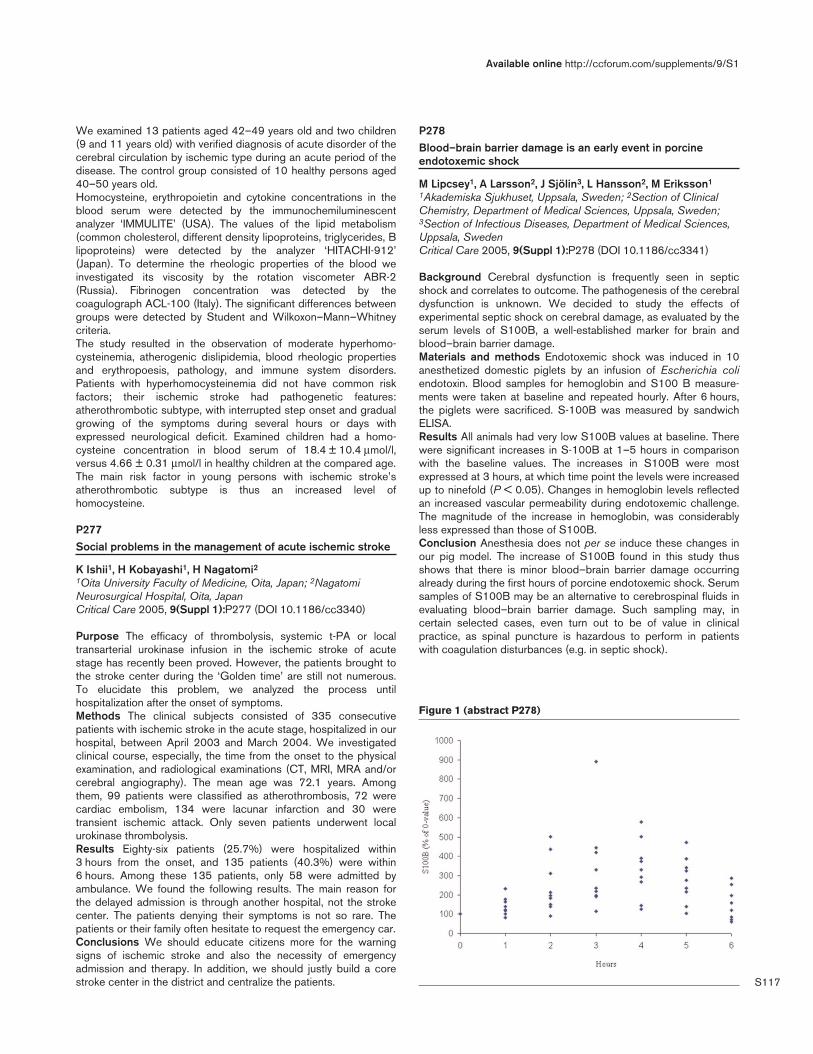
S117
We examined 13 patients aged 42–49 years old and two children(9 and 11 years old) with verified diagnosis of acute disorder of thecerebral circulation by ischemic type during an acute period of thedisease. The control group consisted of 10 healthy persons aged40–50 years old.Homocysteine, erythropoietin and cytokine concentrations in theblood serum were detected by the immunochemiluminescentanalyzer ‘IMMULITE’ (USA). The values of the lipid metabolism(common cholesterol, different density lipoproteins, triglycerides, Blipoproteins) were detected by the analyzer ‘HITACHI-912’(Japan). To determine the rheologic properties of the blood weinvestigated its viscosity by the rotation viscometer ABR-2(Russia). Fibrinogen concentration was detected by thecoagulograph ACL-100 (Italy). The significant differences betweengroups were detected by Student and Wilkoxon–Mann–Whitneycriteria.The study resulted in the observation of moderate hyperhomo-cysteinemia, atherogenic dislipidemia, blood rheologic propertiesand erythropoesis, pathology, and immune system disorders.Patients with hyperhomocysteinemia did not have common riskfactors; their ischemic stroke had pathogenetic features:atherothrombotic subtype, with interrupted step onset and gradualgrowing of the symptoms during several hours or days withexpressed neurological deficit. Examined children had a homo-cysteine concentration in blood serum of 18.4 ± 10.4 µmol/l,versus 4.66 ± 0.31 µmol/l in healthy children at the compared age.The main risk factor in young persons with ischemic stroke’satherothrombotic subtype is thus an increased level ofhomocysteine.
P277Social problems in the management of acute ischemic stroke
K Ishii1, H Kobayashi1, H Nagatomi21Oita University Faculty of Medicine, Oita, Japan; 2NagatomiNeurosurgical Hospital, Oita, JapanCritical Care 2005, 9(Suppl 1):P277 (DOI 10.1186/cc3340)
Purpose The efficacy of thrombolysis, systemic t-PA or localtransarterial urokinase infusion in the ischemic stroke of acutestage has recently been proved. However, the patients brought tothe stroke center during the ‘Golden time’ are still not numerous.To elucidate this problem, we analyzed the process untilhospitalization after the onset of symptoms.Methods The clinical subjects consisted of 335 consecutivepatients with ischemic stroke in the acute stage, hospitalized in ourhospital, between April 2003 and March 2004. We investigatedclinical course, especially, the time from the onset to the physicalexamination, and radiological examinations (CT, MRI, MRA and/orcerebral angiography). The mean age was 72.1 years. Amongthem, 99 patients were classified as atherothrombosis, 72 werecardiac embolism, 134 were lacunar infarction and 30 weretransient ischemic attack. Only seven patients underwent localurokinase thrombolysis.Results Eighty-six patients (25.7%) were hospitalized within3 hours from the onset, and 135 patients (40.3%) were within6 hours. Among these 135 patients, only 58 were admitted byambulance. We found the following results. The main reason forthe delayed admission is through another hospital, not the strokecenter. The patients denying their symptoms is not so rare. Thepatients or their family often hesitate to request the emergency car.Conclusions We should educate citizens more for the warningsigns of ischemic stroke and also the necessity of emergencyadmission and therapy. In addition, we should justly build a corestroke center in the district and centralize the patients.
P278Blood–brain barrier damage is an early event in porcineendotoxemic shock
M Lipcsey1, A Larsson2, J Sjölin3, L Hansson2, M Eriksson1
1Akademiska Sjukhuset, Uppsala, Sweden; 2Section of ClinicalChemistry, Department of Medical Sciences, Uppsala, Sweden;3Section of Infectious Diseases, Department of Medical Sciences,Uppsala, SwedenCritical Care 2005, 9(Suppl 1):P278 (DOI 10.1186/cc3341)
Background Cerebral dysfunction is frequently seen in septicshock and correlates to outcome. The pathogenesis of the cerebraldysfunction is unknown. We decided to study the effects ofexperimental septic shock on cerebral damage, as evaluated by theserum levels of S100B, a well-established marker for brain andblood–brain barrier damage.Materials and methods Endotoxemic shock was induced in 10anesthetized domestic piglets by an infusion of Escherichia coliendotoxin. Blood samples for hemoglobin and S100 B measure-ments were taken at baseline and repeated hourly. After 6 hours,the piglets were sacrificed. S-100B was measured by sandwichELISA.Results All animals had very low S100B values at baseline. Therewere significant increases in S-100B at 1–5 hours in comparisonwith the baseline values. The increases in S100B were mostexpressed at 3 hours, at which time point the levels were increasedup to ninefold (P < 0.05). Changes in hemoglobin levels reflectedan increased vascular permeability during endotoxemic challenge.The magnitude of the increase in hemoglobin, was considerablyless expressed than those of S100B.Conclusion Anesthesia does not per se induce these changes inour pig model. The increase of S100B found in this study thusshows that there is minor blood–brain barrier damage occurringalready during the first hours of porcine endotoxemic shock. Serumsamples of S100B may be an alternative to cerebrospinal fluids inevaluating blood–brain barrier damage. Such sampling may, incertain selected cases, even turn out to be of value in clinicalpractice, as spinal puncture is hazardous to perform in patientswith coagulation disturbances (e.g. in septic shock).
Available online http://ccforum.com/supplements/9/S1
Figure 1 (abstract P278)
S118
P279Antiapoptotic effects of delta opioid peptide [D-Ala2,D-Leu5]enkephalin in brain slices induced by oxygen-glucose deprivation
XR Wang1, YJ Zheng1, HZ Chen2, XJ Wu2, YH Zhao1, DS Su1
1Department of Anesthesiology, Renji Hospital, Shanghai SecondMedical University, Shanghai, China; 2Department ofPharmacology, Shanghai Second Medical University, Shanghai,ChinaCritical Care 2005, 9(Suppl 1):P279 (DOI 10.1186/cc3342)
Background Delta opioid peptide [D-Ala2,D-Leu5]enkephalin(DADLE) has been shown to play a role in neuronal protectionagainst hypoxic ischemia. However, the cellular mechanisms ofthese actions of DADLE on neurons are not totally clear. Being anin vitro model of brain ischemia, oxygen-glucose deprivation(OGD) injury in rat brain slices has the advantages of both in vivoand in vitro models, and therefore can imitate damages induced bybrain I/R injury in intact animals. In the present study, we examinedthe protective mechanism(s) of DADLE against apoptosis using arat brain slice model. In addition, we determined whether delta-opioid receptors are unique and have a specific role inneuroprotection against OGD injury by activating the MAPKpathway, specifically through delta2-opioid receptors.Methods The brain slices were incubated with differentconcentrations of DADLE after injury by OGD. Selective delta2-opioid antagonist or selective inhibitor of ERK kinase was co-incubated with or without DADLE. The effects of DADLE againstapoptosis in neurons were measured by the following biochemicaland morphological assays: LDH release, RT-PCR, western blot,and TUNEL staining.Results OGD caused LDH release in brain slices. DADLEinhibited OGD-induced LDH release. Naloxone, and naltribenmethane sulfonate, partially suppressed the survival of brain slicespromoted by 10 µM DADLE. DADLE at lower concentrations (10–6
and 10–5 M) significantly suppressed Bax mRNA expression andenhanced Bcl-2 mRNA expression. But with the increasedconcentrations of DADLE, the ability to regulate apoptotic geneexpression was attenuated. DADLE induced ERK phosphorylationin brain slices with the increase of concentrations. The withdrawalof oxygen and glucose per se decreased the ERK phosphorylation.In the absence of oxygen and glucose, DADLE at 10–5 Mconcentrations could, however, increase the ERK phosphorylation.Naltrindole reduced the degree of ERK phosphorylation inducedby 10–5 M DADLE. PD98059 abolished the DADLE-inducedphosphorylation of ERK in the condition of OGD. PD98059 notonly abolished the cell survival promoted by DADLE, but alsoapparently led to a higher level of LDH release when comparedwith the DADLE control.Conclusions Our results suggest, therefore, that endogenousopioid peptides may, at low concentrations, promote cell survival viathe MEK–ERK pathway, perhaps through delta2-opioid receptors.
P280The use of transcranial Doppler ultrasonography in earlyoutcome prediction of patients with subarachnoid hemorrhage
A Lavrentieva, B Soulountzi, B Boutsas, A Lampanari, A Mitka,M BitzaniG Papanikolau Hospital — ICU, Theesaloniki, GreeceCritical Care 2005, 9(Suppl 1):P280 (DOI 10.1186/cc3343)
Introduction Transcranial Doppler ultrasonography (TCD) is awell-established technique for detection of cerebral vasospasm in
patients with subarachnoid hemorrhage (SAH). The aim of ourstudy is to investigate whether there is a relationship between TCDfindings during the first 3 days after admission and the neurologicaloutcome of patients.Materials and methods Sixty-seven patients (60 ± 12.9 years,APACHE II score 18.2 ± 6.4) were included in our study. TCDmiddle cerebral artery systolic (SV), diastolic, and mean flowvelocities and pulsatility index (PI) were measured daily followingSAH. Neurological evaluation was performed at ICU dischargeusing the Glasgow Outcome Score (GOS). A GOS score of 1–3was considered a pure outcome (group 1, n = 56), a GOS scoreof 4 or 5 as a good outcome (group 2, n = 11).The Student t testwas used to compare the data and P < 0.05 was consideredsignificant.Results See Table 1. At admission there was no statisticallysignificant difference between the two groups in their flowvelocities and PI. The patients with pure outcome (group 1)exhibited higher systolic flow velocities and higher PI on day 3 afteradmission.
Table 1
TCD findings during the first 3 days after SAH (mean ± standarddeviation)
Day 1 Day 2 Day 3
PI SV PI SV PI SV
Group 1 1.4 ± 0.6 74 ± 30 1.26 ± 0.4 88 ± 35 1.63 ± 0.6 106 ± 4
Group 2 1.17 ± 0.3 60 ± 18 1.21 ± 0.16 68 ± 8 1 ± 0.07 57 ± 9
P NS NS NS NS < 0.05 < 0.05
NS, not significant.
Conclusion The elevation of PI and systolic flow velocity on day 3after admission seems to be related to pure neurological outcomeof patients suffering SAH probably due to early appearance ofvasospasm.
P281OCTOPUS — observation or computed tomography of mildhead injury in Sweden: a randomised clinical trial concerningeffects and costs
J af Geijerstam1, S Oredsson2, M Britton1
1Department of Medicine, Karolinska University Hospital,Stockholm, Sweden; 2Helsingborg Hospital, Helsingborg, SwedenCritical Care 2005, 9(Suppl 1):P281 (DOI 10.1186/cc3344)
Background Mild head injury, defined as short-term loss ofconsciousness and/or amnesia as a result of head trauma, is acommon problem in emergency departments worldwide. Acutecomputed tomography (CT) in triage for admission as well asinhospital observation is suggested as standard practice for thesepatients. However, the outcome, safety and costs of the twostrategies is unclear. To address this question a randomisedcontrolled trial (RCT) was conducted.Methods A multicenter, pragmatic RCT with 39 emergencydepartments throughout Sweden participating to randomise the2500 cases needed. Eligibility criteria Head trauma within the past24 hours, age 6 years or older, confirmed or suspected amnesia orbrief loss of consciousness, GCS 15, normal neurological findingsand no associated injuries that require admission. RandomisationUrgent CT for all patients, early discharge for home care if normalfindings. Admission for inhospital observation. Main hypothesesMedical outcome — the CT strategy is safe and clinically equivalentto one based on inhospital observation. Cost comparison — the CT
Critical Care March 2005 Vol 9 Suppl 1 25th International Symposium on Intensive Care and Emergency Medicine

S119
strategy reduces costs for management of mild head injury in theacute stage. Costs in the follow-up period are equivalent betweenthe strategies. Endpoints Medical outcome — patient function at3 months (Glasgow Outcome Scale Extended). Cost comparison— resource utilisation in acute and follow-up phase.Results From May 2001 through January 2004, 2603 patientsunderwent randomisation. Patient follow-up was completed in June2004. Data on the primary endpoint (GOSE at 3 months) areavailable for 97% of the cases. Data analysis and manuscriptpreparation are currently under way.Conclusions The OCTOPUS study has randomised over 2600patients with mild head injury to acute CT to triage for admission orinhospital observation. Results from the study regarding themedical effects, safety and costs of the two strategies will beavailable in late 2005. Data on group characteristics and practicalperformance will be presented at ISICEM in March 2005.
P282Post-trauma subarachnoid hemorrhage
C Kaddour, R Souissi, M Laamourou, Z HaddadNational Institute of Neurology, Tunis, TunisiaCritical Care 2005, 9(Suppl 1):P282 (DOI 10.1186/cc3345)
Purpose Assessment of the incidence of coronary complicationduring post-trauma subarachnoïd hemorrhage (SAH).Methods An open, double-blind, prospective study, during aperiod of 15 months in the Department of Anesthesia andResuscitation of the National Institute of Neurology of Tunis.Patients Thirty-five patients with head trauma in whom a CT scanreveals a SAH on admission. Patients with cardiovascular history,thorax trauma, neurological coma related to a vascular pathologyand non-neurological coma were excluded.Results The mean age of patients is 39 ± 17 years; 85% had aGlasgow Coma Scale < 8 on admission. The mean ISS was27 ± 14, and 6% and 54% had a Fischer scale between 3 and 4,on admission. Electrocardiogram changes in 57% of patients.Troubles of repolarization are the most frequent; 94% of patientspresented this trouble on the third day. The mean duration of thesetroubles is 4 ± 1 days. Over the three performed measurements,total CPK was increased in 88% of patients, creatine kinase MBisoenzyme was increased in 65% of patients and Troponin Ic waselevated in 34% of patients in a certain time of the evolution withan average of 0.068 (± 0.2). The peak of Troponin Ic occurred onthe third day with an average of 0.113 (± 0.338). The increase ofTroponin Ic is associated with a poor CT scan grade. The elevationof Troponin Ic is correlated with T-wave anomalies in comparisonwith those of the ST segment. But it is not correlated to creatinekinase MB elevation. The T-wave troubles, which are morefrequent, therefore reflect better a myocardic lesion and TroponinIc compared with creatine kinase MB is a better indicator ofmyocardic ischemia during SAH. In our department, no case ofmortality was related to a cardiac cause. The incidence of TroponinIc and T-wave troubles could be considered predictive factors ofmortality.Conclusion According to this study, troubles of repolarizationduring post-trauma SAH are frequent in the acute phase of the first3 days. They could reflect a myocardic lesion as it is indicated bythe increase of Troponin Ic, which is associated with severeneurological injuries.
P283Multimodal neuroelectrophysiological studies to predictoutcome in paediatric patients with traumatic brain injury
M Gray, V Nenadovic, J HutchisonHospital for Sick Children, Toronto, CanadaCritical Care 2005, 9(Suppl 1):P283 (DOI 10.1186/cc3346)
Introduction A prognostic tool is needed in the early assessmentof paediatric patients with traumatic brain injury (TBI). Thecombination of electroencephalography and evoked potentials mayhelp predict outcome.Methods We enrolled patients with TBI admitted to the PaediatricCritical Care Unit with an initial GCS of 12 or less. Patients had anelectroencephalogram (EEG), visual evoked potentials (VEPs) andsomatosensory evoked potentials (SEPs) done within 60 hours ofadmission to the PICU, and repeated within 7 days. A CT scanwas done on admission. EEG, VEP and SEP were scored. TheEEG was analyzed for synchrony using the Hilbert transform.Outcome was measured at 3 months using the Paediatric CerebralPerformance Category Score (PCPC).Results Seven patients aged 3–13 were enrolled. Lowestrecorded GSC ranged from 5 to 12. Three of the seven patientsdeveloped intracranial hemorrhage. Four of the seven patientssustained diffuse axonal injury; these patients had encephalopathicEEG patterns. Children whose EEG had normal backgroundactivity had better outcomes at 3 months. Those who regainednormal background activity on the second EEG had intermediateoutcomes. Children having the worst scores for EEG and SEP hadworst functional outcome. EEG score did not show a correlationwith change in the PCPC. Encephalopathic EEGs showedincreased synchrony compared with EEGs having a normalbackground (Fig. 1). Increased SEP latency in cortical to corticalrelays correlated with poor functional outcome. SEP score didcorrelate with change in the PCPC (Pearson coefficient r = 0.82,P = 0.02). Children whose EEG showed attenuated occipitalbackground patterns had large-amplitude VEPs. Current scoringsystems for VEPs do not account for this observed increase.
Available online http://ccforum.com/supplements/9/S1
Figure 1 (abstract P283)
Synchrony pattern from EEG electrode C3 of patient 2 in the deltafrequency range. Red, maximal synchrony; blue, minimal synchrony.Patient demonstrates synchrony between C3 and between electrodes1–3, 8–11 and 18. Synchrony is pathological.
S120
Conclusions Our results indicate that SEP correlates directly withoutcome. EEG synchrony correlates with outcome. High-amplitudeVEPs may reflect neuronal hyperexcitability The significance of thisneeds further investigation.
P284Comparison of biochemical markers and transcranial Dopplerultrasonography findings as early predictors of outcome inpatients with traumatic brain injury: preliminary report
I Rodini1, A Lavrentieva1, B Soulountzi1, C Fitili2, M Karvouniaris1,M Bitzani11ICU, G Papanikolau Hospital, Theesaloniki, Greece; 2MicrobiologyDepartment, G Papanikolau Hospital, Thessaloniki, GreeceCritical Care 2005, 9(Suppl 1):P284 (DOI 10.1186/cc3347)
Introduction Early brain ischemia after traumatic brain injury (TBI)is predictive of pure outcome. We investigated the time course ofbiochemical markers of brain damage: serum neuron-specificenolase (NSE) and protein S-100 (Pr S-100) during the first5 days after the ICU admission. Brain hemodynamics weresynchronously assessed using transcranial Doppler ultra-sonography (TCD). Each patient was examined with TCD, and themaximum, mean, and the end diastolic velocities and pulsatilityindex (PI) were evaluated.Methods Eleven patients (aged 34 ± 14.7, APACHE II score15.7 ± 9, SAPS II 25 ± 13.7) with TBI were included in thisprospective study. Paired arterial and jugular bulb bloodconcentrations of NSE and Pr S-100 were measured daily for fiveconsecutive days and were correlated with neurological outcomeevaluated by GOS score. TCD middle cerebral artery velocitiesand PI were also measured daily after admision and werecorrelated with biochemical markers and outcome at ICUdischarge. The statistical analysis was performed using theWilcoxon U test and Spearman correlation test.Results NSE and PrS-100 concentrations were significantlyhigher in patients with poor outcome (GOS 1–3) than in patientswith good outcome (GOS 4–5) both in arterial as well as in jugularvenous blood samples during the first 5 days. There weresignificant correlations between these biochemical indicies and theneurological outcome of patients from day 2 after admission(Pr S-100: r = 0.76, P = 0.02, NSE: r = 0.67, P = 0.03) thatpersisted for the following 3 days. TCD findings were notcorrelated neither with neurological outcome nor with biochemicalmarkers of brain damage.Conclusion The biochemical markers of brain injury (NSE,Pr S=100) seem to be better early outcome prediction parametersthan cerebral blood flow velocities and PI measured by TCD inpatients with TBI.
P285Clinical progresses and radiological findings in patients withhead injury
K Ishii, K KitajimaOita University Faculty of Medicine, Oita, JapanCritical Care 2005, 9(Suppl 1):P285 (DOI 10.1186/cc3348)
Purpose X-ray and computed tomography (CT) are currently themost effective tools in the analysis of head trauma patients.However, the standard for operation of X-ray and CT is still unclear.We investigated the clinical courses and radiological findings inpatients with head injury, especially minor trauma.Methods The clinical subjects consisted of consecutive 217patients, treated by our emergency medicine between January
2003 and December 2004. Among them, Glasgow Coma Scale(GCS) scores in 209 patients (96.3%) were 15. We investigatedtheir clinical courses and radiological findings (X-ray and CT).Results Skull X-ray was performed in 90 patients (41.5%), and CTwas performed in 114 patients (52.5%). Skull fracture was shownradiographically in six patients (2.8%), and abnormalities on theinitial CT were seen in four patients (1.8%). Two hundred and ninepatients with GCS scores of 15 had excellent prognosis excepttwo died. One died because of delayed epidural hematoma, andthe other because of crescendo cerebral swelling due to cerebralcontusion in the bilateral frontal lobe.Conclusions Head trauma patients must be treated with attentionand should undergo X-ray and CT examinations, even in minorinjury.
P286The clinical importance of bulbus juguli oxymetry
C Hoffmann, Z Marjanek, Z KisvargaJávorszky Ödön Hospital, Vác, HungaryCritical Care 2005, 9(Suppl 1):P286 (DOI 10.1186/cc3349)
Objective In our study, we attempted to determine thoserepresentative clinical parameters that most exactly correlate withthe final outcome of intensive treatment requiring neurological(ischaemic and haemorrhagic stroke) and neurosurgical (subduraland epidural haematoma) cases.Methods We observed 27 patients retrospectively. The cohortwas divided into a survivor group I (12 cases) and a non-survivorgroup II (15 cases). We introduced a central venous canule intothe internal jugular vein in the direction of the bulbus juguli, andcollected blood samples every 6 hours, at every significantalteration of physical status and at therapeutic intervention. Wemonitored the cerebral perfusion pressure (CPP) and intracranialpressure (ICP) continously, and measured the oxygen saturation ofa blood sample from the bulbus juguli (SvjO2) and the jugulararteriovenous lactate gap every 6 hours, and the GCS every 8hours. We registrated the change of intracranial structure bycomputer tomography.Results In group II (non-survivors) the SvjO2 and lactate gapvalues were 83.83 ± 6.6% and 0.43 ± 0.06 mmol/l, in group I(survivors) the respective values were 71.57 ± 6.26% and0.2 ± 0.1 mmol/l. We could not find a patient in group II with aGCS higher than 5. There was no significant difference in ICP andCPP values between survivor and non-survivor groups(13.6 ± 2.4 mmHg and 72.3 ± 8.4 mmHg versus 16.4 ± 8.1 mmHgand 68.45 ± 4.6 mmHg).Conclusion The bulbus juguli oxymetry is an invasive but cheap,simple and with competent practical skills a safely applicablemonitoring method for the judgement of cerebral oxygenisation.The SvjO2 and lactate gap values were determinative in the aspectof final outcome.
P287Prognostic value of specific neural enolase in head trauma
C Kaddour, N Baffoun, Z Haddad, M HamdaniNational Institute of Neurology, Tunis, TunisiaCritical Care 2005, 9(Suppl 1):P287 (DOI 10.1186/cc3350)
Purpose Searching for a correlation between the plasma level ofspecific neural enolase (SNE) and the prognosis of head trauma inthe short term.Subjects and methods An open prospective study carried out in aHUC of Tunis over 15 months including 35 patients with head
Critical Care March 2005 Vol 9 Suppl 1 25th International Symposium on Intensive Care and Emergency Medicine

S121
trauma causing a subarachnoid hemorrhage. Criteria of non-inclusion were age <15 years and 3 months, and head traumarequiring neurosurgery. The management was standardized for allpatients to avoid secondary cerebral aggression caused bysystemic troubles, as recommended by Maas and colleagues.Clinical basic neurologic survey and CT scan monitoring (Fischerand Marshall classification) were repeatedly carried out. A cerebralCT scan was systematically performed 48 hours after hospitali-zation or in case of aggravation. A surgical operation wasperformed throughout the period of the study whenever required(orthopedics, digestive surgery, etc.). The plasma level of SNE wasmeasured on the first, third and fifth day of hospitalization, usingthe ELISA technique with an immunoenzymatic method, sandwichtype (kit CanAg Diagnostics), in a Dias Orin automate (Eti-max3000). (The normal value of enolase used as a reference in ourstudy was considered the average of 47 volunteers’ plasma level.)Statistical analysis used the chi-squared test and variance analysis,respectively, for qualitative and quantitative variables. Values areexpressed as means ± standard deviation.Results Mean age: 39 ± 17 years; sex ratio = 4; 80% of patientshad a severe head trauma; mortality: 16 patients (45.7%); meanlevel of SNE in a ‘reference population’ was 7.02 ± 3.86 µg/mlwith a statistically significant difference (P = 0.001) (pathologicvalue above 12.5 µg/ml). Seventeen patients (48.56%) had apathologic SNE plasma level in the first day. All patients (nine)whose SNE plasma level had still been elevated until the third orthe fifth day died. The plasma level was higher in patients who diedin comparison with that of surviving patients; 25.74 ± 22.39 µg/mlvs 11.51 ± 4.56 µg/ml, with a significant difference (P = 0.032).All dead patients had a higher plasma level of SNE on the fifth day,six of them had a normal value in the first day and then it increased.Patients whose GCS was < 8 had a first-day plasma level so farhigher, 24.89 ± 25.27 µg/ml vs 15.25 ± 11.6 µg/ml; but with nosignificant difference. SNE plasma level was not correlated to theimportance of the subarachnoïd hemorrhage (Fischer classification).Discussion The persistence of a high plasma level of specificneural enolase or its elevation represents a short-term prognosticfactor. Severe patients have higher plasma levels with nosignificant difference.
P288S100B protein: a possible prognostic factor in patients with asevere head trauma injury
P Lavicka, R Pikner, R Bosman, M Choc, I Chytra, O TopolcanFaculty Hospital, Plzen, Czech RepublicCritical Care 2005, 9(Suppl 1):P288 (DOI 10.1186/cc3351)
Introduction S100B protein is a small protein that belongs to thegroup of calcium binding proteins. It is present in highconcentrations in astroglia cells. The level of this marker increasessignificantly with CNS damage. Normal value is 0–0.2 µg/l.Aim The aim of our research was to evaluate a consequence of adynamic monitoring of the S100B protein levels for a prognosis ofpatients with severe head trauma injury.Method Thirty patients with severe head trauma injury wereprospectively monitored. All patients were admited to theEmergency Department. The time period from injury to admissionwas shorter than 6 hours. All patients had the initial CT scan of thebrain performed with a positive finding. We used these scoringprotocols: at admission GCS, APACHE II, at dismissal GOS,Karnofsky Performance Score (KPS). S100B protein level wasdone by LIA essay on a fully automated immunoanalyser (Liaison;DiaSorin, Sweden).
Results We divided our patients into four groups according tocharacteristics of the S100B protein levels. The average values ofthe particular groups are given in Table 1: group A, patients with arapid decrease of the level; group B, patients with an increasing orpersistent high level; group C, patients with a repeated increase ofthe level after a previous decrease; group D, patients with an initiallevel above 5 µg/l.
Table 1
Patient Level Level Level Group Death number GCS APACHE GOS KPS 0 24 72
A 1 15 9 17 4 80 1.378 0.415 0.245
B 0 5 8 22 3 50 1.45 1.68 0.924
C 2 7 8 26 2,5 40 1.697 3.155 1.18
D 3 3 3 37 1 0 18 – –
Conclusion A rapid decrease of the level in the first 72 hours isassociated with a good prognosis. Increasing or persistent highlevels longer than 72 hours are associated with a poor prognosis.A repeated increase level above 1 µg/l after a previous decrease isassociated with a poorer outcome. An initial level above 5 µg/l isassociated with death.
P289Increased levels of serum S100B protein in critically illpatients without brain injury
C Routsi, E Stamataki, S Nanas, C Psachoulia, M Zervou, A Koroneos, A Stathopoulos, C RoussosEvangelismos Hospital, Athens, GreeceCritical Care 2005, 9(Suppl 1):P289 (DOI 10.1186/cc3352)
Objective S100B, a calcium-binding protein produced andreleased predominantly by astrocytes of the central nervoussystem, has been established as a specific biochemical marker forbrain injury [1]. However, there is increasing evidence that S100Brelease is also induced by other causes or even from tissue outsidethe brain [2]. The aim of this study was to explore the possible roleand the utility of S100B in critically ill patients with respiratoryfailure of various etiologies, but without brain injury.Patients and methods Thirty-eight mechanically ventilatedcritically ill patients aged 62 ± 17 years, 29 males, with respiratoryand/or organ failure, but with no evidence of brain injury or otherneurological disorder, were prospectively studied. Blood samplesfor analysis of serum S100B protein were obtained from the radialartery at the time of laboratory sampling and blood gas analysis.For the determination of S100B, all samples were analyzed using acommercially available immunoluminometric assay (LIASON;Sangtec Medical, Bromma, Sweden). Blood gases and lactatewere analyzed using the ABL 625 analyzer (RadiometerCopenhagen, Denmark) and then the arterial oxygen content wascalculated.Results Serum S100B was measured in 16 medical and 22surgical ICU patients with a SOFA score on admission of 8 ± 3.4.Compared with the normal values (< 0.15 µg/l), almost all patientsexhibited increased serum S100B levels at least once(0.56 ± 1.03 µg/l, mean ± standard deviation, range 0.04–9.25).Pearson’s correlation showed a statistically significant relationbetween S100B and Hb (P < 0.001), O2 content (P = 0.002), andarterial lactate (P < 0.001). Multivariate analysis using a stepwiselogistic regression model showed that the lactate was anindependent variable (F = 9.6, P < 0.001), followed by O2 content(F = –3.25, P = 0.02).
Available online http://ccforum.com/supplements/9/S1

S122
Conclusions Serum levels of S100B protein are elevated incritically ill patients and correlate with increased lactate levels, lowhemoglobin, and abnormal oxygen content. Further researchshould determine the sources of this S100B release and evaluateits significance as a possible marker of tissue hypoperfusion in theICU setting.References1. Br J Neurosurgery 1999, 13:56.2. Neurosurgery 2001, 48:1255.
P290S100B protein in heroin overdose: a pilot study
M Brvar, J Ambrozic, J Osredkar, M Mozina, M BuncUniversity Medical Center Ljubljana, SloveniaCritical Care 2005, 9(Suppl 1):P290 (DOI 10.1186/cc3353)
Objective Heroin overdose could result in hypoxic-ischemic changeswith cerebral edema, ishemic neuronal damage and neuronal lossdue to central nervous depression with significant respiratorydepression and hypotension. Heroin overdose can cause cognitiveimpairment, neurological deficits or even death. S100B, thestructural protein of astroglia, has already been shown to be a usefulneurobiochemical marker of brain damage in circulatory arrest,stroke, traumatic head injury and carbon monoxide poisoning. Theaim of our study was to assess the possible role of S100B as abiochemical marker of brain injury in heroin overdose.Methods The prospective study included patients with heroinoverdose who were admitted to the Emergency Department (ED).Patients were enrolled if they had a documented exposure toheroin only. The physical and neurological examinations werecarried out on the scene and on arrival at the ED.Neuropsychological testing was performed at the ED just beforethe patient’s discharge. Blood samples for S100B determinationwere drawn immediately after arrival at the ED. S100Bconcentrations were measured with a commercial immuno-luminometric assay. Blood samples were also analyzed for creatinekinase, troponin I and creatinine level. A toxicology analysis ofpatient’s blood or urine samples by gas chromatography coupledto mass spectrometry was performed. The control group included10 healthy volunteers. Data are presented as the mean. S100levels between groups were compared using the Mann–Whitney Utest and the Pearson correlation coefficients were calculated forS100B, creatine kinase, troponine I and creatinine levels. A P valueof less than 0.05 was considered significant.Results A total of 20 patients with heroin overdose were enrolled.The mean age of the study cohort was 24.5 years (range,16–39 years). There were four women and 16 men. Heroinoverdose was unintentional in all patients. All patients wereunconscious on the scene and regained conciseness afternaloxone application. 6-Monoacetyl-morphine, a metabolite ofheroin, was detected in all patients. S100B levels of heroinoverdosed patients were significantly higher compared withS100B levels of the control group (0.65 µg/l vs 0.07 µg/l,P < 0.05). Twelve patients (60%) had elevated creatine kinase withmean value 15.30 µkat/l (normal value 0.17–2.08 µkat/l) indicatingrhabdomyolysis and two (10%) of them also had elevatedtroponine I (0.15 and 0.1 µg/l; normal value 0.06 µg/l) indicatingmyocardial necrosis. No correlation was found between serumS100B, creatine kinase, troponine I and creatinine levels(P > 0.05). All patients survived and had no clinically detectableneurological deficits on discharge. Psychological testing wasunsuccessful because the majority of patients refused testing.Conclusion Heroin overdose is associated with elevated S100Blevels. S100B could be useful biochemical marker of heroin
overdose, but its clinical value to predict brain tissue damage withneurological or cognitive deficits after heroin overdose should befurther evaluated.
P291Paired serum S-100B and neuron-specific enolase assay inearly and fast assessment of brain damage after cardiac arrest
C Crippa, G Larosa, R Dorizzi, A Merlini, E Pecci, E Polati, A LuzzaniInstitute of Anesthesiology & Intensive Care, Verona, ItalyCritical Care 2005, 9(Suppl 1):P291 (DOI 10.1186/cc3354)
Background and purpose Early and accurate predictors ofneurological outcome in patients with hypoxic-ischemic encephalo-pathy after cardiac arrest are needed in order to improve medicaldecision-making. The aims of the present study were: to assessserum S-100 protein and neuron-specific enolase (NSE) as earlymarkers of brain damage and outcome after cardiac arrest; and tocompare the diagnostic and prognostic power of these serummarkers to somatosensory evoked potentials (SSEP).Design A prospective study. Clinicians were blinded to S-100 andNSE results.Setting The ICU of a university hospital.Methods Twenty-five patients resuscitated from cardiac arrest(CA) were assessed until they regained consciousness or untildeath/permanent vegetative state. We correlated SSEP, S-100protein and NSE serum concentrations with patient outcome.S-100 protein and NSE serum concentrations were measuredusing an automated chemiluminescent analyzer (Liaison; Diasorin,Saluggia, Italy) in samples collected 24, 48, 72 and 168 hoursafter the cardiopulmonary resuscitation (CPR); SSEP wererecorded at 48 hours and on day 7 after the CPR.Results We enrolled 25 consecutive patients (17 males/eightfemales, mean age 69 ± 17 years, mean APACHE II score26 ± 4.5); eight of them regained consciousness. Median values ofNSE and S-100B serum concentrations were significantly(Mann–Whitney U test) higher in patients who did not regainconsciousness compared with patients who regained conscious-ness at all time intervals (NSE: 53 vs 24 µg/l at 24 hours; 137 vs20 µg/l at 48 hours; 102 vs 16 µg/l at 72 hours, and 39 vs 6 µg/l168 hours after CA; S-100B: 2882 vs 342.5 ng/l at 24 hours;6514 vs 240 ng/l at 48 hours; 3125 vs 198 ng/l at 72 hours, and610 vs 146 ng/L 168 hours after CA).S-100B concentration cut-off of 776 ng/l at 24 hours was foundthe best predictor of not regaining consciousness. The area underthe curve was 0.96 (95% confidence interval = 0.88–1.00);specificity was 100%; sensitivity was 82.4%. NSE concentrationcut-off point of 45 µg/l at 48 hours was found the best predictor ofnot regaining consciousness. The area under the curve was 0.87(95% confidence interval = 0.72–1.00); specificity was 100%;sensitivity was 81.2%.High association between SSEP responses and outcome wasfound at 48 hours (rho = 0.68, P < 0.001) and at 7 days (rho =0.72, P < 0.001) (Spearman correlation).Conclusions According to our study and previous reports [1,2]biochemical markers, and especially S100 protein, provide reliableinformation about patient outcome after acute global cerebralischemia. Moreover, we used a recently marketed analyzer thatmakes available S-100 protein and NSE results within 1 hour; inthis way, clinicians can easily and promptly use them in themanagement of the patient.References1. Hachimi-Idrissi S, Van der Auwera M, Schiettecatte J, et al.:
Resuscitation 2002, 53:251-257.2. Martens P, Raabe A, Johnsson P: Stroke 1998, 29:2363-2366.
Critical Care March 2005 Vol 9 Suppl 1 25th International Symposium on Intensive Care and Emergency Medicine
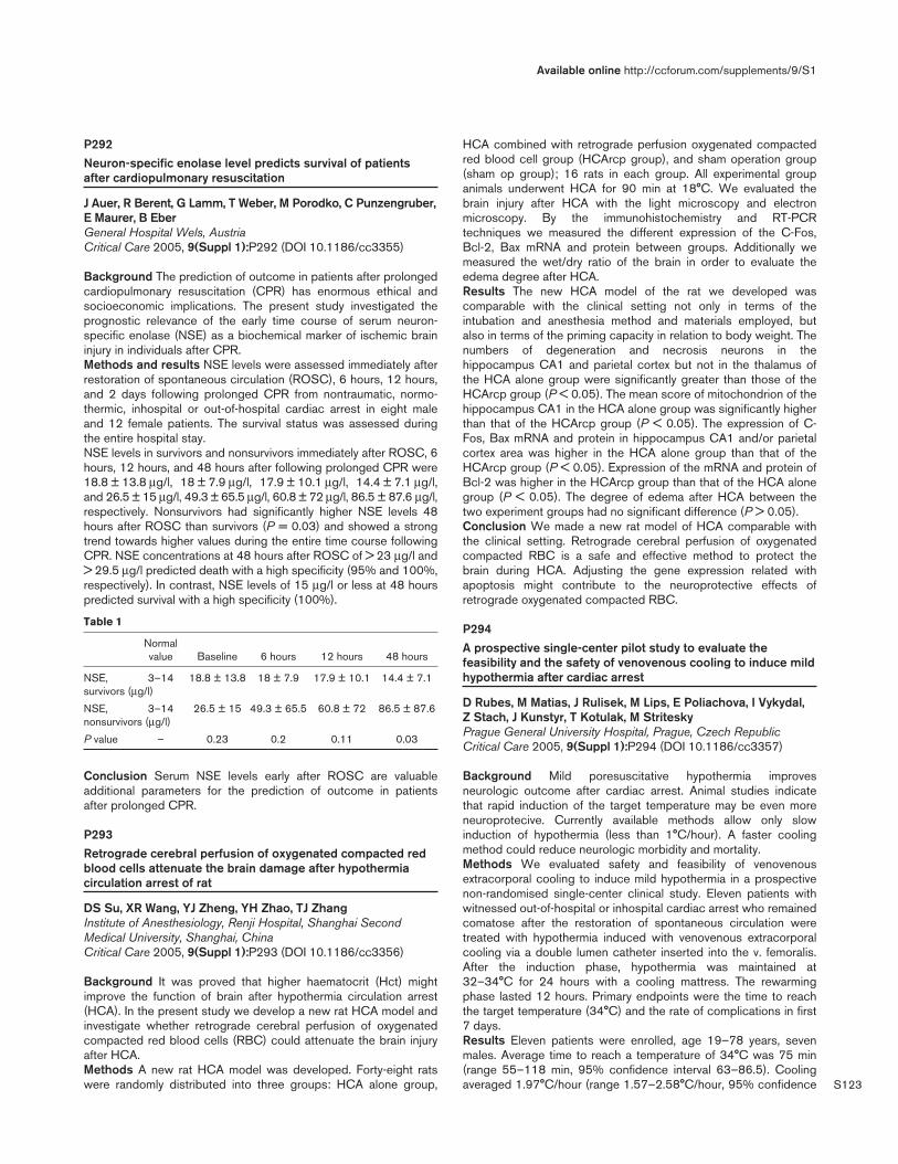
S123
P292Neuron-specific enolase level predicts survival of patientsafter cardiopulmonary resuscitation
J Auer, R Berent, G Lamm, T Weber, M Porodko, C Punzengruber,E Maurer, B EberGeneral Hospital Wels, AustriaCritical Care 2005, 9(Suppl 1):P292 (DOI 10.1186/cc3355)
Background The prediction of outcome in patients after prolongedcardiopulmonary resuscitation (CPR) has enormous ethical andsocioeconomic implications. The present study investigated theprognostic relevance of the early time course of serum neuron-specific enolase (NSE) as a biochemical marker of ischemic braininjury in individuals after CPR.Methods and results NSE levels were assessed immediately afterrestoration of spontaneous circulation (ROSC), 6 hours, 12 hours,and 2 days following prolonged CPR from nontraumatic, normo-thermic, inhospital or out-of-hospital cardiac arrest in eight maleand 12 female patients. The survival status was assessed duringthe entire hospital stay.NSE levels in survivors and nonsurvivors immediately after ROSC, 6hours, 12 hours, and 48 hours after following prolonged CPR were18.8 ± 13.8 µg/l, 18 ± 7.9 µg/l, 17.9 ± 10.1 µg/l, 14.4 ± 7.1 µg/l,and 26.5 ± 15 µg/l, 49.3 ± 65.5 µg/l, 60.8 ± 72 µg/l, 86.5 ± 87.6 µg/l,respectively. Nonsurvivors had significantly higher NSE levels 48hours after ROSC than survivors (P = 0.03) and showed a strongtrend towards higher values during the entire time course followingCPR. NSE concentrations at 48 hours after ROSC of > 23 µg/l and> 29.5 µg/l predicted death with a high specificity (95% and 100%,respectively). In contrast, NSE levels of 15 µg/l or less at 48 hourspredicted survival with a high specificity (100%).
Table 1
Normal value Baseline 6 hours 12 hours 48 hours
NSE, 3–14 18.8 ± 13.8 18 ± 7.9 17.9 ± 10.1 14.4 ± 7.1survivors (µg/l)
NSE, 3–14 26.5 ± 15 49.3 ± 65.5 60.8 ± 72 86.5 ± 87.6nonsurvivors (µg/l)
P value – 0.23 0.2 0.11 0.03
Conclusion Serum NSE levels early after ROSC are valuableadditional parameters for the prediction of outcome in patientsafter prolonged CPR.
P293Retrograde cerebral perfusion of oxygenated compacted redblood cells attenuate the brain damage after hypothermiacirculation arrest of rat
DS Su, XR Wang, YJ Zheng, YH Zhao, TJ ZhangInstitute of Anesthesiology, Renji Hospital, Shanghai SecondMedical University, Shanghai, ChinaCritical Care 2005, 9(Suppl 1):P293 (DOI 10.1186/cc3356)
Background It was proved that higher haematocrit (Hct) mightimprove the function of brain after hypothermia circulation arrest(HCA). In the present study we develop a new rat HCA model andinvestigate whether retrograde cerebral perfusion of oxygenatedcompacted red blood cells (RBC) could attenuate the brain injuryafter HCA.Methods A new rat HCA model was developed. Forty-eight ratswere randomly distributed into three groups: HCA alone group,
HCA combined with retrograde perfusion oxygenated compactedred blood cell group (HCArcp group), and sham operation group(sham op group); 16 rats in each group. All experimental groupanimals underwent HCA for 90 min at 18°C. We evaluated thebrain injury after HCA with the light microscopy and electronmicroscopy. By the immunohistochemistry and RT-PCRtechniques we measured the different expression of the C-Fos,Bcl-2, Bax mRNA and protein between groups. Additionally wemeasured the wet/dry ratio of the brain in order to evaluate theedema degree after HCA.Results The new HCA model of the rat we developed wascomparable with the clinical setting not only in terms of theintubation and anesthesia method and materials employed, butalso in terms of the priming capacity in relation to body weight. Thenumbers of degeneration and necrosis neurons in thehippocampus CA1 and parietal cortex but not in the thalamus ofthe HCA alone group were significantly greater than those of theHCArcp group (P < 0.05). The mean score of mitochondrion of thehippocampus CA1 in the HCA alone group was significantly higherthan that of the HCArcp group (P < 0.05). The expression of C-Fos, Bax mRNA and protein in hippocampus CA1 and/or parietalcortex area was higher in the HCA alone group than that of theHCArcp group (P < 0.05). Expression of the mRNA and protein ofBcl-2 was higher in the HCArcp group than that of the HCA alonegroup (P < 0.05). The degree of edema after HCA between thetwo experiment groups had no significant difference (P > 0.05).Conclusion We made a new rat model of HCA comparable withthe clinical setting. Retrograde cerebral perfusion of oxygenatedcompacted RBC is a safe and effective method to protect thebrain during HCA. Adjusting the gene expression related withapoptosis might contribute to the neuroprotective effects ofretrograde oxygenated compacted RBC.
P294A prospective single-center pilot study to evaluate thefeasibility and the safety of venovenous cooling to induce mildhypothermia after cardiac arrest
D Rubes, M Matias, J Rulisek, M Lips, E Poliachova, I Vykydal, Z Stach, J Kunstyr, T Kotulak, M StriteskyPrague General University Hospital, Prague, Czech RepublicCritical Care 2005, 9(Suppl 1):P294 (DOI 10.1186/cc3357)
Background Mild poresuscitative hypothermia improvesneurologic outcome after cardiac arrest. Animal studies indicatethat rapid induction of the target temperature may be even moreneuroprotecive. Currently available methods allow only slowinduction of hypothermia (less than 1°C/hour). A faster coolingmethod could reduce neurologic morbidity and mortality.Methods We evaluated safety and feasibility of venovenousextracorporal cooling to induce mild hypothermia in a prospectivenon-randomised single-center clinical study. Eleven patients withwitnessed out-of-hospital or inhospital cardiac arrest who remainedcomatose after the restoration of spontaneous circulation weretreated with hypothermia induced with venovenous extracorporalcooling via a double lumen catheter inserted into the v. femoralis.After the induction phase, hypothermia was maintained at32–34°C for 24 hours with a cooling mattress. The rewarmingphase lasted 12 hours. Primary endpoints were the time to reachthe target temperature (34°C) and the rate of complications in first7 days.Results Eleven patients were enrolled, age 19–78 years, sevenmales. Average time to reach a temperature of 34°C was 75 min(range 55–118 min, 95% confidence interval 63–86.5). Coolingaveraged 1.97°C/hour (range 1.57–2.58°C/hour, 95% confidence
Available online http://ccforum.com/supplements/9/S1

S124
interval 1.76–18°C). No procedure-releated adverse eventsoccured. Four patients (36%) had Glasgow Outcome scale 1–214 days after the cardiac arrest.Conclusion Extracorporeal venovenous cooling is a safe and fastmethod for induction of mild hypothermia in patients whounderwent cardiac arrest. To prove the hypothesis that the fasterthe induction of hypothermia the better neurologic outcome, amuch larger study is needed.
P295Therapeutic hypothermia post cardiac arrest: an evidence-based guideline
O Al-Muslim1, K Burns2, C Martin2
1Riyadh Armed Forces Hospital, Riyadh, Saudi Arabia; 2The London Health Sciences Centre, London, CanadaCritical Care 2005, 9(Suppl 1):P295 (DOI 10.1186/cc3358)
Introduction Recent studies have shown that inducedhypothermia improves neurologic outcome in comatose survivorsof cardiac arrest (CA). Our goal was to develop, implement andevaluate an evidence-based guideline for the management ofcomatose survivors of cardiac arrest.Methods A systematic review of the literature was conducted toidentify potentially relevant randomized clinical trials andobservational studies reporting on a strategy of therapeutichypothermia in comatose survivors of out-of-hospital CA. Theresearch design and methodologic quality of all studies meeting ourinclusion criteria were evaluated independently and in duplicate.Results Three randomized clinical trials (RCTs) and sixobservational studies were evaluated. Pooling the RCT data ofgood neurologic outcome showed an odds ratio of 2.06 (95%confidence interval 1.34–3.15; P = 0.001) favoring therapeutichypothermia strategy. Two RCTs support the use of a hypothermiastrategy for the management of patients with witnessed CA due toventricular fibrillation while one small RCT supports its use inpatients with pulseless electrical activity or asystole. Although moststudies used external cooling measures to induce and maintainmild hypothermia (32–34°C), one study used a 30 ml/kg bolus ofcold (4°C) crystalloid during the induction of hypothermia. Durationof cooling ranged from few hours to about 3 days. Cointerventionsin the evaluated studies were anesthetic agents, hemodynamicsupport, mechanical ventilation, glucose management andtreatment of acute coronary syndrome. The evaluated trials wereused to develop a practice guideline. The guideline wasimplemented during a pilot study to assess its feasibility. Duringthis pilot phase, 16 patients were treated. The patients’characteristics and their outcomes data were comparable with theresults of the clinical trials.Conclusion An evidence-based guideline for the management ofcomatose survivors of cardiac arrest is helpful to disseminate thestrategy of induced hypothermia into clinical practice.
P296Therapeutic hypothermia: pitfalls and pearls
M Busch, E SøreideRogaland Central Hospital, Stavanger, NorwayCritical Care 2005, 9(Suppl 1):P296 (DOI 10.1186/cc3359)
Background and goal Mild therapeutic hypothermia (MH)improves neurological recovery after prehospital cardiac arrest(OHCA) [1] and has become a standard procedure in post-resuscitative care. Our goal was to study the complications duringMIH [2].
Materials and methods We evaluated retrospectively 30 patientsafter VF-OHCA with regard to complications associated with theuse of MIH. MIH was induced by use of a surface cooling protocoland maintained for 12–24 hours. Incidence of koagulopathy,electrolyte disorders, pneumonia (within 72 hours after admission),leukopenia, pancreatitis, hemorrhage, elevated amylase andarrhythmias requiring treatment were documented.Results and discussion The median age of the patients was 60.5years (32–75 years), with all patients (100%) having presumedcardiac cause of OHCA and ventricular fibrillation/pulslessventricular tachykardia (VF/VT) as the initial ECG rhythm. Allpatients were intubated at the scene. The presenting arrhythmiasduring MIH were VF in one patient, VT in two patients and rapidatrial fibrillation in four patients.
Table 1
Complication Incidence (%)
Hypokalemia 73 (n = 22)
Severe hypokalemia (< 3.1 mmol/l) 36.6 (n = 11)
Pneumonia 70 (n = 21)
Elevated amylase 53.3 (n = 16)
Arrhythmia 23.3 (n = 7)
Hyperkalemia during rewarming 3.33 (n = 1)
Elevated INR (1.3–1.6) 16.6 (n = 5)
Platelet reduction > 30% 10 (n = 3)
Insulin resistance 16.6 (n = 5)
Leukopenia 0
Thrombocytopenia 0
Pancreatitis 0
Hemorrhage 0
Conclusion MIH is a safe therapy when the clinician is aware ofthe potential side effects. Electrolyte disorders during MIH andpneumonia after MIH treatment should be anticipated and treatedpre-emptively.References1. Nolan J: Resuscitation 2003, 57:231-235.2. Polderman KH: Int Care Med 2004, 30:757-769.
P297The European Resuscitation Council Hypothermia AfterCardiac Arrest Registry: presentation and plans
J ArrichMedical University Vienna, AustriaCritical Care 2005, 9(Suppl 1):P297 (DOI 10.1186/cc3360)
Introduction Resuscitative hypothermia after cardiac arrest hasbeen shown to improve neurological outcome and has been addedto the recommendations for the treatment of cardiac arrest patientsof the Task Force of the International Liaison Committee onResuscitation [1–3]. However, there is clearly a need for furtherinvestigations. A population-based international database, theEuropean Resuscitation Council (ERC) Hypothermia After CardiacArrest Registry (HACA-R), is on its way to provide sufficient data toanswer relevant study questions (see www.erchacar.org,www.erc.edu). It was established in 2002 under the lead of theERC. The goals of the HACA-R are: to document information of allpatients with cardiac arrest admitted to one of the participatingcentres; to set up relevant study protocols in close collaborationwith the clinicians of the ERC to further improve guidelines on
Critical Care March 2005 Vol 9 Suppl 1 25th International Symposium on Intensive Care and Emergency Medicine
S125
application of mild therapeutic hypothermia; and to formulatehypothesis on underlying causes of the benefits of mild therapeutichypothermia on neurological outcome of cardiac arrest patients.Methods Protocols on baseline characteristics, details on cardiacarrest, out-of-hospital procedures and events, cooling andrewarming procedures, neurological and functional outcomescores at discharge, information on possible adverse events anddeath are documented.Results From March 2003 to May 2004, 301 patients across 16sites have been enrolled. Of all enrolled patients 93 (31%)received endovascular cooling, 30 (10%) were cooled with othermethods, 178 (52%) acted as control, 75 (25%) were female, themean age was 60 (STD 15) years. A detailed analysis will beconducted when a sufficient set of patient data has beencompleted.Conclusion With the effort of a growing number of participatingcentres, the ERC HACA-R is expected to become a useful tool tofurther elucidate scientific questions regarding resuscitativehypothermia after cardiac arrest and to realize goals of the ERC.References1. Bernard SA, Gray TW, Buist MD, Jones BM, Silvester W, Gut-
teridge G, Smith K: Treatment of comatose survivors of out-of-hospital cardiac arrest with induced hypothermia. N Engl JMed 2002, 346:557-563.
2. HACA authors: Mild therapeutic hypothermia to improve theneurological outcome after cardiac arrest. N Engl J Med 2002,346:549-556.
3. Nolan JP, Morley PT, Vanden Hoek TL, Hickey RW: Therapeutichypothermia after cardiac arrest. Resuscitation 2003, 57:231-235.
P298Hypothermia in a surgical intensive care unit
F Abelha, N Landeiro, A Neves, C Santos, M CastroHospital de São João, Porto, PortugalCritical Care 2005, 9(Suppl 1):P298 (DOI 10.1186/cc3361)
Background and goal Inadvertent hypothermia is not uncommonin the immediate postoperative period and it is associated withimpairment and abnormalities in various organs and systems thatcan lead to adverse outcomes. The aim of this study was toestimate the incidence, predictive factors and outcome of corehypothermia at admission to a surgical ICU.Materials and methods All 185 adult patients who underwentscheduled or emergency noncardiac surgery admitted to a surgicalICU between April and July 2004. The tympanic membrane coretemperature (Tc) was measured on arrival on the ICU and every2 hours until 6 hours after admission. The following variables werealso recorded: age, sex, body weight and height, preoperativebody temperature, ASA physical status, emergency or scheduledsurgery, magnitude of surgical procedure, anesthesia technique,amount of fluids during anesthesia, use of temperature monitoringand warming techniques, duration of the anesthesia, length of stayin the ICU and in the hospital, and SAPS II score. The incidence ofcore hypothermia at admission to the ICU and its 95% confidenceinterval (CI) were calculated using the cutoff point of Tc < 35.0°C.That was used to classify patients as either hypothermic ornormothermic. The two groups were compared to assess therelationship between each clinical predictor and core hypothermiausing univariate analysis performed by simple binary logisticregression with an odds ratio (OR) and its 95% CI.Results The mean (± standard deviation) admission Tc was34.69 ± 1.02°C. Incidence of core hypothermia on ICU admissionwas 57.84%. Temperature monitoring (OR 0.185, 95% CI0.053–0.651, P = 0.009), use of warming techniques (OR 0.473,
95% CI 0.258–0.865, P = 0.015) and higher previous bodytemperature (OR 0.402, 95% CI 0.206–0.784, P = 0.007) weresignificant protective factors against core hypothermia.Significant independent predictors of hypothermia at admissionwere the magnitude of surgery (OR 3.110, 95% CI 1.235–7.831,P = 0.016 for medium surgery; OR 4.679, 95% CI 2.063–10.613,P < 0.001 for major surgery), use of general anesthesia orcombined epidural and general anesthesia (OR 5.963, 95% CI,1.891–18.804, P = 0.002 for general anesthesia; OR 22.50, 95%CI 2.068–244.838, P = 0.011 for combined epidural and generalanesthesia), amount of crystalloids (OR 1.435, 95% CI1.161–1.773, P = 0.001) and total blood administration (OR1.575, 95% CI 1.142–2.170, P = 0.006), anesthesia longer than3 hours (OR, 1.808; 95% CI, 1.002–3.263, P = 0.049) and SAPSII scores (OR 1.028, 95% CI 1.004–1.052, P = 0.020). Twenty-nine patients (15.70%) died during their hospitalization.Hypothermia was not a risk factor for mortality, not at admission,neither at 2, 4 and 6 hours after arrival on ICU. Statisticallysignificant independent risk factors for mortality were low bodyweight (OR 0.970, 95% CI 0.941–0.999, P = 0.044) and lowbody mass index (OR 0.895, 95% CI 0.815–0.982, P = 0.019),emergency surgery (OR 7.109, 95% CI 2.902–17.419,P < 0.001), major surgery (OR 5.500, 95% CI 1.133–26.690,P = 0.034), high SAPS II scores (OR 1.105, 95% CI1.067–1.144, P < 0.001), longer stay in ICU (OR 8.686, 95% CI3.646–20.692, P < 0.001 for length of stay longer than 2 days)and in the hospital (OR 1.024 per day, 95% CI 1.011–1.038,P < 0.001).Conclusions The incidence of patient hypothermia on arrival inintensive care is very high but is not an independent factor formortality or for staying longer in the ICU. All efforts should be madefor preventing hypothermia that should include temperaturemonitoring and more often use of methods of warming patients andintravascular fluids.
P299Infrared ear thermometry in drowning: not a good choice
U Ehrmann1, E Shank2, B Hauser1, M Gröger1, P Radermacher1,C Muth1
1University of Ulm, Germany; 2Massachusetts General Hospital,Boston, MA, USACritical Care 2005, 9(Suppl 1):P299 (DOI 10.1186/cc3362)
Introduction Hypothermia is a serious problem in many out-of-hospital emergencies [1], one of them being (near-)drowning.Hypothermia per se may threaten the patient, but it can also protectvital organs [1,2]. Since severe hypothermia also impacts thedecision where to bring the victim for further treatment [2], exacttemperature measurement assumes crucial importance in thiscontext. The purpose of this trial was to determine the reliability ofthe tympanic temperature measurement after immersion in water.Materials and methods The trial was approved by the ethicscommittee of the University of Ulm. In 28 volunteers aged 18–49years sublingual (Fixotherm; Tradesell Europe, Eglharting, Germany)and tympanal (First Temp Genius; Sherwood Medical, Sulzbach,Germany) temperature measurements were taken before immersionin water, immediately after the 45 min of immersion, and 15 minafter leaving the water. We measured the oral temperature once ateach of these three time-points. During immersion the volunteershad to swim in a manner that provides a temporary submersion ofthe ears. The control group consisted of 11 volunteers who had toswim for the same time with their heads always above the water.The trial was performed in an indoor swimming pool at 28°C watertemperature and 30°C air temperature.
Available online http://ccforum.com/supplements/9/S1
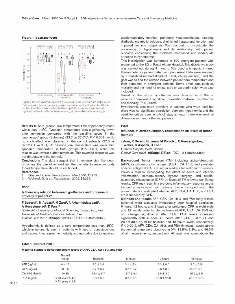
S126
Results In both groups oral temperature time-dependently variedwithin only 0.4°C. Tympanic temperature was significantly lowerafter immersion compared with the baseline values in the‘submerged’ group (Submerg) (33.7 vs 37.5°C, P < 0.001), whileno such effect was observed in the control subjects (37.6 vs37.5°C, P = 0.31). At baseline, oral temperature was lower thantympanic temperature in both groups (P < 0.001), while thisrelation was reversed after immersion. This reversed response wasnot detectable in the controls.Conclusions The data suggest that in emergencies like near-drowning, the use of infrared ear thermometry to measure body(core) temperature should be cautioned.References1. Giesbrecht: Aviat Space Environ Med 2000, 71:733.2. Wollenek G, et al.: Resuscitation 2002, 52:255.
P300Is there any relation between hypothermia and outcome incritically ill patients?
F Bozorgi1, M Abbasi2, M Zare2, A Arhamidolatabadi1, A Hosseinnejad2, D Farsi21Beheshti University of Medical Sciences, Tehran, Iran; 2IranUniversity of Medical Sciences, Tehran, IranCritical Care 2005, 9(Suppl 1):P300 (DOI 10.1186/cc3363)
Hypothermia is defined as a core temperature less than 35°C,which is commonly seen in patients with loss of consciousnessand trauma. It increases the mortality and morbidity due to impaired
cardiorespiratory function, peripheral vasoconstriction, bleedingdiathesis, metabolic acidosis, diminished hepatorenal function andimpaired immune response. We decided to investigate theprevalence of hypothermia and its relationship with patientoutcome considering the problems mentioned and inadequateattention to hypothermia.This investigation was performed in 100 emergent patients whopresented to the ED of Rasol Akram Hospital. This discriptive studywas carried out during 4 months. We used a tympanic infraredthermometer for patient detection upon arrival. Data were analyzedby a statistical method (Student t test, chi-square test), and thegoal was to find the relation between patient core temperature andtheir outcomes in emergent patients. Some other data such asmortality and the need for critical care or ward admission were alsoincluded.Based on this study, hypothermia was detected in 35.3% ofpatients. There was a significant correlation between hypothermiaand mortality (P < 0.002).Hypothermia was more prevalent in patients who were died butthere was no significant correlation between hypothermia and theneed for critical care length of stay, although there was minimaldifference with normothermic patients.
P301Influence of cardiopulmonary resuscitation on levels of tumormarkers
J Auer, R Berent, G Lamm, M Porodko, C Punzengruber, T Weber, G Aspöck, B EberGeneral Hospital Wels, AustriaCritical Care 2005, 9(Suppl 1):P301 (DOI 10.1186/cc3364)
Background Tumor markers (TM) including alpha-fetoprotein(AFP), carcinoembryonic antigen (CEA), CA 15-3, and prostate-specific antigen (PSA) are serum markers for malignant diseases.Previous studies investigating the effect of acute and chronicinflammation, cardiopulmonary bypass surgery, and cardio-pulmonary resuscitation (CPR) on levels of TM showed conflictingresults. CPR may result in a profound inflammatory response and isfrequently associated with severe tissue hypoperfusion. Thepresent study investigated whether AFP, CEA, CA 15-3, and PSAare influenced by CPR.Methods and results AFP, CEA, CA 15-3, and PSA (only in malepatients) were assessed immediately after hospital admission,6 hours, 12 hours, and 2 days after prolonged CPR in eight maleand 12 female patients. Serum levels of AFP, CEA, CA 15-3 didnot change significantly after CPR. PSA levels increasedsignificantly with a peak 48 hours after CPR (3.3 ± 3.1 and28.3 ± 30.5 ng/ml for baseline and 48 hours levels, respectively;P < 0.001). AFP, CEA, CA 15-3, and PSA (in males) values abovethe normal range were observed in 0%, 13.8%, 3.8%, and 46.9%of all measurements, respectively. At least one value above the
Critical Care March 2005 Vol 9 Suppl 1 25th International Symposium on Intensive Care and Emergency Medicine
Figure 1 (abstract P299)
Table 1 (abstract P301)
Mean (± standard deviation) serum levels of AFP, CEA, CA 15-3, and PSA
Normal values Baseline 6 hours 12 hours 48 hours
AFP (ng/ml) 0 – 15 3.2 ± 2.4 3.1 ± 2.4 6.2 ± 8.0 3.0 ± 2.5
CEA (ng/ml) 0 – 5 3.7 ± 3.5 3.7 ± 3.2 3.6 ± 3.0 3.6 ± 3.1
CA 15-3 (U/ml) 0–35 19.4 ± 9.7 18.7 ± 9.4 3.6 ± 3.0 16.5 ± 8.8
PSA (ng/ml) ≤ 70 years < 4.5 3.3 ± 3.1 6.2 ± 8.0 16.8 ± 25.0 28.3 ± 30.5> 70 years < 6.5
S127
normal range was observed in 0%, 20%, 5%, and 75% of allpatients for AFP, CEA, CA 15-3, and PSA (in males), respectively.Baseline values of AFP, CEA, CA 15-3, and PSA (in males) wereabove the normal range in 0%, 15%, 5%, and 10% of all patients,respectively. Levels for all markers did not differ significantlybetween survivors and nonsurvivors.Conclusions Prolonged CPR does not influence AFP, CEA, CA15-3 serum levels, but is frequently associated with increases ofPSA. Thus, in contrast to PSA, interpretation of AFP, CEA, CA15-3 serum levels is not influenced by recent CPR.
P302Descriptive analysis of contributing factors in outcomes ofemergency department CPRs
A Arhami Dolatabadi1, A Setayesh2, M Zare2, A Hosseinnejad2,F Bozorgi1, D Farsi21Beheshti University of Medical Sciences, Tehran, Iran; 2IranUniversity of Medical Sciences, Tehran, IranCritical Care 2005, 9(Suppl 1):P302 (DOI 10.1186/cc3365)
Introduction CPR can avoid 25% of deaths especially out-of-hospital cases. Cases of cardiac arrest are usually studied fromepidemiological aspects. This study is designed to guide usthrough our Emergency Department (ED) and EMS managements.Methods and materials In this observational cross-sectionalstudy, 195 patients admitted to the ED of Rasoul Akram hospitalduring 11 month from June 2003 to April 2004 were included.Age, sex, time, place, cause and witness of arrest, BLS/ALS, out-of-hospital intubations, transportation vehicle, CPR outcome andfinal status of patients were gathered in a data registration form.Results The mean (± standard error of the mean) age was59.33 ± 1.4, there was a significant difference in mean age betweenmales and females (P = 0.003). A total 41.5% of cases arrested inthe ED, 14.9% had no witness, 56.4% were transported to hospitalby ambulance, 53.3% received no life support before arrival and13.3% had been intubated out of hospital. The first rhythm checkedin the ED was asystole in 73.3%. Cardiovascular problemsconstituted 42.1% of causes. There was a significant difference incause of cardiac arrest between age groups (P = 0.000). A total59.5% of CPRs were successful; there was no significant differencein sex (P = 0.199) and age (P = 0.746) groups and different causecategories. CO poisoning and hanging had maximal mortality(100%). Age, sex distribution (P = 0.07), time of cardiac arrest,place of collapse (P = 0.1), cause of arrest, being witnessed,performed BLS/ALS (P = 0.7), intubations (P = 0.76), initial cardiacrhythm, and transportation vehicle had no effects on patients finalstatus. CPR outcome (P = 0.000) and the cause of arrest(P = 0.000) affected the final status of patients. Finally, 93.3% ofpatients expired and 6.7% of patients were discharged from hospital.Discussion Successful CPR was performed in the majority ofcases. But the overall prognosis was poor. The main determiningfactor of final status was the cause of arrest.
P303Identification of agonal breathing by dispatchers to improvethe detection and treatment of cardiac arrest
L Roppolo, P Pepe, A Westfall, A Idris, J WiggintonUniversity of Texas Southwestern Medical Center and the ParklandHealth and Hospital System, Dallas, TX, USACritical Care 2005, 9(Suppl 1):P303 (DOI 10.1186/cc3366)
Background In as many as 40% of applicable out-of-hospitalcardiac arrest (OOHCA) cases, bystanders do not receive
instructions over the telephone from public safety dispatchers toperform cardiopulmonary resuscitation (CPR) because thebystanders report that the patient is ‘breathing’ due to thepresence of agonal respirations.Objective To determine whether a new dispatcher assessmentprotocol could better identify agonal breathing and thus morecases of OOHCA that might benefit from CPR instructions.Methods Study of all dispatcher-assisted OOHCA cases in alarge municipality (population 1.2 million) for the 8 months beforeand 4 months after protocol implementation (November 2003). Inthe protocol, the call-taker asks about gasping defined as either‘slow, barely breathing’ or ‘slow, noisey/gurgling’. If unsure, thecaller places the telephone near the patient’s mouth and thedispatcher listens for breathing, or the bystander is asked to stateeach time the patient takes a breath.Results Among 962 consecutive patients identified as havingOOHCA (57% male, 43% female; mean age 63 ± 17 years, range17–106), the presenting rhythm was ventricular fibrillation orpulseless electrical activity in 85% of those identified with agonalbreaths versus 46% without them (P = 0.002) and such presen-tations had a higher rate of survival to hospital admission(P < 0.0001). After protocol introduction, the percentage ofOOHCA patients thought to be ‘breathing’ normally fell from 28%(168/599) to 19% (68/362; P = 0.0012). Also, in the 8 monthsbefore the protocol, no patient had their agonal respirationsdetected compared with 22 patients detected in the 4 monthsafterwards (P < 0.0001). No adverse problems were reported fromthe subsequent performance of CPR in these agonal breathingcases.Conclusion Introduction of a supplemental dispatcher assessmentprotocol (that includes questioning about agonal respirations) cansignificantly increase cardiac arrest detection over the telephone,thus increasing the opportunity for dispatchers to provide CPRinstructions for those very patients who, in fact, may a higherpropensity for survival than the average OOHCA patient (i.e. thosewith agonal respirations).
P304Cardiopulmonary resuscitation training: the need forcontinued education and training
A Turley, G Bone, L Garcia, M de Belder, J GedneyJames Cook University Hospital, Middlesbrough, UKCritical Care 2005, 9(Suppl 1):P304 (DOI 10.1186/cc3367)
Introduction Cardiopulmonary resuscitation (CPR) has a poorsurvival rate that may in part be due to flawed technique andtraining [1]. Recent recommendations for education and traininghave been issued for staff with responsibility for resuscitatingpatients [2]. These include ‘staff with patient contact should begiven regular resuscitation training appropriate to their expectedabilities and roles’, and ‘cardiopulmonary arrest should bemanaged according to national guidelines’. The use of humanpatient simulators has become increasingly common in medicaleducation [3]. Their use for exposing staff to high-risk complexclinical situations such as CPR is advocated by the department ofhealth [4].Aims and methods The aim of this study was to assess theresuscitation skills of all trainee junior doctors rotating through a20-bed adult critical care unit (ITU/HDU) against current UKadvanced life support (ALS) guidelines. Over an 18-month period,trainees underwent a compulsory CPR competency assessment bysimulation testing, observed by two experienced ALS instructorsfollowing a refresher seminar. The trainees were assessed on basiclife support (BLS), defibrillator safety and ALS skills in a scenario-
Available online http://ccforum.com/supplements/9/S1

S128
based format. Only candidates who passed each section wereassessed on the next. All trainees were active members of thehospital cardiac arrest teams.Results During the study period 32 trainees were assessed, ofwhich 25/32 (78%) held a valid ALS certificate. BLS testing wasinadequate in 2/32 (6%). Safety of defibrillation was failed by 7/30(23%) of trainees. A full CPR scenario was given to 23 trainees. Ofthese 7/23 (30%) failed the scenario. Of the group as a whole16/32 (50%) required further training and re-testing. Of thosecandidates who held a valid ALS certificate 10/25 (40%) failed thecompetency based assessment and required further training andre-testing.Conclusions Junior medical staff require regular scenario-basedCPR training. This study demonstrates that skills acquired duringlife support training are not retained and regular competency-basedassessment is required irrespective of previous resuscitationtraining. Repeated scenario-based competency assessmentsshould be a compulsory requirement of all trainee posts.References1. Resuscitation 1998, 37:173-175.2. Standards for Clinical Practice and Training. Resuscitation
Council (UK); 2004.3. Hospital Med 2003, 64:677-681.4. Building a Safer NHS for Patients. London: Department of
Health; 2001.
P305Effectiveness of an intubating laryngeal mask airway (ILMAFastrach) used by nurses during out-of-hospital cardiac arrestresuscitation
M Vergnion, S Thoumsin, V FraipontCHR Citadelle, Liège, BelgiumCritical Care 2005, 9(Suppl 1):P305 (DOI 10.1186/cc3368)
Introduction ILMA Fastrach is a good alternative to difficultendotracheal intubation even by an untrained physician but hasnever been studied in a out-of-hospital setting. Our aim was toevaluate the efficiency of this device used by untrained nurses inout-of-hospital cardiac arrest (OHCA).Method After a brief instruction course and training on amannequin, 12 nurses used ILMA Fastrach during 71 OHCA(s)resuscitation in a prospective open study. Procedure took placeunder the supervision of a certified emergency physician whileambulance officers applied basic life support. The ILMA size wasselected according to the manufacturer’s instructions and theywere lubricated before insertion. Effective ventilation after maskinsertion was verified by presence of ample chest movement andcorrect intubation was verified by the physician (chestauscultation) and by capnography. A digital voice recorderrecorded speed and success rates. After two unsuccessfulattempts, the physician performed classic intubation.Results Effective ventilation after mask insertion was obtained in97% (65/67); in the two unsuccessful cases, one was hanged andone had a literalised larynx (these data were not collected in fourpatients because of a recording problem). The rate of successfulendotracheal intubation was 75% (53/71) after the first attemptand 89% (63/71) after the second. In the eight unsuccessfulcases, ventilation with the mask was effective. The total mean timeof intubation (since inclusion) was 148 s with a mean time of 53 sto prepare the device. The physician easily intubated all the eightremaining patients: a cephalic larynx was noticed in the five cases(mask size 5 chosen), one was hanged, and no abnormalities werein the two others.
Conclusion The ILMA Fastrach is a good alternative to classicalintubation by untrained nurse OHCA resuscitations. The patientcould be ventilated after mask insertion in most cases and the timeto perform intubation is short. However, 1 min is necessary toprepare the device, so a good training and preparation of thematerial is necessary. Results are not as favourable as in theoperative room and the effectiveness of ventilation with the maskwas not predictive of intubation. However, only two unsuccessfulattempts were permitted before classical intubation and the sizeaccording to the weight was inadequate in five cases. A largemulticenter study is warranted to confirm these results.
P306Use of automated external defibrillators in the House ofRepresentatives Brazil, the first in Latin America: a 2-yearexperience
S Timerman1, A Quilici1, M Gonzalez1, L Hargreaves2, J Ramires1
1Heart Institute (InCor), Sao Paulo, Brazil; 2House ofRepresentatives Medical Department, Brasilia — DF, BrazilCritical Care 2005, 9(Suppl 1):P306 (DOI 10.1186/cc3369)
Background Passengers who have ventricular fibrillation aboardcommercial aircraft rarely survive, owing to the delay in obtainingemergency care and defibrillation.Methods In 2002, after the incorporation of automated externaldefibrillators (AEDs) by other PAD initiatives, a major LatinAmerican House of Representatives (HR) began equipping itsmajor building (Chamber of Deputies) — 513 Representatives,around 5000 people working and more than 20,000 peopletransiting every day — with automated external defibrillators (Lp500). We chose the healthcare professional (more than 30) as thefocus of the training program conducted according to theguidelines of the American Heart Association in the use of thedefibrillator and applied the device when people had a lack ofconsciousness, pulse, or respiration. The AEDs were also used asa monitor for other medical emergencies, generally at the directionof a physician. We placed a separate cardiac monitor without thecapacity for defibrillation, to be used when the strict definition ofcardiac arrest has not been met (Lp 12). The electrocardiogramthat was obtained during each use of the device was analyzed bytwo physicians for appropriateness of use. We analyzed data on allsix instances in which the defibrillators were used between 1February 2002 and 1 September 2004.Results Automated external defibrillators were used for sixpatients, including six patients in ventricular fibrillation withdocumented loss of consciousness. Electrocardiographic datawere available for all patients. The administration of shock wasadvised in all six patients who had electrocardiographicallydocumented ventricular fibrillation (sensitivity and specificity of thedefibrillator in identifying ventricular fibrillation, 100%). One patienthad atrial flutter with high-degree atrioventricular block. All sixepisodes of ventricular fibrillation were properly treated withestablishment of a final rhythm potentially perfusional in five cases.The first shock successfully defibrillated the heart in six patients(100%). No complications arose from use of the automatedexternal defibrillator as a monitor in conscious passengers.Of the six patients, four (66%) left the locality alive and three(50%) were discharged home from hospital. In all cases the initialrhythm was ventricular fibrillation.Conclusions The use of the automated external defibrillator in theHR is effective.
Critical Care March 2005 Vol 9 Suppl 1 25th International Symposium on Intensive Care and Emergency Medicine

S129
P307Synchronized cardioversion in a prehospital setting: a safeand reliable method for urgent treatment of tachyarrhythmias
A Jelatancev1, S Grmec1, P Klemen1, M Kovac2
1Centre for Emergency Medicine, Maribor, Slovenia; 2PrehospitalEmergency Unit, Ljubljana, SloveniaCritical Care 2005, 9(Suppl 1):P307 (DOI 10.1186/cc3370)
Objective Synchronized cardioversion (SCV) is a useful andreliable method of converting atrial or ventricular tachycardia tosinus rhythm. It is a method of choice in patients who are at highrisk of the arrhythmia and therefore require urgent treatment.Conscious patients must be well sedated.Design and setting A prospective clinical investigation.Patients and methods All adult patients (>18 years) were treatedby an emergency physician in the prehospital setting. Indicationsfor SCV were: arrhythmia with severe chest pain and/or shortnessof breath, congestive heart failure, pulmonary congestion,decreased level of consciousness (reduced cerebral blood flow),systolic blood pressure less than 90 mmHg, shock, or acutemyocardial infarction (AMI). Conscious patients were sedated oranaesthetized with midazolam and morphine, etomidate orpropofol.Results Over a period of 2 years, 22 patients have required SCV.Indications for SCV included 10 (45.5%) patients with atrialfibrillation (AF) with high risk, seven (31.8%) patients withsupraventricular tachycardia (SVT) and five (22.7%) patients withventricular tachycardia (VT) with pulse. Heart rates in AF and VTwere more than 150 beats/min and in SVT more than200 beats/min. In all patients two or more adverse signs of tachy-arrhythmia have been observed: congestive heart failure and/orpulmonary congestion in eight (36.4%) patients, hypotensionand/or shock in 15 (68.2%) patients, severe chest pain or AMI in10 (45.5%) patients; decreased level of consciousness in nine(40.9%) patients. The SCV was successful in all cases (in 19cases in the first shock with 100 J, in two cases in the secondshock with 200 J and in one case in the third shock with 360 J). Allpatients were hospitalized and left the hospital without anycomplications.Conclusion SCV is a useful and safe method in treatment ofunstable tachyarrhythmia in a prehospital setting. This is veryimportant information, because in such situations the universalrecommendation of the ILCOR 2000 International Guidelines forimmediate cardioversion is the method of choice rather than a trialof antiarrhythmics.
P308Prehospital transcutaneous cardiac pacing in bradycardia andasystole
C Febra, A LufinhaHospital S Francisco Xavier SA, Lisbon, PortugalCritical Care 2005, 9(Suppl 1):P308 (DOI 10.1186/cc3371)
Background Transcutaneous external cardiac pacing (TECP)provides a noninvasive, safe and rapid ventricular pacing, althoughthere is controversy on the results observed both inhemodynamically significant bradycardia and asystole situations.The purpose of this study is to examine the success of TECP in ourprehospital emergency medical team (VMER) practice, at HospitalS Francisco Xavier.Methods and results In this retrospective cohort study, weexamined 46 patients submitted to TECP between January 2001and November 2004 with a median age of 68.6 years (40–94
years). The mean call-to-arrival time was 7.8 min, and cardiac arrestwas present at our arrival in 43.5% of the cases and bradycardiawith hemodynamic repercussion in 56.5%, manifested, p.e., assyncope (61.5%), chest pain (23%) and/or dyspnoea (15.4%). Inthe cardiac arrest situations, classic advanced cardiac life supportwas always provided, resulting in electromechanical disassociation(40%), asystole (15%) and complete AV blocking (15%), presentimmediately before pacing. There was a survival rate in the field of40% of these patients, of whom 75% were hemodynamicallystable at hospital admission. In the bradycardia cases, completeAV blocking was the most common presenting rhythm (73%), themean heart rate was 33 ppm, and 76.9% of patients had shockcriteria. Atropine was used unsuccessfully in 61.5% of patients.The TECP resulted in hemodynamic stability in 53.8%, death in thefield in 15.4% and sustained shock in 30.8%. The overall successrate of TECP in these two groups, defined as hospital admission,was 65.2%.Conclusions In cardiac arrest events, TECP was associated with abetter survival rate on the field than our global survival rate on thefield (21%). This may be due to its utilisation mainly inelectromechanical disassociation and younger patients. TECPresults in a correct cardiac output in much of severe bradycardiasituations, allowing a stable transportation to the hospital where itmay be kept or changed by other pacing approaches.
P309EMERGE™: a multiprofessional training course in the care ofthe acute coronary syndrome patient
S Timerman1, M Moretti1, F Marques1, R Ramos2, E Mesquita3,E Stefanini4, C Gun2, F Tarasoutchi1, E Paiva5, D Ferreira5, A Timerman2
1Heart Institute, Sao Paulo, Brazil; 2Dante Pazzanese Institute ofCardiology, Sao Paulo, Brazil; 3Hospital Pró Cardiaco, Rio deJaneiro, Brazil; 4UNIFESP, Sao Paulo, Brazil; 5NRC, Sao Paulo,BrazilCritical Care 2005, 9(Suppl 1):P309 (DOI 10.1186/cc3372)
The Acute Coronary Syndrome (ACS) Recognition and Treatment(EMERGE™) course is a 1-day (3 hours) course originally designedto give doctors greater confidence and ability in the recognitionand management of adult patients who have ACS. It may also besuitable for many other groups of health professionals. EMERGE™was developed using principles common to many advanced lifesupport courses and incorporates aspects of clinical governance,multidisciplinary education and interprofessional working. Itincorporates precourse reading, informal and interactive seminars,and role-play during three clinically based scenarios. A novelaspect of EMERGE™ is that participants undertake roleinterchange during scenarios, thereby facilitating mutualunderstanding. At all times during the course, participants areencouraged to reflect on their actions and to pay particularattention to detail.Using initial and final theoretical written test, after the 3-monthcourses (36 courses), we assessed the knowledge of aspects ofACS among 900 doctors. The average (± standard deviation)knowledge score was higher for those who had completed anEMERGE™ course, pre-test (545 students) = 23.8 (8.44 ± 1.30)points and post-test (834 alunos) = 25.3 (9.85 ± 2.12) points(P < 0.05). In addition, those in the post-EMERGE™ group alsoshowed significantly better knowledge about ACS (85.6% [714doctors]) acquired skills in treatment an ACS. We havedemonstrated evidence that doctors’ knowledge of ACS can beimproved by attending courses such as EMERGE™.
Available online http://ccforum.com/supplements/9/S1

S130
P310High mortality rate in critically ill medical patients with new-onsetatrial tachyarrhythmias despite rapid conversion to sinus rhythm
M Sleeswijk, J Tulleken, T v/d Werf, J Ligtenberg, T v Noord, J ZijlstraAZG, Groningen, The NetherlandsCritical Care 2005, 9(Suppl 1):P310 (DOI 10.1186/cc3373)
Introduction New-onset atrial tachyarrhytmia (AT) is frequentlyseen in critically ill surgical patients (incidence 10.2%) andassociated with high mortality rate (23.4%). Whether this alsoapplies to medical ICU patients is unclear. In a follow-up study allmedical patients with AT were evaluated.Methods In a 12-bed medical ICU, all consecutive patients withAT were evaluated during a 4-month period. Characteristics,response to treatment and outcome were registered. All patientswith AT were treated with MgSO4-amiodarone after correction ofpredisposing factors; MgSO4 bolus (0.037 g/kg) followed bycontinuous infusion (0.025 g/kg). Those with persistent AT>110 beats/min after 1 hour were given intravenous amiodarone(bolus 300 mg) followed by continuous infusion (1200 mg/24 hours).Results One hundred and forty-seven patients were admitted formedical reasons. AT was seen in 14 patients. The most commonreason for ICU admission was sepsis (nine patients). Patientcharacteristics are presented in Table 1. Two patients convertedafter fluid repletion. Ten patients received the treatment protocol.Five patients converted after magnesium therapy, and addition ofamiodarone was given to five patients. All patients converted within24 hours (median [range] 2.5 [1–19]). Ten patients died duringtheir hospitalization. Time between the onset of AT and death was7.7 ± 5.5 days (mean ± standard deviation [SD]).
Table 1
Male/female 6/8Age (mean ± SD) 64 ± 20 yearsAPACHE II (mean ± SD) 24 ± 8Rhythm (AF/flutter/SVT) 11/1/2Norepinephrine 11MAP before/after AT (mean ± SD) 82 ± 13/65 ± 13 mmHgHeart rate before/after AT 99 ± 19/148 ± 37 beats/minSerum K+ (mean ± SD) 4.10 ± 0.5 mmol/lSerum Mg2+ (mean ± SD) 0.85 ± 0.12 mmo/l
Conclusion The incidence of AT in this patient cohort was 14/147.Mg-amiodarone is a highly effective regimen for conversion. Despitethis, mortality was high and therefore AT was not the cause of deathbut rather an epiphenomenon. Compared with surgical patients,medical patients had a much higher mortality rate.
P311Troponin incidence, prevalence and prognosis among criticallyill patients
W Lim, L Griffith, M Crowther, P Devereaux, D CookMcMaster University, Hamilton, CanadaCritical Care 2005, 9(Suppl 1):P311 (DOI 10.1186/cc3374)
Background The diagnosis of acute myocardial infarction (AMI) incritically ill patients is usually dependent on the presence ofischemic changes on an electrocardiogram (ECG) and measure-ment of biochemical markers such as troponin. Among patients inthe ICU, there is uncertainty surrounding the significance ofelevated troponin values, their relation to coronary ischemia andinfarction, and the prognostic and therapeutic implications.
Objective To document the prevalence and incidence of elevatedtroponin levels in critically ill patients correlated with ECG findings(presence of ischemic changes [AMI], non-ischemic ECGchanges, and normal ECG), and to evaluate the morbidity andmortality outcomes of each of these categories compared withpatients with normal troponin values.Methods We conducted a retrospective chart review of 198patients from 261 critically ill patients admitted to a university-affiliated, general medical–surgical ICU who were enrolled in aprospective cohort study evaluating the prevalence and incidenceof deep vein thrombosis.Data extraction We reviewed daily medical records during thepatient’s ICU admission for signs and symptoms of AMI, laboratoryand radiologic investigations, and collected morbidity and mortalityoutcomes.Results The prevalence of all elevated troponin levels was 36.4%;19.2% had AMI (elevated troponin with ischemic ECG changes),14.7% had elevated troponin without typical ischemic ECGchanges, and 1.5% had elevated troponin and a normal ECG. Theincidence of AMI over the ICU stay was 6.2%, of which 42% werediagnosed with AMI on admission (i.e. they had recurrent AMI).Morbidity (length of stay [LOS]) and mortality outcomes in patientswith elevated troponin and associated ECG findings comparedwith patients with normal troponin are presented in Table 1.
Table 1
Ischemic Non-ischemic Normal Normal ECG ECG ECG troponin
Outcomes (n = 45) (n = 30) (n = 3) (n = 104)
ICU LOS, 10 8 4 10median (IQR) (8, 18) (4, 14) (3, 39) (6, 18)
Hospital LOS, 20 13.5 40 30median (IQR) (11, 54) (10, 31) (12, 114) (15, 61)
ICU mortality, 15 11 1 21n (%) (33.3) (36.7) (33.3) (20.2)
Hospital mortality, 20 13 1 35n (%) (44.4) (43.3) (33.3) (33.7)
Conclusions Elevated troponin levels are common in the criticallyill, yet it is unclear under what circumstances they represent anadverse prognostic marker for ICU and hospital mortality.Additional studies are needed to determine the significance ofelevated troponin values in the presence and absence of ECGchanges, evaluating long-term outcomes and the efficacy of anti-ischemic treatments in critically ill patients.
P312Clinical determinants of severe bleeding complications in patientswith acute myocardial infarction treated with thrombolytic therapy
J Rossinen1, J Viita2, M Nieminen1, R Lassila1
1Helsinki University Hospital, Helskinki, Finland; 2Medical Faculty,Turku, FinlandCritical Care 2005, 9(Suppl 1):P312 (DOI 10.1186/cc3375)
The aim of the study was to determine the frequency of bleedingcomplications in a clinical setting, and to clarify the practise inlaboratory monitoring of haemostasis (TT%, APTT, HCR) in patientswith acute myocardial infarction who are treated with fibrinolytics. Inthis clinical, retrospective series we included randomly 301 patientswho were treated during the years 2000–2003 due to acutemyocardial infarction in the Helsinki University Central Hospital. Twohundred and forty (80%) of the patients were treated with athrombolytic therapy. Six (3%) patients received thrombolytic therapy
Critical Care March 2005 Vol 9 Suppl 1 25th International Symposium on Intensive Care and Emergency Medicine

S131
despite them having an absolute contraindication, and 75 (31%)received it while having a relative contraindication. The incidence ofcerebral bleeding during the first 48 hours was unexpectedly high(2.5%). The incidences of bleeding complications are presented inTable 1. The parameters of haemostasis could not predict bleedingcomplications in our study, probably due to low rate ofmeasurements prior to fibrinolytic therapy. It is therefore important tostrictly follow the guidelines of thrombolytic therapy in patients withacute ST-elevation myocardial infarction. Routines for laboratorymonitoring of haemostatic variables are to be implemented toimprove safety and early detection of bleeding complications.
Table 1
Thrombolysis group Non-thrombolysis group
Intracerebral bleed 6 (2.5%) 1 (1.6%)GI haemorrhage 21 (8.8%) 3 (4.9%)Unexplained bleed 24 (11%) 14 (23%)
P313Facilitated percutaneous coronary intervention: a newtreatment strategy for acute myocardial infarction
H Khaled Nagi, M Shehata, A ElSherif, M Ashraf, M ElBadry, S MokhtarCairo University, Cairo, EgyptCritical Care 2005, 9(Suppl 1):P313 (DOI 10.1186/cc3376)
Restoring patency of infarct-related artery (IRA) is the ultimate goal ofall cardiologists. Such a goal can be achieved either pharmaco-logically ‘using thrombolytic therapy’ or through percutaneouscoronary intervention (PCI). The latter needs a sophisticated setup, anequipped cath lab and a skilled team. Such a system might not bealways available, and a full-dose thrombolytic therapy is the next bestalternative. There has recently been a trend towards ‘facilitated PCI’whereby low-dose thrombolytic and/or antiplatelet therapies are usedprior to primary PCI, aiming at an early, complete, and sustainedepicardial flow and myocardial perfusion.To compare the efficacy and safety of facilitated PCI with standardprimary PCI, we studied 40 patients with acute myocardialinfarction (AMI) divided into two groups, a study group consistingof 20 patients (18 males, two females, mean age46.3 ± 11.5 years), all received 750,000 u streptokinase combinedwith GP IIb/IIIa receptor inhibitor ‘tirofiban’ 0.4 µg/kg/min over30 min followed by 0.1 µg/kg/min over 48 hours. Twenty patients(15 males, five females, mean age 54 ± 8.6 years) served ascontrol group (no thrombolytic nor antipaltelet therapy). Bothgroups underwent PCI within (73 ± 18 min) from randomization.Angiographic patency was expressed in terms of TIMI flow gradingsystem, ECG criteria comprised extent and rapidity of ST-segmentresolution, and laboratory criteria involved early peaking of CK-MBwithin 12 hours from randomization. Besides clinical evaluation interms of major adverse cardiac events, echocardiographicparameters (LVEDD and LVEF) were used to assess LV functionbefore and after PCI and monthly thereafter for 6 months.Compared with the group subjected to PCI alone, those who hadpreceding adjunctive pharmacological therapy ‘facilitated PCI’exhibited significantly greater TIMI 3 flow (84% vs 60%, P < 0.05),smaller LVEDD (5.0 vs 5.5, P < 0.05), significantly higher LVEF(55.4% vs 50.7%, P < 0.05) and lower rate of MACE (0% vs20%). Patients with facilitated PCI also exhibited significantlyhigher ST-segment resolution (58% vs 45%, P < 0.05) and earlierpeaking of CK-MB (85% vs 35%) compared with control group.In conclusion, facilitated PCI offers an excellent way ofcircumventing the time delay preceding PCI that is frequently
encountered on hospital admission of patients with acute MI.Through combining interventional, fibrinolytic and GP IIb/IIIainhibitor therapy, facilitated PCI provides more rapid, complete andsustained patency of IRA than primary PCI alone without theadverse effects of full-dose thrombolytic therapy and with a betteroutcome in terms of lesser MACE and preserved LV function.
P314Left ventricular remodeling and function after primary anddelayed PCI in patients with recent myocardial infarction
M Hamila, M Badry, S Fawzy, H Nagi, T Gohary, K HusseinFaculty of Medicine, Kasr El Aini Hospital, El Manial Cairo, EgyptCritical Care 2005, 9(Suppl 1):P314 (DOI 10.1186/cc3377)
Background It has been well established that restoration ofpatency of an infract-related artery by percutaneous intervention asearly as possible in patients with recent myocardial infarction maypreserve left ventricular global function and also prevent LVEDVprogressive increase and remodeling,Aim To evaluate and compare the effect of primary and delayedPCI in patients with recent myocardial infarction (MI).Methods Forty patients were included in the study with firstanterior MI; they were divided into group A who had the chance ofundergoing primary PCI within less than 12 hours of start of chestpain with mean 5.4 hours and a door to balloon time of 1.6 hours,and group B who due to out of hand circumstances neitherreceived thrombolytic therapy nor arrived at hospital within 12 hours,but were scheduled as routine PCI with mean 20.7 days. The leftventricular function and dimensions were assessed by serialechocardiographic readings measuring LVEDV, LVESV, EF andRWMI at 24 hours of admission and after 3 months and 6 months.Results were expressed as mean ± standard deviation andP < 0.005 was considered significant.Results First echo results at baseline showed no significantdifference of LVEDV, RWMI and EF in both group A and group B,that indifference was also remarkerable at echo done at 3 monthswith P = 0.6, while at 6 months reading the LVEDV in group Ashowed a significant decrease (115 ± 32.14 vs 138 ± 32.96,P = 0.048). Ejection fraction difference between two groups werenot significant at baseline echo, but improved significantly at 3 and6 months (58.5 ± 4.5 vs 55.2 ± 6.4, P = 0.046 at 3 months and59.5 ± 6.81 vs 51.88 ± 10.7, P = 0.008 at 6 months,respectively). RWMI results started to improve significantly from3 months to 6 months in group A versus group B (1.27 ± 0.13 vs1.38 ± 0.18, P = 0.032 at 3 months) but at 6 months there was nofurther improvement with P = 0.6Conclusion Although the recent evolvement and the theory ofopening of IRA whatever the timing, primary PCI has superiority inpreventing LV remodeling and preserving global LV function
P315Acute myocardial infarction: incidence and outcomes in anintensive care unit
I Qushmaq1, W Lim1, P Devereaux1, M Crowther1, A Tkaczyk2,K Lutz1, E McDonald2, J Hancock1, D Heels-Andsell1, D Cook1,for the Troponin T Trials Group1McMaster University, Hamilton, Canada; 2St Joseph’s Hospital,Hamilton, CanadaCritical Care 2005, 9(Suppl 1):P315 (DOI 10.1186/cc3378)
Objective To enhance the precision of the definition of acutemyocardial infarction (AMI), the European Society of Cardiology/American College of Cardiology (ESC/ACC) recently updated the
Available online http://ccforum.com/supplements/9/S1

S132
diagnostic criteria for AMI. Given this change in AMI diagnosticcriteria, little is known about the current incidence of AMI in theICU and the associated clinical outcomes. We undertook a studyto inform these issues.Design A prospective 2-month cohort study.Setting A 15-bed closed university-affiliated medical–surgical ICU.Methods We enrolled all consecutive ICU patients from 12 July to12 September 2004. All aspects of patient management were atthe discretion of the ICU team, which was unaware of the study tolimit any influence on ECG or troponin test-ordering practices. Asclinically indicated, 12-lead ECGs were performed by atechnologist during the day, or by the ICU bedside nurse foremergencies, during the evenings and weekends. We adapted theJoint ESC/ACC redefinition of AMI for the ICU setting. Twoinvestigators independently classified all patients as AMI or noAMI; discordance was resolved by discussion and criterion review.We used Wilcoxon Two-Sample Rank Sum Test to test fordifferences in continuous variables and Fisher’s Exact Test fordichotomous variables.Result We screened 117 ICU admissions but considered only thefirst admission of two patients who were admitted twice. Weenrolled 115 patients aged 64.1 (± 17.2) years with an APACHE IIscore of 21.9 (± 9.8); 83 (72.2%) patients were medical. OverallICU mortality was 21.7% and hospital mortality was 27.8%. Of 115patients 93 (80.9%) patients had both at least one ECG performedand one troponin measurement during ICU admission, seven (6.1%)patients had at least one ECG performed but no troponinmeasurement, 11 (9.6%) patients had at least one troponinmeasurement but no ECG performed, and four (3.5%) patients hadneither an ECG nor troponin. Using the ESC/ACC criteria adaptedto the ICU setting, 24 of 115 (20.9%) patients developed AMI(crude agreement between adjudicators was 88%).
Table 1
Outcome AMI (n = 24) No AMI (n = 91) P value
Duration of MV 2 (1–6) 2 (0–5) 0.32
Duration of ICU 4.5 (2–8) 4 (2–6) 0.62
ICU mortality 9 (37.5%) 16 (17.6%) 0.05
Hospital stay 18 (4.5–4.1) 12 (7–21) 0.34
Hospital mortality 12 (50%) 20 (22.0%) 0.01
Conclusion One-fifth of these medical–surgical critically ill patientssustained an AMI. Patients with AMI had a significantly highermortality than those who did not. More research is needed onappropriate AMI definitions in the ICU setting, risk factors,detection methods, mechanisms of myocardial necrosis duringcritical illness, optimal prevention and treatment strategies, andlong-term prognosis.
P316Clinical features of acute myocardial infarction in patients withcor pulmonale
Y Zhao, L Lu, L Liou, Q XueInstitute of Geriatric Cardiology, Chinese PLA General Hospital,Beijing, ChinaCritical Care 2005, 9(Suppl 1):P316 (DOI 10.1186/cc3379)
Objective To investigate the clinical features of acute myocardialinfarction (AMI) in patients with cor pulmonale.Methods Manifestations of AMI in 30 patients with cor pulmonale(observation group) was compared with that of 171 patients withAMI only (control group).
Results There were more smokers in the observation group than inthe control group (60.0% vs 35.1%, P < 0.05). The incidence ofchest pain in observation group was significantly lower than that inthe control group (16.7% vs 43.9%, P < 0.001), but the incidenceof dyspnoea was significantly higher than that in the control group(50.0% vs 16.4%, P < 0.001). The serum albumin and ferrohemo-globin were lower in the observation group than those in thecontrol group (33.2 ± 9.1 vs 37.8 ± 6.0 g/l, P < 0.01 and127.2 ± 18.9 vs 134.9 ± 17.7 g/l, P < 0.05, respectively). Acutepulmonary edema was more common in the observation groupthan those in the control group (46.7% vs 22.2%, P < 0.01). Of 30patients in the observation group 33.3% were accompanied withserious lung infection but only 8.8% in the control group. Themortality in the observation group was higher than that in thecontrol group (23.3% vs 13.5%, P = 0.161), but there was nostatistically significant difference between the two groups.Conclusions Infection, anemia and hypoproteinemia are importantprecipitating factors in acute myocardial infarction characteristic ofpainlessness in patients with cor pulmonale. Acute left ventricularfailure can be the initial symptoms of AMI in patients with corpulmonale. Because of its high mortality and poor prognosis, moreattention should be paid to these symptoms.
P317Prospective study on myocardial infarction: short-termprognostic factors
M Zare1, N Kianmehr1, A Hosseinnejad1, D Farsi1, A Arhamidolatabadi2, F Bozorgi21Iran University of Medical Sciences, Tehran, Iran; 2Beheshti University of Medical Sciences, Tehran, IranCritical Care 2005, 9(Suppl 1):P317 (DOI 10.1186/cc3380)
Introduction MI is one of the major causes of mortality andmorbidity worldwide. Our resources in approaching and treatingthis entity are restricted due to financial causes. Studying the riskfactors helps to improve health services and to provide better carewhere most needed.Methods and materials In this observational longitudinal studypatients with diagnosis of acute MI admitted at Rasoul akram EDduring 21 months were included. The risk factors, primary andsecondary endpoint outcomes were registered on a form. Primaryendpoints were defined as death and need to CPR, and secondaryendpoints were defined as death, asymptomatic, CCU admission,need to CPR, and angiography result when it was performed. Twomonths later, secondary endpoints were followed-up by telephone.Results One hundred and fifty-four patients were included.Twenty-one percent of patients died. Low Hgb level (P = 0.037)and leukocytosis (P = 0.011) are related to patient death. Meanage was 70.5 ± 2.28 and 56.5 ± 1.06 years in dead and alivepatients, respectively (P = 0.000). Angiostenosis, previous MI andheart failure increase the mortality risk to 10 times (P = 0.000), 3.8times (P = 0.01) and 3.2 times (P = 0.003), respectively. Meaninterval time between appearance of the symptoms and fibrinolytictherapy in ST-elevation MIs was 2.31 (± 0.22) hours, there was nomortality when fibrinolytic therapy was started less than 0.5 hoursfrom symptoms, and only one mortality when it was started at lessthan 1 hour. Sex distribution was not related to either primary orsecondary endpoint outcomes. There was no relationship betweenpatients’ 2-month survival and HTN, DM, HLP, dysrhythmia, blocks,ST elevation, involved aspect of the heart and fibrinolytic therapy.Discussion Mean age > 70 years, angiostenosis, previous MI,heart failure, low Hgb level and leukocytosis are associated withpoor prognosis in MI patients. Early thrombolytic therapydecreased mortality but has no effect on 2-month outcome.
Critical Care March 2005 Vol 9 Suppl 1 25th International Symposium on Intensive Care and Emergency Medicine

S133
P318Haemoglobin concentration on admission influences the rateof inhospital 30-day mortality and complications in patientswith acute myocardial infarction: a retrospective analysis of637 Chinese patients
Y Zhao, J Ma, S Wang, Q Xue, W Gao, Y LiInstitute of Geriatric Cardiology, Chinese PLA General Hospital,Beijing, ChinaCritical Care 2005, 9(Suppl 1):P318 (DOI 10.1186/cc3381)
Objective To determine the association between haemoglobinconcentrations on admission and inhospital 30-day cardiacmortality and complications among patients with acute myocardialinfarction during their hospital course.Methods We conducted a retrospective study of data on 637Chinese patients who were hospitalized with acute myocardialinfarction. Patients were categorized according to the haemoglobinconcentration on admission (<12 g/dl, mean ± standard deviation10.5 ± 1.4 g/dl; 12–13.9 g/dl, mean ± standard deviation13 ± 0.6 g/dl; 14 g/dl or greater, mean ± standard deviation15.2 ± 0.9 g/dl), and data were evaluated to determine whetherthere was an association between the haemoglobin concentrationson admission and inhospital 30-day mortality and complications.Complications were defined as cardiogenic shock, congestiveheart failure, arrhythmia, ventricular tachycardial or fibrillation andpneumonia.Results Patients with lower haemoglobin concentrations onadmission had higher inhospital 30-day mortality rates (P < 0.001).Inhospital 30-day mortality was 21.4% in patients withhaemoglobin concentrations <12 g/dl, 11.6% in patients withhaemoglobin concentrations of 12–13.9 g/dl, and 5.3% in patientswith haemoglobin concentrations 14 g/dl or greater. The increasein risk of complications associated with a low haemoglobinconcentration was more pronounced in patients with anaemia thanin patients without. Compared with patients with haemoglobinconcentrations 14 g/dl or greater, those with haemoglobinconcentrations <12 g/dl and 12–13.9 g/dl had more inhospitalcomplications, including arrhythmia, congestive heart failure andpneumonia (40.5% versus 33.8% versus 23.1%, P = 0.001;28.2% versus 20.0% versus 8.2%, P < 0.001; and 26.7% versus8.4% versus 2.8%, P < 0.001, respectively). As expected, a signifi-cant inverse correlation between haemoglobin concentrations andages was observed in the patients with acute myocardial infarction(r = –0.51; P < 0.001).Conclusions The traditional belief is that anaemic patients withcoronary artery disease are at high risk for myocardial ischaemia orinfarction because they cannot increase oxygen extraction oraugment coronary arterial flow. It is demonstrated in this study thata low haemoglobin concentration on admission increases the riskof death or serious complications in the patients with acutemyocardial infarction. There is a greater incidence of patients witha low haemoglobin concentration on admission in the elderlypatient than that in the younger one.
P319Protective effects of hemodilution and ischemicpreconditioning against reperfusion injury in pig heart
W XiangruiRenji Hospital, Shanghai, ChinaCritical Care 2005, 9(Suppl 1):P319 (DOI 10.1186/cc3382)
Objective We investigated the protective effects of isovolumetrichemodilution and ischemia preconditioning against myocardial
injury due to reperfusion through a pig myocardial ischemiareperfusion model.Methods Eighteen mini-pigs were used in an acute myocardialischemia model. They were randomly divided into three groups,with six pigs in each group: the control (group I, n = 6), ischemiapre-treatment (group II, n = 6), ischemia pretreatment plus blooddilution (group III, n = 6). Cardiac output (CO), saturation of mixedvenous blood oxygen (SvO2) and blood flow of coronary arterywere measured, and then the oxygen supply to the myocardiumand the oxygen consumption were calculated. The amount ofmalondialdehyde (MDA), the superoxide dismutase (SOD) activity,the creatine phosphokinase (CPK) and creatine phosphokinaseisoenzyme (CK-MB) were also determined 20 min and 60 min,respectively, before and after the forceps clamping. A sample wastaken from the left atrial appendage to determine the rate ofexpression of heat shock proteins Hsp70 mRNA.Results When ischemia lasted for 20 min, the heart rate (HR) ofgroup I and group II slowed down dramatically while that of groupIII accelerated slightly. The MAP, CI and SVR of all three groupssignificantly decreased from control values (P < 0.01), but themagnitude of decrease in groups II and III were lower than those ofgroup I (P < 0.05). Also the SVR of group III was significantly lowerthan group II (P < 0.05). After reperfusion at 20 min and 60 min,the HR of groups I and II was significantly lower than that of thecontrols (P < 0.01). The HR of group III was significantly higherthan that of groups I and II (P < 0.05), but was not significantlyhigher than the control value (P > 0.05). The decreased MAP andCI of groups II and III were significantly lower than those of group I(P < 0.05). In addition the SVR of group III was lower than groups Ior II (P < 0.05). The values of CPK, CPK-MB of all three groupswere significantly higher than the controls after reperfusion at 20min and 60 min (P < 0.05–0.01, respectively). However, groups IIand III increased less than those of group I. The values of CPK andCPK-MB in group III were lower than that of group II (P < 0.05).The MDA value of group I was dramatically higher than that ofgroups II and III after reperfusion at 20 min and 60 min (P < 0.05),while the value of group III was lower than that of group II afterreperfusion at 60 min (P < 0.05).The expression of Hsp70 mRNAin group II and group III was higher than that of group I (P < 0.05),and Hsp70 mRNA in group III was higher than group II (P < 0.05)after reperfusion at 20 min and 60 min.Conclusion Isovolumetric hemodilution can increase theprotection of preconditioning against ischemic myocardial injurycaused by reperfusion.
P320The reliability of electrocardiogram interpretation in critically illpatients
W Lim1, I Qushmaq1, A Tkacyzk1, L Donahoe1, D Heels-Ansdell1,J Hancock1, E McDonald1, M Crowther1, P Devereaux1, R Cook2,D Cook1
1McMaster University, Hamilton, Canada; 2University of Waterloo,CanadaCritical Care 2005, 9(Suppl 1):P320 (DOI 10.1186/cc3383)
Background Critically ill patients are generally unable to reportischemic chest pain due to decreased consciousness,endotracheal intubation, and use of sedatives and narcotics. Thediagnosis of cardiac ischemia is therefore usually based onelevated cardiac enzymes and typical ischemic changes on anelectrocardiogram (ECG). Although key management decisionsdepend on ECG interpretation, the reproducibility of ECGinterpretation is unclear in the ICU.
Available online http://ccforum.com/supplements/9/S1

S134
Objective To estimate the inter-rater and intra-rater reliability of ECGinterpretation for the presence of myocardial ischemia in critically illpatients, with and without knowledge of troponin T values.Design A prospective cohort study.Setting A 15-bed medical–surgical university-affiliated ICU.Patients Consecutive adults admitted to the ICU over a 2-monthperiod.Methods We collated all consecutive 12-lead ECGs performed onall patients during the study period. Using a structured, pre-testedform, and blinded to patient data and each other’s ratings, twointernists interpreted each ECG. Two weeks later, both raters re-interpreted the same ECGs, blinded to their previous interpretationand unblinded to the troponin T values. We report results usingchance-independent agreement (phi), which generates less biasedagreement measures than chance-corrected agreement (kappa).Discrepancies for the two primary outcomes (presence ofpathologic Q waves and the overall assessment of the presence ofmyocardial ischemia) and 13 secondary outcomes (other ECGabnormalities, such as ST segment changes) were resolved by athird internist blinded to the two previous ratings but unblinded totroponin T values.Results We found variable inter-rater reliability of ECGinterpretation when raters were blinded to troponin T levels. Wereport all values with the associated 95% confidence interval (CI).For the two primary outcomes, agreement was substantial (phi0.61 [95% CI 0.17, 0.85]) for the detection of pathologic Q wavesand slight (phi 0.10 [–0.06, 0.25]) for the overall assessment of thepresence of myocardial ischemia. For secondary specific ECGchanges, agreement was highest (phi 0.79 [0.64, 0.89]) fordetection of a new left bundle branch block (LBBB) and lowest(phi 0.30 [0.003, 0.54]) for ST segment depression. Whenunblinded to troponin T values, inter-rater reliability increasedslightly for each primary and secondary outcome. Intra-raterreliability, assessed prior to and following knowledge of troponin Tlevels, was high for detection of pathologic Q waves (phi 0.44[0.08, 0.69] and 0.78 [0.68, 0.85], respectively), and new LBBB(phi 1.0 and 0.95 [0.83, 0.98], respectively), but was lower for theoverall assessment of the presence of ischemia (phi 0.21 [0.06,0.36] and 0.47 [0.34, 0.58], respectively).Conclusions We found that the inter-rater and intra-rater reliabilityof ECG interpretation for the overall detection of specific ECGchanges in critically ill patients was good but variable. However, forthe overall assessment of the presence of myocardial ischemia,reliability was poor. Knowledge of troponin T levels appears toimprove the reliability of ECG interpretation. Further studiesevaluating the difficult diagnosis of myocardial ischemia in thecritically ill are required.
P321Patient selection for the ventricular assist device
D Hall, P VykaranamHarefield Hospital, Harefield, UKCritical Care 2005, 9(Suppl 1):P321 (DOI 10.1186/cc3384)
Introduction In addition to their use as a bridge-to-transplant, abridge-to-recovery and as destination therapy, ventricular assistdevices (VADs) are increasingly being used on patients ‘inextremis’ as a salvage procedure, as a bridge to a long-termdevice. We performed a retrospective analysis of the clinicalcriteria for VAD insertion in patients who have received devices.Methods Data were collected for all patients receiving a VAD inthe last year, excluding those patients with a previous transplant orVAD. On each patient the following data were collected: age anddegree of urgency of referral, diagnosis and duration of illness, pre-
operative systems review within 24 hours of device insertion,device inserted, and outcome.Results Fifteen patients were included. Three groups of fivepatients emerged based on the referral pattern and duration ofillness. Group 1, emergency (less than 2 weeks illness) —ischaemic and postpartum cardiomyopathy, acute myocardialinfarction, myocarditis. Group 2, acute (less than 4 weeks illness) —postpartum and dilated cardiomyopathy. Group 3, elective (assessedfor transplantation) — dilated and ischaemic cardiomyopathy. SeeTable 1.
Table 1
Group 1 Group 2 Group 3
Age (mean) 47.2 29.6 43.6
CI (mean) 1.2 1.3 1.4
LVEF (mean) 19% 16.6% 23.4%
Inotropes 5/5 4/5 4/5
IABP 1/5 0/5 1/5
Ventilated 2/5 0/5 0/5
Acidosis 5/5 0/5 0/5
Renal dysfunction 4/5 1/5 3/5
Liver dysfunction 4/5 3/5 5/5
Device Levitronix Heartmate I Jarvik 2000
Jarvik 2000 Heartmate I
Thoratec
Survival 1/5 4/5 5/5
Conclusion Once patients require inotropic support, early referralis preferred. Recent onset renal and liver dysfunction due to poorventricular function will be improved with a VAD. The onset of ametabolic acidosis is a predictor of poor outcome but a salvageprocedure is possible in most patients with acute ventriculardysfunction.
P322Prognostic factors of percutaneous cardiopulmonary supporttherapy at Osaka City General Hospital, Japan
T Yoshida, M Satani, H Rinka, H Tamiya, T Murai, K Atagi, K Shimadzu, H Ujino, T Miyaichi, M Kan, A Kaji, K HazeEmergency and Critical Care Medical Center, Osaka City GeneralHospital, Osaka, JapanCritical Care 2005, 9(Suppl 1):P322 (DOI 10.1186/cc3385)
Objective The aim of our study was to investigate the prognosticfactors in patients with cardio and/or respiratory failure whoreceived percutaneous cardiopulmonary support (PCPS) therapyin the ICU.Materials and methods The population was a total of 148patients (105 adults, 13 children, 30 newborn infants) whoreceived PCPS therapy from July 1994 to June 2004. Weevaluated the patients’ ages, clinical characteristics, the number ofdays of PCPS therapy, PaO2/FiO2, and hemodynamic parameters,including base excess at the time of discontinuing PCPS. Weclassified the cases into five groups considering the clinicalcharacteristics: cardiac disease, non-cardiac disease includingacute respiratory failure, postoperative cardiac failure, congenitalheart disease and operation-assisted cases (n = 58, n = 38, n = 18,n = 28 and n = 6, respectively).Results Seventy-three of the 148 patients could be taken offPCPS; however, 37 of those 73 patients died within 4 weeks after
Critical Care March 2005 Vol 9 Suppl 1 25th International Symposium on Intensive Care and Emergency Medicine

S135
removing the PCPS. The mortality was 76% (adults 78%, children61%, newborn infants 90%). In 104 adult patients, those whorequired PCPS for more than 7 days exhibited significantly poorprognosis. In the five groups, mortality was 67%, 76%, 95%, 89%and 33%, respectively. Patients who had less than –2 on the baseexcess level and less than 200 on the PaO2/FiO2 level when thePCPS was removed had significantly poor prognoses.Conclusions A low PaO2/FiO2, a low base excess and a longcourse of PCPS therapy are very poor prognostic indicators inpatients receiving PCPS therapy. PCPS therapy has been proveneffective in acute respiratory and cardiac failure cases, but wesuggest that PCPS therapy has to be carefully limited considering allthe clinical characteristics because the mortality rate is still so high.
P323Practice of extracorporeal membrane oxygenation in adultpatients: a single-centre experience
R Tiruvoipati, S Chatterjee, C Harvey, G Faulkner, H Killer, A Sosnowski, R Firmin, G PeekGlenfield Hospital, Leicester, UKCritical Care 2005, 9(Suppl 1):P323 (DOI 10.1186/cc3386)
Extracorporeal membrane oxygenation (ECMO) is used as atreatment modality in patients with severe but potentially reversiblecardiorespiratory failure that is not responding to conventionaltreatments. Practice of ECMO for neonatal respiratory failure hasnow become evidence-based practice supported by randomisedcontrolled trials. Efficacy of ECMO in treating adult patients withsevere respiratory failure is currently being investigated in amulticentre randomised controlled trial in the UK — the CESAR trial(www.cesar-trial.org). The aim of this study is to review the practiceof ECMO in adult patients and to identify complications of ECMOassociated with mortality.This study is a retrospective review of case records, microfilms andour database between August 1989 and May 2004. All adultpatients (aged 18 years or more) who received ECMO for severerespiratory failure at our hospital were included. The indication foruse of ECMO in respiratory failure is a Murray score of more than 3or a pH of less than 7.2 with uncompensated hypercapnoea notresponsive to conventional ventilation. Venovenous ECMO wasused in most of the patients with respiratory failure and venoarterialECMO was used in patients with primary cardiac failure.Complications on ECMO were classified as mechanical (if thecomplication was related to the ECMO circuit used) or patientcomplications.A total of 269 adult patients received ECMO in our unit. ECMOwas used for respiratory failure in 94% (pneumonia 54.6%, primaryARDS 14.5%, secondary ARDS 8%, status asthamaticus 1.86%and other causes in 15% of patients) and cardiac failure in 6% ofpatients. Out of these, 42 patients were excluded from the analysisas they were part of the CESAR trial. The over all survival todischarge was 65%. Complete data on the complications wereavailable in 222 out of 227 patients. The incidence of mechanicaland patient-related complications were as follows.Mechanical complications: clots in oxygenator 94 (42.3%), clots inbladder 55 (24.8%), oxygenator failure 51 (23.0%), clots in bridge47 (21.2%), cracks on connectors 18 (8.1%), pump malfunction15 (6.8%), cannula problems 13 (5.9%), race way/tubing rupture 4(1.8%) and air in circuit 2 (0.9%).Patient-related complications: cardiac failure requiring inotropes115 (51.8%), renal 102 (45.9%), infections 60 (27.0%), arrhyth-mias 50 (22.5%), cannula site bleeding 32 (14.4%), metabolic 32(14.4%), pulmonary 29 (13.1%), gastrointestinal haemorrhage 15(6.8%), clinical brain death 8 (3.6%), hypertension requiring
vasodilator therapy 6 (2.7%), cerebral infarction 2 (0.9%), andintra-cranial bleeding 2 (0.9%).The mean number of complications is 3.06 (standard deviation2.48) per patient. Multiple logistic regression analysis of thesecomplications revealed cannulation site bleeding, hypertension,cardiac arrhythmias and pulmonary complications were indepen-dently associated with mortality and none of the mechanicalcomplications were associated with mortality.ECMO is a viable option in managing adult patients with severe butpotentially reversible respiratory and cardiac failure. Mechanicalcomplications of ECMO are not associated with mortality in suchpatients. This may be because of the expert care of the ECMOcircuit by the dedicated ECMO specialists.
P324Elevated plasma levels of troponin I in surgical patients:analysis of risk factors and outcomes
M Alsahaf1, H Barker2
1Kingston Hospital, Kingston Upon Thames, UK; 2Royal BromptonHospital, London, UKCritical Care 2005, 9(Suppl 1):P324 (DOI 10.1186/cc3387)
A retrospective study of the application of troponin I assays inpostoperative patients admitted to the intensive care and highdependency units in a busy district hospital was performed.Preoperative patient demographics and cardiac risk factors, priormedical history and type and urgency of surgery were correlatedwith the results of troponin assays: only patient gender wassignificantly associated, with more females having elevated serumtroponin I. Outcomes were worse when troponin I was raised, witha significantly higher mortality and trend to more organ systemsfailed. The study was limited by small size (47 patients) and non-standardisation of the timing of troponin assay after admission. Alarger prospective study needs to be performed with assays atagreed time or trigger points.The patient cohort was derived from admissions to the ICU or highdependency units (HDU) in a busy district general hospital during a9-month period from January to September 2003. Data werecollected retrospectively from the biochemistry laboratory databasein order to identify those patients whose troponin I levels wereassayed during this period.Serum troponin I was measured in 61 patients in the ICU or HDUsetting. Of these, 14 were medical patients with myocardialinfarction and were excluded from the study. The other 47 patientswere surgical and they form the cohort for this study. Patientswhose serum troponin was measured comprised 31 males and 16females. Their mean age was 74 years (range 49–93, standarddeviation [SD] 10 years).Among the 47 patients in whom a suspicion of myocardialdysfunction had precipitated the laboratory test, 36 had abnormallyhigh serum levels of troponin I (troponin I > 0.05 units in ourlaboratory). In none of these cases had there been anydemonstration of myocardial infarction on electrocardiography norechocardiography. The remaining 11 patients were classified ashaving normal troponin I levels and no biochemical evidence ofmyocardial injury.All the patients in the normal troponin group were male, but theother cohort comprised 16 females and 20 males. This was asignificant difference in sex distribution (P = 0.006, chi-squaredtest) (Table 1).Analysing the outcomes of patients with raised troponin levelscompared with those with normal values (summarised in Table 2),the presence of an elevated serum troponin I was associated witha shorter duration of stay although this did not reach statistical
Available online http://ccforum.com/supplements/9/S1

S136
significance (P = 0.12, unpaired t test). This contrasts with theirhaving more organ failure as already defined: cases with abnormalserum troponin I levels had a mean of 2.0 organ systems fail duringtheir stay in ICU/HDU, compared with 1.5 organ systems in thenormal troponin I group. However, the apparent differencebetween the groups in numbers of organs failed did not achievesignificance (P = 0.35, t test).Among those with normal troponin tests, two of 11 patients died.This was a lower proportion than in the group with elevated serumtroponin levels, in whom 13 of 36 patients died. This wasstatistically a highly significant difference (P < 0.001, chi-squaredanalysis).
P325Prognostic value of serum troponin levels in patients withpulmonary embolism: a systematic review and meta-analysis
J Latour Pérez1, E Palencia Herrejón2, F Gordo Vidal3, B Sánchez Artola4
1Hospital General de Elche, Alicante, Spain; 2Hospital GregorioMarañón, Madrid, Spain; 3Fundación Hospital de Alcorcón, Madrid,Spain; 4Hospital Gómez Ulla, Madrid, SpainCritical Care 2005, 9(Suppl 1):P325 (DOI 10.1186/cc3388)
Objective Patients with pulmonary embolism (PE) often showelevated plasmatic troponin levels; this condition may beassociated with increased mortality and other adverse outcomes.To clarify the possible association between serum troponin levelsand clinical outcome in patients with PE we performed asystematic review of the literature.Data source The search strategy included electronic search oforiginal studies in Medline, Embase, Cochrane Library, Pascal,Medion and ISI Proceedings, manual retrieval of recoveredbibliographic references and personal communication with authors.Study selection We selected inceptive cohort studies relatingbaseline troponin levels and the occurrence of hospital mortality,unfavourable clinical course or echocardiographic signs of rightventricular dysfunction. The reviewers screened the articlesindependently for eligibility and resolved disagreements byconsensus.Extraction, quality assessment and results Data about oddsratio (OR), sensitivity, specificity, likelihood ratios (LR) withconfidence interval (95% CI), study population, type of test assayused (troponin T or troponin I), and methodological quality (using amodified score after the Lijmer and Altman checklist) wereobtained independently in a duplicate form and disagreementsresolved by consensus. Concordance between reviewers wasassessed using the weighted kappa statistic. We explored
methodological, statistical and clinical heterogeneity, and built ameta-regression model to explore threshold effects, constructedthe Summary Receiver Operating Characteristic (SROC) curvesand quantified the impact of study’s characteristics on themagnitude of effects, using StatsDirect (v 2.3.7), SPSS (v 11.0.1)and Meta-Disc (v 1.1.0) software.Results We identified 13 studies, which included 1293 patients.Agreement for quality assessment previous to consensus for eachpair of reviewers were 0.51 (95% CI 0.30, 0.72) and 0.74 (95%CI 0.58, 0.90), respectively. The quality of recovered studies wasnot optimal. The studies were relatively homogeneous for eachoutcome, and there was no evidence of threshold effect orpublication bias. Overall weighted mortality was 14%. Thepresence of high troponin levels identified a subgroup of patientsat high risk of: dying during the hospitalization (OR 6.20 [95% CI4.04, 9.52]; area under SROC curve [AUC] 0.78 [CI 95% 0.73,0.83]), showing echocardiographic signs of right ventriculardysfunction (OR 8.04 [95% CI 4.40, 16.69]; AUC 0.80 [95% CI0,77, 0,82]), and suffering an unfavourable clinical course (OR14.58 [95% CI 6.82, 31.16]; AUC 0.84 [95% CI 0.75, 0.94]).Normal troponin levels identified patients with favourable clinicalcourse (LR 0.23 [95% CI 0.13, 0.41]).Conclusions Troponin levels are useful to stratify the risk inpatients with PE. In the individual patient, normal troponin levelsindicate a high probability of following a favourable clinical course.
P326Pulmonary embolism in younger and older adults: clinicalpresentation and comparison of right ventricular dysfunction,a new prognostic echocardiographic index
N Parenti, S Bonarelli, A FanciulliM. Bufalini, Cesena, ItalyCritical Care 2005, 9(Suppl 1):P326 (DOI 10.1186/cc3389)
Introduction Previous reports suggested that clinical presentationof pulmonary embolism (PE) could be different in younger ascompared with older patients and the presence of right ventriculardysfunction (RVD) at echocardiography could identify a group ofPE patients with poor short-term prognosis. We tested thesehypotheses and compared in these two groups a new prognosticechocardiographic index: the RVD.Methods This is an observational retrospective study of 80consecutive patients (40 younger, age ≤ 45 years; 40 older, age> 45 years) admitted to the ED of an urban hospital from January1996 to June 2003 for suspected PE, confirmed by high-probability lung scan and/or spiral CT. All patients hadsymptomatic PE and SBP ≥ 100 mmHg at presentation. A 2D
Critical Care March 2005 Vol 9 Suppl 1 25th International Symposium on Intensive Care and Emergency Medicine
Table 1 (abstract P324)
Total (mean ± SD) Normal TI (mean ± SD) Raised TI (mean ± SD) Normal TI vs raised TI
Urgency score (1–3) 1.9 ± 1.3 2.2 ± 0.4 2.1 ± 0.8 P = 0.88
Previous cardiac Hx (0–2) 0.4 ± 0.5 0.5 ± 0.7 0.4 ± 0.5 P = 0.58
Number of cardiac meds 1.4 ± 1.5 1.1 ± 1.9 1.5 ± 1.4 P = 0.81
Table 2 (abstract P324)
Total (mean ± SD) Normal TI (mean ± SD) Raised TI (mean ± SD) Normal TI vs raised TI
Length of stay 13 ± 18.3 22 ± 30 10.5 ± 12.1 P = 0.12
Number of organs failed 1.9 ± 1.2 1.5 ± 1.4 2.0 ± 1.2 P = 0.35
Dead 15/47 2/11 13/36 P < 0.0005

S137
transthoracic echocardiogram was performed on all patients within24 hours of diagnosis of PE. The severity of PE was based on thenumber of non-perfused pulmonary segments on lung scan orspiral CT. We excluded patients with severe COPD, shock/hypo-tension (SBP <100 mmHg) and/or chronic pulmonary hyper-tension. We collected data of history, symptoms, signs, laboratory,chest X-ray, ECG and echocardiographic findings. RVD diagnosiswas made with echocardiography, in the presence of right/leftventricular end-diastolic diameter ratio > 0.7 in the parasternal longaxis view without RV hypertrophy. We used the Student t test orFisher’s exact test to compare the two groups.Results Mean ages were 39 years (range 25–45 years) foryounger and 81 years (range 70–93 years) for older adults. Aftercomparison with older patients, younger patients had less typicalPE risk factors (in particular recent immobilization: 20% vs 35%,P = 0.03; malignancy: 6% vs 36%, P < 0.001); less abnormalECG: 40% vs 70%, P < 0.001 (right BBB was the more frequentabnormality in younger adults 30% vs 17%, P < 0.001); lessabnormal X-ray (20% vs 40%, P < 0.001); lower D-dimer levels:mean of 875.8 µg/l vs 1889.7 µg/l in older adults (P < 0.05).Among younger adults, the more frequent symptoms were:dyspnoea (50%), chest pain (50% vs 30% in older, P = 0.006),cough (20% vs 5% in older, P = 0.003), hemoptysis (15% vs 5%,P < 0.03). At presentation, younger patients showed betterparameters, better air room ABG (PaO2, oxygen alveolar–arterialdifference). Two groups had similar severity of PE: mean of non-perfused segments: 7.9 ± 3.1 standard deviation for youngerpatients vs 9.2 ± 5.9 standard deviation for older patients(P = 0.38). In two groups the number of patients with RVD wasvery similar: 32.5% in older adults vs 30% in younger (P > 0.05).Discussion Our study indicates that PE may present more subtlyamong younger patients. These results could be explained by thedifferent pathophysiology and compensation of PE in youngerpatients. To our knowledge no one has compared the RVD in thesetwo groups. Because of different pathophysiology and compen-sation of PE in these two groups, we were surprised to find asimilar rate of RVD. Maybe the acute onset of PE does not allowcompensation in both populations. The main limitations of thestudy were: few patients, incomplete data on etiology of PE inyounger patients (few laboratory tests on thrombophilias), and theretrospective design of the study.Conclusion This study suggests one should be careful of insidiouspresentations of PE in younger patients and that RVD, a novelprognostic echocardiographic index, could be used in youngerpatients as in older patients.
P327Pulmonary embolism originated from right heart chamberthrombus: the diagnostic role of transesophagealechocardiography
B Volgyes, M Mezei, R Szücs, A TóthBajcsy Zs kh, Budapest, HungaryCritical Care 2005, 9(Suppl 1):P327 (DOI 10.1186/cc3390)
Background Pulmonary embolism (PE) usually arises from thethrombi originating in the deep venous system of the lowerextremities, pelvic or renal vein; however, it may rarely originate inthe upper extremity veins and the right heart chambers. Ninety percent of all pulmonary emboli arise from the deep venous system,usually from the lower extremities. In symptomatic patients, venousultrasonography has a sensitivity of 95%, a specificity of 96%, apositive predictive value of 97%, and a negative predictive value of98%. Rarely the source of emboli may be the right atrium, or thethrombi stagnate in the right heart chambers. Transthoracic or
transesophageal echocardiography may directly visualize embolizedthrombi (right heart chambers or central pulmonary arteries). Theincidence of PE is 0.5–1/1000 cases.Materials and methods Eleven patients with massive pulmonaryembolism, age 36–91 (median 72) years, were treated in the ICUbetween January 2001 and July 2004. Searching for the source ofembolism, venous ultrasonography of lower extremities andtransesophageal echocardiography (TEE) were performed. Theright heart chambers thrombus could be visualized with TEE; distalembolization of the thrombus into the pulmonary vasculature wassuspected. The first and conclusive examination of massive PE isTEE. The presence of intracardial thrombus was enough in itself toindicate thrombolysis therapy. Eleven patients were treated in ourstudy with massive PE. All the patients received tissue plasmino-gen activator (rt-PA), 100 mg over 2 hours, and continuousechocardiographic monitoring demonstrated clot dissolution within1 hour in all patients.Results There was no re-embolism into the pulmonary vesselsduring the thrombolysis. There was in one case moderatehaemorrhagic complication. One patient died in PE because of theinsufficiency of thrombolysis. Nine patients out of 11 should havetreated mechanical ventilation. Ten patients were discharged.Conclusions Due to the low number of patients our statements aretendency and nondescript. The right heart chamber thrombosesverified by TEE are effectively treated with rt-PA. Based on ouropinion the lysis activity of rt-PA was faster and more effective thanthe treatment of streptokinase.
P328The use of type brain natriuretic peptide in a cardiac surgerypostoperative context
A Rouge, RV Gomes, P Nogueira, M Fernandes, C Karam, C Rutherford, L Pinto Carvalho, L Campos, H Rocha DohmannPró-Cardíaco, Rio de Janeiro, BrazilCritical Care 2005, 9(Suppl 1):P328 (DOI 10.1186/cc3391)
Background Several scientific publications have shown theimportance of the brain natriuretic peptide (BNP).Objective To evaluate the BNP kinetics and its correlation withoxyhemodynamic variables at the postoperative (PO) stage ofcardiac surgery (CS).Methods A classical cohort was carried out with 22 patientsundergoing CS, who were consecutively selected from August2003 to January 2004. The mean age of the patients was65.7 ± 8.18 years, and three were females (13.6%). The type BBNP was dosed before (Pre-BP), at the first hour (BNP 1) and thesixth hour (BNP 6) PO. The laboratory and hemodynamic datawere also registered. The quantitative dosage of BNP was done bythe immunofluorescence method (Biosite Triage BNP test). Theresults underwent statistical analysis with the following tests: chi-square, Student t, Mann–Whitney, and Pearson, followed bylogistic regression and stepwise (likelihood ratio).Results No significant difference among Pre-BNP and BNP 1 wasfound (P = 0.84), but the opposite occurred when comparing pre-BNP and BNP6 (P = 0.0004). BNP 1 was significantly related tothe first hour PAM (PAM 1), P = 0.023; with the (DELTAPP),P = 0.292; Spearman rho = 0.475913, which produced thepossible correlation equations: PAM 1 hour = (89.8432 + 0.0356)× BNP and DELTAPP = (12.7283 – 0.0087) × BNP. There wasno correlation between the use of VA (dobutamine ornoradrenaline) and the BNP 1 and BNP 6. The MODS wascorrelated to BNP 6 (P = 0.01 and rho = 0.524) but no significantcorrelation between the BNP 1 and the PO evaluated variableswas found. The BNP 1, BNP 6 and the BNP were not able to
Available online http://ccforum.com/supplements/9/S1
S138
predict a longer length of stay in the surgical ICU or greater MODSat the third day.Conclusion Preliminary data show a BNP kintetics curve at POCS, as well as a linear correlation among BNP 1 and PAM 1 hourand DELTAPP and BNP 6. The MODS score was correlated toBNP 6. The noncorrelation with the outcome variables may berelated to the sample size.
P329Pro-B-type natriuretic peptide predicts mortality in critically illpatients
B Meyer, M Huelsmann, G Delle Karth, P Wexberg, O Wagner,G HeinzMedical University of Vienna, AustriaCritical Care 2005, 9(Suppl 1):P329 (DOI 10.1186/cc3392)
Background Natriuretic peptides have emerged during recentyears as potent prognostic markers in patients with heart failureand other cardiovascular diseases. Moreover, natriuretic peptidesshow strong predictive value in patients with pulmonary embolism,sepsis and renal failure. Also, first data imply that BNP is a goodpredictor of outcome in a selected population (shock) of critically illpatients.Aim To test the hypothesis that N-terminal pro-B-type natriureticpeptide (pro-BNP) serves as a prognostic marker in an unselectedcohort of intensive care patients.Design A prospective, observational study at a tertiary ICU.Methods Eighty-one consecutive patients (age 63 ± 16 years, malen = 51, SAPS 2 score 49 ± 11, mechanical ventilation n = 50,vasopressors n = 56, renal failure n = 19, postoperative n = 23)admitted to the ICU during a 3-month period were evaluated.Plasma samples of all patients were obtained upon ICU admission.Pro-BNP was determined using commercially available kits (RocheDiagnostics). Subgroup analyses comprised main diagnosis(cardiologic/non-cardiologic and medical/postsurgery) andpresence and category of shock. Data were compared afterlogarithmic transformation.Results Pro-BNP levels were significantly higher in patients who died(n = 17) than in ICU survivors (13,871 ± 19,869 vs 5304 ± 8211pg/ml, n = 17 vs n = 64, P = 0.02). There was no significantdifference in pro-BNP between cardiologic and other medicalpatients (9794 ± 14,379 vs 7099 ± 11,208, P = 0.91) but pro-BNP was significantly lower in postoperative patients (3671 ±6600 vs 9131 ± 14,090 pg/ml, P = 0.045, surgical and medicalpatients, respectively). Patients presenting with shock showedsignificantly higher levels of pro-BNP (14,321 ± 17,653 vs 5264 ±8605 pg/ml, P < 0.001); however, no difference was observedbetween cardiogenic and septic shock categories.Conclusion Pro-BNP might be a strong predictor of outcome evenin an unselected population of critically ill patients.
P330Elevated N-terminal pro-B-type natriuretic peptide levels andnormal left ventricular systolic function: an echocardiographicstudy
A Turley, B Kunadian, A Roberts, A Davies, M de Belder, J Drury, M StewartJames Cook University Hospital, Middlesbrough, UKCritical Care 2005, 9(Suppl 1):P330 (DOI 10.1186/cc3393)
Introduction The neurohormone N-terminal pro-B-type natriureticpeptide (NT-proBNP) has been shown to be a useful ‘rule-out’ testfor the diagnosis of left ventricular systolic dysfunction (LVSD). As
NT-proBNP plasma levels are related to left ventricular walltension, NT-proBNP may be elevated in the absence of LVSD.Aim To evaluate echocardiographic abnormalities in patients withelevated levels of NT-proBNP and clinical symptoms and signssuggestive of LVSD but with normal LV systolic function onechocardiography.Methods Within two primary care trusts, NT-proBNP wasmeasured in 1054 patients over a 1-year period (September2003–October 2004). To assess echocardiographic abnormalitiesother than LVSD we retrospectively reviewed our digital archivedatabase.Results NT-proBNP was elevated (>150 ng/l) in 744/1054patients (70.6%), 42% male, median age 76 years (33–100years). Of those patients with an elevated NT-proBNP 492/744(66%) had an echocardiogram. A cut-off value of 150 ng/l wasused, sensitivity 0.97 (confidence interval [CI] 0.94–0.98),specificity 0.45 (CI 0.41–0.48), positive predictive value 0.44 (CI0.41–0.48), negative predictive value 0.97 (CI 0.94–0.98).Three hundred and twenty-eight patients had normal LV systolicfunction and an elevated NT-proBNP. Of these, echocardiographicabnormalities were seen in 252/328 (77%). These included LVH152/328 (46%), aortic stenosis 19/328 (6%), aortic regurgitation48/328 (15%), mitral stenosis 11/328 (3%), mitral regurgitation104/328 (32%), tricuspid regurgitation 80/328 (24%) withsignificant pulmonary hypertension in 57/328 (17%), and 14patients had a previous valve replacement. A significant pericardialeffusion was found in two patients.Conclusion NT-proBNP is a useful tool in the ‘rule-out’ of LVSD.When elevated, many clinically relevant pathologies other thanLVSD may be identified.
P331N-terminal pro-B-type natriuretic peptide and the diagnosis ofleft ventricular systolic dysfunction: what is the optimal cut-off?
A Turley, A Roberts, B Kunadian, A Davies, M de Belder, J Drury, M StewartJames Cook University Hospital, Middlesbrough, UKCritical Care 2005, 9(Suppl 1):P331 (DOI 10.1186/cc3394)
Introduction The neurohormone N-terminal pro-B-type natriureticpeptide (NT-proBNP) is predominantly released from the leftventricle in response to increasing wall tension, and is suggestedas a useful screening test for suspected left ventricular systolicdysfunction (LVSD).Aim To evaluate the predictive value of different NT-proBNPreference cut-off values in patients with clinical symptoms andsigns suggestive of LVSD.Methods NT-proBNP was measured in 1054 patients with clinicalsymptoms and signs suggestive of LVSD over a 1-year period(September 2003–October 2004). We retrospectively reviewedour digital archive echocardiographic database.Results An echocardiogram was performed in 549/1054 (52%)patients. NT-proBNP was >150 ng/l in 744/1054 patients (71%),42% male, median age 76 years (33–100 years) and > 500 ng/l in411/1054 patients (40%), 50% male, median age 76 years(33–100 years). ROC curves are shown for NT-proBNP cut-offvalues of 150 ng/l, positive predictive value (PPV) 0.22(confidence interval [CI] 0.19–0.25), negative predictive value(NPV) 0.97 (CI 0.94–0.98), and 500 ng/l, PPV 0.3 (CI 0.26–0.35),NPV 0.93 (CI 0.9–0.94), for the detection of LVSD.At a cut-off value of 500 ng/l, NT-proBNP has a sensitivity 0.85 (CI0.76–0.91), specificity 0.67 (CI 0.64–0.69), PPV 0.18 (CI0.15–0.22), and NPV 0.98 (CI 0.97–0.99) at detecting moderateto severe LV impairment.
Critical Care March 2005 Vol 9 Suppl 1 25th International Symposium on Intensive Care and Emergency Medicine

S139
Conclusion NT-proBNP is a useful tool in the ‘rule-out’ of LVSD.An NT-proBNP level of 500 ng/l has a similar negative predictivevalue to an NT-proBNP level of 150 ng/l with improved positivepredictive value. This is particularly important in this populationwhere other confounding comorbidities are common.
P332Pro-atrial natriuretic peptide in patients with sepsis, severesepsis and septic shock
M Prucha1, R Zazula2, I Herold3, M Dostal4, B Kavka1
1Hospital Na Homolce, Prague, Czech Republic; 2Thomayer’sHospital, Prague, Czech Republic; 3Klaudian´s Hospital, MladaBoleslav, Czech Republic; 4Institute of Experimental Medicine,Prague, Czech RepublicCritical Care 2005, 9(Suppl 1):P332 (DOI 10.1186/cc3395)
Introduction The natriuretic peptides are counter-regulatoryhormones involved in volume homeostasis and cardiovascularremodeling. Both ANP and BNP are released by the heart inresponse to several stimuli, the most prominent of which beingincreased intravascular volume and increased intraventricularpressure. Recently, interest has focused on their significance inpatients with sepsis and septic shock. A correlation between ANPand IL-6 was found in patients with septic shock [1].Objectives The present study aimed at examining: serum levels ofthe stable midregional prohormone forms of ANP in patients withsepsis, severe sepsis and septic shock; and correlations ofproANP with cytokines involved in the pathogenesis of the sepsis.Patients and methods Serum concentrations of proANP weredetermined using the immunoluminometric assay in 17 consecutivepatients admitted to the medical intensive care unit for sepsis (twopatients) severe sepsis (five patients) and septic shock (10patients). ProANP levels as well as those of procalcitonin, IL-6,IL-10 and lipopolysaccharide binding protein were measured atday 1 and in the course of the stay at the intensive care unit.ProANP was also determined in 12 healthy adults.Results The median APACHE II score of surviving patients was18; minimum 5, maximum 25; of non-survivors median APACHE IIscore was 22.5; minimum 5, maximum 31 on day 1. Eight of the 17patients died (47%). Significantly higher concentrations(P = 0.001) of proANP were measured in survivors (412.1 pmol/l;minimum 89; maximum 1486.1) and non-survivors (447.5 pmol/l;minimum 201.8; maximum 1876) as compared with controls
(50.7 pmol/l; minimum 33.4; maximum 64.5). Significantly higherconcentrations (733.8 pmol/l; minimum 201.8; maximum 1876) ofproANP were in patients with severe sepsis and septic shock ascompared with patients with sepsis (399.9 pmol/l; minimum 89;maximum 897), P = 0.0053. There were no differences of proANPlevels between survivors and non-survivors. There was nosignificant correlation between proANP and cytokine levelsmeasured simultaneously.Conclusions ProANP levels are elevated in the course of sepsis,severe sepsis and septic shock. The ANP levels do not correlatewith the levels of PCT, IL-6, IL-10, LBP.Reference1. Intensive Care Med 2003, 29:1696-1702.
P333Evaluation of a noninvasive hemoglobin and hematocritmonitoring device
D Dvir1, D Geva2, O Amir1, I Fine1, E Monashkin1, G Fostick1, E Segal31Orsense, Rehovot, Israel; 2Kaplan Medical Center, Rehovot,Israel; 3ICU Sheba Medical Center and OrSense Affiliation,Ramat Gan, IsraelCritical Care 2005, 9(Suppl 1):P333 (DOI 10.1186/cc3396)
Introduction Optimal monitoring of critically ill patients remains achallenge. A noninvasive point-of-care test of blood hemoglobinand hematocrit (Hb/Hct) levels may provide rapid patientassessment, including the need for transfusion, less iatrogenicblood loss and cost savings. A new, noninvasive device, theOrSense NBM-100, was tested for continuous monitoring (CM) ofHb/Hct in an ICU and postanesthesia care unit (PACU).Objective The aim of this study was to evaluate the accuracy of anew noninvasive Hb/Hct continuous monitor, compared with thetraditional laboratory technique in critically ill and postsurgery patients.Materials and methods The NBM-100 device utilizes a finger-base ring-like sensor using red–near infrared occlusionspectroscopy to detect and analyze the Hb/Hct concentrationsand blood glucose level.Study design Ten patients were enrolled (four females, six males,ages 24–83): four in the PACU, and six in the ICU. The NBM-100probe was placed on patients’ thumbs and assessed for 2–12 hoursof CM. NBM-100 output was provided every 10–15 min. Results ofthe device were compared with arterial blood samples taken every30–60 min and analyzed by blood gas machine (Nova Biomedical).Results The NBM-100 showed Hb trend tracking for 54 hours of CM(see for example Fig. 1). The mean absolute error was 1.0 g/dl (84points, Hb range: 8.5–12.6 g/dl). An average bias of 0.3 g/dl was
Available online http://ccforum.com/supplements/9/S1
Figure 1 (abstract P331) Figure 2 (abstract P331)
Figure 1 (abstract P333)
NBM-100 hemoglobin (Hb) tracking versus spot Hb values in onepatient when calibrated with the first in vitro measurement.

S140
found between the two methods. When calibrated with the input ofthe first in vitro measurement, the mean absolute error was 0.95 g/dl.Conclusions The OrSense monitor can provide accurate,noninvasive Hb CM in critically ill patients. This may be useful fordiagnosis and treatment of patients with significant Hb changes.The device and sensor provided the capability of long-term CMand early-trend information at the point of care. Particularly, thedevice could be valuable in patients with wide Hb dynamic rangesresulting from blood dilution and hemorrhaging.Preliminary results of ongoing trials evaluating the NBM-100 inother settings (e.g. intermittent Hb/Hct measurements inhematology clinics and in blood banks) show a good agreementbetween NBM-100 and standard invasive Hb/Hct measurementmethods. Further developments may enable noninvasive measure-ment of glucose as well.
P334Hematocrit of 20% versus 25% during normothermiccardiopulmonary bypass for elective coronary artery bypassgraft surgery
C von Heymann, M Sander, A Foer, A Heinemann, J Braun, P Dohmen, W Konertz, C SpiesUniversity Hospital Charité, Berlin, GermanyCritical Care 2005, 9(Suppl 1):P334 (DOI 10.1186/cc3397)
Introduction Much debate still exists on the minimum level ofnormovolemic hemodilutional anemia that is safe to maintain
oxygen delivery within the physiological range [1]. Normothermiaduring cardiopulmonary bypass (CPB) raises the question ofadequate regional and whole body oxygen delivery underconditions of hemodilutional anemia [2]. We performed aprospective, controlled and randomized study to investigateoxygen delivery, oxygen consumption and clinical outcome ofpatients who were randomly allocated to a hematocrit of 20% or25%, respectively, during normothermic cardiopulmonary bypassfor isolated CABG surgery.Patients and methods After approval of the local ethicalcommittee and informed written consent 50 patients wererandomized to a hematocrit of 20 ± 1% versus 25 ± 1% duringnormothermic CPB. Inclusion criteria were: informed consent,isolated CABG surgery, age >18 and < 75 years, Hct > 36% andbodyweight > 70 kg. Prior to CPB patients were subjected toisovolemic hemodilution using HES130/0.4 (Voluven, Fresenius,Germany). Outcome measures of this study were: blood lactate,postoperative drainage loss and transfusion requirements,incidence of organ dysfunction (neurological, cardiac, respiratoryand renal), stay in ICU (hours) and hospital stay (days).Postoperative intensive care therapy followed a standardizedprotocol. Statistical analysis was performed using the chi-squaredtest and Fisher’s exact test for categorical and dichotomousvariables, respectively. The Mann–Whitney U test was applied forintergroup comparison of continuous variables.Results One patient had to be excluded from statistical analysisdue to clot formation in the autologous blood that had to bediscarded. Basic patient characteristics are presented in Table 1,
Critical Care March 2005 Vol 9 Suppl 1 25th International Symposium on Intensive Care and Emergency Medicine
Table 1 (abstract P334)
Basic patient characteristics
Hematocrit 25% Hematocrit 20%
Median IQR Median IQR P
Age (years) 60 56–67 64 58–71 0.28Gender (male/female) 25/2 22/0 0.50Body mass index (kg/m2) 28.1 26.1–32.6 28.6 25.2–29.4 0.36Preoperative hematocrit (%) 41.8 40.2–43.0 41.9 39.4–43.4 0.92Duration of anesthesia (min) 300 285–320 305 290–325 0.28Duration of surgery (min) 195 160–220 205 175–250 0.15CPB time (min) 70 52–82 73 65–81 0.40Cross-clamp time (min) 45 32–57 45 38–49 0.72APACHE II score 14 11–19 17 12–20 0.18
Table 2 (abstract P334)
Outcome variables
Hematocrit 25% Hematocrit 20%
Median IQR Median IQR P
Hematocrit on admission to ICU (%) 25.8 24.0–26.9 23.5 20.8–25.3 0.04DO2 on admission to ICU (ml/m2/min) 819 675–1089 787 665–948 0.43VO2 on admission to ICU (ml/m2/min) 227 190–289 211 185–263 0.78DO2 18 hours after admission to ICU (ml/m2/min) 991 743–1299 910 807–1032 0.66VO2 18 hours after admission to ICU (ml/m2/min) 327 269–399 285 265–347 0.16Blood lactate on admission to ICU (mmol/l) 1.4 1.1–2.1 1.4 1.1–2.0 0.95Blood lactate 18 hours after admission to ICU (mmol/l) 1.4 1.1–2.1 1.8 1.2–2.2 0.69Drainage loss (ml) 390 280–470 400 270–580 0.88Transfused patients (n) 0 0% 2 9% 0.51Agitated arousal reaction (n) 3 13% 3 14% 0.59Myocardial infarction (n) 0 0% 0 0% 0.99CK/CK-MB 0.05 0.04–0.07 0.05 0.04–0.08 0.99Ventilator support (hours) 10 7–13 10 9–12 0.74Creatinine 18 hours after admission to ICU (mg/dl) 0.86 0.80–1.08 1.02 0.82–1.13 0.65Patients with acute renal failure (n) 1 4% 1 5% 0.73ICU stay (hours) 23 22–25 23 21–35 0.87Mortality (n) 0 0% 1 5% 0.47

S141
showing no significant differences between groups. Patients weredischarged from the ICU after a median 23 hours in both groups(P = 0.87). Outcome variables were not significantly differentbetween groups (Table 2) and within the normal range. Duration ofhospital stay was not different between groups. One patient in the20% group died of septic multiorgan failure due to pneumoniaoccurring on day 3 after surgery.Conclusion The results of our study showed that whole bodyoxygen delivery was sufficiently maintained as blood lactate levelswere not different between groups. Clinical outcome after electiveCABG surgery was not impaired by an hematocrit of 20% duringnormothermic CPB. Furthermore, lowering the safe degree ofhemodilutional anemia during CPB may prevent patients beingexposed to blood products.References1. DeFoe GR: Ann Thorac Surg 2001, 71:769-776.2. Warm Heart Investigators: Lancet 1994, 343:559-563.
P335Anaemia at discharge from the intensive care unit isassociated with an inappropriate erythropoietin response
A Bateman, G Mills, T WalshRoyal Infirmary of Edinburgh, UKCritical Care 2005, 9(Suppl 1):P335 (DOI 10.1186/cc3398)
Background and objectives The adoption of a restrictivetransfusion practice in our unit has led to a high prevalence ofsignificant anaemia during the ICU stay, which persists to level 3(L3) discharge. There has been little research exploring the recoveryof anaemia following critical illness. We investigated the aetiology ofthe anaemia in patients at discharge from intensive care.Setting and patients An 18-bed general intensive care unit withina tertiary hospital. Patients who required > 48 hours L3 care andwere anaemic at time of L3 discharge were recruited. Exclusionoccurred if renal replacement therapy or immunosuppression wasrequired at time of discharge, or if patients had a chronichaematological abnormality.Methods Haemoglobin (Hb) and erythropoietin (EPO) levels weremeasured within 5 days of L3 discharge, along with the percen-tage of hypochromic red cells (%HYPO), reticulocyte haemoglobincontent (CHr), reticulocyte count, ferritin and vitamin B12 andfolate levels.Baseline characteristics Presented as median (first and thirdquartiles); range. Twenty male and 10 female patients wererecruited with the following characteristics: age: 66.5 years (55.5,77); 35–83. APACHE II score 21 (16, 24); 7–38. ICU length ofstay (LOS): 12 days (9, 23.25); 2–46.Results Data are presented in Table 1.
Table 1
Parameter Median Range (normal value) (first quartile–third quartile) (minimum–maximum)
Hb (g/l) (>120) 92.0 (87.7–100.2) 78.0–120.0
EPO (mIU/ml) (< 55) 18.2 (9.1–25.9) 4.3–83.4
CHretic (pg) (> 28) 32 (30.2–33.8) 27.0–37.2
%HYPO (< 5%) 4.15 (2.2–6.9) 0.7–19.7
Reticulocyte × 109 107.2 (75.1–145.2) 47.1–331.3
Vitamin B12 (ng/l) (>170) 610 (416–1282) 203–1827
Folate (µg/l) (> 2) 5.7 (3.6–7.5) 2.3–12.6
Conclusions Anaemia persisting at ICU discharge is associatedwith an inappropriate EPO response in the majority of patients. The
elevated reticulocyte count suggests that the erythropoieticresponse has been initiated despite low EPO levels but this isinadequate to correct anaemia. There is no evidence of vitaminB12 or folate deficiency. The normal CHr suggests little evidenceof current functional iron deficiency but the elevated %HYPO in40% of patients suggests that depleted iron stores may havelimited erythropoiesis at some point during the ICU stay. Furtherwork is required to determine factors contributing to theinappropriate EPO response.
P336Recombinant human erythropoietin therapy in critically illpatients
D Georgopoulos1, D Matamis2, N Xirouchaki1, C Routsi3, A Michalopoulos4, A Maggina5, G Dimopoulos6, E Zakynthinos7, G Nakos8, G Thomopoulos9, K Mandragos10, G Vasiliadou11, M Peftoulidou12, A Maniati13
1University Hospital, Heraklion, Greece; 2Papageorgiou Hospital,Thessaloniki, Greece; 3Evagelismos University Hospital, Athens,Greece; 4H Dunan Hospital, Athens, Greece; 5Saint Olga Hospital,Athens, Greece; 6Sotiria Hospital, Athens, Greece; 7UniversityHospital, Larisa, Greece; 8University Hospital, Ioannina, Greece;9Laiko Hospital, Athens, Greece; 10Hellenic Red Cross Hospital,Athens, Greece; 11Papanikolaou Hospital, Thessaloniki, Greece;12AHEPA University Hospital, Thessaloniki, Greece; 13UniversityHospital, Patras, GreeceCritical Care 2005, 9(Suppl 1):P336 (DOI 10.1186/cc3399)
Objective The aim of our study was to assess the efficacy of twodosing schedules of recombinant human erythropoietin (rHuEPO)in increasing hemoglobin (Hb) level and reducing the exposure toallogeneic red blood cells (RBC) transfusion in critically ill patients.Design A prospective, randomized, multicenter trial.Settings A total of 13 intensive care units participated in the study.Patients A total of 148 patients who met eligibility criteria wereenrolled.Intervention Patients were randomly assigned to receiveintravenous (i.v.) iron saccharate alone (control group), i.v. ironsaccharate and subcutaneous rHuEPO 40,000 units once perweek (group A) and i.v. iron saccharate and subcutaneousrHuEPO 40,000 units three times per week (group B). rHuEPOwas given for a minimum of 2 weeks or until ICU discharge ordeath. The maximum duration of therapy was 3 weeks.Results The cumulative number of RBC units transfused, theaverage RBC units transfused per patient and per transfusedpatient, the average volume of RBC transfused per day and thepercent of transfused patients were significantly higher in thecontrol group than those in groups A and B. No significantdifference was observed between group A and group B. The meanincrease in hematocrit (∆Hct) and Hb (∆Hb) from baseline to finalmeasurement were significantly higher in group B than those incontrol group. ∆Hct was significantly higher in group B than that ingroup A. ∆Hct in group A was significantly higher than that incontrols, whereas ∆Hb did not differ significantly between thecontrol group and group A.Conclusion Administration of rHuEPO in critically ill patientssignificantly reduced the need for RBC transfusions. Themagnitude of the reduction did not differ between the low and highdoses of rHuEPO, whereas there was a dose response of Hct andHb to rHuEPO in these patients.
Available online http://ccforum.com/supplements/9/S1

S142
P337A randomised, placebo-controlled, double-blind study toinvestigate the efficacy and safety of rFVIIa as adjunctive therapyfor control of bleeding in patients with severe blunt trauma: a re-analysis following the exclusion of early (< 48 hours) deaths
R Rossaint1, B Riou2, K Boffard3, Y Kluger4, S Rizoli5, B Warren6,P Iau7
1University Hospital, Aachen, Germany; 2Hôpital Pitié Salpêtrière,Paris, France; 3Johannesbourg Hospital, Johannesbourg, SouthAfrica; 4Ichilov Hospital, Tel Aviv, Israel; 5University of Toronto,Canada; 6Tygerberg Hospital, Tygerberg, South Africa; 7NationalUniversity Hospital, SingaporeCritical Care 2005, 9(Suppl 1):P337 (DOI 10.1186/cc3400)
Methods Severely bleeding patients with blunt trauma wererandomised to rFVIIa (200 + 100 + 100 µg/kg) or placebo as anadjunct to conventional treatment. Patients who had incurred bothblunt and penetrating trauma were included in this study. First trialproduct injection followed the eighth RBC unit, with additionalinjections 1 hour and 3 hours later. The new efficacy analyses wereperformed on the patients surviving 48 hours or more after firstadministration of the trial product.Results Of 153 blunt trauma patients randomised, 117 wereeligible for the primary efficacy analysis (10 patients were excludedbecause they did not receive the trial product due to death orarrest of bleeding before transfusion of the eighth unit of RBC, and26 patients were excluded due to death within the 48-hour period).Total RBC transfusion was significantly reduced with rFVIIa relativeto placebo (estimated reduction of 2.6 total RBC units; 90%confidence interval: [0.7; 4.6]; P = 0.02) (results previouslypresented). The need for massive transfusion (> 20 RBC units,inclusive of the eight predose units administered pre trial product)was significantly reduced: 8/56 patients (14%) with rFVIIa versus20/61 patients (33%) with placebo (P = 0.03), along withsignificant reductions in 48-hour requirements for FFP andplatelets. The improved haemostasis was accompanied by asignificant decrease in ARDS (3/56 [5%] of rFVIIa-treated vs11/61 [18%] of placebo-treated patients [P = 0.047]) as well as asignificant decrease in the risk of developing organ failure (eitherMOF or ARDS: 5/56 [9%] of rFVIIa-treated vs 15/61 [25%] ofplacebo-treated patients [P = 0.047]).Conclusions Recombinant FVIIa improved clinical outcome (risk ofdeveloping single or multiple organ failure, especially ARDS) inseverely bleeding blunt trauma patients. No safety issues wereidentified.
P338The use of activated recombinant coagulation factor VII inpatients undergoing major reconstruction surgery fortraumatic fracture pelvis or pelvis and acetabulum: a double-blind, randomised, placebo-controlled trial
R Raobaikady1, J Redman2, J Ball3, G Maloney4, R Grounds1
1St George’s Hospital, London, UK; 2York Hospital, York, UK;3Liverpool Hospital, New South Wales, Australia; 4Mayo GeneralHospital, Co Mayo, IrelandCritical Care 2005, 9(Suppl 1):P338 (DOI 10.1186/cc3401)
Introduction Activated recombinant coagulation factor VII (rFVIIa)effectively prevents and controls bleeding in patients withcoagulopathy. Data show that rFVIIa may reduce blood loss andeliminate the need for transfusion in patients with normalhaemostasis undergoing major surgery. We assessed the efficacy ofrFVIIa in patients with normal haemostasis undergoing repair surgery
of major traumatic fracture of the pelvis or the pelvis and acetabulum,who were expected to have a large volume of blood loss.Methods We performed a double-blind, randomised, placebo-controlled trial involving 48 patients undergoing major pelvic-acetabular surgery. Patients were randomised to receive anintravenous bolus injection of 90 µg/kg rFVIIa or placebo as add-on therapy at the time of the first skin incision. All patients alsoreceived intraoperative salvaged red blood cells (RBC).Results There was no significant difference in the total volume ofperioperative blood loss, the primary outcome variable, betweenthe rFVIIa and placebo groups (Table 1). In addition, there were nodifferences between the two groups in the total volume of bloodcomponents including salvaged RBC transfused, number ofpatients requiring allogeneic blood components, total volume offluids infused, total operating time, time taken after entry to theintensive care unit to reach normal body temperature andacid–base status, and time spent in hospital. No adverse events, inparticular thromboembolic events, were reported in either group.
Table 1
Trial results
rFVIIa Placebo P(n = 24) (n = 24) value
Total perioperative* 2070 1535 0.79blood loss (ml) (431–6774) (714–7057)Intraoperative (ml) 1598 1188 0.57
(331–6574) (514–6657)Postoperative (ml) 240 370 0.022
(0–1210) (80–650)Total perioperative blood 289 706 0.33transfusion requirement (ml) (42–2365) (53–7138) Salvaged RBC (ml) 194 171 0.90
(0–935) (47–676)Allogeneic RBC (units) 0 2 0.34
(0–4) (0–16)Number of patients transfused 11 (46%) 16 (67%) 0.24with allogeneic blood components
Data presented as median (range) when applicable. *Perioperative periodwas defined as the intraoperative period combined with the 48 hours afterthe first dose of rFVIIa or placebo.
Conclusions In patients with normal haemostasis undergoingrepair surgery of traumatic pelvic-acetabular fracture, theprophylactic use of rFVIIa does not decrease the volume ofperioperative blood loss. Nevertheless, the use of rFVIIa appears tobe safe in this patient population.
P339Abstract withdrawn
P340Anti-Xa activity after subcutaneous administration of dalteparinin intensive care unit patients with and without subcutaneousoedema
M Rommers1, N van der Lely2, T Egberts1,3, P van den Bemt1,3
1Hospital Pharmacy Midden-Brabant, TweeSteden Hospital and StElisabeth Hospital Tilburg, The Netherlands; 2Intensive Care, StElisabeth Hospital, Tilburg, The Netherlands; 3Department ofPharmaco-epidemiology and Pharmacotherapy, Institute ofPharmaceutical Sciences, University of Utrecht, The NetherlandsCritical Care 2005, 9(Suppl 1):P340 (DOI 10.1186/cc3403)
Introduction Subcutaneous administration of low molecular weightheparins (LMWHs) is efficient in the prevention of venous
Critical Care March 2005 Vol 9 Suppl 1 25th International Symposium on Intensive Care and Emergency Medicine

S143
thromboembolism (VTE), which is a frequent complication incritically ill patients. ICU patients often suffer from subcutaneousoedema, due to the pathophysiology and large volumes of fluidthey require. Subcutaneous oedema may impair the absorption ofdalteparin, a LMWH, which is given by subcutaneous admini-stration for VTE. To investigate whether indeed the absorption ofdalteparin is impaired because of subcutaneous oedema, wecompared the anti-Xa activity after subcutaneous injection ofdalteparin in ICU patients with subcutaneous oedema with ICUpatients without subcutaneous oedema.Methods The study design is a non-randomised open parallelgroup follow-up study. The study was conducted at the ICUs(mixed medical–surgical) of two teaching hospitals in TheNetherlands, from January 2003 until November 2004. Theinclusion criteria were ICU patients with age >18 years,subcutaneous administration of dalteparin 2500 IU once daily forVTE prophylaxis. The exclusion criteria were use of vitamin Kantagonists, use of therapeutic doses of LMWHs or UFH, severeliver failure, renal insufficiency, signs of disseminated intravascularcoagulation, use of vasopressors and/or inotropics. Anti-Xaactivities were determined at 0, 3, 4, 6, 8, 12 and 24 hours aftersubcutaneous administration of dalteparin. Both the Cmax andAUC(0–24 hours) values of anti-Xa activity were compared.Comparison of continuous variables was done by Student’s t test.The anti-Xa activity (Cmax and AUC(0–24 hours)) was comparedby the non-parametric Mann–Whitney test (mean ± standard errorof the mean). A P value of 0.05 is taken as the cut-off for statisticalsignificance.Results Six ICU patients with and six ICU patients withoutsubcutaneous oedema were studied. The characteristics of theindex group were: age 57 years, male/female ratio 5/1, body massindex at admission 22.7 kg/m2. The characteristics of the referencegroup were: age 48 years, male/female ratio 5/1, body mass indexat admission 23.9 kg/m2. In the index group creatinine clearancewas lower compared with the reference group (81 vs 127 ml/min,P = 0.57). Both mean creatinine clearances were within the normalrange. Sequential organ failure assessment score did not differbetween the index and reference groups (4 vs 5). Mean arterialpressure was comparable between the index and reference group(87 vs 90 mmHg) and within the normal range. Mean plasmaconcentrations of factor anti-Xa activity were comparable in bothgroups. The mean Cmax value was not different between ICUpatients with and without subcutaneous oedema (0.15 ± 0.02 vs0.13 ± 0.03 IU/ml, P = 0.30). In the ICU patients withsubcutaneous oedema the mean AUC(0–24 hours) value washigher (1.41 ± 0.34 vs 0.92 ± 0.11 hours IU/ml, P = 0.23).Conclusions There is no difference in anti-Xa activity aftersubcutaneous administration of 2500 IU dalteparin between ICUpatients with and without subcutaneous oedema. When comparedwith values in healthy volunteers, critically ill patients seem to havelower anti-Xa activity levels. This is found in recent studies as well.More research is necessary to identify the proper dose and routeof LMWHs for VTE prophylaxis in ICU patients.
P341In vitro effect of an antithrombin in platelet activation by HITantibodies
P Eichler, N Lubenow, R Raschke, A GreinacherErnst-Moritz-Arndt-University, Greifswald, GermanyCritical Care 2005, 9(Suppl 1):P341 (DOI 10.1186/cc3404)
Purpose Acute HIT usually precludes treatment with heparin.Clotting factor concentrates that contain heparin have also beenregarded as contraindicated in these patients. Occurrence of HIT
is currently shifting towards severely ill patients. These patientsoften require antithrombin (AT) substitution (e.g. for increasedconsumption in DIC). AT may contain heparin from the manu-facturing process. In this study we assessed by means of animmunological assay whether heparin contaminations in AT cantrigger platelet activation by HIT antibodies. As only 30% ofheparins contain the AT binding pentasaccharide, determination ofFXa activity is not sufficient to detect immunological relevantheparin concentrations.Methods After thawing and heat inactivation, HIT sera were testedin the 14C-serotonin release assay (14C-SRA) in the presence ofheparin or two AT batches at different concentrations (undiluted,1/2, 1/4, 1/8, 1/16, 1/32, 1/64, 1/128; one batch unspiked, onebatch spiked with 1 IU heparin/ml) (Atenativ®; Octapharma AB,Stockholm, Sweden). A reaction in the presence of AT wasconsidered positive if the 14C-SRA was > 20% with at least two offour platelet donors.Results Sera of 10 patients with documented clinical HIT wereinvestigated. Using the unspiked AT batch only one of 10 HIT serashowed weak platelet activation in the 14C-SRA at a dilution of ATof 1:16 but not at less diluted AT. This indicates a false positivereaction rather than a real HIT-dependent platelet activation. Withthe spiked AT batch the sensitivity of the assay was determined todetect at least a concentration of 0.06 IU heparin/ml AT.Conclusion The antithrombin concentrate we tested does notcontain ≥ 0.06 IU/ml heparin. Considering a worst-case scenario,treating a HIT patient with 1000 IU Atenativ® might theoreticallyresult in a final heparin plasma concentration of maximally0.2 × 10–3 IU/ml whole blood. It is unlikely that this amount ofheparin will cause adverse effects in a HIT patient.
P342The effect of fibrinogen and prothrombin complex concentrateon dilutional coagulopathy in a porcine model of uncontrolledhemorrhagic shock
D Fries1, T Haas1, W Streif2, K Ebert1, A Klingler3, P Innerhofer1
1Department for Anesthesiology and Critical Care Medicine,Medical University Innsbruck, Austria; 2Department for Pediatrics,Medical University Innsbruck, Austria; 3Department for TheoreticalSurgery, Medical University Innsbruck, AustriaCritical Care 2005, 9(Suppl 1):P342 (DOI 10.1186/cc3405)
Purpose Dilutional coagulopathy is a common complication inseverely injured patients and a consequence of blood loss,administration of synthetic colloids and red blood cell concentratesto compensate huge blood losses. This study was conducted toassess whether substituting the combination of fibrinogen and pro-thrombin complex concentrate (PCC) enables the reversal ofdilutional coagulopathy, and whether this treatment leads to areduction in further blood loss and mortality.Methods Anesthetized pigs (n = 20) were instrumented for bloodsampling and hemodynamic monitoring. About 66% of thecalculated blood volume was withdrawn and replaced with hydroxy-ethylstarch (130 HES 0.4). Then, pigs were randomized to receiveeither 200 mg/kg fibrinogen and 35 IE/kg PCC (n = 10) or placebo(n = 10). Thereafter, a standard liver laceration was performed toinduce uncontrolled hemorrhage. Modified thrombelastography wasused to assess the dynamics and quality of clot formation.Results After hemodilution both groups showed statisticallysignificant increased clotting times (CT), clot formation times(CFT), and decreased maximum clot firmnesses (MCF). Afterfibrinogen and PCC administration, CT and CFT decreasedwhereas MCF increased statistically significantly. Median bloodloss after liver injury was significantly smaller in the animals treated
Available online http://ccforum.com/supplements/9/S1

S144
with clotting factor concentrates versus the placebo group:240.0 ml (50.0–830.0) vs 1.800 ml (1.500–2.500) (P < 0.0001).All animals, treated with fibrinogen and PCC survived, whereas80% of the placebo group died after liver laceration (P < 0.0001).Conclusion During hemodilution, substitution of fibrinogen andPCC leads to an enhancement in coagulation and final clotstrength. This reversal of dilutional coagulopathy may reduce bloodloss and mortality when large amounts of colloids are needed tomaintain normovolemia during huge blood losses.
P343Consensus protocol for the emergency reversal of coumarin-induced anticoagulation
G Dolan1, D Bevan2, P Giangrande3, K Hampton4, C Hay5, C Ludlam6, B McVerry7, K Pasi8, M Winter9
1University Hospital, Nottingham, UK; 2St George’s Hospital,London, UK; 3Chuchill Hospital, Oxford, UK; 4Royal HallamshireHospital, Sheffield, UK; 5Manchester Royal Infirmary, Manchester,UK; 6Edinburgh Royal Infirmary, Edinburgh, UK; 7St JamesHospital, Leeds, UK; 8Royal London Hospital, London, UK; 9Kent &Canterbury Hospital, Canterbury, UKCritical Care 2005, 9(Suppl 1):P343 (DOI 10.1186/cc3406)
The management of haemorrhage or risk of haemorrhage inanticoagulated patients increasingly involves those providingservices for haemophilia. This is particularly so in the use ofprothrombin complex concentrates where experience of the use ofthese products exists almost entirely within haemophilia centres. Asthe majority of bleeding anticoagulated patients present to hospitalsthat do not have haemophila centres, and where there is littleexperience in the use of PCCs, there is clearly a need for practicalguidelines to guide use of these products. Clinicians must be madeaware of the potential benefits and risks of using concentratedcoagulation factors prepared from large plasma pools.A key aspects of the safe use of PCCs is the careful selection ofthe clinical indications for their use. Only those patients with or atrisk from life-threatening haemorrhage should be treated. Andwhere a licensed product is available, this should be used inpreference to those that are not.Data are lacking on the comparative efficacy of productscontaining factors II, VII, IX (FIX) and X against those lacking factorVII. There is evidence to show that factor VII levels may beextremely low in patients with high INRs, suggesting thatoveranticoagulated patients may benefit more from PCCscontaining all four vitamin K-dependent factors.The optimal dose of PCC required to reverse anticoagulation is notfully established but does depend on the degree of the coagulationdefect. The experience of this group suggests that doses between25 IU FIX/kg and 50 IU FIX/kg are effective.We present a practical protocol for the use of PCC in reversal ofcoumarin-induced anticoagulation.
P344Warfarin overactivity
L Yamanel, B Comert, V Inal, I NaharciGATA, Ankara, TurkeyCritical Care 2005, 9(Suppl 1):P344 (DOI 10.1186/cc3407)
Background The number of patients anticoagulated with warfarinhas rapidly increased over the past decade. Although warfarin is ahighly effective anticoagulant agent, bleeding diathesis is not anuncommon complication of this treatment. Approximately 1% ofthese patients annually experience serious bleeding, and 0.5%
dies. Warfarin is a special drug, which interacts with many drugsand food. These interactions of the drug make its therapeuticmonitoring very problematic.Method The present study was performed at the Medical IntensiveCare Unit (MICU) of Gulhane School of Medicine between 1January and 31 December, and 22 patients with hemorrhagiccomplication-related warfarin usage were enrolled into the study.All of the 22 patients had International Normalized Ratio (INR) > 4.Results The average age was 69.7 years, and 66 (73%) of thesepatients were women. Warfarin was most frequently prescribed forchronic atrial fibrillation (55%). All of the patients with hemorrhagedue to warfarin toxicity had received concomitant drugs, which maybe responsible for the overactivity of warfarin. The INRs of 13patients (59%) were ranged between 4.0 and 10.0, and for theremaining patients were above 10.0. Epistaxis, hematuria, andhemarthrosis (minor bleeding) has occurred in all patients withINRs between 4.0 and 10.0, and these patients were givenaccording to our MICU protocols parenteral vitamin K replace-ments. Patients were discharged from the MICU when hemostasiswas established, and vitamin K administration continued byappropriate on an outpatient basis. Patients with INR of 10.0 andabove (n = 9) were treated with fresh frozen plasma transfusions(2–5 units). Massive upper gastrointestinal bleeding was observedin five (55%) of these patients, which were in the seventh–eighthdecades. One of them has died besides fresh frozen plasma anderythrocyte transfusions and endoscopic procedures applied forall. In one patient with INR of 13.4, bleeding from ears wasinterestingly observed. The average length of ICU stay for allpatients was 3.2 days.Conclusion Although the patients on warfarin treatment werestrictly monitored, hemorrhage is not an uncommon problem andthese patients need ICU support. Many patients receiving warfarintherapy are treated with concomitant drugs that may interact withthe warfarin. The high percentage of patients taking drugs that mayincrease the INR or bleeding risk is a reminder that bleeding eventsare a probable adverse outcome of combining drugs that interactwith warfarin.
P345Role of thromboelastography in septic patients treated withcoagulation inhibitor
A Giarratano, S Raineri, D CanzioAzienda Ospedaliera Universitaria Policlinico, Palermo, ItalyCritical Care 2005, 9(Suppl 1):P345 (DOI 10.1186/cc3408)
Introduction Substitutive treatment with coagulation naturalinhibitors could be associated with severe bleedings in surgicalpatients. We evaluate the role of thromboelastography (TEG) inmonitoring of septic patients during treatment with AT III and aPC.Methods We enrolled all the patients admitted in ICU for more48 hours with surgical severe sepsis associated with severedeficiency of coagulation natural inhibitors. Two groups werecreated. Group A treated with activated PC 24 µg/kg for 96 hoursand Group B with, in addition, AT III administered by the target[(140 – AT III dosed) × BW (kg)]. TEG was registered before, atthe beginning, during the treatment every 12 hours and 24 hoursafter the end of administration. We also registered the SOFA scoreuntil discharge.Results The clinical data and results are presented in Tables 1 and 2.Conclusions TEG allows, at the bedside, the dynamic monitoringof coagulation systems. APC has an anticoagulant and profibrino-litic role without haemorrhagic effects and this is registered byTEG. It is interesting to notice that the use of AT III and aPC in the
Critical Care March 2005 Vol 9 Suppl 1 25th International Symposium on Intensive Care and Emergency Medicine

S145
Table 2 (abstract P345)
24-hour after treatment
n SOFA r MA Ly 30
aPC 6 6 ± 3 8 ± 3 71 ± 22 6 ± 3
AT + aPC 4 7 ± 3 7 ± 4 75 ± 15 7 ± 2
septic patients has no haemorrhagic effects and this is alsoconfirmed by SOFA and by our preliminary data about 28-daymortality. We believe that the TEG could solve a lot of problemsrelated to the correct timing of sepsis and to inhibitor treatment.
P346Thromboelastography during treatment with activated protein C
E Panascia, G Castiglione, P Calabrese, S Saglimbene, B PanasciaAzienda Ospedali Vittorio Emanuele, Catania, ItalyCritical Care 2005, 9(Suppl 1):P346 (DOI 10.1186/cc3409)
Introduction Activated protein C (rhAPC) infusion increasesfibrinolysis, which is very difficult to evaluate with standard tests.We used TEG to monitor the coagulation in our patients during theinfusion of rhAPC.Materials and methods Six patients with severe sepsis weretreated with rhAPC (24 µg/kg/hour for 96 hours). TEG (ETEG3000; Hemoscope Corp., Skokie IL, USA) was carried out onnative blood before (T0), at 24 hours (T1), 48 hours (T2), 72 hours(T3), 96 hours (T4) during and 24 hours after the end (T5) ofrhAPC treatment. Analysis of differences between data was carriedout with the Student t test for paired data (differences statisticallysignificant for P < 0.05).Results Data are expressed as the mean and standard deviation.Results obtained are shown in Table 1 and Figs 1 and 2. * indicatesthe statistically significant results.Discussion TEG is a reliable tool for monitoring fibrinolysis duringrhAPC. Fibrinolyisis is more evident during the first 48 hours ofinfusion, probably because of a new equilibrium in the coagulationsystem reached after that period.
Available online http://ccforum.com/supplements/9/S1
Table 1 (abstract P345)
Pretreatment During treatment
n SOFA r MA Ly 30 r MA Ly 30
aPC 6 9 ± 2 2 ± 1 76 ± 11 2 ± 1 18 ± 6 56 ± 22 9 ± 5
AT + aPC 4 9 ± 3 3 ± 2 77 ± 9 3 ± 1 19 ± 6 57 ± 17 11 ± 5
Table 1 (abstract P346)
T0 T1 T2 T3 T4 T5
R min 29.4 (16.6) 67.4 (70.6) 54.1 (33.6) 47.6 (15.2) 52.9 (34.3) 38.5 (43.6)
K min 11.7 (5.8) 21.4 (29.6) 14 (10.2) 18.5 (18.2) 19.4 (19.4) 23.1 (37.6)
MA mm 64.9 (12.4) 60.7 (20.9) 65.2 (19.6) 66.3 (22.6) 64.8 (18.3) 58.7 (21.5)
ANG ° 43.1 (13.4) 41.8 (22.6) 40.8 (15.7) 40.7 (23.2) 35.8 (20.6) 45.7 (25.7)
Ly 30% 1 (1.1) 30.5* (22.9) 30.8* (26.4) 7.4 (10) 22.8 (25.8) 5.4 (5.6)
Ly 60% 3.1 (3.3) 40* (22.6) 40.3* (26.6) 11.8 (14.6) 24.8 (32.1) 8.1 (4.7)
Figure 1 (abstract P346)
Figure 2 (abstract P346)
S146
P347Coagulation changes during pregnancy and delivery:adjustment of reference ranges for thrombelastography andfor some laboratory tests is necessary
F Polak, H Bedrichova, P Kriz, L Bohacenkova, M Lips, A Parizek, J KvasnickaGeneral University Hospital, Prague, Czech RepublicCritical Care 2005, 9(Suppl 1):P347 (DOI 10.1186/cc3410)
Hypothesis The coagulation status in pregnant women is differentfrom the general population in so far that we must adjust thereference range of coagulation tests for these women.Objective To compare the coagulation parameters in pregnant andparturient women to normal ranges used for the generalpopulation. If there is a significant difference between thesegroups, set new ranges for thrombelastography and/or coagulationlaboratory tests.Participants Sixty healthy pregnant women (group HPW) weredivided into two subgroups: 30 in the third trimester (subgroup TT)and 30 in the first period of delivery (subgroup FP).Design and methods A prospective nonintervention study. In allwomen we performed thrombelastography and coagulationlaboratory tests. Thrombelastography was performed from nativeblood after activation by 1% cellite. Blood samples were obtainedfrom the peripheral vein, before any medical intervention.Results The mean of all thrombelastographic parameters in theHPW group was in the normal range set for the general population,but mostly very close to a ‘procoagulation’ limit (mean time r4.7 ± 1.7; time k 1.5 ± 0.5; alpha angle 69.6 ± 5.5; maximalamplitude 71.3 ± 4.5; coagulation index 2.7 ± 1.8); there was nostatistically significant difference between subgroups TT and FP. Inlaboratory tests, the majority of parameters was also close to the‘procoagulation’ limit of the normal range and, in addition, three ofthe parameters were outside the normal range (mean fibrinogen5.2 ± 0.6; D-dimers 349.9 ± 138.6; protein S 42.4 ± 13.7). Afterthe statistical analysis we adjusted the reference ranges forpregnant women. Some of our recommendations for thromb-elastography are presented in Table 1.
Table 1
Time Time Alpha Maximal Coagulation r k angle amplitude index
Normal population 4–8 1–4 47–74 55–73 –3 to +3
Pregnant women 2–8 1–3 60–77 64–76 0–5
Conclusions For assessment of coagulation status bythrombelastography and for some laboratory tests in pregnant andparturient women, it is necessary to set new reference ranges thatare different from those for the normal population.Acknowledgements Supported by grant IGA MZ No. NR/8157-3and VZ 64165.
P348The histopathologic effects on organs of desmopressinadministered to rats in different doses
G Kutuk, L Dösemeci, M Cingi, T Gelen, H Gülmez, S Atalay, E Nizamoglu, A RamazanogluUniversity of Akdeniz, Antalya, TurkeyCritical Care 2005, 9(Suppl 1):P348 (DOI 10.1186/cc3411)
Objectives Desmopressin is a synthetic analogue of vasopressin.It leads to the release of vWF, FVIII-C, t-PA, P-selectin from
endothelium, microthrombus and thromboembolism. In this study,the histopathologic effects of intravenous (i.v.) desmopressin givenboth in normal and high doses on organs of rats were studied.Materials and methods This study was performed in 5-month-oldfemale rats of WISTAR type with the consent of the EthicsCommittee of Experimental Animals. Three groups each consistingof 10 rats were formed. Following anesthesia and catheterization,the first group did not receive desmopressin (control).Desmopressin was administered slowly by i.v. route to group 2 andgroup 3 at 1.5 µg/kg and 4.5 µg/kg bolus doses, respectively. A25 ml/kg/hour dose of 0.9% NaCl infusion was administered intoeach group. After 4 hours, laparatomy and thoracotomy wereperformed and the kidneys, pancreas, liver and heart were removedwith their vessels using normal procedures (without damaging thestricture of the organs). These organs were examinedhistopathologically. Appearances of specimens were graded andevaluated statistically using the Mann–Whitney U test.Results Normal findings were obtained in the histopathologicexaminations of the liver, kidneys and heart. But mild inflammation,vacuolization, necrosis, microthrombus, and damage ofendothelium were determined in the pancreas examinations of allgroups. A significant difference was found in terms of the damageof the endothelium between groups in the pancreas (group 1 <group 3 and group 2 < group 3). In addition, a significantdifference was found between group 1 and group 2 (group 1 <group 2) in vacuolization. A significant difference was not found inthe appearances of microthrombus, inflammation and necrosisamong groups.Conclusions There is concern that desmopressin, used in thetreatment of diabetes insipidus, may cause formation of thrombus,especially in the microvascular bed, and so this may result indisorders in the perfusion of target organs. But in our study, eventhough desmopressin was administered in high doses, it did notcause thromboembolic damages in several organs and capillaryendothelium, and for this reason it can be used safely.
P349Continuous hemodiafiltration for hypercytokinemia in tumorlysis syndrome
M Nakamura, H Hirasawa, S Oda, H Shiga, K Matsuda, E WatanabeDepartment of Emergency & Critical Care Medicine, GraduateSchool of Medicine, Chiba University, Chiba, JapanCritical Care 2005, 9(Suppl 1):P349 (DOI 10.1186/cc3412)
Tumor lysis syndrome (TLS) is a critical condition being presentedwith hyperuricemia, electrolyte disturbance and acute renal failure,and it is thought to be caused by massive release of cellularbreakdown products due to substantial destruction of tumor cells.Some TLS patients develop multiple organ failure. Bloodpurification, such as hemodialysis is often performed on patientswith TLS as a renal replacement therapy. We noticed that someTLS patients also had hypercytokinemia and speculated that suchhypercytokinemia might be associated with the pathophysiology ofTLS. On the other hand, we reported that continuous hemo-diafiltration using a polymethyl methacryrate membrane hemofilter(PMMA-CHDF) can eliminate cytokines from the blood stream ofpatients. We therefore applied PMMA-CHDF on four patients withTLS with the expectation that PMMA-CHDF also can be aneffective treatment against hypercytokinemia in TLS patients. Allpatients had hematological malignancy and received induction anti-tumor chemotherapy. During the chemotherapy, they developedhyperuricemia, hyperpotassemia and acute renal failure, and werediagnosed to have TLS. Two of them developed multiple organ
Critical Care March 2005 Vol 9 Suppl 1 25th International Symposium on Intensive Care and Emergency Medicine
S147
failure. In one patient the failing organs were the lung and kidney,and in the other patient the failing organs were the heart, lung andkidney. In all TLS patients, blood levels of tumor necrosis factoralpha, IL-6 and IL-10 before the initiation of PMMA-CHDF were102 ± 85 pg/ml, 1097 ± 546 pg/ml and 98 ± 83 pg/ml, respectively(mean ± standard deviation). After 3 days of CHDF treatment,blood levels of those cytokines were 37 ± 55 pg/ml, 326 ± 511 pg/ml,and 9 ± 8 pg/ml, respectively. All cytokine levels were thussignificantly decreased with 3 days of PMMA-CHDF treatment(P < 0.05, P < 0.05, P < 0.05, paired t test). After the PMMA-CHDF treatment, their clinical conditions were improved, and allpatients survived. In one of these patients, the blood levels of eachcytokine before and after the PMMA membrane homofilter weremeasured and the clearance of these cytokines was calculated. Theclearance of each cytokine is as follows; tumor necrosis factor alpha13.9 ± 6.4 ml/min, IL-6 4.9 ± 2.4 ml/min, IL-8 21.6 ± 3.9 ml/min andIL-10 11.1 ± 6.4 ml/min,. From those results, we conclude that TLScould be caused by hypercytokinemia probably due to massivetumor cellular breakdown by chemotherapy and that PMMA-CHDFis an effective treatment for TLS patients not only as renalreplacement therapy, but also as a cytokine modulator.
P350Anticoagulation during continuous renal replacement therapywith lepirudin in patients with heparin-inducedthrombocytopenia
T Schroeder, C Karcher, G Engelmann, W Krueger, K UnertlUniversity of Tuebingen, GermanyCritical Care 2005, 9(Suppl 1):P350 (DOI 10.1186/cc3413)
Introduction Heparin-induced thrombocytopenia type II is aserious complication during heparin therapy. Treatment withlepirudin is an alternative when anticoagulation is indispensable.However, lepirudin accumulates in patients with acute renal failuretreated with renal replacement therapy. This might lead to anincrease in bleeding complications, longer ICU length of stay, andmore therapeutic interventions. We conducted a retrospectivestudy of all patients after cardiothoracic surgery that developedacute renal failure, treated by continuous renal replacementtherapy, during a 17-month study period. We grouped the patientsaccording to anticoagulation with heparin or lepirudin, respectively,and analyzed the groups for differences in length of ICU stay,bleeding complications, scores (APACHE II, SAPS, TISS), andmortality.Methods One hundred and thirty-seven (7.2%) of a total of 1888patients were treated with renal replacement therapy. Of those,eight patients received lepirudin, due to confirmed heparin-inducedthrombocytopenia, and the remaining 129 patients were treatedwith heparin. Continuous heparin and lepirudin treatment wasadjusted to an activated partial thromboplastin time of 1.5 timesbaseline values. Age, sex, type of surgery, re-operation, transfusionrequirements, APACHE II score, SAPS score, TISS76 score,length of ICU stay, and ICU mortality were recorded. Statisticalanalysis was performed by chi-square test, likelihood ratio test, andWilcoxon–Kruskal–Wallis test, where appropriate. Data areexpressed as mean ± standard deviation, or median with25th–75th percentiles.Results Patients that were anticoagulated with lepirudin remainedsignificantly longer in the ICU (18 [13.5–53.25] vs 7 [3–16.5]days; P = 0.013). Lepirudin-treated patients were transfusedsignificantly more frequently with packed red blood cells, andpooled thrombocytes (packed red blood cells 7 [3–15] vs 17
[8.25–27] units; P = 0.037; pooled thrombocytes 3.5 [1–8.5] vs 1[0–2] units; P = 0.025). Additionally, these patients hadsignificantly higher TISS scores (53 [44.75–62.25] vs 50[44–53.5] adjusted for days; P = 0.046). Age, sex, number of re-operations, APACHE II score, SAPS score, type of surgery, andmortality did not differ significantly between the groups.Conclusion Lepirudin treatment after cardiac surgery in patientsthat develop acute renal failure, treated with continuous renalreplacement therapy, leads to longer ICU length of stay, highertransfusion requirements, and more therapeutic interventions, andthus higher costs.
P351Renal replacement therapy for acute renal failure in theintensive care unit is associated with a worse prognosis thanrenal replacement therapy for end-stage renal failure
M Ostermann, R ChangSt George’s Hospital, London, UKCritical Care 2005, 9(Suppl 1):P351 (DOI 10.1186/cc3414)
Background Renal failure in critically ill patients is perceived as aserious problem with high mortality.Objectives To compare the outcome of ICU patients with renalfailure requiring renal replacement therapy (RRT) with that ofpatients without renal failure (defined by a serum creatinine<120 µmol/l) and to determine whether there is a difference inoutcome between ICU patients with acute renal failure (ARF)treated with RRT and ICU patients with pre-existing dialysis-dependent end-stage renal failure (ESRF).Methods Retrospective analysis of the Riyadh ICU Programdatabase of 41,972 patients admitted to 22 ICUs in the UnitedKingdom and Germany between 1989 and 1998.Results Table 1 presents the characteristics and outcome ofpatients with and without renal failure. Table 2 outlines theassociation between the maximum number of other failed organsystems (as defined by criteria of Knaus and colleagues, excludingrenal failure) and ICU mortality in ARF patients and patients withESRF.Conclusions The need for renal replacement therapy in the ICUwas associated with an increased mortality. Patients who neededRRT for ARF had a longer stay in the ICU and a significantly highermortality compared with patients who had RRT for pre-existingESRF. This difference in outcome was most probably due to otherfactors, including number of associated failed organ systems.
Table 1 (abstract P351)
Total No renal ARF and ESRF population failure RRT and RRT
Number of patients 41,972 29,225 1787 797
Mean age (95% CI) 60.5 59 60 55(60.3–60.6) (59–59.4) (60–61) (54–56)
Male sex (%) 63.5 61.8 67.4 59.6
Median APACHE II 13 11 23 21score (day 1)
Length of stay in ICU (days) 2 1 10 2
ICU mortality (%) 13.7* 6.2* 54.6* 20.8*
Hospital mortality (%) 18.8* 10.3* 62.3* 34.5*
Standardized mortality 1.08 0.86 1.49 0.97ratio (%)
* P < 0.0001.
Available online http://ccforum.com/supplements/9/S1

S148
Table 2 (abstract P351)
ARF and RRT ESRF and RRT
Maximum Proportion Proportion number of ICU of ICU of other patients mortality patients mortality failed organs (%) (%) (%) (%) P value
0 9.7 12.1 42 2.7 < 0.0001
1 22.9 35.7 32.4 14.7 < 0.0001
2 34.0 56.8 16.7 48.1 NS
> 2 33.4 78.2 8.9 77.5 NS
P352The effect on renal function in high pulmonary flow infantsunder hypoxic therapy
A Asakura, A Miyaji, H Hayami, O Yamaguchi, H Oki, S Nagai, A Kokawa, K Akita, Y OiYokohama City University Medical Center, Yokohama, JapanCritical Care 2005, 9(Suppl 1):P352 (DOI 10.1186/cc3415)
Objective To evaluate the effect on renal function in highpulmonary flow infants under hypoxic therapy.Patients and methods The patients were eight infants with heartfailure due to high pulmonary flow who needed to use nitrogenbefore surgical correction. Nitrogen was administered through theoxygen inlet line of a Servo 300® ventilator and FiO2 was kept inthe range 0.13–0.18. Creatinine clearance (CCr), CH2O, andFENa were measured daily until the operation.Results CCr increased from 30 ± 1.7 ml/min to 42 ± 24 ml/min,while PO2 decreased from 47.4 ± 8.42 mmHg (FiO2 0.21) to40.0 ± 6.06 mmHg (FiO2 0.16 ± 0.02).Conclusion Hypoxic therapy for high pulmonary flow infantsimproved creatinine clearance as well as pulmonary flow.
P353Prophylaxis of contrast-induced nephropathy in patients withimpaired renal function: double-blind comparison ofacetylcysteine, theophylline, acetylcysteine + theophylline andplacebo
W Huber1, C Schilling1, A Wacker2, M Hennig3, L Greiner1, T Rutz2, A Umgelter2, F Eckel1, B Vogelsang1, A Schoemig2, U Heemann1, R Schmid1
1Second Medical Department, Klinikum Rechts der Isar, Munich,Germany; 21st Medical Department, Munich, Germany; 3Institutefor Medical Statistics and Epidemiology, Munich, GermanyCritical Care 2005, 9(Suppl 1):P353 (DOI 10.1186/cc3416)
Background Contrast-induced nephropathy (CIN) is frequentlyfound in ICU patients. Several recent studies have shown aprophylactic effect of acetylcysteine (A) and theophylline (T). Theresults are contradictory, however, and only one study has so farcompared the effects of both agents [1]. Nevertheless, this studydid not include a placebo group and was not restricted to patientswith impaired renal function.Aim To compare the effects of A, T, a combination of A and T (A +T) and placebo (P) in patients with impaired renal function.Materials and methods Two hundred and fifty-four patients withserum creatinine ≥ 1.3 mg/dl receiving ≥ 100 ml contrast-medium(CM) were randomised to receive A (600 mg, p.o., b.i.d.; start24 hours before CM), T (200 mg, i.v., 30 min before CM), A + T, orP. Primary endpoint The incidence of CIN (increase of serum
creatinine ≥ 0.5 mg/dl within 48 hours). Secondary endpoint Timecourse of serum-creatinine. Statistics Chi-square-test andWilcoxon-test, SAS software version 6.12.Results Patients of groups A, T, A + T and P were comparablewith regard to baseline creatinine (1.49 ± 0.43 vs 1.53 ± 0.56 vs1.57 ± 0.40 vs 1.69 ± 0.77 mg/dl; not significant [NS]), amount ofCM (192.5 ± 104.3 vs 240.2 ± 149.9 vs 222.7 ± 141.9 vs207.6 ± 113.6 ml; NS) and incidence of other risk factors (RF)such as diabetes and hypertension. The incidence of CIN was4/62 (6.5%) in group A, 3/64 (4.7%) in group T, 7/66 (10.6%) ingroup A + T, and 10/62 (16.1%) in group P. Overall, the incidencewas lower under prophylaxis (14/192; 7.3%) than under placebo(10/62, 16.1%; P = 0.0386). Patients of group T had asignificantly lower incidence of CIN than patients under placebo(P = 0.0348), whereas neither group A nor group A + T had alower incidence of CIN than group P.Compared with baseline, serum creatinine levels significantly increasedafter 48 hours in group A (1.54 ± 0.39 vs 1.49 ± 0.43 mg/dl;P = 0.00986), group A + T (1.63 ± 0.41 vs 1.57 ± 0.40 mg/dl;P = 0.0075) and group P (1.80 ± 0.85 vs 1.69 ± 0.77 mg/dl;P = 0.00619). However, there was no significant increase in groupT (1.56 ± 0.55 vs 1.53 ± 0.56 mg/dl; NS).Conclusions In patients with impaired renal function theophyllinesignificantly reduces the incidence of CIN, whereas acetylcysteinealone or in combination with theophylline was not effective.Reference1. Intensive Care Med 2002, 28(suppl 1):S139.
P354Insulin-like growth factor-1 during renal replacement therapyin patients with multiple organ failure
I Yakovleva1, V Timokhov2, G Lialikova1
1Common Reanimatology, Moscow, Russia; 2Russian MedicalAcademy of Postgraduate Edication, Moscow, RussiaCritical Care 2005, 9(Suppl 1):P354 (DOI 10.1186/cc3417)
Introduction Insulin-like growth factor-1 (IGF-1) protein (7650 Da)has a strong anabolic effect. Secretion of IGF-1 fails in a number ofpathologies, and this deficit can increase during renal replacementtherapy (RRT) procedures.Materials Eleven patients (nine males/two females, mean age49.5 ± 3.7 years) with multiple organ failure (MOF) were examined.APACHE II score was 27.8 ± 0.6. Eight patients had sepsis(72.7%). Ten patients underwent mechanical lung ventilation. Sixpatients received an inotropic support (54.5%). The mortality ratewas 54.5%. Hemoprocessors ‘Prisma’ (Hospal, France) and‘Aquarius’ (Edwards, Germany) were used for RRT. Retro-spectively, two groups were selected: Group A (n = 5), treated byhemofiltration (HF), with mean duration of procedures 57.1 ± 6.4hours, effluent volume 66.0 ± 7.1 l/day; and Group B, treated byhemodiafiltration (HDF), mean duration 49.1 ± 5.7 hours, effluentvolume 103.2 ± 3.5 l/day. IGF-1 levels were measured using anELISA assay (DRG, Germany).Results During RRT procedures in Group A the plasmaconcentration of IGF-1 fluctuated on average from 42.6 to46.8 ng/ml, in Group B it ranged from 18.1 to 39.8 ng/ml (inhealthy men — 150–300 ng/ml). The concentration of IGF-1 ineffluent in both groups varied from 14.2 to 25.9 ng/ml and had nostatistically significant difference. Elimination of IGF-1 in Group Aduring HF was 1389.4–1678.9 µg/day and in Group B duringHDF was 1741.3–2446.9 µg/day. Clearance IGF-1 was21.6 ml/min during HF and 56.8 ml/min during HDF due to thelarge volumes of effluent during the procedures.
Critical Care March 2005 Vol 9 Suppl 1 25th International Symposium on Intensive Care and Emergency Medicine

S149
Conclusion A relative deficit in IGF-1 was found in patients withMOF that possibly could be increased by intensive RRT. Thepreliminary data show the necessity of early usage of this growthfactor in patients with MOF.
P355Clearance of Sotalol on continuous venovenous haemofiltration
H Roberts, N StallardUniversity Hospital of Wales, Cardiff, UKCritical Care 2005, 9(Suppl 1):P355 (DOI 10.1186/cc3418)
Introduction Sotalol is a non-selective beta blocker with class IIIantiarrhythmic properties. It is not protein bound and is mostlyexcreted unchanged in the urine. Both peritoneal dialysis [1] andhaemodialysis [2] have been successful in treating Sotalol-inducedarrhythmias in the prescence of renal failure. Clearance of Sotalolby continuous venovenous haemofiltration (CVVH) is notdescribed. We report a case of massive Sotalol overdose in whichCVVH was successfully used.Patient A 53-year-old lady presented following a deliberateoverdose of Sotalol tablets. She was admitted to the ITU inmultiple organ failure requiring mechanical ventilation, vaso-pressors and renal replacement therapy. CVVH was commenced15 hours after the overdose using an exchange of 6000 ml/hour(approximately 70 ml/kg/hour) on a Baxter ‘Aquarius’ machine.High-flow CVVH was continued for 48 hours before reducing to3000 ml/hour. CVVH was required for 21 days in total. The patientwas discharged home without significant morbidity 2 months afteradmission.Methods Paired plasma and filtrate samples were collected atintervals during the first 24 hours of CVVH. These were analysedfor Sotalol concentration using a HPLC assay. Plasma clearancewas estimated using the formula C = UV / P, where C = clearance(ml/min), U = filtrate concentration (mg/ml), V = filtrate volume(ml/min) and P = plasma concentration (mg/ml).Results See Table 1. Mean clearance = 67.00 ± 6.64 ml/min.
Table 1
Plasma Filtrate Time concentration concentration Clearance
15:30 0.0235 0.013 55.32
17:30 0.0156 0.0111 71.15
21:30 0.0177 0.0121 68.36
01:30 0.0142 0.0105 73.94
05:30 0.0114 0.0082 71.93
09:30 0.011 0.0074 67.27
13:30 0.01 0.0061 61.00
Conclusion Clearance of Sotalol by CVVH is significant andshould be considered as an adjunct in cases of Sotalol toxicity andrenal insufficiency. CVVH is likely to cause less cardiovascularinstability than the previously described methods [1,2] ofincreasing drug clearance.Acknowledgement The authors would like to thank Guy’s and StThomas’ Medical Toxicology Unit for their assistance with theSotalol assay.References1. Tang S, et al.: Perit Dial Int 1997, 17:207-208.2. Van Uum SH, et al.: Nephrol Dial Transplant 1997, 12:331-333.
P356Prevalence, incidence and risk factors for acute renal failure inthe intensive care unit
C Rabbat, E McDonald, L Griffith, F Clarke, D CookMcMaster University, Hamilton, CanadaCritical Care 2005, 9(Suppl 1):P356 (DOI 10.1186/cc3419)
Background Acute renal failure (ARF) is a serious complication ofcritical illness in the ICU. When ARF is severe enough to requirerenal replacement therapy, mortality approaches 50%. To evaluateinterventions to prevent and treat renal failure in medical–surgicalcritically ill patients, a better understanding of risk factors for ARFin the ICU is needed.Objective To determine the independent baseline and time-dependent risk factors for ARF in a 15-bed medical–surgicalclosed multidisciplinary ICU in Hamilton, Canada.Design A prospective longitudinal cohort study.Population Consecutive patients admitted to the ICU during 2001for > 72 hours. Patients were excluded if they had an admittingdiagnosis of trauma, orthopedic surgery, cardiac surgery, orpregnancy, or life support withdrawal. We identified all patientsrequiring hemodialysis as prevalent (within 48 hours of ICUadmission) or incident (after 48 hours in ICU) cases of ARF.Methods We recorded 49 potential risk factors for ARF on ICUadmission (e.g. pre-existing comorbidities such as diabetes) anddaily in the ICU (e.g. exposure to nephrotoxic antibiotics, ampho-tericin, contrast media, NSAIDS, diuretics and vasopressors). Wealso recorded strategies to prevent renal failure (e.g. N-acetyl-cysteine before contrast media exposure). We conducted Coxproportional hazard regression analysis to identify patients at riskfor ARF.Results Among 261 admissions, 22 patients with chronic renalfailure were excluded. Of 239 eligible patients, 11 had missingcharts. Of 228 included patients, the mean age was 66.9 (± 15.3)and the mean APACHE II score was 25.2 (± 8.5). Dialysis wasinitiated within the first 48 hours of ICU admission for nine (3.9%,95% confidence interval [CI] 1.8–7.4%) patients, and after48 hours in the ICU for 14 (6.1%, 95% CI 3.4–10.1%) patients.The only independent risk factor for prevalent ARF was beingtransferred from another ICU (hazard ratio [HR] 5.6 [95% CI1.0–30.5]). Vasopressors and/or inotropes (HR 18.2 [95% CI1.5–220.3]) and amphotericin (HR 17.3 [95% CI 2.1–141.6])were the independent risk factors for incident ARF.Conclusions In this medical–surgical critically ill population, theoverall rate of ARF was 10% (prevalence 4%, incidence 6%).Patients at highest increased risk for incident ARF were thosetransferred from another institution; patients at highest risk for ICU-acquired ARF were those who received vasopressors and/orinotropes, or amphotericin.
P357Clinical features and prognosis of the patients with acuterenal failure in the intensive care unit
T Erdogan, G Erdogan, L Dosemeci, M Yilmaz, M Cengiz, A RamazanogluUniversity of Akdeniz, Antalya, TurkeyCritical Care 2005, 9(Suppl 1):P357 (DOI 10.1186/cc3420)
Objective To examine the etiologies of acute renal failure (ARF) andto evaluate the hemodynamic and ventilatory data, mortality, ICUand hospital stays and therapies of the patients with ARF. Wecompared these parameters between the patients who had ARF atICU admission and those who developed ARF during their ICU stay.
Available online http://ccforum.com/supplements/9/S1

S150
Methods Seven hundred and thirty patients admitted to the ICUfrom January 2001 to January 2002 were retrospectively evaluatedin terms of ARF. A serum creatinine level of 2 mg/dl or more wasdefined as ARF, and 72 patients were diagnosed as ARF. Among72 patients with ARF, Group 1 (n = 47) consisted of the patientswho had ARF at ICU admission. Group 2 (n = 25) consisted of thepatients who developed ARF during their ICU stay.Results There was no difference in the demographic data andAPACHE II scores between the two groups (P > 0.05). Themortality rate of the patients who had ARF at ICU admission was72%. The patients who developed ARF in the ICU had a mortalityof 68%. The mean durations of the mechanical ventilation, ICU andhospital stay of the patients in Group 1 and Group 2 were9.04 ± 10.9 and 9.7 ± 9.04 days, 10.6 ± 13.3 and 12.3 ± 9.6 days,and 20.5 ± 20.6 and 17.5 ± 11.8 days, respectively. There was nosignificant difference in terms of the mortality rate, ICU and hospitalstay, hemodynamic and respiratory data between the two groups(P > 0.05). The mean serum creatinine levels were 4.4 ± 2.3 vs3.2 ± 0.7 mg/dl (P < 0.01), and the mean serum lactate levels were2.17 ± 2.8 vs 0.94 ± 0.9 mmol/l (P < 0.01). Hemodialysis wasrequired in 37.5% of all patients with ARF. The most frequentreason causing ARF was sepsis (61%).Conclusion The mortality rate of our critically ill patients with ARFwas approximately 70%, which is comparable with the results ofthe literature. There was no significant difference in terms ofmortality and morbidity between patients who had ARF at ICUadmission and those who developed ARF during their ICU stay.
P358Renal failure in obstetrics: epidemiology and outcome in theintensive care unit
Z Haddad, C Kaddour, M Laamourou, T Chaaoua, R Souissi, S SghaierNeurology, Tunis, TunisiaCritical Care 2005, 9(Suppl 1):P358 (DOI 10.1186/cc3421)
Introduction Acute renal failure (ARF) is a serious complication ofpregnancy. It is associated with an increased mortality.Objective To determine risk factors and outcome of peripartumARF.Design Demographic and obstetric management (transfusion,cesarean section, hysterectomy, anesthetic complications, etc.)data were collected and analysed. ARF was defined as creatininelevels ≥ 100 µmol/l and/or oliguria <150 ml/8 hours or < 500 ml/day.Generalistic scoring systems (APACHE II, APACHE III) and organdysfunction scoring systems were calculated at admission and ona daily basis. Data were computed on SPSS 11.5 XP-Windowscompatible. Results were expressed as means ± standarddeviation. Statistical analysis was based on the chi-squared testand Student t test corrected by the Fisher exact test.Setting A multidisciplinary ICU.Study period January 1996–December 2003.Patients Obstetric patients (n = 541) admitted in the ICU.Measurements and results The mean age was 31.2 ± 5.9 years,mean term was 34.7 ± 4.5 weeks. The major part of our patientswere admitted after delivery. Obstetric complications accountedfor 70% of admissions. Pre-eclampsia, eclampsia and peripartumhaemorrage were the leading causes associated with ARF. Overallmortality was 10.4% (n = 57). ARF was noticed in 68 patients,with a mortality of 33.8% (n = 23). The relative risk (RR) ofmortality when patients developed ARF was 4.7 with an odds ratio(OR) of 6.6. We distinguished two populations: ARF with (Dial+)(n = 22) or without (Dial–) dialysis (n = 46); mortality was respec-tively 10/22 and 13/46. Mean scores for patients with and without
ARF were respectively: 41.1 ± 20.9 and 21.6 ± 13.7 for SAPS II;16 ± 8 and 7.5 ± 6 for APACHE II and 63.3 ± 31.6 and24.4 ± 23.8 for APACHE III (P < 0.01 for all scores). Renal failurewas usually associated with at least another organ dysfunction asdemonstrated by mean SOFA at day 1 (9.3 ± 4.5) whereas withoutARF it was 3.7 ± 3 (P < 0.001). Dial+ and Dial– respectivelyshowed an OR concerning mortality of 8.37 and 4.04 and arespective RR of 6.33 and 3.93 compared with patients withoutARF. Persistant oliguria was the major cause of dialysis. A cut-offpoint of creatinine at 300 µmol/l is associated with a RR ofmortality of 3.5 compared with patients that developed ARF withlower creatinine levels.Univariate analysis found that uterine atonia, transfusion, multiplepregnancy and vaginal delivery were significantly associated withARF, whereas cesarean section showed an OR = 0.455. Multipleregression analysis retained only transfusion prior to ICUhospitalization as significantly associated with ARF. Oliguria andthe level of renal failure are predicting factors of mortality.Conclusion ARF is associated with high mortality (> 30%).Aggressive treatment and prevention of renal failure is necessary toimprove prognosis.
P359Filtration gradient and abdominal perfusion pressure do notcorrelate with urinary output in patients at risk for intra-abdominal hypertension and normal serum creatinine
J De Waele, E Hoste, S Blot, K Colpaert, J Nollet, S Oeyen, C Roosens, J DecruyenaereGhent University Hospital, Gent, BelgiumCritical Care 2005, 9(Suppl 1):P359 (DOI 10.1186/cc3422)
Introduction In patients with intra-abdominal hypertension (IAH),decreased urinary output is an early sign of renal impairment. Theexact mechanisms remain unknown, but decreased perfusionpressure, increased pressure on the venous outflow tract, andincreased tubular pressure are possible explanations. Because theabdominal perfusion pressure (APP = MAP – IAP) does not takethe increased tubular pressure into account, the filtration gradient(FG = glomerular filtration pressure – proximal tubular pressure =MAP – 2 × IAP) has been proposed as a better reflection of renalpressure gradients.Objective The aim of this analysis was to study the correlationbetween urinary output (UO) and APP or FG at the individualpatient level in patients with normal serum creatinine, and tocompare the correlations between FG and UO with APP and UO.Methods We studied 27 surgical ICU patients at risk for IAH.Causes of intra-abdominal hypertension included primary andsecondary IAH. Median age was 60 years (IQR 36–74 years),median APACHE II score on admission 13 (IQR 10–19).IAP was measured transvesically after instillation of 20 ml saline.IAH was defined as a sustained IAP >12 mmHg, according to theWorld Society of Abdominal Compartment Syndrome guidelines.The APP, FG and UO (expressed as ml/kg/hour) were calculatedfor each IAP measurement. For each individual patient, thecorrelation coefficient between urinary output and APP or FG wascalculated. When serum creatinine levels were above 1.2 mg/dl orwhen patients were anuric at the moment of IAP measurements,observations were not taken into consideration for analysis of thecorrelation between the parameters studied.Results Four hundred and thirty-one paired datasets wereavailable for analysis. Mean IAP in all observations was13 ± 5.2 mmHg, mean APP 73 ± 16.0 mmHg and mean FG60 ± 18.7 mmHg. IAH was present in 40% of the observations.
Critical Care March 2005 Vol 9 Suppl 1 25th International Symposium on Intensive Care and Emergency Medicine

S151
There was no significant correlation between APP and UO in24/27 patients, and no significant correlation between FG and UOin 23/27 patients. When the correlation was significant for bothparameters, FG correlated better with UO than APP in all patients.Conclusion In this sample of critically ill patients with normalserum creatinine and at risk for IAH, there was no correlation at theindividual patient level in the majority of the patients. Thecorrelation between FG and UO was better than between APP andUO when there was a significant correlation.
P360Omega 3 fatty acids reduce mortality and length of hospitalstay in a cohort of 661 patients with different diagnoses
A Heller, S Rössler, T KochUniversity Hospital Carl Gustav Carus, Dresden, GermanyCritical Care 2005, 9(Suppl 1):P360 (DOI 10.1186/cc3423)
Background Supplementation with omega 3 fatty acids (n3PUFA)exerts immune-modulating and organ-protective effects, even aftershort-term infusion in both postoperative and critically ill patients[1,2]. The aim of this study was to evaluate dose-dependent effectsof a fish oil (FO) emulsion on the clinical course of patients withdifferent diagnoses. Primary study endpoint was survival; secondaryendpoints were length of hospital stay and use of antibiotics.Materials and methods After approval by the National Board forDrug Safety (BfArM) and the Institutional Ethics Board we analyzeda database containing 661 patients from 82 German hospitals whoreceived total parenteral nutrition (TPN) for at least 3 days. Thecohort was divided into five groups according to the administeredFO dose (Omegaven, Fresenius Kabi).Results The patients of this survey were 62 ± 17 years old (SAPSII 32 ± 14). Diagnoses and patient numbers are shown in Fig. 1. Inmultivariate analysis the absolute dose of FO per kg/day had a2–20 times higher impact on respective outcome parameters thanthe ratio of n3/n6 PUFA, which is considered to be a majordeterminant of beneficial n3 effects in the current literature. TPNincluding FO had most favourable effects on survival, infection ratesand length of stay when administered in doses between 0.1 and0.2 g/kg/day. Diagnosis-related optimum FO doses are given in Fig. 1.Conclusion FO administration may reduce mortality, antibiotic use,and length of hospital stay. Individual optimum FO doses andeffect sizes are diagnosis dependent.
Acknowledgements The authors thank R Koch and J Novotny forbiomathematical advice.References1. Gadeck JE, et al.: Crit Care Med 1999, 27:1409-1420.2. Heller AR, et al.: Int J Cancer 2004, 111:611-616.
P361Probiotic treatment restores short-term fasting-inducedcolonic mucosal atrophy in rats
K Kotzampassi1, D Paramythiotis1, A Voudouris1, E Mioglou2, Z Iakovidou2, E Eleftheriadis1
1Department of Surgery, University of Thessaloniki Medical School,Thessaloniki, Greece; 2Department of Biology, University ofThessaloniki Medical School, Thessaloniki, GreeceCritical Care 2005, 9(Suppl 1):P361 (DOI 10.1186/cc3424)
Background and aim Short-term fasting, although a commonprescription perioperatively, has been found to result in metabolicimpairment and severe alterations in the morphology and functionof the gut. Probiotics have been reported to stimulate epithelialproliferation in the colon and thus might promote restoration of gutmucosa after starvation. The aim of the present study was toinvestigate the effects of probiotics on improving recovery ofcolonic mucosal atrophy (expressed by the mucosal DNA content)after short-term fasting in rats.Materials and methods Twenty-four male Wistar rats were fastedfor 3 days and then, for the next 3 days, were fed with standard ratchow or rat chow plus probiotics (Synbiotic 2000Forte; Medifarm,Sweden), or given parenteral liquids with or without probioticsorally. Six more rats were normally fed with standard rat chow for6 days, and served as controls. Synbiotic 2000Forte contains 1010
CFU of each of Pediococcus pentoseceus 5-33:3, Leuconostocmesenteroides 32-77:1, L. paracasei ssp paracasei 19 and L.plantarum 2362, as well as 2.5 g inulin, oat bran, pectin andresistant starch. At the end of the 3-day refeeding period the entirecolon was opened longitudinally, and the mucosa stripped out,weighed and assayed for DNA contents (µg/g tissue).Results Three days’ feeding with rat chow plus probiotics orparenteral liquids in combination with per oral probiotics was foundto significantly (P = 0.05) increase DNA contents in relation to ratchow or parenteral liquids, respectively. However, it is of interest tomention that there is no difference between mucosal DNAcontents in rats treated parenteral liquids plus per oral probioticsand rats fed rat chow without probiotics.
Table 1
Rat chow + Parenteral + No fasting Rat chow probiotics Parenteral probiotics (controls)
3.15 ± 0.98 3.79 ± 0.82 2.47 ± 0.36 3.11 ± 0.53 4.91 ± 0.91
Conclusion The results of the present study led us to concludethat probiotics enhance the recovery of gut mucosa after short-term starvation-induced atrophy in rats.
Available online http://ccforum.com/supplements/9/S1
Figure 1 (abstract P360)

S152
P362Benefits of Synbiotic 2000 Forte in critically ill patients: a randomized controlled trial
A Voudouris1, P Kazamias2, E Spyridaki3, A Antonopoulou3, E Giamarellos-Bourboulis3, C Skourtis1, K Kotzampassi11Department of Surgery, University of Thessaloniki Medical School,Thessaloniki, Greece; 2ICU of 424 Military Hospital, Thessaloniki,Greece; 3Department of Internal Medicine, University of AthensMedical School, Athens, GreeceCritical Care 2005, 9(Suppl 1):P362 (DOI 10.1186/cc3425)
Background and aim Early probiotics administration wassuggested to reduce the incidence of infections and thus theoverall morbidity and mortality in surgical patients. The aim of thisprospective randomized clinical trial was to assess the effects of acombination formula of probiotics and prebiotics (Synbiotic2000Forte; Medifarm, Sweden) versus prebiotics only (fiber) incritically ill, long-term mechanically ventilated trauma patients.Materials and methods Patients were randomized to eithertreatment for a 2-week treatment period. Synbiotic 2000 Forteconsists of 1010 CFU of each of Pediococcus pentoseceus 5-33:3, Leuconostoc mesenteroides 32-77:1, L. paracasei sspparacasei 19 and L. plantarum 2362, as well as 2.5 g inulin, oatbran, pectin and resistant starch. Blood samples were collected ondays 0, 4, 7 and 15 for measurements of CRP, PCT, MDA, tumornecrosis factor alpha, IL-6. Days under ventilatory support, days oftotal stay in the ICU, gut motility, antibiotic-associated diarrheas,infections, and septic morbidity as well as overall mortality werealso recorded.Results Patients were well matched regarding age and sexdistribution, APACHE II and Glasgow scores. From day 7, allpatients of the Synbiotic group exhibited a progressive reduction ofall inflammatory indexes in relation to the prebiotics only group(P = 0.05). Similarly, they had a significantly shorter ICU stay anddays on mechanical ventilation (P = 0.04), less episodes ofantibiotic-associated diarrheas and less distension and crampsafter enteral nutrition, in relation to the prebiotics group. The overallinfection rate was 34% versus 90%, respectively, while threepatients from the prebiotics group died after sepsis and MODS.Conclusion The administration of the Synbiotic 2000 Forteformula in critically ill ventilated trauma patients favorably alteredthe systemic inflammatory response and seemed to be associatedwith measurable clinical benefits.
P363The effects of enteral feeding with eicosapentaenoic acid,gamma-linolenic acid and antioxidants in patients with sepsis
A Pontes-ArrudaHospital Antônio Prudente, Fortaleza, Ceará, BrazilCritical Care 2005, 9(Suppl 1):P363 (DOI 10.1186/cc3426)
Introduction Nutritional support with diets containingeicosapentaenoic acid (EPA), gamma-linolenic acid (GLA) andantioxidants can improve lung microvascular permeability, oxygena-tion and cardiopulmonary function, by modulating proinflammatoryeicosanoid (leukotriene B4, prostaglandin E, thromboxane B2)production from arachdonic acid [1]. This kind of enteral diet canimprove gas exchange and clinical outcomes in comparison with astandard control diet in patients with ARDS [2]. The scope of thisstudy is to investigate whether an enteral diet enriched with EPA,GLA and elevated antioxidants can improve outcomes and reduceall-cause mortality in patients with sepsis.
Materials and methods This abstract reports the preliminaryresults of a prospective, randomized, controlled trial. Thirty patientswith clinical diagnosis of either sepsis, severe sepsis or septicshock, and under mechanical ventilation were included in this workand randomized for a high-fat, low-carbohydrate enteral nutritionformula or an enteral diet enriched with EPA, GLA and elevatedantioxidants (Oxepa; Abbott Laboratories), isocaloric andisonitrogenous to the control diet, differing only in its lipidcomposition and level of antioxidant vitamins. The patients receivedthe enteral formula during mechanical ventilation in associationwith the sepsis standard of care [3]. Enteral feeding was deliveredat a constant rate to achieve a minimum of 50% of Basal EnergyExpenditure (BEE) (Harris–Benedict equation) × 1.3 within the first24 hours and, if well tolerated, a minimum of 75% of BEE × 1.3within 72 hours of initiation of enteral feeding, until the completeweaning from ventilator. At the time of hospital discharge, thepatients’ charts were reviewed to assess a number of outcomevariables. The primary outcome is 28-day all-cause mortality. Weperform the statistical analysis of the two dietary groups using aone-way analysis of variance.Results Septic patients fed EPA, GLA and elevated antioxidantsmaintained higher oxygenation status (P = 0.001), more ventilator-free days (P = 0.001), more ICU-free days (P = 0.02) and lowermortality rates (P = 0.03).Conclusions This study suggests that an enteral diet containingEPA, GLA and elevated antioxidants can contribute to better ICUand hospital outcomes of septic patients. This can be through adownregulation on the synthesis of proinflammatory mediators thatcontributes to restoring homeostasis of the septic patients. Thebeneficial effects of this diet suggest that this enteral nutritionformula would be a useful adjuvant therapy in the clinicalmanagement of sepsis.References1. Pacht ER, et al.: Crit Care Med 2003, 31:491-500.2. Gadek JE, et al.: Crit Care Med 1999, 27:1409-1420.3. Dellinger RP, et al.: Crit Care Med 2004, 32:858-873.
P364Effects of L-alanyl L-glutamine dipeptide-supplementedparenteral nutrition on lymphocyte subpopulations and inprevalence of nosocomial infection in critically ill patients
P Martins1, L Alves2, M Santos Rosa2, L Lemos2, P Casanova1, J Falcão1, N Devesa1, J Pimentel11University Hospital Coimbra, Portugal; 2Faculty of Medicine ofCoimbra University, PortugalCritical Care 2005, 9(Suppl 1):P364 (DOI 10.1186/cc3427)
Background In critically ill patients glutamine (GLN) becomes anessential amino acid. Several studies showed a relationshipbetween GLN depletion and the increased nosocomial infection inICU patients. There are a kinetics profile of lymphocyte activationfrom early (CD69) to late (CD25 and HLA-DR) cell surface markerexpression that relates directly to the ability to mount an effectiveimmune response to infectious agents. The purpose of the presentstudy is to investigate the effects of glutamine-supplementedparenteral nutrition on lymphocyte subpopulations and the kineticsprofile of lymphocyte expression markers of activation, andcorrelate it with the infectious morbidity in critically ill patients.Patients and methods We performed a blind randomised,controlled study of GLN-enriched parenteral nutrition (GLN-PN).Twenty-eight critically ill patients were randomly assigned to twogroups of nutrition therapies, as either Dipeptiven® (0.40 g/kg/day)supplemented parenteral nutrition (glutamine group n = 16) or anisocaloric and isonitrogenous standard parenteral nutrition
Critical Care March 2005 Vol 9 Suppl 1 25th International Symposium on Intensive Care and Emergency Medicine

S153
(standard group n = 12). Blood samples were obtained forlymphocyte subpopulations (NK, CD3, CD4, CD8, CD56, CD19,CD25, CD69 and HLA-DR) at the beginning and at the fifth and10th day after standard TPN. Flow cytometry analysis wasperformed immediately. Nosocomial infections from entry todischarge from the ICU were determined.Results Baseline data (age, sex and severity score — APACHE II)were similar in the standard and glutamine groups. GLN-PN-treated patients had less nosocomial infections (14 vs 25nosocomial infectious episodes). The CD4/CD8 ratio showed anincrease from day 1 to day 5 in the GLN group. Mean lymphocyteexpression activation marker CD69 and HLA-DR are slightlyincreased at day 5 in the standard group, while the later expressionlymphocyte activation marker CD25 is increased in the GLN group(Fig. 1). The mortality in the ICU is increased in the standard group(25 vs 12.5%).Conclusions We conclude that in critically ill patients GLN-PN canreduce the incidence of infectious complications and mortality.This pilot study showed that the GLN-PN supplement improvednot only lymphocyte activation, but also regulatory mechanisms oflymphocyte proliferation, illustrated by increased lymphocyteexpression surface marker CD25, compared with CD69 and HLA-DR. This effect can apparently decrease nosocomial infections andmortality in ICU patients.
P365Managing diarrhea and fecal incontinence: results of aprospective clinical study in the intensive care unit
T Gallagher, J WishinUniversity of Florida College of Medicine, Gainesville, FL, USACritical Care 2005, 9(Suppl 1):P365 (DOI 10.1186/cc3428)
Introduction Managing fecal incontinence and diarrhea challengesICU staff to control the fecal output and to protect the skin. Thisdiverts resources from other vital patient needs. A new medicaldevice system (FMS) was developed in which an inflated balloonretains a tube within the rectum while an external pouch collectsfecal material, offering an option for temporary continence insubjects with uncontrolled diarrhea.Hypothesis Safety and performance of an innovative system(FMS) for managing fecal incontinence was evaluated in aprospective non-comparative study in two ICUs.Methods Ten subjects with diarrhea and incontinence in two ICUshad the FMS inserted. Endoscopic proctoscopies of the rectalvault assessed the condition of the anorectal mucosa pre-insertionand post-removal. Investigators assessed ease of FMS insertionand removal, device retention and leakage, patient comfort,perineal skin condition and presence or absence of odor duringFMS use.Results The FMS device performed well during use for 1–13 dayswith no safety issues among any of the 10 subjects, although two
died during the study due to non-product-related illnesses. It wasjudged easy to insert and to remove with easy-to-followinstructions. All subjects retained the device without difficulty orexternal securing devices for the duration of the study, except onewith a weakened internal sphincter who expelled it after 8 hours.During a total of 65 daily assessments, nurses reported the FMSeffective and time efficient in managing fecal incontinence, with noodor or discomfort reported and limited leakage. During FMS use,perineal and buttock skin condition was maintained or improved inall but one patient who developed patchy redness on the buttocks.Conclusions The FMS was well accepted, performed well, with nosafety issues, for all patients and helped reduce the risk of perinealand buttock skin breakdown.
P366Luminal lactate and tyrosine release during intestinal ischemiain rabbits
S Lobo, L Contrin, L Navegantes, S Orrico, M Queiroz, P Cury,E Lira, A Carta, A Yamamoto, S Portella, J VincentFamerp — Hospital de Base, Sao José do Rio Preto, BrazilCritical Care 2005, 9(Suppl 1):P366 (DOI 10.1186/cc3429)
Introduction The intestinal tract plays a central role in the proteincatabolic response after injury and infection. Tyrosine (an index ofoverall proteolysis) and lactate release were evaluated in luminalgut perfusate during ligation of the superior mesenteric artery(SMA). The aim of this study is to determine whether tyrosine flowfrom the intracellular compartment to the lumen could occur duringischemia-induced gut injury.Methods Fourteen anesthetized New Zealand rabbits wereallocated into two groups (group I: control, n = 5 and group II:ischemia, n = 9). SMA (QSMA) and aortic (Qaorta) flows weremeasured using ultrasonic flow probes. A segment from the ileumwas isolated using two multilumen tubes with inflated balloons todelimit a closed segment to be perfused. In a second gut segment,a tonometric catheter (TRIP® Tonometry Catheter; Datex, Finland)was placed. Animals in group II were submitted to ligation of SMAafter baseline measurements. The concentrations of lactate andtyrosine were determined in serum and gut luminal perfusate(GLP). Tyronise was assayed by the fluorometric method aspreviously described [1].Results Lactate concentration significantly increased in GLP afterligation of SMA (from 0.15 ± 0.05 mEq/l to 3.5 ± 1.2 mEq/l at2 hours) in comparison with the control (from 0.18 ± 0.43 mEq/l to0.22 ± 0.12 mEq/l at 2 hours) (P < 0.05). Luminal tyrosine signifi-cantly increased during ischemia compared with the control at2 hours (from 11.9 ± 8.9 mM/ml to 84 ± 22 mM/ml, group II; andfrom 6.9 ± 2.9 mM/ml to 15.3 ± 13.8 mM/ml, group I; P < 0.05).Conclusion Ischemia rapidly induces gut-derived proteolysis inrabbits.Reference1. Waalkes TP, Udenfriend S: A fluorometric method for the esti-
mation of tyrosine in plasma and tissues. J Lab Clin Med1957, 50:733-736.
P367
Abstract withdrawn.
Available online http://ccforum.com/supplements/9/S1
Figure 1 (abstract P364)

S154
P368The impact of gastrointestinal failure on intensive care unitmortality
A Reintam1, P Parm1, U Redlich2, L Tooding3, J Starkopf3, C Spies4, H Kern2
1Tartu University Clinics, Tartu, Estonia; 2DRK Kliniken, Berlin,Germany; 3Tartu University, Tartu, Estonia; 4ChariteUniversitätsmedizin, Berlin, GermanyCritical Care 2005, 9(Suppl 1):P368 (DOI 10.1186/cc3431)
Objective To determine the incidence of gastrointestinal failure(GIF) in the ICU and its impact on mortality.Methods A retrospective study on adult patients (n = 2588)admitted to three ICUs (two ICUs at Berlin Charité, Germany andone ICU at Tartu University Hospital, Estonia) during the year 2002was performed. GIF was defined as documented gastrointestinalproblems (including food intolerance, gastrointestinal haemorrhageand ileus). The risk ratio (RR) and odds ratio (OR) were calculatedto evaluate GIF as a risk factor of death.Results A total of 252 patients (9.7%) developed GIF during theICU stay. We showed that in patients who develop GIF the risk ofdeath increases remarkably. The most dramatic rise in risk of deathoccurred in elective cardiosurgical patients in Berlin, with risk ratio23.4 and OR 31.82. The risk of death rose also in surgical andmedical emergency patients (RR 5.5; OR 9.21 in Berlin and RR3.2; OR 7.67 in Tartu).By evaluating patients according to their maximum SOFA score,we demonstrated that patients with GIF had lower survival ratescompared with nonGIF patients. The incidence of GIF significantlyraised the risk of death in patients with SOFAmax <12 (P < 0.001;Table 1), while in patients with SOFAmax of 12 or more pointsdifferences between the GIF and nonGIF cohorts were notsignificant.
Table 1
GIF survival, NonGIF survival, OR (95% CI) SOFAmax n (%) n (%) for death P
< 7 28 (82.4) 1711 (99.2) 26.19 (9.49–72.97) < 0.0018–11 54 (73.0) 404 (89.8) 3.25 (1.77–5.99) < 0.00112–14 49 (66.2) 70 (70.0) 1.19 (0.20–2.27) 0.624>15 11 (15.9) 10 (30.3) 0.44 (0.16–1.17) 0.118
Conclusions Development of GIF has an impact on ICU mortality.Using GIF together with the SOFA score might be helpful forestimating the risk of death in ICU patients. Further studies areneeded for establishing a proper definition of GIF.
P369Incidence of gastrointestinal failure in intensive care unitpatients: retrospective and prospective study
A Reintam1, P Parm1, H Kern2, J Starkopf1
1Tartu University Clinics, Tartu, Estonia; 2DRK Kliniken, Berlin,GermanyCritical Care 2005, 9(Suppl 1):P369 (DOI 10.1186/cc3432)
Objective To describe the incidence of gastrointestinal failure(GIF) and ICU performance according to GIF in a comparison oftwo studies.Design A retrospective study of year 2002 and a prospectivestudy in 2004.Patients A total of 468 adult patients of surgical and medicalemergency were admitted to the ICU of Tartu University Hospital in
2002 and 201 patients were admitted in 2004. Patients’ meanAPACHE II score at admission was 13.7 in 2002 and 16.4 in2004, the mean SOFA score at admission was 6.8 and 6.7 points,respectively. Sixteen percent of patients in 2002 and 19% in 2004were transferred from other hospitals. A total 20.0% and 17.5%,respectively, were reanimated before admission.Measurements For 2002, data from patients’ charts wereretrospectively recorded in a computerized database. In 2004 anexisting database was used in a prospective manner. In bothstudies GIF was defined as documented gastrointestinal problems(including food intolerance, gastrointestinal haemorrhage andileus) in patient data. Comparative analysis was performed with thechi-square test.Results Incidence of GIF was 15.6% in the retrospective studyand 29.9% in the prospective study (P < 0.001). Patients with GIFhad significantly higher mortality, longer ICU stay (Table 1) andlonger mechanical ventilation period. Mortality of GIF patients was67.1% in 2002 and 51.7% in 2004 (P = 0.051), while overallmortality was 28.2% versus 24.4% (P = 0.343).
Table 1
GIF GIF NonGIF NonGIF alive dead alive dead
Number of patients (%)
2002 24 (32.9) 49 (67.1) 312 (79.0) 83 (21.0)
2004 29 (48.3) 31 (51.7) 123 (87.2) 18 (12.8)
ICU days, mean ± SEM
2002 15.0 ± 2.3 10.8 ± 1.6 4.4 ± 0.3 3.0 ± 0.7
2004 17.0 ± 3.9 8.9 ± 2.3 6.0 ± 0.6 0.8 ± 0.1
Conclusions Gastrointestinal failure is a relevant clinical problemwith high mortality, prolonged ICU stay and mechanical ventilation.Without special attention, part of the GIF cases remain unnoticed.Development of universal definition and further studies aretherefore needed.
P370Effects of dobutamine on hepatic metabolism in paediatricpatients evaluated by LiMON®
C Cecchetti, C TomaselloHospital Bambino Gesu, Rome, ItalyCritical Care 2005, 9(Suppl 1):P370 (DOI 10.1186/cc3433)
Introduction Multiple organ dysfunction syndrome (MODS) is amajor therapeutic challenge for intensive care physicians treatingcritically ill patients. The mortality rate in MODS still exceeds 50%despite recent improvements in supportive treatment in theintensive care unit. Abnormal perfusion of the liver can persist afterresuscitation in critically ill children, leading to hepatic dysfunction.Since systemic haemodynamic measurements provide a poorestimate of splancnic blood flow, specific tools are required toassess the adequacy of tissue perfusion and oxygenation incritically ill children. These tools have allowed detection ofsplancnic hypoperfusion even in the absence of altered systemichemodynamics.Aim To evaluate the effects of dobutamine on hepatosplacnicperfusion and hepatocytic clearance in critically ill children.Patients and methods After resuscitation involving volumeexpansion with NaCl 0.9%, 14 haemodynamically stable patients(median age: 9 years, median weight 28 kg, height 140 cm) weregiven an infusion of 6 µg/kg/min dobutamine for 1 hour. Hepato-cytic clearance was assessed with plasma disappearance rate of
Critical Care March 2005 Vol 9 Suppl 1 25th International Symposium on Intensive Care and Emergency Medicine
S155
indocyanine green (PDR dye) elimination at basal time (B0) andafter 1 hour (B1). All measurements of PDR dye were made beforedobutamine infusion and 1 hour after dobutamine infusion. Analgo-sedation was standard with remifentanyl (0.25 µg/kg/min)–midazolam (0.03 mg/kg/hour) and all patients were in mechanicalventilation with standard settings: pressure controlled with peakinspiratory pressure < 20 cmH2O, mean airway pressure 10 cmH2O,PEEP 5 cmH2O.Results In all patients at baseline the PDR dye was: median28.8% (range: 37–23.09%); PDR dye changed significantly afterdobutamine infusion: 35.9% (range: 28.1–49.8%), with anincrease in PDR dye of 22.14%. Haemodynamic values showed:baseline PVC median value 5 (range 2–14), mean blood pressure(MBP) 70 mmHg (range 47–84), HR 110 beats/min (range78–150); after dobutamine infusion PVC median value 6 (range4–15), MBP 84 mmHg (range 65–96), HR 121 beats/min (range93–145)Discussion We have used the indocyanine green (ICG)elimination technique to asses hepatic perfusion. This approachrelies on the assumption that hepatic elimination of ICG is flow-dependent. Variations in the plasma ICG disappearance ratetherefore directly reflect variations in hepatic blood flow. Insummary, our preliminary data show that dobutamine increaseshepatocytic clearance in critically ill children.
P371Therapeutic application of the molecular adsorbentrecirculating system in chronic severe hepatitis patientscomplicated with multiorgan failure
X Hu, Y Zhou, Y Zhao, M Liao, M He, M WangDepartment of Severe Liver Diseases, Guangzhou No 8 Hospital,Guangzhou, ChinaCritical Care 2005, 9(Suppl 1):P371 (DOI 10.1186/cc3434)
Objective The aim of this study is to evaluate the therapeuticeffectiveness of the molecular adsorbent recirculating system(MARS) in patients with chronic severe hepatitis complicated withmultiorgan failure.Methods The present study randomized 82 patients of chronicsevere hepatitis complicated with multiorgan failure into a MARStherapy group of 40 patients and the remaining 42 patients of thecontrol group, respectively. One hundred and ten sessions ofMARS treatments were performed with an average of 2.75sessions per patient. All were evaluated clinically by the model forend-stage liver diseases (MELD) and liver function, hemogram,ammonia lever, coagulopathy, BUN and creatinine levels beforeand after treatment.Results MARS therapy resulted in remarkable improvement of theprognosis assessment model of MELD (27.1 ± 2.81 to19.5 ± 3.66, P < 0.01) and finally benefited the improved survivalin the MARS group (19/40, 47.5% vs 10/42, 23.8% of the controlgroup, P < 0.05), clinically presented in significant therapeuticeffectiveness in hepatic encephalopathy or brain edema, renaldysfunction, obstinate ascites as well as reversal development ofsystematic inflammatory response syndrome and improvement ofhemodynamic and respiratory function by selective elimination ofaccumulated metabolic toxins and management of electrolyte, fluidand acid–base balance with a safe record.Conclusions MARS therapy can be applied safely as preferableliver support for liver failure patients in therapeutic management forcomplications and multiorgan failure.
P372Control of intra-abdominal pressure in severe acute pancreatitiswith continuous hemodiafiltration using a polymethylmethacrylate membrane hemofilter and colloid fluid therapy
K Matsuda, H Hirasawa, S Oda, H Shiga, M Nakamura, E WatanabeDepartment of Emergency and Critical Care Medicine, GraduateSchool of Medicine, Chiba University, Chiba, JapanCritical Care 2005, 9(Suppl 1):P372 (DOI 10.1186/cc3435)
Aim It is generally accepted that vascular permeability is increasedby the pathologic effects of various humoral mediators, leading toreduction of circulating blood volume and fluid collection in theabdominal cavity, or intestinal edema in the early stage of severeacute pancreatitis (SAP). The hyperpermeability-induced patho-physiological conditions thus increase the intra-abdominal pressure(IAP), and eventually cause intra-abdominal hypertension (IAH).IAH causes organ dysfunctions such as respiratory failure,circulatory failure, and renal failure, and such a condition is referredto as abdominal compartment syndrome. If the causative humoralmediators can be removed and the vascular permeability can benormalized, the administration of oncotic agent can increasecolloid osmotic pressure (COP) and reduce IAP. We have claimedthat continuous hemodiafiltration using a polymethyl methacrylatemembrane hemofilter (PMMA-CHDF) can remove various humoralmediators from the blood stream and that PMMA-CHDF andcolloid fluid administration can reduce interstitial edema. Thepresent study was undertaken to investigate the efficacy of PMMA-CHDF and colloid fluid therapy for the control of IAP in SAP.Patients and methods Fourteen patients with SAP who weretreated with PMMA-CHDF and colloid fluid therapy in the periodfrom May 2000 to December 2003 were included in this study.IAP, COP and the blood level of IL-6 were measured for the initial3 days. The correlations between the degree of the changes in IAP(dIAP), COP (dCOP), IL-6 (dIL-6) and cumulative water balances(CWB) for the initial 3 days were investigated.Results There was significant and positive correlation betweenIAP and the blood level of IL-6 before the treatment (r = –0.75,P < 0.01). There was also significant and negative correlationbetween the blood level of IL-6 and COP before the treatment(r = 0.75, P < 0.01). The blood level of IL-6 significantly decreasedand COP significantly increased with 3 days of PMMA-CHDF andcolloid fluid therapy. IAP also decreased significantly and380 ± 2350 ml water could be removed from the patients for theinitial 3 days. There were significant and negative correlationsbetween dCOP and dIL-6, and between dIAP and dCOP(r = –0.65, r = –0.70, P < 0.05). On the contrary, there was nosignificant correlation between dIAP and CWB.Conclusions These results indicate that IAP significantlycorrelates with the blood level of IL-6 and COP in patients withSAP, and that PMMA-CHDF and colloid fluid therapy cansignificantly decrease the blood level of IL-6, increase COP andeffectively reduce IAP. We thus conclude that PMMA-CHDF andcolloid fluid therapy can control IAP in patients with SAP, and thatit should be applied in the early stage of SAP.
P373Impact of abdominal banding on splanchnic organ perfusion
P Suk, M Masek, M Fencl, V SramekSt. Ann’s University Hospital, Brno, Czech RepublicCritical Care 2005, 9(Suppl 1):P373 (DOI 10.1186/cc3436)
Introduction Abdominal banding is still recommended by somesurgeons to prevent eventeration after abdominal surgery. We
Available online http://ccforum.com/supplements/9/S1

S156
assessed the impact of increased abdominal pressure (IAP)caused by abdominal banding on perfusion of splanchnic organs,ventilation and hemodynamics.Methods The study protocol was approved by the local EC. Fivemechanically ventilated patients after major abdominal surgerywere studied. IAP, gastric tonometry (pCO2gap) with a tonometrymodule (Datex Ohmeda, Finland), plasma disappearance ofindocyanine green (ICG-PDR) noninvasively with LiMON (PulsionMedical Systems, Germany), pulmonary compliance (C-dyn) with aspirometry module (Datex Ohmeda, Finland), hemodynamics anddiuresis were recorded (T1). Then IAP was increased by 10 mmHgwith an abdominal belt and data measured after 60 min (T2). Thenthe abdominal bandage was released and a control measurementwas done after 60 min (T3). Data are presented as median (range).Friedman analysis of variance was used for statistical analysis andP < 0.05 was considered significant.Results IAP increased from 12 (3; 15) to 19 (10; 21) mmHg. Afterrelease of banding, IAP decreased to 11 (4; 15) mmHg. PgCO2increased from 1.61 (1.39; 1.93) kPa (T1) to 2.70 (2; 3.15) kPa(T2) and stayed increased at 2.43 (1.25; 3.10) kPa (T3) (showed atrend). ICG-PDR decreased from 19.1 (7.5; 39.3)%/min to 15.8(7.7; 20.5)%/min (T2) and increased to 18.2 (6.0; 22.9)%/min(T3) (not significant). C-dyn decreased from 67 (32; 80)ml/cmH2O to 56 (26; 66) ml/cmH2O and slightly improved to 59(32; 70) ml/cmH2O (showed a trend). HR, MAP, CVP, CO(measured in three patients) and diuresis did not change.Conclusion An increase in IAP caused by abdominal banding cancompromise splanchnic organ perfusion and respiratory function insurgical patients.Acknowledgement Supported by IGA of the Czech Ministry ofHealth 7144-3.
P374A comparison of the effect on gastric emptying of propofol ordexmedetomidine in critically ill patients
D Memis, D Dokmeci, B Karamanlioglu, A Turan, M TureAnaesthesiology, Edirne, TurkeyCritical Care 2005, 9(Suppl 1):P374 (DOI 10.1186/cc3437)
Background Propofol and dexmedetomidine are widely used forsedation in the ICU, but there are limited data on the effects ongastric motility. In our study, we were to establish whether or notthe propofol and dexmedetomidine effect of gastric emptying ispreserved in critically ill patientsMethod The study involved 24 critically ill, enterally fed adultpatients. Each patient received enteral feeding via a nasogastric tubeat 50 ml/hour throughout the 5-hour study period — either propofol2 mg/kg/hour (n = 12, Group P) or dexmedetomidine 0.2 µg/kg/hour(n = 12, Group D) intravenously over 5-hour infusion. Gastric motilitywas measured indirectly by analysis of the absorption over time 1.5 gparacetamol administered into the stomach at the start of the studyperiod. At the beginning and end of the study, the residual gastricvolume and pH were measured. The rate of gastric emptying isproportional to the area under the line plot of serum paracetamolconcentration over 120 min (AUC120).Results The gastric residual volume measured at the end ofpropofol infusion (11.33 ± 4.84) was found to be higher whencompared with the volume measured before infusion(19.33 ± 11.33) and after dexmedetomidine infusion (9.17 ± 4.54).But, there was no difference between groups in the gastricemptying time (AUC 120 894.53 ± 499.39 vs 1113.46 ± 598.09,Group P and Group D, respectively).Conclusion In patients with high gastric residual volume,dexmedetomidine infusion can be the preferred sedative agent.
P375Does diabetes mellitus affect the mortality and morbidity ratesafter coronary artery bypass surgery?
Y Bicer, N Yapici, Y Altuntas, F Izgi, O Aydin, Z AykacSiyami Ersek Thoracic and Cardiovascular Surgery Center,Istanbul, TurkeyCritical Care 2005, 9(Suppl 1):P375 (DOI 10.1186/cc3438)
Introduction Although diabetes mellitus (DM) is one of the major riskfactors of developing atherosclerosis, its effects on mortality andmorbidity after cardiac surgery are still not clear. We aimed todetermine the effects of DM on hospital mortality and morbidity rates.Methods A total of 1245 consecutive patients undergoingcoronary artery operations with or without cardiopulmonary bypassfrom September 2003 to July 2004 were followed to determinerisk factors on mortality and morbidity in patients with diabetes andnondiabetes. The statistical analysis was realised by it test forparametric variables and chi-square test for nonparametricvariables. P < 0.05 was considered statistically significant.Results Twenty percent of all patients were diabetics. Accordingto demographic data and coexisting diseases, there weresignificant differences between the diabetics and nondiabetics, insex (female), body mass index (obesity), carotid arterial disease,hypertension, preoperative urea and creatinine levels (P < 0.001).Although we found that the inotropic usage of the diabetics weresignificantly higher (P < 0.005), there were no differences inoperation, bypass and cross-clamping time between the twogroups, In the ICU period, we could not find any difference inintubation time, blood products usage, reoperation and ICU stay.Duration of hospital stay was significantly longer in diabetics(P < 0.01). Postoperative complications and mortality rates arepresented in Table 1.Conclusion It was interesting to see that although there were nodifferences in operative and postoperative data between the twogroups, factors that could affect the morbidity and the mortalityrate were significantly higher in diabetics. In our opinion this wasbecause diabetics unfortunately have more coexisting diseasessuch as hypertension or renal insufficiency.
Table 1
Postoperative complications and mortality rates
Diabetics Nondiabetics Complication (%) (%) P value
Stroke 6 0.6 < 0.001Renal problems 6.5 2.4 < 0.001Total infection 7.7 5.1 0.1Profound surgical site infection 1.2 1.8 0.5Superficial surgical site infection 3.2 1.1 < 0.001Mortality 4.8 2.1 < 0.01
P376Hyperglycemia and mortality in high-risk surgical patients
A Tasiou, M Christodoulou, E Lykoudi, A Vlachou, N MaguinaCritical Care Department, Konstantopoulio General Hospital AgiaOlga, Athens, GreeceCritical Care 2005, 9(Suppl 1):P376 (DOI 10.1186/cc3439)
Introduction Hyperglycemia and insulin resistance are common incritically ill patients, even if they have not previously had diabetesmellitus. Maintenance of normoglycemia with insulin reducesmortality and morbidity of critically ill patients.
Critical Care March 2005 Vol 9 Suppl 1 25th International Symposium on Intensive Care and Emergency Medicine

S157
Study objective To investigate the relationship between hyper-glycemia and intensive care mortality in a group of surgical criticallyill patients.Methods We prospectively studied 21 patients (seven females, 14males, aged 66 ± 14 years old) admitted to a general ICU afterhigh-risk non-cardiac surgery. The exclusion criteria were: knownprior diabetes mellitus requiring insulin therapy and hemodynamicinstability before or during surgery and during the first hour afteradmission to the ICU. Demographic data, serum chemistries, typeof operation, length of ICU stay (LOS) and mortality wererecorded. APACHE II score, blood glucose levels (minimum andmaximum levels), PaO2/FiO2 ratio, heart rate and mean arterialpressure were recorded upon admission to the ICU.Results Non-survivors (n = 4) had higher minimum blood glucoselevels (217 ± 69 mg/dl vs 128 ± 46, P < 0.005) and higher maximumblood glucose levels (270 ± 120 mg/dl vs 192 ± 61 mg/dl,P < 0.05) compared with the survivors (n = 17). The ICU LOS(17 ± 11 days vs 4 ± 3 days, P < 0.001) and days of mechanicalventilation (17 ± 11 days vs 3 ± 3 days, P < 0.001) were longer fornon-survivors compared with survivors. The APACHE II score onadmission was higher for non-survivors than for survivors (22 ± 4vs 15 ± 9, P < 0.05). Non-survivors (n = 4) had similar PaO2/FiO2ratio levels (202 ± 172 mmHg vs 234 ± 99 mmHg in survivors,P = non-significant). There was no statistically significant differencein mean arterial pressure and heart rate.Conclusion Elevated blood glucose levels during the first 24 hoursof the ICU stay are associated with a higher mortality rate in stablesurgical patients.
P377Admission hyperglycemia increases mortality in burns
A Messaadi, K Bousselmi, T Lahouali, S OueslatiAziza Othmana Hospital, Tunis, TunisiaCritical Care 2005, 9(Suppl 1):P377 (DOI 10.1186/cc3440)
Introduction Stress hyperglycemia is a common event in acutecritical illness. Hyperglycemia may contribute to morbidity andmortality after burns. The purpose of this study was to assess theincidence of admission hyperglycemia in burn patients and itsrepercussion on mortality rates.Patients and methods A survey of the medical records fromJanuary 2000 to January 2004 identified 855 patients. Onlyunknown diabetic patients with admission blood glucose levels>1.4 g/l were considered. The group of hyperglycemia patients(H+) was compared with a control patient group deemed to havenormal glucose level and the same epidemiological data (H–). Dataanalysis was performed with EpiInfo softwareResults Admission hyperglycemia was observed in 8.6% ofpatients (74/855 admissions). Only 15 patients had the benefit ofinsulin therapy. Epidemiological characteristics and gravity scoreswhere similar in the two groups, H+ and H–, as shown in Table 1.The mortality rate in group H+ (46%), however, was significantlyhigher than in group H– (28.4%), P = 0.04.
Table 1
H+ (n = 74) H– (n = 74) P
Age 43 ± 19 43 ± 20 0.9
Sex ratio 1.15/1 1.15/1
TBSA 39 ± 24 36 ± 25 0.24
ABSI 8 ± 2.6 7.8 ± 3 0.34
I BAUX 83 ± 27 81 ± 25 0.7
UBS 94 ± 70 82 ± 25 0.28
Conclusion Admission hyperglycemia was observed in 8.6% ofour patients. It is a bad prognosis factor in burn patients. Only anaggressive insulin therapy allowing a strict control of blood glucoselevel can reduce the mortality rate in this group.
P378Intensive insulin therapy does not change the inflammatoryresponse in non-diabetic patients undergoing electivecoronary artery bypass grafting
C Hoedemaekers, P Pickkers, M Netea, M van Deuren, J van der HoevenUniversity Hospital Nijmegen, The NetherlandsCritical Care 2005, 9(Suppl 1):P378 (DOI 10.1186/cc3441)
Introduction Strict control of plasma glucose in diabetic and non-diabetic patients has been shown to improve outcome in severalclinical settings. A large randomized trial in critically ill patientstreated with intensive insulin therapy resulted in a 42% reduction inmortality compared with conventional treatment. In patients withacute myocardial infarction, maintenance of blood glucose levelsbelow 11.9 mmol/l has been shown to increase the success rate ofthrombolysis, to preserve myocardial function and to improve long-term outcome. There is extensive evidence that glucose canstimulate the production of proinflammatory cytokines such astumour necrosis factor (TNF) and IL-6, with no effect on the anti-inflammatory cytokine IL-10. We hypothesized that strict glucoseregulation results in modulation of cytokine production, resulting ina change in cytokine balance from a proinflammatory state to amore balanced anti-inflammatory condition.Methods We performed a randomized, controlled study in 20 non-diabetic patients undergoing elective coronary bypass surgery.Cardiopulmonary bypass increases proinflammatory and anti-inflammatory cytokines and is associated with the development ofSIRS. After surgery patients were randomly assigned to intensiveinsulin therapy (to maintain blood glucose between 4.4 and6.1 mmol/l) or conventional insulin therapy (only instituted whenblood glucose exceeded 11.1 mmol/l). At 0, 1, 2, 4, 8, 12, 16, and24 hours after admission to the intensive care unit, plasma samplesand samples from the tubes draining the mediastinal cavity weredrawn. In these samples levels of the proinflammatory cytokinesTNF and IL-6 and the anti-inflammatory cytokine IL-10 weremeasured by ELISA.Results Both patient groups were comparable with respect todemographics, clinical characteristics and operative data. In theintensive treatment group glucose levels were significantly lowercompared with the conventionally treated group. The cumulativeinsulin dose was significantly higher in the intensive therapy group.No differences were found in levels of TNF, IL-6 and IL-10 inplasma samples or in fluid draining the mediastinal cavity betweenboth groups. Levels of IL-6 and IL-10 were significantly higher inmediastinal fluid samples compared with plasma samples,suggesting a compartmentalized production of cytokines. Nodifferences in TNF levels between plasma and mediastinal fluidswere found.Conclusions The protective effect of intensive insulin therapy incritically ill patients is not related to a change in cytokine balancefrom a proinflammatory to an anti-inflammatory pattern. Systemiccytokine levels are not representative measures for localinflammatory processes.
Available online http://ccforum.com/supplements/9/S1

S158
P379Blood glucose control during cardiac surgery: an evaluation ofa fixed versus a variable insulin regime
A Vohra, A Chan, N OdomManchester Royal Infirmary, Manchester, UKCritical Care 2005, 9(Suppl 1):P379 (DOI 10.1186/cc3442)
Introduction Intensive insulin therapy (blood glucose < 6.1 mmol/l)may reduce mortality after cardiac surgery (by 42%) [1]. Aneffective regime would keep blood glucose < 6 mmol/l for 80% ofthe time; < 8 for 90% of the time and <12 for 99% of the time, andno hypos (< 3.0).Method Ninety elective heart surgery patients were recruited, andrandomly allocated a ‘Fixed’ regimen (F) (Table 1) or a ‘Variable’regimen (V) (Table 2) (n = 45). Blood sugars were recorded 1–2hourly postoperatively for 48 hours. In each patient the mean andstandard deviation were calculated. Episodes of hypoglycaemia(< 3 mmol/l), and mild (6.1–8.0), moderate (8.1–12) or severe(>12) hyperglycaemia were identified.
Table 1 (abstract P379)
Blood sugar (mmol/l) Infusion rate (IU/hour) Additional bolus (units)
< 4.0 0 0
4.1–6.9 1 0
7.0–8.9 2 0
9.0–11.9 3 5
>12 4 10
Results and conclusion The variable better controlled bloodglucose (V = 6.5 ± 0.74; F = 7.0 ± 0.90) (P = 0.004) especially inpatients with a high BMI (> 25) (Fig. 1). There were more hypos in
F (n = 4) than V (n = 1). Glucose < 6 mmol/l was found for 33% ofthe time in F, 48% in V; < 8 mmol/l for 75% in F and 82% in V;<12 mmol/l for 99% in F and 99% in V.Reference1. Van den Berghe G, et al.: Intensive insulin therapy in the criti-
cally ill patients. N Engl J Med 2001, 345:1359-1367.
P380Accuracy of blood glucose measurements in the intensivecare unit
F Staric, U Kovacic, B Ozek, R KapsGeneral Hospital Novo Mesto, SloveniaCritical Care 2005, 9(Suppl 1):P380 (DOI 10.1186/cc3443)
Introduction Strict glucose control in critically ill patients mayimprove survival. According to the Van den Berghe protocolfrequent glucose measurements are needed. In the originalprotocol, however, measurements are made by a blood gasanalyzer that is not accessible to our unit. Measurements in thecore chemistry laboratory are time and staff consuming andunpractical because of a time delay of about half an hour.According to some other protocols, bedside glucometry is aproposed solution, which is easily accessible and not timeconsuming. In our preliminary phase of adopting the afore-mentioned protocol we observed some clinically suspected casesof hypoglycemia, which were not confirmed by the bedsideglucometry. However, glucose measurements in the corelaboratory confirmed our suspicion. The question arouse whetherbedside glucose measurements are accurate enough to beperformed in so strict a protocol as that of Van der Berghe.Methods The blood samples were obtained by arterial line.Capilary blood was taken simultaneously. Analysis was performedby the core chemistry laboratory in arterial plasma as the goldstandard (GS). Arterial whole blood and capillary blood sampleswere analysed by four bedside glocometers: Accu-Chek Active(ACA), Accu-Check Inform (ACI), HemoCue (HC) and GlucoTouch (GT). Results were compared with the GS and presentedas the correlation coefficient. Data were expressed as the percentof difference to GS and the precision for each glucometer wasexpressed as the percent of data in the range of ± 10%. Theproportion of overestimated values was calculated for each groupand the difference between the groups was evaluated by chi-square test. The average percent of difference to GS for alloverestimated values for each group was calculated andexpressed as the mean ± SD. The difference between the groupswas tested by analysis of variance.Results One hundred and forty series of measurements wereperformed from blood samples of four male and four femalecritically ill patients. The correlation coefficients and the precisionas already defined for each group (a — arterial, c — capillary) were:ACAa — 0.91 and 53%; ACIa — 0.96 and 93%; HCa — 0.91 and44%; GTa — 0.93 and 13%; ACAc — 0.91 and 2%; ACIc — 0.94and 91%; HCc — 0.91 and 42%; GTc — 0.85 and 25%. Theproportion of overestimated values for ACAa, ACIa and HCa were27%, 30% and 23%, respectively, and this was significantly less
Critical Care March 2005 Vol 9 Suppl 1 25th International Symposium on Intensive Care and Emergency Medicine
Table 2 (abstract P379)
Blood sugar (mmol/l) Start rate (IU/hour) Sugar falling > 2/hour Sugar fall 0.5–2/hour Sugar stable Sugar rising > 0.5/hour
> 9.1 4 No change No change Increase by 50% Double rate
6.6–9.0 2 Decrease by 30% No change Increase by 50% Increase by 50%
5.1–6.5 1 Decrease by 50% Decrease by 30% No change Increase by 50%
3.0–5.0 0 Decrease by 80% Decrease by 50% Decrease by 30% No change
Figure 1 (abstract P379)

S159
than in the GTa group (98%, P < 0.05). The average percent ofdifference to the GS of all overestimated values in groups ACAaand GTa were 16 ± 15% and 30 ± 20%, which is significantlygreater than in groups HCa and ACIa (4 ± 3% for both; P < 0.05).Conclusion In the settings where measurements by blood gasanalyzers are not possible and core chemistry laboratory data havean unacceptable time delay, AccuCheck Inform measurements areaccurate enough to be performed in strict protocols. HemoCueseems also to be acceptable because of its low probability ofoverestimating the glucose level. The other two analyzers proved tobe inaccurate. The best way to obtain a blood samples is anarterial line; however, one should be aware of physiologic andanalytic reasons for discrepancy between results from plasma andwhole blood samples. Capillary blood sampling is inaccurate,especially in circumstances such as septic shock.
P381Evaluation of intensive insulin therapy
J Cordingley, S Kemp, A EliaRoyal Brompton Hospital, London, UKCritical Care 2005, 9(Suppl 1):P381 (DOI 10.1186/cc3444)
Introduction Intensive insulin therapy has been shown to improveoutcomes in critically ill patients [1]. We evaluated a protocolaimed at achieving a blood glucose concentration of 4–6 mmol/l[2] in patients in an adult cardiothoracic intensive care unit.Methods Records of 552 consecutive patient admissions over a6-month period were reviewed and compared with 523 patientsadmitted over a similar time prior to introduction of the protocol.Results Age (median 64 years), sex and APACHE II scores(median 12) and reasons for admission were not significantlydifferent between the groups. Use of intravenous insulin infusionsincreased from 57% to 87% of patients (P < 0.01) and medianinsulin infusion rate increased from 0.4 to 1.6 units/hour(P < 0.01). The mean glucose concentration decreased from 8.3to 7.2 mmol/l (P < 0.01) and the hyperglycaemic index [3] decreasedfrom 2.2 to 1.1 mmol/l.Intensive care length of stay decreased from 1.8 to 1.0 days(median, P < 0.01). Intensive care mortality was not significantlydifferent (4.2% [post] vs 5.2% [pre], P > 0.05). There were nosignificant differences in requirements for blood transfusion, use ofinotropes, haemofiltration or the number of patients with bilirubinconcentration > 32 µmol/l. The area under the curve for C-reactiveprotein concentrations corrected for length of stay was significantlylower in the intensive insulin protocol group (89.6 vs 62.0 mg/l,median, P < 0.01).Conclusion The mean glucose concentration was not in the targetrange despite a significant increase in insulin administration. In thisobservational study the use of an intensive insulin protocol wasassociated with decreased C-reactive protein concentrations andshorter intensive care unit length of stay. Tighter control of glucoseconcentrations may be necessary to achieve other previouslydescribed benefits of intensive insulin therapy [4].References1. Van den Berghe G, et al.: Intensive insulin therapy in critically
ill patients. N Engl J Med 2001, 345:1359-1367.2. Laver S, et al.: Implementing intensive insulin therapy. Anaest
Int Care 2004, 32:311-316.3. Vogelzang M, et al.: Hyperglycaemic index as a tool to assess
glucose control: a retrospective study. Crit Care 2004, 8:R122-R127.
4. Van den Berghe G, et al.: Outcome benefit of intensive insulintherapy in the critically ill: insulin dose versus glycaemiccontrol. Crit Care Med 2003, 31:359-366.
P382Intensive insulin therapy in the intensive care unit:assessment by continuous glucose monitoring
C De Block1, P Rogiers2, B Manuel y Keenoy1, K Bervoets2, M Smet2, L Van Gaal11University of Antwerp, Belgium; 2General Hospital Middelheim,Antwerp, BelgiumCritical Care 2005, 9(Suppl 1):P382 (DOI 10.1186/cc3445)
Background Intensive insulin therapy to keep glycaemia at80–110 mg/dl reduced mortality and morbidity in a surgical ICU. Itis not known whether these findings are applicable to a medicalICU. Continuous glucose monitoring (CGM) might improve adjust-ment of insulin therapy.Aims To assess whether insulin regimens (subcutaneous [s.c.] vsintavenous [i.v.]), based on discontinuous glucose monitoring, areadequate in achieving euglycaemia in a medical ICU. Secondaryoutcome measures were the influence of glycaemia oninflammatory, renal, and metabolic parameters.Methods So far, 28 patients (male/female: 16/12, mean age64 ± 16 years, non-diabetic/type 2 DM/type 1 DM: 16/9/3, i.v. vss.c. insulin: 13/15; median APACHE II score: 20, range 12–32,SOFA score 6 [2–15]; 10 septic shock patients) were recruited ina single-center, prospective, observational study. Forty-eight-hourCGM was initiated within 3 days after admission at ICU, using asubcutaneous glucose sensor (GlucoDay®; Menarini Diagnostics)and compared with arterial blood glucose samples.Results During 48-hour continuous monitoring, glycaemia was>140 mg/dl in 37 ± 28% of the time, and < 60 mg/dl in 5 ± 10%of the time. Target glycaemia (80–110 mg/dl) was reached in only25 ± 16% of the time. The mean insulin dose per day was 72 units(range 0–276). Patients on s.c. insulin spent more time atglycaemia >110 mg/dl (70 ± 27% vs 47 ± 16% on i.v. insulin,P = 0.016), but similar time at glycaemia < 60 mg/dl. Patients withseptic shock had higher insulin needs (119 ± 90 vs 46 ± 55 units/day,P = 0.013), and tended to spend less time at glycaemia>110 mg/dl (47 ± 22 vs 66 ± 25%, P = 0.062). Diabetic patientsspent more time at hyperglycaemia (%time >140 mg/dl: 51 ± 27vs 26 ± 24%, P = 0.019). Patients who died at ICU had a higherSOFA score (9 ± 4 vs 6 ± 3, P = 0.027), a higher lactate(P = 0.003), a higher ferritin (P = 0.043), a lower total proteinconcentration (P = 0.023), but similar glycaemic profiles (% time> 200, >140, >110, < 80 and < 60 mg/dl) and insulin dose.Presence of diabetes, insulin regimen and dose did not influencemortality. Glycaemia correlated with leucocyte count (r = 0.37,P = 0.05), total protein concentration (r = 0.37, P = 0.049), but notwith renal and liver parameters. CGM detected peaks (e.g. whenstarting total parenteral nutrition) and dales of glycaemia muchearlier than discontinous monitoring. CGM data correlated witharterial blood glucose. There were no adverse events with the useof the GlucoDay®.Conclusions The GlucoDay® system, a good method to monitorglucose excursions in ICU patients, was well tolerated. Continuousglucose monitoring is mandatory for optimal titration of insulintherapy in the ICU, as target glycaemia (80–110 mg/dl) was onlyreached in 25% of the time. An i.v. insulin therapy is better than as.c. regimen to obtain target glycaemia.
Available online http://ccforum.com/supplements/9/S1
S160
P383Insulin sensitivity and hepatic glucose production in sepsis
M Zourek, Z Jankovec, Z Rusavy, I NovakUniversity Hospital Charles University, Plzen, Czech RepublicCritical Care 2005, 9(Suppl 1):P383 (DOI 10.1186/cc3446)
Introduction We designed our pilot study to determine to whatextent insulin influences carbohydrate metabolism and theassociated thermogenesis in septic patients.Methods Five septic patients complying with the inclusion criteria(clinical, laboratory and haemodynamical signs of sepsis havingpositive blood cultures) underwent the study procedure. Illnessseverity was assessed using the APACHE II scoring system.Exclusion criteria were hemodynamic instability treated byadministration of catecholamines and cortisol, changes in pH andlactate level, hemodialysis or CVVHD.The glucose rate of appearance (RA) was determined using thestable isotope technique [6,6-2H2]-glucose, and tracer/traceeratio was measured by gas chromatography–mass spectrometry(GCMS). A priming dose followed by a constant infusion of stableisotope was used to maintain stable plasma enrichment. Basalplasma glucose enrichment was measured after 3 hours of thebaseline period. Two step insulin clamps each with 120-minduration were performed using a primed continuous regular insulininfusion. This provided a steady-state infusion rate of250 IU/min/m2 for the first step, and a five times higher infusionrate for the second step. Arterial blood glucose concentration wasmaintained at 5 mmol/l. There were taken seven blood samples at5-min intervals for assessment of the average glucose enrichmentbefore the end of each clamp. A continuous recording of theindirect calorimetry was performed using a Deltatrac II throughoutthe baseline period of 60 min and for all steady-state periods ofclamps (last 40 min of each clamp step).Results Statistical analysis was not performed due to the lownumber of patients. Results are expressed as mean ± standarddeviation at basal conditions versus steady state of each clamp.Total glucose disposal (mg/kg/min): was not calculated vs2.57 ± 1.05 vs 4.25 ± 1.98, hepatic glucose production(mg/kg/min): 2.48 ± 0.69 vs 0.31 ± 0.53 vs 0.66 ± 0.47, energyexpenditure (kJ/min): 8.23 ± 0.95 vs 8.52 ± 0.96 vs 8.99 ± 0.98,glucose oxidation (mg/kg/min): was not calculated vs 1.08 ± 0.94vs 2.69 ± 1.45.These preliminary data show that basal gluconeogenesisdecreases in septic patients (in comparison with healthy people —literature). Further decrease of hepatic glucose production ispresent during both clamps, but it is not completely diminished inspite of hyperinsulinaemia.Conclusion Hyperinsulinaemia in septic patients does notcompletely suppress hepatic glucose production, and glucoseremains the main energy substrate in sepsis.
P384Insulin signaling in critical illness: intensive versusconventional insulin therapy
L Langouche, S Vander Perre, P Wouters, G Van den BergheKatholieke Universiteit Leuven, BelgiumCritical Care 2005, 9(Suppl 1):P384 (DOI 10.1186/cc3447)
Insulin resistance and hyperglycemia are common in critical illness.We recently demonstrated that strict maintenance ofnormoglycemia with intensive insulin therapy during intensive carereduced morbidity and mortality of surgical ICU patients [1]. Littleis known, however, about insulin signaling in critical illness. There
are two main insulin signaling pathways: the metabolic IRS-phosphatidylinositol-3-kinase (PI3K) pathway, which is impaired intype 2 diabetes, and the mitogenic MAPK pathway. The aim of thepresent study is to examine the effect of intensive insulin therapyon insulin signaling.A random selection of 36 non-survivors, who had been randomizedto intensive (normoglycemic) or conventional (hyperglycemic)insulin therapy, was comparable for age and severity, duration andtype of critical illness. The mean blood glucose levels were5.6 ± 0.4 and 9.9 ± 0.9 mmol/l (P < 0.001) on a median dailyinsulin dose of 44.2 and 14.4 IU (P = 0.005), respectively. Snap-frozen postmortem liver and skeletal muscle biopsies werehomogenized and protein levels of signaling molecules werequantified with immunoprecipitation (IRS1 + PI3K and SHC +Grb2), western blotting (phosphorylated Akt) and ELISA(phosphorylated p42/44MAPK).In the muscle and liver, intensive insulin therapy significantlystimulated the association of IRS with the p85 subunit of PI3K ascompared with conventional therapy. The phosphorylation of thedownstream signaling molecule Akt was increased in the musclebut not in the liver. To study the effect of intensive insulin therapyon the MAPK pathway, we measured the association of SHC withGrb2 and the phosphorylation of the downstream activator p42/44MAPK. The two therapy groups did not differ in the amount ofassociated SHC-Grb2, in neither the muscle nor the liver. Therewas also no detectable difference in phosphorylated p42/44MAPK levels.In conclusion, metabolic insulin resistance in the critically ill, asrevealed by an impaired PI3K pathway, can be overcome withintensive insulin therapy in skeletal muscle but not in the liver.Interestingly, liver insulin resistance is associated with an impairedactivation of Akt, but not its upstream regulator PI3K. The MAPKpathway is not activated by intensive insulin therapy. Whetherunresponsiveness of the mitogenic signal transduction is due tomaximal stimulation of this pathway via other signals during criticalillness is being explored.Reference1. Van den Berghe G, et al.: N Engl J Med 2001, 345:1359.
P385Influence of blood glucose-related factors to glucosetolerance in acutely ill severe patients evaluated by means ofbedside-type artificial pancreas
M Hoshino1, Y Haraguchi2, I Mizushima1, H Ueno1, C Tanaka1
1Tokyo Police Hospital, Tokyo, Japan; 2National Hospital TokyoDisaster Medical Center, Tokyo, JapanCritical Care 2005, 9(Suppl 1):P385 (DOI 10.1186/cc3448)
Purpose To verify the importance of the role of blood glucose(BG)-related factors to glucose tolerance in acutely ill severepatients with glucose intolerance.Materials and methods Thirty-four patients in whom BG levelswere controlled by means of a bedside-type artificial pancreas(STG-22; NIKKISO Corporation, Japan) were investigated. Thetarget of BG control by STG-22 was set to 150 mg/dl. Glucosetolerance was evaluated by the following five parameters: M value(mg/kg/min), glucose disposal rate measured by the glucoseclamp method (GC) using STG-22; M/I value (ml mg/kg/min/mU),M value divided by the blood insulin level (I), which indicates insulinsensitivity; daily mean BG level (BGm) (mg/dl); I/E ratio, theamount of administered insulin from STG-22 divided byadministered energy (glucose) (mU/kcal); and insulin clearance(IC) (ml/kg/min), measured by GC with a two-step insulin infusionrate (IIR). GC was performed with a clamped BG level of 80 mg/dl
Critical Care March 2005 Vol 9 Suppl 1 25th International Symposium on Intensive Care and Emergency Medicine

S161
and IIR of 1.12 and 3.36 mU/kg/min. M1, M3 and I1, I3 indicatethe M value and I value when IIR is 1.12 and 3.36, respectively(normal value of M1: 5–10). the first measurement of GC wasperformed in acute conditions for all the patients, and the secondmeasurement was done 1 week after the first measurement for 20patients. Organ dysfunction and endogenous insulin productionwere evaluated by SOFA score and blood C-peptide reactivity(CPR), respectively. Nutritional support for all the patients wasperformed with total parenteral nutrition. Patients were classified infour groups (A, B, C and D) according to M1 and BGm. Patientswith decreased M value (M1 < 5) were subdivided into two groups(A [n = 23], increased BGm, 165 < BGm; B [n = 11], BGm<165). Patients with normal M value (5 < M1) were subdividedinto two groups (C [n = 14], BGm <165; D, 165 < BGm).Results (1) The BGm and I/E ratio in group A versus group B were184 ± 15 vs 150 ± 15 and 51 ± 22 vs 24 ± 18 (P < 0.005),respectively. However, there was no significant difference betweengroup A and group B in the amount of administered glucose,SOFA score, M value (M1, M3), M/I value (M1/I1, M3/I3), IC, andCPR. (2) The BGm and I/E ratio in group C versus group D were142 ± 19 vs 172 ± 12 and 13 ± 11 vs 50 ± 46 (P < 0.01),respectively. However, there was no significant differencesbetween group C and group D in the amount of administeredglucose, SOFA score, M value (M1, M3), M/I value (M1/I1, M3/I3),IC, and CPR.Interpretation There was discrepancy between the M value andBGm, I/E ratio in group B and group D. The M value was measuredunder a BG level of 80 mg/dl, while the target of the BG controlwas set to 150 mg/dl. Therefore, the influence of BG-relatedfactors (BG itself or some factors closely related to BG — ex.function of glucose transporter-2) to glucose tolerance wasconsidered in both group B and group D (increased [decreased]glucose metabolism by those factors in group B [group D]).Conclusions BG-related factors seem to have a significant role forglucose tolerance. The artificial pancreas, STG-22, wasconsidered to be useful not only for BG control, but also for furtherunderstanding and evaluation of complicated glucose metabolismin acute severely ill patients.
P386Randomized controlled study for the evaluation of automatedblood glucose control in critically ill patients
J Plank1, M Ellmerer1, W Toller1, B Rigler1, A Amegah-Sakotnik1,R Hovorka2, M Willinskaja2, O Agbaje1, A Plasnik1, L Schaupp3,T Pieber1
1Medical University Graz, Austria; 2University of Cambridge, UK;3Joanneum Research, Graz, AustriaCritical Care 2005, 9(Suppl 1):P386 (DOI 10.1186/cc3449)
Introduction Numerous guidelines have been developed andtested to implement tight glycaemic control in intensive care units.However, most of these guidelines still require user interventionsand/or intuitive decisions of intensive care unit staff and do notallow the establishment of an automated glycaemic control system.To develop Closed Loop Insulin Infusion for Critically Ill Patients(CLINICIP), the objective of the present study was to perform arandomised controlled trial testing a fully automated algorithm forthe establishment of tight glycaemic control in critically ill patientsand to compare the results with routine treatment of glycaemia.Methods In a randomized controlled fashion, 20 patients wereincluded into the trial. Patients were investigated after cardiacsurgery and for a period of up to 48 hours. Ten patients weretreated according to routine care of glycaemia (change of insulininfusion rate according to actual glucose concentration in 1–4
hourly intervals) or using the fully automated model predictivecontrol (MPC) algorithm as implemented on a laptop computerwith hourly adjustment of the insulin infusion rate according toinformation from blood glucose measurements and underconsideration of enteral and parenteral carbohydrate infusions. Theglucose target range for both treatments was defined between 80and 110 mg/dl. In all patients, hourly arterial blood gas analysiswas performed and intravenous insulin regimen was adjustedaccording to general routine care or MPC algorithm, respectively.Analysis according to the last value carried forward was applied asnot all patients could be investigated for a period of 48 hours.Results Glucose concentrations, after start of glycaemictreatment, were comparable between both groups (routine care:136 ± 35 mg/dl; MPC: 147 ± 32 mg/dl; mean ± standarddeviation [SD], not significant). No hypoglycaemic event (bloodglucose < 54 mg/dl) occurred in either group of the patients andnone of the experiments in the MPC group had to be terminatedbecause of a malfunction of the algorithm. During routine caretreatment an average blood glucose concentration of 145 ± 31 mg/dl(mean ± SD) was measured. Average glucose concentration wereclearly lower in the MPC group with 109 ± 25 mg/dl (mean ± SD).Confirming this result, 9.8% of the glucose measurements asobtained using routine care compared with 51.6% of the glucoseconcentrations as obtained using MPC treatment were within theglycaemic target range.Discussion These findings suggest that the software algorithmMPC allows save implementation of tight glycaemic control inpostcardiac surgery critically ill patients.
P387Implementation of intensive insulin therapy improvesglycaemic control in patients managed with conventionalinsulin therapy
C Krieger, G Dempsey, T JonesUniversity Hospital Aintree, Liverpool, UKCritical Care 2005, 9(Suppl 1):P387 (DOI 10.1186/cc3450)
Introduction The implementation of intensive insulin therapy in thecritically ill has recently been shown to improve morbidity andmortality [1,2].Patients and methods We have performed a prospectiveobservational study to assess the effects, on all patients, of the first6 months of an intensive insulin therapy protocol. We have studied91 patients staying in critical care > 48 hours. This extended to1233 patient-days and 7987 individual sugar estimations. Forintensive insulin therapy blood sugars were maintained between 4and 7 mmol/l.Results Fifty-two patients received intensive insulin therapy duringtheir admission, 39 did not. The demographics of the two groupswere well matched. The impact of the institution of the protocolwas assessed in two 3-month blocks. The mean blood glucose inthe intensive insulin therapy group was significantly lower than theconventional therapy group (P < 0.0001, unpaired t test).Glycaemic control failed to improve in the second 3 months in theintensive therapy group (Table 1). There was a significant reductionin blood sugars in the second 3 months for conventional control(Table 1).Conclusions Intensive insulin therapy resulted in sugar controlcomparable with that published previously [2]. Sugar control in theconventional group improved over the study period. This may bedue to the increased awareness of the importance of sugar control.Use of intensive insulin therapy may lead to better sugar controland improved morbidity and mortality in all patients, regardless ofthe insulin therapy protocol used.
Available online http://ccforum.com/supplements/9/S1

S162
Table 1
Number of sugars Mean (SD) (mmol/l)
Intensive control
First 3 months 1300 6.7 (2.4)*
Second 3 months 2133 6.7 (2.2)*
Conventional control
First 3 months 1545 7.9 (3.0)**
Second 3 months 1936 7.2 (1.95)**
*P = 0.48, ** P = 0.0001.
References1. Van den Berghe G, Wouters P, Weekers F, et al.: N Engl J Med
2001, 345:1359-1367.2. Krinsley JS: Mayo Clin Proc 2004, 79:992-1000.
P388Tight glycemic control increases the incidence ofhypoglycemia in intensive care unit patients
H Moeniralam1, P Spronk2, M Graat1, M Vroom1, J Rommes2, M Schultz1
1Academic Medical Center, Amsterdam, The Netherlands; 2GelreHospital, Apeldoorn, The NetherlandsCritical Care 2005, 9(Suppl 1):P388 (DOI 10.1186/cc3451)
Introduction Hyperglycemia is common in critically ill patients.Tight glycemic control (aimed at blood glucose levels of 4.4–6.1mmol/l) improved the mortality and morbidity in postoperativeintensive care patients [1]. Tight glycemic control, however, baresthe risk of hypoglycemia. The present study was performed toinvestigate whether the introduction of a nurse-driven tightglycemic control protocol in two general ICUs increases theincidence of (severe) hypoglycemia.Methods All measured plasma glucose levels in all patientsadmitted to a 28-bed academic ICU and to a 10-bed ICU of anaffiliated teaching hospital from 1999 to 2003 were retrospectivelycollected from the hospital information systems. Severe hypo-glycemia was defined as blood glucose levels < 2.2 mmol/l, andhypoglycemia as blood glucose levels < 4.4 mmol/l. Comparisonswere made between the period before (1999–2001) and after(2002–2003) implementation of tight glycemic control. Directlyafter analysis of the aforementioned study results, an adjusted tightglucose protocol was implemented in daily ICU practice at the endof 2001.Statistics Data are means ± standard deviation. Comparisonsbetween years were made by chi-square test. Statisticalsignificance was accepted for P < 0.05.
Table 1 (abstract P388)
Mean blood glucose values (mmol/l) and percentage of patientsdeveloping (severe) hypoglycemia
Hospital 1999 2000 2001 2002 2003
Mean blood glucoseAMC 9.2 ± 3.2 9.0 ± 3.2 8.8 ± 3.0 7.8 ± 2.5* 7.4 ± 2.5*GH 9.0 ± 3.6 9.2 ± 5.4 9.2 ± 4.8 8.8 ± 4.1* 8.5 ± 3.5*
% of patients with glucose < 2.2 mmol/lAMC 0.6 2.1 1.9 3.6* 5.1*GH 1.4 1.0 0.7 1.6* 2.2*
% of patients with glucose < 4.4 mmol/lAMC 13.4 13.1 15.8 30.0* 38.8*GH 12.4 12.0 11.4 17.3* 17.1*
* P < 0.05 compared with the period before tight glycemic control.
Results For all years, > 99% of data were retrieved in bothcentres. In the AMC approximately 220,000 blood glucose levelswere collected (1999–2002: 3869 patients, 2002–2003: 2613patients); in the GH approximately 35,000 blood glucose levelswere collected (1999–2001: 1639 patients, 2002–2003: 1098patients).Conclusions Tight glycemic control dramatically raises theincidence of hypoglycemia. In parallel the incidence of severehypoglycemia also increases. Better glucose control regimens aretherefore needed to reduce the incidence of hypoglycemia duringtight glycemic control.Reference1. van den Berghe, et al.: N Engl J Med 2001, 345:1359-1367.
P389Impact of continuous versus bolus low-dose hydrocortisoneapplication on blood glucose in septic shock patients
S Weber-Carstens, M Deja, F Hokema, A Dimroth, U Kaisers, K Falke, D KehCharite Hospital, Berlin, GermanyCritical Care 2005, 9(Suppl 1):P389 (DOI 10.1186/cc3452)
Tight glycemic control in critically ill patients has been shown toreduce ICU and hospital morbidity and mortality [1]. Adjunctive lowdose hydrocortisone (HC) therapy in septic shock (200–300mg/day) might impair tight glycemic control. The degree ofmetabolic impairment is possibly influenced by the way of HCadministration. However, recent Surviving Sepsis Campaignguidelines do not favour either continuous HC administration(cHC) or bolus HC administration (bHC) (4 × 50 mg or 3 × 100 mg)[2]. The purpose of this observational study was to investigate theeffects of bHC on blood glucose (Glc) levels in patients withseptic shock. The protocol was approved by the local ethicscommittee. Sixteen patients receiving cHC (200 mg/day) wereincluded. The course of Glc after discontinuing cHC followed byadministration of a bolus of 50 mg HC was investigated. Glcvalues were recorded from charts 12 hours prior to bHCapplication, straight before bolus application (baseline), and hourlyduring a 6-hour period. Afterwards, cHC was resumed and Glcmeasured three times 4 hours apart. Insulin dosage was notadjusted as long as Glc remained <180 mg/dl. Nutritional supportwas not changed during study period. Mean Glc calculated from allvalues 12 hours prior to baseline was 131 mg/dl (mean, 95%confidence interval: 121, 142). At baseline, Glc was 128 mg/dl
Critical Care March 2005 Vol 9 Suppl 1 25th International Symposium on Intensive Care and Emergency Medicine
Figure 1 (abstract P389)
S163
(114, 141). Glc increased significantly from baseline until 6 hours(P < 0.01, analysis of variance) with peak levels of 154 mg/dl(132, 178) after 5 hours (P < 0.05 compared with baseline), andreturned to baseline values after 14 hours. The presented dataindicate that a bolus of 50 mg HC significantly aggravatesimpairment of glucose homeostasis. It is conceivable that repetitiveHC application three or four times per day would make adequateinsulin therapy and glucose monitoring much more time consumingand difficult. In conclusion, although a comparative study onoutcome between cHC and bHC does not exist, it seems prudentto administer HC as a continuous infusion in septic shock patientsin order to maintain normoglycemia in these patients as tight aspossible.Reference1. van den Berghe, et al.: Intensive insulin therapy in the critically
ill patients. N Engl J Med 2001, 345:1359-1367.2. Dellinger RP, et al.: Surviving Sepsis Campaign guidelines for
management of severe sepsis and septic shock. IntensiveCare Med 2004, 30:536-555.
P390Cortisol metabolism in critical illness: effects of intensiveinsulin therapy
I Vanhorebeek1, R Peeters2, S Vander Perre1, I Jans1, P Wouters1, G Van den Berghe1
1UZ Gasthuisberg, Katholieke Universiteit Leuven, Belgium;2Erasmus University Medical Center, Rotterdam, The NetherlandsCritical Care 2005, 9(Suppl 1):P390 (DOI 10.1186/cc3453)
An appropriate activation of the hypothalamic–pituitary–adrenalaxis and cortisol response to critical illness is essential for survival.Indeed, both high and low cortisol levels are associated withincreased mortality. We investigated the effect of intensive insulintherapy (IIT), a treatment recently shown to reduce mortality andmorbidity of surgical ICU patients [1], on the cortisol responseversus conventional insulin therapy (CIT).All patients included in the large randomized trial [1] who weredependent on intensive care for more than 5 days (n = 451) wereselected for this study. Serum levels of total cortisol and cortisol-binding globulin (CBG) were measured upon ICU admission, day5, day 15 and the last day of intensive care. Free cortisol levelswere calculated from these data. Baseline characteristics weresimilar for both patient groups. The two levels of blood glucosecontrol were well maintained throughout the study.Total cortisol, CBG and free cortisol levels were comparable inboth groups on admission to the ICU. On day 5, day 15 and thelast day, IIT significantly lowered total cortisol levels. CBG levelswere similar except for slightly lower levels in the IIT group on thelast day. IIT thus significantly decreased free cortisol on day 5 andday 15. Multivariate logistic regression analysis revealed thatreducing cortisol at least in part explained the survival benefit of IIT[1]. Total and free cortisol levels were several-fold higher incritically ill patients receiving exogenous hydrocortisone in so-called ‘replacement dose’ as compared with patients who did notrequire such a therapy. IIT appeared to largely exert similar effectsin patients with or without hydrocortisone therapy.In conclusion, by reducing serum cortisol levels, intensive insulintherapy beneficially affected outcome of critically ill patients.Studies are ongoing to elucidate the mechanisms underlying thiseffect.Reference1. Van den Berghe, et al.: N Engl J Med 2001, 345:1359.
P391Cortisol elimination properties during intermittinghemofiltration in patients with acute renal failure
V Timokhov1, I Yakovleva2
1Russian Medical Academy of Postgraduate Edication, Moscow,Russia; 2Common Reanimatology, RAMS, Moscow, RussiaCritical Care 2005, 9(Suppl 1):P391 (DOI 10.1186/cc3454)
Introduction Application of intermitting hemofiltration (IHF) intreatment of patients with acute renal failure (ARF) has significantstress influence and leads to elimination of essential amounts ofcortisol and other adaptive hormones.Materials Nineteen patients (11 males/eight females) 11–62 yearsof age with ARF were examined. Forty-six IHF procedures wereperformed with the mean duration 6.5 ± 0.6 hours; filtration volume50.2 ± 2.6 (18–85) l; index 1.03 ± 0.8 (0.45–1.53). The filtrationrate was 128 ± 9 (27–260) ml/min. The cortisol concentration inthe plasma and the filtrate was measured by radioimmune assay.Results The cortisol content in the plasma of controls (n = 32) was334 ± 20 nM/l. At the beginning of the IHF procedures the cortisolconcentration in plasma of patients with ARF was 672 ± 83 nM/l(n = 36; P < 0.001). At the end of the procedures the meanplasma cortisol concentration increased to 1197 ± 199 nM/l(P < 0.001). Cortisol concentrations in the effluent were8303 ± 543, 7993 ± 532 and 8414 ± 416 nM/l at the beginningof the procedure, at the end and in total effluent volume accordingly.Conclusion The phenomenon of cortisol ‘hyperfiltration’ revealedin this study may be explained by filtration of significant amounts ofthis hormone from the surface of erythrocytes that need to beinvestigated further.
P392Effect of a single dose of etomidate on adrenal function inpatients with trauma
K Price, U Allen, G Mandersloot, P Shirley, D McAuleyRoyal London Hospital, London, UKCritical Care 2005, 9(Suppl 1):P392 (DOI 10.1186/cc3455)
Studies investigating the relationship between adrenal insufficiencyin sepsis and the potential need for steroids have been complicatedby the use of etomidate, an anaesthetic agent that has been shownto cause adrenal impairment. Controversy still exists regarding theeffects of a single dose of etomidate on adrenal function and atemporal or dose-dependent effect may occur. In an attempt toremove the confounding effect of sepsis on adrenal function, westudied a group of severely injured trauma patients to determinewhether a single dose of etomidate effects adrenal function.Trauma patients admitted to the intensive care unit of the RoyalLondon Hospital who had received etomidate as their inductionagent were enrolled within 36 hours of admission. Patientsreceived varying doses of etomidate on induction. Plasma cortisolwas measured at baseline and 60 min following a short ACTHstimulation test (250 µg synthetic ACTH). Responders weredefined as having an increment in cortisol of > 250 nmol/l. Datawere analysed using an unpaired t test or chi-square test asappropriate.Twenty-two patients (17 male) with polytrauma were recruited.Nine patients had impaired adrenal function. Overall, comparedwith responders, there was no difference between dose ofetomidate received or time from induction (Table 1).There was no difference in incidence of adrenal impairment inthose patients who had their adrenal function assessed pre or post18 hours from receiving etomidate.
Available online http://ccforum.com/supplements/9/S1

S164
In conclusion, patients with severe polytrauma who have receivedetomidate commonly have adrenal impairment. The clinicalsignificance of these findings is unclear but it is not possible toexclude an inhibitory effect of etomidate on adrenal function. Priorto commencing steroids in the critically ill with adrenalinsufficiency, it is important to consider whether patients havereceived etomidate.
Table 1 (abstract P392)
Non-responders Responders
n 9 13
Etomidate dose (mg) 15 (4) 15 (4)
Time to test (hours) 17 (9) 21 (6)
P393Influence of hydrocortisone on platelet receptor expressionand aggregation in vitro
T Schuerholz1, O Keil2, M Vonnemann2, L Friedrich2, G Marx1,D Scheinichen2
1Friedrich-Schiller-University, Jena, Germany; 2Medical School,Hannover, GermanyCritical Care 2005, 9(Suppl 1):P393 (DOI 10.1186/cc3456)
Introduction Hydrocortisone differentially modulates inflammatorymediators and expression of adhesion molecules. Previously, arandomised controlled trial showed that hydrocortisone attenuatescell interactions in septic shock [1]. However, the effect of ‘low’-dose hydrocortisone treatment on platelet activation has not yetbeen evaluated systematically.Objective To evaluate the influence of hydrocortisone levels onplatelet activation in vitro.Methods Citrated blood samples were drawn from healthy blooddonors (n = 25, 36% male, age: 40 ± 10 [mean ± standarddeviation]). Exclusion criteria were smoking, diabetes mellitus,diseases and drugs interfering with cortisone levels or platelets.After measuring the morning cortisol level, blood samples wereadjusted with hydrocortisone (Pharmacia, Erlangen, Germany) tofinal concentrations of 4.5 µg/ml (group 1), 9 µg/ml (group 2, lowdose) and 90 µg/ml (group 3), respectively. The control groupreceived no additional hydrocortisone. Samples were incubated for10 min at 37°C with fluorescence-labeled monoclonal antibodiesagainst CD62P, CD41, CD45, CD42b (all: Beckman-Coulter,Krefeld, Germany) or PAC-1 (Becton Dickinson, San Jose, CA,USA). To evaluate platelet reactivity 5 µM thrombin-receptor-agonist-peptide-6 (TRAP-6; Bachem, Germany) or 2.5 µMadenosine-di-phosphate (ADP; Sigma, Germany) were added.Analyses were performed in a flow cytometer. The meanfluorescence intensity (MFI) and percentage of CD45/41+
complexes was calculated. Determination of platelet aggregationwas performed by turbidimetric procedure (BCT; Dade Behring,Marburg, Germany). Aggregation was induced with ADP (200µM/l; Dade Behring), collagen (2 mg/l; Dade Behring) andepinephrine (100 µM/l; Dade Behring). Intergroup differenceswere compared by one-way analysis of variance.Results The initial cortisol level was 11.6 ± 3.5 µg/dl. Hydro-cortisone administration had no significant influence on expressionof PAC-1, CD62P and CD45/41+ complexes, with and withoutstimulation. In contrast, we observed a significant lower expressionof CD42b in group 3 compared with the control group only withoutactivation (P = 0.047). A similar observation was made for CD41expression (group 3 vs control: unstimulated: P = 0.035).Differences between the control and groups 1 and 2 were not
significant with either activator. Aggregometry showed significantlater onset of maximum aggregation after activation with ADP andcollagen (group 3 vs control: collagen: P = 0.001; ADP:P = 0.048).Conclusion This in vitro study demonstrates that administration oflow-dose hydrocortisone neither reduces expression ofinvestigated platelet receptors nor attenuates begin of maximumaggregation in doses recommended in septic shock. We thereforeconclude that ‘low’-dose hydrocortisone might not result inimpaired platelet function.Reference1. Keh D, et al.: Am J Respir Crit Care Med 2003, 167:512-520.
P394Outcome of septic shock patients with relative adrenalinsufficiency
G Friedman1, R da Silva2, M Becker2, L Gehrke2
1Hospital de Clinicas, Porto Alegre, Brazil; 2Complexo HospitalarSanta Casa, Porto Alegre, BrazilCritical Care 2005, 9(Suppl 1):P394 (DOI 10.1186/cc3457)
Introduction Septic shock may be associated with relative adrenalinsufficiency. A replacement therapy with low doses ofcorticosteroids has been proposed to treat septic shock.Objective To describe the outcome of a cohort of septic shockpatients with a treatment strategy guided by a short corticotropintest.Design and setting An observational and prospective cohort ofsevere septic shock patients treated in a general ICU of auniversity hospital.Materials and methods Thirty-three patients who fulfilled criteriafor severe septic shock. In all patients a short corticotropin test(250 µg) with cortisol measured at 0, 30 and 60 min wasperformed. The response to the test was considered whenever thedifference between baseline cortisol and pick cortisol was greaterthan 9 mg/dl. Low-dose corticosteroids (hydrocortisone 50 mg,intravenously, 6/6 hours) was given to all patients and maintainedonly when the patient was a non-responder.Main outcome measure Twenty-eight-day mortality.Results The total mortality rate was 65% (21/32). The non-survivors where older (68.5 ± 11.5 vs 53.9 ± 20.2, P = 0.014),and had a higher baseline cortisol (27.5 ± 17.7 vs 17.0 ± 12.9 µg/dl,P = 0.043). APACHE II score, pick cortisol, delta cortisol andalbumin where not different. There were 15 responders and 17non-responders to the corticotropin test. In non-responders, themortality rate was 64% (11/17) and in responders it was 66%(10/15). Responders had a higher baseline cortisol (29.1 ± 20.7vs 19.2 ± 11.1 mg/dl, P = not significant), higher pick cortisol(45.4 ± 21.9 vs 23.7 ± 10.3 µg/dl, P = 0.001) and higher deltacortisol (16.3 ± 5.9 vs 4.7 ± 2.3 µg/dl, P < 0.001). Age, APACHEII score and albumin were similar. Vasopressor therapy waswithdrawn in 20 patients and eight patients were responders(40%). Among 12 patients not weaned from vasopressors, seven(58%) were responders. Hypoalbuminemic (albumin < 2.5 g/dl)and non-hypoalbuminemic patients had no differences amongmeasured variables. However, 60% (15/25) of hypoalbuminemicbut only 28% (2/7) of normoalbuminemic patients were non-responders.Conclusions Mortality rates among responders and non-responders were equivalent and may indicate a beneficial effect oflow-dose hydrocortisone. Hypoalbuminemia may influence theresponse to the corticotropin test once most hypoalbuminemicpatients were non-responders.
Critical Care March 2005 Vol 9 Suppl 1 25th International Symposium on Intensive Care and Emergency Medicine

S165
P395Low-dose and high-dose corticotropin stimulation test inseptic shock patients
J Schweitzer, C Broux, J Brun, P Lavagne, J Payen, C JacquotGrenoble Hospital, Grenoble, FranceCritical Care 2005, 9(Suppl 1):P395 (DOI 10.1186/cc3458)
Introduction Septic shock is frequently associated with relativeadrenal insufficiency (RAI). RAI increases mortality andhydrocortisone supplementation improves survival in septic shockpatients. There is no consensus about RAI diagnosis criteria. Withthe standard corticotropin stimulation test (SCST) (250 µg) ACTHlevels reaches 100 times the physiological concentration. A low-dose corticotropin stimulation test (LDCST) (1 µg) is closer tophysiological ACTH levels.Objective To determine whether a LDCST (1 µg) can reveal RAI inseptic shock patients when the SCST (250 µg) does not diagnoseRAI.Methods A prospective cohort study conducted between October2003 and May 2004 in an adult intensive care unit of a teachinghospital in France. A baseline cortisol concentration was obtainedin septic shock patients. An intravenous injection of 1 µg cortico-tropin (LDCST) followed 90 min later by an injection of 249 µgcorticotropin (SCST) was performed. Cortisol concentrations wereobtained 30 and 60 min after each test. All patients receivedhydrocortisone (50 mg/6 hours intravenous bolus) and fludro-cortisone (50 µg tablet once daily) while awaiting the results ofcortisol assesment. Patients were considered without RAI if Deltamax > 9 mg/dl. Delta max is defined as the difference between T0and the highest value between T30 and T60 of each test.Hydrocortisone and fludrocortisone were delivered for 7 days if thepatient had RAI on SCST and stopped in the other cases.Results Twenty-three patients with septic shock were enrolled.Their mean age was 60 ± 14 years; 16 were male. IGS II was52 ± 21 and the SOFA score at admission was 9 ± 3. Sixteenpatients (63%) were admitted for nosocomial infection, 14 (61%)for peritonitis, six (26%) for pulmonary infection and three for othersites of infection. Seven patients died at day 28 (33%) and nine inthe intensive care unit (40%). Baseline cortisol levels were12.69 ± 7.24 µg/dl. Twenty-one patients met the diagnosis criteriaof RAI with the LDCST and 18 with the SCST (Delta max LDCST:10.78 ± 1.37; Delta max SCST: 17.44 ± 4.53). Three patients hadRAI with the LDCST but not with the SCST.Conclusion The incidence of RAI was very high in our studypopulation with the SCST (78%). We found three patients withoutRAI according to the SCST but showing impaired response to theLDCST. The small number of patients in this situation does notallow any conclusion about the best test to choose. It seems ofinterest to enlarge our cohort of patients to draw clinically relevantinformation.
P396Sepsis-related adrenal dysfunction in HIV-positive andnegative critically ill patients using a 1 µµg short synacthen test
S Omar, P Ferrao, S Caroline, L Mathivha, S RajahUniversity of Witwatersrand, Johannesburg, South AfricaCritical Care 2005, 9(Suppl 1):P396 (DOI 10.1186/cc3459)
Introduction Sepsis is a common problem in our ICU, oftenleading to hypotension requiring vasopressor support. Adrenaldysfunction is thought to be common in septic patients. HIV-related opportunistic infections are known to be the most commoncause of adrenal dysfunction. The prevalence of HIV in the
community we service is around 20%. We will present here thepreliminary results from a study on septic patients admitted to ourICU. We have assessed HIV status and CD4 counts onconsecutive ICU admissions over a 4-month period All patientsrequiring vasopressors have undergone a synacthen test.Method A prospective, observational, experimental study at anacademic ICU. A presenting sample was taken over a 4-monthperiod. Ethics approval was obtained from the University EthicsCommittee.Testing A 1 µg low-dose short synacthen test (SST). Baselinebloods for renin, aldosterone, HIV and CD4. The laboratory wasblinded to patient details and the physician blinded to results. Onehundred and twenty-six patients enrolled and 40 synacthen testswere carried out. Results were available for 68 patients and 20synacthen tests as of 14 December 2004. No renin, aldosteroneand CD4 counts were available at submission of abstract.Results Sixty-eight patients admitted to the ICU were enrolled andfollowed up to D28 in 6 weeks. Twenty SSTs were performed forrefractory hypotension within 24 hours of initiating vasopressors.The incidence of adrenal dysfunction (AD) was highest with astress cortisol (SC) < 690 nmol/l or a delta cortisol (D30/60)< 250 nmol/l (89% in either case). Primary AD was more commonthen secondary AD and tertiary AD combined (89% vs 11%). ASC < 690 or a (D30/60) < 250 during a SST were more sensitivethan a SC of < 550 nmol/l in detecting AD (89% vs 74%). Hemo-dynamic response (HR) is defined as an increase in MAP ordecrease in vasopressor requirements in the 24 hours post steroidinitiation. At a threshold of 10%, 68% responded to steroidtreatment. The median increase in MAP or vasopressor decreaseduring the 24 hours post steroid initiation was 28%. There was nosignificant difference in mortality between steroid-treated AD andpatients without AD (P = 0.91). Of the 68 patients, 17 were HIV-positive and 51 were HIV-negative. The period prevalence(1.5 months) is 25%. There was no significant difference inmortality between HIV-negative and HIV-positive (without AIDS-defining illnesses) patients (P = 0.29).Conclusion There is a high incidence of AD in septic shock. Themost sensitive means of detection is a SC < 690 nmol/l or aD30/60 < 250 nmol/l. Primary AD is much more common thansecondary AD. The increased mortality and haemodynamicinstability of AD is eliminated by steroid treatment. Finally, HIVstatus alone does not impact on mortality in disease unrelated toHIV/AIDS. On completion we will be able to comment on thediagnostic performance of renin and aldosterone and theirrelationship to AD and ARF and the CD4 count, and its ability topredict mortality in HIV-positive patients
P397The time course of adrenocortical hormones and cytokines insepsis and/or septic shock
I Dimopoulou, S Tsagarakis, M Zervou, E Douka, S Orfanos, A Kotanidou, O Livaditi, C Augustatou, M Tzanella, N Thalassinos, A Armaganidis, C RoussosUniversity of Athens Medical School, Athens, GreeceCritical Care 2005, 9(Suppl 1):P397 (DOI 10.1186/cc3460)
The present study was carried out to evaluate the time course ofserum adrenocortical hormones and cytokines in critically illpatients with sepsis and/or septic shock. To this end, 56 patients(42 men) with a median age of 53 years (range 17–82 years) wereinvestigated. At study entry, the mean APACHE score was 20 andthe median SOFA score was 11. Blood was drawn to measurecortisol, corticotropin (ACTH), dehydroepiandrosterone-sulphate(DHEAS), IL-6 and tumor necrosis factor alpha (TNF-α) levels
Available online http://ccforum.com/supplements/9/S1
S166
within 48 hours of the onset of sepsis and/or septic shock, andthereafter every second day for 14 days, or until death or ICUdischarge. Median cortisol and DHEAS run in parallel andremained unchanged during the study period (17, 18, 17, 14, 17and 18 µg/dl, P = 0.42, 1.188, 1.145, 1.190, 1.220 and 873;1.202 ng/ml, P = 0.97, respectively). Median ACTH increasedsignificantly during the entire observation period (21, 24, 28, 34,32 and 43 pg/ml, P = 0.008). TNF-α levels were high anddecreased gradually (2, 2, 1, 0, 0 and 0 pg/ml, P < 0.001). IL-6concentrations were high initially, but decreased abruptly duringthe first week; thereafter, they reached a plateau (344, 215, 169,118, 128 and 93 pg/ml, P < 0.001).In conclusion, during the course of sepsis and/or septic shock,serum glucocorticoids and adrenal androgens do not change.During the first week, the hypothalamic–pituitary–adrenal axis isactivated by both ACTH and IL-6; the axis is subsequentlystimulated primarily by ACTH.
P398Adrenal insufficiency in septic shock: what should one do atthe present time?
J Sales, S Lima, P Souza1Hospital de Clínicas Niterói, Brazil; 2Hospital da VOT Penitência,Rio de Janeiro, BrazilCritical Care 2005, 9(Suppl 1):P398 (DOI 10.1186/cc3461)
Introduction In the past years many studies have demonstratedthe occurrence of relative adrenal insufficiency and immunologicand hemodynamic effects of low-dose hydrocortisone in septicshock. Despite this evidence, the optimal approach to ensure thediagnoses is not yet clear.Methods We conducted a prospective cohort study in two ICUsfrom March 2003 to March 2004. The patients who developedseptic shock and were under mechanical ventilation had theirserum cortisol measured. The diagnoses of septic shock weremade in accordance with the criteria proposed by ACCP/SCCM.The hydrocortisone was administrated every 6 hours (200 mgintravenously daily) during 7 days or less, according to thehemodynamic response and the relationship with the serumcortisol level (≤ 25 µg/dl). Two criteria were used to make thediagnoses: serum cortisol level ≤ 25 µg/dl and the weaning ofvasoative drugs in the first 24 hours or dose reduction in at least50%. The APACHE II score and SOFA score were used toevaluate the seriousness.Results Fifty-nine patients were evaluated. Average age was69 years, 30 (50.8%) were male. The average APACHE II scorewas 24 and the SOFA score on the first day was 10.5. Theaverage plasma cortisol was 27.5 µg/dl (4–114). Cortisol≤ 25 µg/dl was identified in 30 (50.8%) patients. The weaningfrom vasoative drugs or the dose reduction in at least 50% wasobserved in 25 patients. The sensitivity of a baseline cortisol≤ 25 µg/dl associated with hemodynamic response to predictadrenal insufficiency was 83.3% (95% confidence interval;72.5–94). According to this criterion, relative adrenal insufiencywas diagnosed in 33.8% in this group.
Conclusion Adrenal insufiency appears to be common in septicshock patients. Hemodynamic response to hydrocortisone linkedto a serum cortisol ≤ 25 µg/dl seems to be a good way to make thediagnosis. Until we have the best cut-off value of serum cortisoland a definitive response about the validity of the corticotropin testin septic patients, the clinical response to corticosteroids shouldbe taken into consideration to make that diagnosis.
P399Adrenal failure in neurological and septic critically ill patients
D Salgado1, R Paiva1, M Rodrigues1, J Rocco2, J Verdeal11Hospital Barra D’or, Rio de Janeiro, Brazil; 2Hospital UniversitarioClementino Fraga Filho, Rio de Janeiro, BrazilCritical Care 2005, 9(Suppl 1):P399 (DOI 10.1186/cc3462)
Introduction Adrenal failure (AF) is known to develop during thecourse of sepsis and to adversely affect the prognosis of thepatients. Neurological critically ill patients may additionally besusceptible to development of AF due to disruption of thehypotalamic–pituitary–adrenal axis.Objectives To determine the response to the corticotropinstimulation test with low dose (1 µg) and high dose (249 µg) inseptic patients and to compare the frequency of AF in theneurological and non-neurological subsets.Materials and methods Patients admitted to an ICU from May2002 to November 2004, with septic shock, additionally registeredwhether they had associated neurological diseases (head trauma,acute cerebrovascular disorders, postoperative) or not, weresubmitted to the corticotropin stimulation test. A baseline cortisollevel was first obtained, then patients received an intravenousinjection of 1 µg corticotropin (low dose) followed 60 min later byan injection of 249 µg (high dose). The cortisol level was obtained60 min after low-dose and high-dose corticotropin. The differencebetween the higher and baseline cortisol level was registered as∆max. We defined AF when the baseline cortisol level was lowerthan 25 µg/dl or ∆max was lower than or equal to 9 µg/dl. TheEPiINFO version was used for data analysis, and P < 0.05 wasconsidered significant.Results Eighty-two patients were enrolled, 47 were female. Themean age was 71. The overall rate of baseline cortisol level lowerthan 25 µg/dl was 79.3% (65/82) and ∆max lower than or equal to9 µg/dl was 19.5% (16/82). The cortisol levels were significantlyhigher in the neurological group than in the non-neurologicalgroup, as presented in Table 1. The frequency of cortisol levellower than 25 µg/dl was similar in the neurological and non-neurological disease groups (33/43 vs 32/39 — odds ratio 1.37[95% confidence interval, 0.41–4.83]), but the frequency of ∆max< 9 µg/dl was higher in the neurological patients (9/39 vs 7/43),although non-statistically significant (P = 0.2).Conclusion Patients with septic and neurological diseases havehigher baseline and post-corticotropin cortisol levels than non-neurological patients, but the frequency of AF is similar betweenboth groups.
Critical Care March 2005 Vol 9 Suppl 1 25th International Symposium on Intensive Care and Emergency Medicine
Table 1 (abstract P399)
Baseline cortisol Low-dose cortisol High-dose cortisol n (µg/dl) (µg/dl) (µg/dl) ∆max (µg/dl)
Neurological disease 39 21.6 ± 13.1 37.5 ± 16.9 49.2 ± 21 27.5 ± 17.9
Non-neurological disease 43 15 ± 8.6 25.2 ± 10.7 33.7 ± 13.5 18.6 ± 11
Not significant P = 0.006 P = 0.0006 P = 0.001 P = 0.03

S167
P400Relative adrenal insufficiency in critically ill hematologypatients
J Cerman, M Cermanova, R Mottl, L Sobotka, J MalyFaculty Hospital, Hradec Kralove, Czech RepublicCritical Care 2005, 9(Suppl 1):P400 (DOI 10.1186/cc3463)
Aim Sepsis is the most common cause of death of hematologypatients in intensive care units. The adrenal gland is potentiallyinvolved in sepsis. Impaired functions of the adrenal gland can becaused by hemorrhage, tumor infiltration, drugs and suppressionafter glucocorticoid treatment. The goal of the study was to evaluateadrenal functions in a group of critically ill hematology patients.Patients and methods Forty critically ill patients withhematological disease, mean age 57 ± 14 years, 22 of themwomen. Mortality was 48%. The mean APACHE II score was 37(22–48). We performed a 250 µg short intravenous corticotropintest and we determined plasma concentrations of cortisol beforeand 30 and 60 min after the test. According to the results patientswere classified as having adrenal insufficiency or not.Results The incidence of the adrenal insufficiency depends on thecriteria used. Using a basal cortisol level lower than 414 nmol/l(15 mg/dl), the incidence of adrenal insufficiency was 27.5% (11patients). Using a basal cortisol level or cortisol response tocorticotropin lower than 250 nmol/l, the incidence of adrenalinsufficiency was 60% (20 patients). When we use both thesecriteria, the incidence of adrenal insuficiency was 12.5% (fivepatients) and all these patients died. Patients with adrenalinsufficiency had higher mortality.Conclusion There is a high incidence of adrenal insufficiency incritical ill hematology patients. These patients should undergoevaluation of adrenal functions to reveal patients with a need ofhydrocortisone treatment.
P401The relative adrenal insufficiency syndrome in the intensivecare unit: incidence and relationship to outcome
I Dimopoulou, K Stamoulis, P Lyberopoulos, E Douka, P Alevizopoulou, A Armaganidis, N Thalassinos, S TsagarakisUniversity of Athens Medical School, Athens, GreeceCritical Care 2005, 9(Suppl 1):P401 (DOI 10.1186/cc3464)
The aim of the current study was to investigate the incidence ofrelative adrenal insufficiency in a large cohort of critically illpatients. Furthermore, we wished to assess the relationship ofadrenal function tests to outcome of patients admitted to an adultmedical and surgical ICU.To this end, 151 (119 men) critically ill patients having variousprincipal diagnoses were studied. The median age was 52 years(range 17–84 years). The median APACHE II score was 11. Bloodwas sampled within 48 hours of admission for measurement ofserum concentrations of baseline cortisol and corticotropin(ACTH). Immediately after, a low-dose (1 µg) ACTH stimulationtest was performed. Patients having stimulated cortisol levelsbelow 18 µg/dl were defined as non-responders to the test.Median values for baseline cortisol and ACTH were 16.0 µg/dl(range 0.3–61.3 µg/dl) and 16.8 pg/ml (range 0.9–166.4 pg/ml)respectively. The median stimulated cortisol value was 22.4 µg/dl(range 4.7–70.0 µg/dl) and the median increment in cortisol was7.1 µg/dl (range 0.2–25.6 µg/dl). There was a positive correlationbetween ACTH and baseline cortisol (r = 0.42, P < 0.001). Thirty-nine (26%) patients were non-responders to the low-dose ACTHstimulation test. Overall mortality was 26%. Non-survivors were
older (64 vs 24 years, P = 0.002), and had a higher APACHE IIscore (13 vs 11, P = 0.007) compared with survivors. There wereno differences between the two groups with regard to baseline orstimulated cortisol levels. Similarly, ACTH and the increment incortisol was comparable in the two groups.In conclusion, this study involving a large number of severely illpatients showed that the relative adrenal insufficiency syndrome isquite common in the ICU; however, baseline or stimulated cortisolconcentrations do not differ between survivors and non-survivorson admission to the ICU.
P402Incidence of adrenal insufficiency within 10 days of traumaticbrain injury
F Bernard, J Outtrim, D Menon, B MattaAddenbrooke’s NHS Trust, Cambridge, UKCritical Care 2005, 9(Suppl 1):P402 (DOI 10.1186/cc3465)
Adrenal insufficiency (AI) is common in septic shock, and thephysiologic replacement of hydrocortisone improves outcome [1].Head injury (HI) has been shown to cause AI in the subacutephase (>10 days) with a reported incidence of approximately 15%[2,3], but there are no data on early AI, at times when it is mostlikely to influence ICU management. Furthermore, analysis ofavailable data is confounded by variations in the definition of AI[4,5]. We aim to describe adrenal function in the first 10 days afterinjury using various suggested definitions of AI in critical care [4,5].We retrospectively analysed patients who had a stimulation testperformed within 10 days of admission to our neurointensive careunit. Tetracosactide (Synacthen) was administered as a 250 µgbolus and cortisol levels measured at 0, 30 and 60 min. AI wasdefined as a baseline cortisol level below 414 mmol/l and failure torise by 250 mmol/l after Synacthen administration. All patients whoshowed AI or responded clinically to a trial of steroid therapyreceived 200–400 mg hydrocortisone per day while onvasopressor therapy.One hundred and thirteen patients were analysed. Mean age was35 ± 15 and 79% were male. Four percent, 25% and 70% hadmild, moderate and severe HI defined by the GCS on admission.Mean APACHE II and ISS scores were 15 ± 6 and 24 ± 10. Theincidence of AI according to baseline cortisol level was 78%. Thecortisol level failed to rise in 48% and 27% of patients at time 30and 60 min, respectively. Primary adrenal insufficiency defined byboth low baseline cortisol and failure to react to synacthen waspresent in 28% and 13% of patients at time 30 and 60 min. Allpatients had baseline cortisol levels below 690 mmol/l. Cortisolremained below 500 mmol/l after stimulation in 49% and 22% ofpatients at 30 and 60 min. The incidence of AI, using any of thedefinitions in the literature, was unrelated to APACHE II or ISSscore or GCS on admission, or to ICU length of stay or outcome(defined using the Glasgow Outcome Score).A 60-min sampling time point may be more appropriate to evaluatethe response to Synacthen stimulation. During the first 10 daysafter HI, secondary AI was present in 78% of patients and primaryAI in at least 15%. These incidences might be higher if lessstringent definitions are used. Injury and/or illness severity indicesdo not predict AI. While AI was not predictive of outcome in thisstudy, all subjects with AI received steroid replacement. Such ahigh incidence of AI may have major therapeutic implications.References1. Annane D: JAMA 2002, 288:862.2. Dimopoulou I: Crit Care Med 2004, 30:1216.3. Dimopoulou I: Intensive Care Med 2004, 32:404.4. Cooper MS: N Engl J Med 2003, 348:727.5. Marik PE: Chest 2002, 122:1784.
Available online http://ccforum.com/supplements/9/S1

S168
P403Endocrine responses to major abdominal surgery
I Dimopoulou, E Douka, I Mavrou, P Kopterides, M Lignos, P Lyberopoulos, C Augustatou, M Tzanella, S Orfanos, A Macheras, G Kostopanagiotou, P Trivizas, E Potoula, N Thalassinos, S Tsagarakis, A ArmaganidisUniversity of Athens Medical School, Athens, GreeceCritical Care 2005, 9(Suppl 1):P403 (DOI 10.1186/cc3466)
The purpose of the current study was to assess the pattern ofendocrine responses in patients undergoing elective majorabdominal surgery. To this end, 16 patients (nine women) with amean age of 71 ± 9 years (range, 48–87 years) were studied.Underlying diagnoses included carcinoma of the pancreas (n = 5),stomach (n = 4), colon (n = 4) and rectum (n = 3). Blood was takenbefore surgery (day –1), immediately after the operation (day 0), andon the two subsequent days (day 1 and day 2). At each sample timecortisol (F), corticotropin (ACTH), triiodothyronine (T3), freethyroxine (fT4), thyroid-stimulating hormone (TSH), and insulin-likegrowth factor 1 (IGF-1) were determined. Serial measurementswere analyzed by one-way analysis of variance followed by Dunn’stest for multiple comparisons, and the results are presented in Table1 (median values or mean ± standard deviation).
Table 1
Day –1 Day 0 Day 1 Day 2 P value
F (µg/dl) 15 20 17 13 0.03
ACTH (pg/ml) 21 129 10 8 0.001
T3 (ng/dl) 100 ± 22 82 ± 18 72 ± 17 69 ± 15 0.001
fT4 (ng/dl) 1.3 ± 0.2 1.5 ± 0.3 1.4 ± 0.3 1.4 ± 0.3 NS
TSH (µIU/ml) 1.0 1.1 0.9 1.0 NS
IGF-1 (ng/ml) 75 46 58 49 0.001
NS, not significant.
In conclusion, major surgical stress results in a profound, albeittransient, activation of the hypothalamic–pituitary–adrenal axis.Furthermore, surgery leads to a suppression of thyroid andsomatotroph function, and this effect persists for more than 2 days.The relationship between hormonal responses and patients’outcome remains to be determined.
P404Acute rhabdomyolysis in the intensive care unit
M Pedonomos, M Tsirantonaki, G Psoma, C Mandila, G Koukoulitsios, N Katsarelis, H Angelopoulou, G Stathopoulos,N Noulas, S Phousphoukas, G Theodoropoulos, A KarabinisGeneral Hospital, Athens, GreeceCritical Care 2005, 9(Suppl 1):P404 (DOI 10.1186/cc3467)
Introduction The aim of this clinical trial is to examine the factorsthat influence prognosis of acute rhabdomyolysis (ARM) in criticallyill ICU patients.Methods We studied retrospectively 39 ICU patients with ARM.Mean age: 47.8 ± 24.5 years, mean stay: 20.3 ± 11.5 days.Underlying diseases: multiple trauma 26, complicated surgery 9,status epilepticus 2, drug poisoning 2. All patients underwentmechanical ventilation. Laboratory data included initial and peakvalues, as well as frequent measurements at least twice a day, of:serum creatinine (CR), creatine phoshokinase (CPK), blood ureanitrogen (BUN), potassium, sodium, calcium, magnesium,phosphate, white blood cell count (WBC), hematocrit (HT),
transaminases, bilirubin, lactate dehydrogenase (LDH), andmyoglobulin of urine and urine toxicology screen.Results Intensive therapy began very early upon diagnosis withintravenous administration of fluids, furosemide and/or mannitoland bicarbonates besides etiological treatment, where waspossible. The initial value of CR was normal (≤ 1.3mg%) in 22patients (56.4%). The mean percentage decrease in HT was 13.1± 2.8% after 24 hours of hydration (showing approximately thepercentage of initial volume depletion). In 11 patients (28.2%)CPK was >10,000 IU/l and in five patients (12.8%) > 20,000 IU/l.BUN ≤ 50 mg% was initially found in 20 patients (51.3%) andWBC <11,000/mm3 in 19 (48.7%). The urine pH (measured every3–6 hours) had a value < 6.0 in two patients (5.1%), while 23(59.0%) developed acute renal failure (ARF) and requiredhemodialysis. Mortality rates: 8/39 (20.5%).Conclusion (1) Normal initial CR, initial BUN < 50 mg%, initialWBC <11,000/mm3 and less than 15% decrease of HT (volumedepletion) were associated with either normal renal function or aslight ARF not requiring hemodialysis (P < 0.05). (2) An elevatedinitial value of CPK >10,000 IU/l was associated with ARFrequiring hemodialysis (P < 0.01) [1], longer duration ofmechanical ventilation (P < 0.05), longer ICU stay (P < 0.05) andhigher mortality rates (< 0.05). (3) Contrarily, high values of urinemyoglobulin were not associated with severe ARF or elevatedmortality rates (P < 0.1).Reference1. de Meijer AR, Fikkers BG, de Keijzer MH, van Engelen BG, Drenth
JP: Serum creatine kinase as predictor of clinical course inrhabdomyolysis: a 5-year intensive care survey. Intensive CareMed 2003, 29:1121-1125.
P405Early definitive stabilization versus damage controlorthopaedics for femur shaft fractures in the multiply injuredpatient
S Morgan1, M Tuttle1, W Smith1, C Hartshorn2, D Ciesla1, A Williams1, E Moore1
1Denver Health Medical Center, Denver, CO, USA; 2University ofColorado, Denver, CO, USACritical Care 2005, 9(Suppl 1):P405 (DOI 10.1186/cc3468)
Background The optimal timing and treatment of patients withfemoral shaft fractures and associated head, thoracic, orabdominal injury remains controversial. This study examines theacute patient outcomes associated with the evolution of early totalcare to damage control orthopaedics for multiply injured patientswith femoral shaft fractures.Methods This study was a retrospective comparison of twosequential protocols for the care of multiply injured patients withfemoral shaft fractures. From 1993 to 1998 our institutionalprotocol for these patients consisted of early total care (ETC) withintramuscular nailing or femoral traction within 24 hours. From 1998to 2003 we placed femoral external fixation on all polytraumapatients meeting specific injury criteria. Study inclusion criteriaincluded severely injured skeletally mature patients (closed physes)with femoral shaft fractures. Patients were determined to beseverely injured if they presented with an ISS ≥ 25, AIS of the head≥ 3, or hypotension. All included patients received fracturestabilization within 24 hours of presentation. The cohort populationwas compared with respect to age, sex, and ISS. Primary outcomemeasures for comparison between these groups included mortality,pulmonary complications (ARDS score), and multiple organ failure(MOF score). The groups were also compared regarding operativetime, ICU length of stay, and hospital length of stay.
Critical Care March 2005 Vol 9 Suppl 1 25th International Symposium on Intensive Care and Emergency Medicine

S169
Results During the ETC treatment period 102 patients with femurshaft fractures were identified, 27 of which met the inclusioncriteria. During the DCO period 202 patients were identified, 38 ofwhich met the inclusion criteria. The patient groups werecomparable regarding age, gender, and injury severity score. Therewas no difference between the groups in terms of ARDSincidence, lung scores, MOF incidence, MOF score, ICU LOS, orhospital LOS. The DCO group had a significantly shorter OR time.Conclusion In our experience the method of fracture fixation (DCOvs ETC) did not appear to impact the incidence of systemiccomplications (ARDS and MOF) in the polytrauma patient with anassociated femoral shaft fracture. However, we feel that multi-disciplinary care is facilitated with DCO and that the decreased initialoperative exposure appears to benefit specific subsets of patients.The debate over the optimal timing and method of femur fixation willcontinue until a treatment protocol can be validated based on a high-quality study with a large number of patients. This study provides thebasis for a prospective, randomized, multicenter trail of DCO versusETC for femur shaft fractures in multiply injured patients.
P406The efficacy of HPLC for the diagnosis of ‘illegal drug’intoxication
J Yuzawa, T Suzuki, M Ishikawa, M Takeda, N Sasaki, AYaguchiTokyo Women’s Medical University, Tokyo, JapanCritical Care 2005, 9(Suppl 1):P406 (DOI 10.1186/cc3469)
Intoxicated patients are not uncommonly admitted to theemergency room (ER) due to unconsciousness. In Japan, ‘illegaldrug’ abuse has increased recently among young people.Diagnosis of ‘illegal drug’ intoxication is often limited to informationfrom patients or surroundings. The purpose of this study was toevaluate the efficacy of HPLC for the diagnosis of ‘illegal drug’intoxication. The clinical records from six patients with ‘illegal drug’intoxication who were admitted to our ER between 2000 and 2004were reviewed. The HPLC system was performed and analyzedusing the reversed-phase isolated method. Results are presentedin Table 1. HPLC showed the spectrum of drugs from the patients’samples. The chemical component of Rush and Sex Hyper is thesame, but they showed different peak retention times. MagicMushrooms made the varied peaks. Pure White has two chemicalcomponents and the two peaks were recognized independently.Pinky made two different peaks in urine. HPLC could be useful inthe diagnosis of ‘illegal drug’ intoxication. However, HPLC haslimitations. First, a control drug or a control spectrum is necessaryto compare with the spectrum of the patient’s sample. Second,HPLC seems to be influenced by additional materials even if theyhave the same chemical component. Third, we should choose asuitable extract method and column. Because we used acetnitrilenot alcohol to extract Magic Mushrooms, HPLC showed manypeaks. Finally, urine is not adequate for ‘illegal drug’ analysis. 2C-I
in urine showed different peaks. Serum or gastric juice, before thechemical component is metabolized, should be used.
P407Lethal intoxication with hydrofluoric acid
C Marx, S Trautmann, M Halank, M WeiseUniversity Hospital Carl Gustav Carus, Dresden, GermanyCritical Care 2005, 9(Suppl 1):P407 (DOI 10.1186/cc3470)
Intoxication with hydrofluoric acid is a very rare but mostly lethalclinical condition. It is mainly caused by dermal exposure, withingestion extremely rare. Hydrofluoric acid is one of the strongestacids known that has a strong lipophilic ability. Ingestions of morethan 20 mg/kg body weight are considered a lethal dose. Evenafter dermal exposure, the fatal prognosis is caused by severeelectrolyte disturbances, especially hypocalcemia.We report on a 50-year-old female with endogenous depressionwho swallowed approximately 30 ml hexafluorosilicate (hydrofluoricacid) during a night-time attempt to commit suicide. In the earlymorning, the woman was found somnolent in a condition ofcirculatory shock. She complained of severe abdominal pain. Shehad to be intubated by the emergency doctor and was admitted tothe hospital. Physical examination of the mechanically ventilatedpatient only revealed conjunctival irritation. Laboratory investiga-tions showed severe electrolyte disturbances (hypokalemia,hypocalcemia, hypophosphatemia decreased magnesium levels)as well as severe metabolic acidosis. Within the next few hours,disseminated intravascular coagulation and hyopalbuminemiadeveloped. Serum fluoride levels were 0.099 mmol/l or 1.98 mg/l(normal values 0.0005–0.02 mmol/l or 0.1–0.4 mg/l). Urine fluoridelevels were 1.94 mg/l (normal values <1.0 mg/l). Gastroscopyshowed necrotic gastric mucosa without signs of bleeding. Topicalinstillation of calcium was not performed due to the long timeinterval from burn to treatment but intravenous calciumgluconatewas substituted immediately.Despite fluid resuscitation, correction of the electrolyteabnormalities, stabilisation of coagulation and substitution ofalbumin, the clinical condition worsened. The patient had to bedefibrillated due to ventricular fibrillation about 30 times. Later on,these episodes of ventricular fibrillation were becoming lessresponsive to defibrillation although the electrolyte threshold hadalready been corrected. After 12 hours, the patient died undercardiopulmonary resuscitation.On autopsy, severe burns were found in the esophageal andgastric mucosa, hyperemic lesions were seen in the renal tubularsystem as well as necrotic lesions in the cardiac musculature.Besides the poor prognosis of a fluoride intoxication in this dosis,the long time interval until treatment could be started seems to bethe main reason for the fatal outcome of this case.References1. Mayer TG, Gross PL: Fatal systemic fluorosis due to hydroflu-
oric acid burns. Ann Emerg Med 1985, 14:149-153.
Available online http://ccforum.com/supplements/9/S1
Table 1 (abstract P406)
Case Age (years) Sex Common name Component HPLC spectrum Peak time (min)
1 26 Male Rush 5-MEO-DIPT Recognized 7.6
2 24 Female Magic Mushrooms Psilocin Recognized Many peaks
3 20 Male Sex Hyper 5-MEO-DIPT Recognized 11.1
4 24 Male Pure White 2CT-2, 2CT-7 Recognized 11.3, 12.4
5 28 Male Sex Hyper 5-MEO-DIPT Recognized 11.1
6 17 Male Pinky 2C-I Recognized 10.6, 10.8

S170
2. Mullett T, Zoeller T, Bingham H, Pepine CJ, Prida XE, Castenholz R,Kirby R: Fatal hydrofluoric acid cutaneous exposure with refrac-tory ventricular fibrillation. J Burn Care Rehabil 1987, 8:216-219.
3. Chan BS, Duggin GG: Survival after a massive hydrofluoricacid ingestion. J Toxicol Clin Toxicol 1997, 35:307-309.
P408Dispersion of the corrected QT interval in acute carbonmonoxide poisoning
B Yelken, B Tanriverdi, F CetinbasOsmangazi University Medical Faculty, Eskisehir, TurkeyCritical Care 2005, 9(Suppl 1):P408 (DOI 10.1186/cc3471)
Carbon monoxide (CO) poisoning is known to cause myocardialtoxicity and life-threatening arrhythmias. Corrected QT dispersion(QTdc) of the ECG is an indirect measure of heterogeneity ofventricular repolarisation, which may contribute to ventriculararrhythmias.The aim of the study was to evaluate the relationship between thecarboxyhemoglobin (COHb) level and QTdc during carbonmonoxide poisoning.This study was prospectively performed in 104 patients whodiagnosed CO intoxication. Patients were assigned to two groupsaccording to COHb levels < 25% (Group I, n = 32) or COHb> 25% (Group II, n = 72). In each ECG lead, the QT interval andQRS duration were measured in the two groups and corrected forheart rate (QTc) using Bazett’s formula (QT/RR1/2). QTdc is thenthe difference between the leads with the shortest and longestQTc intervals. Measurement of QT intervals were calculated atadmission and in 24-hour and 48-hour ECGs after admission.Cardiac enzymes were measured at each stage. The myocardialperfusion after exercise and at rest 4 hours after exercise weredetermined in all patients 1 week after admission.QT, QTc and QTd intervals were not significantly different in bothof groups each stage whereas the values of the QTdc after24 hours were significantly greater than QTdc at admission inGroup II but not in Group I. Comparison of QTdc for the twogroups demonstrated significant differences after 24 hours(P < 0.05) (Table 1). Plasma creatinine phosphokinase (CPK) andCPK-MB levels at admission, and 24 and 48 hours later in Group IIwere significantly higher than the other group (P < 0.05). Sevenpatients in Group I and 23 patients in Group II had an extent ofmyocardial ischemia on scintigraphy.
Table 1
Group I Group II (COHb < 25%) (COHb > 25%) P value
QTdc interval
Admission 95.68 ± 5.9 93.90 ± 7.8 0.44
24 hours after admission 90.55 ± 5.5 105.89 ± 12.1 0.005
48 hours after admission 90.30 ± 9.9 85.93 ± 11.7 0.32
QTc interval
Admission 450.49 ± 6.7 455.41 ± 4.35 0.13
24 hours after admission 429.29 ± 6.0 439.61 ± 6.9 0,69
48 hours after admission 431.61 ± 6.9 422.69 ± 8.1 0.68
The reduced threshold for arrhythmias in CO poisoning may bedue to inhomogeneous repolarization of ventricles, assessed byQTdc. Measuring QTdc for monitoring after CO poisoning may besuggestive of the potential danger of high levels of COHb tocardiac-disabled individuals
P409A new electronic urine meter (UREXACT) is more accurate inmeasuring urine output than the standard Urinometer: acomparative study
M Hersch, L KanterShaare Zedek Medical Centre, Jerusalem, IsraelCritical Care 2005, 9(Suppl 1):P409 (DOI 10.1186/cc3472)
Introduction Urine output (UO) measurement is an essential partof fluid balance management while caring for critically ill patients.Moreover, it is the most reliable reflection of organ perfusion. Thetraditional way of monitoring UO is by visual hourly readings of theamount of urine accumulated in a ‘scaled container’ = Urinometer.These vary according to nurses’ technical and visual inaccuracies.Hypothesis An electronic flow meter (Urexact) will measure UO ina much more accurate, efficient, user-friendly and ‘hands-off’manner than the nurses/Urinometer.Methods Adult ICU patients who were expected to be in the ICU≥ 24 hours were enrolled. UO was measured hourly by thestandard Urinometer (nurse) or by the Urexact, for 10 hours each.Hourly urine volume was validated in both techniques by a‘measuring cylinder’ operated by a laboratory technician (accuracyof ± 1 ml). Medical personnel completed a questionnaire regardingsatisfaction using the Urexact.Results Eighty percent of the Urexact measurements wereaccurate within ± 10% of the actual ‘cylinder measurement’ versusonly 65% of the Urinometer measurements. Eighty-six percent of
Critical Care March 2005 Vol 9 Suppl 1 25th International Symposium on Intensive Care and Emergency Medicine
Figure 1 (abstract P409)
Figure 2 (abstract P409)

S171
the nurses graded the Urexact as a very-easy/easy instrument tooperate reliably, while saving time and avoiding urine contact.Conclusions Urexact is an accurate, easy-to-use and time-savingdevice, compared with the standard Urinometer. It is well acceptedby the ICU staff and allows them to measure UO without urinecontact. It has the potential to allow for fully computerized real-timemanagement of fluid balance.
P410A computerized alert system for hypokalemia can decreasetime to treatment
E Segal, L Toderis, D Livingstone, A Axel, Y Waheeb, B Lavie, A PerelSheba Medical Center, Ramat Gan, IsraelCritical Care 2005, 9(Suppl 1):P410 (DOI 10.1186/cc3473)
Introduction Computerized systems are becoming increasinglyimportant in critical care. Systems can record physiological dataautomatically, leading to improved charting and reducing timerequirement for charting by nurses [1], and can perhaps impactfavorably on clinical decisions.An additional potential advantage of computerized systems is theability to alert clinicians to various clinical occurrences, thusimproving care. This has been shown regarding hypokalemia in ahospital-wide computerized system [2]. We examined the impactof a computerized alert for a low potassium level in our generalICU. We hypothesized that an alert provided by the computerizedsystem will reduce the response time of the clinical staff tohypokalemia.Methods The general ICU at the Sheba Medical Center is a 12-bed ICU in a 1600-bed tertiary hospital. Three years ago acomputerized system was implemented (Metavision; IMDsoft Ltd,Israel). The system receives and charts data automatically. Thesystem has an ‘Event Manager’ module that enables clinicians todetermine various indicators that will trigger an alert to the staff.We created an alert that responds to a low potassium level bypresenting a message at the patient workstation indicating that alow potassium measurement has been detected and considerationshould be given for supplemental potassium. The clinical staffreceived a general update regarding the implementation of theevent without specific training. The event was introduced almost2 years after the staff had been working with the computerizedsystem. We retrospectively looked at the time to treat (TTT) frommeasurement of a potassium level lower than 2.9 meq/l during theyear before the event was implemented (Before event) andcompared this with the time period after the event wasimplemented (After event). There was a statistically significantreduction in the time to treat between the two time periods (Table 1).
Table 1
n Mean (min) Median (min)
Before event 542 85 52
After event 423 70 34
P < 0.01 P < 0.0001
Discussion A computerized alert that notifies clinicians of a lowpotassium measurement can decrease the time to recognition andtreatment of hypokalemia, and thus improve care.Conclusions Computerized alerts regarding laboratory results mayimpact clinical care.Acknowledgements ES and AP are consultants to IMDsoft.
References1. Bosman RJ, Rood E, Oudemans-van Straaten HM, et al.: Inten-
sive care information system reduces documentation time ofthe nurses after cardiothoracic surgery. Intensive Care Med2003, 29:83-90.
2. Paltiel O, Gordon L, Berg D, et al.: Effect of a computerizedalert on the management of hypokalemia in hospitalizedpatients. Arch Intern Med 2003, 163:200-204.
P411Acetazolamide-mediated decrease in strong ion differenceaccounts for the correction of metabolic alkalosis in intensivecare unit patients
P Pickkers, M Moviat, H Van der HoevenUniversity Hospital Nijmegen, The NetherlandsCritical Care 2005, 9(Suppl 1):P411 (DOI 10.1186/cc3474)
Introduction Metabolic alkalosis is a well-known acid–basederangement in the ICU. Treatment with the carbonic anhydraseinhibitor acetazolamide is indicated in selected cases. Accordingto the quantitative physicochemical approach described byStewart, correction of serum pH due to carbonic anhydraseinhibition in the proximal tubule cannot be explained by excretion ofbicarbonate. pH is exclusively mediated by three independentvariables: pCO2, total concentration of weak acids, and thedifference between dissociated anions and cations (SID). Wetherefore studied the mechanism of action of acetazolamide incritically ill patients with a metabolic alkalosis according to theStewart approach.Methods The study was approved by the local ethics committeeand the need for informed consent waived. Fifteen consecutiveICU patients with metabolic alkalosis (pH > 7.48 and HCO3> 28 mmol/l) were treated with a single administration of 500 mgacetazolamide intravenously. Serum levels of strong ions,creatinine, lactate, weak acids, pH and pCO2 were measured at 0,12, 24, 48 and 72 hours. Main strong ions of the urine (sodium,chloride and potassium) and pH were measured at 0, 3, 6, 12, 24,48 and 72 hours. Strong ion difference (SID), strong ion gap (SIG)and the Na–Cl effect were calculated. Effects of acetazolamidewere analyzed by comparing baseline variables and the time-dependent changes by one-way analysis of variance for repeatedmeasures.Results Eighty-seven percent of patients were mechanicallyventilated and 47% were treated with diuretics. Ventilator settingsand diuretic dose were not changed during the study period. MeanAPACHE II score was 21 (range 12–30) and hospital mortalitywas 20% (SMR 0.57). Correction of serum pH (7.49 ± 0.01 to7.46 ± 0.01; P = 0.001) was maximal at 24 hours and sustainedduring the observation period. The parallel decrease in pCO2 wasnot significant (5.7 ± 0.2 to 5.3 ± 0.2 kPa, P = 0.08). Also, therewas no significant change in the total concentration of weak acids.Serum SID significantly decreased during the observation period(41.5 ± 1.3 to 38.0 ± 1.0 mEq/l, P = 0.03) due to an increase inserum chloride (105 ± 1.2 to 110 ± 1.2 mmol/l, P < 0.0001).There was a very strong relation between the serum SID and theNa–Cl effect (R2 = 0.99, P = < 0.001) The decrease in serum SIDwas explained by a significant increase in the urinary excretion ofsodium without chloride in the first 24 hours (urinary [Na]/[Cl] 1.3± 0.3 to 2.5 ± 0.5, P = 0.02).Conclusions A single dose of acetazolamide effectively correctsmetabolic alkalosis in critically ill patients by decreasing the serumSID. This effect is completely explained by the increased renalexcretion ratio of sodium/chloride, resulting in an increase in serumchloride.
Available online http://ccforum.com/supplements/9/S1

S172
P412Acid–base changes in meningococcal sepsis revealed bypartitioning the base deficit
S Tibby, E O’Dell, A Durward, I MurdochGuy’s Hospital, London, UKCritical Care 2005, 9(Suppl 1):P412 (DOI 10.1186/cc3475)
Introduction Base deficit (BD) quantifies a metabolic acidosiswithout defining aetiology. Factors contributing to a metabolicacidosis include lactic acid and unmeasured anions (togetherknown as tissue acid), hyperchloraemia and plasma albuminconcentration. An equation exists to quantify the effect of albuminon BD; we have recently derived a similar equation for the effect ofchloride. After partitioning the BD for these factors, the residual BDshould reflect the contribution from tissue acid.Aim In patients with meningococcal sepsis, we aimed to: (1)validate this approach by comparing the agreement between tissueacids derived from the partitioned BD with that from theStewart–Fencl method; and (2) use the partitioned BD to examinethe temporal profile of acid base disturbance during the first48 hours of treatment.Methods Sixty patients admitted to the intensive care unit withmeningococcal sepsis over 2 years were studied, median(interquartile) weight 13 kg (10–20), median PIM risk of mortality10.3% (6–16). Arterial blood was drawn at admission, and at 4, 8,12, 18, 24 and 48 hours. BD was calculated from a standardalgorithm in the blood gas analyser, and tissue acid via theStewart–Fencl method.Results (1) For the group as a whole, there was a strongassociation between tissue acid derived from the partitioned BDand the Stewart–Fencl method (r2 = 0.83). (2) A substantialmetabolic acidosis was present at admission (mean pH 7.31,standard error of the mean 0.01, mean BD –8.9, standard error ofthe mean 0.5), which persisted at 24 hours. The admission BDwas predominantly due to tissue acid, which was offset by analkalinising effect of hypochloraemia and hypoalbuminaemia(Fig. 1). By 8 hours, chloride was now exhibiting an acidifyingeffect, which was related to the amount of chloride administered inthe resuscitation fluids (r2 = 0.41).Conclusion The partitioned BD provides an accurate measure oftissue acid, and can be used to identify changes in the cause of ametabolic acidosis over time.
P413Peripheral oedema may not reflect capillary leak in severesepsis: pilot study
J Tóth, T Szakmány, Z MolnárUniversity of Pécs, HungaryCritical Care 2005, 9(Suppl 1):P413 (DOI 10.1186/cc3476)
Objectives Whether systemic capillary leak can be monitored orwhether it exists at all remains controversial [1]. The aim of thisstudy was to investigate the origin of peripheral oedema in patientswith severe sepsis.Materials and methods Ten patients fulfilling the severe sepsiscriteria [2], requiring mechanical ventilation, and having a high(> 2 mm) oedema score were recruited [3], and observed daily, for3 days. Subcutaneous oedema fluid (5 ml) was drained from thepatients’ back of the hand using a special, perforated 20Gintravenous cannula once a day. At the same time arterial blood andurine were collected (5 and 10 ml, respectively). From oedema andblood samples total protein, albumin and inflammatory markers(PCT, CRP, tumour necrosis factor alpha, IL-6, IL-8), and from urinemicroalbuminuria (MCR) levels were determined. Oedema wasconsidered due to capillary leak if the oedema protein (TPoe)/serumtotal protein (TPs) ratio ≥ 0.65, and hydrostatic if < 0.65 [4]. Dataare presented as the mean ± standard deviation, or the median andinterquartile range (IQR) according to the data distribution.Results In addition to clinical signs, all patients proved to be septicas indicated by high inflammatory marker levels (Table 1). Increasedcapillary leak was suggested by high oedema score, 3.3 ± 0.6 mm,and elevated MCR. However, the TPoe/TPs ratio was 0.21 ± 0.16,indicating mainly hydrostatic forces as a reason for peripheraloedema
Table 1
Median IQR Normal
PCT (ng/ml) 8.0 1.0–23.3 < 0.5
CRP (mg/l) 135 89–234 <10
Tumour necrosis factor alpha (pg/ml) 14.2 6.1–43.2 < 8.1
IL-8 (pg/ml) 49.0 22.7–196.0 < 2.0
MCR (mg/mmol) 5.3 3.0–12.0 < 4.28
Discussion To our knowledge this is one of the first studies toaddress the question of the origin of the oedema fluid in severesepsis. The low TPoe/TPs ratio found indicates mainly hydrostaticforces in oedema formation. These preliminary results suggest that,despite evidence of severe sepsis, the diagnosis of ‘increasedcapillary leak’ cannot be made on the basis of the clinical signs ofsevere peripheral oedema.References1. Molnar Z, Szakmany T, Heigl P: Intensive Care Med 2003, 29:391-
395.2. ACCP/SCCM Consensus Conference: Crit Care Med 1992,
20:864-875.3. Diskin CJ, Stokes TJ, Dansby LM, et al.: BMJ 1999, 318:1610-
1613.4. Sue RD, Matthay MA, Ware LB: Intensive Care Med 2004,
30:1921-1926.
Critical Care March 2005 Vol 9 Suppl 1 25th International Symposium on Intensive Care and Emergency Medicine
Figure 1 (abstract P412)

S173
P414Comparative evaluation of extracellular mass and body cellmass between patients with SIRS and severe sepsis bybioelectrical impedance analysis
G Tsoros1, M Paraschos1, M Michail1, A Amygdalou1, M Moukas1, M Vassiliou2, C Mandragos1
1Red Cross Hospital of Athens, Greece; 2PneumonologyDepartment, School of Medicine, University of Ioannina, GreeceCritical Care 2005, 9(Suppl 1):P414 (DOI 10.1186/cc3477)
Introduction The present study compares the changes ofextracellular mass (ECM) and body cell mass (BCM) in patientswith SIRS and severe sepsis with the aid of bioelectricalimpedance analysis.Methods In the study, 32 mechanically ventilated SIRS patientswere included. The total body water balance and nitrogen balance(NB) were evaluated everyday. ECM and BCM were measured inkilograms, by bioelectrical impedance analysis at the first and 10thICU hospitalisation day. On the second measurement, 12 patients(SIRS group) remained in SIRS with stable health status orimprovement, while 18 patients had severe sepsis (SEPSISgroup). Differences between the first and second measurementwere expressed as ∆ECM and ∆BCM. Alterations of all studiedparameters were compared between the two groups with thestudent paired t test (P < 0.05).Results BCM was significantly reduced from 23.7 ± 7.21 to19.0 ± 7.01 (P = 0.048). The reduction in the SEPSIS group wasinsignificant (from 21.8 ± 6.28 to 20.3 ± 5.74, P = 0.252). Thedifference between the corresponding changes was not significant(P = 0.2180).ECM decreased non-significantly in the SIRS group (from36.6 ± 9.91 to 35.3 ± 7.88, P = 0.666). On the contrary, ECMincreased significantly in the SEPSIS group (from 37.7 ± 11.94 to49.2 ± 13.09, P < 0.0001). Changes of ECM differed significantlybetween the two groups (P = 0.0011). The total body waterbalance in the SIRS group (602.9 ± 2938.85) was significantlylower than in the SEPSIS group (12,779.4 ± 8025.75) (P < 0.0001).The NB was negative in both groups (SIRS: 1.5 ± 3.74; SEPSIS:4.3 ± 4.23), but their difference was not significant (P = 0.7308).Conclusions The present study shows that BCM and NB follow asimilar reduction pattern in both groups, although it is notcorrelated. This finding could be related to the multivariateinfluence on BCM (catabolism, immobilization, medication). Itseems that ECM changes reflect the process of the disease sinceit decreased in the rather stable SIRS group and increased in thedeteriorating SEPSIS group.
P415Strong ion acid–base abnormality and lactate:pyruvate ratio inchildren following cardiopulmonary bypass
M Hatherill, M Salie, Z Waggie, J Lawrenson, J Hewitson, L Reynolds, A ArgentRed Cross Children’s Hospital, Cape Town, South AfricaCritical Care 2005, 9(Suppl 1):P415 (DOI 10.1186/cc3478)
Objectives To describe post-cardiopulmonary bypass (post-CPB)acid–base derangement in terms of the Fencl–Stewart strong ionapproach and the lactate:pyruvate ratio (LPR).Methods A prospective observational study set in the PICU of auniversity hospital. Arterial blood gas, serum lactate, pyruvate, andelectrolytes were measured on admission to the PICU followingcardiac surgery with CPB. LPR, corrected chloride (cCl =140 × Cl/Na), and unmeasured anions or cations (strong ion gap)
were calculated using modified Fencl–Stewart equations. CPBtime, percent predicted mortality (PIM I), PICU mortality, andduration of: ventilation; inotropic support; and PICU stay wererecorded. Data are reported as the median (range), or n (%), andare analysed by Mann–Whitney and Fisher’s Exact tests. Ninety-four children, median age 51 months (0.03–166), median weight14 kg (2.1–50), were enrolled.Results Surgery was performed for cyanotic heart disease in 41children (44%) and acyanotic heart disease in 53 children (56%).Median CPB time was 80 min (17–232). One child died (PICUmortality 1%). Predicted mortality was 2% (SMR 0.50). Themedian pH was 7.38 (7.17–7.61). Of 69 children with standardbicarbonate < 22 mmol/l, the primary factor causing metabolicacidosis was elevated cCl in 32 patients (46%), lactate in threepatients (4%), unmeasured anions in two patients (4%), and mixedin 32 patients (46%). The median base excess (BE) was–5.0 meq/l (–12.9 to –2.5), with chloride effect of –10.8 meq/l(–24 to +1.2), free water effect of –0.6 meq/l (–3.3 to +1.8),albumin effect of +3.5 meq/l (–0.6 to +6.8), and unmeasuredcation effect of +3.7 meq/l (–10.7 to +17). The median cCl was113 mmol/l (101–126). Hyperchloraemia (cCl >110 mmol/l)occurred in 66 children (70%) and was negatively associated withuse of adrenaline by infusion (P = 0.005). Median lactate was1.9 mmol/l (0.7–9.1) and hyperlactataemia (> 2 mmol/l) occurredin 41 children (44%). The median LPR was 18.8 (5.4–35) and LPRwas raised (> 20) in 42 children (45%). Hyperlactataemia plusraised LPR was associated with use of adrenaline by infusion(P = 0.0009), CPB time (P = 0.04), percent predicted mortality(P = 0.05) and PICU stay (P = 0.03). Median albumin was 30 g/l(16-44) and hypoalbuminaemia (< 35 g/l) occurred in 81 children(86%). The median calculated strong ion gap was –3.8 mmol/l(–18.4 to +9.1) (i.e. unmeasured cation predominance).Conclusion In this group of children with low mortality post-CPB,hyperchloraemia was primarily responsible for metabolic acidosis,although both hyperlactataemia and raised LPR were common.Both hypoalbuminaemia and unmeasured cations limited themagnitude of the base deficit.
P416Magnesium at admission: is it an outcome marker in thecritically ill patients?
M Passakiotou, C Lampiri, E Kopatzidis, N Sounidakis, M Asimaki, N Gritsi-GerogianniIntensive Care Unit, Hippokration Hospital, Thessaloniki, GreeceCritical Care 2005, 9(Suppl 1):P416 (DOI 10.1186/cc3479)
Introduction Magnesium (Mg) is the fifth most common cation inthe body, considered, however, as the ‘forgotten cation’ in clinicalpractice [1]. Mg deficiency is rather common in critically ill patientsand recent studies found that reductions in total serum Mg atadmission are associated with increased mortality [2]. Loss of Mgcan lead to vasoconstriction, especially of cerebral and coronaryarteries, while Mg appears to play a role in the scavenging of freeradicals and the prevention of reperfusion injury [3].Aim An observational study on the evaluation of total serum Mg ina tertiary 10-bed intensive care unit.Patients Ninety-five patients admitted to the ICU over a 6-monthperiod: 30 polytrauma, 11 COPD, 12 stroke, nine liver transplan-tation, 13 postoperative, five eclampsia and 15 miscellaneous.Mean age: 43.2 years, length of stay: 9.2 days, APACHE II score:11.3 ± 7.5, mortality: 34%.Measurements and results Total serum Mg, calcium, phosphorusand proteins were measured at admission and then every day untildischarge from the ICU. At admission, 15/30 polytrauma, 6/13
Available online http://ccforum.com/supplements/9/S1
S174
postoperative, 4/12 stroke, 5/5 eclampsia, 8/9 liver transplanta-tion, 2/11 COPD and 5/15 miscellaneous patients had <1.3 mg/dltotal serum Mg. Calcium and phosphorus deficiency was notremarkable in any of the groups, while total proteins were found inrelatively low levels (< 3.5 mg/dl) in polytrauma and postoperativepatients. Low APACHE II score and negative outcome had arelative correlation with low Mg levels in all groups, whereas Mgreplacement had not affected patients’ outcome. ICU length of stayhad no correlation to low Mg levels at admission.Conclusion Hypomagnesemia at admission is relatively associatedwith a worse prognosis, considering, however, that other majorfactors (e.g. sepsis, MODS) play the first role in critically illpatients’ outcome.References1. Elin RJ: Magnesium: the fifth but forgotten electrolyte. Clin
Chem 1994, 102:616-622.2. Rubeiz GJ, Thill-Baharozian M: Association of hypomagnesemia
and mortality in acutely ill medical patients. Crit Care Med1993, 21:203-209.
3. Noronha JL, Matuchak GM: Magnesium in critical illness:metabolism, assessment and treatment. Intensive Care Med2002, 28:667-679.
Critical Care March 2005 Vol 9 Suppl 1 25th International Symposium on Intensive Care and Emergency Medicine
Copyright © 2022 FDOKUMEN