Liver transplantation in the critically ill: a Canadian collaboration
Upload
khangminh22Category
view
0download
0
Healing Environments for Critically Ill Children: Development of a multidisciplinary and integrated approach to sleep, sedation, delirium and
early mobilization
by
Sapna R. Kudchadkar, MD
A dissertation submitted to Johns Hopkins University in conformity with the requirements for the degree of Doctor of Philosophy
Baltimore, MD March 2018
© 2018 Sapna R. Kudchadkar All Rights Reserved

ii
ABSTRACT Pediatric Intensive Care Unit mortality rates have decreased significantly in the last decade,
but the proportion of children surviving with significant functional morbidity is rising.1-4
For decades, hospital routines have been in place that disrupt normal sleep-wake patterns,
which may have negative effects on the developing brain. Sleep disturbances during
childhood negatively impact learning, memory, and psychological well-being.5-7 During
critical illness, children are exposed to both intrinsic and extrinsic factors that disturb sleep,
potentially increasing the risk of delirium and post-intensive care syndrome.8-11 Intubated
children frequently are heavily sedated to prevent inadvertent extubation or other
complications, leading a culture of immobility in the pediatric critical care setting.12,13
Furthermore, acute rehabilitation may be delayed to the perception that a child is too
critically ill to engage in early mobilization activities, further fueling the cycle of
immobility and increasing the risk of intensive care unit-acquired weakness and potentially
impacting quality of life after discharge. While optimizing sedation approaches, sleep
promotion, delirium prevention and early mobilization individually is a viable option,
integrating these components to create healing environments for children recovering from
critical illness is a practical approach to optimize both short and long-term outcomes.
The work described in this dissertation systematically explores current PICU
practice and clinician perceptions in each of these areas, and characterizes temporal sleep
and activity patterns of critically ill children admitted to the PICU through hospital
discharge. The dissertation concludes by demonstrating the safety and feasibility of a
multicomponent, multidisciplinary early mobilization program which integrates titrated

iii
sedation, sleep promotion, and delirium prevention. The overarching goal of my research
is to create a paradigm shift in PICU care prioritizing minimal but effective sedation with
sleep promotion to prevent delirium and facilitate early mobilization through
multidisciplinary collaboration. I hope that transforming PICU culture to liberate the
critically ill child from these iatrogenic risk factors will improve both short and long-term
outcomes for children undergoing active neurocognitive development.
Dissertation Advisors and Readers
Naresh M. Punjabi, MD, PhD (Research Mentor)
N. Franklin Adkinson, MD (Academic Advisor)
Ciprian Crainiceanu, PhD
Dale M. Needham, MD, PhD
Sujatha Kannan, MD
Alternates: Joseph Bienvenu, MD and Lainie Rutkow, PhD

iv
PREFACE AND ACKNOWLEDGMENTS As I reflect upon the work contained within this final dissertation, I am awestruck by
the sheer number of incredible individuals that have inspired and played a crucial
role in each of these projects.
My Family
Thank you to my incredibly supportive family for making all of this work possible.
You have tolerated countless hours of me being away from home (and being on the
computer at home!) in order to do this work that I love. My husband, Raj
Kudchadkar, is truly my rock, always encouraging me to take on the next research
challenge even if it means making our family life even more complicated. My
amazing children, Kishen and Asha, inspire me each and every day and are the
reason I do what I do. Finally to my fabulous parents Shaila and Kananur Ravi- you
will never know just how much your love and encouragement have impacted my life.
I am who I am because of you and the sacrifices you made to give me the world—
thank you from the bottom of my heart.
My Mentors and Advisors
I am indebted to my mentors and advisors for their encouragement and support over
the course of my training in clinical investigation. My research path was lit by Dr.
Myron Yaster, who, by asking one simple question, inspired me to tackle sleep
disturbances in the PICU. He has been my champion and continues to challenge me
to push the envelope. With Dr. Punjabi’s mentorship, I have been able to forge

v
forward in an underdeveloped area of research in pediatric critical care-sleep in the
pediatric intensive care unit (PICU). His guidance has facilitated several key
collaborations within an incredible research environment at Johns Hopkins. Dr.
Adkinson has always been generous with his time and advice during my academic
course as well as providing important career mentorship. Dr. Crainiceanu’s support
and mentorship have been instrumental in building on my initial sleep research and
biostatistical techniques. Dr. Needham’s guidance inspired me to translate ongoing
work into pediatric delirium and early mobilization and has been a continuous
support as I tackle multicenter projects. Mentorship within the PICU clinical
environment by Dr. Sujatha Kannan has been so integral to my progress and she is
always available for life and career advice.
The PICU Up! Early Mobilization Taskforce
The work described herein is an amalgamation of the passion, creativity, patience
and initiative of a truly interdisciplinary team committed to optimizing outcomes for
critically ill children. Each and every team member in the PICU has been essential to
creating a culture of mobility for children admitted to the hospital. While I cannot
thank every single contributor individually due to the sheer number involved, I
would like to thank the follow colleagues who have been instrumental in the PICU
Up! initiative.
Beth Wieczorek, DNP, PNP-AC Julie Quinn, PT, MSed, PCS Hallie Lenker, PT, DPT, STAR/C

vi
Yun Kim, MS, OTR/L Emily Warren, MSN, RN, CCRN Judy Ascenzi, DNP, RN, CCRN Caroline Potter, MS, CCLS, CIMI Krista Hajnik, BHSc, RRT Meghan Shackelford, MSN, CRNP-AC
Department of Anesthesiology & Critical Care Medicine (ACCM) and the Division of Pediatric ACCM at Johns Hopkins Medicine I am truly lucky to be part of a department that is so supportive of research and fostering the development of junior faculty, and am so grateful to current and past departmental and pediatric divisional leadership for supporting my research endeavors. Colleen Koch, MD, MBA John McCloskey, MD, MBA Donald H. Shaffner, MD John Ulatowski, MD, MBA
Pediatric Intensive Care Unit Colleagues at Johns Hopkins Medicine
David Nichols, MD, MBA: My first attending during my PICU residency rotation, who inspired me to pursue a combined career as a pediatric anesthesiologist and intensivist and continues to be a supportive mentor even after he transitioned to be the President of the American Board of Pediatrics.
Melania Bembea, MD, MPH, PhD: Mela was my senior fellow on my first week as a PICU fellow, and my first close-up example of success as an early-career clinician-scientist. Much of my research path has been following her example.
The following PICU faculty have been extremely supportive of my research goals and provided critical feedback and mentorship along the way.
o Ivor Berkowitz, MD o James Fackler, MD o Jennifer Schuette, MD: o Elizabeth Hunt, MD, PhD o Lewis Romer, MD
Resident/Fellow/Student Collaborators/Mentees One of the greatest gifts of embarking on a research career is the opportunity to mentor and learn from emerging young scientists.
Ahmad Alharbi, MD Othman Aljohani, MD, MPH Eman Alsafi, MD

vii
Sean Barnes, MD, MBA Sarah Bertrand, PhD Jillian Gregory, MD Elizabeth Herrup, MD Ebaa Jastaniah, MD Shinya Miura, MD, MPH Anisha Nadkarni, MD Alex Parra, PT Ruchit Patel Nehal Shata, MD Tracie Walker, MD

viii
SOURCES OF FUNDING 7/1/11–6/30/13 “Sleep, Sedation, and Delirium in the Pediatric Intensive Care Unit” 5KL2RR025006 NIH/NCRR KL2/Johns Hopkins Clinical Research Scholars Award
Role: PI, 75% https://www.hopkinsmedicine.org/clinicalresearchscholars/index.html
7/1/13–6/30/16 Sommer Scholar Award for PhD Candidates Johns Hopkins University Bloomberg School of Public
Health/Sommer Scholar Fund http://www.jhsph.edu/admissions/scholarships/institutional-scholarships/sommer-scholars/
7/1/15–6/30/17 “Alterations in Temporal Sleep-Wake Patterns of Critically Ill
Children” Ross Physician Scientist Endowment Johns Hopkins Clinician Scientist Award Role: PI, 75% https://www.hopkinsmedicine.org/research/resources/offices-
policies/ora/handbook/csaawards.html 1/31/16–1/31/18 “Alterations in Temporal Sleep-Wake Patterns of Critically Ill
Children” Outstanding Early Career Investigator Award American Thoracic Society Foundation https://www.thoracic.org/professionals/research/research-program-
portfolio/unrestricted-grants-loi.php

ix
TABLE OF CONTENTS Front Matter
Title page i Abstract ii Preface and acknowledgments iv Sources of Funding viii Table of Contents, with titles and page references ix List of Tables, titles and page references x List of Figures, titles and page references xi
Text Introduction 1 Chapter 1: Sleep of Critically Ill Children in the Pediatric Intensive Care 5 Unit: A systematic review Chapter 2: Sedation, Sleep Promotion, and Delirium Screening Practices in 30 the Care of Mechanically Ventilated Children: A wake up call for the pediatric critical care community
Chapter 3: Temporal Characteristics of the Sleep EEG Power Spectrum in 49 Critically Ill Children Chapter 4: Day-night Activity Patterns in Children Admitted to the Hospital 62 after Major Surgery Chapter 5: Nurses’ Perceptions of the Pediatric ICU Environment Before and 71 After a Transition to Single-patient Rooms Chapter 6: Identifying Barriers to Delirium Screening and Prevention in the 83 Pediatric ICU: Evaluation of PICU Staff Knowledge Chapter 7: PICU Up!: Impact of a quality improvement program to promote 90 early mobilization in critically ill children Conclusions and Future Directions 105
References Bibliography 112 Brief Biography 125 Curriculum Vitae 126

x9
LIST OF TABLES, TITLES AND PAGE REFERENCES
Table Title Page Table 1.1 Included studies 13-14
Table 2.1 Pediatric intensivist and ICU demographics 34
Table 2.2 Pediatric intensive care unit sedation scoring and protocols 35
Table 3.1 Demographics of PICU sample 54
Table 3.2 Average EEG spectral power during nocturnal period
55
Table 4.1 Baseline Patient Characteristics
66
Table 4.2 Model results for daytime activity ratio
67
Table 5.1 Demographics of pediatric intensive care nurses who responded
76
Table 5.2 Nursing perceptions of the pediatric intensive care unit environment
77
Table 5.3 Free-text responses summarizing common themes after move to all private rooms
78
Table 6.1 Survey answers
86
Table 7.1 PICU Up! levels and activities 94
Table 7.2 Criteria to “Rest and Reassess” 95
Table 7.3 Patient characteristics before and after PICU UP! implementation
98
Table 7.4 Early mobilization activities- First 3 days after PICU admission 99
Table 7.5 Barriers to activities- First 3 days after PICU admission 100

xi
LIST OF FIGURES, TITLES AND PAGE REFERENCES Figure Title Page Figure 1.1 Proposed causal pathway for changes in sleep
behavior as a modulator of outcomes in critically ill children
7
Figure 2.1 Continents represented by survey participants
34
Figure 2.2 Preferred opioid for analgesia in mechanically ventilated children
37
Figure 2.3 Preferred benzodiazepine for sedation in mechanically ventilated children
37
Figure 2.4 Delirium screening, sleep promotion, and pediatric intensive care unit layout
38
Figure 3.1 Nocturnal EEG δ power in PICU patients and age and gender-matched healthy children.
56
Figure 3.2 Nocturnal EEG θ power in PICU patients and age and gender-matched healthy children.
54
Figure 3.3 Twenty-four hour EEG δ power in PICU patients
57
Figure 3.4 Twenty-four hour EEG θ power in PICU patients
57
Figure 4.1 Histogram of daytime activity ratio for all subjects
67
Figure 5.1 Comparison of nurses’ views on work environment in single patient rooms vs. multipatient rooms
78

1
INTRODUCTION Over 250,000 children are admitted to the Pediatric Intensive Care Unit (PICU) each year.1
The modern PICU cares for infants and children from 1 day of age through adulthood, with
a wide range of diagnoses encompassing cardiac and respiratory disease, trauma, and major
surgery including transplants, sepsis and severe burns. While PICU mortality rates have
decreased significantly in the last decade due to advanced medical technology, the
proportion of children surviving with significant functional morbidity is rising, heightening
awareness about modifiable risk factors impacting PICU outcomes.1-3 Children’s
experiences in the PICU are affected by multiple exposures that can be, but often are not,
optimized in the face of critical illness, leading to iatrogenic harm. Minimizing the
exposures that increase harm can be achieved by sleep promotion, sedation optimization,
delirium prevention and management and early mobilization.
With considerable heterogeneity in ages, development and diagnoses in the PICU,
the implications of sleep disturbances on the developing brain during critical illness are
unknown. Sleep needs are in a constant state of change as a child grows, reflecting
neurologic maturation.4-8 The PICU environment is typically noisy with brightly lit
environments where care providers make multiple interventions throughout the day and
night for the benefit of a child’s recovery.9 In adult studies, frequent peaks in noise lead to
arousals from sleep and adversely affect cardiovascular function.10,11 Exposure to natural
light during the day and minimizing light exposure at night are essential for regulation of
melatonin secretion and optimization of the sleep-wake cycle.12,13 However, it is often
assumed that constant noise and bright lights are simply an unfortunate consequence of
ICU care that cannot be remedied. For staffing convenience, often baths are given and

2
weights are obtained in the middle of the night. Because of a lack of education about the
sleep process and the physical and psychological benefits of sleep, hospital routines have
been in place for years that disrupt sleep in critically ill children.
Intubated children are often heavily sedated for comfort and safety to prevent
inadvertent extubation or decannulation of catheters.14-16 Opioids and benzodiazepines, the
most common sedative-analgesic combination used in children, have both been shown to
decrease slow-wave and REM sleep in adults.17-19 Doses are often increased to improve the
care provider’s subjective assessment of the patient’s sleep, fueling tolerance and
dependence and potentially leading to further agitation and deterioration of sleep quality.20
Systematic approaches to sedation in the PICU have not shown improvement in PICU
outcomes.15 Thus, the role of sleep-wake homeostasis in the PICU must be investigated to
develop targets for early intervention to improve outcomes and create healing
environments for critically ill children.
Sleep disturbance is a major risk factor for the development of delirium, and left
untreated, delirium during critical illness is associated with worse functional outcome,
longer hospitalization, and higher mortality rates.21-23 Delirium is an acute neurologic
dysfunction characterized by disturbances in attention, alertness, and cognition that
develop rapidly and fluctuate throughout the day.23 Interest in pediatric delirium has grown
over the last five years, with a recent international study demonstrating a point prevalence
of 38% among children in the PICU greater than six days.24 Like in adults, the cognitive
consequences of delirium in children can last well after the hospitalization and may impact
the child’s educational progress.25
In 2010, the concept of the ‘ABCDE’ bundle was described, which has now evolved

3
to the ‘ABCDEF’ bundle (Assess, prevent and manage pain, Both spontaneous awakening
and breathing trials, Choice of Sedation, Delirium monitoring/management, Early
exercise/mobility, Family engagement and empowerment). The ABCDEF bundle is a
multicomponent approach to “liberate” the mechanically ventilated ICU patient through
organizational change to promote culture change.26 In addition to addressing sedation
optimization (C= Choice of sedation) and delirium prevention and management (D), the
ABCDEF bundle integrates early mobility as a key component of ICU liberation.27 Early
mobilization in the adult ICU is associated with improved functional outcomes, less days
on the ventilator, and decreased length of ICU and hospital stay.28 The concept of PICU
Liberation and early mobilization has emerged just in recent years. Given the positive
impact of activity on sleep and circadian rhythms29, early mobilization in the PICU could
play an important role in maintaining normal sleep-wake patterns and decrease the
incidence of ICU-acquired weakness to improve short and long-term functional outcomes.
The inspiration for the work described in this dissertation came in 2010 when I was
a pediatric critical care fellow caring for children in the midst of a winter with a high burden
of respiratory infections and mechanically ventilated patients. Universally all of our
intubated patients were on opioid and benzodiazepine infusions, with escalating doses each
day. The word “sleep” was used synonymously on rounds with “sedation”, and all of these
patients were in physical restraints for safety due to intermittent agitation, which may have
been what we now know as delirium. Given the vital importance of sleep for the developing
brain, I wanted to learn more about whether these children were experiencing natural,
restorative sleep, and if sleep disturbances due to the chaotic PICU environment and
pharmacologic therapies could be contributing to a vicious cycle. Exploring the literature,

4
I found that sleep in critically ill children was a severely underdeveloped area of research.
In this dissertation, I present the work that progressively developed into a
multidisciplinary and integrated approach to sleep, sedation, delirium and early
mobilization. Each chapter describes a key step in building the foundation for a research
program that focuses on creating healing environments for critically ill children to optimize
short and long-term functional outcomes.
Chapter One is a synthesis of the literature surrounding sleep in critically ill
children. Chapter Two presents the results of an international survey of sedation, sleep
promotion, and delirium screening practices in the care of mechanically ventilated children,
highlighting significant heterogeneity in practice and a paucity of sleep promotion and
delirium screening practices worldwide in 2012. In Chapter Three, the results of an
observational study are described characterizing temporal patterns in the sleep EEG power
spectrum in PICU patients, which demonstrates a lack of circadian and ultradian variability
compared to healthy controls. Chapter Four describes data on activity monitoring in PICU
patients to estimate sleep-wake patterns in children after major surgery. In Chapter Five,
we surveyed nurses about their perception of the PICU environment before and after the
transition to a new building with all single-patient rooms to understand how it impacts
children’s care as well as nurses’ work satisfaction and well-being. Chapter Six builds on
the association between sleep and delirium and investigates barriers to delirium screening
through a survey of nursing staff. Finally, Chapter Seven integrates the work described in
the previous chapters and culminates with a multidisciplinary, multicomponent quality
improvement study to demonstrate the safety and feasibility of an early mobilization
program for all critically ill children admitted to the PICU.

5
CHAPTER 1
Sleep of Critically Ill Children in the Pediatric Intensive Care Unit: A systematic
review30
(Reprinted with permission from Sleep Medicine Reviews)

6
INTRODUCTION
Approximately 250,000 children are admitted to the Pediatric Intensive Care Unit
(PICU) each year in the United States.1 Critical illnesses in children encompass a range of
medical and surgical diagnoses, such as multi-system organ failure requiring
extracorporeal support, complex congenital heart disease, and severe trauma. The modern
PICU admits all critically ill infants and children ages 0-18 with the exception of critically
ill neonates, who are admitted to the Neonatal Intensive Care Unit (NICU). As a result, the
PICU provides care for a heterogeneous age range of patients at vastly different
developmental stages and biological needs. Thus, during any given shift over a 24 hour
period, physicians and nurses in the PICU may be concurrently responsible for the care of
a one month old infant and a 12-year old adolescent. Admission to the PICU is a stressful
experience for children who are going through active neurocognitive development, and
sleep disturbances are often an unavoidable result of the critical illness and associated
management.
Although the importance of sleep in the intensive care unit setting has become a
topic of significant research interest in recent years, there is a paucity of scientific evidence
investigating sleep as a modulator of outcomes in critically ill children.31 Studies that have
used polysomnography in adults admitted to the ICU have demonstrated a decrease in sleep
efficiency, an increase in arousal frequency, and a decrease or absence of slow wave sleep
(SWS) and rapid eye movement (REM) sleep.32-35 Furthermore, poor sleep quality and
sleep onset and maintenance insomnia are the most frequent complaints noted by adult
survivors of critical illness – impairments that persist even after discharge from the ICU.36
When a child becomes critically ill, admission to the PICU brings with it a multitude of

7
risk factors for disruption of the normal rhythm of the sleep-wake cycle, including a chaotic
environment, administration of centrally acting medications, pain associated with the
underlying illness, interruptions for nursing care, and invasive medical interventions
(Figure 1.1). The resulting disruption in sleep continuity and reduction in sleep duration
can interfere with a myriad of fundamental physiologic processes that, in turn, can lead to
delirium, impaired immunity, catabolism and respiratory compromise - undesirable effects
when a child is critically ill and recovery and healing are the goal.31,37-39 Despite obvious
similarities between the pediatric and adult ICU environments, key differences exist in the
exposures experienced by the critically ill child and adult. Moreover, across the age
spectrum, there is substantial biological variability in normal sleep-wake behavior which
may modify the detrimental impact of the ICU environment.
Figure 1.1: Proposed causal pathway for changes in sleep behavior as a modulator of outcomes in critically ill children
It is well known that sleep needs are in a constant state of change as a child matures,
reflecting neurologic maturation. Newborns typically sleep up to 18 hours per day on an

8
irregular schedule, with periods of wakefulness that are limited to one to three hours. A
newborn’s sleep cycle ranges from 50 to 60 minutes and is comprised of an equal
proportion of REM and non-REM (NREM) sleep. Over the first twelve months of life,
sleep becomes consolidated in infants, and by five years of age sleep patterns stabilize to
one continuous period of sleep at night.4-6 Healthy children between 3 to 12 years old sleep
between nine to ten hours each night on average, and spend a greater proportion of the
night in slow wave sleep than adults, from 20-38% of total sleep time.5,7,8
To understand key factors that disrupt the normal sleep-wake cycle in children
admitted to the PICU, it is imperative to recognize that there are several features of critical
care in children that are distinct from adults. Perhaps one of the most challenging
components of pediatric critical care is sedation of mechanically ventilated children.
Mechanically ventilated children universally receive sedative and analgesic drugs for pain
and anxiety associated with invasive instrumentation, the most common being the
endotracheal tube. The inability to communicate and the developmental limitations in
understanding the need for ICU interventions compounds the challenges involved in
adequately sedating children to maintain safety and prevent inadvertent extubation or
decannulation of catheters.40-42 Common medications used for sedation include opioids,
benzodiazepines, ketamine, barbiturates and alpha-agonists such as dexmedetomidine and
clonidine. Often these medications are used in combinations of two or more classes, the
most common combination consisting of an opioid and a benzodiazepine after the initiation
of mechanical ventilation. Opioids and benzodiazepines have been shown to decrease slow
wave and REM sleep in adults.17,18 Paradoxically, sedative and hypnotic medications doses
are often increased in critically ill children to improve the subjective assessment of sedation

9
and sleep, leading to further agitation and deterioration of sleep quality. In a study of
“difficult to sedate”, mechanically ventilated children in a tertiary care PICU (n=47), over
50% of these children received four or more distinct sedative or analgesic agents
simultaneously highlighting the challenge of balancing patient comfort and medication side
effects.43 Indeed, excessive sedation is a risk in the critically ill child given the efforts that
are usually made to help the patient “sleep” and maintain comfort and safety. Part of the
difficulty in optimizing the use of centrally acting medications in critically ill children is
the use of subjective measures to assess sedation such as the COMFORT scale, the State
Behavioral Scale (SBS), and the Richmond Agitation and Sedation Scale (RASS). While
helpful to some degree, these scoring systems are subjective and rely on the nurse’s
assessment of patient comfort and sedation. Negative consequences of the escalating doses
of sedative medications include lengthened time to extubation, withdrawal syndromes, and
need for detoxification from sedatives due to pharmacological dependence. The addition
of neuromuscular blockade as an adjunct to the sedation regimen adds an additional layer
of complexity to the assessment of sedation and potentially compounds the problem of
achieving adequate sleep quality in the ICU. Neuromuscular blockade diminishes the
ability of care providers to use physical movement as an indicator of sleep state, and may
lead to oversedation in an effort to ensure that the child is amnestic while receiving muscle
relaxant. The role of impaired PICU sleep quantity and quality in escalating sedative needs
requires further evaluation.
Previous research on sleep in the PICU has relied predominantly upon subjective
assessments. As a result, assessment of sleep quality and quantity in the PICU remains an
area for much needed research. An obvious challenge in characterizing sleep in the ICU

10
results from the need to provide life-saving interventions and care for the critical illness
that can interfere and confound the assessment of sleep. Interestingly, recommendations
by the Society for Critical Care Medicine guidelines on sedation monitoring suggest that
sleep assessment be a part of routine care.33 Both subjective and objective sleep assessment
tools are available for use in the ICU. Techniques for objectively characterizing sleep in
the ICU include polysomnography, actigraphy, and bispectral index monitoring. There are
also subjective methods that are based on observations of patient’s behavioral state.
Due to a lack of general awareness about the physical and psychological
significance of sleep, hospital routines have been in place for decades that disrupt sleep
continuity in the adult and pediatric ICU. Interestingly, many neonatal intensive care units
nationally have adopted protocols to minimize sleep disruption, but neonates have distinct
sleep physiology compared to children and adults, and many neonates do not require
sedatives while mechanically ventilated. Lack of adequate sleep duration and quality has a
known association with delirium, and the vast majority of PICUs internationally do not
have protocols in place for sleep promotion and optimization. Given the limited
information on sleep in critically ill children, the objective of the current study was to: (a)
summarize the current evidence base; (b) highlight the challenges of sleep research in the
PICU; and (c) demonstrate avenues for future research on sleep as a potential modulator of
outcomes in critically ill children.
METHODS
Criteria for Selecting Studies
All prospective studies investigating sleep in children admitted to the pediatric
intensive care unit (ages 1 month-18 years, inclusive) were included. Broad criteria for

11
inclusion were employed to capture the entire breadth of sleep-related studies performed
in the PICU environment, given the heterogeneous methodologies used and research
questions posed. There were no exclusions for neurologic injuries. Studies in neonates were
excluded because classification and evaluation of sleep in newborns is performed utilizing
different criteria than in children, and the neonatal intensive care unit environment varies
significantly from that of the PICU. Many newborns in the NICU are in incubators, and
most of these patients do not require high doses of sedative medications to maintain
invasive instrumentation such as the endotracheal tube and intravenous or intra-arterial
catheters.
Literature Search Methodology
To identify relevant articles, the MEDLINE and EMBASE databases were
searched. The search strategy focused on two main concepts: “sleep” and “pediatric
intensive care unit”. There were no study period restrictions, and studies were limited to
those performed in human subjects. The databases were last searched on August 20th,
2012. The search strategy for MEDLINE was as follows: (("sleep"[MeSH Terms] OR
"sleep"[All Fields] OR "sleeping"[All Fields]) AND ("Intensive Care Units"[Mesh] OR
"Intensive Care Units, Pediatric"[MeSH] OR "Intensive Care Units, Neonatal"[MeSH] OR
"intensive care units"[All Fields] OR "intensive care unit"[All Fields] OR "pediatric
intensive care units"[All Fields] OR "pediatric intensive care unit"[All Fields] OR
"neonatal intensive care units"[All Fields] OR "neonatal intensive care unit"[All Fields]
OR "critical care"[tw] OR "Burn Units"[Mesh] OR "burn units"[All Fields] OR "burn
unit"[All Fields]). For the EMBASE database the following search approach was utilized:
(('sleep'/exp OR 'sleeping'/exp OR sleep OR sleeping AND ('intensive care units'/exp OR

12
'intensive care unit'/exp OR 'burn units'/exp OR 'burn unit'/exp OR 'pediatric intensive care
units' OR 'pediatric intensive care unit'/exp OR 'neonatal intensive care units' OR 'neonatal
intensive care unit' OR 'critical care units' OR 'critical care unit' OR 'newborn intensive
care'/exp OR 'newborn intensive care'). A total of 3,153 articles were identified and title
and abstract screening yielded 141 articles for full-text review. Furthermore, reference lists
of the included studies, as well as review articles, were also examined to identify additional
studies for inclusion.
RESULTS
Nine studies of sleep in children admitted to the PICU were identified and included in the
current review (Table 1.1).4,40,42,44-49 Seven of the published studies utilized PSG for sleep
measurement, although four resulted from the same randomized-controlled trial.40,42,45-49
Two studies used the Patient Sleep Behavior Observation Tool (PSBOT) as the primary
method of sleep measurement.4,44 Because of the significant heterogeneity in research
objective, patient population (e.g., age, illness), and methodology among the nine studies
identified, a quantitative synthesis of the published findings was not possible. Thus, in the
following sections, available studies have been categorized by the method used for
assessing sleep and summarized by specific patient subgroups. The subgroups focused on
by the included studies were children with severe burns admitted to the PICU and
mechanically ventilated children.

13
Table 1.1. Included Studies
Authors (year) Design N Age Patient Sample Sleep Assessment Main findings
Al-Samsam & Cullen, 200540
Cross-sectional 11
3-21 mo
Intubated children on sedatives
24 hour PSG
↓ REM sleep No diurnal variation in
TST or sleep stages Armour, Gottschlich et al, 2008, 2009, 2011,2011* 46-49
Randomized Crossover Study
40 3-18 years PICU patients with severe burns randomized to zolpidem or haloperidol
Nocturnal PSG (22:00-07:00) for two 3-day periods in the 7-20 days postburn injury First night of PSG= control night (no treatment) Sleep assessment by observers q15 minutes over nighttime PSG periods
↑ Wakefulness on control and treatment nights
↓ REM sleep Zolpidem: ↑ stage 3 and
REM sleep Haloperidol: ↓ sleep
latency and ↑ TST & N2 sleep
Zolpidem + Haloperidol: ↑ sleep continuity
Ketamine: ↓ REM sleep
Carno et al., 200442
Cross-sectional 2 3 years PICU patients on sedation and neuromuscular blockade after laryngotracheoplasty
PSG for 96 hours beginning 2 hours after surgery
↑ Stage 1 and 2 sleep and ↓slow wave sleep
Cureton-Lane and Fontaine, 199744
Cross-sectional
9 15 mo- 10.5 years
Children in the PICU for at least 24 hours
PSBOT for a 10 hour nighttime period
Frequent awakenings Mean length of nighttime
sleep less than at home ↑ Noise levels associated
with wakefulness Abrupt changes in noise
increased arousals ↑ Light levels and
caregiver contact correlated with wakefulness

14
Table 1.1 (cont)
Corser et al., 19964 Cross-sectional
12
13 mo- 35 mo
PICU patients PSBOT for 12 hour Nighttime Period Sleep Follow-up Interview Guide
Arousals after sleep onset more than home baseline
Benzodiazepines: ↑TST No correlation between
time to return to pre-illness sleep pattern and PRISM score, ICU, or hospital length of stay
Gottschlich et al., 199445
Cross-sectional
11 1.4-16 years
PICU patients with burns
Biweekly 24-hour PSG measurements through discharge
Mean TST over 24-hour period of 10.5 hours
Absence of stage 3/4 in 40% of PSG periods
↑ Stage 1 early in hospitalization
Progressive ↓ Stage 2 and ↑ Stage 3 and REM sleep with recovery
Normalization of sleep associated with clinical improvement

15
Polysomnography in Children Admitted to the PICU with Severe Burns
Studies have demonstrated that severe thermal injury has detrimental effects on
sleep quality and quantity, and animal studies suggest that disruption of sleep continuity in
the presence of severe burns may contribute to delayed wound healing and poor pain
tolerance.50-52 Data from subjective assessments and objective measurements with
actigraphy have also shown poor sleep quality in adult burn patients in the hospital.51,53
Gottschlich and colleagues have previously summarized the entirety of PICU sleep
literature in burned children.45-49 The first study on sleep in critically ill children was
published in 1994 and included children with burn injury that involved greater than 20%
BSA admitted to the ICU with an endotracheal tube.45 Using biweekly 24-hour eight-
channel polysomnography in 11 children enrolled over 16 months, the observed total sleep
time was, on average, 10.5 hours of sleep time/patient/24 hour period, which did not change
significantly as days post-burn increased. Of the 43 sleep studies performed, 40% showed
no evidence of slow wave sleep, and 19% demonstrated complete absence of REM sleep.
Interestingly, slow wave and REM sleep percentages increased over the course of the
hospital stay concomitant with a decrease in stage 1 and 2 sleep. Although the
aforementioned findings confirm the disturbance of sleep in burn patients admitted to the
PICU, the clinical implications for recovery after thermal injury and overall ICU stay
remains to be determined.
Four of the identified manuscripts for the current review resulted from the same
prospective, randomized controlled crossover study investigating the effects of two sleep-
inducing medications in 40 pediatric acute burn patients admitted to the PICU.46-49 The
overarching objective was to evaluate the effects of zolpidem, a non-benzodiazepine short-

16
acting hypnotic, and haloperidol, an antipsychotic agent frequently used to treat delirium
in the adult ICU setting, on sleep in children ages 3-18 admitted within seven days with
burns that covered greater than 20% body surface area. Children were randomized to
zolpidem or haloperidol in the second week post-burn for two nights, and crossed over to
the other drug for two nights in the third week post-burn. Each subject had a control night
on the night prior to administration of each drug. Nocturnal polysomnography was
performed for three nights for each of medications from 9:30 p.m. to 7 a.m., resulting in
six nighttime PSG recordings per subject. All medications administered within 72 hours of
the PSG periods were recorded. Sleep variables used for analysis were time in sleep stage,
number of awakenings and longest periods of wake and sleep.46 Thirteen of the forty
patients enrolled were mechanically ventilated and the mean BSA burned was 50% (SD:
2.9%). Study subjects underwent anywhere from 2 to 21 surgical procedures during their
hospitalization and 65% of subjects had inhalational injury. A majority of patients (82%)
were receiving midazolam, while 42% and 44% were on methadone and morphine,
respectively. Results from the control nights demonstrated a mean number of awakenings
was 24.9 events/hr (SD: 10.3), with an average total sleep time of 4.2 hours (SD: 0.2 hours).
At baseline, a five-fold increase in the proportion of time spent in stage 1 sleep was noted
as compared to published normal values (13.1% vs. 2.8%). The greatest reduction was
observed in the percentage of slow wave sleep, with an average 1.6% (SD: 2.5) on control
nights in burned children compared to 21% in normal children. REM sleep amount was
also reduced at 5.25% (SD: 5.0%). Zolpidem increased the number of awakenings
compared to control nights (28.8 vs. 24.9 events/hr, p=0.03), whereas haloperidol had no
significant effect on awakenings (p=0.72). Total sleep time was not statistically

17
significantly improved by zolpidem, with only a 12% increase, while children receiving
haloperidol had an average of 23% higher total sleep than controls (p=0.02). Neither
zolpidem or haloperidol had a significant effect on the average time spent in stage 1 sleep
or REM sleep, but zolpidem was noted to increase the percentages of slow wave and REM
sleep compared to controls (0.81 vs. 0.62 hrs., p=0.02). The overarching inference was that
sleep duration and architecture are significantly affected among pediatric burn patients and
that accelerated drug metabolism in burn patients may have had an effect on the outcome
of the interventions, necessitating inclusion of pharmacokinetic parameters in future
studies.
In a secondary analysis from the same randomized crossover trial, the correlation
between the burn care provider’s subjective evaluation of sleep quality with simultaneous
polysomnographic data was evaluated.47 Trained field observers carried out sleep
assessments with concurrent polysomnographic recordings, and recorded their
observations every 15 minutes over each nine hour nighttime period. The observers
classified the subjects as “awake”, “drowsy” or “asleep”, based on behavioral
characteristics such as eye closure, vital sign changes, and body movement. Sedation
scores were not monitored in order avoid waking the patients. Sleep study epochs were
analyzed in 2-minute and 10-minute intervals, and categorized as sleep, wakefulness or a
combination of both. In order to be classified as sleep, all the epochs in the 2-minute and
10-minute intervals must be designated as either stage 1, 2, slow wave or REM sleep. The
percentage of polysomnographic intervals rated as awake, asleep or mixed were calculated
and averaged for all 40 patients. A secondary classification to facilitate statistical analysis
was comprised of asleep (for all contiguous epochs) and not asleep (either mixed or awake).

18
Correlation of visual observation and polysomnography showed a poor agreement, with a
kappa statistic of 0.21. Children judged to be asleep by visual assessment were awake by
polysomnography 56.3% of the time. The sensitivity and specificity of visual assessment
of sleep state were 97% and 28%, respectively. Administration of zolpidem or haloperidol
did not improve the intraclass correlation between subjective and objective measurements.
Patients on ventilators were noted to have a larger amount of wake time compared to non-
ventilated patients (p=0.001), and mechanical ventilation altered the level agreement
between subjective and objective measures (p<0.05), although the direction of the
association was not reported.
A third subanalysis of data collected from the randomized crossover study
investigated the effect of ketamine administration on the quantity and quality of sleep in
the pediatric burn patient.48 Patients were grouped retrospectively into those who received
ketamine on their first control day/night (a period of 30 hours beginning at midnight the
day of the control PSG) and those who did not. Twenty-three of 40 patients received
ketamine during the study period, and demographics including age, percentage of body
surface area burned, sex, length of stay and mortality were similar between the ketamine
and non-ketamine groups. Ketamine administration was associated with reduced REM
sleep when compared to the non-ketamine group (4.54% vs. 1.66%, p=0.04). Ketamine
had no effect on nocturnal total sleep time, frequency of awakenings, or the percentages of
stages 1, 2, or slow wave sleep.
Finally, Gottschlich et al. evaluated the association between hormonal
abnormalities and sleep stage distribution post-burn, as well as the effects of zolpidem and
haloperidol on measures of endocrine function in these subjects.49 At 6:00 a.m. on each

19
study day, epinephrine, norepinephrine, and serotonin levels were measured, along with
dehydroepiandrosterone (DHEA), growth hormone (GH), melatonin and cortisol. DHEA
was the only hormone significantly affected by haloperidol or zolpidem. Serum levels of
DHEA were higher than in the control group (p<0.03) but still below normal, and increases
in DHEA were not associated with increases in slow wave or REM sleep. There was a
significant inverse correlation between the amount of REM sleep and epinephrine levels
(r=-0.34, p=0.004). Those subjects who had at least 2.5% REM sleep demonstrated reduced
epinephrine levels, and groups with negligible REM had elevated concentrations of
epinephrine. The same inverse association also existed between REM sleep percentage and
norepinephrine, with subjects who experienced greater than 10% REM having normal
norepinephrine levels compared to increased levels in those with <10% REM. Serotonin
had a positive correlation with slow wave (r=0.24, p=0.01) and REM sleep (r=0.48,
p=0.01). No significant associations were noted between GH, melatonin, or cortisol and
slow wave sleep.
Polysomnography in Mechanical Ventilated Children
While the studies by Gottschlich and colleagues included a proportion of
mechanically ventilated patients, two separate studies have focused on sleep in
mechanically ventilated children using polysomnography.40,42 In a 2004 feasibility study
of polysomnography in children receiving neuromuscular blockade, recordings were
obtained for four days in two children following laryngotracheoplasty.42 These post-
operative patients were chosen due to their uniform need for immobility after repair and
the difficulty in subjective assessment of sleep in children receiving neuromuscular
blockade. Both subjects’ had a greater proportion of sleep during the daytime, and their

20
sleep became more consolidated by day 4. There was a marked decrease of SWS and a
pronounced shift to stages 1 and 2. REM sleep could not be documented due to
neuromuscular blockade and resulting inability to record any eye movement and muscle
activity data. There was significant variability in sedative and neuromuscular blockade
dosing which may explain the lack of correlations between dose of drug of administered
and sleep.
Al-Samsam and colleagues conducted 24-hour polysomnography, noise
monitoring, and logging of staff interventions in infants from 3-21 months of age.40 No
interventions were made on medical management including sedative medications, and staff
interventions were logged by the interveners on a bedside log, categorized as mildly,
moderately or severely intrusive. Data collected by polysomnography were manually
scored in 30-second epochs as quiet sleep, active sleep, wake or indeterminate using criteria
for infants.54,55 Sixty percent of subjects were post-operative cardiac surgery patients, and
all were mechanically ventilated and sedated with morphine and midazolam infusions.
Chloral hydrate and trimeprazine were also used in all patients, and four patients were
receiving dopamine. Active sleep was reduced to a mean of 3.0% (SD: 4.0%), although the
24-hour total sleep time was longer than expected for age, at 19 hours (SD: 2.6 hours). The
average frequency of wake episodes per night was 40 (SD: 20), and the average of the
longest sustained sleep period was 194 minutes (SD: 79 minutes). No statistically
significant differences in percentages of total sleep time, wake, quiet or active sleep were
present between daytime and nighttime, and the mean ratio of day/night total sleep time
was 1.56 (SD: 2.3). Noise levels were ≥75 dB (A) for 19% of the nights, with minimum
24-hour Leq level of 48 dB (A). With regard to staff interventions, medical staff was in

21
contact with subjects for a mean duration of 240±90 minutes in a 24-hour period. Nighttime
severely intrusive interventions were associated with wake states 88±13% of the time.
Studies on Subjective Assessments of Sleep in the PICU
Two studies utilized observation of sleep state as the primary method of sleep
measurement in the PICU.4,44 Both of these measured sleep with the Patient Sleep
Behavior Observation Tool (PSBOT), which was developed in 1968 by Echols as part of a
master’s thesis (Catholic University of America, Washington, DC). The PSBOT is an
instrument which describes four tiers of cortical vigilance, identifying patient behaviors in
each category. These tiers include awake, drowsy, paradoxical (REM) and orthodox
(NREM) sleep. Using polysomnography, validity and reliability has been demonstrated for
sleep latency, midsleep awakenings and waking after sleep onset when PSBOT is used for
evaluation every five minutes.4,44,56 Corser and colleagues examined the sleep of twelve 1-
2 year old children during and after PICU admission, as well as environmental stimuli with
sound and light measurement.4 The study recruited subjects from a limited age group to
minimize the potential confounding effects of psychosocial, cognitive and neurological
development. In addition to the PSBOT which was performed for one 12-hour nighttime
period every five minutes with light and sound monitoring, the Caregiver Activity Rating
Scale (CARS) was developed to measure caregiver actions during the study period. The
Sleep History Interview Guide and Sleep Follow-up Interview Guide were used to elicit
sleep patterns before admission and after hospital discharge, respectively, and follow-up
interviews were conducted weekly with parents for a six week period. Mean sleep time for
children in the PICU was 436 minutes (SD: 167 minutes), and children awoke a mean of 9
times per 12-hour night (SD: 4.4). Sleep periods averaged 52 minutes and benzodiazepine

22
use was associated with increased total sleep time (p=0.045). A negative correlation was
noted between individual sleep state observations and noise, light, caregiver activity and
pain (all p<0.05). Return to pre-illness total sleep time occurred at an average of 3.5 weeks
(SD: 2.2), and the number of awakenings after discharge returned to baseline at 3.6 weeks
(SD: 1.8). There was no association between time to return to baseline sleep patterns and
extent of sleep change, disease severity or length of PICU stay or hospital stay.
One year after publication of the above study, Cureton-Lane and colleagues
observed nine children from 15 months to 10 years of age admitted to the PICU for at least
24 hours.44 The PSBOT was used for one 10-hour period, with simultaneous light and
sound monitoring. The technician/staff member performing the PSBOT also recorded
interactions between healthcare workers and the subject, as well as information about
sources of noise and parental activities likely to influence variables. Mean total sleep time
in that study was 4.7 hours (SD: 0.5 hours) over the 10 hour period observed, with a mean
length of sleep episode of 27.6 minutes (SD: 25.9 minutes). Average number of
awakenings was 9.8 per night (SD: 2.5). Noise levels averaged 55.1 dB (A) (SD: 6.8), and
descriptions of noise exposures included frequent sharp elevations due to ventilators,
cardiac monitors, oximeters, IV pumps, and staff conversations. Light levels, measured in
foot candles, did vary distinctly according to the time of night, and remained low until 6
a.m. Caregivers were in direct contact with subjects 13.4 % (SD: 34.2) of all 5-minute
observation points, and 22.5 % (SD: 41.2) of the time between observations. PRISM score,
a measure of severity of illness and risk of mortality for PICU patients, was low or
moderate for all subjects, due to exclusion of severely ill PICU patients in the convenience
sample. Associations between sleep and metrics of noise, light, caregiver contact, parental

23
presence and PRISM score indicated that noise (p<0.001), light (p<0.02), and caregiver
contact (p<0.001) were predictive of sleep state.
DISCUSSION
In the characterization of sleep in critically ill children admitted to the PICU, it is
important to note the complexity of sleep assessment in an environment with a multitude
of confounding variables that include the age, nature of the critical illness, invasive
instrumentation such as the endotracheal tube, mechanical ventilation, noise, light, staff
interventions and medications. Each child admitted to the PICU has a unique sleep
environment considering all the factors that are associated with the child’s illness. In the
current study, nine publications were reviewed on sleep-related measurement in the PICU
setting, resulting from six unique studies. The included studies assessed sleep in a wide
range of ages, 3 months to 18 years, which is a major source of heterogeneity given the
changes in sleep architecture that occur in development from infancy to adolescence.7 All
of the included studies excluded children with neurologic disorders such as traumatic brain
injury or baseline severe neurologic deficits. The effects of traumatic brain injury represent
a fertile area for additional research given evidence that children and adults with traumatic
brain injury demonstrate sleep disturbances after discharge from the hospital.57,58 Specific
sites of neurologic injury may impact the sleep-wake cycle differentially, with resultant
consequences for short and long-term prognosis. In addition, all but one study excluded
patients who required neuromuscular blockade. Although exclusion criteria were
consistently imposed on all of the included studies, six varied considerably with regards to
inclusion criteria with only two focusing solely on children undergoing mechanical

24
ventilation, while the remainder included both spontaneously breathing and mechanically
ventilated patients. There was also significant heterogeneity with regards to inclusion of
children who had recently undergone surgery, which may play an important role when pain
from the surgical intervention is factored into analgesic requirements.
Five of the included publications resulted from two studies focusing on sleep in
children with severe burns admitted to the PICU.45-49 In addition to the physical,
environmental and pharmacologic factors that contribute to sleep disturbances in critically
ill children admitted to the PICU, the child with severe burns has experienced a major
traumatic event that can have significant implications for the child’s stress response in the
acute and chronic phase of the injury.59 These children have been shown to meet criteria
for post-traumatic stress disorder soon after the injury that can extend months to years
later.44,60 Much of the psychological distress is due to pain associated with the injury, but
fear and anxiety may be difficult for medical providers to distinguish from pain,
particularly in younger children.61 As a result, children with severe burns are potentially
at the highest risk for sleep disturbances in the PICU.
Perhaps one of the most interesting factors common to all studies that included
polysomnography was the use of historical controls, normative age-based sleep stage and
duration data for analysis of the sleep parameters observed in the PICU. Overall, these
studies demonstrated that children in the PICU experience changes in sleep-wake behavior.
Mechanical Ventilation and Sedation
Children requiring mechanical ventilation in the PICU are the most critically ill
patients and have the longest hospital length of stay, morbidity and mortality. Therefore,
when considering sleep in the PICU, a special focus must be made on this vulnerable

25
population. Many adult studies have focused on the effects of mechanical ventilation on
sleep using polysomnography, demonstrating marked differences between the
mechanically ventilated and spontaneously breathing patients.31,62 As mechanically
ventilated patients spend a disproportionate amount of time in stage 1 sleep compared to
the spontaneously breathing patient it is important to note that mechanically ventilated
patients have the influence of an endotracheal or tracheostomy tube in addition to the other
interventions (e.g., sedative medications, pulmonary toilet) from care providers.36,37 As a
result, it is difficult to assess the true interplay between mechanical ventilation and sleep
in the critically ill.
Mechanical ventilation modes may also have an effect on sleep. The irregularities
in respiration and heart rate and the accompanying paralysis of respiratory muscles
excepting the diaphragm during REM sleep may further influence mechanical ventilation
and patient-ventilator synchrony.31 Studies in adults have demonstrated that sleep
fragmentation is more frequent with pressure support ventilation, and sleep efficiency is
higher with assist control ventilation.34,62 Mechanically ventilated patients demonstrate
more disruption of the natural diurnal fluctuation of 6-sulfatoxymelatonin and a decrease
in excretion of the metabolite when compared to the spontaneously breathing patient.63
Circadian fluctuation of melatonin can be completely abolished, but may not correspond
to the level of sedation measured by a BIS monitor or sedation agitation scale, suggesting
that sedative medications may play a greater role in circadian rhythmicity.64
Although the effects of sedative and analgesic medications on sedation depth have
been given much attention, little is known of their effects on sleep in the critically ill
population, particularly in children.65 For example, benzodiazepines are known to increase

26
sleep efficiency and duration while decreasing sleep latency and awakenings. Yet,
benzodiazepines have been observed to decrease and suppress REM and slow wave sleep,
the most restorative components of the sleep cycle. In addition, benzodiazepines decrease
EEG frequency and amplitude at high doses and increase cortical EEG frequency at low
doses.66 The definition of high and low doses of these medications in children is complex
given the plasticity of the developing brain and the wide range of ages and doses used in
these critically ill children. Continuously changing dosing regimens add another
confounding factor to the influence of mechanical ventilation, sedation and environmental
stimuli on sleep architecture in the critically ill child.
Before strategies to improve sleep quality can begin in critically ill children,
pediatric intensivists must begin to understand the physiology of normal sleep and the
effect of sedative management on sleep. The majority of children in the ICU setting who
require sedation are intubated; therefore a clear distinction between sleep and sedation
becomes important. Sedation can be a form of augmented or artificial sleep, but it can also
be used to define a patient’s level of unresponsiveness, or level of sedation, a complex
interplay.67
Challenges in PICU Sleep Research
Given the data summarized and the growing evidence for the role of sleep as a
potential modulator of outcomes in critically ill adults, there is a great need for increased
sleep research in the PICU, where each patient is undergoing active neurocognitive
development. The call for increased research is not without significant challenges. First,
there is the challenge of measuring sleep in a complex and uncontrolled environment. In
addition to varying ages and developmental levels, the potential confounding of each

27
child’s individual illness, severity, medication exposures and external environment need to
be adequately considered. Though some of these factors (e.g., noise, light, and
medications) can be measured and accounted for, it is the effect of the child’s critical illness
on the sleep process that is unknown. Added to the complex and heterogeneous milieu of
the critically ill child are the effects of various medications (e.g., sedatives) on the
neurobiology of the sleep process. Objective measurement of sleep in the PICU setting is
ideal, utilizing the gold standard of polysomnography, but this approach has several
operational limitations. Equipment needed for polysomnography is expensive and
cumbersome and may be viewed by care providers as interfering with care of critically ill
children. Parents may be reluctant to consent to non-therapeutic assessments which involve
application of additional diagnostic sensors. In addition, to understand the sleep process of
a child in the PICU, more than 24 hours of continuous recordings are likely required given
that sleep may occur any time during the day and total sleep time during the night may not
necessarily reflect a child’s experience. Studies that require extended monitoring periods
are challenging particularly because critical illness increases the risk of sensor loss leading
to missing data. Actigraphy, an alternative and simpler option given its ease of
implementation and non-invasive nature, has not been validated in the ICU population, and
results can be affected by movement not initiated by the child, such as routine nursing care.
Furthermore, actigraphy offers no information about sleep quality in a sedated child, which
is a compelling question in this population.
Aside from implementation of the sleep assessment tools, specifically
polysomnography, there are significant challenges in the analysis of the data obtained. The
electroencephalogram of a critically ill child may be confounded by the derangements of

28
their critical illness, in addition to the neurologic effects of medications administered.
Neuromuscular blockade limits polysomnography given the inability to record muscle
activity. Children add the additional layer of variability in normative sleep depending on
age. Given these factors, traditional sleep staging may not be appropriate in the critically
ill population.
Future Directions
This review has synthesized the available body of evidence in sleep in critically ill
children admitted to the PICU and highlighted many of the confounders and challenges
associated with research in this patient population. The role of sleep as a possible modulator
of outcomes in these children needs to be investigated in a systematic fashion in order to
understand the complex neurobiology at play. Given that there are a limited number of
objective assessment tools available for use in the PICU setting, it is imperative that these
are implemented in a rigorous fashion to obtain the most valid information possible.
Although polysomnography and specifically the sleep EEG may be affected by
independent factors such as critical illness, the variables that may prove to be the most
dependent are dose and duration of sedative and analgesic medications. If a critically ill
child’s sleep is adversely affected by centrally acting medications and these medications
are being perhaps utilized to induce sleep, there is a clear need for developing methods for
sleep optimization in the PICU. The potential effects of decreased REM sleep on
autonomic function, inflammation and immunity present multiple avenues of future
investigation.68 Modalities that may help improve the sleep-wake cycle in the PICU may
include non-invasive, non-pharmacologic approaches such as earplugs, noise reduction
protocols, lighting optimization, and pharmacologic approaches including melatonin.
Although these preventative and treatment approaches may be viewed as simple and

29
inexpensive to implement, there is a major culture change that must occur in the pediatric
critical care community to maintain these approaches. Scientific and clinical evidence is
imperative to demonstrate that optimizing sleep in critically ill children can reduce
morbidity through decreases in sedative medications, neuroinflammation, and hospital
length of stay. Characterizing sleep and understanding the biology of circadian rhythmicity
in critically ill children are possible avenues for research, using EEG, actigraphy, and
biomarkers, in addition to long-term follow-up of neurodevelopmental outcomes. Finding
the optimal balance between analgesia, anxiolysis and sleep is integral to the care of
critically ill children in the PICU.

30
CHAPTER 2
Sedation, Sleep Promotion, and Delirium Screening Practices in the Care of Mechanically Ventilated Children: A wake up call for the pediatric critical care
community14
(Reprinted with permission from Critical Care Medicine)

31
INTRODUCTION
Optimal sedation management is an integral component of the comprehensive medical care
of a mechanically ventilated child. The heterogeneity in ages and diagnoses in the pediatric
intensive care unit (PICU) can create particular challenges in sedation of intubated children,
with a myriad of physiologic considerations for each sedative or analgesic medication
administered in a given clinical situation. Adequate sedation and analgesia is required for
the comfort and safety of the child, as well as to promote patient-ventilator synchrony. The
frequent noise and bright lights of the PICU environment and the recurrent interventions by
the medical care team add to the stressors that a child experiences when critically ill, and
unlike adults, many children cannot cooperate with or understand the need for medical
instrumentation and interventions.30 These factors combined with the development of
physiologic tolerance often lead to a cycle of increasing sedative and analgesic medications
to maintain a child’s comfort and safety and improve sleep.
Most medications used for sedation and analgesia in the PICU, commonly opioids
and benzodiazepines, are known to decrease slow-wave sleep and rapid-eye movement
sleep (REM sleep).31,69 In addition, benzodiazepines are a strong independent risk factor for
the development of delirium. which is common in adults treated with these medications70,71
ICU delirium increases morbidity and mortality in critically ill adults, and emerging
evidence suggests that delirium may be clinically relevant in critically ill children.72-75
Normal sleep-wake homeostasis has a critical role in immunity and thermoregulation, as
well as prevention of delirium and the development of a catabolic state, which may influence
the rate of recovery from critical illness.31,76 In addition to the clinical variability within the
PICU patient population itself, approaches to sedation and analgesia of mechanically

32
ventilated children vary across PICUs and pediatric intensivists.
There is no universally accepted goal-directed approach to sedation of mechanically
ventilated children. The sedative and analgesic medications available for use in the PICU
can vary from hospital to hospital, and choice of specific medications may differ in different
areas of the world. As care providers change over the course of a child’s PICU admission,
variations in sedation goals and approaches may be introduced from both physicians and
nurses. In addition to the pharmacologic management of sedation and analgesia, many non-
pharmacologic adjunctive approaches have been described in adult and pediatric critical
care literature, including sleep promotion and early delirium recognition.30,31,74,77,78 To
characterize the current state of practice internationally, we designed a detailed survey to
describe the experiences and approaches of pediatric intensivists with regard to sedative
availability, preferences and strategies, PICU environment, sleep optimization, and delirium
recognition and treatment. We hypothesized that there is significant variability in the
approaches to sedation of the child requiring long-term mechanical ventilation, and predict
that sleep promotion and delirium screening are not routinely practiced in PICUs
internationally.
33
METHODS
On July 5, 2012, the World Federation of Pediatric Intensive and Critical Care Societies
(WFPICCS) invited all member societies to send the electronic survey to their membership.
The survey was administered by Survey Monkey (www.surveymonkey.com) and was also
made available by a direct link from the WFPICCS internet homepage. In September 2012,
a reminder was sent in the WFPICCS newsletter. All participants were informed that
individual responses would remain anonymous and confidential. The survey closed to
further responses on January 15, 2013. Survey questions and topics were developed by
content experts in the fields of pediatric critical care medicine and pediatric anesthesiology,
and the survey was pilot tested among multiple pediatric intensivists for feedback regarding
question clarity and the survey interface. WFPICCS leadership and the Johns Hopkins
Institutional Review Board approved the study and final survey for distribution.
The survey was in English and consisted of 40 questions divided into four sections
by concept. The first section ascertained particular demographics, including the size and
type of hospital, number of years the intensivist had been in practice, and if the institution
had a fellowship training program. Respondents were also asked about the physical layout
of their PICU. The second section asked about medications available in the PICU, whether
any sedative or analgesic agents had restrictions, and their preferences in medication for
children who they anticipated to require mechanical ventilation longer than 24 hours. The
third section had questions regarding sedation protocols/algorithms used in their PICU and
methods used for sedation assessment. The last section gathered information about PICU
sleep promotion and delirium screening practices. Questions were closed-ended, multiple-

34
choice design, but many included an “other”
option for a free-text response.
Data were analyzed with the statistical
software package STATA version 11.0
(StataCorp LP, College Station, TX).
Characteristics of respondents were
summarized with frequencies and proportions
for categorical variables and means and
standard deviations for continuous variables.
Medication preferences and availability were
compared across intensivist demographics and
other survey responses by using chi-square
analysis.
RESULTS
Intensivist and PICU Demographics
Demographic data for intensivist respondents
and the PICU settings are shown in Table 2.1.
In total, 341 respondents participated in the
electronic survey through the e-mail link or
online invitation. The majority of attending
physician respondents (81%) had more than
five years of experience caring for critically ill
Table 2.1: Pediatric Intensivist and ICU demographics
Figure 2.1: Continents represented by survey participants

35
children, and most respondents (56%) reported their primary practice setting as an academic
university. Across the sample, the PICUs were reported to accommodate an average of 17.6
(SD, 12.1) critically ill children. As shown in Figure 2.1, North America was the continent
with the largest proportion of respondents (70%), followed by Europe (14%) and Asia (9%).
Sedation Monitoring of Mechanically Ventilated Children
Written sedation protocols with treatment algorithms were in place in only 27% of the
respondents’ PICUs, and of those, 52% of the sample reported that the protocols were
physician-driven (Table 2.2). North American respondents reported a higher percentage of
nursing-driven protocols (58%; p=0.02). A physician-driven protocol was defined as one
Table 2.2: Pediatric Intensive Care Unit Sedation Scoring and Protocols

36
from which the physician directs all medication and dosing changes, in contrast to a nursing-
driven protocol where the nurse has the ability to independently titrate medications to the
desired level of sedation, within set limits as defined by an initial physician order.
Sedation scoring systems, defined as a tool utilized to assess depth of sedation and
set patient-specific goals for sedation, were in place in 70% of the respondents’ PICUs, but
only 42% stated that they were used as a routine part of daily rounds and patient-care goals.
Eleven percent reported that although a sedation scoring system existed in their PICU, it
was never used. Most commonly used sedation scoring scales included COMFORT (37%),
State Behavioral Scale (SBS; 24%), and Ramsay (18%). While 72% reported that the
bispectral index monitor (BIS) was not available in their unit, 4% reported consistent use of
this modality for mechanically ventilated children.
Sedative-Analgesic Medication Preference
In response to a question regarding preferred initial sedative regimen for a child with
primary respiratory failure, 72% of survey participants chose a combination of opioid and
benzodiazepine, whereas 12% preferred an opioid alone, 8% preferred using an opioid with
dexmedetomidine, and only 1% used a combination of benzodiazepine and
dexmedetomidine. Less than 1% used dexmedetomidine alone when initiating a sedation
regimen. Interestingly, 2% reported the routine use of propofol in the initial sedation
regimen, and this was always in combination with an opioid and benzodiazepine. Similarly,
2% used ketamine routinely along with an opioid and benzodiazepine.
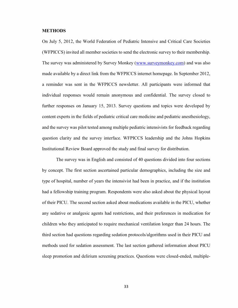
Most respondents (66%) preferred fentanyl as the opioid for analgesia during
sedation, whereas 28% preferred morphine, although respondents from countries outside of
North America demonstrated a more even preference in comparison—42% for fentanyl and
37
46% for morphine (Fig. 2.2; p<0.001). The majority (93%) chooses to administer opioid as
a maintenance infusion when it is used as part of a sedation regimen. Midazolam was the
Figure 2.2: Preferred opioid for analgesia in mechanically ventilated children (%); p<0.001 when comparing opioid preference between North America and all other countries.
Figure 2.3: Preferred benzodiazepine for sedation in mechanically ventilated children (%): p<0.001 when comparing benzodiazepine preference between North America and all other countries

38
benzodiazepine of choice for most respondents (86%; Fig. 2.3) followed by lorazepam
(12%).
Similar to opioid administration, most intensivists initiate benzodiazepines as a
maintenance infusion (80%), with equal proportions choosing scheduled interval dosing and
as-needed dosing (10% each). Propofol and dexmedetomidine were the most commonly
restricted drugs for sedation in PICUs. Of 246 respondents who have dexmedetomidine
available at their institution, 25% stated they have either duration or indication restrictions.
PICU Layout, Sleep Promotion, and Delirium Screening
As shown in Figure 2.4, the majority of North American respondents (62%) reported that
their PICU consists of all private rooms, in contrast to 11% for all other countries (p<0.001).
Figure 2.4: Delirium screening, sleep promotion, and pediatric intensive care unit layout (%). p-value for comparison between North America and all other countries *p=0.01 **p <0.001

39
Seventy-seven percent of all respondents reported that all patient rooms had windows, and
4% reported no windows in any of the patient rooms. A small proportion reported the
presence of unit protocols to optimize noise (16%) and light exposure (9%) for sleep
promotion. Of those surveyed, 78% had never observed earplug use in their PICU, and 65%
had never observed use of eye masks. Of those who had observed eye mask or earplug use,
5% and less than 1%, respectively, reported consistent use for all mechanically ventilated
children.
Seventy-one percent of respondents reported that their unit does not perform routine
delirium screening, and only 2% reported that delirium screening is performed on every
child at least once per shift. Of the respondents who reported frequent or occasional delirium
screening, the only validated delirium screening tool reported was the Pediatric Confusion
Assessment Method-ICU (pCAM-ICU, n=6). Multiple respondents listed withdrawal
assessment tools such as the WAT-1, SOS (Sophia Observation withdrawal Symptoms
scale) and Finnegan as their method of delirium screening. Four respondents reported that
their PICU utilizes a unit-specific delirium screening tool.
Free-text responses
Fifty-four respondents provided detailed comments regarding sedation of mechanically
ventilated children. One common theme was frustration over inconsistencies in sedation
goals. Many intensivists voiced concerns that bedside nurses often want a child to be
immobile, without any signs of awareness, leading to oversedation. Several expressed
interest in pursuing delirium education due to concerns that agitated, critically ill children
may be receiving additional doses of psychoactive medications instead of therapy for what
may actually be undiagnosed delirium.

40
DISCUSSION
Our study is the first international survey to characterize sedation, sleep promotion, and
delirium screening practices in the pediatric population. We found that despite the need for,
and near universal application of, pharmacologic sedation in the PICU, a minority of
programs have formalized methods of sedation assessment, drug choice and delivery, or
therapeutic goal direction. Unlike the adult and neonatal ICUs, sleep promotion is
exceedingly uncommon in the PICU. Finally, the interplay between sedation, sleep, and
ICU delirium is not prioritized in PICU management, even though delirium prevention with
nonpharmacologic behavioral interventions including sleep promotion has become a major
therapeutic initiative in the care of critically ill adults. Thus, we found significant variation
in practice, with many opportunities for future study and therapeutic intervention in a
population undergoing active neurocognitive development.
Our finding that less than one-third of respondents utilize a standardized, unit-wide,
written sedation protocol for mechanically ventilated children is different from that of a
previous survey of PICU fellowship programs in which 66% reported the use of a written
sedation policy.79 This difference is likely due to the survey questions themselves, as we
defined the written sedation protocol to be a specific algorithm that is consistently used in
the PICU, in contrast to a written sedation policy, which may be more general and used as
a guideline. The current study was an international survey that encompassed teaching and
non-teaching hospitals, whereas the previous study was limited to only American fellowship
program directors with a 59% response rate. Nevertheless, we found that even in PICUs
with fellowship programs, the minority (35%) utilized standardized sedation protocols, and

41
sub-group analysis demonstrated no significant difference in the use of sedation protocols
(p=0.73) and sedation scoring (p=0.13) between private and university-based respondents.
A growing body of literature has demonstrated the benefit of standardized sedation
treatment algorithms in critically ill adults. Sedation protocols in the adult ICU have been
shown to decrease days of mechanical ventilation, reduce hospital length of stay, and
promote earlier ambulation. Perhaps most importantly, it reduces the incidence and severity
of delirium, a negative consequence of critical care.80-82 A small number of published studies
suggest that these benefits extend to pediatrics, and a large multicenter randomized
controlled trial is ongoing.83-85
Much to our surprise, we found that most intensivists do not utilize formalized
methods of sedation assessment to guide drug choice and delivery, although several
validated measures exist. The COMFORT score continues to be the most commonly used
scoring system overall, although North American respondents report increased usage of the
SBS and Richmond Agitation-Sedation Scale (RASS) compared to that in a previous U.S.
survey.79 The SBS was designed and validated in 2006 to define the sedation-agitation
continuum in critically ill infants and children to guide goal-directed therapy.86 RASS, the
sedation scale most commonly used in critically ill adults, has not been validated in critically
ill children.87
Even when respondents stated there was a specific sedation scoring system
designated in their PICU, the majority do not consistently use them to set goals for sedation
management on a day-to-day basis. This finding is concerning because sedation is not one-
size-fits-all. Rather, it requires titration to effect. Thus, the failure to measure the level of
sedation and to actually use these measures to guide and titrate therapy may result in

42
oversedation or undersedation, both of which have the potential to produce significant
patient morbidity and mortality. Within a complex team framework of nurses, residents,
nurse practitioners, fellows, respiratory therapists, and attendings who provide and manage
sedation, the use of a streamlined sedation scoring tool in conjunction with a goal-directed
treatment algorithm is imperative to facilitate consistent communication and optimal
therapy.
A small proportion of care providers utilize automated tools such as the BIS for
bedside monitoring of sedation. Initially designed to decrease the incidence of intraoperative
awareness during general anesthesia in adults, the BIS monitor uses an algorithmic analysis
of the electroencephalogram (EEG) to provide a single, dimensionless number to guide
titration of anesthetic agents.88 Because the algorithm of the BIS monitor is derived from
adult EEG data and the EEG of children changes as the brain matures, it remains
controversial whether BIS can be generalized to infants and children in the operating room
and ICU settings.89-92
We found substantial differences in how sedation is provided, particularly in regard
to drug preferences and delivery. Although most providers choose a combination of opioid
and benzodiazepine as their initial regimen for sedation and analgesia, many opt for opioid
alone. Opioids are primarily analgesics that produce sedation and either euphoria or
dysphoria as wanted or unwanted side effects. Opioid alone may be chosen by PICU
providers because they are less likely to adversely affect hemodynamics and are perceived
as short-acting, an erroneous notion given the increased context-sensitive half-life when
administered as a continuous infusion.93,94 This is especially true for fentanyl, now the most
commonly prescribed opioid in North American PICUs, supplanting morphine.79 Opioid

43
use as a primary sedative agent in the PICU warrants concern due to a lack of anxiolytic and
amnestic properties at low doses, leading to dose escalation to achieve the desired level of
sedation and increased risk of tolerance and withdrawal. In addition, the child receiving
opioid as a primary sedative is exposed to an increased risk of all the adverse effects
associated with both long-term and high-dose opioid use, including gastrointestinal
dysmotility that may lead to feeding intolerance during a period when optimal nutrition is
crucial.
Fentanyl may also be chosen in the PICU due its hepatic clearance, facilitating use
in children who have renal insufficiency. Although data are limited, the choice of fentanyl
over morphine in the PICU may also be influenced by morphine’s association with
histamine release, which may concern care providers when potential hypotension is an
undesirable risk.95,96 Morphine is the most inexpensive opioid available. Emerging evidence
has shown that when used as an infusion it is more favorable than fentanyl with regard to
need for dose escalation, incidence of withdrawal, and length of hospital stay.97,98 Opioid
tolerance and withdrawal pose major challenges for all providers in the PICU, and the
predilection for fentanyl may be a trend that needs further evaluation through a randomized
controlled trial.
Midazolam is the benzodiazepine most commonly used for sedation in mechanically
ventilated children. Benzodiazepines are GABA(A) agonists that are
sedative/hypnotic/amnestics with little to no analgesic property. Midazolam has a short
plasma half-life, rapid onset, and is amenable to titration as a continuous infusion. Although
benzodiazepines provide the benefits of amnesia, anxiolysis, and sedation, one major
disadvantage is the increased risk of delirium with prolonged administration.70,71,78,99 Adult

44
guidelines for long-term sedation recommend lorazepam, based on high-grade evidence that
lorazepam infusions require fewer dosage adjustments, require less time to achieve adequate
sedation, and provide more predictability for awakening times and time to extubation
compared to midazolam.80,100,101 Midazolam pharmacokinetics and pharmacodynamics
have been shown to change with age, leading to significant inter-individual
variability.92,102,103 To date, direct comparisons of midazolam and lorazepam have not been
made in the PICU population, and data are limited on the use of diazepam in the ICU
setting.104 Consistent with other ICU surveys, we found that diazepam is rarely used as a
first-line sedative agent in mechanically ventilated children.79,104,105
Dexmedetomidine, an alpha-2 adrenergic agonist with analgesic, sedative, and
anxiolytic properties, has emerged over the last decade as an attractive option for sedation
of critically ill children because of its favorable hemodynamic profile and preservation of
respiratory function.106 Compared to all sedatives and analgesics available,
dexmedetomidine has been shown to induce an EEG pattern with the most similarities to
natural sleep.107-109 Most respondents did not report restrictions on the use of
dexmedetomidine in their units, yet only 10% include it as part of the initial regimen.
Although the currently patent-protected dexmedetomidine is more expensive than the
commonly used benzodiazepines, studies have demonstrated comparatively less cost
because time to extubation and ICU and hospital length of stay are reduced.110
Dexmedetomidine has been associated with decreased opioid and benzodiazepine
administration in critically ill children, as well as decreased inotropic support.111-113 Despite
these favorable qualities, anecdotal evidence suggests that long-term use of
dexmedetomidine infusions can result in physiologic tolerance, withdrawal, and adrenal

45
insufficiency.114-117 Therefore, additional study is warranted.
Our survey demonstrates a clear preference for continuous infusions over scheduled,
intermittent dosing of sedatives and analgesics during mechanical ventilation. Continuous
infusions are advantageous because they are titratable, maintain steady-state plasma drug
levels, decrease line breaks and infection risk, and are less labor-intensive for a busy critical
care nurse. Disadvantages include potential for oversedation from drug accumulation and,
perhaps more important, an increased risk for tolerance and escalation of sedative and
analgesic doses.118-120 Intermittent, scheduled dosing of sedative and analgesic medications
in the appropriate target population may effectively provide many of the purported benefits
of sedation interruption, while exposing a child to decreased overall doses of psychoactive
medications.121,122 These benefits must be considered alongside with the potential increase
in nursing workload necessary to care for an awake, critically ill child.123
Finally, this study underlines an impending and critical need for sleep promotion
and delirium screening in critically ill pediatric patients. Sedatives and analgesics are often
increased to improve the subjective assessment of “sleep,” yet these very medications
decrease restorative sleep, leading to a vicious cycle of sleep disturbances that manifest as
agitation and delirium.31 A small number of studies that have objectively evaluated sleep in
critically ill children have shown significant decreases in slow-wave sleep and REM sleep,
which are integral to neuronal development in childhood.30 Despite the availability of
proven, noninvasive, and inexpensive modalities such as earplugs and eye masks to decrease
sleep interruptions during nighttime hours, such methods are rarely used in adult and
pediatric ICU settings.77,124,125
A minority of PICUs employ noise-reduction strategies to target WHO-

46
recommended levels of noise (<30 dBA Leq day and nighttime). Several studies have
demonstrated that ICU noise levels are frequently greater than 50 dBA throughout a 24-hr
period, with several intermittent peaks to >80 dBA.126,127 Private rooms provide some
protection from general ICU noise and may improve the quality of patient sleep.128 Light
levels are also integral to maintaining circadian rhythmicity during critical illness.
Receiving exposure to natural light during the daytime and minimizing nighttime light
exposure are necessary for hormonal regulation, specifically melatonin secretion, to
optimize the sleep-wake cycle.12,13
Delirium in critically ill children is emerging as an extremely important diagnosis
that must be recognized by all pediatric intensivists.38,74,75,129,130 A combination of critical
illness, sedative-analgesic medications, and sleep disruption place the mechanically
ventilated child at increased risk for delirium. Consequences can include increased
morbidity and mortality from prolonged intubation and weaning of medications. Despite the
availability of validated tools for delirium screening and diagnosis, our study draws
attention to a PICU culture that does not prioritize this important diagnostic consideration.
The low prevalence of PICU delirium screening may be related to multiple factors,
including knowledge gaps surrounding the importance of delirium in critically ill children,
availability of resources to perform unit-wide screening, and accessibility of therapies for
treatment of delirium. Several respondents listed withdrawal screening tools such as the
SOS and WAT-1 as their method of delirium screening, highlighting the need for
widespread delirium education. Under-recognition of delirium may also be secondary to the
reluctance of intensivists to engage the pediatric neuropsychiatrist as yet another
consultation team, whose recommendations for nonpharmacologic and pharmacologic

47
therapies may seem challenging for an already burdened patient, family and PICU team.
Most survey results were similar across respondents internationally, although there
were some interesting differences. The preference for fentanyl as the first-line opioid is
higher in North America compared to all other countries, which may be due to differences
in cost considerations or simply PICU culture that has been propagated based on fentanyl’s
purported advantages over morphine. North American respondents also reported a higher
percentage of nursing-driven sedation protocols, which may reflect an increasing focus on
nursing autonomy in patient care as a predictor of greater job satisfaction and improved
patient care within a team framework.131-133
The current study has some notable limitations. First, the respondents may not be
representative of PICU practitioners internationally, as the 341 respondents are a small
proportion of all intensivists caring for critically ill children. It is also possible that, as a
result of “chain sampling,” several respondents work in the same PICU, which may lead to
bias; other PICUs may not have been represented if no staff member received the survey
link. Second, a respondent’s practice may differ significantly from others in their center,
potentially decreasing generalizability. Additionally, there was a higher proportion of
responses from North American intensivists, which may decrease the generalizability of
data on the international level. Finally, another limitation stems from questions the survey
did not ask. For example, issues such as sedation interruption, rotation, weaning practices,
or use of neuromuscular blockade were not included in the survey. Nevertheless, our study
delineates the international heterogeneity of practice and differing approaches to sedation,
sleep, and delirium in critically ill children.

48
CONCLUSION
Despite the need for, and near universal application of, pharmacologic sedation in
the PICU, only a minority of intensivists utilize formalized methods of sedation assessment,
drug choice and delivery, or even therapeutic goal direction. This study highlights the
heterogeneity in sedation practices among pediatric intensivists, as well as a paucity of sleep
promotion and delirium screening in PICUs worldwide. Thus, numerous opportunities exist
for future study and therapeutic intervention in a population undergoing active
neurocognitive development.

49
CHAPTER 3
Temporal Characteristics of the Sleep EEG Power Spectrum in Critically Ill Children134
(Reprinted with permission from the Journal of Clinical Sleep Medicine)

50
INTRODUCTION
During infancy, childhood and adolescence, the sleep experience evolves over time,
reflecting ongoing neurologic maturation.135 Sleep plays an integral role in homeostasis of
multiple organ systems, including the respiratory, cardiac, gastrointestinal, endocrine and
neurologic systems. Critically ill children in the Pediatric Intensive Care Unit (PICU) are
exposed to a multitude of risk factors for sleep disruption, during a time when the
restorative benefits of sleep are of fundamental importance for neurocognitive
development. In fact, a number of studies in children have shown that slow wave activity
in the sleep electroencephalogram (EEG) is associated with high levels of synaptic density
and activity, a vital component of neurologic maturation.136-138 Critically ill children are at
higher risk for altered sleep-wake homeostasis resulting from not just the underlying illness
but also from exposure to the PICU environment. In addition, centrally acting medications
used for sedation in critically ill children are detrimental to the biology of sleep.30,31,107
Internationally, the most common combination of medications used for sedation of
mechanically ventilated children is opioid and benzodiazepine.15,19 Thus, given the number
of environmental and iatrogenic exposures in the PICU, the normal ultradian variability of
sleep EEG activity is likely to be absent. However, empirical evidence on whether the
temporal organization of sleep is indeed disrupted is lacking.
Sleep is traditionally assessed using visual inspection of the sleep EEG from
polysomnography (PSG).139,140 Although conventional sleep staging is clinically useful for
assessing sleep architecture, its use in characterizing sleep of patients admitted to the ICU
is of limited value.141,142 The EEG of a critically ill child is confounded by the
derangements induced by the critical illness and by the central effects of various

51
medications that are typically administered for sedation. Furthermore, neuromuscular
blockade limits the use of PSG as muscle activity is pharmacologically inhibited. Children
add an additional layer of variability in normative sleep depending on age. Given these
factors, alternative methods for characterizing the sleep EEG in critically ill children are
needed.143 Additionally, sleep may occur any time during the day or night while in the
PICU, therefore assessment of a 24 hour period is necessary to capture the temporal
characteristics of sleep in these patients. The objective of the current study was to
characterize the sleep EEG in a sample of critically ill children without baseline neurologic
disease. It was hypothesized that critically ill children will not demonstrate day-night
organization of sleep and will lack the normal characteristics of the sleep EEG that are
typical in healthy children.
METHODS
Study Sample
Children with respiratory failure requiring mechanical ventilation were recruited
from the PICU of the Johns Hopkins Children’s Center. Each bed in the PICU is positioned
adjacent to a window with adjustable shades. Patients were eligible for the study on the
first day of mechanical ventilation if ventilator support was expected to continue for least
24 hours. Exclusion criteria included a history of sleep-disordered breathing (SDB),
surgery within the last seven days, seizures, neurological injury and severe developmental
delay. To rule out SDB, a detailed history was obtained from the parent(s) including a
history of snoring. In addition, the medical record was reviewed for previous sleep studies
and parents were asked to complete the Children’s Sleep Habits Questionnaire.144 Finally,
patients were excluded if it was deemed that the monitoring would interfere with medical

52
care. Eligible patients underwent informed consent with a parent or legal guardian. To
compare the data collected during critical illness, healthy age- and gender-matched
children without central or obstructive sleep apnea (OSA) from the Tucson Children’s
Assessment of Sleep Apnea (TuCASA) study were used. The TuCASA study is a
community-based study aimed at assessing the prevalence of sleep apnea in children.145
OSA was defined if the respiratory disturbance index, using the 3% desaturation criteria,
was 1 or more events per hour of total sleep time. Hypopneas were scored if the magnitude
of any ventilation signal decreased to below approximately 70% of the baseline amplitude
for at least 6 seconds or for 2 or more consecutive breaths. The study protocol was approved
by the Institutional Review Board (IRB) on human subjects research of the Johns Hopkins
University School of Medicine.
EEG Monitoring and Power Spectral Analysis
The recording montage for the PICU patients included bilateral central and occipital
EEG leads sampled at 125 Hz, right and left-sided electrooculograms (EOG) and submental
electromyogram (EMG) using the Embla N7000 system (Embla, Denver, Colorado
U.S.A.). The montage was accompanied by high-resolution video monitoring for assessing
medical interventions (e.g., suctioning, dressing changes) and occurrence of movement or
activity. The recording was continued for at least 24 hours and discontinued when the
patient was extubated or at 72 hours after the initiation of data collection. Home nocturnal
PSG for the healthy children in the TuCASA sample included central leads as well as
bilateral EOG leads and chin EMG. Detailed methodology for the TuCASA cohort have
been previously described.146 Power spectral analysis of EEG recordings from the PICU
and age and gender-matched subjects was conducted using the fast Fourier transform (FFT)

53
in Matlab (Ver. R2014A, Matlab, Inc.). The FFT was conducted on an EEG record length
of 5-s to obtain a frequency resolution of 0.2 Hz. The power content (expressed as μV2) for
each 30-s epoch of sleep was determined as the average power across the six 5-s segments
of the EEG. The resulting spectral distribution from the FFT was categorized into the
following frequency bands: δ (0.8 to 4.0 Hz); θ (4.1 to 8.0 Hz); α (8.1 to 13.0 Hz); β1 (13.1
to 16.0 Hz); and β2 (16.1 to 20.0 Hz). The power in each frequency bandwidth was
expressed as absolute power in each 30-s epoch of sleep. Frequencies < 0.8 Hz were
excluded to remove the effects of low-frequency artifacts (e.g., sweating and respiration).
Statistical Analysis
Data are summarized as means ± standard deviation or medians (interquartile
range) as appropriate. Differences in mean nocturnal EEG power between the PICU
patients and matched healthy children were tested using the Wilcoxon signed-rank test
given the small sample size and skewed distribution of EEG spectral power. Matching was
performed using the ccmatch command in Stata 12 (StatacorpLP; College Station, TX).
The nocturnal period was defined as the time from sleep onset for healthy children, and the
hours of 2200-0600 for the PICU patients. Given well-established temporal patterns in the
EEG spectra during the night in healthy children, analyses were also undertaken to
characterize temporal patterns of EEG spectral power for each frequency band using a
Locally Weighted Scatterplot Smoothing (LOWESS) spline technique for both the PICU
patients and healthy children. The LOWESS method depicts the association between two
variables (i.e. EEG δ power activity vs. time) using a smoothing coefficient to provide a
robust fit to the data without eliminating changes in slope over time. Statistical tests were

54
2-sided, and a p-value less than 0.05 was defined as statistically significant. All calculations
were performed in Stata 12 (StatacorpLP; College Station, TX).
RESULTS
Eight critically ill PICU patients with respiratory failure (median age 8 years, range
6-16 years) underwent limited montage EEG monitoring. Each patient was receiving a
continuous background infusion of both opioid and benzodiazepine at the time of
enrollment and monitoring, with a goal State Behavioral Score between -1 and 1.86
Demographic data and diagnoses for the study sample are shown in Table 3.1. Two of the
eight patients were receiving a low-dose infusion of one sympathomimetic agent for
hemodynamic support. Patient D received dopamine at 5 mcg/kg/hr while Patient F
received phenylephrine at 2 mcg/kg/hr during the 24-hr EEG recording period. Although
the full 24-h period was recorded in all patients, only 18 hours were included in two patients
due to clinical instability. However, EEG power spectral analysis included the recordings
Table 3.1: Demographics of PICU sample

55
of the full
nocturnal period in
all patients. EEG
data from the first
eight consecutive
nocturnal hours
(10:00 PM – 6:00
AM) were analyzed for each PICU patient and compared to nocturnal EEG of an age and
gender-matched healthy child. The average EEG power in each of the frequency bands
(i.e., δ, θ, α, β1, β2) was lower in the PICU patients compared to healthy patients (Table
3.2). Furthermore, unlike healthy children, PICU patients did not demonstrate the
characteristic temporal EEG patterns in δ or θ power spectral bands during the night as
evident in the healthy children. As shown in Figure 3.1, healthy children demonstrated the
typical ultradian pattern in EEG activity with an increase in δ power during NREM sleep,
Figure 3.1: Nocturnal EEG δ power in PICU patients and age and gender-matched healthy children.
Table 3.2: Average EEG spectral power during nocturnal period, expressed as μV2 (mean ± SD).

56
particularly in the first third of the night. Moreover, healthy children demonstrated a
decline in overall EEG δ power over the course of the night, while PICU patients
demonstrate little variability as evidenced by their flattened profiles. Subject A’s profile
differed from the other PICU patients in that the baseline delta activity was maintained at
a high level throughout the recording period. All other patients had peaks in EEG δ power
well below 500 µV2.
Alterations in the nocturnal EEG θ power profile (4–7 Hz) were similar to the
findings for δ power, with lack of an ultradian rhythm during the night (Figure 3.2).
Healthy children demonstrated the expected temporal variability in EEG θ power, which
gradually declined in the latter part of the night. In contrast, θ power in the EEG was at
very low levels and did not display temporal variability in PICU patients. Mean absolute θ
power in the EEG in the healthy children was 30.7 μV2, while mean EEG θ power in PICU
patients was 5.6 μV2 (p < 0.001). Given that critically ill children in the PICU may
Figure 3.2: Nocturnal EEG θ power in PICU patients and age and gender-matched healthy children.

57
experience sleep during any part of the day or night, EEG δ and θ power were evaluated in
the 6 children with complete 24-h data. As seen in Figure 3.3, there was evidence of
oscillations in EEG δ power over the 24-h period for PICU patients, most obvious for
Patient G. In contrast, with the exception of Patients A and E, most PICU patients did not
demonstrate ultradian variability in EEG θ power (Figure 3.4). Mean EEG δ and θ power
of PICU patients were compared between the day (07:00 through 19:00) and night (19:00
through 07:00) hours. Mean EEG δ power was higher during the day (237 ± 243 μV2) than
night (211 ± 223 μV2; p < 0.001). Similarly, EEG θ power during the day was higher than
the night (10 ± 31 vs. 8 ± 17 μV2, p < 0.001). Among PICU patients, nocturnal δ and θ
power was not associated with cumulative nocturnal morphine (p = 0.4 and 0.5,
respectively) or midazolam equivalents (p = 0.6 and 0.3, respectively). There was also no
significant association among the PICU patients between cumulative 24-h morphine and
midazolam equivalents and mean 24-h δ and θ power.
Figure 3.3/3.4: Twenty-four hour EEG δ and θ power in PICU patients.

58
DISCUSSION
The results of this study demonstrate that, over a 24-hour period, there are
significant differences in the EEG power spectra between critically ill and healthy children.
Critically ill children have no day-night organization of sleep and exhibit decreases in slow
wave activity (i.e., δ power) during the nighttime hours when compared to healthy
children. Although sleep has previously been investigated in PICU patients, available
studies have relied on subjective assessment with visual scoring of the polysomnogram.30
In a recent systematic review on sleep in critically ill children, nine studies were identified,
with four resulting from the same randomized controlled trial. Of the studies which focused
on critically ill children and utilized objective measurements of sleep, common findings
included decreases in REM sleep and slow wave sleep, with evidence of frequent nocturnal
arousals.147,148 The findings of the current study add to the existing body of evidence by
providing insights regarding the temporal trends of sleep EEG power spectral activity in
critically ill children without baseline neurologic disease who are admitted with respiratory
failure.
Although the true function of sleep remains elusive, a great deal is now known
regarding the physiologic effects and consequences of altered sleep including effects on
protein synthesis, cellular and humoral immunity and increased catabolism.31 Much
research has been devoted to characterizing sleep in the adult ICU.142,143,149-151 Yet, the
negative impact of sleep disruption in the critically ill pediatric patient undergoing rapid
advances in growth, cognition and behavior has not been well characterized. Infancy and
childhood are marked by sleep as the predominant behavioral state, and sleep structure and
organization are in a constant state of change, paralleling central nervous system (CNS)

59
growth and differentiation.152 However, the ontogeny of sleep may not only be a correlate
of CNS development, but a reflection of the reciprocal interactions between brain plasticity
and sleep.153 Indeed, natural sleep may play a dynamic role in brain development, laying
the groundwork for future learning.
Healthy children demonstrate a well-characterized temporal pattern of slow wave
activity (EEG δ-power) activity when asleep, with a significant concentration of δ power
in the first NREM period, which generally occurs within the first two hours after sleep
onset.154 Data from the age and gender-matched healthy children in this investigation
demonstrate that consistent pattern. With increasing age, EEG δ power exhibits a steep
decline across adolescence after reaching a peak in childhood, again reflecting the role of
NREM sleep in brain function.155 Findings from the patient sample in the current
investigation show that critically ill children demonstrate major differences in nocturnal
EEG patterns when compared to normal children, despite demonstrating a behavioral state
(eyes closed, resting state) that is characteristic of sleep. The observed differences
including the lack of day-night organization among sedated, critically ill children may, in
part, be related to use of sedative medications. Pharmacologic management of sedation and
analgesia for intubated children in the PICU primarily consists of opioids and
benzodiazepines.15,19 A recent international survey of pediatric intensivists determined that
fentanyl is the most commonly used opioid, and midazolam is the most commonly used
benzodiazepine. Although opioids increase total sleep time in healthy subjects, they
decrease slow wave and REM sleep.31 Benzodiazepines also significantly suppress slow
wave and REM sleep, and are known independent risk factors for the development of ICU
delirium in adults.70 The risks of continuous sedative and analgesic exposure are

60
compounded by polypharmacy and environmental factors such as noise, light and patient
care interventions, leading to further negative effects on sleep architecture. Additionally,
sympathomimetic medications frequently used in the critically ill setting are known to
decrease both REM and slow wave sleep.31,67
Poor sleep quality is a risk factor for delirium, which can lead to a vicious cycle of
increasing sedation to maintain safety and help the patient “sleep”. The results of our study
challenge the general notion that children in the PICU are experiencing natural, restorative
sleep.19 Lack of day-night cycles in conjunction with loss of natural sleep among PICU
patients is an important area for future research. Simple non-pharmacologic interventions
such as earplugs and eye masks at night, consistently opening shades to achieve sunlight
exposure during the day and implementation of noise reduction protocols may go a long
way to promote sleep and decrease sedation needs.19,124,156,157 Therefore, it is essential that
future studies investigating sleep in the PICU include continuous noise and light
measurements. Before pharmacologic interventions such as melatonin are considered, it is
imperative to further characterize dim light melatonin onset (DLMO) in critically ill
children to better delineate the underlying pathophysiology of altered sleep in the PICU.
Furthermore, sleep-wake problems that begin in the PICU may persist long after discharge
from the PICU and hospital into the home setting, a long-term consequence that deserves
further investigation as it relates to neuropsychological outcomes.
There are several limitations in the current study. First, the use of healthy children
as a comparison group for patients in the PICU may not be the most suitable given that
healthy children were monitored only overnight and not for the extended period as the
PICU sample. The second limitation is a small sample size and all of the children in the

61
PICU had respiratory failure. Therefore, the results of the current study may not be
generalizable to pediatric patients with other causes of critical illness. The same sample
size may also explain the lack of an association between δ and θ power with sedative
medication doses among PICU patients. Third, the sample of healthy children from the
TuCASA cohort likely had greater exposure to sunlight than the PICU patients, which was
not measured. Fourth, differences in the platform used to record the EEG in the PICU
patients (Embla™) and the TuCASA cohort (Compumedics™) may lead to quantitative
differences in EEG power spectra between the two groups. Finally, the results presented
herein need to be interpreted with the knowledge that there are unmeasurable confounders
that may affect the EEG recordings, including the severity of critical illness and underlying
neurobiologic activity of the patients.
In summary, critically ill children in the PICU demonstrate differences in the EEG
including the lack of ultradian increase in slow wave activity during the nighttime hours.
The importance of sleep during recovery from critical illness deserves continued attention,
particularly for children who are undergoing active neurologic maturation when sleep plays
a vital role in normal development. Furthermore, disruption in sleep architecture has
implications for how children are cared for during the most acute phase of their illness.
Future research must focus on the modifiable risk factors for sleep disturbances in the
critically ill child, including sedative approaches and non-pharmacologic behavioral
interventions to promote natural sleep during recovery.

62
CHAPTER 4
Day-night Activity Patterns in Pediatric Patients after Major Surgery

63
INTRODUCTION
Sleep plays an integral role in normal neurodevelopment, which is reflected by the
constantly evolving sleep needs of infants and children as they grow. Children admitted to
the hospital, and in particular the Pediatric Intensive Care Unit (PICU) are exposed to
several factors that can lead to disruption of the normal sleep-wake cycle, including
invasive medical interventions, pain, noise, light, interruptions for nursing cares, and the
underlying illness itself.30 It is routine for children who undergo major surgery to be
admitted to the PICU in the post-operative period, and the majority of these children are
exposed to sedative and analgesic medications that are known to have negative effects on
restorative sleep and circadian rhythms.31
Actigraphy is a non-invasive, well-tolerated, reliable and objective method to
assess 24-hour patterns of day-night activity over several days to weeks. Validated for sleep
assessment in infants and children, this wristwatch-like device produces minute-to-minute
activity counts to monitor activity patterns over the course of hospitalization. While the
actigraph can distinguish sleep from wake using algorithms to quantify the reduced
movement associated with sleep, a more robust assessment of differences in day and night
activity patterns can be performed with raw motor activity data.
To date, there have been no studies describing objectively assessing day-night
activity patterns in children in the hospital after major surgery. The primary objective of
this study was to characterize the day-night activity patterns of infants and children after
major surgery, and describe the evolution of these children’s activity patterns from the
PICU until discharge from the hospital.

64
MATERIALS AND METHODS In this prospective, observational study, children were eligible for inclusion if they were
admitted to the Johns Hopkins Pediatric Intensive Care Unit (PICU) after major cardiac,
orthopedic or urologic surgery. Informed consent was obtained on the day after surgery
from the child’s parent or legally authorized representative, and assent was obtained from
the child when age or developmentally appropriate, unless the child was mechanically
ventilated and receiving sedatives. Patients were only excluded if they had evidence of
seizure activity at the time of screening or a history of traumatic brain injury or ischemic
or hemorrhagic stroke. The study was approved by the Johns Hopkins Institutional Review
Board.
Instruments and Measures
Clinical Variables
Demographic and clinical data collected through daily chart review during the duration of
actigraphy recording included age, gender, type of surgery (cardiac/orthopedic/urologic),
length of PICU, floor and hospital length of stay, pediatric risk of mortality score (PRISM-
3), intubation status, length of mechanical ventilation, and sedative-analgesic medication
administration.
Actigraphy
After informed consent, an actigraphy watch (MicroMotionlogger, Ambulatory
Monitoring, Inc) was placed on the child’s wrist or ankle on post-operative day #1, and
remained in place until the day of discharge from the hospital. The MicroMotionlogger has
been used widely to document rest-activity patterns in infants and children. Motion sensors
inside the accelerometry device capture data on patient movement using the Zero Crossing

65
Mode, which is measured as the number of accelerations per minute. The devices used in
this study are capable of collecting data for up to twelve weeks. If the child was intubated
at the time of actigraphy initiation, data analysis began at the time of extubation. The Act
Millenium software was used for initializing and downloading actigraph data, and data
processing was performed using the ActionW 2.7 software.
Data Analysis/Statistical Methods
For subjects with complete 24-hour actigraphy data, we analyzed activity profiles
represented by 1440 minute-level (24 hours) activity counts. A log (count+1)
transformation was applied to ensure symmetry of activity count distributions.
Transformed activity counts were averaged within two specific time periods: daytime
(08:00-20:00) and nighttime (20:00-08:00). Day-night activity patterns were estimated
using the daytime activity ratio (DAR), defined as the ratio of the average daytime activity
to total 24-hour activity. DAR = �̅�𝑑 (�̅�𝑑 + �̅�𝑛)⁄ , where �̅�𝑑 and �̅�𝑛 are the average log
activity counts (per minute) for an individual during the daytime and nighttime hours
respectively. A DAR of 50% suggests that the level of activity was equally distributed
between day and night periods, while a DAR of 100% indicates that all activity occurred
during the daytime period.
We utilize a mixed effect model with random intercepts for each subject to account
for the variation within subjects due to the subjects having multiple days of activity data
recorded. We examined the fixed effects for the PICU, gender, age, race, Pediatric Risk of
Mortality Score (PRISM), cohort, and the time spent intubated measured in minutes. Due
to the distribution of time intubated being extremely skewed, we transformed it by taking
the log (log[int. min. + 1]) to allow it to be less skewed in order to conform to the model

66

67
When all patients (n=221) and hospital days were included (n=2271), median daytime
activity represented 58% of total activity while in the hospital (Figure 4.1). The mean
daytime activity ratio
during 1346 PICU
days was 0.559, while
the average DAR on
the floor was 0.608
over 905 days
(p<0.0001). Children
in the cardiology
cohort had the highest
mean DAR (58.2%)
when compared to the orthopedic (57.5%) and urology cohorts (57.0%).
In order to determine if daytime and nighttime activity are different between the PICU
and the floor settings, a mixed model with random intercept for each subject was fit
Variable Coefficient Standard Error
T-statistic P-value
PICU -0.0530 0.0047 -11.341 <0.0001 Gender (F) 0.0001 0.0067 0.086 0.9931 PRISM 0.0000 0.0007 -0.047 0.9628 Race
Black -0.0081 0.0086 -0.941 0.3479 Other 0.0063 0.0081 0.775 0.4390
Age 0.0018 0.0006 2.732 0.0068 Cohort
Ortho -0.0331 0.0091 -3.627 0.0004 Urology -0.0495 0.0124 -3.977 0.0001
Time Intubated (log - scale)
-0.0025 0.0010 -2.569 0.0109
Figure 4.1: Histogram of daytime activity ratio for all subjects (n=221) Histogram of DAR
0.2 0.4 0.6 0.8 1.0
01
23
45
DAR
Mean = 0.578
Table 4.2: Model Results for daytime activity ratio

68
controlling for age, gender, surgical cohort, PRISM score, log of time intubated, and race
(Table 4.2). PICU days were associated with a significantly lower DAR (5.3%,
p<0.0001) compared to days when the subject was on the floor. Older children had
increased DAR, with a 0.18% increase with each year of age.
After adjustment, the urology cohort demonstrated the lowest DAR when compared
to both the cardiology and orthopedic cohorts (-4.95%, p=0.0001). The log intubation time
was associated with the DAR. As an example, for each 20% increase in intubation time,
the DAR decreases on average by 0.05, or 0.05%. Severity of illness at PICU admission
was not associated with the DAR.
DISCUSSION
The results of this study confirm that children recovering from major surgery experience
disturbances in day-night activity patterns both in the PICU and after transfer to the floor.
In addition to being in the PICU location, duration of intubation and orthopedic or urologic
surgery were predictors of lower DAR. These findings provide important insights into how
children’s rest-activity cycles are distributed throughout the 24-hour period, suggesting
that the hospital setting may be severely disruptive to the normal sleep-wake patterns.
In healthy children, activity is consolidated during the daytime hours. We found
that even older children in our cohort had significant increases in nighttime activity,
consistent with alterations in rest-activity patterns or sleep fragmentation. In lieu of using
algorithms to distinguish sleep from wake, we quantified raw motor activity data to analyze
the daytime activity ratio and characterize how sleep patterns may be altered in the hospital
setting. Raw motor activity provides a quantitative measure that as a percentage (DAR)
can be easily compared across subjects regardless of age, disease process, or

69
neuromuscular strength. Additionally, algorithmic sleep-wake categorization can
underestimate the amount of wake time and overestimate the amount of time in sleep due
to a state of bedrest or immobility in the ICU.159
Day-night differences were indistinguishable in 38% of our subjects (<50%), which
is consistent with adult literature utilizing actigraphy in the hospital setting. Most
pronounced in the PICU but also present in the floor setting, nursing and physician
interventions, light, and noise from alarms and voices are risk factors for sleep
disturbances. In a study of patients hospitalized after traumatic brain injury, rest-activity
cycles were consolidated on only 46.6% of all hospital days, however a linear trend of
improvement was observed over time.160 In our cohort of children without acute
neurologic disease, the average DAR on the floor setting was slightly lower than that of
the PICU setting, suggesting that sleep-wake patterns do improve over time, however
whether this is a consequence of the environment, improvement in illness process, or both
is not known.
There are several limitations to this study. First, it is a single-center cohort and may
not be generalizable to other pediatric hospital settings. Secondly, we did not account for
children’s other comorbidities such as neuromuscular weakness in assessment of the
daytime activity ratio- however, utilizing this ratio measure facilitates the child being his
or her own control. Third, our analysis did not include pain, sedative and anesthetic
medications before or during the period of actigraphic analysis, which may play a
significant role in activity patterns and sleep quality.

70
CONCLUSION
Children admitted to the PICU after major surgery experience disruptions in day-night
activity patterns during their hospital stay which may likely reflect fragmented sleep-wake
patterns. Given the potential impact of sleep disturbances on the developing brain and
importance of sleep during recovery from surgery further research is needed to identify
methods of optimizing sleep hygiene in the perioperative period.

71
CHAPTER 5
Nurses’ Perceptions of the Pediatric ICU Environment Before and After a Transition to Single-patient Rooms9
(Reprinted with permission from American Journal of Critical Care)

72
INTRODUCTION
Every year in the United States, over 250,000 children are admitted to the pediatric
intensive care unit (PICU).1 The chaotic PICU environment, including constant sound and
bright light, exposes critically ill children to multiple risk factors that can fragment sleep
during their hospital stay.30 Sleep is a basic human need. Integral to normal
neurodevelopment in a healthy child, sleep may play an even more important role in the
recovery of children with critical illness.31 Several studies have demonstrated that both
adult and pediatric ICU patients experience circadian rhythm disturbances and decreases
in slow-wave sleep.134,143,149 Although there are multiple risk factors for sleep
fragmentation in critically ill patients, the most modifiable is the external environment, as
sound and light levels play a key role in sleep-wake homeostasis. 30,143,149,161,162
Disturbances in sleep continuity may be associated with an increased risk of
delirium in patients admitted to the ICU.38,74,163,164 Critical illness implies the need for
invasive medical instrumentation and frequent interruptions for nursing care and
monitoring. However, modifying the external environment can help optimize the sleep
experience and potentially decrease a child’s sedation needs during mechanical ventilation.
A lack of natural sunlight and periodicity in light-dark exposure can lead to adverse effects
on circadian rhythms and sleep architecture.165 Constant loud and sudden sounds interrupt
sleep, increase sedation needs, and affect physiologic parameters such as heart rate and
respiratory rate.166
In addition to potential benefits for patients, modifying the external environment to
promote sleep for patients may also have favorable effects on nursing stress and job
satisfaction.167-169 Caring for critically ill children and their families has a strong emotional

73
impact. Nursing has been well-established as a stressful profession, and studies of ICU
nurses have shown that work satisfaction and stress levels are strongly correlated.170.
Higher light levels during the day, achieved with exposure to natural sunlight, is integral
to human health.171 Outside of the healthcare environment, previous studies have
demonstrated that the presence of windows and sunlight improves job satisfaction.169,172
Furthermore, sound levels in most ICUs are universally higher than those recommended
by the World Health Organization.30,126,166,173-176 Noisy work environments in the both the
PICU and adult ICUs contribute to higher heart rates, fatigue and annoyance among
nurses.168,177
Most PICUs internationally have a layout with multi-patient rooms, which, though
convenient for workflow of daily patient care, may not be conducive for sleep continuity
or the preservation of circadian rhythm.19 In the past two 2 decades, hospital designs have
been shifting from multi-patient to single-patient room configurations. In the acute care
setting, single-patient rooms are associated with lower rates of medical errors, increased
control of room light and temperature, lower sound levels, and even decreased delirium
prevalence.178 Moreover, patients and their families prefer the private-room design.179
Literature from pediatric ICUs have largely focused on neonatal ICU (NICU) design, while
the heterogeneous population of the PICU (ages 0-21) and impact of PICU design have not
been investigated.29-31 Compounding the challenges of caring for critically ill children are
the vastly different neurodevelopmental needs of PICU patients. The same nurse, for
example, may be assigned to care for both a one-month-old infant and a sixteen-year-old
adolescent on the same shift.
The primary objective of the current study was to examine changes in nursing

74
perceptions about the PICU environment with the transition from an ICU with multi-patient
rooms to one with single-patient rooms. Specifically, we sought to examine the effect of
having all private rooms on a nurse’s ability to control the external environment in a way
that optimizes sleep for critically ill children and the effect of this change in environment
on the nurses’ perception of their own personal work experience. We hypothesized that a
private-room setting would improve a nurse’s ability to control the external environment
in ways that would promote sleep in patients, decrease nursing stress and enhancing the
overall work experience.
METHODS
A cross-sectional survey study was conducted during a transition of the PICU from
multi-patient to single-patient rooms. The original PICU was a 32-bed unit that had four
private rooms and four semi-private rooms; the remainder of the unit consisted of several
beds in larger rooms. All of the private rooms and semi-private rooms had one window
with shades, whereas the large multi-bed rooms had one window for four beds. Nurses’
stations were located in open spaces within the patient-care areas. The current PICU is a
40-bed unit with all private rooms and a window in each room. Each room includes lighting
on dimmer switches and a movable boom which enables the patient to be positioned in any
direction (facing window, nurses’ station or wall). There is also a sliding door which can
be incrementally opened, whereas in the original PICU the door was either closed or open
completely.
After receiving approval from the Institutional Review Board, we electronically
mailed a survey to all PICU nurses 12 weeks before and 12 weeks after the PICU transition.
We informed all participants that individual responses would remain confidential and

75
assigned respondents a numeric identifier to allow pre-post survey matching. To minimize
reporting bias, we did not inform respondents about the follow-up survey at the time of the
first survey. The survey was pilot tested among a committee of nurses in the PICU for
feedback regarding question clarity and the survey interface. The final survey consisted of
20 questions that were divided into perceptions of the PICU sound and lighting
environment for patient care and perceptions of the environment for the nurses' personal
work experience. Questions were closed-ended, multiple-choice design measured on a 5-
point Likert scale. Respondents were also asked to provide any specific comments or
additional information in free-text form.
Statistical Analysis
Responses were collected in Survey Monkey. McNemar’s chi-square tests were
used to compare responses to the questions regarding perceptions about the sleep
environment and work experience before and after the move to the single-patient room
layout. All tests were two-tailed, and a value of 0.05 or less was considered statistically
significant. The data were analyzed with Stata 12 (StataCorp, College Station, TX).
RESULTS
Demographics
Demographic data of respondents are summarized in Table 5.1. Eighty-three
percent of nurses (100/120) completed the pre-transition questionnaire, and of these
respondents 90 completed the post-transition survey (75% overall response rate). The
majority of the 90 respondents (63%) were 21-29 years old and 99% of the respondents
(89/90) were female. Forty-three percent of nurses had 1 to 3 years of work experience in

76
the PICU, with 41% reporting greater than 3 years. The average percentages of the nurses’
work time spent on day shifts, night shifts, and administrative/education duties were 49%,
45%, and 6%, respectively.
Perceptions of Sleep Environment: Sound and Light
As shown in Table 5.2, nurses reported that a single-patient private room layout
was overall more conducive to patients sleeping well at night and to promoting a normal
sleep-wake cycle than was a multi-patient layout (p<0.001). More nurses agreed that they
had increased control of light and sound for sleep promotion during both day and night
(p<0.001) than they did with a multi-patient layout. However, in both the single- and multi-
patient room settings, monitors and alarms were the greatest factor that affected sound
levels during the day and night. Conversations between clinical staff were the second most
commonly reported factor that affected sound exposure in both layouts, although it was
cited less often after the move to single-patient rooms. With respect to light exposure, in
both layouts the most important factors cited for increasing light at night were safety issues,
followed by requirements of the charge nurse/unit culture to keep the lights on. Patient care
Table 5.1: Demographics of pediatric intensive care nurses who responded.

77
was also a persistent reason for increased light exposure at night in both PICU layouts.
Nursing Perceptions of Annoyance and Stress
Nurses were less annoyed by sound in the all-private room layout (79% vs. 33%;
p<0.001; Figure 5.1). In addition, sound contributed less to the stress levels of nurses in
Table 5.2: Nursing perceptions of the pediatric intensive care unit environment

78
the single-patient room layout than in the open layout (90% vs. 44%; p<0.001) and fewer
nurses desired a quieter unit after the move (89% vs. 27%; p<0.001). Finally, desire for
Table 5.3: Free-text responses summarizing common themes after move to all private rooms.
Figure 5.1: Comparison of nurses views on work environment in single patient rooms vs multipatient rooms.

79
more sunlight exposure was significantly reduced after the move to the new layout (95%
vs. 50%; p<0.001). Table 5.3 lists free-text responses from PICU nurses which summarize
common themes that were present after the move to single-patient rooms.
DISCUSSION
In this cross-sectional survey study of PICU nurses, a change from multi-patient
rooms to a single-patient room layout improved nurses' perceptions of the PICU
environment for sleep promotion, particularly in regard to sound and lighting optimization.
Additionally, nurses reported feeling significantly less stress and annoyance as a result of
the PICU environment after a transition to the single-patient room layout. To our
knowledge this is the first study to investigate the role of PICU architectural design on
nursing perceptions of the patient care and work environment. In addition to results
suggesting the single-patient room design has benefits for sleep promotion and decreasing
nursing stress, we identified issues for ongoing improvement that must be addressed
beyond a change in PICU design.
It is not surprising that monitors and alarms persisted in the single-patient room
layout as the most important factor affecting sound levels in the PICU during the day and
night, followed by clinical staff conversations. Private rooms provide a level of protection
from general ICU sounds only. Although alarms are a necessary part of ICU care to
anticipate and manage acute changes in a child’s clinical condition, a large body of
evidence is mounting to support technological and institutional culture changes to
minimize false alarms and alarm fatigue.180 Electronic sounds from intermittent alarms lead
to the most arousals from sleep in studies of healthy volunteers in simulated ICU

80
environments.11 Therefore, quality improvement initiatives to individualize alarm
thresholds and minimize false alarms are crucial to optimize the balance between patient
safety and a healing ICU environment, while simultaneously decreasing nursing stress.181-
184 Similarly, the institution of nighttime “quiet hours” both in patient rooms and general
areas can change culture and minimize sound levels due to staff conversations, enabling
adherence to WHO recommendations recommending no more than 30 dB at night.30
However, current evidence suggests that most PICUs internationally do not have either
noise or light optimization protocols in place for sleep promotion.19
Another important issue raised by the questionnaire was the struggle between
optimizing patient safety and promoting sleep. Dimming lights in the rooms of the most
critically ill children may not be feasible because staff need to make continual visual
assessments of the patient and provide frequent care. Even with the ability to increase
sunlight exposure during the day or dim lights at night, family and staff preferences can be
major obstacles to lighting optimization. Nevertheless, the results of our study enabled
physician and nursing leadership to identify areas for culture change and the need to create
specific patient criteria for which noise and lighting protocols should be utilized.
Finally, transitioning to a single-patient layout improved PICU nurses’ perceptions
of the work environment and decreased job stress. Mealer and colleagues found that 20-
30% of adult ICU nurses had symptoms of post-traumatic stress disorder (PTSD)
secondary to their work, significantly higher than the lifetime prevalence of 6.8% among
the U.S. general population.185 PICU nurses spend their entire shift at the bedside of
critically ill children, which has great psychological impact and increases the risk for
secondary traumatic stress.170,185 In a study of pediatric acute care nurses, 21% of survey

81
respondents demonstrated strong PTSD symptoms which were also associated with
comorbid symptoms of anxiety, depression and burnout.186 Younger ICU nurses (ages 20-
29), the most highly represented demographic in our PICU, have reported higher job stress
levels in previous studies than their older and more experienced colleagues.187,188
Therefore, the architectural design of an ICU where nurses care for critically ill pediatric
patients may have important implications for nursing retention and wellness.189 Sound
levels are known to increase stress and annoyance levels among nurses in the ICU
environment.168,190 Sunlight exposure during the day optimizes circadian rhythms for
nurses who are frequently transitioning between day- and night-shift, and also improves
mood and energy.191 Thus, having the ability and control to minimize noise and increase
their own sunlight exposure during the day was beneficial to nurses’ perceptions of the
work environment.
There are limitations to this single-center study. First, perceptions of the
environment may have been affected by anticipation and completion of the change. That
is, it is possible that the new building and environment in general worsened perceptions of
the old unit. Second, the ages and experience levels of the nurses who responded were very
heterogeneous, although this heterogeneity is generalizable to the majority of adult and
pediatric ICUs. Third, the survey that we disseminated was not a previously validated tool.
Finally, we do not have quantitative sound and light level data to correlate the change in
perceptions with actual changes in sound and light levels before and after the move.
In summary, a single-patient private room layout led to improved perceptions of
the patient environment for sleep promotion and decreased stress levels among pediatric
ICU nurses. As more ICUs shift their architectural designs to the single-patient room

82
model, it is important to continue to focus on issues that are not completely addressed by
a new design. These issues include patient alarms, staff conversations, and the perception
that an optimized sleep environment may detract from patient safety. Future quality
improvement and research initiatives should systematically address methods to optimize
sound and light in the ICU environment so that patients can benefit from optimized sleep
continuity and staff can work in an ICU that promotes a healing environment.
83
CHAPTER 6
Identifying Barriers to Delirium Screening and Prevention in the Pediatric ICU: Evaluation of PICU Staff Knowledge192
(Reprinted with permission from Journal of Pediatric Nursing)

84
BACKGROUND AND OBJECTIVES
Delirium in the pediatric intensive care unit (PICU) setting is often unrecognized
and undertreated. As defined in the Diagnostic and Statistical Manual of Mental Disorders:
Fifth Edition (DSM-5) delirium is a disturbance and change in attention and awareness
from baseline, developed over a short time, with a fluctuating course. The change from
baseline includes a disturbance in cognition, which is not better explained by another
neurocognitive disorder.193 In adults, Intensive Care Unit (ICU) delirium is associated with
increased hospital length of stay, morbidity and mortality.194-196 Due to heterogeneity in
ages, development and diagnoses, delirium screening in critically ill children can be
challenging. Despite validated tools for screening delirium in this population, few pediatric
ICUs (PICUs) internationally perform screening.14
Pediatric delirium has been more recently highlighted in literature, with the
development and evolution of pediatric screening tools.74,197 Pediatric studies have shown
that delirium is present in at least 30% of critically ill children and adolescents.74,198 There
is limited data on pediatric patient outcome measures after delirium, but a few retrospective
studies describe longer length of stay and similarities to an adult delirium course.199
However, many PICU’s internationally are still not screening for delirium. In a recent
survey study characterizing international PICU practices, 71% of respondents reported that
their unit does not perform routine delirium screening, and only 2% reported that delirium
screening is performed on every child at least once per shift.14
Knowledge of etiology and risk factors for delirium in adults is growing. One study
by Ouimet in 2007 showed that hypertension, alcoholism, severity of illness score
(APACHE II), and use of sedatives and analgesics to induce coma were independently

85
associated with the incidence of delirium.200 Another study by Aldemir in 2001 indicated
that respiratory disease, infection, fever, anemia, hypotension and metabolic derangements
were associated with incidence of delirium.201 While there are less documented
investigations into risk factors for pediatric delirium, it could be assumed that similar risk
factors might be indicated in delirium for the pediatric critically ill patient.
In order to tailor an educational intervention to facilitate consistent and reliable
screening, it is important to determine current knowledge gaps and barriers to delirium
screening and prevention. Our goal was to implement twice-daily delirium screening with
the Pediatric Confusion Assessment Method-ICU (pCAM-ICU) in our large, tertiary-care
PICU. The overall objective of this study was to determine current knowledge regarding
delirium and its risk factors among PICU nurses prior to beginning targeted education. In
adult literature, similar surveys have been done to evaluate knowledge and opinions about
delirium screening.202 We hypothesized that prior to a targeted educational intervention,
PICU nursing staff do not have an adequate knowledge base for accurate screening and
diagnosis of delirium in critically ill children.
METHODS
A 17-item questionnaire was administered to all nurses in a 36-bed tertiary care
PICU to assess current staff knowledge about pediatric delirium. The survey was sent to
all PICU nursing staff, and responding to the survey was voluntary. Survey questions were
formulated based on available evidence surrounding risk factors, screening methods,
treatments, and diagnostic criteria for adult and pediatric delirium.
After the survey was piloted for feedback to PICU nursing leadership, the
questionnaire was administered online to all PICU nurses before targeted education and

86
implementation of the pCAM-ICU tool. All participants were informed that individual
responses would remain anonymous and confidential, and participation in the survey was
considered consent to be involved in the study. The institutional review board approved
the questionnaire and study. Data were summarized as the proportion answering correctly
for each of the items.
RESULTS
Of the 143 nurses who received the survey link, 105 completed the survey (73.4%).
The percentage of nurses who answered each question correctly ranged from 35% to 100%.
Only one nurse scored 100%. The answers to each item are summarized in Table 6.1.
Several concepts reviewed in the survey revealed a strong knowledge base. Out of 105
respondents, over 95% recognized that poor nutrition and dehydration increases the risk of
delirium. Additionally, almost all nurses (103/105; 98%) confirmed that delirium does not
always manifest as a hyperactive, confused state. All respondents correctly identified that
altered sleep/wake cycles may be a symptom of delirium, and 91% recognized that delirium
is characterized by fluctuations in orientation and disorientation.
Table 6.1: Survey answers
87
In contrast, there were specific concepts that identified significant knowledge gaps
and areas for education. Eleven percent of nurses (12/105) believed the Glascow Coma
Scale (GCS) is an appropriate method for delirium screening. Furthermore, 38% surveyed
(40/105) answered that benzodiazepines are beneficial in the treatment of delirium. When
questioned about the presence of a urinary catheter as a risk factor, thirteen percent
(14/104) incorrectly answered that the presence of a catheter can reduce the risk of
delirium. Forty-three percent incorrectly responded (45/104) that delirium usually lasts
several hours. Sixty-three percent believe that gender has no effect on the development of
delirium. Finally, the majority of respondents (62%) believed that children generally don’t
remember being delirious.
DISCUSSION
The results of this survey demonstrate specific areas where there is a deficiency in
knowledge about risk factors and treatment of pediatric delirium among PICU nursing
staff. In a recent international survey of pediatric intensivists by our group, less than 2%
of pediatric intensivists report consistent delirium screening in their PICU. When asked
what forms of delirium screening tools were most commonly used, many listed pain and
withdrawal assessment tools such as the Finnegan, WAT, and SOPHIA assessments.14
Therefore, targeted multidisciplinary educational interventions are critical to successfully
implement delirium prevention and treatment programs in the PICU.
This survey highlighted several particularly concerning areas where PICU nursing
staff knowledge was deficient about delirium. Lorazepam has been identified as an
independent risk factor for delirium.70,203 However, thirty-eight percent of the nurses

88
surveyed believed that benzodiazepines were an effective treatment option for delirium.
The GCS score is a neurological scale that gives a reliable, description of state of
consciousness. Education about screening tools for delirium became an important focus
after observing that eleven percent of our nurse respondents believed that the GCS was an
appropriate method to screen for delirium. Presence of a foley catheter can be a functional
restraint, increase risk of infections, and be a precipitating factor for delirium204, however
thirteen percent of nurses responded incorrectly that placement of a foley catheter can help
reduce the incidence of delirium. Finally, several studies have found that both adult and
pediatric patients have memories of their delirious state after discharge from the ICU,
which can have long-term psychological sequelae205-208. We identified the importance of
education surrounding potential long-term effects of delirium, as 37% of nursing staff
believed that children don’t remember being delirious. The misconception that delirium
isn’t remembered may lead to decreased urgency and emphasis in prevention and
treatment.
Many publications have demonstrated that prior to implementation of a
protocolized screening program, the nursing or physician staff may use simple observation
to diagnose delirium.203,209 Given that delirium has a waxing and waning course, the
bedside nurse is the ideal front line provider to evaluate presence or absence of delirium.
Knowledge about the importance of delirium screening and the ability to accurately
perform a rapid and efficient delirium assessment that is uniform in the PICU is vitally
important to patient care. Barriers to evaluation include lack of education, concern about
the time involved, or complicated nature of the assessment tool. Another barrier which
should also be considered when implementing new screening is the investment of the

89
physician team. Physicians are more likely to take notice and act upon a new diagnosis of
delirium when a known assessment tool is used on a regular basis, validating the nursing
efforts to screen.
There are notable limitations to this study. First, given the survey was administered
only to nursing staff in a single-center, it is possible that the results are not generalizable
to nurses in other PICUs. In addition, although the respondents represented a wide range
of education and experience levels with critically ill children, there is also the potential for
selection bias if the non-responders were different from the responders in years of
experience or knowledge of delirium.
CONCLUSIONS
Pediatric delirium is a significant issue for the critically ill child, with a need for close
monitoring, recognition, and treatment. Our study highlighted areas for which targeted
education needed to be done. Prior to implementation of unit-wide delirium screening, it
was determined that the staff required further education regarding the importance, risk
factors, and treatments for pediatric delirium. Furthermore, the results obtained may be
considered applicable to most pediatric critical care units who employ staff with a wide
variation of experience and education. It is likely that most critical care units, whether
pediatric or adult, will have similar deficits in knowledge without targeted education for
the staff about how to diagnose delirium, appropriate screening and associated risk factors.

90
CHAPTER 7
PICU Up!: Impact of a quality improvement program to promote early mobilization in critically ill children210
(Reprinted with permission from Pediatric Critical Care Medicine)

91
INTRODUCTION The central focus of care in the pediatric intensive care unit (PICU) is on resuscitation,
stabilization, management of critical disease processes, and reversal of organ failure. As a
result, children are often sedated, restrained, and confined to bed for prolonged periods due
to perceived benefits of safety, comfort, and hemodynamic stability 14. A culture of
immobility may have several short- and long-term implications for critically ill children
and their families 211-213, negatively affecting circadian rhythms and potentially increasing
the risk of delirium 30,162,214-217. Ineffective positioning and limited mobility contribute to a
high incidence of pressure ulcers in both infants and children 218-220. Moreover, children
who survive their illness can experience residual perceptual-motor, psychiatric, and
behavior problems as sequelae of critical illness 215.
Data from adult ICUs demonstrate that structured and interdisciplinary approaches
to early mobilization are associated with reduced intensive care and hospital LOS,
improved muscle strength and self-perception of functional status, and decreased sedation,
delirium, and length of mechanical ventilation 221-224. Although pediatric literature
regarding early mobilization practices is just emerging, available data indicate that early
mobilization activities for the critically ill child are likely safe and feasible and may have
short and long-term benefits 16,225-227.
The overall objective of this quality improvement (QI) project was to evaluate a
structured and interdisciplinary early rehabilitation and progressive mobility protocol,
“PICU Up!,” for all critically ill children in a large, academic, tertiary-care PICU.
Previously, the referral of children for therapy and mobilization was at the discretion of the
medical providers without standardization or guidelines. The goals of PICU Up! were to

92
provide a standardized, evidence-based, interdisciplinary mechanism to increase each
child’s activity level safely while simultaneously initiating and promoting a culture of
mobility.
MATERIALS AND METHODS Overview of Project Setting, Design and Patients
The PICU Up! Initiative was developed using an established and structured QI framework
(described below in “Quality Improvement Process”) and evaluated using a pre-post design
228. The setting was the Johns Hopkins PICU, a 40-bed academic, tertiary care, combined
medical-surgical unit with single-patient rooms that provides care for children from ages 1
day to 21 years. Registered nurse-to-patient ratios are 1:1 or 1:2 depending on patient
acuity. Pediatric occupational therapist (OT), physical therapist (PT), and speech/language
pathologist consultations and treatments are available when ordered by a medical provider.
The PT and OT staff who care for children in the PICU also staff other units in the hospital.
PICU Up! was implemented without any additional personnel or equipment resources.
A non-probability, convenience sampling strategy was used for program
evaluation. Inclusion criteria were children aged 1 day to 17 years who required PICU
admission for 3 or more days. Exclusion criteria included ECMO, open chest or abdomen,
unstable fractures, or medical orders specifying alternate activities. Charts of the first
consecutive 100 children who met inclusion criteria were reviewed retrospectively both
before (July-August 2014) and after (July-August 2015) implementation of the mobility
protocol. PICU Up! was implemented in March-May 2015. This QI project was reviewed
and approved by the Johns Hopkins Institutional Review Board.

93
Quality Improvement Process
The structured QI model (22) used for the PICU Up! initiative aimed to create a change in
practice through the “4 Es” approach: engage, educate, execute and evaluate. To achieve
this, a collaborative interdisciplinary QI team, the PICU Up! Working Group, was created.
Participation in the PICU Up! Working Group was open to all interested PICU staff. Active
participants included at least one champion from each of the following groups: physicians,
nurse practitioners, nursing, OT, PT, child life specialists, speech language pathology,
respiratory therapy, and rehabilitation medicine. This core group of individuals met
together on a weekly basis over one year to plan (engage and educate) the QI project prior
to its execution and evaluation.
First, champions from the interdisciplinary team conducted focus groups with all
stakeholders from their respective disciplines to present the problem and identify PICU-
based facilitators and barriers to early mobilization. Second, the group worked together to
develop guidelines for mobilization in the PICU based on the feedback provided from the
focus groups and prior literature regarding early mobilization (14,22). Third, educational
resources were developed for all PICU staff (“Staff Educational Resources”, below)
Finally, the interdisciplinary team determined valid and feasible outcome measures based
on previously published adult early mobilization QI studies to evaluate performance.
The PICU Up! Program
Components of PICU Up! included a tiered activity plan based on clinical parameters and
exclusion criteria (Table 7.1). A foundation of PICU Up! was implementation of sleep
hygiene promotion and routine delirium screening for all children. Each activity level was

94
connected with a set of interventions, written to promote individualization based on the
child’s unique needs. Also included were criteria for pausing activity and reassessing the
child before continuing the activity (Table 7.2).
Table 7.1: PICU Up! levels and activities

95
It was determined that each
child’s activity level would be
discussed and established
during morning rounds, with
the bedside nurse reporting the
PICU Up! level (1, 2 or 3)
based on the established
criteria (Table 7.1). The
interdisciplinary team caring
for the patient considered the
activity level and activity goal
for the day during rounds. The bedside nurse recorded this information in the electronic
record daily goals note, which was accessible to all providers, and assessed the
appropriateness of the activity level throughout the day. Activities could be implemented
by nursing and/or therapists based on their specific skills and the child’s needs. The PICU
Up! levels were intended to provide guidelines for appropriate activities, with the option
to perform more advanced activities (i.e. ambulation of an intubated patient) with
interdisciplinary team discussion individualized to the child.
Staff Educational Resources
Educational resources were presented in meetings, presentations and online
communications to ensure involvement of all PICU stakeholders. A formal written
procedure was available to staff via the Johns Hopkins online policies portal. Formal
educational materials included a required 10-minute online learning module about early
Table 7.2: Criteria to “Rest and Reassess”

96
rehabilitation in critically ill patients, including a brief overview of relevant adult and
pediatric literature. The module then transitioned into interactive case-based scenarios to
illustrate application of the PICU Up! criteria. Because we identified a simultaneous need
for pediatric delirium education among the staff 192, a parallel educational module for
pediatric delirium utilizing the same interactive cases was developed. A pocket card that
summarized the levels and activities was distributed to all staff members.
PICU Outcome Measures
Early mobilization was defined by the team as any passive or active activity that occurred
within the first 3 days of PICU admission and was intended to maintain or restore
musculoskeletal strength and function. At our institution, both PT and OT must be
formally consulted by physicians or nurse practitioners to work with patients. PTs facilitate
ambulation and mobility, while splinting, range of motion, seating, and positioning are the
purview of OTs, with PT collaboration as needed. The primary outcome measures were
1) the proportion of patients with OT and/or PT consultations by PICU Day 3 and 2) the
number and types of mobilization activities performed by PICU Day 3. Activities were
categorized as in-bed therapies or mobility therapies. In-bed therapies included passive
range of motion, active range of motion, and active or passive bed positioning. Mobility
activities included sitting at edge of bed, sit to stand, transfer, pre-gait activities, mobility
device, ambulation, and play. Secondary outcome measures were 1) the number of times
and reasons that activities were stopped, 2) barriers to activities, and 3) mobilization-
related adverse events, including inadvertent extubation or line removal. Data for all
outcome measures, including mobilization activities, adverse events and barriers were
collected for each day of PICU admission in all patients. After implementation, a Likert-

97
style four-question, voluntary and anonymous response questionnaire was sent to the entire
PICU staff to collect general feedback about PICU Up! and ongoing barriers to mobilizing
the critically ill child.
Statistical Analysis
Data were summarized as proportions for categorical variables and median and
interquartile range for continuous variables. Unadjusted comparisons before and after
implementation of PICU Up! were performed by using chi-square analysis, Fisher’s exact
test, and Wilcoxon rank-sum test, as appropriate. All data were analyzed with the statistical
software package STATA version 11.0 (StataCorp LP, College Station, TX).
RESULTS
Patient Characteristics Data was collected from 100 children who met inclusion criteria
in the pre-implementation group (675 PICU days) and 100 children in the post-
implementation group (737 PICU days). Characteristics of the pre- and post-
implementation groups are summarized in Table 7.3. The groups were similar with regard
to age, weight, PICU admission diagnoses, and Pediatric Risk of Mortality (PRISM)
scores. The PICU Up! level did not differ between pre- and post-implementation groups at
PICU day 3 (p= 0.79).
Occupational and Physical Therapy Consultations
OT consultations increased significantly from the pre- to post-implementation phase by
PICU day 3 (44% vs 59%; p=0.034). The number of PT consultations by PICU day 3
increased but did not reach significance (54% vs. 66%; p= 0.08).

98
Activities
There was no difference in the participation in at least one mobility activity prior to and
after PICU UP on PICU Day 3, while the proportion of children receiving in-bed activities
increased significantly from 50% to 93% (Table 7.4; p=0.001). There was an increase in
the number of children who transitioned from sit to stand (14% vs. 33%; p= 0.002) and the
number of children who ambulated. (11% vs. 24%; p = 0.016). Three children were orally
intubated at the time of ambulation after PICU Up! implementation Before PICU Up!
implementation, no children with endotracheal tubes ambulated.
Fifty-six percent and 53% of the study sample remained in the PICU for 5 or more
days in the pre- and post-implementation phases, respectively. After implementation of
PICU Up!, this group had increased passive range of motion (2% vs. 12%; p = 0.037) and
Table 7.3: Patient characteristics before and after PICU Up! implementation

99
passive bed positioning, (29% vs. 49%; p = 0.028) in the 24 hours prior to PICU discharge.
There was also an increase in active bed positioning (head of bed elevation >90%) in the
post-implementation phase (18% vs. 45%; p = 0.002), and more children were reported to
have at least one bed activity (52% vs. 94%; p < 0.001).
Table 7.4: Early mobilization activities- First 3 days after PICU admission
Barriers to Activities and Adverse Events
Barriers to activities before and after PICU Up! implementation in the first 3 days of PICU
admission are shown in Table 7.5. Pre-implementation, the most frequently reported
barrier to early mobilization activities as documented by nursing and/ or therapists included
procedures (n = 19), patient condition (n = 10), and bed rest orders (n = 3). Post-
implementation, the most frequently reported barrier was lack of equipment, specifically
age and size-appropriate seating devices and positioning materials (n = 22), which was
reported only twice preimplementation. Procedures were less frequently reported as a
barrier (n = 10) after PICU Up! implementation and patient condition continued to be
Table 7.4: Early mobilization activities- First 3 days after PICU admission

100
reported with similar frequency (n = 11). No adverse events related to mobility, including
unplanned extubations or line displacements, were reported in either phase of the program.
In addition, there were no documented events of aborting mobilization activities once they
had been initiated for hemodynamic, respiratory, pain, behavior, or pain criteria.
Table 7.5: Barriers to activities- First 3 days of PICU admission
Feedback from PICU Staff
Ninety-five PICU staff responded to the post-implementation feedback questionnaire, and
58% of respondents were nurses. Most staff (95%) indicated that the PICU Up! levels were
helpful in planning activities. Seventy-three percent reported that the pocket card was the
most useful resource for daily use to increase knowledge regarding mobilization of the
critically ill child, and 68% agreed that they had the support needed from team members
to increase patient activity. Support from PTs and OTs was reported as a valuable
personnel resource for mobilization. Barriers included not having adequate staff to
mobilize the child while continuing to care for the other patients. Respondents also
indicated that the medical team was not always sensitive to competing demands of the
bedside caregivers when discussing mobilization.

101
DISCUSSION
Through a structured QI process, the PICU Up! Early Rehabilitation and Progressive
Mobility Program established an interdisciplinary, multicomponent, and stratified
intervention for critically ill children admitted to the PICU. Implementation of a bundled
intervention to create a culture of mobility in the PICU was safe and feasible and facilitated
the formal involvement of physical and occupational therapists in the care of critically ill
children. Although PICU Up! led to increases in early mobilization activities of critically
ill children, our QI initiative identified several areas for ongoing education and
modification.
A structured and stratified approach to adult ICU rehabilitation has been shown to
increase the success of mobility program implementation 26,27,221,223,229-231. In line with the
adult literature, key factors that fostered a culture of mobility after PICU Up!
implementation included 1) standardization of workflow, 2) facilitation of discussions
regarding mobility goals during morning work rounds, and 3) identification of safe
activities for each patient based on his/her acuity level and specific needs. Unlike the adult
literature, data to support early rehabilitation in the PICU are limited to pilot studies, case
series, and case reports 16. To our knowledge, this is the first report of a large-scale,
multifaceted QI intervention aimed at promoting a culture of mobility in the PICU setting.
PICU Up! significantly increased the formal involvement of occupational and
physical therapists in the care of critically ill children. These resources were determined to
be the most valuable by all staff in creating a culture of PICU mobility. Like many adult
and pediatric intensive care units nationally, our PICU does not have dedicated PT/OT staff
232, and because PTs and OTs care for children throughout the hospital, they must be

102
formally consulted by physicians or nurse practitioners to work with PICU patients.
Therefore, prior to implementation, we observed that PT/OT consultations were commonly
delayed for the most critically ill children. This observation is consistent with a recent
Canadian survey in which physiotherapists identified the need for a physician order as the
major barrier to PICU rehabilitation 233. Before PICU Up! implementation, even when
PT/OT consults were ordered early, nurses and/or physicians often would turn away
therapy staff because they perceived that the patient’s acuity was a contraindication to
rehabilitation therapies. This was true particularly for intubated children. In addition to
creating a framework for timely PT/OT consultation for all PICU patients, educating staff
about the breadth of early mobilization activities and empowering PT/OT staff to advocate
for therapy was crucial. Nurses also used PT/OT sessions to learn simple interventions
from therapists that they could facilitate between sessions. Guidelines for stopping and
reassessing children during therapy activities also enabled all staff involved to have a
shared mental model.
The increase in OT consultations by PICU day 3 is consistent with prior pre-post
implementation mobility studies investigating early activity in ICUs. Unlike programs
demonstrating increased mobility activities as measured by billable PT units, we did not
observe a significant increase in PT referrals by PICU day 3 234. However, more children
ambulated, suggesting that PICU Up! facilitated a transition to early ambulation through
streamlined workflow and discussions.
Like other pediatric studies, our large-scale QI intervention did not result in any
mobilization-related adverse events, such as inadvertent extubation 225-227. There were also
no reports of a child needing to terminate activities early due to a decline in physiologic

103
status or behavior. This finding is likely related to the fact that we designed the mobility
levels based on the physiologic and developmental level of each child.
Although PICU Up! led to an increase in mobilization activities on PICU day 3 for
both intubated and non-intubated children, activities on the day before PICU discharge
were not significantly different before and after program implementation. Even if children
were designated as Level 3 on the day before discharge, a longer LOS may be associated
with the patient being more sedated and/or debilitated, requiring additional recovery time
and resources for mobilization. In this study, additional resources were not allotted for
personnel or equipment, and, consistent with the adult literature, PICU staff indicated that
lack of equipment was a deterrent to mobilizing patients 224. Equipment challenges are
further magnified in the pediatric population because of the heterogeneous nature of
patients’ heights and weights. Barriers reported in the staff survey were consistent with the
chart review and supported the importance of appropriate equipment and personnel in
mobilizing the child.
Finally, the exclusion criteria for PICU Up! were based on several factors. First, in
order to ease the implementation phase, address safety concerns, and optimize acceptability
of the program among staff, we chose to exclude ECMO patients in the initial PICU Up!
rollout given the unique needs of this patient population. After the completion of this study,
we have begun translating the PICU Up! principles to ECMO patients and are successfully
increasing mobilization activities in this population. The PICU Up! criteria will be
modified after a pilot phase with ECMO-specific activity guidelines developed in
conjunction with the ECMO team. Second, post-surgical patients with specific orders
outlining mobilization restrictions were excluded from the initial implementation, although

104
sleep hygiene and delirium screening were in place for all patients. The same applied for
open chests and open abdomens. All in all, the vast majority of surgical patients were
included in the PICU Up! program which was well-received by our surgeons.
This study had several important limitations. First, data collected from a
convenience sample in a single academic pediatric hospital may limit the generalizability.
Second, the retrospective nature of data collection is limited by the quality of
documentation, specifically with regards to barriers. Third, only children who remained in
the PICU for 3 or more days were included, limiting generalizability for short-stay patients.
Fourth, we were unable to determine changes in delirium incidence given that delirium
screening was not yet routine in the pre-implementation phase. Fifth, we did not measure
the impact of PICU Up! implementation on resource utilization and workload, an important
future direction for research given that most PICUs do not have dedicated PTs and OTs.
Finally, data on sedative-analgesic dosing was not collected.
CONCLUSION
A multicomponent and interdisciplinary approach to early mobilization of children
admitted to the PICU is safe and feasible. PICU Up! increased the formal involvement of
PTs and OTs and increased early mobilization activities including ambulation, without
adverse events. Promoting and maintaining a culture of mobility in the PICU creates a
strong foundation to investigate the effects of early mobilization on functional outcomes
in critically ill children to potentially improve short and long-term outcomes while
providing a healing PICU environment.

105
CONCLUSIONS AND FUTURE DIRECTIONS The work in this dissertation systematically examined the state of sedation, sleep
promotion, and delirium screening practices internationally and identified major
knowledge gaps in need of investigation to identify interventions to decrease iatrogenic
harm and optimize functional outcomes in critically ill children. In two single-center,
observational studies, we demonstrated that children admitted to the PICU for respiratory
failure or post-surgical management experience severe disruptions in day-night activity
patterns as shown by EEG and accelerometry. We determined ongoing barriers to sleep
promotion despite optimization of the architectural modification to single-patient rooms
through a single-center survey, and also investigated barriers to accurate and effective
delirium screening and management through a single-center survey of PICU staff. Finally,
we integrated sedation, sleep promotion, and delirium screening with early mobilization to
create a multidisciplinary program, and tested the safety and feasibility of this program in
a large, tertiary care PICU. This quality improvement demonstrated that a streamlined
approach to early mobilization in the PICU is indeed safe and feasible, and increased the
engagement of PT and OT early in the course of critical illness. The long-term goal of this
research is to create a paradigm shift in the care of critically ill children internationally and
facilitate a culture of mobility with minimal but effective sedation, attention to sleep
hygiene, and consistent and accurate delirium screening.
Over the last five years, there has been heightened awareness and attention to
outcomes of both adult and pediatric survivors of critical illness after discharge from the
hospital. In 2010, the Society of Critical Care Medicine convened a conference of
stakeholders involved in the care of intensive care unit survivors after discharge. At this

106
conference, the term post-intensive care syndrome (PICS) was agreed upon as the
recommended term to define as new or worsening impairment in physical, cognitive or
mental health status arising after critical illness and persisting beyond discharge from the
acute care setting.235,236
Physical disability after ICU discharge is most commonly manifested as ICU-acquired
neuromuscular weakness, described in greater than 25% of adult ICU survivors.237,238
Prolonged mechanical ventilation, sepsis, and prolonged bedrest are common risk factors.
Psychiatric symptoms include depression, post-traumatic stress disorder (PTSD), and
anxiety, affecting up to 62% of adult patients.239,240 Cognitive impairment is demonstrated
by new or worsened deficits in global cognition or executive function, which can persist
beyond one year after ICU discharge.241 These manifestations of PICS have significant
impact on quality of life. In a recent study of adult ARDS survivors, nearly one-third of
patients who were previously employed never returned to work across five years of follow-
up.242
Although PICS was defined with the lens of adult survivors of critical illness, there is
also increased focus on outcomes in pediatric ICU survivors. Our recent systematic review
synthesized the available evidence focusing on morbidities in pediatric survivors of critical
illness that fall within the construct of PICS in adults.25 We found that PICS-related
morbidities impact a significant proportion of children discharged from PICUs, however,
substantial heterogeneity in functional outcome assessment tools and patient populations
studied limited the generalizability of the findings. Long-term functional outcomes are
associated with use of mechanical ventilation, ventilator days, use of vasoactive
medications, and PICU length of stay.243

107
The primary goal of critical care medicine will always be to resuscitate and stabilize
patients in the face of severe illness. However, there are several aspects of the care we
provide that we can change and optimize to create an environment that promotes the best
outcomes possible for PICU survivors. This dissertation work has laid the foundation for
several projects that will continue to help us understand how the care we provide critically
ill infants and children and their families impacts our patients both in the short and long-
term.
Prevalence of Acute Rehabilitation for Kids in the PICU (PARK-PICU)
Through the PICU Early Mobilization Study, we demonstrated that a multidisciplinary
program with formal PT and OT involvement to facilitate PICU mobilization is safe and
feasible. Although PICU Up! led to a modest increase in PT/OT consultations within the
first 72 hours of admission, perhaps most impactful was the two-fold increase in
mobilization activities per child. These data suggest that mobilization activities were being
conducted by nursing and families even when therapists weren’t at the bedside, similar to
data from a recent ARDSnet point prevalence study of adult patients with respiratory
failure.244 Staff received the program very positively, and felt that PT/OT were the most
valuable educational resource available to them. However, data from one large academic
PICU may not be generalizable to all PICUs.
Adult point-prevalence studies have demonstrated that although physical and
occupational therapy (PT/OT) involvement is predictive of achieving greater mobility
levels and improved outcomes, physical and occupational therapy-provided mobility is
infrequent.244 Yet, adult studies cannot simply be extrapolated to the PICU due to wide
range of ages and development in this population. Before large-scale trials can be designed

108
to investigate the effects of early mobilization therapies on outcomes in critically ill infants
and children, it is critical to understand the current state of PICU practice for early
mobilization and rehabilitation.
Building on what we learned from PICU Up!, we designed the PARK-PICU study:
Prevalence of Acute Rehabilitation for Kids in the PICU. PARK-PICU is a 2-day cross-
sectional point prevalence study of PICUs in the United States. The overall objective of
this national point prevalence study is to characterize the prevalence and characteristics of
mobility as part of routine clinical care for critically ill children in the PICU. We
hypothesize that PT/OT-provided mobilization activities are infrequent, with severity of
critical illness and mechanical ventilation predicting a decrease in mobilization activities.
We are testing our hypothesis with the following aims:
Specific Aim 1: Determine the prevalence of mobilization activities facilitated by physical
and occupational therapists for critically ill children admitted to PICUs in the United States.
Specific Aim 2: Determine the demographic and treatment-related barriers to the formal
involvement of PT and OT in the care of critically ill children in the PICU.
Specific Aim 3: Determine the frequency and characterize the types of mobilization
activities in the PICU and identify factors associated with mobility progression.
With the collaboration of the Pediatric Acute Lung Injury Network and Pediatric
Neurocritical Care Research Group, PARK-PICU has enrolled 80 PICUs encompassing
cardiac, medical-surgical, and neurointensive care units across the United States in 60
hospitals. We anticipate a sample size of >1500 over the two point prevalence days, which
will provide rich information about the current landscape of PICU acute rehabilitation.
Because rehabilitation staffing and practice varies from continent to continent to continent

109
and even country to country, PARK-PICU is being conducted separately in Canada, Brazil
and Europe, with over 75 sites already on board in the planning stages. PARK-PICU Asia
is also in its initial planning stages.
Families PICU Up!
While the ‘ABCDE’ bundle was first introduced in 2010, it has been since been augmented
to include ‘F’ for family engagement.245-247 There is perhaps no more crucial role for
families to engage in ICU care than in the pediatric ICU. While the traditional role of
parents and guardians is to be with their child at the bedside and discuss their child’s care
with the medical team, in an era of patient and family-centered care it becomes even more
critical for families to directly engage in their child’s progress. To facilitate a culture of
mobility in the PICU, it is not only the staff who need to be educated, informed and engaged
about the importance of sleep promotion, minimal sedation, delirium prevention and early
mobilization. Family members play a key role in implementing practices to support a
healing environment for their child. However, the stress of having a child that is critically
ill in an unfamiliar and fast-paced environment can be overwhelming for families, leading
to a feeling of being secluded and uninformed.248 Parents may want their child to be heavily
sedated in order to ensure that their child does not remember a stressful time, or keep the
lights off and the shades closed in the morning to “let them sleep”.
“Families PICU Up!” is a quality improvement study to investigate the role of a
family-specific education and orientation program on family satisfaction and engagement
in daily care activities. In this pre-post implementation study, we are surveying fifty
families of critically ill children to characterize the barriers and facilitators to family
engagement. Additionally, we are learning about what activities families feel comfortable

110
with, and which activities cause anxiety or concern. Based on this information, we will
create a Families PICU Up! menu modeled after the work of the adult ICUs at Johns
Hopkins.248 In addition, a multidisciplinary team comprised of physicians, child life
specialists, nurses and patient services coordinators will create a PICU Family Orientation
Program that includes an introduction to the family menu as well as education about the
importance of sleep hygiene, delirium prevention, minimal but effective sedation and early
mobility. The orientation on the family’s role in optimizing the PICU environment and
how they can partner with the critical care team to advocate for their child. After a 3-month
implementation phase, 50 families will be surveyed in the post-implementation phase to
characterize how the Families PICU Up! program impacted their perceptions of inclusion
and comfort in their child’s PICU care. The Families PICU Up! study will provide critical
information to guide multicomponent programs incorporating families to facilitate PICU
liberation.
A stepped-wedge cluster randomized trial of a multidisciplinary, multicomponent early
mobilization intervention
We have developed a sustainable and low-cost strategy to facilitate early mobilization in
the PICU setting, and the results from our large-scale, multidisciplinary PICU quality
improvement (QI) initiative (PICU Up!) has demonstrated that a streamlined,
multicomponent early mobilization program is safe and feasible. However, there are no
clinical trials to date investigating the impact of PICU early mobilization programs on
short and long-term outcomes. Therefore, we propose to conduct the first trial of its kind
building on the foundation of work in this dissertation.
The overall objective of the proposed study is to establish that an interdisciplinary

111
multicomponent early mobilization program improves outcomes of critically ill children
admitted to the PICU in a stepped wedge cluster randomized controlled trial. Our central
hypothesis is that implementation of a multicomponent early mobilization program will
increase the direct involvement of PT/OT early in PICU admission (first 72 hours) and
translate to increase in mobilization activities, decreased sedative administration, and
improved functional outcomes compared to usual care.
At the completion of this project, it is our expectation that we will have elucidated
the effectiveness of a multicomponent early mobilization program with quantitative and
qualitative outcome measures, in addition to identifying barriers and facilitators to
widespread implementation in the pediatric critical care setting. A large-scale randomized
controlled-trial has not been conducted to demonstrate the impact of EM on short and long-
term PICU outcomes, therefore it is imperative that the pediatric critical care community
simultaneously identify effective methods for implementation of early mobilization
interventions that are generalizable, pragmatic and efficacious. Goal-directed sedation,
sleep hygiene and delirium prevention are key contributors to creating a healing
environment in the ICU, and incorporating these interventions into an early mobilization
program is practical and low-cost. The primary positive impact of our anticipated findings
would be evidence-based verification that multidisciplinary, multicomponent approaches
to early mobilization are feasible and generalizable, with important benefits for children
recovering from critical illness.

112
BIBLIOGRAPHY 1. Randolph AG, Gonzales CA, Cortellini L, Yeh TS. Growth of pediatric intensive care units
in the United States from 1995 to 2001. J Pediatr 2004;144:792-8. 2. Pollack MM, Holubkov R, Funai T, et al. Simultaneous Prediction of New Morbidity,
Mortality, and Survival Without New Morbidity From Pediatric Intensive Care: A New Paradigm for Outcomes Assessment. Crit Care Med 2015;43:1699-709.
3. Pollack MM, Holubkov R, Funai T, et al. Pediatric intensive care outcomes: development of new morbidities during pediatric critical care. Pediatr Crit Care Med 2014;15:821-7.
4. Corser NC. Sleep of 1- and 2-year-old children in intensive care. Issues in comprehensive pediatric nursing 1996;19:17-31.
5. Williams RL, Karacan I, Hursch CJ. Electroencephalography (EEG) of human sleep: clinical applications. New York,: Wiley; 1974.
6. Ross JJ, Agnew HW, Jr., Williams RL, Webb WB. Sleep patterns in pre-adolescent children: an EEG-EOG study. Pediatrics 1968;42:324-35.
7. Coble PA, Kupfer DJ, Taska LS, Kane J. EEG sleep of normal healthy children. Part I: Findings using standard measurement methods. Sleep 1984;7:289-303.
8. Palm L, Persson E, Elmqvist D, Blennow G. Sleep and wakefulness in normal preadolescent children. Sleep 1989;12:299-308.
9. Kudchadkar SR, Beers MC, Ascenzi JA, Jastaniah E, Punjabi NM. Nurses' Perceptions of Pediatric Intensive Care Unit Environment and Work Experience After Transition to Single-Patient Rooms. Am J Crit Care 2016;25:e98-e107.
10. Hume KI, Brink M, Basner M. Effects of environmental noise on sleep. Noise Health 2012;14:297-302.
11. Buxton OM, Ellenbogen JM, Wang W, et al. Sleep disruption due to hospital noises: a prospective evaluation. Ann Intern Med 2012;157:170-9.
12. Shilo L, Dagan Y, Smorjik Y, et al. Patients in the intensive care unit suffer from severe lack of sleep associated with loss of normal melatonin secretion pattern. The American journal of the medical sciences 1999;317:278-81.
13. Perras B, Meier M, Dodt C. Light and darkness fail to regulate melatonin release in critically ill humans. Intensive Care Med 2007;33:1954-8.
14. Kudchadkar SR, Yaster M, Punjabi NM. Sedation, Sleep Promotion, and Delirium Screening Practices in the Care of Mechanically Ventilated Children: A Wake-Up Call for the Pediatric Critical Care Community*. Critical Care Medicine 2014;42:1592-600.
15. Curley MA, Wypij D, Watson RS, et al. Protocolized Sedation vs Usual Care in Pediatric Patients Mechanically Ventilated for Acute Respiratory Failure: A Randomized Clinical Trial. JAMA 2015.
16. Wieczorek B, Burke C, Al-Harbi A, Kudchadkar SR. Early mobilization in the pediatric intensive care unit: a systematic review. J Pediatr Intensive Care 2015;2015:129-70.
17. Johnson LC, Spinweber CL, Seidel WF, Dement WC. Sleep spindle and delta changes during chronic use of a short-acting and a long-acting benzodiazepine hypnotic. Electroencephalography and clinical neurophysiology 1983;55:662-7.
18. Knill RL, Moote CA, Skinner MI, Rose EA. Anesthesia with abdominal surgery leads to intense REM sleep during the first postoperative week. Anesthesiology 1990;73:52-61.
19. Kudchadkar SR, Yaster M, Punjabi NM. Sedation, sleep promotion, and delirium screening practices in the care of mechanically ventilated children: a wake-up call for the pediatric critical care community*. Crit Care Med 2014;42:1592-600.
20. Anand KJ, Clark AE, Willson DF, et al. Opioid analgesia in mechanically ventilated

113
children: results from the multicenter Measuring Opioid Tolerance Induced by Fentanyl study. Pediatr Crit Care Med 2013;14:27-36.
21. Mu JL, Lee A, Joynt GM. Pharmacologic agents for the prevention and treatment of delirium in patients undergoing cardiac surgery: systematic review and metaanalysis*. Crit Care Med 2015;43:194-204.
22. Zaal IJ, Devlin JW, Peelen LM, Slooter AJ. A Systematic Review of Risk Factors for Delirium in the ICU*. Crit Care Med 2015;43:40-7.
23. Morandi A, Pandharipande PP, Jackson JC, Bellelli G, Trabucchi M, Ely EW. Understanding terminology of delirium and long-term cognitive impairment in critically ill patients. Best Pract Res Clin Anaesthesiol 2012;26:267-76.
24. Traube C, Silver G, Reeder RW, et al. Delirium in Critically Ill Children: An International Point Prevalence Study. Crit Care Med 2017;45:584-90.
25. Herrup EA, Wieczorek B, Kudchadkar SR. Characteristics of postintensive care syndrome in survivors of pediatric critical illness: A systematic review. World J Crit Care Med 2017;6:124-34.
26. Pandharipande P, Banerjee A, McGrane S, Ely EW. Liberation and animation for ventilated ICU patients: the ABCDE bundle for the back-end of critical care. Crit Care 2010;14:157.
27. Balas MC, Vasilevskis EE, Olsen KM, et al. Effectiveness and safety of the awakening and breathing coordination, delirium monitoring/management, and early exercise/mobility bundle. Crit Care Med 2014;42:1024-36.
28. Tipping CJ, Harrold M, Holland A, Romero L, Nisbet T, Hodgson CL. The effects of active mobilisation and rehabilitation in ICU on mortality and function: a systematic review. Intensive Care Med 2017;43:171-83.
29. Tahara Y, Aoyama S, Shibata S. The mammalian circadian clock and its entrainment by stress and exercise. J Physiol Sci 2017;67:1-10.
30. Kudchadkar SR, Aljohani OA, Punjabi NM. Sleep of critically ill children in the pediatric intensive care unit: A systematic review. Sleep Med Rev 2014;18:103-10. doi: 10.1016/j.smrv.2013.02.002. Epub May 21.
31. Kudchadkar S, Sterni L, Yaster M, Easley RB. Sleep in the Intensive Care Unit. Contemporary Critical Care 2009;7:1-12.
32. Friese RS. Sleep and recovery from critical illness and injury: A review of theory, current practice, and future directions. Critical Care Medicine 2008;36:2963.
33. Bourne RS, Minelli C, Mills GH, Kandler R. Clinical review: Sleep measurement in critical care patients: research and clinical implications. Crit Care 2007;11:226.
34. Parthasarathy S, Tobin MJ. Effect of ventilator mode on sleep quality in critically ill patients. Am J Respir Crit Care Med 2002;166:1423-9.
35. Parthasarathy S, Tobin MJ. Sleep in the intensive care unit. Intensive care medicine 2004;30:197-206.
36. Freedman NS, Gazendam J, Levan L, Pack AI, Schwab RJ. Abnormal sleep/wake cycles and the effect of environmental noise on sleep disruption in the intensive care unit. Am J Respir Crit Care Med 2001;163:451-7.
37. Gabor JY, Cooper AB, Crombach SA, et al. Contribution of the intensive care unit environment to sleep disruption in mechanically ventilated patients and healthy subjects. American journal of respiratory and critical care medicine 2003;167:708-15.
38. Schieveld JN, Leroy PL, van Os J, Nicolai J, Vos GD, Leentjens AF. Pediatric delirium in critical illness: phenomenology, clinical correlates and treatment response in 40 cases in the pediatric intensive care unit. Intensive Care Med 2007;33:1033-40.

114
39. Smith HA, Fuchs DC, Pandharipande PP, Barr FE, Ely EW. Delirium: an emerging frontier in the management of critically ill children. Anesthesiol Clin 2011;29:729-50.
40. Al-Samsam RH, Cullen P. Sleep and adverse environmental factors in sedated mechanically ventilated pediatric intensive care patients. Pediatr Crit Care Med 2005;6:562-7.
41. Carno MA, Connolly HV. Sleep and sedation in the pediatric intensive care unit. Crit Care Nurs Clin North Am 2005;17:239-44.
42. Carno MA, Hoffman LA, Henker R, Carcillo J, Sanders MH. Sleep monitoring in children during neuromuscular blockade in the pediatric intensive care unit: a pilot study. Pediatr Crit Care Med 2004;5:224-9.
43. Rapan K VM, Easley RB. Use of dexmedetomidine in a Pediatric Intensive Care Unit. Crit Care 2007;35:A876.
44. Cureton-Lane RA, Fontaine DK. Sleep in the pediatric ICU: an empirical investigation. Am J Crit Care 1997;6:56-63.
45. Gottschlich MM, Jenkins ME, Mayes T, et al. The 1994 Clinical Research Award. A prospective clinical study of the polysomnographic stages of sleep after burn injury. The Journal of burn care & rehabilitation 1994;15:486-92.
46. Armour A, Gottschlich MM, Khoury J, Warden GD, Kagan RJ. A randomized, controlled prospective trial of zolpidem and haloperidol for use as sleeping agents in pediatric burn patients. J Burn Care Res 2008;29:238-47.
47. Armour AD, Khoury JC, Kagan RJ, Gottschlich MM. Clinical assessment of sleep among pediatric burn patients does not correlate with polysomnography. J Burn Care Res 2011;32:529-34.
48. Gottschlich MM, Mayes T, Khoury J, McCall J, Simakajornboon N, Kagan RJ. The effect of ketamine administration on nocturnal sleep architecture. J Burn Care Res 2011;32:535-40.
49. Gottschlich MM, Khoury J, Warden GD, Kagan RJ. An evaluation of the neuroendocrine response to sleep in pediatric burn patients. JPEN J Parenter Enteral Nutr 2009;33:317-26.
50. Raymond I, Nielsen TA, Lavigne G, Manzini C, Choiniere M. Quality of sleep and its daily relationship to pain intensity in hospitalized adult burn patients. Pain 2001;92:381-8.
51. Raymond I, Ancoli-Israel S, Choiniere M. Sleep disturbances, pain and analgesia in adults hospitalized for burn injuries. Sleep Medicine 2004;5:551-9.
52. Gumustekin K, Seven B, Karabulut N, et al. Effects of sleep deprivation, nicotine, and selenium on wound healing in rats. The International journal of neuroscience 2004;114:1433-42.
53. Lawrence JW, Fauerbach J, Eudell E, Ware L, Munster A. The 1998 Clinical Research Award. Sleep disturbance after burn injury: a frequent yet understudied complication. The Journal of burn care & rehabilitation 1998;19:480-6.
54. Stern E, Parmelee AH, Akiyama Y, Schultz MA, Wenner WH. Sleep cycle characteristics in infants. Pediatrics 1969;43:65-70.
55. Hoppenbrouwers T, Hodgman J, Arakawa K, Geidel SA, Sterman MB. Sleep and waking states in infancy: normative studies. Sleep 1988;11:387-401.
56. Fontaine DK. Measurement of nocturnal sleep patterns in trauma patients. Heart and Lung: Journal of Critical Care 1989;18:402-10.
57. Baumann CR, Werth E, Stocker R, Ludwig S, Bassetti CL. Sleep-wake disturbances 6 months after traumatic brain injury: a prospective study. Brain 2007;130:1873-83.
58. Li YC, Jin HQ, Owens JA, Hu CL. The association between sleep and injury among school-

115
aged children in rural China: A case-control study. Sleep Medicine 2008;9:142-8. 59. Stoddard FJ, Ronfeldt H, Kagan J, et al. Young burned children: the course of acute stress
and physiological and behavioral responses. Am J Psychiatry 2006;163:1084-90. 60. Saxe GN, Stoddard F, Hall E, et al. Pathways to PTSD, part I: Children with burns. Am J
Psychiatry 2005;162:1299-304. 61. Arceneaux LL, Meyer WJ, 3rd. Treatments for common psychiatric conditions among
children and adolescents during acute rehabilitation and reintegration phases of burn injury. Int Rev Psychiatry 2009;21:549-58.
62. Parthasarathy S. Sleep during mechanical ventilation. Curr Opin Pulm Med 2004;10:489-94.
63. Frisk U, Olsson J, Nylen P, Hahn RG. Low melatonin excretion during mechanical ventilation in the intensive care unit. Clinical Science 2004;107:47-53.
64. Olofsson K, Alling C, Lundberg D, Malmros C. Abolished circadian rhythm of melatonin secretion in sedated and artificially ventilated intensive care patients. Acta Anaesthesiol Scand 2004;48:679-84.
65. Sessler CN, Gosnell MS, Grap MJ, et al. The Richmond Agitation-Sedation Scale: validity and reliability in adult intensive care unit patients. Am J Respir Crit Care Med 2002;166:1338-44.
66. Wauquier A. EEG and neuropharmacology. In: Niedermeyer E, Da Silva F, eds. Electroencephalography: Basic Principles, Clinical Applications, and Related Fields. Baltimore: Williams and Wilkins; 1993:619-29.
67. Bourne RS, Mills GH. Sleep disruption in critically ill patients--pharmacological considerations. Anaesthesia 2004;59:374-84.
68. Gomez-Gonzalez B, Dominguez-Salazar E, Hurtado-Alvarado G, et al. Role of sleep in the regulation of the immune system and the pituitary hormones. Ann N Y Acad Sci 2012;1261:97-106.
69. Friese RS. Sleep and recovery from critical illness and injury: A review of theory, current practice, and future directions. Critical Care Medicine 2008;36:697-705.
70. Pandharipande P, Shintani A, Peterson J, et al. Lorazepam is an independent risk factor for transitioning to delirium in intensive care unit patients. Anesthesiology 2006;104:21-6.
71. Aydogan MS, Korkmaz MF, Ozgul U, et al. Pain, fentanyl consumption, and delirium in adolescents after scoliosis surgery: dexmedetomidine vs midazolam. Paediatr Anaesth 2013;23:446-52.
72. Inouye SK, Rushing JT, Foreman MD, Palmer RM, Pompei P. Does delirium contribute to poor hospital outcomes? A three-site epidemiologic study. J Gen Intern Med 1998;13:234-42.
73. Kelly P, Frosch E. Recognition of delirium on pediatric hospital services. Psychosomatics 2012;53:446-51.
74. Smith HA, Boyd J, Fuchs DC, et al. Diagnosing delirium in critically ill children: Validity and reliability of the Pediatric Confusion Assessment Method for the Intensive Care Unit. Crit Care Med 2011;39:150-7.
75. Schieveld JN, Lousberg R, Berghmans E, et al. Pediatric illness severity measures predict delirium in a pediatric intensive care unit. Crit Care Med 2008;36:1933-6.
76. Gabor JY, Cooper AB, Crombach SA, et al. Contribution of the intensive care unit environment to sleep disruption in mechanically ventilated patients and healthy subjects. American Journal of Respiratory and Critical Care Medicine 2003;167:708-15.
77. Van Rompaey B, Elseviers MM, Van Drom W, Fromont V, Jorens PG. The effect of

116
earplugs during the night on the onset of delirium and sleep perception: a randomized controlled trial in intensive care patients. Crit Care 2012;16:R73.
78. Pandharipande P, Ely EW. Sedative and Analgesic Medications: Risk Factors for Delirium and Sleep Disturbances in the Critically Ill. Critical Care Clinics 2006;22:313-27.
79. Twite MD, Rashid A, Zuk J, Friesen RH. Sedation, analgesia, and neuromuscular blockade in the pediatric intensive care unit: survey of fellowship training programs. Pediatr Crit Care Med 2004;5:521-32.
80. Jacobi J, Fraser GL, Coursin DB, et al. Clinical practice guidelines for the sustained use of sedatives and analgesics in the critically ill adult. Crit Care Med 2002;30:119-41.
81. Brook AD, Ahrens TS, Schaiff R, et al. Effect of a nursing-implemented sedation protocol on the duration of mechanical ventilation. Crit Care Med 1999;27:2609-15.
82. Girard TD, Kress JP, Fuchs BD, et al. Efficacy and safety of a paired sedation and ventilator weaning protocol for mechanically ventilated patients in intensive care (Awakening and Breathing Controlled trial): a randomised controlled trial. Lancet 2008;371:126-34.
83. Grant MJ, Scoppettuolo LA, Wypij D, Curley MA. Prospective evaluation of sedation-related adverse events in pediatric patients ventilated for acute respiratory failure. Crit Care Med 2012;40:1317-23.
84. Deeter KH, King MA, Ridling D, Irby GL, Lynn AM, Zimmerman JJ. Successful implementation of a pediatric sedation protocol for mechanically ventilated patients. Crit Care Med 2011;39:683-8.
85. Hartman ME, McCrory DC, Schulman SR. Efficacy of sedation regimens to facilitate mechanical ventilation in the pediatric intensive care unit: a systematic review. Pediatr Crit Care Med 2009;10:246-55.
86. Curley MA, Harris SK, Fraser KA, Johnson RA, Arnold JH. State Behavioral Scale: a sedation assessment instrument for infants and young children supported on mechanical ventilation. Pediatr Crit Care Med 2006;7:107-14.
87. Ely EW, Truman B, Shintani A, et al. Monitoring sedation status over time in ICU patients: reliability and validity of the Richmond Agitation-Sedation Scale (RASS). JAMA 2003;289:2983-91.
88. O'Connor MF, Daves SM, Tung A, Cook RI, Thisted R, Apfelbaum J. BIS monitoring to prevent awareness during general anesthesia. Anesthesiology 2001;94:520-2.
89. Kim HS, Oh AY, Kim CS, Kim SD, Seo KS, Kim JH. Correlation of bispectral index with end-tidal sevoflurane concentration and age in infants and children. Br J Anaesth 2005;95:362-6.
90. Davidson AJ, McCann ME, Devavaram P, et al. The differences in the bispectral index between infants and children during emergence from anesthesia after circumcision surgery. Anesth Analg 2001;93:326-30.
91. Malviya S, Voepel-Lewis T, Tait AR, Watcha MF, Sadhasivam S, Friesen RH. Effect of age and sedative agent on the accuracy of bispectral index in detecting depth of sedation in children. Pediatrics 2007;120:e461-70.
92. Hammer GB. Sedation and analgesia in the pediatric intensive care unit following laryngotracheal reconstruction. Paediatric Anaesthesia 2009;19:166-79.
93. Hughes MA, Glass PS, Jacobs JR. Context-sensitive half-time in multicompartment pharmacokinetic models for intravenous anesthetic drugs. Anesthesiology 1992;76:334-41.
94. Santeiro ML, Christie J, Stromquist C, Torres BA, Markowsky SJ. Pharmacokinetics of continuous infusion fentanyl in newborns. J Perinatol 1997;17:135-9.

117
95. Flacke JW, Flacke WE, Bloor BC, Van Etten AP, Kripke BJ. Histamine release by four narcotics: a double-blind study in humans. Anesth Analg 1987;66:723-30.
96. Grossmann M, Abiose A, Tangphao O, Blaschke TF, Hoffman BB. Morphine-induced venodilation in humans. Clin Pharmacol Ther 1996;60:554-60.
97. Franck LS, Vilardi J, Durand D, Powers R. Opioid withdrawal in neonates after continuous infusions of morphine or fentanyl during extracorporeal membrane oxygenation. Am J Crit Care 1998;7:364-9.
98. Anand KJ, Clark AE, Willson DF, et al. Opioid Analgesia in Mechanically Ventilated Children: Results from the Multicenter Measuring Opioid Tolerance Induced by Fentanyl Study. Pediatr Crit Care Med 2012.
99. Pandharipande PP, Pun BT, Herr DL, et al. Effect of sedation with dexmedetomidine vs lorazepam on acute brain dysfunction in mechanically ventilated patients: the MENDS randomized controlled trial. JAMA 2007;298:2644-53.
100. Pohlman AS, Simpson KP, Hall JB. Continuous intravenous infusions of lorazepam versus midazolam for sedation during mechanical ventilatory support: a prospective, randomized study. Crit Care Med 1994;22:1241-7.
101. Swart EL, van Schijndel RJ, van Loenen AC, Thijs LG. Continuous infusion of lorazepam versus medazolam in patients in the intensive care unit: sedation with lorazepam is easier to manage and is more cost-effective. Crit Care Med 1999;27:1461-5.
102. de Gast-Bakker DA, van der Werff SD, Sibarani-Ponsen R, Swart EL, Plotz FB. Age is of influence on midazolam requirements in a paediatric intensive care unit. Acta Paediatr 2007;96:414-7.
103. Swart EL, Slort PR, Plotz FB. Growing up with midazolam in the neonatal and pediatric intensive care. Curr Drug Metab 2012;13:760-6.
104. Gesin G, Kane-Gill SL, Dasta JF, Birrer KL, Kolnik LJ, Cheatham ML. Diazepam as a component of goal-directed sedation in critically ill trauma patients. J Crit Care 2011;26:122-6.
105. Mehta S, Burry L, Fischer S, et al. Canadian survey of the use of sedatives, analgesics, and neuromuscular blocking agents in critically ill patients. Crit Care Med 2006;34:374-80.
106. Tobias JD. Dexmedetomidine: applications in pediatric critical care and pediatric anesthesiology. Pediatr Crit Care Med 2007;8:115-31.
107. Huupponen E, Maksimow A, Lapinlampi P, et al. Electroencephalogram spindle activity during dexmedetomidine sedation and physiological sleep. Acta Anaesthesiol Scand 2008;52:289-94.
108. Sanders RDM, M. Contribution of sedative-hypnotic agents to delirium via modulation of the sleep pathway. Canadian Journal of Anesthesia 2011;58:149-56.
109. Mason KP, O'Mahony E, Zurakowski D, Libenson MH. Effects of dexmedetomidine sedation on the EEG in children. Paediatr Anaesth 2009;19:1175-83.
110. Riker RR, Shehabi Y, Bokesch PM, et al. Dexmedetomidine vs midazolam for sedation of critically ill patients: a randomized trial. JAMA 2009;301:489-99.
111. Tobias JD, Berkenbosch JW. Sedation during mechanical ventilation in infants and children: dexmedetomidine versus midazolam. South Med J 2004;97:451-5.
112. Gupta P, Whiteside W, Sabati A, et al. Safety and efficacy of prolonged dexmedetomidine use in critically ill children with heart disease*. Pediatr Crit Care Med 2012;13:660-6.
113. Fagin A, Palmieri T, Greenhalgh D, Sen S. A comparison of dexmedetomidine and midazolam for sedation in severe pediatric burn injury. J Burn Care Res 2012;33:759-63.

118
114. Tobias JD. Dexmedetomidine: are tolerance and withdrawal going to be an issue with long-term infusions? Pediatr Crit Care Med 2010;11:158-60.
115. Weber MD, Thammasitboon S, Rosen DA. Acute discontinuation syndrome from dexmedetomidine after protracted use in a pediatric patient. Paediatr Anaesth 2008;18:87-8.
116. Darnell C, Steiner J, Szmuk P, Sheeran P. Withdrawal from multiple sedative agent therapy in an infant: is dexmedetomidine the cause or the cure? Pediatr Crit Care Med 2010;11:e1-3.
117. Tucker EW, Cooke DW, Kudchadkar SR, Klaus SA. Dexmedetomidine infusion associated with transient adrenal insufficiency in a pediatric patient: a case report. Case Rep Pediatr 2013;2013:207907.
118. Hovav E, Weinstock M. Temporal factors influencing the development of acute tolerance to opiates. J Pharmacol Exp Ther 1987;242:251-6.
119. Katz R, Kelly HW. Pharmacokinetics of continuous infusions of fentanyl in critically ill children. Crit Care Med 1993;21:995-1000.
120. Tobias JD. Tolerance, withdrawal, and physical dependency after long-term sedation and analgesia of children in the pediatric intensive care unit. Crit Care Med 2000;28:2122-32.
121. Kress JP, Pohlman AS, O'Connor MF, Hall JB. Daily interruption of sedative infusions in critically ill patients undergoing mechanical ventilation. N Engl J Med 2000;342:1471-7.
122. Salgado DR, Favory R, Goulart M, Brimioulle S, Vincent JL. Toward less sedation in the intensive care unit: a prospective observational study. J Crit Care 2011;26:113-21.
123. Mehta S, Burry L, Cook D, et al. Daily sedation interruption in mechanically ventilated critically ill patients cared for with a sedation protocol: a randomized controlled trial. JAMA 2012;308:1985-92.
124. Hu RF, Jiang XY, Zeng YM, Chen XY, Zhang YH. Effects of earplugs and eye masks on nocturnal sleep, melatonin and cortisol in a simulated intensive care unit environment. Critical Care 2010;14.
125. Zahr LK, de Traversay J. Premature infant responses to noise reduction by earmuffs: effects on behavioral and physiologic measures. J Perinatol 1995;15:448-55.
126. Liu YCC, C. H.; Wang, T. M.; Chi, C. S. Noise distribution in closed and open spaces in the neonatal intensive care unit. Clinical Neonatology 2005;12:26-9.
127. Busch-Vishniac IJ, West JE, Barnhill C, Hunter T, Orellana D, Chivukula R. Noise levels in Johns Hopkins Hospital. J Acoust Soc Am 2005;118:3629-45.
128. Van Eijk M, Van Den Bossche R, Zwolle S, et al. Quality and quantity of sleep in multiple versus single patient room intensive care units. Critical Care Medicine 2010;38:A206.
129. Schieveld JN, van der Valk JA, Smeets I, et al. Diagnostic considerations regarding pediatric delirium: a review and a proposal for an algorithm for pediatric intensive care units. Intensive Care Med 2009;35:1843-9.
130. Esseveld MM, Leroy PL, Leue C, et al. Catatonia and refractory agitation in an updated flow chart for the evaluation of emotional-behavioral disturbances in severely ill children. Intensive Care Med 2013;39:528-9.
131. Upenieks V. The relationship of nursing practice models and job satisfaction outcomes. J Nurs Adm 2000;30:330-5.
132. Finn CP. Autonomy: an important component for nurses' job satisfaction. Int J Nurs Stud 2001;38:349-57.
133. DuBose JJ, Nomoto S, Higa L, et al. Nursing involvement improves compliance with tight blood glucose control in the trauma ICU: a prospective observational study. Intensive

119
Crit Care Nurs 2009;25:101-7. 134. Kudchadkar SR, Yaster M, Punjabi AN, et al. Temporal Characteristics of the Sleep EEG
Power Spectrum in Critically Ill Children. J Clin Sleep Med 2015;11:1449-54. 135. Curcio G, Ferrara M, De Gennaro L. Sleep loss, learning capacity and academic
performance. Sleep Med Rev 2006;10:323-37. 136. Tarokh L, Van Reen E, LeBourgeois M, Seifer R, Carskadon MA. Sleep EEG provides
evidence that cortical changes persist into late adolescence. Sleep 2011;34:1385-93. 137. Feinberg I, Campbell IG. Sleep EEG changes during adolescence: an index of a
fundamental brain reorganization. Brain Cogn 2010;72:56-65. 138. Kurth S, Jenni OG, Riedner BA, Tononi G, Carskadon MA, Huber R. Characteristics of
sleep slow waves in children and adolescents. Sleep 2010;33:475-80. 139. Grigg-Damberger M, Gozal D, Marcus CL, et al. The visual scoring of sleep and arousal in
infants and children. J Clin Sleep Med 2007;3:201-40. 140. Iber C, American Academy of Sleep Medicine. The AASM manual for the scoring of sleep
and associated events : rules, terminology, and technical specifications. Westchester, IL: American Academy of Sleep Medicine; 2007.
141. Pisani MA, Friese RS, Gehlbach BK, Schwab RJ, Weinhouse GL, Jones SF. Sleep in the Intensive Care Unit. Am J Respir Crit Care Med 2015.
142. Ambrogio C, Koebnick J, Quan SF, Ranieri M, Parthasarathy S. Assessment of sleep in ventilator-supported critically III patients. Sleep 2008;31:1559-68.
143. Watson PL, Pandharipande P, Gehlbach BK, et al. Atypical Sleep in Ventilated Patients: Empirical Electroencephalography Findings and the Path Toward Revised ICU Sleep Scoring Criteria. Crit Care Med 2013;41:1958-67.
144. Owens JA, Spirito A, McGuinn M. The Children's Sleep Habits Questionnaire (CSHQ): psychometric properties of a survey instrument for school-aged children. Sleep 2000;23:1043-51.
145. Budhiraja R, Quan SF. Outcomes from the Tucson Children's Assessment of Sleep Apnea Study (TuCASA). Sleep Med Clin 2009;4:9-18.
146. Goodwin JL, Enright PL, Kaemingk KL, et al. Feasibility of using unattended polysomnography in children for research--report of the Tucson Children's Assessment of Sleep Apnea study (TuCASA). Sleep 2001;24:937-44.
147. Carno MAH, L. A.; Henker, R.; Carcillo, J.; Sanders, M. H. Sleep monitoring in children during neuromuscular blockade in the pediatric intensive care unit: a pilot study. Pediatr Crit Care Med 2004;5:224-9.
148. Al-Samsam RH CP. Sleep and adverse environmental factors in sedated mechanically ventilated pediatric intensive care patients. Pediatr Crit Care Med 2005;6:562-7.
149. Gehlbach BK, Chapotot F, Leproult R, et al. Temporal disorganization of circadian rhythmicity and sleep-wake regulation in mechanically ventilated patients receiving continuous intravenous sedation. Sleep 2012;35:1105-14.
150. Kamdar BB, Yang J, King LM, et al. Developing, implementing, and evaluating a multifaceted quality improvement intervention to promote sleep in an ICU. Am J Med Qual 2014;29:546-54.
151. Kamdar BB, Niessen T, Colantuoni E, et al. Delirium Transitions in the Medical ICU: Exploring the Role of Sleep Quality and Other Factors*. Crit Care Med 2015;43:135-41.
152. Tong NK, Beran J, Kee SA, et al. Immunogenicity and safety of an adjuvanted hepatitis B vaccine in pre-hemodialysis and hemodialysis patients. Kidney Int 2005;68:2298-303.
153. Tarullo AR, Balsam PD, Fifer WP. Sleep and Infant Learning. Infant Child Dev 2011;20:35-46.

120
154. Campbell IG, Darchia N, Higgins LM, et al. Adolescent changes in homeostatic regulation of EEG activity in the delta and theta frequency bands during NREM sleep. Sleep 2011;34:83-91.
155. Darchia N, Campbell IG, Tan X, Feinberg I. Kinetics of NREM delta EEG power density across NREM periods depend on age and on delta-band designation. Sleep 2007;30:71-9.
156. Richardson AA, M.; Coghill, E.; Turnock, C. Earplugs and eye masks: do they improve critical care patients' sleep? Nursing in critical care 2007;12:278-86.
157. Scotto CJ, McClusky C, Spillan S, Kimmel J. Earplugs improve patients' subjective experience of sleep in critical care. Nursing in critical care 2009;14:180-4.
158. Lindstrom MJ, Bates DM. Newton-Raphson and EM Algorithms for Linear Mixed-Effects Models for Repeated-Measures Data. Journal of the American Statistical Association 1988;83:1014-22.
159. Sleep monitoring by actigraphy in short-stay ICU patients. Crit Care Nurs Q 2013;36:169-73. doi: 10.1097/CNQ.0b013e318283cff3.
160. Duclos C, Dumont M, Blais H, et al. Rest-Activity Cycle Disturbances in the Acute Phase of Moderate to Severe Traumatic Brain Injury. Neurorehabil Neural Repair 2014;28:472-82.
161. Hardin KA, Seyal M, Stewart T, Bonekat HW. Sleep in critically ill chemically paralyzed patients requiring mechanical ventilation. Chest 2006;129:1468-77.
162. Kudchadkar SR, Yaster M, Punjabi AN, et al. Temporal Characteristics of the Sleep EEG Power Spectrum in Critically Ill Children. J Clin Sleep Med 2015.
163. Figueroa-Ramos MI, Arroyo-Novoa CM, Lee KA, Padilla G, Puntillo KA. Sleep and delirium in ICU patients: A review of mechanisms and manifestations. Intensive Care Medicine 2009;35:781-95.
164. Flaigle MC, Ascenzi J, Kudchadkar SR. Identifying Barriers to Delirium Screening and Prevention in the Pediatric ICU: Evaluation of PICU Staff Knowledge. J Pediatr Nurs 2016;31:81-4.
165. Glotzbach SFR, E. A.; Edgar, D. M.; Moffat, R. J.; Ariagno, R. L. Light variability in the modern neonatal nursery: Chronobiologic issues. Medical Hypotheses 1993;41:217-24.
166. Salandin A, Arnold J, Kornadt O. Noise in an intensive care unit. J Acoust Soc Am 2011;130:3754-60.
167. The impact of light on outcomes in healthcare settings. 2006. at http://www.healthdesign.org/research/reports/light.php.)
168. Morrison WE, Haas EC, Shaffner DH, Garrett ES, Fackler JC. Noise, stress, and annoyance in a pediatric intensive care unit. Crit Care Med 2003;31:113-9.
169. Edwards L, Torcellini PA. A literature review of the effects of natural light on building occupants: National Renewable Energy Laboratory Golden, CO; 2002.
170. Myhren H, Ekeberg O, Stokland O. Job Satisfaction and Burnout among Intensive Care Unit Nurses and Physicians. Crit Care Res Pract 2013;2013:786176.
171. Shepley MM, Gerbi RP, Watson AE, Imgrund S, Sagha-Zadeh R. The impact of daylight and views on ICU patients and staff. HERD 2012;5:46-60.
172. Boyce P, Hunter C, Howlett O. The benefits of daylight through windows. Troy, New York: Rensselaer Polytechnic Institute 2003.
173. Konkani A, Oakley B. Noise in hospital intensive care units--a critical review of a critical topic. J Crit Care 2012;27:522 e1-9.
174. Cordova AC, Logishetty K, Asuku ME, et al. Ambient sound levels in a burn critical care unit. Journal of Burn Care and Research 2011;32:S59.

121
175. Kahn DM, Cook TE, Carlisle CC, Nelson DL, Kramer NR, Millman RP. Identification and modification of environmental noise in an ICU setting. Chest 1998;114:535-40.
176. Tainter CR, Levine AR, Quraishi SA, et al. Noise Levels in Surgical ICUs Are Consistently Above Recommended Standards. Crit Care Med 2015.
177. Ryherd EE, Waye KP, Ljungkvist L. Characterizing noise and perceived work environment in a neurological intensive care unit. J Acoust Soc Am 2008;123:747-56.
178. Caruso P, Guardian L, Tiengo T, Dos Santos LS, Junior PM. ICU architectural design affects the delirium prevalence: a comparison between single-bed and multibed rooms*. Crit Care Med 2014;42:2204-10.
179. Carter BS, Carter A, Bennett S. Families' views upon experiencing change in the neonatal intensive care unit environment: from the 'baby barn' to the private room. J Perinatol 2008;28:827-9.
180. Christensen M, Dodds A, Sauer J, Watts N. Alarm setting for the critically ill patient: a descriptive pilot survey of nurses' perceptions of current practice in an Australian Regional Critical Care Unit. Intensive Crit Care Nurs 2014;30:204-10.
181. Sendelbach S, Wahl S, Anthony A, Shotts P. Stop the Noise: A Quality Improvement Project to Decrease Electrocardiographic Nuisance Alarms. Crit Care Nurse 2015;35:15-22; quiz 1p following
182. Karnik A, Bonafide CP. A framework for reducing alarm fatigue on pediatric inpatient units. Hosp Pediatr 2015;5:160-3.
183. Konkani A, Oakley B, Penprase B. Reducing hospital ICU noise: a behavior-based approach. J Healthc Eng 2014;5:229-46.
184. Konkani A, Oakley B, Bauld TJ. Reducing hospital noise: a review of medical device alarm management. Biomed Instrum Technol 2012;46:478-87.
185. Mealer ML, Shelton A, Berg B, Rothbaum B, Moss M. Increased prevalence of post-traumatic stress disorder symptoms in critical care nurses. Am J Respir Crit Care Med 2007;175:693-7.
186. Czaja AS, Moss M, Mealer M. Symptoms of posttraumatic stress disorder among pediatric acute care nurses. J Pediatr Nurs 2012;27:357-65.
187. Sacco TL, Ciurzynski SM, Harvey ME, Ingersoll GL. Compassion Satisfaction and Compassion Fatigue Among Critical Care Nurses. Crit Care Nurse 2015;35:32-43; quiz 1p following
188. Burtson PL, Stichler JF. Nursing work environment and nurse caring: relationship among motivational factors. J Adv Nurs 2010;66:1819-31.
189. Olausson S, Ekebergh M, Osterberg SA. Nurses' lived experiences of intensive care unit bed spaces as a place of care: a phenomenological study. Nurs Crit Care 2014;19:126-34.
190. Applebaum D, Fowler S, Fiedler N, Osinubi O, Robson M. The impact of environmental factors on nursing stress, job satisfaction, and turnover intention. J Nurs Adm 2010;40:323-8.
191. Mead MN. Benefits of sunlight: a bright spot for human health. Environ Health Perspect 2008;116:A160-7.
192. Flaigle MC, Ascenzi J, Kudchadkar SR. Identifying Barriers to Delirium Screening and Prevention in the Pediatric ICU: Evaluation of PICU Staff Knowledge. J Pediatr Nurs 2015.
193. American Psychiatric Association. Desk reference to the diagnostic criteria from DSM-5. Washington, DC: American Psychiatric Publishing; 2013.
194. Brummel NE, Jackson JC, Pandharipande PP, et al. Delirium in the ICU and subsequent long-term disability among survivors of mechanical ventilation. Crit Care Med 2014;42:369-77.

122
195. Ely EW, Gautam S, Margolin R, et al. The impact of delirium in the intensive care unit on hospital length of stay. Intensive Care Med 2001;27:1892-900.
196. Girard TD, Jackson JC, Pandharipande PP, et al. Delirium as a predictor of long-term cognitive impairment in survivors of critical illness. Crit Care Med 2010;38:1513-20.
197. Traube C, Silver G, Kearney J, et al. Cornell Assessment of Pediatric Delirium: a valid, rapid, observational tool for screening delirium in the PICU*. Crit Care Med 2014;42:656-63.
198. Silver G, Traube C, Kearney J, et al. Detecting pediatric delirium: development of a rapid observational assessment tool. Intensive Care Med 2012;38:1025-31.
199. Turkel SB, Tavare CJ. Delirium in children and adolescents. J Neuropsychiatry Clin Neurosci 2003;15:431-5.
200. Ouimet S, Kavanagh BP, Gottfried SB, Skrobik Y. Incidence, risk factors and consequences of ICU delirium. Intensive Care Med 2007;33:66-73.
201. Aldemir M, Ozen S, Kara IH, Sir A, Bac B. Predisposing factors for delirium in the surgical intensive care unit. Crit Care 2001;5:265-70.
202. Hare M, Wynaden D, McGowan S, Landsborough I, Speed G. A questionnaire to determine nurses' knowledge of delirium and its risk factors. Contemp Nurse 2008;29:23-31.
203. Flagg B, Cox L, McDowell S, Mwose JM, Buelow JM. Nursing identification of delirium. Clin Nurse Spec 2010;24:260-6.
204. Inouye SK, Charpentier PA. Precipitating factors for delirium in hospitalized elderly persons. Predictive model and interrelationship with baseline vulnerability. JAMA 1996;275:852-7.
205. Colville G, Kerry S, Pierce C. Children's factual and delusional memories of intensive care. Am J Respir Crit Care Med 2008;177:976-82.
206. Parker AM, Sricharoenchai T, Raparla S, Schneck KW, Bienvenu OJ, Needham DM. Posttraumatic stress disorder in critical illness survivors: a metaanalysis. Crit Care Med 2015;43:1121-9.
207. Svenningsen H, Tonnesen EK, Videbech P, Frydenberg M, Christensen D, Egerod I. Intensive care delirium - effect on memories and health-related quality of life - a follow-up study. J Clin Nurs 2014;23:634-44.
208. Wade DM, Brewin CR, Howell DC, White E, Mythen MG, Weinman JA. Intrusive memories of hallucinations and delusions in traumatized intensive care patients: An interview study. Br J Health Psychol 2015;20:613-31.
209. Boot R. Delirium: A review of the nurses role in the intensive care unit. Intensive and Critical Care Nursing 2012;28:185-9.
210. Wieczorek B, Ascenzi J, Kim Y, et al. PICU Up!: Impact of a Quality Improvement Intervention to Promote Early Mobilization in Critically Ill Children*. Pediatric Critical Care Medicine 2016;17:e559-e66.
211. Desai SV, Law TJ, Needham DM. Long term complications of critical care. Crit Care med 2011;39:371-0.
212. King L. Developing a progressive mobility activity protocol. Orthopedic nursing 2012;31:253-62; quiz 63-4.
213. Knoester H, Bronner MB, Bos AP. Surviving pediatric intensive care: physical outcome after 3 months. Intensive care medicine 2008;34:1076-82.
214. Smeets IA, Tan EY, Vossen HG, et al. Prolonged stay at the paediatric intensive care unit associated with paediatric delirium. Eur Child Adolesc Psychiatry 2010;19:389-93.
215. Silver G, Traube C, Kearney J, et al. Detecting pediatric delirium: development of a rapid

123
observational assessment tool. 2012;38:1025-31. 216. Smith HA, Boyd J, Fuchs C, et al. Diagnosing delirium in critically ill children: Validity and
reliability of the Pediatric Confusion Assessment Method for the Intensive Care Unit. Crit Care Med 2011;39:150-7.
217. Smith HA, Gangopadhyay M, Goben CM, et al. The Preschool Confusion Assessment Method for the ICU: Valid and reliable delirium monitoring for critically ill infants and children. Crit Care Med 2016.
218. Schuler A, Schols JM, Halfens RJ. Risk and associated factors of pressure ulcers in hospitalized children over 1 year of age. J Spec Pediatr Nurs 2014;19:80-9.
219. Manning MJ, Gauvreau K, Curley MA. Factors Associated With Occipital Pressure Ulcers in Hospitalized Infants and Children. Am J Crit Care 2015;24:342-8.
220. Curley MA, Quigley SM, Lin M. Pressure ulcers in pediatric intensive care: incidence and associated factors. Pediatric critical care medicine : a journal of the Society of Critical Care Medicine and the World Federation of Pediatric Intensive and Critical Care Societies 2003;4:284-90.
221. Engel HJ, Needham DM, Morris PE, Gropper MA. ICU early mobilization: from recommendation to implementation at three medical centers. Critical Care Medicine 2013;41:S69-80.
222. Kayambu G, Boots R, Paratz J. Physical therapy for the critically ill in the ICU: a systematic review and meta-analysis. Critical Care Medicine 2013;41:1543-54.
223. Morris PE, Goad A, Thompson C, et al. Early intensive care unit mobility therapy in the treatment of acute respiratory failure. Critical Care Medicine 2008;36:2238-43.
224. Needham DM, Korupolu R. Rehabilitation quality improvement in an intensive care unit setting: implementation of a quality improvement model. Topics in stroke rehabilitation 2010;17:271-81.
225. Abdulsatar F, Walker RG, Timmons BW, Choong K. "Wii-Hab" in critically ill children: a pilot trial. Journal of pediatric rehabilitation medicine 2013;6:193-204.
226. Hollander SA, Hollander AJ, Rizzuto S, Reinhartz O, Maeda K, Rosenthal DN. An inpatient rehabilitation program utilizing standardized care pathways after paracorporeal ventricular assist device placement in children. The Journal of heart and lung transplantation : the official publication of the International Society for Heart Transplantation 2014;33:587-92.
227. Jacobs BR, Salman BA, Cotton RT, Lyons K, Brilli RJ. Postoperative management of children after single-stage laryngotracheal reconstruction. Critical Care Medicine 2001;29:164-8.
228. Pronovost PJ, Berenholtz SM, Needham DM. Translating evidence into practice: A model for large scale knowledge translation. BMJ 2008;337:a1714.
229. Engel HJ, Tatbe s, alonzo PB, Mustille RL, Rivera MJ. Physical therapist-established intensive care unit early mobilization program: Quality improvement project for critical care at the University of California San Francisco Medical Center. Phys Ther 2013;93:975-85.
230. Honiden S, Connors GR. Barriers and challenges to the successful implementation of an intensive care unit mobility program: understanding systems and human factors in search of practical solutions. Clin Chest Med 2015;36:431-40.
231. Liu V, Herbert D, Foss-Durant A, et al. Evaluation Following Staggered Implementation of the "Rethinking Critical Care" ICU Care Bundle in a Multicenter Community Setting. Crit Care Med 2016;44:460-7.
232. Lord RK, Mayhew CR, Korupolu R, et al. ICU early physical rehabilitation programs:

124
financial modeling of cost savings. Crit Care Med 2013;41:717-24. 233. Choong K, Koo KK, Clark H, et al. Early mobilization in critically ill children: a survey of
Canadian practice. Crit Care Med 2013;41:1745-53. 234. Clark DE, Lowman JD, Griffin RL, Matthews HM, Reiff DA. Effectiveness of an early
mobilization protocol in a trauma and burns intensive care unit: a retrospective cohort study. Phys Ther 2013;93:186-96.
235. Hashem MD, Nallagangula A, Nalamalapu S, et al. Patient outcomes after critical illness: a systematic review of qualitative studies following hospital discharge. Crit Care 2016;20:345.
236. Needham DM, Davidson J, Cohen H, et al. Improving long-term outcomes after discharge from intensive care unit: report from a stakeholders' conference. Crit Care Med 2012;40:502-9.
237. Fan E, Dowdy DW, Colantuoni E, et al. Physical complications in acute lung injury survivors: a two-year longitudinal prospective study. Crit Care Med 2014;42:849-59.
238. Hermans G, Van Mechelen H, Clerckx B, et al. Acute outcomes and 1-year mortality of intensive care unit-acquired weakness. A cohort study and propensity-matched analysis. Am J Respir Crit Care Med 2014;190:410-20.
239. Rawal G, Yadav S, Kumar R. Post-intensive Care Syndrome: an Overview. J Transl Int Med 2017;5:90-2.
240. Bienvenu OJ, Friedman LA, Colantuoni E, et al. Psychiatric symptoms after acute respiratory distress syndrome: a 5-year longitudinal study. Intensive Care Med 2018;44:38-47.
241. Pandharipande PP, Girard TD, Jackson JC, et al. Long-term cognitive impairment after critical illness. N Engl J Med 2013;369:1306-16.
242. Kamdar BB, Sepulveda KA, Chong A, et al. Return to work and lost earnings after acute respiratory distress syndrome: a 5-year prospective, longitudinal study of long-term survivors. Thorax 2018;73:125-33.
243. Pinto NP, Rhinesmith EW, Kim TY, Ladner PH, Pollack MM. Long-Term Function After Pediatric Critical Illness: Results From the Survivor Outcomes Study. Pediatr Crit Care Med 2017;18:e122-e30.
244. Jolley SE, Moss M, Needham DM, et al. Point Prevalence Study of Mobilization Practices for Acute Respiratory Failure Patients in the United States. Crit Care Med 2016.
245. Balas MC, Devlin JW, Verceles AC, Morris P, Ely EW. Adapting the ABCDEF Bundle to Meet the Needs of Patients Requiring Prolonged Mechanical Ventilation in the Long-Term Acute Care Hospital Setting: Historical Perspectives and Practical Implications. Semin Respir Crit Care Med 2016;37:119-35.
246. Ely EW. The ABCDEF Bundle: Science and Philosophy of How ICU Liberation Serves Patients and Families. Crit Care Med 2017;45:321-30.
247. Morandi A, Piva S, Ely EW, et al. Worldwide Survey of the "Assessing Pain, Both Spontaneous Awakening and Breathing Trials, Choice of Drugs, Delirium Monitoring/Management, Early Exercise/Mobility, and Family Empowerment" (ABCDEF) Bundle. Crit Care Med 2017.
248. Wyskiel RM, Weeks K, Marsteller JA. Inviting families to participate in care: a family involvement menu. Jt Comm J Qual Patient Saf 2015;41:43-6.
125
Brief Biography Sapna Ravi Kudchadkar was born on October 25th, 1977 to Shailaja and Kananur Ravi in
Warner Robins, GA. She grew up in Charleston, IL, where her father was employed with
the United States Department of Agriculture. Dr. Kudchadkar attended Charleston High
School followed by college at Washington University in St. Louis. She graduated from
medical school at the University of Chicago and completed all of her clinical training at
Johns Hopkins, including pediatric and anesthesiology residencies and pediatric critical
care and pediatric anesthesiology fellowships.
After joining the faculty at The Johns Hopkins University School of Medicine, Dr.
Kudchadkar is now an Associate Professor of Anesthesiology and Critical Care Medicine,
Pediatrics and Physical Medicine and Rehabilitation. She is board certified in Pediatrics
and Pediatric Critical Care Medicine by the American Board of Pediatrics, and board-
certified in Anesthesiology and Pediatric Anesthesiology by the American Board of
Anesthesiology. Dr. Kudchadkar directs the PICU Clinical Research Program and the
PICU Up! Taskforce at Johns Hopkins, where her clinical practice covers all aspects of
pediatric critical care and anesthesiology.
Dr. Kudchadkar’s research focus is the interplay of sedation, sleep, delirium and
early mobilization on outcomes in critically ill children. She has received funding from the
NIH, American Thoracic Society and Foundation for Anesthesia Education and Research.
She has authored over 40 peer-reviewed articles as well as several invited editorials and
chapters in the field.
Dr. Kudchadkar is married to Janardan “Raj” Kudchadkar and has two incredible
children, Kishen and Asha.

126
ABMF 12/16/15 CURRICULUM VITAE The Johns Hopkins University School of Medicine (Signature) _____________________________ February 5th, 2018 (Typed Name) Sapna R. Kudchadkar, MD Date of this version DEMOGRAPHIC AND PERSONAL INFORMATION Current Appointments 2011-18 Assistant Professor, Department of Anesthesiology & Critical Care Medicine,
Johns Hopkins University School of Medicine 2018- Associate Professor, Department of Anesthesiology & Critical Care Medicine,
Johns Hopkins University School of Medicine Pediatric Intensivist Johns Hopkins Hospital Pediatric Anesthesiologist Johns Hopkins Hospital
2013-present Department of Pediatrics, Johns Hopkins University School of Medicine 2017-present Department of Physical Medicine & Rehabilitation, Johns Hopkins University
School of Medicine Personal Data
Johns Hopkins Charlotte R. Bloomberg Children’s Center 1800 Orleans Street, Suite 6318B Baltimore, MD 21205
Tel (410) 955-6412 Fax (410) 502-5312 E-mail [email protected] Education and Training Undergraduate 1999 B.A., Washington University, St. Louis, MO; graduated magna cum laude Doctoral/graduate 2003 M.D., University of Chicago Pritzker School of Medicine, Baltimore, MD 2011-present Ph.D., Johns Hopkins University Bloomberg School of Public Health, Baltimore,
MD; anticipated graduation October 2017 Postdoctoral 2003-2006 Residency, Pediatrics, Johns Hopkins Hospital, Baltimore, MD 2006-2009 Residency, Anesthesiology, Johns Hopkins Hospital, Baltimore, MD 2009-2011 Fellowship, Pediatric Anesthesiology and Pediatric Critical Care Medicine, Johns
Hopkins Hospital, Baltimore, MD

127
Professional Experience 1997-1999 Research Assistant, Division of Reproductive Endocrinology, Washington
University School of Medicine, St. Louis, MO 2011-present Assistant Professor, Department of Anesthesiology and Critical Care Medicine,
Johns Hopkins University School of Medicine, Baltimore, MD 2014-2015 Co-director, Johns Hopkins Pediatric Critical Care Clinical Research Program,
Baltimore, MD 2015-present Director, Johns Hopkins Pediatric Critical Care Clinical Research Program,
Baltimore, MD 2016-present Associate Director, ACCM Clinical Research Core, Baltimore, MD PUBLICATIONS Original Research [OR] (Total citations= 388; h-index= 9; Google scholar)
1. Jablonka-Shariff A, Ravi S, Beltsos AN, Murphy LL, Olson LM: Abnormal estrous cyclicity after disruption of endothelial and inducible nitric oxide synthase in mice. Biol Reprod. 1999;61(1):171-177. PMID: 10377046.
2. Kudchadkar SR, Aljohani OA, Punjabi NM. Sleep of critically ill children in the pediatric intensive care unit: a systematic review. Sleep Med Rev. 2014;18(2):103-10. PMID: 23702219; PMCID: PMC3883975.
3. Kudchadkar SR, Yaster M, Punjabi NM. Sedation, sleep promotion, and delirium screening practices in the care of mechanically ventilated children: a wake-up call for the pediatric critical care community. Crit Care Med. 2014;42(7):1592-600. PMID: 24717461; PMCID: PMC4061156.
4. Maxwell LG, Buckley GM, Kudchadkar SR, Ely E, Stebbins EL, Dube C, Morad A, Jastaniah EA, Sethna NF, Yaster M. Pain management following major intracranial surgery in pediatric patients: a prospective cohort study in three academic children's hospitals. Paediatr Anaesth. 2014;24(11):1132-1140. PMID: 25069627; PMCID: PMC4184973.
5. Wieczorek B, Burke C, Al-harbi A, Kudchadkar SR. Early mobilization in the pediatric intensive care unit: a systematic review. J Pediatr Intensive Care. 2015:129-170. PMID 26380147.
6. Miller KM, Kim AY, Yaster M, Kudchadkar SR, White E, Fackler J, Monitto CL. Long-term tolerability of capnography and respiratory inductance plethysmography for respiratory monitoring in pediatric patients treated with patient-controlled analgesia. Paediatr Anaesth. 2015;25(10):1054-1059. PMID 26040512.
7. Kudchadkar SR, Yaster M, Quan SF, Goodwin J, Punjabi AN, Easley RB, Punjabi NM. Temporal characteristics of the sleep EEG power spectrum in critically ill children. J Clin Sleep Med. 2015;11(12):1449-1454. PMID 26194730.
8. Cooper-Flaigle M, Ascenzi J, Kudchadkar SR. Identifying barriers to delirium screening and prevention in the pediatric ICU: evaluation of PICU staff knowledge. J Pediatr Nurs. 2016;31(1):81-4. PMID 26362671.
9. Kudchadkar SR, Beers C, Ascenzi J, Jastaniah E, Punjabi NM. Nurses’ perceptions of the pediatric ICU environment before and after a transition to single-patient rooms. Am J Crit Care. 2016;25:e98-e107.
10. Wieczorek B, Ascenzi J, Kim Y, Lenker H, Potter C, Shata NJ, Mitchell L, Haut C, Berkowitz I, Pidcock F, Hoch J, Malamed M, Kravitz T, Kudchadkar SR. PICU Up!: impact of a quality improvement program to promote early mobilization in critically ill children. Pediatr Crit Care Med. 2016;17(12):e559-e566. PMID: 27759596. [SI/QI]

128
11. Noje C, Bernier ML, Costabile PM, Klein BL, Kudchadkar SR. Pediatric critical care transport as a conduit to terminal extubation at home: a case series. Pediatr Crit Care Med. 2017;18(1):e4-e8. PMID 27801708.
12. Kudchadkar SR, Barnes S, Anton B, Gergen D, Shata NJ, Punjabi NM. Non-pharmacological interventions for sleep promotion in hospitalized children (Protocol). Cochrane Database of Systematic Reviews 2017, Issue 12. Art. No.: CD012908.
13. Traube C, Silver G, Reeder RW, Doyle H, Hegel E, Wolfe HA, Schneller C, Chung MG, Dervan LA, DiGennaro JL, Buttram SDW, Kudchadkar SR, Madden K, Hartman ME, deAlmeida ML, Walson K, Ista E, Baarslag MA, Salonia R, Beca J, Long D, Kawai Y, Cheifetz IM, Gelvez J, Truemper EJ, Smith RL, Peters ME, Iqbal O’Meara AM, Murphy S, Bokhary A, Greenwald BM, Bell MJ. Pediatric delirium in critically-ill children: an international point prevalence study. Crit Care Med 2017;45(4):584-590. PMID: 28079605.
14. Willson DF, Hoot M, Khemani R, Carrol C, Kirby A, Schwarz A, Gedeit R, Nett ST, Erickson S, Flori H, Hays S, Hall M; Ventilator-Associated INfection (VAIN) Investigators (Site PI). Pediatric ventilator-associated infections: the Ventilator-Associated INfection Study. Pediatr Crit Care Med 2017;18(1):e24-e34. PMID: 27828898.
15. Herrup EA, Wieczorek B, Kudchadkar SR. Characteristics of post-intensive care syndrome in survivors of pediatric critical illness: a systematic review. World J Crit Care Med 2017;6(2):124-134. PMID:28529914.
16. Kannan G, Kambhampati SP, Kudchadkar SR. Effects of anesthetics on microglial activation and nanoparticle uptake: implications for drug delivery in traumatic brain delivery. J Control Release 2017. 2017 Mar 21. pii: S0168-3659(17)30143-8. PMID: 28336376
17. Shapiro MC, Donohue PK, Kudchadkar SR, Hutton N, Boss RD. Professional responsibility, consensus and conflict: physician decisions for the chronically critically ill in neonatal and pediatric ICUs. Pediatr Crit Care Med. 2017 Sep;18(9):e415-e422. PMID: 28658198
18. Hamrick JT, Hamrick JL, Bhalala U, Armstrong JS, Lee JH, Kulikowicz E, Lee JK, Kudchadkar SR, Hunt EA, Shaffner DH. End-tidal carbon dioxide-guided chest compression delivery improves survival in a neonatal asphyxial cardiac arrest model. Pediatr Crit Care Med. 2017 Nov;18(11):e575-e584. PMID: 24113478
19. Caprarola SD, Kudchadkar SR, Bembea MM. Neurologic outcomes following care in the pediatric intensive care unit. Curr Treat Options Pediatr. 2017 Sep;3(3):193-207. PMID: 29218262
20. Sibilla A, Nydahl P, Greco N, Mungo G, Ott N, Unger I, Rezek S, Gemperle S, Needham DM, Kudchadkar SR. Mobilization of Mechanically Ventilated Patients in Switzerland. Journal of Intensive Care Medicine 2017. In press. PMID: 28847238
21. Miura S, Wieczorek B, Lenker H, Kudchadkar SR. Normal Baseline Function is a Risk Factor for Delayed Rehabilitation in Critically Ill Children. Journal of Intensive Care Medicine 2017. In press.
Guidelines/Protocols, Consensus Statement, Expert Opinion, Consortium Articles [GL]
1. Choong K, Canci F, Clark H, Hopkins RO, Kudchadkar SR, Lati J, Morrow B, Neu C, Wieczorek B, Zebuhr C. Practice recommendations for early mobilization in critically ill children. J Pediatr Intensive Care 2017. In press.

129
Review Articles [RA] 1. Cohen SP, Maine DN, Shockey SM, Kudchadkar SR, Griffith S. Inadvertent disk
injection during transforaminal epidural steroid injection: steps for prevention and management. Pain Med. 2008;9(6):688-694. PMID: 18657222.
2. Kudchadkar SR, Sterni L, Yaster M, Easley RB. Sleep in the intensive care unit. Contemp Crit Care. 2009;7(1)1-12.
3. Saliski M, Kudchadkar SR. Optimizing sedation to promote early mobilization in critically ill children. J Pediatr Intensive Care. 2015;4(4):188-193. PMID 26702363.
4. Hopkins RO, Choong K, Zebuhr CA, Kudchadkar SR. Transforming PICU culture to facilitate early rehabilitation. J Pediatr Intensive Care. 2015;4(4):204-211. PMID: 27134761.
5. Barnes S, Yaster M, Kudchadkar SR. Pediatric sedation management. Pediatr Rev. 2016; 37(5):203-212. PMID 27139328.
6. Hosey MM, Jaskulski J, Mantheiy E, Kudchadkar SR, Wegener S, Needham DM. Animal assisted activity in the intensive care setting. ICU Management and Practice. 2017. 17(3).
7. Neubauer DN, Kudchadkar SR. Impatient for Inpatient Sleep: Treating Sleep Disturbances in the Hospital Setting. Curr Sleep Medicine Rep. 2017 Dec;3(4):333-341.
Case Reports [CR]
1. Kudchadkar SR, Cohen B. A hairy predicament. Contemp Pediatr. 2005;22(12):31-32. 2. Tucker EW, Cooke DW, Kudchadkar SR, Klaus SA. Dexmedetomidine infusion
associated with transient adrenal insufficiency in a pediatric patient: a case report. Case Rep Pediatr 2013;2013:207907. PMID: 23762715; PMCID: PMC3670516.
3. Kudchadkar SR, Hamrick JT, Mai CL, Berkowitz I, Tunkel D. The heat is on... thermal epiglottitis as a late presentation of airway steam injury. J Emerg Med. 2014;46(2):e43-e46. PMID: 24113478.
4. Wadia RS, Schwartz JM, Kudchadkar SR. Acute renal and hepatic failure in an adolescent: an unusual presentation of multiple aortic aneurysms. Pediatr Emerg Care. 2015;31(9):649-651 PMID: 25411855.
5. Jones KL, Greenberg RS, Ahn ES, Kudchadkar SR. Elevated prothrombin time on routine preoperative laboratory results in a healthy infant undergoing craniosynostosis repair: diagnosis and perioperative management of congenital factor VII deficiency. Int J Surg Case Rep. 2016;24:77-79 PMID: 27218201 PMCID: PMC4885010
Book Chapters, Monographs [BC]
1. Kudchadkar SR. Residency basics. In: Heitmiller E, Schwengel D (eds.) Johns Hopkins Anesthesiology Handbook. Philadelphia, PA: Elsevier; 2009: 1-7.
2. Kudchadkar SR, Easley RB. Just don’t stop my achy breaky heart. In: Gallagher CJ, Lewis MC, Schwengel DA (eds.) Clinical Competencies in Anesthesiology: A Case-based Approach. Cambridge: Cambridge University Press; 2010: 375-381.
3. Kudchadkar SR, Njoku, D. Subclavian steal syndrome. In: Roizen M, Fleisher L (eds.) Essence of Anesthesia Practice. Philadelphia: Saunders, 2010:345.
4. Charnovich EA, Kudchadkar SR. Neonatal and pediatric resuscitation. In: Lockman J (ed.) Pediatric Anesthesiology Board Review. Naerthwyn Press, 2013.
5. Kudchadkar SR, Easley RB, Brady KM, Yaster M. Pain and sedation management in the critically ill child. In: Nichols DG (ed.) Rogers Textbook of Pediatric Intensive Care. Philadelphia: Lippincott Williams and Wilkins, 2015:132-163.

130
6. Kudchadkar SR. Applied anatomy for anaesthesia and intensive care. Crit Care Med. 2016;44(12):e1264–e1266.
7. Kudchadkar SR, Smith HA, Traube C. Sedation, sleep, delirium and rehabilitation in the pediatric cardiac patient. In: McMillan KN (ed.) Critical Heart Disease in Infants and Children (3rd edition). Philadelphia, PA: Mosby. In press.
8. Kudchadkar SR. Promoting sleep hygiene in the PICU. In: Current Concepts in Pediatric Critical Care. Society of Critical Care Medicine. In press.
Other Publications Invited Editorials [ED]
1. Kudchadkar SR. Pediatric intensivists and elective procedural sedation: paradox or perfect pair? Pediatr Crit Care Med. 2015;16(1):77-78. PMID: 25560282.
2. Kudchadkar SR, Shaffner DH. Pharmacologic recipes for tracheal intubation in the PICU: What's on the menu? Pediatr Crit Care Med. 2015: 16(3): 290-292. PMID 25738927
3. Barnes S, Kudchadkar SR. Sedative choice and ventilator-associated patient outcomes: Don’t sleep on delirium. Ann Transl Med. 2016; 4(2):34. PMID: 26889487
4. Kochanek PK, Kudchadkar SR, Kissoon N. Guiding Pediatric Critical Care Medicine toward a bigger “impression” in 2017 and beyond. Pediatr Crit Care Med. 2017;18(5):403-404. PMID 28475530
5. Kudchadkar SR. Benzodiazepines and delirium in the young and old: truth be told or still not sold? Crit Care Med. 2017 Sep;45(9):1562-1564. PMID: 28816839
Letters, Correspondence [LT] 1. Kudchadkar SR, Nemergut ME. Propofol monotherapy for neonatal intubation provides
high risk but few benefits. Acta Paediatr. 2014;103(11):e465. PMID: 24990432. 2. Kudchadkar SR. Beautiful chaos. Southwest Airlines Magazine. October 2014. 3. Barnes S, Greenberg RS, Kudchadkar SR. Propofol infusion in children. Anaesthesia.
2016;71(8):986-987. PMID: 27396263 Media Releases or Interviews [MR] 1/27/13 Interviewed by Reporter Juliet Farmer for the Student Doctor Network “20
Questions with Sapna Kudchadkar, MD- Anesthesiology”: www.studentdoctor.net/2013/.../20-questions-sapna-kudchadkar-md/
7/22/14 Media Release from European Society of Intensive Care Medicine: “A Wake-up Call for the Paediatric Critical Care Community” reviewing findings from 2014 Crit Care Med publication. http://www.esicm.org/news-article/ARTICLE-REVIEW-NEXT-paediatric-study-LUTZ-WEISS
1/29/15 Interviewed by Reporter Rebecca Akkermans for the News section of the Lancet Respiratory Medicine re: the RESTORE Clinical Trial, titled “Sedation in Children with Acute Respiratory Failure”: http://www.thelancet.com/pdfs/journals/lanres/PIIS2213-2600(15)00034-X.pdf
2/23/15 Interviewed by Lavinia Edmunds for “Better Zzzs, Less Delirium For Young Patients,” Hopkins Medicine Magazine. http://www.hopkinsmedicine.org/news/publications/hopkins_medicine_magazine/features/delirium
4/29/15 Interviewed by writer Mat Edelson for Johns Hopkins Children’s Center Magazine, “Asleep or Not, in the PICU”: http://www.hopkinsmedicine.org/news/articles/asleep-or-not

131
5/19/15 Interviewed by writer Judith Minkove for Johns Hopkins “Restore” Newsletter, “Mission Possible: Moving Kids in the ICU”: http://www.hopkinsmedicine.org/news/publications/restore/restore_spring_2015/mission_possible_moving_kids_in_the_icu
6/2/16 Interviewed by Reporter Shefali Luthra, “Bright Lights, Big Hassles for Children in the Hospital.” Article published by NPR, Kaiser News, USA Today, Associated Press. http://www.npr.org/sections/health-shots/2016/06/02/480432053/bright-lights-big-hassles-for-children-in-the-hospital
6/24/16 Interviewed by host Piya Chattopadya on Canadian Broadcast Radio’s “The Current”—Live and podcast, “Sleep is Crucial to Hospitalized Children’s Healing: But They’re Not Getting Enough”: http://www.cbc.ca/radio/thecurrent/the-current-for-june-24-2016-1.3650616/sleep-crucial-to-hospitalized-children-s-healing-and-they-re-not-getting-enough-1.3650670
10/13/16 Interviewed by reporter Chanapa Tantibanchachai for Hopkins Medicine News on publication of PICU Up QI Study, “Hopping Around Johns Hopkins: New Early Mobilization Program, PICU Up!, Gets Pediatrics Patients Moving,” http://www.hopkinsmedicine.org/news/media/releases/hopping_around_johns_hopkins_new_early_mobilization_program_picu_up_gets_pediatrics_patients_moving
11/4/16 Interviewed by reporter Carrie Wells for Baltimore Sun Medicine and Science feature article on PICU Up! Initiative, “New program at Hopkins pediatric ICU gets children moving sooner,” http://www.baltimoresun.com/health/bs-hs-picu-up-20161103-story.html
1/26/17 Interviewed by reporter Lisa Fields for Johns Hopkins Medicine International’s Magazine Leader on PICU Up! Initiative and QI work. In press.
3/1/2017 Interviewed by Evelyn Zuberbuhler for WFPICCS newsletter on #PedsICU initiative and new Associate Editor position: http://www.wfpiccs.org/qa-with-sapna-kudchadkar-md/
Other Media [OM] (Videos, Websites, Blogs, Social Media, etc.) 2015 Kudchadkar SR. Social media: www.twitter.com/SapnaKmd. Twitter
account creator and manager. 2015 Kudchadkar SR. Social media: www.twitter.com/picu_up. Twitter
account creator and manager for the Hopkins PICU Up! Program. Dissemination of ICU Rehab content relevant to pediatrics.
2015 Kudchadkar SR. Social media: www.twitter.com/HopkinsPICU. Twitter account creator and manager for the Johns Hopkins PICU. Dissemination of content and announcements relevant to pediatric critical care medicine and PICU staff.
2015 Kudchadkar SR. Social media: www.twitter.com/PedsPainMed. Twitter account creator and manager for the Society for Pediatric Pain Medicine
2016 Kudchadkar SR on behalf of PICU Up! Working Group. “PICU Up!: Promoting Early Rehabilitation for Pediatric Patients, Johns Hopkins Healthcare Solutions website: https://www.johnshopkinssolutions.com/solution/picu-up/

132
FUNDING EXTRAMURAL Funding Research Extramural Funding – Current 1/31/16–1/31/18 Alterations in Temporal Sleep-Wake Patterns of Critically Ill Children Outstanding Early Career Investigator Award American Thoracic Society Foundation $40,000 Role: PI, 75%-- The objective of this career development grant is to obtain
pilot data for the development of an R01 proposal investigating the impact of the PICU environment and sedative medications on the sleep-delirium pathway.
7/1/17-6/30/18 Diagnosing ICU-acquired Weakness to Optimize Functional Outcomes in
Critically Ill Children Thomas Wilson Sanitarium Research Grant $15,000 Role: PI, 3%--The objective of this study is to validate methods for
diagnosis of critical illness polyneuropathy and myopathy in the PICU using surface EMG and ultrasound.
7/1/17-6/30/19 RESTORE resilience (R2) in Critically Ill Children R21HD093369/ NIH(NICHD)
$70,410 Role: MPI, 3%- The overall objective of this study is to pilot-test the effect of a 7-item individualized circadian rhythm-restoring bundle on the critically ill child’s circadian activity pattern.
Research Extramural Funding – Previous 6/1/10-6/1/11 The Impact of Pain and Pain Management on Sleep in Mechanically
Ventilated, Critically Ill Children RFG-02/15/2010-Kudchadkar (Sapna) $75,000 Role: PI, 75%-- In this study, polysomnography was used to characterize
sleep disturbances in the PICU and develop analysis techniques for EEG in mechanically ventilated, critically ill children
7/1/11–6/30/13 Impact of Opioids and Sedatives on Sleep in Intubated Children L40 HL110231-01 National Institutes of Health/NHLBI Role: PI, 75%-- Loan repayment support during period of clinical research
training. 2/1/13–2/1/14 Measuring and Optimizing Sleep in Mechanically Ventilated, Critically Ill
Children Sleep Services of America/Sleep Research Training Advancement Award $50,000 Role: PI, 10%-- The objective of this study was to measure sleep
fragmentation in children after major surgery using actigraphy in the PICU

133
and after transfer to the floor to determine the circadian activity ratio (CAR) of children admitted to the hospital.
1/1/15–05/31/17 The Role of Sleep Disturbances on Neuroinflammation in the Developing
Brain Society for Anesthesia and Sleep Medicine $20,000 Role: PI, 10%-- This study investigated associations between sleep
fragmentation and both short- and long-term neuroinflammation in a neonatal rabbit model.
4/1/15–8/30/17 Intravenous Clonidine for Infants and Children Who Are Mechanically
Ventilated: A Dose-finding Study Thomas Wilson Sanitarium Research Grant $50,000 PI: Estelle Gauda/Sapna Kudchadkar Role: Co-PI, 5%-- In this pilot study, we are comparing the effectiveness
of intravenous clonidine to that of dexmedetomidine for use as a sedative in infants admitted to the PICU, in addition to obtaining plasma clonidine levels to guide dosing for a large-scale therapeutic intervention.
Educational Extramural Funding – Previous 9/1/16–9/9/17 Impact of a Targeted Educational Program for Delirium Screening and
Prevention in Critically Ill Children American Academy of Pediatrics Section on Critical Care $3,000 Role: PI/Mentor (Sean Barnes, MD, awardee), 2%--The objective of this
proposal is to investigate the effectiveness of an online learning module about pediatric delirium for PICU staff that incorporates the psCAM-ICU (preschool confusion assessment method-ICU).
INTRAMURAL Funding Research Intramural Funding – Previous 7/1/11–6/30/13 Sleep, Sedation, and Delirium in the Pediatric Intensive Care Unit 5KL2RR025006 NIH/NCRR KL2/Johns Hopkins Clinical Research Scholars Award $80,000/year
Role: PI, 75%-- This grant provided funding for clinical research training through the Johns Hopkins Bloomberg School of Public Health and the Graduate Training Program in Clinical Investigation Ph.D. track to provide the foundation for ongoing research and new projects in pediatric anesthesia and critical care medicine.
7/1/13–6/30/16 Sommer Scholar Award for PhD Candidates Johns Hopkins University Bloomberg School of Public Health/Sommer
Scholar Fund Role: PI, 75%-- This grant provided ongoing funding for PhD coursework through the GTPCI program.
7/1/16–6/30/18 Alterations in Temporal Sleep-Wake Patterns of Critically Ill Children Ross Physician Scientist Endowment

134
Johns Hopkins Clinician Scientist Award $80,000/year Role: PI, 75%-- The objective of this career development grant is to obtain
pilot data for the development of an R01 proposal investigating the impact of the PICU environment and sedative medications on the sleep-delirium pathway.
System Innovation or Quality Improvement Intramural Funding – Previous 4/1/15–3/30/17 Cycled Lighting in the Neonatal Intensive Care Unit Dorothy Evans Lyne Fund for Collaborative Nursing Research/JHU
$10,000 Role: Co-I, 5%-- The objective of this study was to investigate the impact of a cycled lighting protocol on sleep-wake patterns in critically ill neonates.
CLINICAL ACTIVITIES Clinical Focus As a pediatric intensivist and anesthesiologist, I am devoted in my clinical practice to the care of critically ill children in the pediatric intensive care unit and to the perioperative care of children undergoing surgery, including general anesthesia. I am dedicated to improving PICU and pediatric anesthesia care through a multidisciplinary approach to ICU liberation, with special emphasis on optimizing sleep and sedation, preventing delirium, and increasing early rehabilitation. As the Director of the PICU Clinical Research Program and creator of the “PICU Up!” initiative, I am focused on translating the complementary knowledge obtained from translational, human subject, and quality improvement studies to advance care of the pediatric patient. Certification Medical, other state/government licensure 2009-present Maryland Medical License, D69587 Boards, other specialty certification 10/06 Diplomate, American Board of Pediatrics (#669241) 10/10 Diplomate, American Board of Anesthesiology 10/12 Pediatric Critical Care Board Certification, American Board of Pediatrics 10/14 Pediatric Anesthesiology Board Certification, American Board of Anesthesiology Clinical (Service) Responsibilities 2011-present Attending Physician, Pediatric Intensive Care Unit (40 beds), 6-7 weeks/year and
25 in-house calls/year 2011-present Attending Physician, Pediatric Anesthesiology/operating room, 1-2 days/week Clinical Productivity My targeted clinical effort assignment is 25%, with my actual clinical effort averaging 35-40% over the last three years.
PICU: Approximately 2,500 patients between the ages of 1 day and 22 years are admitted annually to the Johns Hopkins PICU. As PICU faculty, I supervise and direct teams of

135
residents, fellows, medical students, and nurse practitioners in the care of critically ill pediatric patients with life-threatening medical and surgical conditions. My responsibilities also include medical direction for pediatric critical care transport. I also provide consultation, direction, and airway management for pediatric traumas and oversee pediatric rapid response team activations and medical emergencies throughout the hospital.
Pediatric Anesthesia: In my role as a pediatric anesthesiologist, I supervise and direct residents, fellows, medical students, and nurse anesthetists in the provision of general anesthesia in the operating room and remote locations, including the cardiac catheterization lab, MRI, and interventional radiology.
Clinical Program Building / Leadership 2013–present Collaborated with Nick Dalesio, MD, (Chair, Pediatric DART program) as a co-
chair in planning and implementing the Pediatric Difficult Airway Program, which subsequently led to development of the Pediatric Airway Consultation Service.
2013–present Developed and implemented the PICU Up! Early Mobilization Program in collaboration with the PICU Up! Working Group, a multidisciplinary taskforce I created to initiate the quality improvement process and promote acute rehabilitation for all critically ill children admitted to the PICU. The PICU Clinical Program impacts approximately 2,500 children per year in the Johns Hopkins PICU, and is a direct conduit to the physical and occupational therapy program (see System Innovation section). In addition, we created a streamlined process for the diagnosis and treatment of pediatric delirium using the pediatric CAM-ICU. Under my leadership, the PICU Up! Working Group was nominated for and awarded the 2016 Johns Hopkins Health System Clinical Teamwork & Collaboration Award.
Clinical Demonstration Activities to external audience, on or off campus 2016–present Johns Hopkins Health Care Solutions, PICU Up! Train the Trainer Program.
Through this program, physicians, therapists, and nurses have the opportunity to be trained in group sessions on specific early mobilization interventions. I am the Director of this program and we currently have three tertiary-care children’s hospitals scheduled for training sessions either at Johns Hopkins or at their own sites in the next 12 months. PICU Up! has been adapted in Scotland and Brazil, with multiple other institutions currently developing their programs in Europe and Asia in addition to North America.
EDUCATIONAL ACTIVITIES My educational focus includes the topics of ICU liberation for the pediatric population, specifically pediatric sedation, sleep promotion, pediatric delirium, and early rehabilitation. I am also passionate about difficult airway management as it relates to the pediatric patient. Finally, a major educational focus is clinical research design, biostatistics, and ethics in human subjects research. Teaching Classroom instruction for clinical trainees 2011–present Resident lecture for pediatric, anesthesiology, and emergency medicine residents
during PICU rotation, lecture ~2X/month 2011–present “Sedation and Airway Management in the PICU: Practical Applications”- PICU
fellow annual orientation and quarterly lecture series 2011–present “Scoliosis and the Pediatric Patient: Anesthetic Considerations”- Pedi-20 lecture
series, ~5X/year

136
2011–present PICU fellow lecture series- core topics: ~8X/year 2012–present Anesthesia Resident College Day- One-day lecture and workshops ~ 6X/year
(intraosseous access, standardized patient interviews, resuscitation) 2015–present Biostatistics and Epidemiology for PICU Fellows: Designed and implemented an
educational curriculum around core biostatistics and epidemiology concepts including, evidence-based medicine. I facilitate and/or teach 10 sessions per year.
Classroom instruction for Johns Hopkins University students 2013–2015 Instructor, Clinical Epidemiology- Johns Hopkins University School of Medicine 2014–2016 Faculty Preceptor, Semester Medical Tutorials- Johns Hopkins University School
of Medicine 2015–present Course Leader, Introduction to Clinical Research (ME.800.274), Cell and
Molecular Medicine Graduate Program/Johns Hopkins ICTR 2015–present Lecturer, Professional Goals and Objectives- JHSPH GTPCI Program 2017 Lecturer, Neuromuscular Blockade and Local Anesthetics- JHUSOM 1st year
medical students Clinical instruction 2011-present Pediatric Resident Clinical Rotation, supervise/teach residents (pediatrics,
anesthesiology, adult emergency medicine) during PICU rotation (~4 residents per month)
2011-present Pediatric Critical Care Fellows, supervise/teach subspecialty fellows during clinical service in PICU (~3 fellows/week)
2011-present Pediatric Anesthesia Fellows, supervise/teach in the operating room 2012-present Transition to Wards: Clinical Skills, 2nd year medical student
instruction/mentoring in the history and physical Workshops / seminars JHMI/Regional 2011-2015 Speaker, Sadie Abell Annual Senior PICU fellow boot camp: Advanced Airway
Workshop 2014-2015 Speaker and Organizer, Johns Hopkins Annual Pediatric Multidisciplinary Airway
Workshop National 2017 Organizer and Faculty, Research Workshop, 2017 Society for Pediatric Pain
Medicine Annual Meeting; Houston, TX Mentoring Pre-doctoral Advisees /Mentees 2015-present Anisha Nadkarni, MD [medical student], currently combined pediatrics-anesthesia
resident at Johns Hopkins; mentored research investigating sleep-wake patterns in post-op PICU patients while she was in medical school. Manuscript in submission.
2015-present Gokul Kannan [high school student], currently freshman at UC Berkeley. Mentored research investigating interactions between dendrimers, glia, and anesthetics as potential therapy for traumatic brain injury. First author on OR 16. Awarded American Academy of Neurology Neuroscience Research Prize, Finalist in Regeneron Science Talent Search.

137
2017-present Ruchit Patel [undergraduate student], current sophomore at Johns Hopkins University. Mentoring research project qualitatively assessing the sustainability of early mobilization programs in the PICU. Awarded a Provost’s Undergraduate Research Award (PURA) 11/17.
Post-doctoral Advisees /Mentees 2011-2013 Othman Aljohani, MBBS [Research Fellow], currently 1st year pediatric
cardiology fellow at UC San Diego; mentored his research in determining sleep quality of postoperative pediatric cardiac surgery patients with actigraphy. Two oral presentations at World Pediatric Critical Care Congress in Istanbul, Turkey (2012). Co-author on OR 2.
2013-2014 Ebaa Jastaniah, MBBS, MPH [Research Fellow], currently 3rd year pediatric resident at Tufts University, Boston, MA. Investigated role of environment, opioids, and sedatives on sleep patterns in the PICU. Oral presentation at AAP 2016 in San Diego, CA. Co-author on OR 9.
2013-2015 Melanie Cooper, DO [PICU Fellow], currently PICU attending at Pediatrix Medical Group, Spokane, WA. Investigated barriers to delirium screening in the PICU. Co-author on OR 8.
2013-present Beth Wieczorek, NP, DNP [PICU nurse practitioner]. Developed and implemented a systematic approach to early mobilization for critically ill children. Co-author on OR 5, 10; GL 1.
2015-present Sean Barnes, MD, MBA [PICU/Pediatric Anesthesia fellow]; Mentoring research in risk factors for delirium in the PICU and strategies for delirium education. Awarded AAP Section on Critical Care Small Projects Grant and American Thoracic Society Travel Award (2016). Co-author on OR 12; RA 4; ED 3; LT 3.
2015-present Nehal Shata, MBBS [Research Fellow]; currently 1st year pediatric resident at McGill University. Investigated sleep-wake patterns in postoperative patients and worked on PICU Up! QI. Abstract presentation at PICC 2016 in Toronto, Co-author on OR 10.
2015-present Sarah Bertrand, PhD [Postdoctoral Fellow]; currently 2nd year on multi-institutional T32 for sleep research; mentoring her translational research on sleep fragmentation-induced neuroinflammation in a pediatric rabbit model.
2015-present Elizabeth Herrup, MD [PICU fellow]; Mentoring research on post-intensive care syndrome in PICU patients. Co-author on OR 15.
2016-present Meghan Shackelford, CRNP [PICU nurse practitioner]; Current Armstrong Institute Leadership Academy Fellow. Mentoring QI work on sedation scoring in mechanically ventilated PICU patients.
2017-present Alex Parra [Pediatric Physical Therapy Resident]; Mentoring research validating use of surface EMG in pediatric critical illness to diagnose critical illness polyneuropathy and myopathy. Awarded Thomas Wilson Sanitarium Grant.
Thesis committees 2015 Beth Wieczorek, DNP; “Impact of an Early Mobilization Program for Critically Ill
Children,” Research Mentor 2016 Shinya Mura, MPH “Barriers to Early Mobilization in Critically Ill Children”
Capstone Research Mentor 2017 Marjorie Birdsong, RN, “Prevention of Pressure Ulcers in Critically Ill Children”
DNP Research Mentor

138
Educational Program Building / Leadership 2015 Designed two myLearning modules in collaboration with Hopkins myLearning on
Pediatric Delirium and PICU Early Mobilization. These modules are taken by all PICU staff and are now being disseminated nationally through a collaboration with Johns Hopkins Health Care Solutions and the Armstrong Institute.
RESEARCH ACTIVITIES Research Focus My primary research focus is the role of sleep disturbances as modulator of outcomes in critically ill children, and the effects of sedation optimization, delirium prevention, and early rehabilitation in the management of children in the PICU. My current projects as PI include investigations of sleep in mechanically ventilated children using quantitative techniques and describing the evolution of sleep-wake cycles after critical illness as evidenced by long-term actigraphic monitoring. Additionally, I conduct translational work to study the role of sleep fragmentation on neuroinflammation in a neonatal rabbit model. Finally, I am the Lead PI for the international PARK-PICU (Prevalence of Acute Rehab for Kids in the PICU) study, which includes >130 sites in the US, Canada, Brazil, Europe and Asia. I have methodological expertise in clinical trials and statistical expertise in the time-series analysis of bio-signal data including accelerometry and electroencephalography (EEG). I have devoted my career as a clinician-scientist to advancing science in pediatric critical care through rigorous clinical research. Research Program Building / Leadership 2015–present Director, Pediatric Critical Care Clinical Research Program. I am responsible for
oversight of all human subjects research conducted in the Johns Hopkins PICU. I also act as a resource for PIs conducting this research to ensure that studies are implemented smoothly and that multicenter trial requirements are being met. My roles include a) increasing participation of the Johns Hopkins PICU in national collaborative network studies (i.e., PALISI, CPPCRN, PNCRG); b) guiding fellows and faculty in study methodology and statistical analysis approaches and securing specific resources; c) centralizing screening for sponsored and non-sponsored single and multicenter studies; d) hiring and supervising full-time clinical research coordinator and pediatric research nurse.
2016–present Associate Director, ACCM Clinical Research Core (CRC). Work in close collaboration with Dr. Nauder Faraday (Director) to create and maintain infrastructure for clinical research in the Department of Anesthesiology & Critical Care Medicine. Allocate resources (coordinator support, statistical support, database management) of ACCM CRC staff after review of applications from ACCM faculty and fellows. Work closely with ACCM Grants Management to streamline financing of studies and staff salaries while providing needed support for ACCM faculty seeking pilot funding for new studies or preliminary data.
Inventions, Patents, Copyrights 7/26/16 Trademark: “PICU Up” [Kudchadkar SR, Wieczorek B, Berkowitz I]. #86736071.
Notice of Allowance Issued. 9/13/16 Co-Inventor [Kannan GK, Kamphambati SP, Kudchadkar SR]. Methods for
Treatment of Brain Injury in the Presence of Anesthesia. Pending.

139
SYSTEM INNOVATION AND QUALITY IMPROVEMENT ACTIVITIES System Innovation Focus My system innovation focus is multicomponent approaches to early mobilization for the critically ill child. Through the PICU Up! Working Group at Johns Hopkins, I have led a multidisciplinary team of PICU staff in creating a standardized and streamlined approach to early mobilization and progressive rehabilitation for all PICU patients, changing the traditional paradigm of immobility and sedation to a culture of mobility. As such, improving family engagement to create healing environments in the PICU with augmentation of patient communication is a natural progression of this work. System Innovation and Quality Improvement efforts within JHMI: 2013-present Chair (10%); PICU Up! Early Mobilization Initiative, Johns Hopkins PICU. In this
QI project, data were collected and analyzed from July to August 2014 (preimplementation phase) and July to August 2015 (post-implementation). The study sample included 200 children aged 1 day through 17 years who were admitted to the PICU and had a length of stay of at least 3 days. PICU Up! implementation led to an increase in occupational therapy consultations (44% vs 59%; p=0.034) and physical therapy consultations (54% vs. 66%; p=0.08) by PICU day 3. The median number of mobilizations per patient by PICU day 3 increased from 3 to 6 (p<0.001). More children engaged in mobilization activities after the PICU Up! intervention by PICU day 3, including active bed positioning (p<0.001) and ambulation (p=0.04). No adverse events occurred as a result of early mobilization activities. In summary, implementation of a structured and stratified early mobilization program in the PICU was feasible and resulted in no adverse events. PICU Up! increased physical therapy and occupational therapy involvement in the children’s care and increased early mobilization activities, including ambulation.
System Innovation and Quality Improvement efforts outside of JHMI: 2016–present Site Lead, 3%; SCCM Peds ICU Liberation Initiative. This national quality
improvement collaborative, which includes the Johns Hopkins PICU, aims to implement the ABCDEF bundle as part of a multisite quality improvement initiative. The ABCDEF bundle is a multimodal approach for liberating the ICU patient that incorporates analgesia (A), ventilator weaning (B), sedation coordination (C), delirium prevention (D), early mobilization (E), and family engagement (F). This collaborative has completed the baseline data collection phase, and is in the midst of rolling out subsequent components of the bundle with post-implementation data collection.
2015–present Pediatric ICU Social Media Initiative. As the Social Media ambassador for the
American Academy of Pediatrics Section on Critical Care, I have led an initiative for system innovation in social media use within the pediatric critical care, anesthesiology, and pain medicine communities. Through the creation of #PedsICU, #PedsAnes, and #PedsPain, I am creating an international online community for research collaboration, dissemination, and education. My work to build these social media communities in medicine has led to over 20 million impressions since January 2016 for #PedsICU. I currently have ~2200 Twitter followers on my personal professional account, and the @HopkinsPICU and @PICU_Up! pages that I manage have 1000 followers each and are growing.

140
Based on my success with these social media initiatives and history of scholarly productivity in research, I was asked to be the Associate Editor of Pediatric Critical Care Medicine (PCCM) in February 2017. PCCM is the specialty’s primary journal and is the official pediatric journal of the Society of Critical Care Medicine and of the World Federation of Pediatric and Intensive Critical Care Societies. Since I have started in this role, PCCM has tripled its Twitter followers (700→>2000) and the number of unique visitors to the PCCM web page per month has doubled.
System Innovation and Quality Improvement Program Building/Leadership: 2016–present Leading collaboration with Johns Hopkins Health Care Solutions and the
Armstrong Institute to provide early rehabilitation quality improvement tools to other institutions internationally, including online modules and bedside tools. https://www.johnshopkinssolutions.com/solution/picu-up/
ORGANIZATIONAL ACTIVITIES Institutional Administrative Appointments 2014-present Director of the Johns Hopkins PICU Clinical Research Program 2015-present Associate Fellowship Director (Research), PICU Fellowship Program 2013-present Pediatrics-Anesthesia Combined Training Liaison 2013-2016 Co-Chair, Pediatric Difficult Airway Taskforce 2013-present Director: PICU UP! Early Mobilization Team 2015-present Johns Hopkins Faculty Senate: Departmental Representative 2016-present Associate Director, ACCM Clinical Research Core 2016-present Johns Hopkins Anesthesia & Analgesia Pharmacy Specialty Panel 2017-present ACCM Research Committee, Co-chair ACCM Resident Research Editorial Board Appointments 2017-present Associate Editor, Pediatric Critical Care Medicine Journal peer review activities 2012-present Pediatric Critical Care Medicine 2012-present Sleep 2014-present Journal of Clinical Anesthesia 2014-present Lancet Neurology 2015-present Pediatrics 2015-present Pediatric Drugs 2015-present Pediatric Transplantation 2015-present Journal of Neurotrauma 2016-present Critical Care Nurse 2016-present Hospital Medicine 2016-present Critical Care Medicine 2016-present Journal of Intensive Care Medicine 2017-present Patient Centered Outcomes Institute (PCORI) Review groups/Study Sections 03/2017 Ad hoc reviewer, National Institutes of Health NINDS/NSD-K Study Section

141
Professional Societies 2009-present Society of Critical Care Medicine -Pediatric section member -ICU Liberation Task Force -Chair Elect, Social Media Task Force 2009-present Society of Pediatric Anesthesia -Education Committee Member -Research Committee Member 2010-present American Academy of Pediatrics -Social Media Ambassador, Section on Critical Care (Appointed) 2011-present American Thoracic Society -Sleep & Respiratory Neurobiology Assembly Member 2012-present American Delirium Society -Research Committee Member (Appointed) 2014-present Society for Pediatric Pain Medicine -Chair, Research Committee Conference Organizer JHMI/Regional 11/14 Johns Hopkins Multidisciplinary Pediatric Airway Course, Conference
Organizing Committee 11/16, 11/17 Johns Hopkins Critical Care Rehabilitation Conference, Chair and Organizer for
Pediatric Critical Care Track 10/18 2018 Pediatric Critical Care Colloquium, Activity Co-Director. Session Chair/Facilitator JHMI/Regional 12/15 Co-chair, ACCM Research Day, Baltimore, MD National 1/17 Session Chair, Society for Critical Care Medicine Annual Meeting, “From Toxic
to Healing PICU Environments,” Honolulu, HI 5/16 Thematic Poster Session Facilitator, American Thoracic Society Meeting 4/17 Session Chair, Society for Pediatric Pain Medicine Annual Meeting, “Pediatric
Pain Research: Year in Review,” Austin, TX 5/16, 5/17 Thematic Poster Session Facilitator, American Thoracic Society Meeting 2/18 Session Chair, Society of Critical Care Medicine Annual Congress, “Social Media
in Critical Care,” San Antonio, TX International 6/16 Session Chair, World Pediatric Intensive and Critical Care Societies Congress,
“PICU Outcomes and Rehabilitation,” Toronto, Canada 6/18 Session Chair, World Pediatric Intensive and Critical Care Socieities Congress,
“Delirium in the PICU,” Singapore Awards and Honors 1999 William Greenleaf Eliot Distinguished Leadership and Service Award,
Washington University in St. Louis 2001 Soros Foundation Open Society Fellowship Award 2003 American College of Physicians (ACP) National Medical Student Poster Winner 2003 American Medical Women’s Association Leadership Award

142
2010 Foundation for Anesthesia Education and Research (FAER) Research Fellowship Grant
2010 Best Poster- 12th Annual Anesthesiology and Critical Care Medicine (ACCM) Research Day
2011 American Thoracic Society Fellow Travel Award 2011 Johns Hopkins KL2 Clinical Research Scholars Award 2011 NIH Extramural Pediatric Loan Repayment Program (LRP) Award 2013 Alfred Sommer Scholar Award-Graduate Training Program in Clinical
Investigation, Johns Hopkins Bloomberg School of Public Health 2015 Ross Physician Scientist Endowment Award 2016 American Thoracic Society Outstanding Early Career Investigator Recognition
Award 2016 Johns Hopkins Health System Clinical Teamwork and Collaboration Award 2017 Baltimore Magazine “Top Doctor” in Pediatric Anesthesiology Invited Talks JHMI/Regional 4/12 Speaker at Pediatric Resident Case Conference, “Pediatric Anesthesia Pearls: IV
access,” Baltimore, MD 10/12 Speaker at Pediatric Critical Care Lecture Series for Visiting Professor Dr. Pat
Kochanek, “Sleep in the Pediatric Intensive Care Unit,” Baltimore, MD 11/12 Speaker at PICU Research Series for Visiting Professor Dr. Carol Nicholson
(NIH), “Sleep Research in Pediatric Critical Care,” Baltimore, MD 3/13 Keynote Speaker, Johns Hopkins Delirium Consortium, “Diagnosis and
Management of Delirium in the Pediatric ICU,” Baltimore, MD 3/13 Guest Speaker for Pediatric Cardiology Faculty, “Sleep-Wake Cycles of Children
after Cardiac Surgery,” Baltimore, MD 10/13 Keynote speaker for Johns Hopkins Sleep Medicine Lecture Series,
“Characterizing Sleep in the PICU: Applications of the Fast Fourier Transform,” Baltimore, MD
4/14 Guest speaker for Johns Hopkins Pediatric Pulmonary Divisional Conference,“Sleep Disturbances in the PICU,” Baltimore, MD
8/14 Speaker for JHH Bench-to-Bedside Research Training Course, “Hypothesis and Rationale Building in Clinical Research,” Baltimore, MD
10/14 Speaker at Johns Hopkins Critical Care Rehabilitation Conference, “The PICU UP! Early Mobilization Initiative,” Baltimore, MD
11/14 Speaker for Johns Hopkins Neurocritical Care Seminar,“To Sleep or Not to Sleep in the PICU: Characterizing the Sleep EEG in Critically Ill Children,” Baltimore, MD
4/15 Speaker for Johns Hopkins Sleep Medicine Seminar,“Sleep, Critical Illness and the Developing Brain,” Baltimore, MD
11/15 Speaker at Johns Hopkins Critical Care Rehabilitation Conference, “Pediatric ICU Mobility: Case Studies,” Baltimore, MD
4/16 Speaker at JHU Anesthesiology & Critical Care Medicine Grand Rounds, “Sedation, Sleep, Delirium and the Critically Ill Child: We DID start the fire!” Baltimore, MD
6/16 Speaker at JHSPH Centennial Celebration introducing guest of honor Michael Bloomberg, Baltimore, MD

143
3/16 Grand Rounds Speaker at University of Maryland Department of Pediatrics, “Sleep, Sedation, Delirium and the Hospitalized Child,” Baltimore, MD
11/16 Speaker at Johns Hopkins Critical Care Rehabilitation Conference, “PICU Early Mobilization: Where we’ve come and where we’re headed,” Baltimore, MD
4/17 Speaker for Johns Hopkins Sleep Research Seminar, “Sleep and the Inpatient: More than meets the eye?” Baltimore, MD
6/17 Speaker for ACCM 39th Biennial Meeting, “Lessons Learned from PICU Up!: Successfully Educating Patients, Families and Colleagues to Implement a Paradigm Changing Program” Baltimore, MD
10/17 Speaker for ACCM Discovery Rounds, “The Quest to Liberate the Critically Ill Child” Baltimore, MD
National 2/12 Problem Based Learning Discussion, Society of Pediatric Anesthesia Annual
Conference, “Factor VII deficiency and Craniofacial Reconstruction,” Tampa, FL 2/12 Speaker on panel discussion, Society of Pediatric Anesthesia Annual Conference,
“Trends in Pediatric Anesthesia,” Tampa, FL 2/14 Speaker at University of Pennsylvania Anesthesia Research Seminar, “To Sleep or
Not to Sleep in the Pediatric ICU,” Philadelphia, PA 3/15 Keynote Speaker at Society for Pediatric Pain Medicine Annual
Conference,“Opioids and Sleep,” Phoenix, AZ 4/15 Guest Speaker at Children’s National Medical Center PICU Research
Series,“Sedation, Sleep, and Delirium: Friends or Foes?” Washington, DC 6/15 Invited talk/panelist at American Delirium Society Annual Meeting, “Sleep
Promotion for Delirium Prevention in Children,” Baltimore, MD 6/15 Invited plenary at American Delirium Society Annual Meeting,“Year in Review-
Pediatric Delirium,” Baltimore, MD 10/15 Invited talk at Society for Anesthesia and Sleep Medicine,“Sleep and
Neuroinflammation in the Developing Brain,” San Diego, CA 2/16 Grand Rounds Speaker at St. Joseph’s Medical Center, “PICU Up!: Early
Mobilization and the Critically Ill Child,” Paterson, NJ 2/16 Grand Rounds Speaker at Cornell University Department of Pediatrics, “Sleep and
Hospitalized Children—Paradox?” New York, NY 5/16 Speaker for Postgraduate Course at American Thoracic Society Meeting, “Sleep,
Critical Illness and the Developing Brain,” San Francisco, CA 6/16 Invited speaker for symposia at American Delirium Society Meeting, “Delirium
Prevention in the PICU: Where does sleep fit in?” Nashville, TN 6/16 Speaker for keynote panel at American Delirium Society Meeting, “Pediatric
Delirium: Year in Review,” Nashville, TN 6/16 Grand Rounds Speaker at Medical College of Wisconsin Departments of Physical
Medicine & Rehab and Pediatrics, “PICU Up!: Promoting a Culture of Mobility for Critically Ill Children,” Milwaukee, WI
9/16 Speaker at Pediatric Neurocritical Care Research Group Meeting, “Sleep Disturbances in Traumatic Brain Injury,” Washington, DC
9/16 Speaker at Pediatric Acute Lung Injury Network Conference, “Early Mobilization in the PICU: A point-prevalence study,” Washington, DC
11/16 Speaker at Annual Pediatric Critical Care Colloquium, “Early Mobilization in the PICU: But first a good night’s sleep!” New York, NY
1/17 Speaker at Society for Critical Care Medicine Congress, “Sleep, Delirium, and the ABCDEF Bundle”

144
1/17 Speaker at Society for Critical Care Medicine Congress and Session Chair, “From Toxic to Healing PICU Environments—Sleep Promotion in the PICU,” Honolulu, HI
3/17 Speaker at Pediatric Acute Lung Injury & Sepsis Investigators Meeting, “PARK-PICU: Prevalence of Acute Rehab for Kids in the PICU,” Park City, UT
3/17 Speaker at the Northwell Health ABCDEF Bundle Collaborative Meeting, “PICU Up! and the ABCDEF Bundle,” Lake Success, NY (web conferencing)
4/17 Grand Rounds Speaker at SUNY Stonybrook Department of Pediatrics, “Promoting a Culture of Mobility for Critically Ill Children,” Stonybrook, NY
4/17 Grand Rounds Speaker at University of Virginia Department of Pediatrics, “To Sleep or not to Sleep in the PICU,” Charlottesville, VA
5/17 Grand Rounds Speaker at Nationwide Children’s Hospital Department of Anesthesia, “Delirium: We did start the fire,” Columbus, OH
9/17 Keynote Speaker, Richard and Coral A. Tegley Memorial Lecture, LLUCH Pediatric Critical Care Symposium, “PICU UP! Teaming up and Transforming to a Culture of Mobility for the Critically Ill Child,” Loma Linda, California
12/17 Grand Rounds Speaker at Emory University Department of Pediatrics, Children’s Healthcare of Atlanta: Children’s at Egleston
12/17 Grand Rounds Speaker at Emory University Department of Pediatrics, Children’s Healthcare of Atlanta: Children’s at Scottish Rite
1/11/18 Grand Rounds Speaker at Case Western University/Rainbow Babies and Children’s Hospital, Cleveland, OH
Upcoming 2/23/18 Keynote Speaker at the Society for Critical Care Medicine Current Concepts in
Pediatric Critical Care Course, San Antonio, TX “Sleep Hygiene in the PICU” 3/1/18 Grand Rounds Speaker at University of Iowa Department of Pediatrics, Iowa City,
IA 3/19/18 Grand Rounds Speaker at Connecticut Children’s Medical Center, Hartford, CT 4/5/18 Grand Rounds Speaker at Children’s Hospital of Philadelphia/University of
Pennsylvania, Philadelphia, PA 10/12/18 Speaker, 32nd Annual Meeting of the Society for Pediatric Anesthesia, San
Francisco, CA “Progress Mobility in the Perioperative Period” International 6/16 Speaker at World Pediatric Intensive and Critical Care (PICC) Congress, “Early
Rehabilitation in the PICU: Practical Applications,” Toronto, Canada 6/16 Speaker at World Pediatric Intensive and Critical Care (PICC) Congress “ABCDE
in the PICU: Ready for prime-time?” Toronto, Canada Upcoming 6/18 Speaker at pre-conference workshop; World Pediatric Intensive and Critical Care
(PICC) Congress, “Current Concepts in Pediatric Sedation Outside the Operating Room” Singapore
6/18 Speaker at World Pediatric Intensive and Critical Care (PICC) Congress, “Optimizing outcomes with PICU Rehabilitation” Singapore
6/18 Speaker and Session Chair at World Pediatric Intensive and Critical Care (PICC) Congress, “Social Media Use in Pediatric Critical Care” Singapore
Copyright © 2022 FDOKUMEN