RIFLE classification can predict short-term prognosis in critically ill cirrhotic patients
Upload
khangminh22Category
view
0download
0
Review
Mechanical Ventilation Strategies in the Critically Ill BurnPatient: A Practical Review for Clinicians
Jared S Folwell 1,2, Anthony P Basel 1,2,*, Garrett W Britton 1,2, Thomas A Mitchell 1,2, Michael R Rowland 1,2,Renford Cindass 1,2, David R Lowery 1,2, Alicia M Williams 1,2, David S Lidwell 1,2, Linda Hong 1,2,Jason J Nam 2,3 , Jonathan B Lundy 1,2 , Jeremy C Pamplin 2,4 and Leopoldo C Cancio 1
�����������������
Citation: Folwell, J.S.; Basel, A.P.;
Britton, G.W.; Mitchell, T.A.;
Rowland, M.R.; Cindass, R.; Lowery,
D.R.; Williams, A.M.; Lidwell, D.S.;
Hong, L.; et al. Mechanical
Ventilation Strategies in the Critically
Ill Burn Patient: A Practical Review
for Clinicians. Eur. Burn J. 2021, 2,
140–151. https://doi.org/10.3390/
ebj2030011
Academic Editors: Naiem Moiemen
and Peter M. Vogt
Received: 28 June 2021
Accepted: 25 August 2021
Published: 7 September 2021
Publisher’s Note: MDPI stays neutral
with regard to jurisdictional claims in
published maps and institutional affil-
iations.
Copyright: © 2021 by the authors.
Licensee MDPI, Basel, Switzerland.
This article is an open access article
distributed under the terms and
conditions of the Creative Commons
Attribution (CC BY) license (https://
creativecommons.org/licenses/by/
4.0/).
1 U.S. Army Institute of Surgical Research, Fort Sam Houston, San Antonio, TX 78234, USA;[email protected] (J.S.F.); [email protected] (G.W.B.);[email protected] (T.A.M.); [email protected] (M.R.R.);[email protected] (R.C.); [email protected] (D.R.L.);[email protected] (A.M.W.); [email protected] (D.S.L.);[email protected] (L.H.); [email protected] (J.B.L.);[email protected] (L.C.C.)
2 Department of Medicine, Uniformed Services University School of Medicine, Bethesda, MD 20814, USA;[email protected] (J.J.N.); [email protected] (J.C.P.)
3 Division of Pulmonary, Allergy, and Critical Care Medicine, Duke University Hospital,Durham, NC 27710, USA
4 U.S. Army Telemedicine and Advanced Technology Research Center, Ft. Detrick, Frederick, MD 21702, USA* Correspondence: [email protected]
Abstract: Burn patients are a unique population when considering strategies for ventilatory support.Frequent surgical operations, inhalation injury, pneumonia, and long durations of mechanical venti-lation add to the challenging physiology of severe burn injury. We aim to provide a practical andevidence-based review of mechanical ventilation strategies for the critically ill burn patient that istailored to the bedside clinician.
Keywords: burn; thermal injury; critical care; inhalation injury; respiratory failure; mechanicalventilation; acute respiratory distress syndrome; high-frequency percussive ventilation; airway-pressure-release ventilation; noninvasive ventilation; continuous positive airway pressure; bi-levelpositive airway pressure; high-flow nasal cannula
1. Introduction
Management of critically ill burn patients in the intensive care unit (ICU) is complex.Up to 33% of burn patients admitted to the ICU will require mechanical ventilation (MV) [1].Burn patients are a unique population when considering strategies for ventilatory support.These challenging patients commonly have concomitant inhalation injury, long durationsof MV, and increased incidence of ventilator-associated pneumonia; furthermore, theytypically require multiple surgical operations. We aim to provide an evidence-based,clinical review for the bedside clinician.
2. Common Pulmonary Problems Associated with Burn Injury2.1. Inhalation Injury
Inhalation injury is an independent predictor of mortality after thermal injury irrespec-tive of burn size or age [2,3] and occurs in 10–15% of patients admitted to burn centers [4,5].In the United States, in-hospital mortality related to inhalation injury can exceed 50% inpatients with thermal injuries of greater than 50% of the total body surface area (TBSA) [2].Inhalation injury can be subdivided into 3 categories: upper airway injury, lower airwayand parenchymal injury, and metabolic toxicity caused by carbon monoxide or hydrogencyanide [6]. Smoke inhalation induces cellular injury, igniting an inflammatory cascade
Eur. Burn J. 2021, 2, 140–151. https://doi.org/10.3390/ebj2030011 https://www.mdpi.com/journal/ebj
Eur. Burn J. 2021, 2 141
that results in bronchorrhea, formation of obstructing tracheobronchial casts, atelectasis,ventilation-perfusion mismatch, and hypoxic respiratory failure. At the same time, it causesimpairment of pulmonary immune function and sets the stage for bacterial pneumonia [2,7].Inhalation injury is both one of the leading indications for mechanical ventilation in burninjury, and a leading contributor to prolonged mechanical ventilation [2–5].
2.2. Acute Respiratory Distress Syndrome
ARDS occurs in 2–17% of burn patients. Age, injury severity score (ISS), acute kid-ney injury, and pneumonia are risk factors for the development of moderate-to-severeARDS [8,9]. Compared to other causes of ARDS, the onset of ARDS in burn patients maybe delayed up to 24 days [10,11]. The timing of postburn ARDS varies with etiology. Earlyafter burn, ARDS may be caused by severe inhalation injury and/or by extensive cutaneousburns. Later postburn, ARDS is often caused by pneumonia or other infections [8]. ARDSis another common pulmonary complication in burn patients that contributes to prolongedmechanical ventilation [8–10].
3. Who Requires Intubation?
During the management of critically ill burn patients, the bedside clinician must re-main vigilant for airway compromise and establish an airway in any burn patient with thefollowing: burns over 40% total body surface area (TBSA), respiratory distress with signs ofaccessory muscle use or fatigued appearance, or a Glasgow Coma Scale (GCS) < 8 [6,12,13].Clinicians should be leery of patients with “classic signs” of inhalation injury (stridor,head/neck burns, carbonaceous sputum, hoarseness, intraoral mucosal injury), especiallyin combination with burns ≥ 20% TBSA, large areas of full-thickness burns, and/or cir-cumferential burns to the chest or abdomen. These patients can decompensate quicklyand without warning during their resuscitation. Importantly, edema formation typicallypeaks at 12–24 h after injury [14] and is an important factor to consider when evaluatingfor impending respiratory failure.
4. Conventional Lung-Protective Ventilation
Mechanisms that lead to ventilator-induced lung injury (VILI) include high tidalvolumes (volutrauma), high inspiratory pressures (barotrauma), repeated opening andclosing of alveoli (atelectrauma), high mechanical power (ergotrauma), oxygen toxicity,and inflammatory cytokine release (biotrauma) [15–18]. Low-tidal-volume ventilationreduces the incidence of VILI by minimizing these mechanisms. Targeting 6 mL/kg of idealbody weight (IBW) reduces mortality in patients with acute respiratory distress syndrome(ARDS) [15,16]. The ALVEOLI study demonstrated that high- vs. low-PEEP strategieshad similar outcomes if TV was 6 mL/kg IBW and plateau pressure (Ppl) was limited to30 cm H2O [15]. Recently, minimizing driving pressure (∆P = Ppl-PEEP) and targeting∆P ≤ 15 cm H2O demonstrated an improvement in mortality [16].
Notwithstanding those findings in other patient populations, low-tidal-volume venti-lation is associated with some practical challenges in burn patients. Inhalation injury causesmucosal sloughing, copious secretions, and hemorrhage requiring aggressive pulmonarytoilet to clear the airway [19,20]. Fluid resuscitation may result in pulmonary edema orintraabdominal hypertension. Hypermetabolism from extensive burn injury increases car-bon dioxide production, making adherence to low-tidal-volume ventilation difficult. Thus,alternative modes such as high-frequency percussive ventilation or airway-pressure-releaseventilation are used in many burn centers [5,21,22].
5. Alternative Modes of Ventilation5.1. High-Frequency Percussive Ventilation
The Volumetric Diffusive Respirator (VDR-4; Percussionaire, Corp., Sandpoint, ID) isa pneumatically driven, flow-regulated, and time-cycled ventilator which provides high-frequency percussive ventilation (HFPV). It is pneumatically driven in that the ventilator
Eur. Burn J. 2021, 2 142
itself (as opposed to the monitor) requires only a pressurized gas source for operation. Itis flow regulated in that the user adjusts not the inspiratory pressure or the tidal volume,but rather adjusts the inspiratory flow delivered to the patient to achieve a given peak-inspiratory pressure. This means that changing resistance (e.g., stiffer lungs) can affectdelivered pressures and volumes, mandating periodic checks by the respiratory therapist.It is time cycled in that the user determines the set respiratory rate by adjusted inspiratoryand expiratory times. Timing is not determined by the patient. Rather, patient-ventilatorsynchrony is facilitated by other design features (see below).
In a sense, HFPV can be conceptualized as similar to a pressure-controlled mode inwhich a high-frequency rate (e.g., 400–600 breaths per min) is superimposed—throughoutthe respiratory cycle—on a low-frequency rate (e.g., 0–30 breaths per min). More accu-rately, HFPV interfaces with the patient’s endotracheal tube via a sliding venturi (thePhasitron) which entrains gas from a bias-flow circuit. The Phasitron delivers successivehigh-frequency, flow-interrupted, sub-tidal breaths which are stacked until an oscillatoryapneustic plateau is reached at full inspiration. At the conclusion of the inspiratory phase,passive exhalation then occurs.
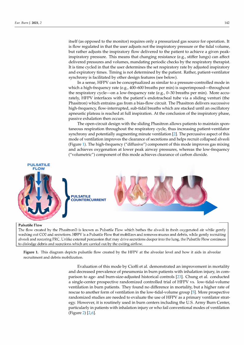
The open-circuit design with the sliding Phasitron allows patients to maintain spon-taneous respiration throughout the respiratory cycle, thus increasing patient-ventilatorsynchrony and potentially augmenting minute ventilation [2]. The percussive aspect of thismode of ventilation improves the clearance of secretions and helps recruit collapsed alveoli(Figure 1). The high-frequency (“diffusive”) component of this mode improves gas mixingand achieves oxygenation at lower peak airway pressures, whereas the low-frequency(“volumetric”) component of this mode achieves clearance of carbon dioxide.
Eur. Burn J. 2021, 2, FOR PEER REVIEW 3
5. Alternative Modes of Ventilation 5.1. High-Frequency Percussive Ventilation
The Volumetric Diffusive Respirator (VDR-4; Percussionaire, Corp., Sandpoint, ID) is a pneumatically driven, flow-regulated, and time-cycled ventilator which provides high-frequency percussive ventilation (HFPV). It is pneumatically driven in that the ven-tilator itself (as opposed to the monitor) requires only a pressurized gas source for opera-tion. It is flow regulated in that the user adjusts not the inspiratory pressure or the tidal volume, but rather adjusts the inspiratory flow delivered to the patient to achieve a given peak-inspiratory pressure. This means that changing resistance (e.g., stiffer lungs) can af-fect delivered pressures and volumes, mandating periodic checks by the respiratory ther-apist. It is time cycled in that the user determines the set respiratory rate by adjusted in-spiratory and expiratory times. Timing is not determined by the patient. Rather, patient-ventilator synchrony is facilitated by other design features (see below).
In a sense, HFPV can be conceptualized as similar to a pressure-controlled mode in which a high-frequency rate (e.g., 400–600 breaths per min) is superimposed—throughout the respiratory cycle—on a low-frequency rate (e.g., 0–30 breaths per min). More accu-rately, HFPV interfaces with the patient’s endotracheal tube via a sliding venturi (the Phasitron) which entrains gas from a bias-flow circuit. The Phasitron delivers successive high-frequency, flow-interrupted, sub-tidal breaths which are stacked until an oscillatory apneustic plateau is reached at full inspiration. At the conclusion of the inspiratory phase, passive exhalation then occurs.
The open-circuit design with the sliding Phasitron allows patients to maintain spon-taneous respiration throughout the respiratory cycle, thus increasing patient-ventilator synchrony and potentially augmenting minute ventilation [2]. The percussive aspect of this mode of ventilation improves the clearance of secretions and helps recruit collapsed alveoli (Figure 1). The high-frequency (“diffusive”) component of this mode improves gas mixing and achieves oxygenation at lower peak airway pressures, whereas the low-fre-quency (“volumetric”) component of this mode achieves clearance of carbon dioxide.
Figure 1. This diagram depicts pulsatile flow created by the HFPV at the alveolar level and how it aids in alveolar recruit-ment and debris mobilization.
Figure 1. This diagram depicts pulsatile flow created by the HFPV at the alveolar level and how it aids in alveolarrecruitment and debris mobilization.
Evaluation of this mode by Cioffi et al. demonstrated an improvement in mortalityand decreased prevalence of pneumonia in burn patients with inhalation injury, in com-parison to age- and burn-size-adjusted historical controls [23]. Chung et al. conducteda single-center prospective randomized controlled trial of HFPV vs. low-tidal-volumeventilation in burn patients. They found no difference in mortality, but a higher rate ofrescue to another form of ventilation in the low-tidal-volume group [5]. More prospectiverandomized studies are needed to evaluate the use of HFPV as a primary ventilator strat-egy. However, it is routinely used in burn centers including the U.S. Army Burn Center,particularly in patients with inhalation injury or who fail conventional modes of ventilation(Figure 2) [2,6].

Eur. Burn J. 2021, 2 143Eur. Burn J. 2021, 2, FOR PEER REVIEW 5
Figure 2. The U.S. Army Burn Center’s algorithm of initiating, managing and weaning HFPV. Figure 2. The U.S. Army Burn Center’s algorithm of initiating, managing and weaning HFPV.

Eur. Burn J. 2021, 2 144
High-frequency oscillatory ventilation (HFOV) is a mode of MV which shares somefeatures with HFPV [24]. Although HFOV is no longer recommended for the treatment ofARDS in adults [25,26] or children [27], it has been used for children with burns and/orinhalation injuries [28–30]. Most literature regarding the use of HFOV in this populationhas been limited to small, single-center, retrospective studies [28]. In those studies, HFOVwas associated with reduced prevalence of pneumonia, lower peak-inspiratory pressures,improved oxygenation, and decreased work of breathing when applied to pediatric burnpatients early in their disease course [28–30].
Most of these centers state that HFOV should be considered in pediatric patients withgreater than 40% TBSA in the first 36 h postburn [28,31]. As with HFPV, future prospectiveevaluation would be needed to more clearly define HFOV utility [31].
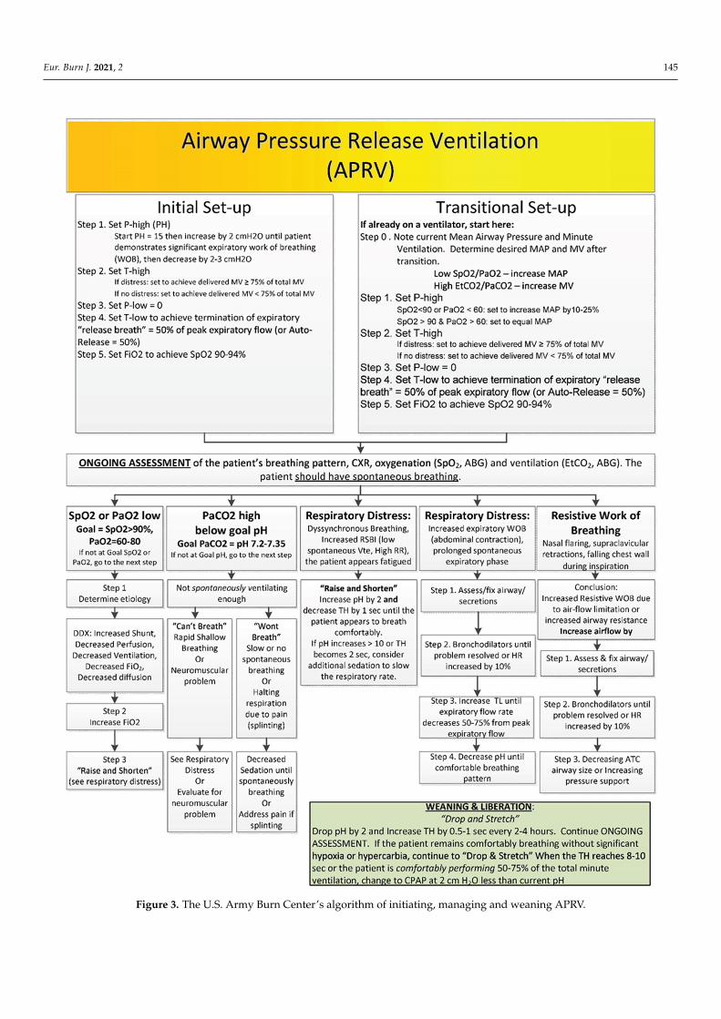
5.2. Airway-Pressure-Release Ventilation
Airway-pressure-release ventilation (APRV) is an option for burn patients, particularlyfor those with ARDS or who fail conventional modes. Stock et al. initially described APRVin an animal model with induced acute lung injury (ALI) as a continuous positive-airway-pressure (CPAP) mode with periodic releases in the pressure to enhance ventilation [32].Habashi et al. described “personalized” APRV by prolonging Thigh, with a brief Tlowtargeting expiratory flow [33].
Specific evidence supporting the use of APRV in the burn population is lacking. Inter-estingly, in a porcine model of severe smoke inhalation injury, Batchinsky and colleaguesfound that APRV-treated animals developed ARDS faster than those treated with conven-tional volume-controlled ventilation [7]. Proponents argue that APRV improves alveolarrecruitment and stability, improves ventilation-perfusion matching, increases mean airwaypressure, minimizes peak and plateau pressures, preserves spontaneous breathing, andreduces sedation requirements [34–36]. Reduced sedation preserves airway reflexes andcough, and has the theoretical benefits in burn patients of reducing ventilator-associatedpneumonia and increasing rehabilitation performance. Further study of APRV, with clearlydefined standards and protocols is needed to establish a clear benefit over conventionalstrategies. We routinely use APRV at the US Army Burn Center (Figure 3).
Eur. Burn J. 2021, 2 145Eur. Burn J. 2021, 2, FOR PEER REVIEW 6
Figure 3. The U.S. Army Burn Center’s algorithm of initiating, managing and weaning APRV.
Figure 3. The U.S. Army Burn Center’s algorithm of initiating, managing and weaning APRV.

Eur. Burn J. 2021, 2 146
6. Spontaneous Awakening and Breathing Trials
Unless otherwise contraindicated, mechanically ventilated burn patients should un-dergo daily sedation interruptions and spontaneous breathing trials [13]. A spontaneousawakening trial (SAT) is a period during which sedating medications are held in order todetermine the need for ongoing sedation [37]. A spontaneous breathing trial (SBT) is aperiod during which MV is decreased to a minimal level of support to determine whetherthe patient requires ongoing MV [37]. In the landmark ABC Trial, a daily paired SAT andSBT protocol for mechanically ventilated ICU patients improved ventilator-free days, ICUlength of stay, and 1-year mortality [38]. Similar results were seen in a retrospective reviewat one burn ICU after the implementation of a combined SAT and SBT protocol [39].
At the U.S. Army Burn Center, all mechanically ventilated patients undergo a dailysafety screen followed by an SAT/SBT performed by the bedside nurse and respiratorytherapist (RT) (Table 1). Safety screening criteria are consistent with those used in the ABCTrial [38] and result in cancellation of the protocol if any of the failure criteria are met. Thenurse then begins the SAT by withholding all sedative and analgesic medications, exceptthose analgesics required for active pain control. If the patient is still anxious, agitated, orin pain, infusions can be titrated back on to a Richmond Agitation Sedation Score (RASS)score of −1 or 0. For 30 to 60 min, the patient is observed for any failure criteria. Patientsthat fail the SAT are placed back on sedatives at half the previous doses then titrated toeffect.
Table 1. U.S. Army Burn Center Spontaneous Awakening and Breathing Trial Protocol †.
Spontaneous Awakening Trial (SAT) Safety Screen [38,40]Actively receiving neuromuscular blockers (paralytics)?Increased ICP at time of SAT?Any medical intervention to reduce ICP in previous 24 h (ex: mannitol, hypertonic saline bolus,barbiturates)?Presence of myocardial ischemia in previous 24 h?Actively on Hypothermia Protocol?Pre-existing ventilatory dependence prior to admission?Planned transport out of ICU in subsequent 4 h?Receiving sedative infusions for active seizures or alcohol withdrawal?Actively requiring vasopressors?Mechanically restrained?
Spontaneous Breathing Trial (SBT) Parameters [38,40] ‡
Sustained anxiety, agitation, or pain?RR > 35 for >5 min BPM?SpO2 < 88% for >5 min?SBP > 200 or <90 mmHg; DBP > 100 or <30 mmHg; MAP > 140 or <60 mmHg?ICP > 20 mmHg?Acute dysrhythmia?Existing safety concerns by any member of healthcare team?Presence of two or more signs of respiratory distress? ¶
Actively requiring vasopressors?Mechanically restrained?Rapid shallow breathing index (RSBI) ≤ 105?Negative inspiratory force (NIF) < −30 cm H2O?Patient intrinsic tidal volume < 4 mL/kg ideal body weight?Presence of acute or abrupt change in mental status from baseline?Presence of secretions requiring > hourly suctioning or copious amount of thick airway casts?Cough and gag reflex present?≥25% endotracheal cuff leak?
† Presence of any failing criteria results in trial termination or consultation with provider before proceedingwith SAT [38,40]. ‡ Performed on pressure-support ventilation with PS of 5 cmH2O and PEEP 5 cmH2O 40. Ifall parameters are sufficient, proceed with extubation. If 1 or more parameters are insufficient, consultationwith provider is required for decision to extubate. ¶ Tachycardia (HR > 130 bpm); bradycardia (HR < 50 bpm);accessory respiratory muscle use; paradoxical abdominal breathing; diaphoresis; marked dyspnea. Abbreviations:ICP—intracranial pressure; ICU—intensive care unit; RR—respiratory rate; BPM—breaths per minute; SpO2—percentage of oxygen saturation; SBP—systolic blood pressure; DBP—diastolic blood pressure; MAP—meanarterial pressure; mmHg—millimeters of mercury; PS—pressure support; cmH2O—centimeters of water; PEEP—positive end expiratory pressure; HR—heart rate; bpm—beats per minute.
Eur. Burn J. 2021, 2 147
Patients that pass the initial safety screening and SAT undergo an SBT by the RT for30–60 min. SBT ventilator settings are typically a pressure-support mode (PS) of 5 cmH2O and PEEP of 5 cm H2O. Varying pressures of PEEP and PS can be used; however,the American College of Chest Physicians/American Thoracic Society Clinical PracticeGuidelines do not recommend SBT via T-piece or CPAP [40]. In addition to those usedin the ABC Trial [38], our SBT failure criteria also address circumstances unique to burnpatients with upper respiratory tract edema or inhalation injury: cough strength, secretionproduction, and cuff leak (Table 1). In a single-center prospective study, burn patients withcough peak flow of less than 60 L/min or requiring hourly suctioning were 8–9 times morelikely to fail extubation [41].
Burn patients who fail extubation have significantly longer duration of MV and lengthof stay [42]. Since both delayed and premature removal from MV are associated withincreased mortality [43], the use of SAT and SBT protocols to determine the optimal time ofextubation is beneficial and should be done daily.
7. High-Risk Extubations
Extubating patients is not without risk. To succeed, a patient should meet the fol-lowing prerequisites: ability to protect their airway; appropriate mental status; adequateoxygenation and ventilation; Glasgow Coma Scale (GCS) score ≥ 8; closed abdomen; andcardiovascular stability [44,45]. Hernandez et al. divided critically ill patients into lowversus high risk for extubation, based on several demographic and clinical criteria [46].Patients with a large burden of injury, large areas of deep partial or full-thickness burns,inhalation injury, and/or who have undergone a large-volume resuscitation are generallyconsidered high risk for extubation (Table 2). When extubating patients who are high risk,airway carts, or reintubation supplies should be readily available.
Table 2. Characteristics of Burn Patients placing them at High Risk for Reintubation.
Age > 65 Years [47]
Heart Failure as primary indication for mechanical ventilation [47]Moderate-to-Severe COPD [47]
APACHE II Score > 12 on day of extubation [47]BMI > 30 kg/m [41,47]
Difficult or prolonged weaning [47]Mechanical ventilation > 7 days [41,47]Burn Shock with ongoing resuscitation
>40% TBSA>20% TBSA mostly deep partial/full-thickness burns
Inhalational Injury [47]Increased Volume Status (Na < 140)
Cuff leak < 25%Requiring suctioning equal or more frequently than hourly
8. Noninvasive Modes of Mechanical Ventilation8.1. Continuous Positive Airway Pressure (CPAP)
Noninvasive CPAP is the administration of positive airway pressure during bothinspiration and expiration in spontaneously breathing patients. The purpose of positive-airway-pressure therapy is to improve functional residual capacity (FRC), increase lungcompliance, and correct VQ mismatch in hypoxemic patients [48]. CPAP results in therecruitment and expansion of partially collapsed alveoli. The subsequent decrease inpulmonary shunt improves oxygenation, reduces work of breathing, and reduces left-ventricular afterload [49]. Additionally, CPAP does not require an artificial airway. Candi-dates for CPAP should be awake, spontaneously breathing, able to protect their airway,and hemodynamically stable. CPAP should be limited to less than 15 cmH2O to mitigatethe risk of gastric distention and aspiration. Excessive CPAP at pressures of greater than

Eur. Burn J. 2021, 2 148
20 cmH2O can result in pulmonary barotrauma from alveolar overdistention, increaseddead-space ventilation, and decreased lung compliance [50].
8.2. Bilevel Positive Airway Pressure (BiPAP)
BiPAP is a pressure-support mode of ventilation with PEEP in spontaneously breathingpatients without an artificial airway. Patient-triggered inspiratory effort results in a setamount of pressure supplied by the ventilator (inspiratory positive airway pressure orIPAP) followed by passive expiration with PEEP (expiratory positive airway pressure orEPAP), providing a baseline of positive pressure at all times. The difference between IPAPand EPAP is the driving pressure, or amount of total pressure support. In this mode,patients control their respiratory rate in addition to inspiratory and expiratory times.
The additional pressure support during inspiration augments TV, increases FRC, anddecreases the WOB [49]. Although CPAP improves oxygenation, the pressure support inBiPAP improves both oxygenation and ventilation, which makes BiPAP especially helpfulin hypercarbic patients [49]. Similar to CPAP, patients on BiPAP must be awake andbreathing spontaneously with intact airway reflexes. Adverse effects of BiPAP secondaryto excessive pressure support include auto-PEEP, gastric distention with the potential foraspiration, pneumothorax, and delayed cycling.
There are no universally applicable recommendations for initiating BiPAP, but individ-ual titration should be performed to improve oxygenation and decrease work of breathing.To achieve the best results when transitioning from invasive ventilation to BiPAP, attemptsshould be made to match the patient’s required driving pressure and wean from there [51].Meta-analyses reviewing noninvasive positive-pressure ventilation after extubation foundreductions in mortality, length of stay, and rates of ventilator-associated pneumonia inpost-operative patients with COPD [52].
There is currently a lack of research examining the impact of extubation to nonin-vasive positive-pressure modalities in the burn population. Anatomic barriers of burnpatients such as facial burns, facial trauma, and facial dressings may preclude the use ofCPAP or BiPAP, due to an inability to form an adequate mask seal. Prophylactic use ofCPAP or BiPAP in appropriate patients may reduce respiratory insufficiency during burnresuscitation. On the other hand, fluid resuscitation in the initial period after a burn mayresult in precipitous development of airway edema. Much work remains to determine theoptimal timing and application of noninvasive positive-pressure ventilation in the burnpopulation.
8.3. High-Flow Nasal Cannula
High-flow nasal cannula (HFNC) has become a staple of oxygen delivery in ICUs.HFNC produces flow rates from 8 L/min to 60 L/min with FiO2 of 0.21 to 1.0 FiO2 isadjusted as gas passes through an oxygen blender where it is heated and saturated withwater via an in-line humidifier. Both flow and FiO2 are allowed to be adjusted indepen-dently of each other [53]. The humidified air is provided to the patient through soft nasalprongs providing a more comfortable experience [54]. HFNC continues to be associatedwith increased comfort, lower dyspnea scores and reduced mouth dryness [55]. Otherbenefits of HFNC include decreased secretions and enhanced secretion mobilization [56,57],reduced work of breathing [53,58], improved ventilation mechanics due to washout ofupper airway dead space [59], and PEEP.
Recently, several large multicenter clinical trials evaluated the use of HFNC af-ter extubation. Five hundred and 27 critically ill patients who were deemed low risk(age < 65 years; APACHE II < 12 on day of extubation, BMI < 30, absence of heart failure ormoderate-to-severe COPD) were randomized to either high-flow or conventional oxygentherapy for 24 h after extubation. The use of HFNC oxygen compared with conventionaloxygen therapy reduced the risk of reintubation within 72 h [46]. A follow-on study eval-uated whether HFNC was noninferior to noninvasive MV in patients who were deemedhigh risk for extubation. Although reintubation rates were the same, the HFNC group had
Eur. Burn J. 2021, 2 149
significantly less postextubation respiratory failure (27% vs. 40%) [47]. These 2 studiesremain the largest and most compelling arguments for the use of HFNC upon extubationof critically ill patients.
No studies evaluating the use of HFNC in the burn population currently exist.Most critically ill burn patients fit into the high-risk category described by Hernandez(Table 2) [46,47]. Currently at the U.S. Army Burn Center, all burn patients who have beenintubated for longer than 12 h are extubated to HFNC for 24 h unless another form of NIVis deemed more clinically appropriate.
9. Conclusions
In critically ill burn patients, lung-protective ventilation can be challenging but islikely beneficial and should be implemented if possible. No single ventilator strategy isconsidered standard of care, as the complicated aspects of burn physiology have dictated amore individualized approach [60]. Early implementation of alternative modes such asHFPV or APRV is considered accepted practice in most burn centers [60]. Further studieswill be helpful to confirm clinical benefit. Upon extubation, many burn patients are athigh risk for reintubation or postextubation respiratory failure, and will likely benefit fromextubation to noninvasive modes such as HFNC.
Author Contributions: Conceptulazation: J.S.F., A.P.B., G.W.B., Methedology: A.P.B., G.W.B., L.C.C.,Investigation: J.S.F., A.P.B., T.A.M., G.W.B., M.R.R., R.C., D.R.L., L.H., J.J.N., J.C.P., L.C.C., Writing—Original Draft Preparation: J.S.F., A.P.B., T.A.M., G.W.B., M.R.R., R.C., D.R.L., L.H., J.B.L., J.J.N., J.C.P.,L.C.C., Writing—Review and Editing: J.S.F., A.P.B., T.A.M., G.W.B., A.M.W., D.S.L., J.B.L., L.C.C.,Visualization: A.P.B., L.C.C., Supervision: A.P.B., L.C.C., Project Administration: A.P.B., L.C.C. Allauthors have read and agreed to the published version of the manuscript.
Funding: No external funding was received.
Conflicts of Interest: Leopoldo C. Cancio has received speaker fees from Percussionaire, Inc.
Disclaimer: The opinions or assertions contained herein are the private views of the authors, andare not to be construed as official or as reflecting the views of the Department of the Army or theDepartment of Defense.
References1. Belenkiy, S.M.; Buel, A.R.; Cannon, J.; Sine, C.R.; Aden, J.K.; Henderson, J.L.; Liu, N.T.; Lundy, J.B.; Renz, E.M.; Batchinsky, A.I.;
et al. Acute respiratory distress syndrome in wartime military burns: Application of the Berlin criteria. J. Trauma Acute Care Surg.2014, 76, 821–827. [CrossRef] [PubMed]
2. Miller, A.C.; Ferrada, P.A.; Kadri, S.S.; Nataraj-Bhandari, K.; Vahedian-Azimi, A.; Quraishi, S.A. High-frequency ventilationmodalities as salvage therapy for smoke inhalation–Associated acute lung injury: A systematic review. J. Intensive Care Med. 2018,33, 335–345. [CrossRef] [PubMed]
3. Walker, P.F.; Buehner, M.F.; Wood, L.A.; Boyer, N.L.; Driscoll, I.R.; Lundy, J.B.; Cancio, L.C.; Chung, K.K. Diagnosis andmanagement of inhalation injury: An updated review. Crit. Care 2015, 19, 1–12. [CrossRef] [PubMed]
4. Chao, K.-Y.; Lin, Y.-W.; Chiang, C.-E.; Tseng, C.-W. Respiratory management in smoke inhalation injury. J. Burn Care Res. 2019, 40,507–512. [CrossRef]
5. Chung, K.K.; Wolf, S.; Renz, E.M.; Allan, P.F.; Aden, J.K.; Merrill, G.A.; Shelhamer, M.C.; King, B.T.; White, C.E.; Bell, D.G.; et al.High-frequency percussive ventilation and low tidal volume ventilation in burns: A randomized controlled trial. Crit. Care Med.2010, 38, 1970–1977. [CrossRef] [PubMed]
6. Cancio, L.C. Airway management and smoke inhalation injury in the burn patient. Clin. Plast. Surg. 2009, 36, 555–567. [CrossRef]7. Batchinsky, A.I.; Burkett, S.E.; Zanders, T.B.; Chung, K.K.; Regn, D.D.; Jordan, B.S.; Necsoiu, C.; Nguyen, R.; Hanson, M.A.; Morris,
M.J.; et al. Comparison of airway pressure release ventilation to conventional mechanical ventilation in the early management ofsmoke inhalation injury in swine. Crit. Care Med. 2011, 39, 2314–2321. [CrossRef] [PubMed]
8. Dancey, D.R.; Hayes, J.; Gomez, M.; Schouten, D.; Fish, J.; Peters, W.; Slutsky, A.S.; Stewart, T.E. ARDS in patients with thermalinjury. Intensive Care Med. 1999, 25, 1231–1236. [CrossRef]
9. Sine, C.R.; Belenkiy, S.M.; Henderson, J.L.; Aden, J.K.; Batchinsky, A.; Cancio, L.C.; Chung, K.K.; Buel, A.R.; Waters, J.A.; Lundy,J.B.; et al. Acute respiratory distress syndrome in burn patients: A comparison of the Berlin and American-European definitions.J. Burn Care Res. 2016, 37, e461–e469. [CrossRef]
10. Pruitt, B.A.; Flemma, R.J.; DiVincenti, F.C.; Foley, F.D.; Mason, A.D.; Young, W.G., Jr. Pulmonary complications in burn patients.A comparative study of 697 patients. J. Thorac. Cardiovasc. Surg. 1970, 59, 7–20. [CrossRef]

Eur. Burn J. 2021, 2 150
11. Tranbaugh, R.F.; Elings, V.B.; Christensen, J.M.; Lewis, F.R. Effect of inhalation injury on lung water accumulation. J. Trauma Inj.Infect. Crit. Care 1983, 23, 597–604. [CrossRef] [PubMed]
12. Driscoll, I.M.-S.E.; Pamplin, J.; Cancio, L. Burn Care (CPG ID: 12); Joint Trauma System, United States of America Department ofDefense: 2016. Available online: https://jts.amedd.army.mil/assets/docs/cpgs/Burn_Care_11_May_2016_ID12.pdf (accessed on24 August 2021).
13. Herndon, D.N. Total Burn Care; Elsevier: Amsterdam, The Netherlands, 2018.14. Demling, R.H. The burn edema process: Current concepts. J. Burn. Care Rehabil. 2005, 26, 207–227.15. Acute Respiratory Distress Syndrome, N.; Brower, R.G.; Matthay, M.A.; Morris, A.; Schoenfeld, D.; Thompson, B.T.; Wheeler, A.
Ventilation with lower tidal volumes as compared with traditional tidal volumes for acute lung injury and the acute respiratorydistress syndrome. N. Engl. J. Med. 2000, 342, 1301–1308. [CrossRef]
16. Amato, M.B.P.; Barbas, C.; Medeiros, D.M.; Magaldi, R.B.; Schettino, G.P.; Lorenzi-Filho, G.; Kairalla, R.; Deheinzelin, D.; Munoz,C.; Oliveira, R.; et al. Effect of a protective-ventilation strategy on mortality in the acute respiratory distress syndrome. N. Engl. J.Med. 1998, 338, 347–354. [CrossRef] [PubMed]
17. Brower, R.G.; Lanken, P.N.; MacIntyre, N.; Matthay, M.A.; Morris, A.; Ancukiewicz, M.; Schoenfeld, D.; Thompson, B.T. Higherversus Lower positive end-expiratory pressures in patients with the acute respiratory distress syndrome. N. Engl. J. Med. 2004,351, 327–336. [CrossRef] [PubMed]
18. Marini, J.J.; Jaber, S. Dynamic predictors of VILI risk: Beyond the driving pressure. Intensive Care Med. 2016, 42, 1597–1600.[CrossRef]
19. Albright, J.M.; Davis, C.S.; Bird, M.D.; Ramirez, L.; Kim, H.; Burnham, E.L.; Gamelli, R.L.; Kovacs, E.J. The acute pulmonaryinflammatory response to the graded severity of smoke inhalation injury. Crit. Care Med. 2012, 40, 1113–1121. [CrossRef]
20. Head, J.M. Inhalation injury in burns. Am. J. Surg. 1980, 139, 508–512. [CrossRef]21. Glas, G.J.; Horn, J.; Van Der Hoeven, S.M.; Hollmann, M.W.; Cleffken, B.; Colpaert, K.; Juffermans, N.P.; Knape, P.; Loef, B.G.;
Mackie, D.P.; et al. Changes in ventilator settings and ventilation–induced lung injury in burn patients—A systematic review.Burns 2019, 46, 762–770. [CrossRef]
22. Palazzo, S.; James-Veldsman, E.; Wall, C.; Hayes, M.; Vizcaychipi, M. Ventilation strategies in burn intensive care: A retrospectiveobservational study. Burn. Trauma 2014, 2, 29–35. [CrossRef]
23. Cioffi, W.G., Jr.; Rue, L.W. Diagnosis and treatment of inhalation injuries. Crit. Care Nurs. Clin. N. Am. 1991, 3, 191–198. [CrossRef]24. Pillow, J. High-frequency oscillatory ventilation: Mechanisms of gas exchange and lung mechanics. Crit. Care Med. 2005, 33,
S135–S141. [CrossRef]25. Fan, E.; Del Sorbo, L.; Goligher, E.C.; Hodgson, C.L.; Munshi, L.; Walkey, A.J.; Adhikari, N.K.; Amato, M.B.; Branson, R.; Brower,
R.G.; et al. An Official American Thoracic Society/European Society of Intensive Care Medicine/Society of Critical Care MedicineClinical Practice Guideline: Mechanical ventilation in adult patients with acute respiratory distress syndrome. Am. J. Respir. Crit.Care Med. 2017, 195, 1253–1263. [CrossRef]
26. Narendra, D.K.; Hess, D.R.; Sessler, C.N.; Belete, H.M.; Guntupalli, K.K.; Khusid, F.; Carpati, C.M.; Astiz, M.E.; Raoof, S. Updatein management of severe hypoxemic respiratory failure. Chest 2017, 152, 867–879. [CrossRef]
27. Gupta, P.; Green, J.W.; Tang, X.; Gall, C.M.; Gossett, J.M.; Rice, T.B.; Kacmarek, R.M.; Wetzel, R.C. Comparison of high-frequencyoscillatory ventilation and conventional mechanical ventilation in pediatric respiratory failure. JAMA Pediatr. 2014, 168, 243–249.[CrossRef] [PubMed]
28. Rowan, C.M.; Cristea, O.; Greathouse, S.T.; Coleman, J.J.; Nitu, M.E. Preemptive use of high-frequency oscillatory ventilation inpediatric burn patients. J. Burn Care Res. 2013, 34, 237–242. [CrossRef] [PubMed]
29. Greathouse, S.T.; Hadad, I.; Zieger, M.; Nitu, M.; Rowan, C.M.; Coleman, J.J. High-frequency oscillatory ventilators in burnpatients: Experience of riley hospital for children. J. Burn Care Res. 2012, 33, 425–435. [CrossRef]
30. Jackson, M.P.; Philp, B.; Murdoch, L.J.; Powell, B.W.E.M. High frequency oscillatory ventilation successfully used to treat a severepaediatric inhalation injury. Burns 2002, 28, 509–511. [CrossRef]
31. Sen, S. Pediatric inhalation injury. Burn. Trauma 2017, 5, 31. [CrossRef]32. Stock, M.C.; Downs, J.B.; Frolicher, D.A. Airway pressure release ventilation. Crit. Care Med. 1987, 15, 462–466. [CrossRef]33. Habashi, N.M. Other approaches to open-lung ventilation: Airway pressure release ventilation. Crit. Care Med. 2005, 33,
S228–S240. [CrossRef]34. Fredericks, A.S.; Bunker, M.P.; Gliga, L.A.; Ebeling, C.G.; Ringqvist, J.R.; Heravi, H.; Manley, J.; Valladares, J.; Romito, B.T. Airway
pressure release ventilation: A review of the evidence, theoretical benefits, and alternative titration strategies. Clin. Med. InsightsCirc. Respir. Pulm. Med. 2020, 14, 1179548420903297. [CrossRef]
35. Jain, S.V.; Kollisch-Singule, M.; Sadowitz, B.; Dombert, L.; Satalin, J.; Andrews, P.; Gatto, L.A.; Nieman, G.F.; Habashi, N.M. The30-year evolution of airway pressure release ventilation (APRV). Intensive Care Med. Exp. 2016, 4, 1–18. [CrossRef]
36. Kollisch-Singule, M.; Andrews, P.; Satalin, J.; Gatto, L.A.; Nieman, G.F.; Habashi, N.M. The time-controlled adaptive ventilationprotocol: Mechanistic approach to reducing ventilator-induced lung injury. Eur. Respir. Rev. 2019, 28, 180126. [CrossRef][PubMed]
37. Bell, L. ICU Liberation: The Power of Pain Control, Minimal Sedation, and Early Mobility; Balas, M., Clemmer, T., Hargett, K., Eds.;Society of Critical Care Medicine: Mount Prospect, IL, USA, 2015.

Eur. Burn J. 2021, 2 151
38. Girard, T.D.; Kress, J.P.; Fuchs, B.D.; Thomason, J.W.; Schweickert, W.D.; Pun, B.T.; Taichman, D.B.; Dunn, J.G.; Pohlman, A.S.;Kinniry, P.A.; et al. Efficacy and safety of a paired sedation and ventilator weaning protocol for mechanically ventilated patientsin intensive care (Awakening and Breathing Controlled trial): A randomised controlled trial. Lancet 2008, 371, 126–134. [CrossRef]
39. Lee, Y.-L.L.; Sims, K.D.; Butts, C.C.; Frotan, M.A.; Kahn, S.; Brevard, S.B.; Simmons, J.D. The combination of SAT and SBTprotocols may help reduce the incidence of ventilator-associated pneumonia in the burn intensive care unit. J. Burn Care Res.2017, 38, e574–e579. [CrossRef]
40. Ouellette, D.R.; Patel, S.; Girard, T.; Morris, P.E.; Schmidt, G.A.; Truwit, J.D.; Alhazzani, W.; Burns, S.M.; Epstein, S.K.; Esteban, A.;et al. Liberation from mechanical ventilation in critically ill adults: An Official American College of Chest Physicians/AmericanThoracic Society Clinical Practice Guideline: Inspiratory Pressure augmentation during spontaneous breathing trials, protocolsminimizing sedation, and noninvasive ventilation immediately after extubation. Chest 2017, 151, 166–180. [CrossRef]
41. Smailes, S.T.; McVicar, A.; Martin, R. Cough strength, secretions and extubation outcome in burn patients who have passed aspontaneous breathing trial. Burns 2013, 39, 236–242. [CrossRef] [PubMed]
42. Smailes, S.T.; Martin, R.V.; McVicar, A.J. The incidence and outcome of extubation failure in burn intensive care patients. J. BurnCare Res. 2009, 30, 386–392. [CrossRef] [PubMed]
43. Frutos-Vivar, F.; Ferguson, N.; Esteban, A.; Epstein, S.K.; Arabi, Y.; Apezteguía, C.; González, M.; Hill, N.S.; Nava, S.; D’Empaire,G.; et al. Risk factors for extubation failure in patients following a successful spontaneous breathing trial. Chest 2006, 130,1664–1671. [CrossRef]
44. Mitchell, V.; Dravid, R.M.; Patel, A.; Swampillai, C.; Higgs, A. Difficult airway society guidelines for the management of trachealextubation. Anaesthesia 2012, 67, 318–340. [CrossRef]
45. Sturgess, D.J.; Greenland, K.B.; Senthuran, S.; Ajvadi, F.A.; Van Zundert, A.; Irwin, M. Tracheal extubation of the adult intensivecare patient with a predicted difficult airway—A narrative review. Anaesthesia 2017, 72, 248–261. [CrossRef] [PubMed]
46. Hernández, G.; Vaquero, C.; González, P.; Subira, C.; Frutos-Vivar, F.; Rialp, G.; Laborda, C.; Colinas, L.; Cuena, R.; Fernandez,R. Effect of postextubation high-flow nasal cannula vs conventional oxygen therapy on reintubation in low-risk patients: Arandomized clinical trial. JAMA 2016, 315, 1354–1361. [CrossRef]
47. Hernández, G.; Vaquero, C.; Colinas, L.; Cuena, R.; González, P.; Canabal, A.; Sanchez, S.; Rodriguez, M.L.; Villasclaras, A.;Fernandez, R. Effect of postextubation high-flow nasal cannula vs noninvasive ventilation on reintubation and postextubationrespiratory failure in high-risk patients: A randomized clinical trial. JAMA 2016, 316, 1565–1574. [CrossRef] [PubMed]
48. Butterworth, J.F.; Wasnick, J.D.; Mackey, D.C. Morgan and Mikhail’s Clinical Anesthesiology, 5th ed.; McGraw-Hill Education:New York, NY, USA, 2013.
49. Jaber, S.; Chanques, G.; Jung, B.; Riou, B. Postoperative noninvasive ventilation. Anesthesiology 2010, 112, 453–461. [CrossRef]50. Endorf, F.W.; Dries, D.J. Noninvasive ventilation in the burned patient. J. Burn Care Res. 2010, 31, 217–228. [CrossRef]51. Amato, M.B.P.; Meade, M.O.; Slutsky, A.S.; Brochard, L.; Costa, E.L.V.; Schoenfeld, D.A.; Stewart, T.E.; Briel, M.; Talmor, D.S.;
Mercat, A.; et al. Driving pressure and survival in the acute respiratory distress syndrome. N. Engl. J. Med. 2015, 372, 747–755.[CrossRef]
52. Glossop, A.J.; Shepherd, N.; Bryden, D.C.; Mills, G. Non-invasive ventilation for weaning, avoiding reintubation after extubationand in the postoperative period: A meta-analysis. Br. J. Anaesth. 2012, 109, 305–314. [CrossRef]
53. Spoletini, G.; Alotaibi, M.; Blasi, F.; Hill, N.S. Heated humidified high-flow nasal oxygen in adults: Mechanisms of action andclinical implications. Chest 2015, 148, 253–261. [CrossRef] [PubMed]
54. Maggiore, S.M.; Idone, F.A.; Vaschetto, R.; Festa, R.; Cataldo, A.; Antonicelli, F.; Montini, L.; De Gaetano, A.; Navalesi, P.;Antonelli, M. Nasal high-flow versus venturi mask oxygen therapy after extubation. Effects on oxygenation, comfort, and clinicaloutcome. Am. J. Respir. Crit. Care Med. 2014, 190, 282–288. [CrossRef] [PubMed]
55. Roca, O.; Riera, J.; Torres, F.; Masclans, J.R. High-flow oxygen therapy in acute respiratory failure. Respir. Care 2010, 55, 408–413.56. Hasani, A.; Chapman, T.; McCool, D.; Smith, R.; Dilworth, J.; Agnew, J. Domiciliary humidification improves lung mucociliary
clearance in patients with bronchiectasis. Chronic Respir. Dis. 2008, 5, 81–86. [CrossRef] [PubMed]57. Williams, R.; Rankin, N.; Smith, T.; Galler, D.; Seakins, P. Relationship between the humidity and temperature of inspired gas and
the function of the airway mucosa. Crit. Care Med. 1996, 24, 1920–1929. [CrossRef]58. Dysart, K.; Miller, T.L.; Wolfson, M.R.; Shaffer, T.H. Research in high flow therapy: Mechanisms of action. Respir. Med. 2009, 103,
1400–1405. [CrossRef]59. Frizzola, M.; Miller, T.L.; Rodriguez, M.E.; Zhu, Y.; Rojas, J.; Hesek, A.; Stump, A.; Shaffer, T.H.; Dysart, K. High-flow nasal
cannula: Impact on oxygenation and ventilation in an acute lung injury model. Pediatr. Pulmonol. 2011, 46, 67–74. [CrossRef][PubMed]
60. Chung, K.K.; Rhie, R.Y.; Lundy, J.B.; Cartotto, R.; Henderson, E.; Pressman, M.A.; Joe, V.C.; Aden, J.K.; Driscoll, I.R.; Faucher, L.D.;et al. A survey of mechanical ventilator practices across burn centers in North America. J. Burn Care Res. 2016, 37, e131–e139.[CrossRef] [PubMed]
Copyright © 2022 FDOKUMEN