Astroglial plasticity and glutamate function in a chronic mouse model of Parkinson's disease
Upload
independentCategory
view
4download
0
ORIGINAL ARTICLE
P2Y receptors on astrocytes and microglia mediate oppositeeffects in astroglial proliferation
Clara Quintas & Sónia Fraga & Jorge Gonçalves &
Glória Queiroz
Received: 18 January 2011 /Accepted: 28 April 2011# Springer Science+Business Media B.V. 2011
Abstract Nucleotides released upon brain injury signal toastrocytes and microglia playing an important role inastrogliosis, but the participation of microglia in thepurinergic modulation of astrogliosis is still unclear. Highlyenriched astroglial cultures and co-cultures of astrocytesand microglia were used to investigate the influence ofmicroglia in the modulation of astroglial proliferation medi-ated by nucleotides. In highly enriched astroglial cultures,adenosine-5’-triphosphate (ATP), adenosine 5’-O-(3-thio)-triphosphate (ATPγS), adenosine 5’-O-(3-thio)-diphosphate(ADPβS; 0.01–1 mM), and adenosine-5’-diphosphate (ADP;0.1–1 mM) increased proliferation up to 382%, an effectabolished in co-cultures containing 8% of microglia. Theloss of ATP proliferative effect in co-cultures is supported byits fast metabolism and reduced ADP accumulation, anagonist of P2Y1,12 receptors that mediate astroglial prolifer-ation. No differences in ADPβS and ATPγS metabolism orP2Y1,12 receptors expression were found in co-cultures thatcould explain the loss of their proliferative effect. However,conditioned medium from microglia cultures or co-culturestreated with ADPβS, when tested in highly enrichedastroglial cultures, also prevented ADPβS proliferativeeffect. None of the uracil nucleotides tested had any effect
in proliferation of highly enriched astroglial cultures, buturidine-5′-triphosphate (UTP; 0.1–1 mM) inhibited prolifer-ation up to 66% in co-cultures, an effect that was dependenton uridine-5’-diphosphate (UDP) accumulation, coincidentwith a co-localization of P2Y6 receptors in microglia anddue to cell apoptosis. The results indicate that microgliacontrol astroglial proliferation by preventing the proliferativeresponse to adenine nucleotides and favouring an inhibitoryeffect of UTP/UDP. Several microglial P2Y receptors maybe involved by inducing the release of messengers thatrestrain astrogliosis, a beneficial effect for neuronal repairmechanisms following brain injury.
Keywords Astroglial proliferation . P2Y receptors .
Nucleotide metabolism . P2Y1,6,12 expression . P2Y1,6,12
cell-type localization . Astrocyte–microglia communication
Introduction
Astrogliosis and microglia activation are common featuresof neurodegenerative diseases and acute pathologicalepisodes of trauma, stroke, seizure or infection [1, 2].ATP and other nucleotides are massively released underthese conditions and by activation of P2Y receptors initiateastrogliosis, a response that is characterised by an increasein glial fibrillary acidic protein (GFAP) expression, cellstellation and astroglial proliferation [3, 4]. P2Y receptorsalso mediate astrocyte migration [5] and modulate therelease of cytokines [6, 7] and prostaglandins [8], support-ing the astroglial reactive phenotype observed duringastrogliosis. In injury models, astroglial proliferation wasfound to be mediated by P2Y1 receptors [9, 10], andrecently, we have shown that P2Y12 receptors may also beinvolved in this response [11]. Additionally, nucleotides
C. Quintas : J. Gonçalves :G. Queiroz (*)Laboratory of Pharmacology, Department of Drug Sciences,REQUIMTE, Faculty of Pharmacy, University of Porto,Rua Aníbal Cunha 164,4050-047, Porto, Portugale-mail: [email protected]
S. FragaLaboratory of Toxicology, Department of Biological Sciences,REQUIMTE, Faculty of Pharmacy, University of Porto,Rua Aníbal Cunha 164,4050-047, Porto, Portugal
Purinergic SignallingDOI 10.1007/s11302-011-9235-x
activate microglia P2 receptors which induce chemotaxis[12, 13], phagocytosis [14], the release of trophic factors[15] and cytokines [16] that may have protective effects incerebral injury [17, 18]. Several cytokines and growthfactors released by microglia, such as interleukin-1β,interleukin-6, interferon-γ, tumour necrosis factor-α andfibroblast growth factor-2 stimulate astrogliosis [19–21]whereas others, such as interleukin-10, attenuate astroglialreactivity through a decrease in microglia activation [22,23]. Furthermore, the in vitro and in vivo demonstration thatmicroglia activation precedes astrogliosis lead to theproposal that these cells are of major relevance in themodulation of this response [24].
Most of the known effects of nucleotides in astrogliosisare based on results obtained from studies in astroglialcultures which, regardless of the protocols used, containedmicroglia in different proportions, but the influence ofmicroglia in the purinergic trophic effects was rarelyaddressed [25]. Microglia, even when present in smallamounts, may regulate astroglial responses and may beresponsible for some of the effects attributed to astrocytes[26].
In this study, we investigated the influence of microgliain the modulation of astroglial proliferation mediated bynucleotides using two types of primary astroglial cultures:highly enriched astroglial cultures and co-cultures ofastrocytes and microglia. In a first approach to understandthe differences observed in the effects of nucleotides inboth types of cultures, several factors that could offer animmediate explanation were investigated: (1) the metabo-lism of nucleotides, (2) the expression and cellularlocalization of the P2Y receptors potentially involved inthe modulation of astroglial proliferation and (3) the releaseof soluble messengers by microglia that could haveinfluenced astroglial proliferation. With this experimentalapproach, we aimed to start disclosing the purinergicmechanisms that influence the astrocyte–microglia commu-nication during astrogliosis, a hallmark of brain injury.
Materials and methods
Drugs and antibodies
The following antibodies and drugs were used: goat anti-mouse IgG conjugated to Alexa Fluor 488 from Invitrogen(Barcelona, Spain); rabbit polyclonal anti-P2Y1 and anti-P2Y6 from Alomone Laboratories (Jerusalem, Israel); mousemonoclonal anti-CD11b, rabbit polyclonal anti-actin andgoat anti-rabbit IgG conjugated to horseradish peroxidasefrom Santa Cruz Biotechnology (Santa Cruz, CA, USA);rabbit polyclonal anti-P2Y12, rabbit and mouse anti-glialfibrillary acidic protein (anti-GFAP), goat anti-rabbit IgG
conjugated to crystalline tetramethylrodamine isothiocyanate(TRITC), adenosine, adenosine-5’-monophosphate (AMP),adenosine-5’-diphosphate tetrasodium (ADP), adenosine 5’-O-(3-thio)-diphosphate tetralithium (ADPβS), adenosine-5’-triphosphate disodium (ATP), adenosine 5’-O-(3-thio)-triphosphate tetralithium (ATPγS), cytosine β-D-arabino-furanoside (Ara-C), 2′-(4-hydroxyphenyl)-5-(4-methyl-1-piperazinyl)-2,5′-bi-1 H-benzimidazole trihydrochloridehydrate (Hoechst 33258), hypoxanthine, inosine, L-leucinemethyl ester hydrochloride (LME), penicillin, streptomy-cin, uracil, uridine, uridine-5’-monophosphate disodium(UMP), uridine-5’-diphosphate sodium (UDP), uridine-5’-triphosphate trisodium (UTP) and uridine 5′-diphospho-glucose disodium (UDP-glucose) and methyl green fromSigma-Aldrich (Sintra, Portugal); methyl-[3H]-thymidine(specific activity 80–86 Ci.mmol-1) and enhanced chemi-luminescence Western blotting system from AmershamBiosciences (Lisbon, Portugal); Sulfo-NHS-SS-Biotin andUltralink Immobilized Neutravidin from Pierce (Rockford,IL, USA). Stock solutions of drugs were prepared withdimethylsulphoxide or distilled water and kept at −20°C.Solutions of drugs were prepared from stock solutionsdiluted in culture medium immediately before use.
Cell cultures
Animal handling and experiments were conducted accord-ing to the guidelines of the Directive 2010/63/EU of theEuropean Parliament and the Council of the EuropeanUnion. Primary cortical astroglial cultures were preparedfrom offspring of Wistar rats (Charles River, Barcelona,Spain) as previously described [27], with minor modifica-tions. Briefly, the brains were placed in ice-cold Dulbecco’sphosphate buffered calcium-free saline solution (DPBS)containing 0.2% glucose. The hemispheres were free ofmeninges and blood vessels, and after washing twice withice-cold DPBS, they were cut into small pieces in culturemedium, i.e., Dulbecco’s modified Eagle medium contain-ing 3.7 g/L NaHCO3, 1.0 g/L D-glucose and stableglutamine, supplemented with 50 U/ml penicillin and50 μg/ml streptomycin. Tissue from two hemispheres wasdissociated by triturating in 10 ml culture medium. The cellsuspension obtained was passed through a 40-μm porenylon mesh and then centrifuged at 200×g for 5 min andthe supernatant discharged. Centrifugation followed by cellsuspension was repeated twice, and the pellet obtained wassuspended in culture medium supplemented with 10% foetalbovine serum (FBS) and seeded at a density of 2×10⁵cells/ml.Cultures were incubated at 37°C in a humidified atmosphereof 95% air, 5% CO2 and the medium was replaced 1 dayafter preparation and subsequently, twice a week.
Highly enriched astroglial cultures were obtained bytreating confluent cultures, after 20 days in vitro (DIV),
Purinergic Signalling
with 8 μM Ara-C for 4 days followed by treatment with50 mM L-LME for 90 min [28]. At DIV28, two types ofcultures were obtained: co-cultures of astrocytes andmicroglia, when no treatment was applied and highlyenriched astroglial cultures, when cultures were treatedwith Ara-C plus LME. In both types of cultures, astrocyteswere the main cell type, but the number of microgliapresent differed between the two types of cultures (seebelow). Cultures were synchronised to a quiescent phase ofthe cell cycle, by shifting serum concentration to 0.1% FBSfor 48 h, being used in experiments at DIV30.
Cultures of microglia were obtained from confluent co-cultures that were shaken overnight at 200 rpm. Super-natants containing detached cells were centrifuged at 290×gfor 10 min. The pellet obtained was suspended in culturemedium containing 10% FBS at a density of 3×10⁴ cells/ml. Cells were seeded in 24-well plates, and the mediumwas changed 1 h later, allowing a selective attachment ofmicroglia [29]. After cell synchronisation for 48 h, micro-glia cultures and co-cultures were treated with solvent orADPβS (0.1 mM) for 8 h. After this period of incubation,the medium was discarded and replaced by fresh medium,which was collected 24 h later to be tested in highlyenriched astroglial cultures. This medium was namedmicroglia conditioned medium (MCM) or co-culturesconditioned medium (CCCM). Conditioned mediumobtained from cells treated with solvent (MCM-S orCCCM-S) or with ADPβS (MCM-ADPβS or CCCM-ADPβS) was tested in proliferation assays of highlyenriched astroglial cultures.
Immunocytochemistry
Cell cultures were fixed with a solution containing 4%formaldehyde and 4% sucrose in phosphate buffered saline(PBS; 100 mMNaH2PO4, 50 mM NaCl, pH adjusted to 7.3)and then treated with PBS containing 0.3% Triton X-100.For double-labelling astrocytes and microglia, cultures wereincubated with the primary antibodies rabbit anti-GFAP(1:600) and mouse anti-CD11b (1:50), overnight at 4°C. ForP2Y receptors localization, cultures were incubated with theprimary antibodies mouse anti-GFAP (1:300) or mouse anti-CD11b (1:50) and rabbit anti-P2Y1 (1:400), anti-P2Y6
(1:200) or anti-P2Y12 (1:400), overnight at 4°C. Visual-isation of GFAP, CD11b and P2Y receptors positive cellswas accomplished upon 1 h incubation, at room temperature,with the secondary antibodies anti-rabbit IgG conjugated tocrystalline TRITC (1:100 and 1:400 for GFAP and P2Yreceptors detection, respectively) and anti-mouse IgG conju-gated to Alexa Fluor 488 (1:400). In negative controls, theprimary antibody was omitted. Cell nuclei were labelled withHoechst 33258 (5 μg/ml) for 30 min at room temperature.To evaluate the percentage of microglia, the two types of
cultures were processed in parallel, and about 200 cells werecounted in each culture. The number of CD11b-positive cellswas expressed as percentage of the total number of cellscounted. Images were captured with a Digital Sight DS-5Mccamera (Nikon, Japan) coupled to an Eclipse E400 fluores-cence microscope (Nikon, Japan).
DNA synthesis
At DIV30, the cultures grown in 24-well plates wereincubated with nucleotides or solvent for 48 h (tested induplicate in each plate), and methyl-[3H]-thymidine wasadded in the last 24 h, at a concentration of 1 μCi/ml. WhenMCMs and CCCMs were tested in highly enrichedastroglial cultures, they were added simultaneously withthe nucleotides. Cells were then rinsed with PBS, fixedwith 10% of trichloroacetic acid (TCA) for 30 min at 4°C,washed with ice-cold 5% TCA and rinsed again with PBS.Protein content and methyl-[3H]-thymidine incorporationwere evaluated after cell lysis with 0.2 M NaOH. The effectof drugs in cell proliferation was determined by methyl-[3H]-thymidine incorporation which was quantified byliquid scintillation spectrometry (Beckman LS 6500, Beck-man Instruments, Fullerton, USA) and normalised by theprotein content determined by the Bradford method.
Metabolism of nucleotides
Cultures were rinsed three times with buffer at 37°C withthe following composition (mM): 135 NaCl, 5 KCl, 0.8MgSO4, 1.8 CaCl2, 10 HEPES, 10 glucose and 1 sodiumpyruvate, with pH adjusted to 7.4 with NaOH (1 M).Nucleotides were added at zero time at concentration of0.1 mM, and samples collected at 0, 1, 3, 8, 24 and 48 hwere immediately stored at −20°C. Nucleotides and theirmetabolites were separated by ion-pair-reverse-phase highperformance liquid chromatography with UV detection(HPLC-UV), as previously described [30]. Standards wereanalysed in the same conditions, and the retention timeidentified was (minutes): uracil (0.95), hypoxanthine (1.19),uridine (1.32), inosine (1.86), UMP (2.15), adenosine(3.93), UDP (4.40), AMP (4.76), UTP (6.40), ADP(6.63), ADPβS (7.70), ATP (7.87) and ATPγS (8.10). Theconcentration of nucleotides and metabolites was calculatedby peak area integration followed by interpolation incalibration curves obtained with standards.
Western blot analysis
Cells were rinsed with ice-cold PBS and total cell proteinextracted in lysis buffer with protease inhibitors (1 mMNa3VO4, 1 mM NaF, 1 mM PMSF, 2 μg/ml aprotinin and2 μg/ml leupeptin). After a brief sonication (10 s), the
Purinergic Signalling
lysate was incubated on ice for 1 h and then centrifuged at20,000×g for 45 min at 4°C. The protein concentrationwas determined in the supernatant, and equal amounts ofprotein (50 μg) were boiled at 95°C for 5 min in 6×sample buffer (0.35 M Tris–HCl at pH 6.8, 10% sodiumdodecyl sulfate (SDS), 30% glycerol, 9.3% dithiothreitoland 0.01% bromphenol blue) and subjected to 12% SDS-PAGE (SDS-polyacrylamide gel electrophoresis). Pro-teins were electrotransferred onto nitrocellulose mem-branes for 2 h at 40 V in a transfer buffer. Membraneswere blocked overnight at 4°C with 5% of non-fat drymilk in PBS and then probed for 2 h at room temperaturewith appropriately diluted primary polyclonal antibodies:rabbit anti-P2Y1, rabbit anti-P2Y6 (both at 1:300) andrabbit anti-P2Y12 (1:400) followed by secondary antibodygoat anti-rabbit IgG conjugated to horseradish peroxidase(1:10,000). Immunoblots were then stripped by incuba-tion in stripping buffer (62.5 mM Tris–HCl, 100 mM 2-mercaptoethanol and 2% SDS, pH adjusted to 6.8) for15 min at 50°C and blocked overnight with 5% of non-fatdry milk in PBS. Subsequently, membranes were re-probed with the primary polyclonal antibody rabbit anti-actin (1:200) for 2 h at room temperature, followed by thesecondary antibody. Immunocomplexes were detected byenhanced chemiluminescence system. Quantification ofP2Y protein levels, obtained in arbitrary density units,was performed by densitometric analysis using Bio-Rad’sQuantity One software (Basic version 4.6.5), and totalP2Y receptors expression was normalised to actin.
Cell surface biotinylation
Cell surface protein biotinylation was performed to deter-mine membrane expression of P2Y1 and P2Y6 receptors.Briefly, cultures were rinsed twice with ice-cold PBS with0.1 mM CaCl2 and 1.0 mM MgCl2 (PBS-Ca-Mg). Theapical surface was then exposed to 1 mg/ml of Sulfo-NHS-SS-biotin in biotinylation buffer (10 mM triethanolamine,2 mM CaCl2, 150 mM NaCl at pH 8.0) for 50 min withhorizontal motion at 4°C. After labelling, the cells wererinsed with quenching solution (PBS-Ca-Mg with 100 mMglycine) and then extracted in lysis buffer with proteaseinhibitors. Precipitation of biotinylated proteins wasattained by adding Neutravidin-agarose beads to approxi-mately 850 μg of total cell protein, with end-over-endrotation overnight at 4°C. Then, beads were centrifugedthree times at 6,000×g for 2 min at 4°C, washed with PBSand bound proteins solubilised with SDS sample buffer(0.0625 M Tris–HCl at pH 6.8, 2% SDS, 10% glycerol,2.5% 2-mercaptoethanol and 0.01% bromphenol blue).Samples were subjected to SDS-PAGE and blotting asdescribed in the previous section (see Western blotanalysis).
Lactate dehydrogenase assays
Necrotic cell death was assessed by measuring the lactatedehydrogenase (LDH) release with an enzymatic assayaccording to the manufacturer’s instructions (SigmaAldrich). The assay was based in the oxidation of lactateto pyruvate by LDH with the formation of NADH, whichreduces tetrazolium to coloured formazan that was mea-sured at a wavelength of 490 nm. Following incubationwith nucleotides or solvent for 48 h, culture supernatantswere collected, and the respective extracts were obtainedupon incubation of astrocytes with a lysis solution for45 min at 37°C. Samples were then centrifuged at 250×gfor 4 min, and LDH activity was determined in thecollected supernatants and respective extracts. The LDHreleased into the culture medium was expressed aspercentage of total LDH.
Terminal transferase-mediated dUTP nick end-labellingassays
Apoptotic cell death was evaluated using the indirect terminaltransferase-mediated dUTP-digoxigenin nick end-labelling(TUNEL) to detect DNA fragmentation using an ApopTagperoxidase detection kit (Millipore, Madrid, Spain). Culturestreated with nucleotides or solvent for 48 h were fixed in 4%paraformaldehyde in PBS pH 7.4, for 10 min at roomtemperature and subsequently post-fixed in pre-cooledethanol/acetic acid (2:1, v/v) for 15 min at −20°C. Theendogenous peroxidase activity was quenched with 3%hydrogen peroxide. Cultures were incubated with equilibra-tion buffer and treated with terminal deoxynucleotidyltrans-ferase plus digoxigenin-dNTPs for 1 h at 37°C. The anti-digoxigenin antibody conjugated to peroxidase was addedfor 30 min at room temperature, and colour was developedwith 3,3′-diaminobenzidine substrate. Counterstaining of thenuclei was accomplished with 0.5% methyl green, and cellswere visualised by bright field microscopy. The cell bodieswere also labelled with Hoechst 33258 (5 μg/ml) to confirmthe results obtained with the TUNEL assay. The number ofTUNEL-positive cells was evaluated by analysing eighthigh-power fields (×400) in each culture and expressed aspercentage of total cell number counted.
Statistical analysis
Data are expressed as means±standard errors of the mean(SEM) from n number of experiments, unless otherwisestated. Statistical analysis was carried out using theunpaired Student’s t test or one-way analysis of variance(ANOVA) followed by Dunnett’s multiple comparison test.The Western blot data analysis was performed by ANOVArepeated measures followed by Bonferroni’s multiple
Purinergic Signalling
comparison test. P values lower than 0.05 were consideredto indicate significant differences.
Results
Characterization of cell cultures
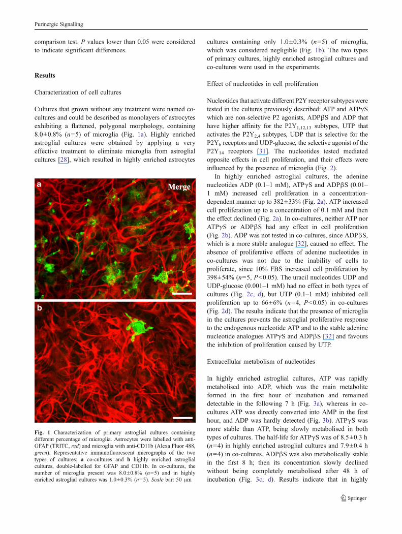
Cultures that grown without any treatment were named co-cultures and could be described as monolayers of astrocytesexhibiting a flattened, polygonal morphology, containing8.0±0.8% (n=5) of microglia (Fig. 1a). Highly enrichedastroglial cultures were obtained by applying a veryeffective treatment to eliminate microglia from astroglialcultures [28], which resulted in highly enriched astrocytes
cultures containing only 1.0±0.3% (n=5) of microglia,which was considered negligible (Fig. 1b). The two typesof primary cultures, highly enriched astroglial cultures andco-cultures were used in the experiments.
Effect of nucleotides in cell proliferation
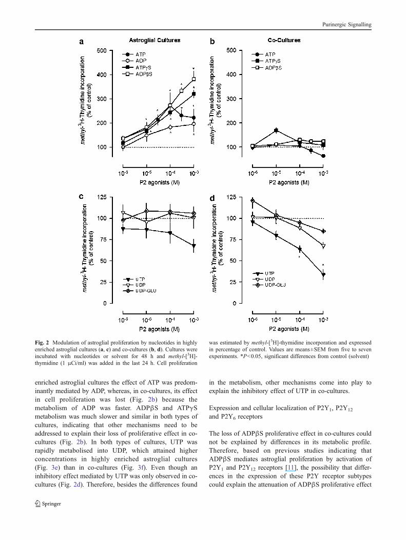
Nucleotides that activate different P2Y receptor subtypes weretested in the cultures previously described: ATP and ATPγSwhich are non-selective P2 agonists, ADPβS and ADP thathave higher affinity for the P2Y1,12,13 subtypes, UTP thatactivates the P2Y2,4 subtypes, UDP that is selective for theP2Y6 receptors and UDP-glucose, the selective agonist of theP2Y14 receptors [31]. The nucleotides tested mediatedopposite effects in cell proliferation, and their effects wereinfluenced by the presence of microglia (Fig. 2).
In highly enriched astroglial cultures, the adeninenucleotides ADP (0.1–1 mM), ATPγS and ADPβS (0.01–1 mM) increased cell proliferation in a concentration-dependent manner up to 382±33% (Fig. 2a). ATP increasedcell proliferation up to a concentration of 0.1 mM and thenthe effect declined (Fig. 2a). In co-cultures, neither ATP norATPγS or ADPβS had any effect in cell proliferation(Fig. 2b). ADP was not tested in co-cultures, since ADPβS,which is a more stable analogue [32], caused no effect. Theabsence of proliferative effects of adenine nucleotides inco-cultures was not due to the inability of cells toproliferate, since 10% FBS increased cell proliferation by398±54% (n=5, P<0.05). The uracil nucleotides UDP andUDP-glucose (0.001–1 mM) had no effect in both types ofcultures (Fig. 2c, d), but UTP (0.1–1 mM) inhibited cellproliferation up to 66±6% (n=4, P<0.05) in co-cultures(Fig. 2d). The results indicate that the presence of microgliain the cultures prevents the astroglial proliferative responseto the endogenous nucleotide ATP and to the stable adeninenucleotide analogues ATPγS and ADPβS [32] and favoursthe inhibition of proliferation caused by UTP.
Extracellular metabolism of nucleotides
In highly enriched astroglial cultures, ATP was rapidlymetabolised into ADP, which was the main metaboliteformed in the first hour of incubation and remaineddetectable in the following 7 h (Fig. 3a), whereas in co-cultures ATP was directly converted into AMP in the firsthour, and ADP was hardly detected (Fig. 3b). ATPγS wasmore stable than ATP, being slowly metabolised in bothtypes of cultures. The half-life for ATPγS was of 8.5±0.3 h(n=4) in highly enriched astroglial cultures and 7.9±0.4 h(n=4) in co-cultures. ADPβS was also metabolically stablein the first 8 h; then its concentration slowly declinedwithout being completely metabolised after 48 h ofincubation (Fig. 3c, d). Results indicate that in highly
Fig. 1 Characterization of primary astroglial cultures containingdifferent percentage of microglia. Astrocytes were labelled with anti-GFAP (TRITC, red) and microglia with anti-CD11b (Alexa Fluor 488,green). Representative immunofluorescent micrographs of the twotypes of cultures: a co-cultures and b highly enriched astroglialcultures, double-labelled for GFAP and CD11b. In co-cultures, thenumber of microglia present was 8.0±0.8% (n=5) and in highlyenriched astroglial cultures was 1.0±0.3% (n=5). Scale bar: 50 μm
Purinergic Signalling
enriched astroglial cultures the effect of ATP was predom-inantly mediated by ADP, whereas, in co-cultures, its effectin cell proliferation was lost (Fig. 2b) because themetabolism of ADP was faster. ADPβS and ATPγSmetabolism was much slower and similar in both types ofcultures, indicating that other mechanisms need to beaddressed to explain their loss of proliferative effect in co-cultures (Fig. 2b). In both types of cultures, UTP wasrapidly metabolised into UDP, which attained higherconcentrations in highly enriched astroglial cultures(Fig. 3e) than in co-cultures (Fig. 3f). Even though aninhibitory effect mediated by UTP was only observed in co-cultures (Fig. 2d). Therefore, besides the differences found
in the metabolism, other mechanisms come into play toexplain the inhibitory effect of UTP in co-cultures.
Expression and cellular localization of P2Y1, P2Y12
and P2Y6 receptors
The loss of ADPβS proliferative effect in co-cultures couldnot be explained by differences in its metabolic profile.Therefore, based on previous studies indicating thatADPβS mediates astroglial proliferation by activation ofP2Y1 and P2Y12 receptors [11], the possibility that differ-ences in the expression of these P2Y receptor subtypescould explain the attenuation of ADPβS proliferative effect
Fig. 2 Modulation of astroglial proliferation by nucleotides in highlyenriched astroglial cultures (a, c) and co-cultures (b, d). Cultures wereincubated with nucleotides or solvent for 48 h and methyl-[3H]-thymidine (1 μCi/ml) was added in the last 24 h. Cell proliferation
was estimated by methyl-[3H]-thymidine incorporation and expressedin percentage of control. Values are means±SEM from five to sevenexperiments. *P<0.05, significant differences from control (solvent)
Purinergic Signalling
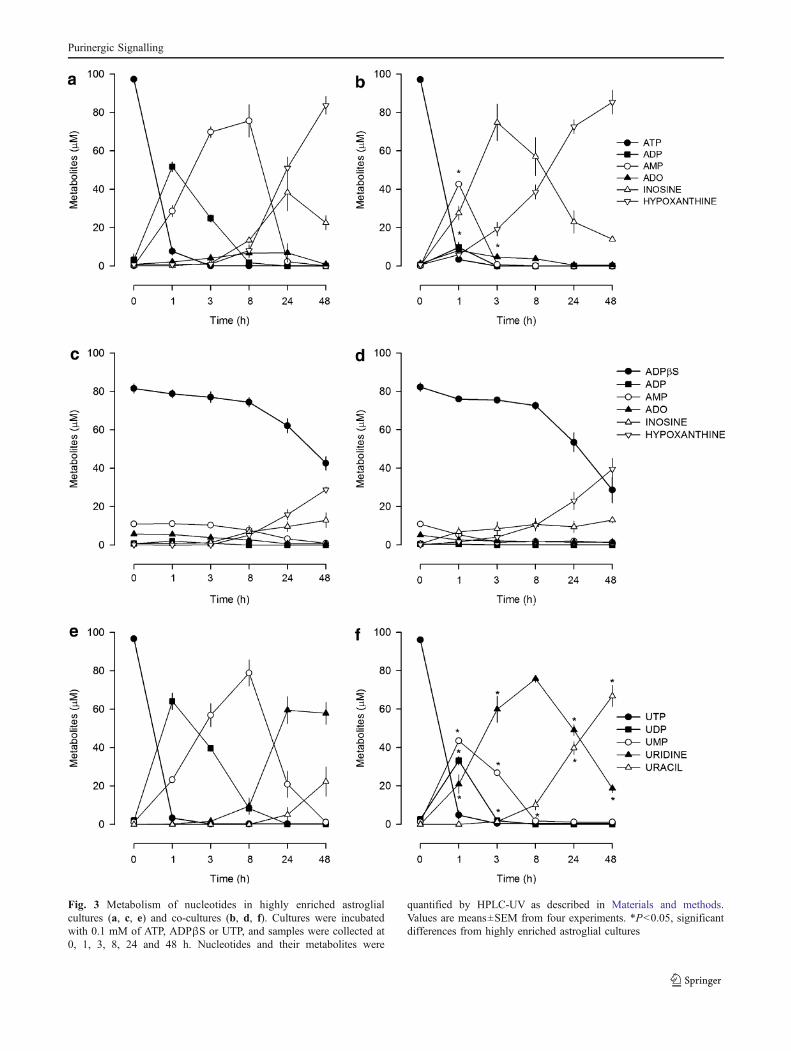
Fig. 3 Metabolism of nucleotides in highly enriched astroglialcultures (a, c, e) and co-cultures (b, d, f). Cultures were incubatedwith 0.1 mM of ATP, ADPβS or UTP, and samples were collected at0, 1, 3, 8, 24 and 48 h. Nucleotides and their metabolites were
quantified by HPLC-UV as described in Materials and methods.Values are means±SEM from four experiments. *P<0.05, significantdifferences from highly enriched astroglial cultures
Purinergic Signalling
observed in co-cultures was investigated. Additionally, therapid conversion of UTP into UDP, which is selective forP2Y6 receptors [33], also raised the question of whetherP2Y6 receptor expression could be increased in cultureswith microglia, thus favouring the inhibitory effect medi-ated by UTP/UDP in co-cultures.
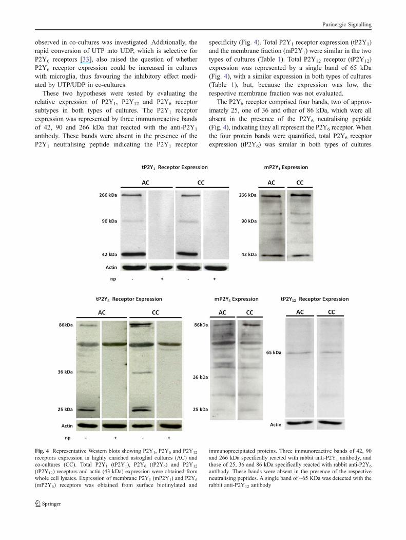
These two hypotheses were tested by evaluating therelative expression of P2Y1, P2Y12 and P2Y6 receptorsubtypes in both types of cultures. The P2Y1 receptorexpression was represented by three immunoreactive bandsof 42, 90 and 266 kDa that reacted with the anti-P2Y1
antibody. These bands were absent in the presence of theP2Y1 neutralising peptide indicating the P2Y1 receptor
specificity (Fig. 4). Total P2Y1 receptor expression (tP2Y1)and the membrane fraction (mP2Y1) were similar in the twotypes of cultures (Table 1). Total P2Y12 receptor (tP2Y12)expression was represented by a single band of 65 kDa(Fig. 4), with a similar expression in both types of cultures(Table 1), but, because the expression was low, therespective membrane fraction was not evaluated.
The P2Y6 receptor comprised four bands, two of approx-imately 25, one of 36 and other of 86 kDa, which were allabsent in the presence of the P2Y6 neutralising peptide(Fig. 4), indicating they all represent the P2Y6 receptor. Whenthe four protein bands were quantified, total P2Y6 receptorexpression (tP2Y6) was similar in both types of cultures
Fig. 4 Representative Western blots showing P2Y1, P2Y6 and P2Y12
receptors expression in highly enriched astroglial cultures (AC) andco-cultures (CC). Total P2Y1 (tP2Y1), P2Y6 (tP2Y6) and P2Y12
(tP2Y12) receptors and actin (43 kDa) expression were obtained fromwhole cell lysates. Expression of membrane P2Y1 (mP2Y1) and P2Y6
(mP2Y6) receptors was obtained from surface biotinylated and
immunoprecipitated proteins. Three immunoreactive bands of 42, 90and 266 kDa specifically reacted with rabbit anti-P2Y1 antibody, andthose of 25, 36 and 86 kDa specifically reacted with rabbit anti-P2Y6
antibody. These bands were absent in the presence of the respectiveneutralising peptides. A single band of ~65 KDa was detected with therabbit anti-P2Y12 antibody
Purinergic Signalling
(Table 1). However, the analysis of individual bands revealedthat expression of the 86 kDa band was higher in co-culturesthan in highly enriched astroglial cultures (Table 1). More-over, when the membrane fraction (mP2Y6) was quantified, ahigher expression was detected in co-cultures, consideringeither the four bands or only that of 86 kDa (Table 1).
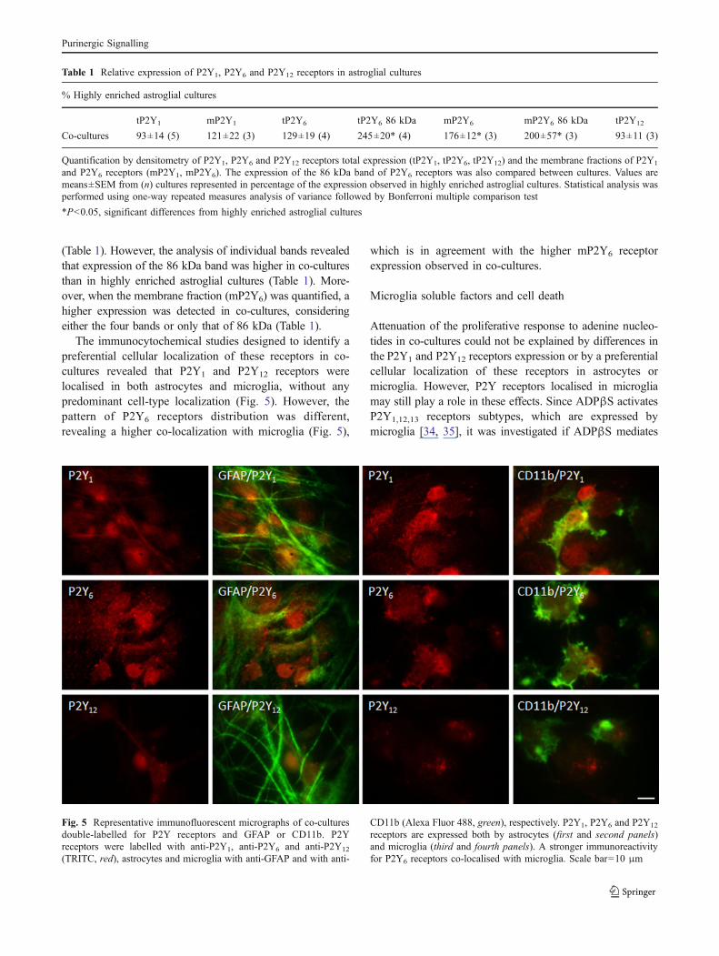
The immunocytochemical studies designed to identify apreferential cellular localization of these receptors in co-cultures revealed that P2Y1 and P2Y12 receptors werelocalised in both astrocytes and microglia, without anypredominant cell-type localization (Fig. 5). However, thepattern of P2Y6 receptors distribution was different,revealing a higher co-localization with microglia (Fig. 5),
which is in agreement with the higher mP2Y6 receptorexpression observed in co-cultures.
Microglia soluble factors and cell death
Attenuation of the proliferative response to adenine nucleo-tides in co-cultures could not be explained by differences inthe P2Y1 and P2Y12 receptors expression or by a preferentialcellular localization of these receptors in astrocytes ormicroglia. However, P2Y receptors localised in microgliamay still play a role in these effects. Since ADPβS activatesP2Y1,12,13 receptors subtypes, which are expressed bymicroglia [34, 35], it was investigated if ADPβS mediates
Table 1 Relative expression of P2Y1, P2Y6 and P2Y12 receptors in astroglial cultures
% Highly enriched astroglial cultures
tP2Y1 mP2Y1 tP2Y6 tP2Y6 86 kDa mP2Y6 mP2Y6 86 kDa tP2Y12
Co-cultures 93±14 (5) 121±22 (3) 129±19 (4) 245±20* (4) 176±12* (3) 200±57* (3) 93±11 (3)
Quantification by densitometry of P2Y1, P2Y6 and P2Y12 receptors total expression (tP2Y1, tP2Y6, tP2Y12) and the membrane fractions of P2Y1
and P2Y6 receptors (mP2Y1, mP2Y6). The expression of the 86 kDa band of P2Y6 receptors was also compared between cultures. Values aremeans±SEM from (n) cultures represented in percentage of the expression observed in highly enriched astroglial cultures. Statistical analysis wasperformed using one-way repeated measures analysis of variance followed by Bonferroni multiple comparison test
*P<0.05, significant differences from highly enriched astroglial cultures
Fig. 5 Representative immunofluorescent micrographs of co-culturesdouble-labelled for P2Y receptors and GFAP or CD11b. P2Yreceptors were labelled with anti-P2Y1, anti-P2Y6 and anti-P2Y12
(TRITC, red), astrocytes and microglia with anti-GFAP and with anti-
CD11b (Alexa Fluor 488, green), respectively. P2Y1, P2Y6 and P2Y12
receptors are expressed both by astrocytes (first and second panels)and microglia (third and fourth panels). A stronger immunoreactivityfor P2Y6 receptors co-localised with microglia. Scale bar=10 μm
Purinergic Signalling
the release of messengers from microglia that might interferewith its proliferative effect. This hypothesis was tested bycomparing the effect of ADPβS (0.1 mM) in highly enrichedastroglial cultures, in the absence and in the presence ofconditioned medium obtained from microglia cultures or co-cultures that were previously treated with solvent (MCM-Sor CCCM-S, respectively) or with ADPβS (MCM-ADPβSor CCCM-ADPβS, respectively).
The proliferative effect of ADPβS (0.1 mM) was notmodified by MCM-S but was abolished in the presenceof MCM-ADPβS (Table 2). Furthermore, in the presenceof CCCM-S the proliferative effect of ADPβS (0.1 mM)was attenuated, an effect even more evident when thecultures were treated with CCCM-ADPβS (Table 2). Innone of the conditions tested was astroglial proliferationnear its maximum, as previously demonstrated theproliferative effect mediated by ADPβS (0.3–1 mM) inhighly enriched astroglial cultures (see Fig. 2a). Theseresults suggest that ADPβS activates microglia P2Y1,12
and/or P2Y13 receptors, which induce the release ofdiffusible messengers that attenuate its proliferative effectin astrocytes. They further suggest that the co-presence ofastrocytes and microglia, and eventually their physicalcontact, facilitates the release of these inhibitory messen-gers since CCCM-S, but not MCM-S, attenuated ADPβSproliferative effect (Table 2). The same experimentalapproach was adopted for UTP, but no effects could bedetected (not shown), possibly because of its highermetabolic instability, but, since UTP caused an inhibitionof proliferation in co-cultures, its contribution to cell deathwas investigated. UTP (1 mM) caused no change in LDHrelease (not shown), but it increased the number of cellspresenting DNA fragmentation, detected by the TUNELassay, from 1.25±0.13% (n=3) in solvent treated culturesto 6.21±0.72% (n=3, P<0.05). Results suggest that, inco-cultures, the presence of microglia may favour UTP/UDP-induced cell apoptosis, contributing to the inhibitionof cell proliferation.
Discussion
Astrogliosis and microglia activation are two closely relatedresponses involved in the brain repairing mechanisms toinjury, and research in the field has been focusing theattention on how astrocytes and microglia influence eachother activity during this process [24]. Astroglial prolifer-ation induced by nucleotides was previously demonstratedboth in vitro [3, 4] and in vivo [9]. However, whereas the invivo studies did not discriminate the influence of microglia,the in vitro studies were performed in astroglial cultures,with the special concern of working without microgliainterference. When the influence of microglia was to beexplored, the mostly used strategy consisted in theevaluation of microglia conditioned medium influence inastroglial cultures [36, 37]. Although being an interestingand valid approach, it only evaluates astroglial response tosoluble factors released by microglia, considering aunidirectional communication between both types of cells,which excludes the possibility that astrocytes may alsorelease soluble factors that regulate and/or influence micro-glia responses [38, 39]. In the present study, the influenceof microglia in the astroglial proliferative response tonucleotides was investigated by comparing their effects intwo types of cultures: highly enriched astroglial cultureswith a negligible presence of microglia and co-cultures ofastrocytes containing 8% of microglia. This approach hasthe advantage of preserving the physical contact betweenastrocytes and microglia, facilitating the action of solublemessengers and a bidirectional communication betweenboth cell types.
As expected, in highly enriched astroglial cultures, theadenine nucleotides increased astroglial proliferation in aconcentration-dependent manner [3, 4]. The concentration–response curve to ATP was biphasic; it caused an increasein astroglial proliferation, mainly by activation of P2Y1,A2A and A2B receptors [11], but at the highest concen-trations tested (0.3–1 mM), the proliferative response
Table 2 Influence of conditioned medium from microglia cultures or co-cultures in the ADPβS-induced astroglial proliferation in highly enrichedastroglial cultures
Methyl-[3H]-thymidine incorporation (% of control)
Medium/drugs No treatment MCM-S MCM-ADPβS CCCM-S CCCM-ADPβS
ADPβS 0.1 mM 222±10* (16) 193±17* (6) 114±18**, *** (6) 157±7*, *** (8) 128±5*, **, *** (8)
Conditioned medium from microglia cultures or co-cultures treated with solvent (MCM-S and CCCM-S, respectively) or with 0.1 mM ADPβS(MCM-ADPβS and CCCM-ADPβS, respectively) was tested in highly enriched astroglial cultures in combination with 0.1 mM ADPβS for 48 hand methyl-[3 H]-thymidine (1 μCi/ml) was added in the last 24 h. The effects in cell proliferation were estimated by methyl-[3 H]-thymidineincorporation and expressed in percentage of respective control (solvent, MCM-S or CCCM-S). Values are means±SEM from (n) experiments
*P<0.05, significant differences from respective control
**P<0.05, from the effect of MCM-S or CCCM-S
***P<0.05, from the effect of ADPβS alone
Purinergic Signalling
declined, which may be explained by activation ofinhibitory adenosine A1 receptors [3] or by activation ofP2X7 receptors that have opposite effects to those of P2Yreceptors in cell proliferation [40]. Several uracil nucleo-tides were also tested but did not change astroglialproliferation. Even the P2Y14 receptors that are highlyexpressed in glial cells [41] were not involved in themodulation of astroglial proliferation since the selectiveagonist at these receptors, UDP-glucose, had no effect.Previous studies have shown that UTP may contribute toastroglial proliferation but only in synergism with thefibroblast growth factor-2 [40].
In co-cultures of astrocytes and microglia, the proliferativeeffect of adenine nucleotides was abolished, whereas UTP orits metabolite UDP inhibited astroglial proliferation.
This study demonstrates that microglia modify theastroglial response to nucleotides. In order to explain thedifferences observed in the proliferative responses tonucleotides in the two types of cultures, several hypotheseswere considered: (1) the nucleotides metabolic profile wasdifferent in the presence of microglia, (2) the P2Y receptorsexpression could be different in the presence of microglia,(3) P2Y receptors could have a preferential cellularlocalization or (4) microglia P2Y receptors induced therelease of messengers that modified the astroglial responseto nucleotides and/or reduced cell viability.
The time course of ATP and UTP metabolism wassimilar in both types of cultures, but the accumulation ofADP and UDP differed. The presence of microglia incultures accelerated the metabolism of the intermediates,having a higher influence on ADP than on UDP metabo-lism. Therefore, in co-cultures, ADP was hardly detected,which can explain the loss of ATP proliferative effect,whereas in highly enriched astroglial cultures the accumu-lation of ADP favoured the activation of P2Y1 receptors,which mediate astroglial proliferation [11]. However, thedifferences observed in the metabolism of UTP are insuffi-cient to explain why it only caused inhibition of proliferationin co-cultures. Additionally, the metabolic profile of the stablenucleotides ADPβS and ATPγS failed to explain the loss oftheir proliferative effects in co-cultures.
The nucleotide ADPβS induced astroglial proliferationthrough the activation of P2Y1 and P2Y12 receptors, andthis effect was lost in co-cultures, therefore the relativeexpression of these receptors in both types of cultures andtheir cell-type localization were evaluated. P2Y1 receptorexpression presented a multiple band pattern: a band of42 kDa that corresponds to the P2Y1 receptor monomer[42] and additional bands with molecular weights of 90 and266 kDa. Similar bands were previously described and mayrepresent homomeric forms of the P2Y1 receptor, orheteromers with A1 receptors [43–45]. P2Y1 and P2Y12
receptors expression was similar in both types of cultures
(Table 1), as well as the pattern of distribution in astrocytes(not shown), arguing against these two factors as relevantcontributors to the loss of proliferative effects mediated by thestable nucleotides in co-cultures. The most feasible explana-tion for the differences in proliferation found in both types ofcultures resides in the influence mediated by the microgliaP2Y receptors. Our findings show that microglia exert aninhibitory influence in the P2Y receptor-mediated prolifera-tive effects in astrocytes. Furthermore, this inhibitory influ-ence of microglia is more pronounced when in contact withastrocytes, since CCCM-S, but not MCM-S, attenuated theproliferative effect of ADPβS in highly enriched astroglialcultures. In agreement with this observation, previous studieshave demonstrated that in co-cultures the spontaneous releaseof ATP by astrocytes may enhance the release of messengersby microglia, which may be involved in the regulation/controlof astroglial proliferation [38]. The CCCM-ADPβS andMCM-ADPβS prevented ADPβS-induced astroglial prolif-eration in highly enriched astroglial cultures. Therefore,besides the inhibitory influence provided by the presence ofmicroglia, activation of microglia P2Y receptors sensitive toADPβS seem to contribute to the release of messengers thatinterfere with the proliferative effects mediated by astroglialP2Y receptors. Microglia express P2Y1, P2Y12 and P2Y13
receptors [34, 35], which are activated by ADPβS and mayregulate the release of messengers, such as interleukin-10[46]. These messengers may regulate astroglial proliferationmediated by P2Y receptors through a functional interactionoccurring at the receptor level and/or at the intracellularsignal transduction pathways. For example, interleukin-1βwas shown to decrease the activity of P2Y1 receptors by amechanism that involves an interaction with connexin 43[47, 48], but interleukin-1β and tumour necrosis factor-αmay also activate intracellular signalling pathways shared byP2Y receptors [49] and consequently regulate astroglialresponse. Other messengers such as transforming growthfactor-β or interleukin-4 mediate opposite effects to adeninenucleotides [50, 51] and may prevent astroglial proliferation.Therefore, several candidates exist to mediate this interactionbetween astrocytes and microglia in the modulation ofastroglial proliferation and it is likely that several mediatorsparticipate in this process.
A comparative study of the P2Y6 receptors expressionand their cell-type localization helped to have clearerpicture of the differences found in the UTP effect betweenthe two types of cultures. P2Y6 receptor expression alsorevealed a multiple band pattern; besides the band of36 kDa predicted for this receptor [52], additional bands oflower molecular weight were also detected, which maycorrespond to degradation products, and a band of 86 kDathat was previously reported to represent a homomericassociation of P2Y6 receptors or a heterodimeric associa-tion between P2Y6 and P2Y4 receptors [53]. The tP2Y6
Purinergic Signalling
expression was similar in both types of cultures, but the86 kDa band and the mP2Y6 had a higher expression in co-cultures. Additionally, P2Y6 receptors were highly localisedin microglia with a more diffuse and less intense distribu-tion in the astrocyte net. These results indicate that UTPmetabolism with UDP formation and a higher expression ofP2Y6 receptors by microglia may have favoured activationof microglia P2Y6 receptors, mediating UTP/UDP-inducedapoptosis through the release of inhibitory messengers.These messengers may include several cytokines describedto inhibit proliferation (see above); a short-lived messengersuch as nitric oxide [54] or other messengers not yetidentified that may be released by microglia P2Y6 receptor-stimulation.
Our results indicate that microglia present in these co-cultures are sufficient to influence the effects of modulatorsof astroglial proliferation and underline the importance ofstudying the contribution of microglia P2Y receptors to themodulation of astroglial proliferation induced by nucleo-tides. This modulation is mediated through the release ofmessengers, not yet identified, but whose identity iscurrently under investigation. Activation of P2Y1, P2Y12
and/or P2Y13 receptors attenuates the proliferative effect ofadenine nucleotides, and activation of P2Y6 receptorsmediates cell apoptosis triggered by uracil nucleotides. Inaddition to the roles previously described for P2Y6
receptors, i.e. secretion of cytokines and phagocytosis[55], these receptors also modulate astroglial proliferation,a mechanism that is important to prevent excessiveastrogliosis that may compromise neuronal repair.
Acknowledgements This study is supported by Fundação para aCiência e a Tecnologia Projects (PTDC/SAU-TOX/115597/2009 andREQUIMTE/CEQUP) and Grant SFRH/BD/23907/2005.
References
1. Sofroniew MV, Vinters HV (2010) Astrocytes: biology andpathology. Acta Neuropathol 119:7–35
2. Lucin KM, Wyss-Coray T (2009) Immune activation in brainaging and neurodegeneration: too much or too little? Neuron64:110–122
3. Ciccarelli R, Di Iorio P, Ballerini P, Ambrosini G, Giuliani P,Tiboni GM, Caciagli F (1994) Effects of exogenous ATP andrelated analogues on the proliferation rate of dissociated primarycultures of rat astrocytes. J Neurosci Res 39:556–566
4. Neary JT, Baker L, Jorgensen SL, Norenberg MD (1994)Extracellular ATP induces stellation and increases glial fibrillaryacidic protein content and DNA synthesis in primary astrocytecultures. Acta Neuropathol 87:8–13
5. Wang M, Kong Q, Gonzalez FA, Sun G, Erb L, Seye C, WeismanGA (2005) P2Y2 nucleotide receptor interaction with αv integrinmediates astrocyte migration. J Neurochem 95:630–640
6. Kucher BM, Neary JT (2005) Bi-functional effects of ATP/P2receptor activation on tumor necrosis factor-alpha release inlipopolysaccharide-stimulated astrocytes. J Neurochem 92:525–535
7. Fujita T, Tozaki-Saitoh H, Inoue K (2009) P2Y1 receptorsignaling enhances neuroprotection by astrocytes against oxida-tive stress via IL-6 release in hippocampal cultures. Glia 57:244–257
8. Xu J, Chalimoniuk M, Shu Y, Simonyi A, Sun AY, Gonzalez FA,Weisman GA, Wood WG, Sun GY (2003) Prostaglandin E2
production in astrocytes: regulation by cytokines, extracellularATP, and oxidative agents. Prostaglandins Leukot Essent FattyAcids 69:437–448
9. Franke H, Krügel U, Grosche J, Heine C, Härtig W, Allgaier C,Illes P (2004) P2Y receptor expression on astrocytes in thenucleus accumbens of rats. Neuroscience 127:431–441
10. Neary JT, Kang Y, Willoughby KA, Ellis EF (2003) Activation ofextracellular signal-regulated kinase by stretch-induced injury inastrocytes involves extracellular ATP and P2 purinergic receptors.J Neurosci 23:2348–2356
11. Quintas C, Fraga S, Goncalves J, Queiroz G (2011) Oppositemodulation of astroglial proliferation by adenosine 5'-O-(2-thio)-diphosphate and 2-methylthioadenosine-5'-diphosphate: mecha-nisms involved. Neurosci 182:32–42
12. Honda S, Sasaki Y, Ohsawa K, Imai Y, Nakamura Y, Inoue K,Kohsaka S (2001) Extracellular ATP or ADP induce chemotaxisof cultured microglia through Gi/o-coupled P2Y receptors. JNeurosci 21:1975–1982
13. De Simone R, Niturad CE, De Nuccio C, Ajmone-Cat MA,Visentin S, Minghetti L (2010) TGF-β and LPS modulate ADP-induced migration of microglial cells through P2Y1 and P2Y12
receptor expression. J Neurochem 115:450–45914. Koizumi S, Shigemoto-Mogami Y, Nasu-Tada K, Shinozaki Y,
Ohsawa K, Tsuda M, Joshi BV, Jacobson KA, Kohsaka S, InoueK (2007) UDP acting at P2Y6 receptors is a mediator of microglialphagocytosis. Nature 446:1091–1095
15. Ulmann L, Hatcher JP, Hughes JP, Chaumont S, Green PJ,Conquet F, Buell GN, Reeve AJ, Chessell IP, Rassendren F (2008)Up-regulation of P2X4 receptors in spinal microglia afterperipheral nerve injury mediates BDNF release and neuropathicpain. J Neurosci 28:11263–11268
16. Färber K, Kettenmann H (2006) Purinergic signaling and micro-glia. Pflugers Arch 452:615–621
17. Choi HB, Ryu JK, Kim SU, McLarnon JG (2007) Modulation ofthe purinergic P2X7 receptor attenuates lipopolysaccharide-mediated microglial activation and neuronal damage in inflamedbrain. J Neurosci 27:4957–4968
18. Yanagisawa D, Kitamura Y, Takata K, Hide I, Nakata Y,Taniguchi T (2008) Possible involvement of P2X7 receptoractivation in microglial neuroprotection against focal cerebralischemia in rats. Biol Pharm Bull 31:1121–1130
19. Eclancher F, Kehrli P, Labourdette G, Sensenbrenner M (1996)Basic fibroblast growth factor (bFGF) injection activates theglial reaction in the injured adult rat brain. Brain Res 737:201–214
20. Balasingam V, Tejada-Berges T, Wright E, Bouckova R, YongVW (1994) Reactive astrogliosis in the neonatal mouse brain andits modulation by cytokines. J Neurosci 14:846–856
21. Klein MA, Moller JC, Jones LL, Bluethmann H, Kreutzberg GW,Raivich G (1997) Impaired neuroglial activation in interleukin-6deficient mice. Glia 19:227–233
22. Balasingam V, Yong VW (1996) Attenuation of astroglialreactivity by interleukin-10. J Neurosci 16:2945–2955
23. Woiciechowsky C, Schöning B, Stoltenburg-Didinger G, Stock-hammer F, Volk HD (2004) Brain-IL-1β triggers astrogliosisthrough induction of IL-6: inhibition by propranolol and IL-10.Med Sci Monit 10:BR325–BR330
24. Zhang D, Hu X, Qian L, O'Callaghan JP, Hong JS (2010)Astrogliosis in CNS pathologies: is there a role for microglia? MolNeurobiol 41:232–241
Purinergic Signalling
25. Ciccarelli R, Di Iorio P, D'Alimonte I, Giuliani P, Florio T,Caciagli F, Middlemiss PJ, Rathbone MP (2000) Culturedastrocyte proliferation induced by extracellular guanosine involvesendogenous adenosine and is raised by the co-presence ofmicroglia. Glia 29:202–211
26. Saura J (2007) Microglial cells in astroglial cultures: a cautionarynote. J Neuroinflammation 4:26
27. Queiroz G, Gebicke-Haerter PJ, Schobert A, Starke K, von KugelgenI (1997) Release of ATP from cultured rat astrocytes elicited byglutamate receptor activation. Neuroscience 78:1203–1208
28. Hamby ME, Uliasz TF, Hewett SJ, Hewett JA (2006) Character-ization of an improved procedure for the removal of microgliafrom confluent monolayers of primary astrocytes. J NeurosciMethods 150:128–137
29. Giulian D, Baker TJ (1986) Characterization of ameboid micro-glia isolated from developing mammalian brain. J Neurosci6:2163–2178
30. Cunha RA, Sebastiao AM, Ribeiro JA (1998) Inhibition by ATPof hippocampal synaptic transmission requires localized extracel-lular catabolism by ecto-nucleotidases into adenosine and chan-neling to adenosine A1 receptors. J Neurosci 18:1987–1995
31. Abbracchio MP, Burnstock G, Boeynaems JM, Barnard EA,Boyer JL, Kennedy C, Knight GE, Fumagalli M, Gachet C,Jacobson KA, Weisman GA (2006) International Union ofPharmacology LVIII: update on the P2Y G protein-couplednucleotide receptors: from molecular mechanisms and pathophys-iology to therapy. Pharmacol Rev 58:281–341
32. Gendaszewska-Darmach E, Maszewska M, Zaklos M, Koziolkie-wicz M (2003) Degradation of extracellular nucleotides and theiranalogs in HeLa and HUVEC cell cultures. Acta Biochim Pol50:973–984
33. Communi D, Parmentier M, Boeynaems JM (1996) Cloning,functional expression and tissue distribution of the human P2Y6
receptor. Biochem Biophys Res Commun 222:303–30834. Fumagalli M, Brambilla R, D'Ambrosi N, Volonte C, Matteoli M,
Verderio C, Abbracchio MP (2003) Nucleotide-mediated calciumsignaling in rat cortical astrocytes: role of P2X and P2Y receptors.Glia 43:218–230
35. Fumagalli M, Trincavelli L, Lecca D, Martini C, Ciana P,Abbracchio MP (2004) Cloning, pharmacological characterisationand distribution of the rat G-protein-coupled P2Y13 receptor.Biochem Pharmacol 68:113–124
36. Röhl C, Lucius R, Sievers J (2007) The effect of activatedmicroglia on astrogliosis parameters in astrocyte cultures. BrainRes 1129:43–52
37. Tilleux S, Berger J, Hermans E (2007) Induction of astrogliosis byactivated microglia is associated with a down-regulation ofmetabotropic glutamate receptor 5. J Neuroimmunol 189:23–30
38. Bianco F, Pravettoni E, Colombo A, Schenk U, Moller T, MatteoliM, Verderio C (2005) Astrocyte-derived ATP induces vesicleshedding and IL-1β release from microglia. J Immunol 174:7268–7277
39. Saura J, Angulo E, Ejarque A, Casadó V, Tusell JM, Moratalla R,Chen JF, Schwarzschild MA, Lluis C, Franco R, Serratosa J(2005) Adenosine A2A receptor stimulation potentiates nitricoxide release by activated microglia. J Neurochem 95:919–929
40. Neary JT, Shi YF, Kang Y, Tran MD (2008) Opposing effects ofP2X7 and P2Y purine/pyrimidine-preferring receptors on prolifer-ation of astrocytes induced by fibroblast growth factor-2:implications for CNS development, injury, and repair. J NeurosciRes 86:3096–3105
41. Abbracchio MP, Verderio C (2006) Pathophysiological roles of P2receptors in glial cells. Novartis Found Symp 276:91–103
42. Ayyanathan K, Webbs TE, Sandhu AK, Athwal RS, Barnard EA,Kunapuli SP (1996) Cloning and chromosomal localization of thehuman P2Y1 purinoceptor. Biochem Biophys Res Commun218:783–788
43. Wang L, Karlsson L, Moses S, Hultgardh-Nilsson A, AnderssonM, Borna C, Gudbjartsson T, Jern S, Erlinge D (2002) P2 receptorexpression profiles in human vascular smooth muscle andendothelial cells. J Cardiovasc Pharmacol 40:841–853
44. Yoshioka K, Saitoh O, Nakata H (2002) Agonist-promotedheteromeric oligomerization between adenosine A1 and P2Y1
receptors in living cells. FEBS Lett 523:147–15145. Tonazzini I, Trincavelli ML, Montali M, Martini C (2008)
Regulation of A1 adenosine receptor functioning induced byP2Y1 purinergic receptor activation in human astroglial cells. JNeurosci Res 86:2857–2866
46. Seo DR, Kim SY, Kim KY, Lee HG, Moon JH, Lee JS, Lee SH,Kim SU, Lee YB (2008) Cross talk between P2 purinergicreceptors modulates extracellular ATP-mediated interleukin-10production in rat microglial cells. Exp Mol Med 40:19–26
47. Striedinger K, Scemes E (2008) Interleukin-1β affects calciumsignaling and in vitro cell migration of astrocyte progenitors. JNeuroimmunol 196:116–123
48. Scemes E (2008) Modulation of astrocyte P2Y1 receptors by thecarboxyl terminal domain of the gap junction protein Cx43. Glia56:145–153
49. Liu JS, John GR, Sikora A, Lee SC, Brosnan CF (2000)Modulation of interleukin-1β and tumor necrosis factor αsignaling by P2 purinergic receptors in human fetal astrocytes. JNeurosci 20:5292–5299
50. Estes ML, Iwasaki K, Jacobs BS, Barna BP (1993) Interleukin-4down-regulates adult human astrocyte DNA synthesis andproliferation. Am J Pathol 143:337–341
51. Lindholm D, Castrén E, Kiefer R, Zafra F, Thoenen H (1992)Transforming growth factor-β1 in the rat brain: increase after injuryand inhibition of astrocyte proliferation. J Cell Biol 117:395–400
52. Chang K, Hanaoka K, Kumada M, Takuwa Y (1995) Molecularcloning and functional analysis of a novel P2 nucleotide receptor.J Biol Chem 270:26152–26158
53. D'Ambrosi N, Iafrate M, Saba E, Rosa P, Volonte C (2007)Comparative analysis of P2Y4 and P2Y6 receptor architecture innative and transfected neuronal systems. Biochim Biophys Acta1768:1592–1599
54. Ohtani Y, Minami M, Satoh M (2000) Expression of induciblenitric oxide synthase mRNA and production of nitric oxide areinduced by adenosine triphosphate in cultured rat microglia.Neurosci Lett 293:72–74
55. Di Virgilio F, Ceruti S, Bramanti P, Abbracchio MP (2009)Purinergic signalling in inflammation of the central nervoussystem. Trends Neurosci 32:79–87
Purinergic Signalling
Copyright © 2022 FDOKUMEN