Chemokine-mediated inflammation in the degenerating retina is coordinated by Müller cells,...
15
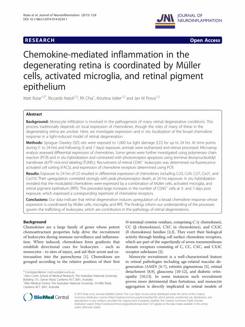
RESEARCH Open Access Chemokine-mediated inflammation in the degenerating retina is coordinated by Müller cells, activated microglia, and retinal pigment epithelium Matt Rutar 1,2* , Riccardo Natoli 1,2 , RX Chia 1 , Krisztina Valter 1,2 and Jan M Provis 1,2 Abstract Background: Monocyte infiltration is involved in the pathogenesis of many retinal degenerative conditions. This process traditionally depends on local expression of chemokines, though the roles of many of these in the degenerating retina are unclear. Here, we investigate expression and in situ localization of the broad chemokine response in a light-induced model of retinal degeneration. Methods: Sprague–Dawley (SD) rats were exposed to 1,000 lux light damage (LD) for up to 24 hrs. At time points during (1 to 24 hrs) and following (3 and 7 days) exposure, animals were euthanized and retinas processed. Microarray analysis assessed differential expression of chemokines. Some genes were further investigated using polymerase chain reaction (PCR) and in situ hybridization and contrasted with photoreceptor apoptosis using terminal deoxynucleotidyl transferase dUTP nick-end labeling (TUNEL). Recruitment of retinal CD45 + leukocytes was determined via fluorescence activated cell sorting (FACS), and expression of chemokine receptors determined using PCR. Results: Exposure to 24 hrs of LD resulted in differential expression of chemokines including Ccl3, Ccl4, Ccl7, Cxcl1, and Cxcl10. Their upregulation correlated strongly with peak photoreceptor death, at 24 hrs exposure. In situ hybridization revealed that the modulated chemokines were expressed by a combination of Müller cells, activated microglia, and retinal pigment epithelium (RPE). This preceded large increases in the number of CD45 + cells at 3- and 7-days post exposure, which expressed a corresponding repertoire of chemokine receptors. Conclusions: Our data indicate that retinal degeneration induces upregulation of a broad chemokine response whose expression is coordinated by Müller cells, microglia, and RPE. The findings inform our understanding of the processes govern the trafficking of leukocytes, which are contributors in the pathology of retinal degenerations. Background Chemokines are a large family of genes whose potent chemoattractant properties help drive the recruitment of leukocytes during immune surveillance and inflamma- tion. When induced, chemokines form gradients that establish directional cues for leukocytes - such as monocytes - to sites of injury, and aid their arrest and ex- travasation into the parenchyma [1]. Chemokines are grouped according to the relative position of their first N-terminal cysteine residues, comprising C ( γ chemokines), CC (β chemokines), CXC (α chemokines), and CX3C (δ chemokines) families [2,3]. They exert their biological activity through binding cell surface chemokine receptors, which are part of the superfamily of seven transmembrane domain receptors consisting of C, CC, CXC, and CX3C receptor subclasses [2]. Monocyte recruitment is a well-characterized feature in retinal pathologies including age-related macular de- generation (AMD) [4-7], retinitis pigmentosa [5], retinal detachment [8,9], glaucoma [10-12], and diabetic retin- opathy [10,13]. In some instances such recruitment proves more detrimental than fortuitous, and monocyte aggregation is directly implicated in retinal models of * Correspondence: [email protected] 1 John Curtin School of Medical Research, The Australian National University, Building 131, Garran Road, Canberra ACT 2601, Australia 2 ANU Medical School, The Australian National University, 54 Mills Road, Canberra ACT 2601, Australia JOURNAL OF NEUROINFLAMMATION © 2015 Rutar et al.; licensee BioMed Central. This is an Open Access article distributed under the terms of the Creative Commons Attribution License (http://creativecommons.org/licenses/by/4.0), which permits unrestricted use, distribution, and reproduction in any medium, provided the original work is properly credited. The Creative Commons Public Domain Dedication waiver (http://creativecommons.org/publicdomain/zero/1.0/) applies to the data made available in this article, unless otherwise stated. Rutar et al. Journal of Neuroinflammation (2015) 12:8 DOI 10.1186/s12974-014-0224-1
Transcript of Chemokine-mediated inflammation in the degenerating retina is coordinated by Müller cells,...
JOURNAL OF NEUROINFLAMMATION
Rutar et al. Journal of Neuroinflammation (2015) 12:8 DOI 10.1186/s12974-014-0224-1
RESEARCH Open Access
Chemokine-mediated inflammation in thedegenerating retina is coordinated by Müllercells, activated microglia, and retinal pigmentepitheliumMatt Rutar1,2*, Riccardo Natoli1,2, RX Chia1, Krisztina Valter1,2 and Jan M Provis1,2
Abstract
Background: Monocyte infiltration is involved in the pathogenesis of many retinal degenerative conditions. Thisprocess traditionally depends on local expression of chemokines, though the roles of many of these in thedegenerating retina are unclear. Here, we investigate expression and in situ localization of the broad chemokineresponse in a light-induced model of retinal degeneration.
Methods: Sprague–Dawley (SD) rats were exposed to 1,000 lux light damage (LD) for up to 24 hrs. At time pointsduring (1 to 24 hrs) and following (3 and 7 days) exposure, animals were euthanized and retinas processed. Microarrayanalysis assessed differential expression of chemokines. Some genes were further investigated using polymerase chainreaction (PCR) and in situ hybridization and contrasted with photoreceptor apoptosis using terminal deoxynucleotidyltransferase dUTP nick-end labeling (TUNEL). Recruitment of retinal CD45+ leukocytes was determined via fluorescenceactivated cell sorting (FACS), and expression of chemokine receptors determined using PCR.
Results: Exposure to 24 hrs of LD resulted in differential expression of chemokines including Ccl3, Ccl4, Ccl7, Cxcl1, andCxcl10. Their upregulation correlated strongly with peak photoreceptor death, at 24 hrs exposure. In situ hybridizationrevealed that the modulated chemokines were expressed by a combination of Müller cells, activated microglia, andretinal pigment epithelium (RPE). This preceded large increases in the number of CD45+ cells at 3- and 7-days postexposure, which expressed a corresponding repertoire of chemokine receptors.
Conclusions: Our data indicate that retinal degeneration induces upregulation of a broad chemokine response whoseexpression is coordinated by Müller cells, microglia, and RPE. The findings inform our understanding of the processesgovern the trafficking of leukocytes, which are contributors in the pathology of retinal degenerations.
BackgroundChemokines are a large family of genes whose potentchemoattractant properties help drive the recruitmentof leukocytes during immune surveillance and inflamma-tion. When induced, chemokines form gradients thatestablish directional cues for leukocytes - such asmonocytes - to sites of injury, and aid their arrest and ex-travasation into the parenchyma [1]. Chemokines aregrouped according to the relative position of their first
* Correspondence: [email protected] Curtin School of Medical Research, The Australian National University,Building 131, Garran Road, Canberra ACT 2601, Australia2ANU Medical School, The Australian National University, 54 Mills Road,Canberra ACT 2601, Australia
© 2015 Rutar et al.; licensee BioMed Central. TCommons Attribution License (http://creativecreproduction in any medium, provided the orDedication waiver (http://creativecommons.orunless otherwise stated.
N-terminal cysteine residues, comprising C (γ chemokines),CC (β chemokines), CXC (α chemokines), and CX3C(δ chemokines) families [2,3]. They exert their biologicalactivity through binding cell surface chemokine receptors,which are part of the superfamily of seven transmembranedomain receptors consisting of C, CC, CXC, and CX3Creceptor subclasses [2].Monocyte recruitment is a well-characterized feature
in retinal pathologies including age-related macular de-generation (AMD) [4-7], retinitis pigmentosa [5], retinaldetachment [8,9], glaucoma [10-12], and diabetic retin-opathy [10,13]. In some instances such recruitmentproves more detrimental than fortuitous, and monocyteaggregation is directly implicated in retinal models of
his is an Open Access article distributed under the terms of the Creativeommons.org/licenses/by/4.0), which permits unrestricted use, distribution, andiginal work is properly credited. The Creative Commons Public Domaing/publicdomain/zero/1.0/) applies to the data made available in this article,

Rutar et al. Journal of Neuroinflammation (2015) 12:8 Page 2 of 15
neovascular-AMD [14], light-induced damage [15-17],diabetic retinopathy [18,19], and glaucoma [20,21]. Che-mokine signaling is believed to play a role in mediatingmonocyte migration in several CNS disorders includingmultiple sclerosis, Alzheimer’s disease, and brain ische-mia and trauma (reviewed in [2,22-24]). In the retina,upregulation of α and β chemokines, such as Ccl2,Cxcl1, and Cxcl10 have been detected by gene expressionanalyses in both wet and dry forms of age-related maculardegeneration (AMD) [25]. Ccl2 is particularly well-characterized in the retina, and knockout studies indicatethat ablation of Ccl2 or its receptor Ccr2 reduces mono-cyte infiltration and retinal degeneration in experimentalchoroidal neovascularization (CNV) [26,27] and in light-damaged Cx3cr1−/− mice [28]. Additionally, we haveshown that expression of Ccl2 is upregulated in Müllercells in light-induced retinal degeneration [29], and tar-geted knockdown of Ccl2 with siRNA reduces recruitmentof microglia/monocytes and photoreceptor death [30].Despite a growing understanding of the Ccl2 axis in
relation to retinal dystrophies, there is a relative paucityin knowledge of the greater chemokine milieu of theretina and the cellular events that contribute to their ex-pression. In this study, we aimed to investigate transcrip-tional regulation and spatiotemporal distribution of thechemokine response in the retina in situ, following light-induced degeneration. We find that multitude α and βchemokines are upregulated following light damage, incorrelation with photoreceptor death. Further, we show,using in situ hybridization, that a coordinated trio ofMüller cells, retinal pigment epithelium (RPE), and micro-glia express a suite of α and β chemokines following injury,for which recruiting CD45+ cells bear corresponding che-mokine receptors.
MethodsAnimals and light damage paradigmAll experiments conducted were in accordance with theARVO Statement for Use of Animals in Ophthalmicand Vision Research; the study was approved by theAnimal Experimentation Ethics Committee (AEEC) ofthe Australian National University (R.BSB.05.10). Youngadult Sprague–Dawley (SD) rats aged were exposed to1,000 lux of light damage (LD) following a previousprotocol [31]. Animals were exposed to LD in incrementsof 1, 3, 6, 12, 17, or 24 hrs. Additionally, some animalswere returned to dim-light (5 lux) conditions immediatelyfollowing 24 hrs of LD for a period of 3 or 7 days, to assesspost exposure changes. All time points were comparedback to age-matched, dim-reared animals.
Tissue collection and processing of whole retinasAnimals were euthanized using an overdose of barbitur-ate, which was administered via intraperitoneal injection
(Valabarb; Virbac, NSW, Australia). The left eye fromeach animal was marked for orientation then enucleatedfor cryosectioning (n = 6), while the retina from the righteye was excised through a corneal incision for RNA ex-traction (n = 6). Retinas were processed using previouslydescribed protocols [31].
Fluorescence-activated cell sorting of retinal microgliaRats at each time point were euthanized as describedpreviously. Retinas from both eyes were promptly re-moved through a corneal incision. Retinas from eachanimal were pooled and immediately placed in chilledHank’s balanced salt solution (HBSS) (n = 5 per timepoint) and then subjected to light mechanical separationusing a razor blade. Samples were transferred into 0.2%papain digestion cocktail as described in a previousprotocol [32] with minor modifications, and incubatedat 8°C for 45 minutes, then 28°C for 7 minutes. Theresulting homogenate was centrifuged at 250 g for 5 mi-nutes at 4°C, and the pellet was resuspended inneutralization buffer [32]. The homogenate was centri-fuged again at 420 g for 5 minutes at 4°C, and the pelletresuspended in staining buffer containing 1.0% bovineserum albumin (BSA), and 0.1% azide. The samples wereincubated in staining buffer containing a CD45 antibodyconjugated to Alexa 647 (Biolegend, San Diego, CA,USA) for 45 minutes at 4°C, then washed twice in HBBSand resuspended in staining buffer. The resultant CD45-stained samples were run through a fluorescence-activatedcell sorter (FACS) (BD FACSAria II; BD Biosciences,Franklin Lakes, NJ, USA). Viability of the sorted cells wasassessed by labeling with DAPI. The isolated CD45+ cellswere collected in staining buffer and kept chilled on iceuntil RNA extraction could be commenced.To prepare for RNA extraction, isolated samples were
centrifuged at 420 g for 5 minutes at 4°C, and the super-natant removed. RNA extraction was performed witha combination of TRIzol Reagent (Life Technologies,Carlsbad, CA, USA) and an RNAqueous-small scale kit(Life Technologies, Carlsbad, CA, USA) utilized in tan-dem to extract and purify the RNA respectively, as de-scribed previously [33]. Isolated total RNA was analyzedfor quantity and purity with a ND-1000 spectrophotometer(Nanodrop Technologies, Wilmington, DE, USA).
Microarray experimentation and analysisMicroarray analysis of RNA from whole retinas was con-ducted using raw microarray data derived from an inves-tigation by our group [31], using Rat Gene 1.0 ST arrays(Affymetrix, Santa Clara, CA, USA); the microarray datais accessible from the NCBI Gene Expression Omnibusrepository (GSE22818). Analysis compared samples fromdim-reared and 24-hrs of LD experimental groups (n = 3for each). The microarray data was analyzed with Partek

Rutar et al. Journal of Neuroinflammation (2015) 12:8 Page 3 of 15
Genomics Suite 6.4 software (Partek Inc., St. Louis, MO,USA), using the same parameters as in our previous in-vestigation [31].Statistical analysis was conducted using the analysis of
variance (ANOVA) statistic with the threshold of signifi-cance set at P <0.05, with a differential expression cut-offof >50%. The differentially expressed genes were screenedfor those pertaining to chemokine activation, and weregrouped according to pathway information summarizedfrom the Gene Ontology Consortium [34].
Polymerase chain reactionQuantitative real-time polymerase chain reaction (qPCR)was used to validate the expression of genes identified inthe microarray analysis, over the protracted light damagetime course (n = 6). First-strand cDNA synthesis wasperformed as described previously [29]. Expression wasmeasured using commercially available TaqMan hydroly-sis probes (Life Technologies, Carlsbad, CA, USA); theparticulars are provided in Table 1. The hydrolysisprobes were applied in the same fashion as our pre-vious study [29]. Fold change was determined usingthe ΔΔCq method, where the expression of the targetgene was normalized relative to the expression of thereference gene glyceraldehyde-3-phosphate dehydro-genase (GAPDH). Expression of GAPDH does not changewith respect to retinal light damage, as indicated by severalinvestigations [31,35,36].Standard PCR was performed on RNA samples purified
from FACS-isolated monocytes/microglia, using primersspecific to chemokine receptor genes (Table 2). Primerswere designed using the Primer3 web-based design pro-gram [37], and tailored to transverse an intron splice site.First-strand cDNA synthesis was performed from 50 ngof RNA using the Tetro cDNA synthesis kit (Bioline,London, UK), and applied according to the manufacturer’s
Table 1 Taqman hydrolysis probes
Gene symbol Gene name
CCL3 Chemokine (C-C motif) ligand 3
CCL4 Chemokine (C-C motif) ligand 4
CCL7 Chemokine (C-C motif) ligand 7
CXCL1 Chemokine (C-X-C motif) ligand 1
CXCL10 Chemokine (C-X-C motif) ligand 10
CXCL11 Chemokine (C-X-C motif) ligand 11
ADAM17 ADAM metallopeptidase domain 17
IL1B Interleukin 1β
MYD88 Myeloid differentiation primary response gene 88
TLR2 Toll-like receptor 2
TNF Tumor necrosis factor
SIGIRR Single immunoglobulin and toll-interleukin 1 recep
instructions. Standard PCR was then conducted on thesamples using MyTaq DNA polymerase; the presence ofPCR product and specificity of the reaction were assessedby gel electrophoresis.
In situ hybridizationA number of chemokines (Ccl3, Ccl4, Ccl7, Cxcl1, andCxcl10) were cloned from PCR products (540-bp, 212-bp, 540-bp, 487-bp, and 504-bp amplicons, respectively)using cDNA prepared from rat retinas (as described inthe qPCR section). Cloning was performed using thepGEM-T DNA vector system (Promega, Madison, WI,USA) and JM109 competent E.coli (Promega, Madison,WI, USA). A DIG RNA Labeling Kit SP6/T7 (Roche, Basel,Switzerland) was used to transcribe linearized plasmid andgenerate DIG-labeled antisense and sense riboprobes. Thein situ hybridization was performed using a previouslyestablished protocol [38]; individual riboprobes were hy-bridized overnight at 57°C and then washed in decreasingconcentrations of saline sodium citrate (pH 7.4) at 60°C.The bound probe was visualized with either NBT/BCIP orHNNP/Fast-Red (Roche, Basel, Switzerland).Following hybridization, sections stained with HNNP/
Fast-Red were also counter-stained using immunohisto-chemistry. In situ-stained sections were incubated withprimary antibody overnight at 4°C, which was raisedagainst either IBA1 (1:500; Wako, Osaka, Japan), Vimentin(1:100; Sigma-Aldrich, St. Louis, MO, USA), or RPE65(1:200; Abcam, Cambridge, UK). Sections were then incu-bated with biotinylated antibodies raised against eitherrabbit or mouse IgG’s (Life Technologies, Carlsbad, CA,USA) for 2 hrs at room temperature and followed by incu-bation in streptavidin conjugated to Alexa488 (LifeTechnologies, Carlsbad, CA, USA) for 1.5 hours at roomtemperature. Slides were then cover slipped with AquaPoly/Mount (Polysciences, Warrington, PA, USA), and
Catalog Entrez Gene ID
Rn00564660_m1 NM_013025.2
Rn00587826_m1 NM_053858.1
Rn01467286_m1 NM_001007612.1
Rn00578225_m1 NM_030845.1
Rn01413889_g1 NM_139089.1
Rn00788262_g1 NM_182952.2
Rn00571880_m1 NM_020306.1
Rn00580432_m1 NM_031512.2
Rn01640049_m1 NM_198130.1
Rn02133647_s1 NM_198769.2
Rn00562055_m1 NM_012675.3
tor (TIR) domain Rn01501616_g1 NM_001024887.1

Table 2 Standard PCR primers
Gene symbol NCBI RefSeq Forward primer (5′ - 3′) Reverse primer (5′ - 3′)
Ccr1 NM_020542.2 GTTGGGACCTTGAACCTTGA TGTGGTTGTGGGGTAGGTTT
Ccr2 NM_021866.1 CCAGTGTGAAGCAAATTGGA TGGAAAATAAGGGCCACAAG
Ccr5 NM_053960.3 GTCAAACGCTTCTGCAAACA CTTGTTCCCAGCCTTCTCAG
Cxcr2 NM_017183.1 CAGAGACTTGGGAGCCACTC TCAGCAAAGTCACCAGAACG
Cxcr3 NM_053415.1 AAGTTCCCAACCACAAGTGC GGCAGGAAGGTTCTGTCAAA
Rutar et al. Journal of Neuroinflammation (2015) 12:8 Page 4 of 15
immunofluorescence was viewed using a Zeiss laser scan-ning microscope (Zeiss, Oberkochen, Germany), and ac-quired using PASCAL software (Zeiss, v4.0).
Analysis of cell deathTUNEL labeling was used to quantify photoreceptorapoptosis in cryosections, and utilized a protocol thathas been documented previously [39]. Counts weremade of TUNEL positive cells in the outer nuclear layer(ONL), and were performed along the full-length of retinalsections cut in the vertical meridian (superio-inferior), in-cluding the optic disc. The final count from each animal isthe average at comparable locations in two nonsequentialsections.
Statistical analysisStatistical analysis was performed using the Kruskal-Wallisone-way analysis of variance, with Dunn’s multiple com-parison post-test applied where desired; differences with aP value <0.05 were considered statistically significant.
ResultsMicroarray analysis for chemokine-related genes following24 hours of light damageAnalysis of microarray data compared gene expressionin retinas of animals reared in dim-light conditionswith those exposed to 24 hrs of LD. The reliability ofthe microarray data was assessed with hierarchical clus-tering and principal component analysis in our previousinvestigation [31]; both analyses indicated high repro-ducibility for samples in their respective treatmentconditions. From the microarray data, a list of differ-entially expressed chemokine-related genes (P <0.05)was compiled (Table 3), which were grouped accordingto their functional roles outlined in the gene ontologyconsortium [34].Chemokine ligands (GO:0008009) were the most prom-
inent among the differentially expressed genes and in-cluded upregulation in a multitude of α (Ccl2, Ccl3, Ccl4,Ccl7, Ccl12, Ccl20), and β (Cxcl1, Cxcl9, Cxcl10, Cxcl11,Cxcl116) chemokines. Conversely, expression of Ccl9 andCxcl6 was found to decrease following LD. Another highlyrepresented group comprised genes involved in promotingchemokine synthesis (GO:0045080/GO:0032722), which
included upregulation in Il1β, Tnfα, Adam17, Tlr2, andMyd88, and a decrease in Nod2. There was also anincrease in expression of Sigirr, which is associatedwith inhibition of chemokine synthesis (GO:0045079/GO:0032682). Expression of chemokine receptors (GO:0019956) showed little wide modulation, although an in-crease in Ccr5 was observed, and the expression of Cxcr7was reduced somewhat.The validity of the microarray data was assessed by
analyzing the expression of 12 genes from Table 3 usingqPCR. These included a selection of chemokine ligands(Ccl3, Ccl4, Ccl7, Cxcl1, Cxcl10, Cxcl11) and regulators(Il1β, Tnfα, Adam17, Tlr2, Myd88, Sigirr). Differentialexpression of these genes at 24 hrs of LD, was found byPCR to be in agreement with the corresponding data ob-tained from the microarray (Figure 1).
Relation of chemokine expression to photoreceptorcell deathThe expression of the 12 gene selections was furtherexamined over the protracted LD time course, and con-trasted with the number of TUNEL+ photoreceptors(Figure 2). The time course encompassed incrementalperiods during- (1, 3, 6, 12, 17, and 24 hrs) and post-LDexposure (3 and 7 days).Large increases in the number of TUNEL+ photore-
ceptors were observed from 12 hrs of LD onward andreached a peak at 24 hrs (Figure 2A), as reported previ-ously [29,31]. By 3- and 7-days post-exposure, the num-ber of TUNEL+ nuclei had decreased substantially.Expression of α (Ccl3, Ccl4, Ccl7), and β (Cxcl1, Cxcl10,Cxcl11) chemokine ligands was upregulated at 12 hrs ofLD, before reaching a peak at 24 hrs - consistent withthe emergence of TUNEL+ cells (Figure 2B-C). By 3-days postexposure, expression of all chemokine ligandswas reduced, with only small upregulation evident by7 days, compared to dim-reared controls. Expression ofchemokine promoters Il1β, TNFα, Tlr2, and Myd88 andthe inhibitor Sigirr exhibited similar trends in upregula-tion over the LD time course to the chemokine ligands(Figure 2D). Il1β and TNFβ were the most strongly up-regulated, beginning at 12 hrs of LD; this upregulationdecreased in the post exposure period, although the de-creases were smaller for Tlr2 and Myd88. In contrast,
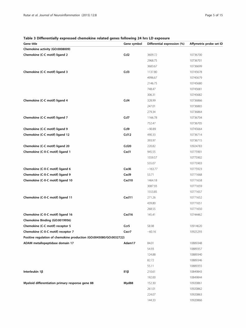
Table 3 Differentially expressed chemokine related genes following 24 hrs LD exposure
Gene title Gene symbol Differential expression (%) Affymetrix probe set ID
Chemokine activity (GO:0008009)
Chemokine (C-C motif) ligand 2 Ccl2 3609.72 10736700
2968.75 10736701
3683.67 10736699
Chemokine (C-C motif) ligand 3 Ccl3 1137.80 10745678
4996.67 10745679
2146.75 10745680
748.47 10745681
306.31 10745682
Chemokine (C-C motif) ligand 4 Ccl4 328.99 10736866
247.01 10736865
279.34 10736864
Chemokine (C-C motif) ligand 7 Ccl7 1166.78 10736704
752.47 10736705
Chemokine (C-C motif) ligand 9 Ccl9 −90.89 10745664
Chemokine (C-C motif) ligand 12 Ccl12 490.33 10736714
393.97 10736715
Chemokine (C-C motif) ligand 20 Ccl20 220.82 10924783
Chemokine (C-X-C motif) ligand 1 Cxcl1 945.55 10775901
1559.57 10775902
555.07 10775903
Chemokine (C-X-C motif) ligand 6 Cxcl6 −163.77 10775923
Chemokine (C-X-C motif) ligand 9 Cxcl9 53.71 10771668
Chemokine (C-X-C motif) ligand 10 Cxcl10 1464.18 10771658
3087.93 10771659
1553.85 10771657
Chemokine (C-X-C motif) ligand 11 Cxcl11 271.26 10771652
439.80 10771651
268.55 10771650
Chemokine (C-X-C motif) ligand 16 Cxcl16 145.41 10744462
Chemokine Binding (GO:0019956)
Chemokine (C-C motif) receptor 5 Ccr5 58.98 10914620
Chemokine (C-X-C motif) receptor 7 Cxcr7 −60.16 10925293
Positive regulation of chemokine production (GO:0045080/GO:0032722)
ADAM metallopeptidase domain 17 Adam17 84.01 10889348
54.93 10889357
124.88 10889340
82.72 10889346
55.11 10889355
Interleukin 1β Il1β 210.61 10849843
192.00 10849844
Myeloid differentiation primary response gene 88 Myd88 152.30 10920861
261.01 10920862
224.07 10920863
144.33 10920866
Rutar et al. Journal of Neuroinflammation (2015) 12:8 Page 5 of 15

Table 3 Differentially expressed chemokine related genes following 24 hrs LD exposure (Continued)
Nucleotide-binding oligomerization domain containing 2 Nod2 −52.42 10809667
Toll-like receptor 2 Tlr2 123.65 10823972
Tumor necrosis factor (TNF superfamily, member 2) Tnfα 106.36 10828025
119.80 10828023
111.45 10828024
Negative regulation of chemokine production (GO:0045079/GO:0032682)
Single immunoglobulin and toll-interleukin 1 receptor (TIR) domain Sigirr 71.95 10726694
92.00 10726700
Rutar et al. Journal of Neuroinflammation (2015) 12:8 Page 6 of 15
Adam17 was upregulated over the entire time course,with a modest peak in expression at 24 hrs of LD.
In situ localization of chemokines following light damageexposureChemokine ligands Ccl3, Ccl4, Ccl7, Cxcl1, and Cxcl10were selected for further characterization in 24-hr LDretinas, by in situ hybridization (Figures 3, 4, 5, 6). No
Figure 1 Validation of differentially expressed microarray genes usingassess robustness of the microarray data, a selection of differentially exprescontrasted with their qPCR counterparts (n = 6). Increases were observed inthough there was some variation in the extent of upregulation.
expression of chemokines was detected in retinas of dim-reared animals (Figures 3, 4, 5, 6). For Ccl3 and Ccl4,mRNA staining was evident by 24 hrs of LD in irregular-shaped nuclei/processes (Figure 3A-D, J-M), scatteredthroughout the ONL. These cells were more numerous inthe superior part of the retina, which is the focal region forLD-mediated degeneration (data not shown). Counterimmunolabeling for IBA1 revealed a strong co-localization
quantitative real-time polymerase chain reaction (qPCR). Tosed microarray genes (‘*’ for each probe set, where P <0.05) wereall in genes by qPCR, which corroborated the microarray analysis,

Figure 2 Expression of selected chemokines and chemokine-modulators in relation to apoptotic markers, following light damage.A: Increases in TUNEL+ photoreceptors were observed from 12 hrs light damage (LD), and peaked at 24 hrs. The number of TUNEL+ nuclei thendecreased profoundly during the post exposure period. B-C: Expression of both Ccl- (B), and Cxcl- (C) ligands exhibited concurrent upregulationbeginning at 12 hrs of LD, which then peaked at 24 hrs. Expression decreased sharply in all chemokines by 3- and 7-days postexposure.D: Chemokine regulators Il1β, TNFα, Tlr2, Myd88, and Sigirr showed emerging upregulation between 12 hrs and 24 hrs of LD, and wasparticularly robust in the instance Il1β, and TNFα. Upregulation of Il1β, TNFα, Tlr2, Myd88, and Sigirr peaked at 24 hrs of LD and tapered off duringthe post-exposure period. Adam17, in contrast to the other regulators, exhibited consistent upregulation over the entire time course, with a minorpeak at 24 hrs of LD. The trend in expression for all genes, as well as TUNEL, was significant by one-way ANOVA (P <0.05); n = 6.
Rutar et al. Journal of Neuroinflammation (2015) 12:8 Page 7 of 15
of both Ccl3 (Figure 3E-I) and Ccl4 (Figure 3N-R) withactivated IBA1+ microglia in the ONL at 24 hrs postexposure/LD.In situ hybridization for Ccl7 also evident in cells scat-
tered throughout the ONL at 24 hrs of LD (Figure 4B-D),but in addition was detected in cell processes radially ori-ented in the INL. As with the Ccl3 and Ccl4, this stainingwas more frequent in the superior retina (data not shown).Counter immunolabeling for IBA1 indicated that stainingfor Ccl7 in the ONL coincided with activated microglialcells (Figure 4H-K). In the INL, Ccl7 mRNA labeling of cellprocesses was IBA1-negative (Figure 4O-P). Instead, thesecells co-localized Ccl7 mRNA with immunoreactivity forvimentin (Figure 4L-N), consistent with an identification ofMüller cell processes.
The localizations of Cxcl1 and Cxcl10 resembled eachother (Figures 5 and 6). Following 24 hrs of LD, Cxcl1(Figure 5B-C) and Cxcl10 (Figure 6B-C) mRNA waslabeled in putative RPE cells, predominately in the su-perior retina (Figures 5D, 6D). The identity of theseCxcl1/Cxcl10 expressing cells was confirmed by posi-tive counter immunolabeling for RPE65 (Figures 5I-K,6I-K). Cxcl1 and Cxcl10 mRNA labeling was also de-tected in radial processes within the INL, mainly inthe superior retina, at 24 hrs of LD (Figures 5E, 6E).In this location, Cxcl1 and Cxcl10 mRNA labeling co-localized strongly with vimentin immunoreactivity con-sistent with an identification of Müller cell processes.(Figures 5G-H, 6G-H). A summary of these data is pro-vided in Table 4.

Figure 3 In situ hybridization for Ccl3 and Ccl4 mRNA in the retina following exposure to 24 hours of light damage. In situ hybridizationfor Ccl3 is documented in A-I, while Ccl4 is shown in J-R. A: In retinas from dim-reared animals, staining for Ccl3 was not observed in the retina.B-D: After 24 hrs light damage (LD), there was staining for Ccl3 in irregularly shaped nuclei - ranging from elongated to globular - traversing theouter nuclear layer (ONL) of the superior retina (arrowheads). Conversely, few Cc3-expressing cells were found in the inferior retina (data not shown).E-I: Nuclei stained for Ccl3 mRNA (red) showed strong correlation (arrowheads) to activated microglia, which were counterimmunolabeled with anantibody to IBA1 (green). J: Ccl4 mRNA staining was not apparent in sections of dim-reared retinas. J-M: At 24 hrs, LD staining for Ccl4 was detectedamong clusters of irregular nuclei in the ONL from the superior retina (arrowheads); little-to-none were observed in the inferior retina (data not shown).N-R: Ccl4-expressing nuclei (red) in retinal sections show strong immunofluorescence (arrowheads) for IBA1+ microglia (green), much like Ccl3 (E-I).INL, inner nuclear layer; IHC, immunohistochemistry; ISH, in situ hybridization; ONL, outer nuclear layer; OS, outer segments.
Rutar et al. Journal of Neuroinflammation (2015) 12:8 Page 8 of 15
Recruitment of CD45+ and expression of chemokinereceptorsChanges in the number of CD45+ monocytes/microgliafollowing LD were identified using FACS; representativegating strategies and scatter blots are noted in Figure 7A.In dim-reared retinas, CD45+ cells comprised a relativelysmall population of the gated retinal isolates, at approxi-mately 0.098% (Figure 7B). After 24 hrs of LD, the retinalCD45+ population rose substantially to 0.340% (P <0.05),then reaching 0.875% by 3 days (P < 0.05). A further in-crease to 1.24% was observed at 7 days, although this wasnot significantly different from the population size at 3 days(P >0.05).PCR of RNA from the CD45-FACS isolates at each
time point showed differential expression of the chemokinereceptors Ccr1, Ccr2, Ccr5, Cxc2, and Cxcr3 (Figure 7),which are known to bind the differentially expressedligands validated by qPCR in Figure 1 [40]. CD45 isolates
express all chemokine receptors assessed (Figure 7C-G),although the density of the PCR bands varies dependingon the time point. Ccr1, Ccr2, Ccr5, and Cxcr2 appear tobe lowly expressed in dim-reared animals and more highlyexpressed following 24 hrs of LD and at the post-exposuretime points (Figure 7C-F). Cxcr3 showed no amplificationin dim-reared samples, was highly expressed at 24 hrs ofLD, and was low by 7-days post-exposure (Figure 7F).
DiscussionThese findings identify a complex network of chemokineactivity elicited by light-induced retinal degeneration andinclude several novel findings. First, we show that a suiteof α (Ccl3, Ccl4, Ccl7) and β (Cxcl1, Cxcl10, Cxcl11)chemokines are upregulated following 24 hrs of LD, andthese chemokines correlate strongly with the emergenceof photoreceptor death and upregulation of chemokineregulatory genes (Il1β, Tnfα, Tlr2, Myd88, Adam17, Sigirr).

Figure 4 In situ hybridization for Ccl7 mRNA in the retina following exposure to 24 hours of light damage. A: Staining for Ccl7 was notobserved in the retinas of dim-reared animals B-D: In retinas exposed to 24 hrs light damage (LD), Ccl7 expression was found in infrequent clustersof nuclei within the outer nuclear layer (ONL) of the superior retina (arrowheads), with few in the inferior (data not shown). E-F: Ccl7 expression wasalso apparent in radial processes (arrowhead) dispersed within inner nuclear layer (INL) of the superior retina, following 24 hrs of LD. G: Ccl7 stainingwas less evident in the inferior portion of the retina following LD. H-K: Ccl7-stained nuclei (red) in the ONL were found to coincide withactivated IBA1-immunolabeled microglia (green; arrowheads). L-N: Staining for Ccl7 mRNA (red) in the INL showed strong co-localization forvimentin-immunreactive Müller cell processes (arrowheads). O-P: Ccl7-expressing cells in the INL (red; arrowheads) were negative for IBA1immunostaining. INL, inner nuclear layer; IHC, immunohistochemistry; ISH, in situ hybridization; ONL, outer nuclear layer; OS, outer segments.
Rutar et al. Journal of Neuroinflammation (2015) 12:8 Page 9 of 15
Second, in situ hybridization demonstrates that Müllercells, RPE, and activated microglia comprise a ‘trio’ of cellsthat express select chemokine ligands (Ccl3, Ccl4, Ccl7,Cxcl1, Cxcll0) after LD. Third, we show that LD-inducedupregulation of chemokines is proceeded by infiltra-tion of CD45+ monocytes/microglia, which bear corre-sponding receptors (Ccr1, Ccr2, Ccr5, Cxcr2, Cxcr3)at 3-days postexposure.Several investigations have previously identified upregu-
lation in chemokine ligands following retinal light damage,including Ccl2, Ccl3, Ccl4, Ccl7, Cxcl1, and Cxcl10through PCR and microarray analysis [35,41-43]. However,these studies did not identify the source of their expressionin the retinal environment in situ, nor did they closely re-late chemokine expression to photoreceptor cell death andregulatory factor expression. To our knowledge, this is thefirst investigation to demonstrate a preferential localizationof chemokine mRNA in activated microglia (Ccl3, Ccl4,
Ccl7), Müller cells (Ccl7, Cxcl1, Cxcl10), and RPE (Cxcl1,Cxcl10) in the degenerating retina. These observations aresupported by a number of in vitro studies, which have re-ported expression of Ccl3 by microglia [43], and Cxcl1[44-46] and Cxcl10 [47,48] in Müller cells or RPE in re-sponse to various stimuli including cytokines and bacterialpathogens. The expression profiles of chemokine ligandsby microglia, Müller cells, and RPE following LD werehighly defined and cell-specific. The precise roles of thesediscrete chemokine phenotypes in the degenerating retinaare unclear, despite a bourgeoning understanding of che-mokine activity in recent years. Functional significancemay be inferred, however, based on current understandingof the chemokines identified.
Chemokine secretion by activated microgliaWe find that Ccl3, Ccl4, and Ccl7 are expressed by acti-vated microglia after LD. Previous in vitro and in vivo

Figure 5 In situ hybridization for Cxcl1 mRNA in the retina following exposure to 24 hours of light damage. A: In sections from dim-rearedanimals, staining for Cxcl1 was absent. B-D: Retinas exposed to 24 hrs of LD showed staining for Cxcl1 in putative retinal pigment epithelium (RPE)cells (arrowheads) in the superior retina (B-C), and few-to-none in the inferior (D). E-F: Staining for Cxcl1 mRNA was also present in the innernuclear layer (INL) of superior retina following 24 hrs of LD (E; arrowheads), and mostly absent in inferior retina (F). G-H: Retinas with stain forCxcl1 mRNA in the INL (red; arrowhead), which was found to co-localize with vimentin-immunolabeled Müller cell processes (green; arrowheads).I-J: Localized Cxcl1 stain in putative RPE cells (red; arrowheads) correlated strongly with immunolabeling for RPE65 (green; arrowheads); Cxcl1 stainappeared to be localized in the nucleus, rather than cytoplasm. C, choroid; INL, inner nuclear layer; IHC, immunohistochemistry; ISH, in situhybridization ONL, outer nuclear layer; OS, outer segments.
Rutar et al. Journal of Neuroinflammation (2015) 12:8 Page 10 of 15
studies indicate that Ccl3 is involved in the proliferationand mobilization of mature myeloid progenitor cells, aswell as in recruitment of bone marrow-derived mono-cytes [49-52]. This function may be exerted through thebinding of receptor Ccr1 [53,54], although Ccl3 also in-teracts with Ccr5, which mediates monocyte recruit-ment, and mobilization of Th1 T cells [52,55,56]. In theretina, deficiencies in Ccl3 reduce progressive photo-receptor death and monocyte recruitment in degenerativeAbca4−/− Rdh8−/− mice, and in the Mertk−/− mouse modelof retinitis pigmentosa [43]. Ccl4 also interacts with Ccr5,to promote recruitment of monocytes into the retina whensubjected to oxygen induced retinopathy [57].In contrast, Ccl7 is a ligand of the better documented
receptor Ccr2, and is major determinant in chemotaxisof monocytes. Deficiencies in Ccr2 impair monocyte re-cruitment to tissues in disease models ranging fromarthrosclerosis [58] and autoimmune encephalitis [59],to choroidal neovascularization [26]. Several studies in-dicate that Ccl2 and Ccl7 are the primary agonists ofCcr2, in which ablation of either of these ligands re-duced monocyte recruitment from bone marrow to per-ipheral vessels in thioglycollate-induced peritonitis [60]and Listeria monocytogenes infection [61]. Taken together,the findings suggest that activated ‘resident’ microglia are
potent drivers of monocyte filtration from the vascula-ture to the parenchyma following LD. The present re-sults suggest that this activity is mediated, at least inpart, by activated microglia through the expression ofspecific chemokines. This expression is possibly trig-gered by migrating microglia that encounter stressedphotoreceptors in the ONL. This suggestion is supportedby our findings that recruited CD45+ cells bear the corre-sponding Ccr1, Ccr2, and Ccr5 receptors following LDand found to be expressed by monocytes and macro-phages in other studies [56,62,63].
Chemokine secretion by retinal pigment epithelium andMüller cellsIn contrast to the monocyte-centric chemotactic profileof activated microglia, chemokine expression by Müllercells and RPE suggests they have a broader role inmodulation of the leukocyte response. Müller cells andRPE shared a similar expression profile of upregulationof Cxcl1 and Cxcl10 following LD, while Müller cells alsoexpressed Ccl7. We have shown previously that Müllercells also express Ccl2 following LD [29].Cxcl1, and its cognate receptor Cxcr2, are commonly
associated with recruitment of neutrophils from the vas-cular supplies [64-66], and signaling via this receptor is

Figure 6 In situ hybridization for Cxcl10 mRNA in the retina following exposure to 24 hours of light damage. A: Expression of Cxcl10 wasnot apparent in dim-reared sections. B-C: Sections from the superior retina (B) showed stain for Cxcl10 mRNA in putative retinal pigmentepithelium (RPE) cells (arrowheads) following 24 hrs light damage (LD), while no staining was observed in the inferior retina (D). D-E: Following24 hrs of LD, stain for Cxcl10 mRNA appeared in the INL within superior retina (D-E; arrowheads). F: Staining for Cxcl10 was not readily apparent inthe inferior retina. G-I: Sections with labeling for Cxcl10 mRNA (red; arrowheads) in the INL, which correlated with Müller cells immunolabeled withvimentin (green; arrowheads). J-K: Putative RPE cells, which were positive for Cxcl10 mRNA labeling (red; arrowheads), were also immunoreactivefor the marker RPE65 (green; arrowheads). C, choroid; INL, inner nuclear layer; IHC, immunohistochemistry; ISH, in situ hybridization ONL, outernuclear layer; OS, outer segments.
Rutar et al. Journal of Neuroinflammation (2015) 12:8 Page 11 of 15
implicated in the pathogenesis of rheumatoid arthritis[67,68], lung injury [69], and adenoviral keratitis [70].Furthermore, neutrophil recruitment contributes to path-ology in choroidal neovascularization [71] and in lossof blood retinal barrier integrity in RPE-choroid explants[72]. Cxcl1 and Cxcr2 are also implicated in mobilizationof monocytes, in models of atherosclerosis [73,74] andis Cxcr2 expressed by CNS- and retinal-derived mono-cytes [75,76].On the other hand, Cxcl10 plays a role in chemotaxis
of T cells via Cxcr3 signaling. Cxcr3 signaling is requiredfor recruitment of cytotoxic T cells in West Nile virusencephalitis [77,78], and in the eye, Cxcl10 mediatesTh1 T cell trafficking in response to chronic ocular
Table 4 Summary of retinal Ccl and Cxcl localizationfollowing light damage
Gene Müller cells IBA1+ microglia Retinal pigment epithelium
Ccl3 - + -
Ccl4 - + -
Ccl7 + + -
Cxcl1 + - +
Cxcl10 + - +
Toxoplasmosis [79]. Besides T cells, Cxcr3 is also ex-pressed on monocytes/microglia as described in vitroand in vivo [80-82] and may modulate their recruit-ment in response to noxious stimuli. Supplementationwith Cxcl10 also promotes the survival of photorecep-tor in vitro and in retinal explants, which suggestadditional neurotrophic properties [48]. Neutrophiland T cell activity are poorly characterized in light-induced retinal degeneration, possibly because, in in-stances where it has been investigated, their numbersare relatively small compared monocytes/microglia [43].Therefore, the precise recruitment and function of neutro-phils and T cells in retinal light damage warrants furtherinvestigation.Synthesis of Cxcl1 and Cxcl10 by RPE and Müller cells
and their juxtaposition to the choriocapillaris and retinalvasculature, respectively, possibly reflects their role as ef-ficient mediators of chemotaxis of monocytes, neutro-phils, and T cells from the circulation. The additionalsecretion of Ccl7 by Müller cells - in conjunction withCcl2 [29] - may suggest a stronger emphasis on monocyterecruitment from the retinal vessels following LD, consist-ent with observation of an influx of bone marrow-derivedmonocytes from the retinal vasculature in retinal injuryfollowing exposure to bright light [83].

Figure 7 Changes the number of CD45+ cells using fluorescence activated cell sorting (FACS), and their expression of chemokine receptors.A: Representative FACS plots, with gating strategies, for a 7-days postexposure sample stained for CD45. Gating methodology was applied equally forall samples. B: Histogram depicts changes in the population of retinal CD45+ cells following light damage (LD). Proportion of CD45+ cells roughlytripled following 24 hrs of LD (P <0.05), and continued to increase substantially during the post-exposure period after 3 days (P <0.05); a furtherincrease at 7 days was not significant (P >0.05). The overall trend was significant by one-way ANOVA (P <0.05); n = 5. C-G: Representative imagesof PCR products via electrophoresis for Ccr1, Ccr2, Ccr5, Cxcr2, Cxcr3, in samples of CD45-sorted cells. Receptor expression was low in dim-rearedcontrol samples (C-F), and absent for Cxcr3 (G). Expression increased substantially following 24 hrs of LD for every receptor assessed (C-G), andwas maintained through the post exposure period, with the exception of Cxcr3 (G).
Rutar et al. Journal of Neuroinflammation (2015) 12:8 Page 12 of 15
Factors that promote chemokine expression in thedegenerating retinaWe also find in this study the upregulation of factorsthat have broad modulatory roles in chemokine secre-tion, including the pro-inflammatory cytokines IL1β andTNFα. These factors induce expression of both α and βchemokines in vitro [84] and under various conditionsincluding mycobacterial infection [85], nephrotoxicity[86], LPS-induced endotoxemia [87], and arthritis [88].Other genes identified in this study that may induce che-mokine expression are Tlr2 [89,90] and Myd88 [91,92].The events leading to collective synthesis of che-
mokines by microglia, Müller cells and RPE followingLD are uncertain, although recent evidence conductedin vitro points to an interactive role with microglia in
chemokine secretion. Müller cells co-cultured with LPS-activated microglia were found to upregulate expressionof Ccl2 and Ccl3 [93]. These microglia-stimulatedMüller cells in turn induced reciprocal activation of un-stimulated microglial cultures, including upregulationof Il1β. Co-culturing with activated microglia also in-duced upregulation chemokines in RPE cultures, suchas Ccl2 and Ccl5 [94]. The data suggest that the trio ofcells identified as expressing chemokines in the LDmodel co-activate; whether direct interaction betweencell types is required is unclear, although, if so, would bemost likely mediated by activated microglia, which arethe only motile cell of the trio. It should be noted thatin vivo Müller cells and RPE do not express Ccl3 andCcl2, respectively.

Rutar et al. Journal of Neuroinflammation (2015) 12:8 Page 13 of 15
ConclusionsThe study shows that microglia, Müller cells, and RPEeach contribute to the trafficking of leukocytes followingretinal damage through coordinated chemokine secre-tion. We show that chemokine expression by this trio ofcells precedes mass recruitment of CD45+ cells, whichbear the corresponding chemokine receptors and whichincreased in number in the retina over the time courseof the experiment. Our findings provide valuable insightinto chemokine activity in retinal degenerations.
AbbreviationsAdam17: ADAM metallopeptidase domain 17; AEEC: Animal ExperimentationEthics Committee; AMD: age-related macular degeneration; C: choroid;Ccl12: Chemokine (C-C motif) ligand 12; Ccl2: Chemokine (C-C motif) ligand2; Ccl20: Chemokine (C-C motif) ligand 20; Ccl3: Chemokine (C-C motif)ligand 3; Ccl4: Chemokine (C-C motif) ligand 4; Ccl7: Chemokine (C-C motif)ligand 7; Ccl9: Chemokine (C-C motif) ligand 9; Ccr5: Chemokine (C-C motif)receptor 5; CNS: central nervous system; Cxcl1: Chemokine (C-X-C motif)ligand 1; Cxcl10: Chemokine (C-X-C motif) ligand 10; Cxcl11: Chemokine(C-X-C motif) ligand 11; Cxcl16: Chemokine (C-X-C motif) ligand 16;Cxcl6: Chemokine (C-X-C motif) ligand 6; Cxcl9: Chemokine (C-X-C motif)ligand 9; Cxcr7: Chemokine (C-X-C motif) receptor 7; FACS: fluorescenceactivated cell sorting; GAPDH: glyceraldehyde-3-phosphate dehydrogenase;HBSS: Hank’s balanced salt solution; IHC: immunohistochemistry; Il1β: Interleukin1β; INL: inner nuclear layer; ISH: in situ hybridization; LD: light damage;Myd88: Myeloid differentiation primary response gene 88; Nod2: Nucleotide-binding oligomerization domain containing 2; ONL: outer nuclear layer;OS: outer segments; PCR: polymerase chain reaction; qPCR: quantitative real-time polymerase chain reaction; RPE: retinal pigment epithelium;SD: Sprague–Dawley; Tlr2: Toll-like receptor 2; Tnfα: Tumor necrosis factor(TNF superfamily, member 2); TUNEL: terminal deoxynucleotidyl transferasedUTP nick-end labeling.
Competing interestsThe authors declare that they have no competing interests.
Authors’ contributionsMVR designed the experiments, conducted the experiments, conducted theanalysis, and wrote the paper; RCN designed the experiments and conductedthe experiments; RXC conducted the experiments and conducted the analysis;KV designed the experiments; and JMP designed the experiments and wrotethe paper. All contributing authors have read and approved the final version ofthe manuscript.
Received: 9 October 2014 Accepted: 18 December 2014
References1. Ransohoff RM. Chemokines and chemokine receptors: standing at the
crossroads of immunobiology and neurobiology. Immunity. 2009;31:711–21.2. Bajetto A, Bonavia R, Barbero S, Schettini G. Characterization of chemokines
and their receptors in the central nervous system: physiopathologicalimplications. J Neurochem. 2002;82:1311–29.
3. Zlotnik A, Yoshie O. Chemokines: a new classification system and their rolein immunity. Immunity. 2000;12:121–7.
4. Ezzat MK, Hann CR, Vuk-Pavlovic S, Pulido JS. Immune cells in the humanchoroid. Br J Ophthalmol. 2008;92:976–80.
5. Gupta N, Brown KE, Milam AH. Activated microglia in human retinitispigmentosa, late-onset retinal degeneration, and age-related maculardegeneration. Exp Eye Res. 2003;76:463–71.
6. Penfold PL, Killingsworth MC, Sarks SH. Senile macular degeneration. Theinvolvement of giant cells in atrophy of the retinal pigment epithelium.Invest Ophthalmol Vis Sci. 1986;27:364–71.
7. Cherepanoff S, McMenamin P, Gillies MC, Kettle E, Sarks SH. Bruch’s membraneand choroidal macrophages in early and advanced age-related maculardegeneration. Br J Ophthalmol. 2009;94:918–25.
8. Lewis GP, Sethi CS, Carter KM, Charteris DG, Fisher SK. Microglial cellactivation following retinal detachment: a comparison between species.Mol Vis. 2005;11:491–500.
9. Nakazawa T, Hisatomi T, Nakazawa C, Noda K, Maruyama K, She H, et al.Monocyte chemoattractant protein 1 mediates retinal detachment-inducedphotoreceptor apoptosis. Proc Natl Acad Sci U S A. 2007;104:2425–30.
10. Vrabec F. Activated human retinal microglia under pathological conditions.Albrecht Von Graefes Arch Klin Exp Ophthalmol. 1975;196:49–60.
11. Yuan L, Neufeld AH. Activated microglia in the human glaucomatous opticnerve head. J Neurosci Res. 2001;64:523–32.
12. Neufeld AH. Microglia in the optic nerve head and the region ofparapapillary chorioretinal atrophy in glaucoma. Arch Ophthalmol.1999;117:1050–6.
13. Zeng HY, Green WR, Tso MO. Microglial activation in human diabeticretinopathy. Arch Ophthalmol. 2008;126:227–32.
14. Kataoka K, Nishiguchi KM, Kaneko H, van Rooijen N, Kachi S, Terasaki H.The roles of vitreal macrophages and circulating leukocytes in retinalneovascularization. Invest Ophthalmol Vis Sci. 2011;52:1431–8.
15. Ni YQ, Xu GZ, Hu WZ, Shi L, Qin YW, Da CD. Neuroprotective effects ofnaloxone against light-induced photoreceptor degeneration throughinhibiting retinal microglial activation. Invest Ophthalmol Vis Sci.2008;49:2589–98.
16. Chang CJ, Cherng CH, Liou WS, Liao CL. Minocycline partially inhibitscaspase-3 activation and photoreceptor degeneration after photic injury.Ophthalmic Res. 2005;37:202–13.
17. Yang LP, Zhu XA, Tso MO. A possible mechanism of microglia-photoreceptorcrosstalk. Mol Vis. 2007;13:2048–57.
18. Ibrahim AS, El-Shishtawy MM, Pena Jr A, Liou GI. Genistein attenuates retinalinflammation associated with diabetes by targeting of microglial activation.Mol Vis. 2010;16:2033–42.
19. Krady JK, Basu A, Allen CM, Xu Y, LaNoue KF, Gardner TW, et al. Minocyclinereduces proinflammatory cytokine expression, microglial activation, andcaspase-3 activation in a rodent model of diabetic retinopathy. Diabetes.2005;54:1559–65.
20. Bosco A, Inman DM, Steele MR, Wu G, Soto I, Marsh-Armstrong N, et al.Reduced retina microglial activation and improved optic nerve integritywith minocycline treatment in the DBA/2 J mouse model of glaucoma.Invest Ophthalmol Vis Sci. 2008;49:1437–46.
21. Neufeld AH. Pharmacologic neuroprotection with an inhibitor of nitric oxidesynthase for the treatment of glaucoma. Brain Res Bull. 2004;62:455–9.
22. Luster AD. Chemokines–chemotactic cytokines that mediate inflammation.N Engl J Med. 1998;338:436–45.
23. Oppenheim JJ, Zachariae CO, Mukaida N, Matsushima K. Properties of thenovel proinflammatory supergene “intercrine” cytokine family. Annu RevImmunol. 1991;9:617–48.
24. Ransohoff RM, Glabinski A, Tani M. Chemokines in immune-mediatedinflammation of the central nervous system. Cytokine Growth Factor Rev.1996;7:35–46.
25. Newman AM, Gallo NB, Hancox LS, Miller NJ, Radeke CM, Maloney MA, et al.Systems-level analysis of age-related macular degeneration reveals globalbiomarkers and phenotype-specific functional networks. Genome Med.2012;4:16.
26. Tsutsumi C, Sonoda KH, Egashira K, Qiao H, Hisatomi T, Nakao S, et al. Thecritical role of ocular-infiltrating macrophages in the development of choroidalneovascularization. J Leukoc Biol. 2003;74:25–32.
27. Luhmann UF, Robbie S, Munro PM, Barker SE, Duran Y, Luong V, et al. Thedrusenlike phenotype in aging Ccl2-knockout mice is caused by anaccelerated accumulation of swollen autofluorescent subretinal macrophages.Invest Ophthalmol Vis Sci. 2009;50:5934–43.
28. Sennlaub F, Auvynet C, Calippe B, Lavalette S, Poupel L, Hu SJ, et al. CCR2(+)monocytes infiltrate atrophic lesions in age-related macular disease and mediatephotoreceptor degeneration in experimental subretinal inflammation in Cx3cr1deficient mice. EMBO Mol Med. 2013;5:1775–93.
29. Rutar M, Natoli R, Valter K, Provis JM. Early focal expression of the chemokineCcl2 by Müller cells during exposure to damage-inducing bright continuouslight. Invest Ophthalmol Vis Sci. 2011;52(5):2379–88.
30. Rutar MV, Natoli RC, Provis JM. Small interfering RNA-mediated suppressionof Ccl2 in Muller cells attenuates microglial recruitment and photoreceptordeath following retinal degeneration. J Neuroinflammation. 2012;9:221.
31. Rutar M, Natoli R, Valter K, Provis JM. Analysis of complement expression inlight-induced retinal degeneration: Synthesis and deposition of C3 by

Rutar et al. Journal of Neuroinflammation (2015) 12:8 Page 14 of 15
microglia/macrophages is associated with focal photoreceptor degeneration.Invest Ophthalmol Vis Sci. 2011;52(8):5347–58.
32. Ma W, Cojocaru R, Gotoh N, Gieser L, Villasmil R, Cogliati T, et al. Geneexpression changes in aging retinal microglia: relationship to microglialsupport functions and regulation of activation. Neurobiol Aging.2013;34:2310–21.
33. Natoli R, Provis J, Valter K, Stone J. Gene regulation induced in the C57BL/6J mouse retina by hyperoxia: a temporal microarray study. Mol Vis.2008;14:1983–94.
34. Ashburner M, Ball CA, Blake JA, Botstein D, Butler H, Cherry JM, et al.Gene ontology: tool for the unification of biology. The Gene OntologyConsortium. Nat Genet. 2000;25:25–9.
35. Chen L, Wu W, Dentchev T, Zeng Y, Wang J, Tsui I, et al. Light damageinduced changes in mouse retinal gene expression. Exp Eye Res.2004;79:239–47.
36. Rohrer B, Guo Y, Kunchithapautham K, Gilkeson GS. Eliminating complementfactor D reduces photoreceptor susceptibility to light-induced damage. InvestOphthalmol Vis Sci. 2007;48:5282–9.
37. Rozen S, Skaletsky H. Primer3 on the WWW for general users and forbiologist programmers. Methods Mol Biol. 2000;132:365–86.
38. Cornish EE, Madigan MC, Natoli R, Hales A, Hendrickson AE, Provis JM.Gradients of cone differentiation and FGF expression during developmentof the foveal depression in macaque retina. Vis Neurosci. 2005;22:447–59.
39. Maslim J, Valter K, Egensperger R, Holländer H, Stone J. Tissue oxygenduring a critical developmental period controls the death and survival ofphotoreceptors. Invest Ophthalmol Vis Sci. 1997;38:1667–77.
40. Comerford I, McColl SR. Mini-review series: focus on chemokines. ImmunolCell Biol. 2011;89:183–4.
41. Zhang C, Shen JK, Lam TT, Zeng HY, Chiang SK, Yang F, et al. Activation ofmicroglia and chemokines in light-induced retinal degeneration. Mol Vis.2005;11:887–95.
42. Natoli R, Zhu Y, Valter K, Bisti S, Eells J, Stone J. Gene and noncoding RNAregulation underlying photoreceptor protection: microarray study of dietaryantioxidant saffron and photobiomodulation in rat retina. Mol Vis.2010;16:1801–22.
43. Kohno H, Maeda T, Perusek L, Pearlman E, Maeda A. CCL3 production bymicroglial cells modulates disease severity in murine models of retinaldegeneration. J Immunol. 2014;192:3816–27.
44. Wood LD, Richmond A. Constitutive and cytokine-induced expression of themelanoma growth stimulatory activity/GRO alpha gene requires both NF-kappaB and novel constitutive factors. J Biol Chem. 1995;270:30619–26.
45. Singh PK, Shiha MJ, Kumar A. Antibacterial responses of retinal Muller glia:production of antimicrobial peptides, oxidative burst and phagocytosis.J Neuroinflammation. 2014;11:33.
46. Portillo JA, Van Grol J, Zheng L, Okenka G, Gentil K, Garland A, et al. CD40mediates retinal inflammation and neurovascular degeneration. J Immunol.2008;181:8719–26.
47. Elner SG, Delmonte D, Bian ZM, Lukacs NW, Elner VM. Differential expressionof retinal pigment epithelium (RPE) IP-10 and interleukin-8. Exp Eye Res.2006;83:374–9.
48. von Toerne C, Menzler J, Ly A, Senninger N, Ueffing M, Hauck SM.Identification of a novel neurotrophic factor from primary retinalMuller cells using SILAC. Mol Cell Proteomics. 2014;13:2371–81.
49. Didier PJ, Paradis TJ, Gladue RP. The CC chemokine MIP-1alpha induces aselective monocyte infiltration following intradermal injection intononhuman primates. Inflammation. 1999;23:75–86.
50. Broxmeyer HE, Sherry B, Cooper S, Lu L, Maze R, Beckmann MP, et al.Comparative analysis of the human macrophage inflammatory proteinfamily of cytokines (chemokines) on proliferation of human myeloidprogenitor cells. Interacting effects involving suppression, synergisticsuppression, and blocking of suppression. J Immunol. 1993;150:3448–58.
51. Davatelis G, Tekamp-Olson P, Wolpe SD, Hermsen K, Luedke C, Gallegos C,et al. Cloning and characterization of a cDNA for murine macrophageinflammatory protein (MIP), a novel monokine with inflammatory andchemokinetic properties. J Exp Med. 1988;167:1939–44.
52. Weber C, Weber KS, Klier C, Gu S, Wank R, Horuk R, et al. Specialized roles ofthe chemokine receptors CCR1 and CCR5 in the recruitment of monocytesand T(H)1-like/CD45RO(+) T cells. Blood. 2001;97:1144–6.
53. Broxmeyer HE, Cooper S, Hangoc G, Gao JL, Murphy PM. Dominantmyelopoietic effector functions mediated by chemokine receptor CCR1.J Exp Med. 1999;189:1987–92.
54. Amat M, Benjamim CF, Williams LM, Prats N, Terricabras E, Beleta J, et al.Pharmacological blockade of CCR1 ameliorates murine arthritis and alterscytokine networks in vivo. Br J Pharmacol. 2006;149:666–75.
55. Braunersreuther V, Zernecke A, Arnaud C, Liehn EA, Steffens S,Shagdarsuren E, et al. Ccr5 but not Ccr1 deficiency reducesdevelopment of diet-induced atherosclerosis in mice. ArteriosclerThromb Vasc Biol. 2007;27:373–9.
56. Zhao Q. Dual targeting of CCR2 and CCR5: therapeutic potential forimmunologic and cardiovascular diseases. J Leukoc Biol. 2010;88:41–55.
57. Ishikawa K, Yoshida S, Nakao S, Sassa Y, Asato R, Kohno R, et al. Bonemarrow-derived monocyte lineage cells recruited by MIP-1beta promotephysiological revascularization in mouse model of oxygen-inducedretinopathy. Lab Invest. 2012;92:91–101.
58. Boring L, Gosling J, Cleary M, Charo IF. Decreased lesion formation inCCR2−/− mice reveals a role for chemokines in the initiation ofatherosclerosis. Nature. 1998;394:894–7.
59. Izikson L, Klein RS, Charo IF, Weiner HL, Luster AD. Resistance toexperimental autoimmune encephalomyelitis in mice lacking the CCchemokine receptor (CCR)2. J Exp Med. 2000;192:1075–80.
60. Tsou CL, Peters W, Si Y, Slaymaker S, Aslanian AM, Weisberg SP, et al. Criticalroles for CCR2 and MCP-3 in monocyte mobilization from bone marrowand recruitment to inflammatory sites. J Clin Invest. 2007;117:902–9.
61. Jia T, Serbina NV, Brandl K, Zhong MX, Leiner IM, Charo IF, et al. Additiveroles for MCP-1 and MCP-3 in CCR2-mediated recruitment of inflammatorymonocytes during Listeria monocytogenes infection. J Immunol.2008;180:6846–53.
62. Mack M, Cihak J, Simonis C, Luckow B, Proudfoot AE, Plachy J, et al.Expression and characterization of the chemokine receptors CCR2 andCCR5 in mice. J Immunol. 2001;166:4697–704.
63. Ribeiro S, Horuk R. The clinical potential of chemokine receptor antagonists.Pharmacol Ther. 2005;107:44–58.
64. Eash KJ, Greenbaum AM, Gopalan PK, Link DC. CXCR2 and CXCR4antagonistically regulate neutrophil trafficking from murine bonemarrow. J Clin Invest. 2010;120:2423–31.
65. Opdenakker G, Fibbe WE, Van Damme J. The molecular basis ofleukocytosis. Immunol Today. 1998;19:182–9.
66. Burdon PC, Martin C, Rankin SM. Migration across the sinusoidal endotheliumregulates neutrophil mobilization in response to ELR + CXC chemokines. Br JHaematol. 2008;142:100–8.
67. Koch AE. Chemokines and their receptors in rheumatoid arthritis: futuretargets? Arthritis Rheum. 2005;52:710–21.
68. Koch AE, Kunkel SL, Shah MR, Hosaka S, Halloran MM, Haines GK, et al.Growth-related gene product alpha. A chemotactic cytokine for neutrophilsin rheumatoid arthritis. J Immunol. 1995;155:3660–6.
69. Reutershan J, Morris MA, Burcin TL, Smith DF, Chang D, Saprito MS, et al.Critical role of endothelial CXCR2 in LPS-induced neutrophil migration intothe lung. J Clin Invest. 2006;116:695–702.
70. Chintakuntlawar AV, Chodosh J. Chemokine CXCL1/KC and its receptorCXCR2 are responsible for neutrophil chemotaxis in adenoviral keratitis.J Interferon Cytokine Res. 2009;29:657–66.
71. Zhou J, Pham L, Zhang N, He S, Gamulescu MA, Spee C, et al.Neutrophils promote experimental choroidal neovascularization.Mol Vis. 2005;11:414–24.
72. Zhou J, He S, Zhang N, Spee C, Zhou P, Ryan SJ, et al. Neutrophilscompromise retinal pigment epithelial barrier integrity. J BiomedBiotechnol. 2010;2010:289360.
73. Soehnlein O, Drechsler M, Doring Y, Lievens D, Hartwig H, Kemmerich K,et al. Distinct functions of chemokine receptor axes in the atherogenicmobilization and recruitment of classical monocytes. EMBO Mol Med.2013;5:471–81.
74. Boisvert WA, Rose DM, Johnson KA, Fuentes ME, Lira SA, Curtiss LK, et al.Up-regulated expression of the CXCR2 ligand KC/GRO-alpha in atheroscleroticlesions plays a central role in macrophage accumulation and lesion progression.Am J Pathol. 2006;168:1385–95.
75. Lindner M, Trebst C, Heine S, Skripuletz T, Koutsoudaki PN, Stangel M. Thechemokine receptor CXCR2 is differentially regulated on glial cells in vivobut is not required for successful remyelination after cuprizone-induceddemyelination. Glia. 2008;56:1104–13.
76. Goczalik I, Ulbricht E, Hollborn M, Raap M, Uhlmann S, Weick M, et al.Expression of CXCL8, CXCR1, and CXCR2 in neurons and glial cells of thehuman and rabbit retina. Invest Ophthalmol Vis Sci. 2008;49:4578–89.

Rutar et al. Journal of Neuroinflammation (2015) 12:8 Page 15 of 15
77. Klein RS, Lin E, Zhang B, Luster AD, Tollett J, Samuel MA, et al. NeuronalCXCL10 directs CD8+ T-cell recruitment and control of West Nile virusencephalitis. J Virol. 2005;79:11457–66.
78. Zhang B, Chan YK, Lu B, Diamond MS, Klein RS. CXCR3 mediates region-specific antiviral T cell trafficking within the central nervous system duringWest Nile virus encephalitis. J Immunol. 2008;180:2641–9.
79. Norose K, Kikumura A, Luster AD, Hunter CA, Harris TH. CXCL10 is requiredto maintain T-cell populations and to control parasite replication duringchronic ocular toxoplasmosis. Invest Ophthalmol Vis Sci. 2011;52:389–98.
80. Vargas-Inchaustegui DA, Hogg AE, Tulliano G, Llanos-Cuentas A, Arevalo J,Endsley JJ, et al. CXCL10 production by human monocytes in response toLeishmania braziliensis infection. Infect Immun. 2010;78:301–8.
81. Nieto JC, Canto E, Zamora C, Ortiz MA, Juarez C, Vidal S. Selective loss ofchemokine receptor expression on leukocytes after cell isolation. PLoS One.2012;7:e31297.
82. Flynn G, Maru S, Loughlin J, Romero IA, Male D. Regulation of chemokinereceptor expression in human microglia and astrocytes. J Neuroimmunol.2003;136:84–93.
83. Joly S, Francke M, Ulbricht E, Beck S, Seeliger M, Hirrlinger P, et al. Cooperativephagocytes: resident microglia and bone marrow immigrants remove deadphotoreceptors in retinal lesions. Am J Pathol. 2009;174:2310–23.
84. Oh JW, Schwiebert LM, Benveniste EN. Cytokine regulation of CC and CXCchemokine expression by human astrocytes. J Neurovirol. 1999;5:82–94.
85. Roach DR, Bean AG, Demangel C, France MP, Briscoe H, Britton WJ. TNFregulates chemokine induction essential for cell recruitment, granulomaformation, and clearance of mycobacterial infection. J Immunol.2002;168:4620–7.
86. Ramesh G, Reeves WB. TNF-alpha mediates chemokine and cytokineexpression and renal injury in cisplatin nephrotoxicity. J Clin Invest.2002;110:835–42.
87. Calkins CM, Bensard DD, Shames BD, Pulido EJ, Abraham E, Fernandez N,et al. IL-1 regulates in vivo C-X-C chemokine induction and neutrophilsequestration following endotoxemia. J Endotoxin Res. 2002;8:59–67.
88. Chou RC, Kim ND, Sadik CD, Seung E, Lan Y, Byrne MH, et al. Lipid-cytokine-chemokine cascade drives neutrophil recruitment in a murine model ofinflammatory arthritis. Immunity. 2010;33:266–78.
89. Niebuhr M, Baumert K, Werfel T. TLR-2-mediated cytokine and chemokinesecretion in human keratinocytes. Exp Dermatol. 2010;19:873–7.
90. Kochan T, Singla A, Tosi J, Kumar A. Toll-like receptor 2 ligand pretreatmentattenuates retinal microglial inflammatory response but enhances phagocyticactivity toward Staphylococcus aureus. Infect Immun. 2012;80:2076–88.
91. Chen CJ, Shi Y, Hearn A, Fitzgerald K, Golenbock D, Reed G, et al. MyD88-dependent IL-1 receptor signaling is essential for gouty inflammation stimulatedby monosodium urate crystals. J Clin Invest. 2006;116:2262–71.
92. Jia T, Leiner I, Dorothee G, Brandl K, Pamer EG. MyD88 and Type I interferonreceptor-mediated chemokine induction and monocyte recruitment duringListeria monocytogenes infection. J Immunol. 2009;183:1271–8.
93. Wang M, Ma W, Zhao L, Fariss RN, Wong WT. Adaptive Muller cell responsesto microglial activation mediate neuroprotection and coordinateinflammation in the retina. J Neuroinflammation. 2011;8:173.
94. Ma W, Zhao L, Fontainhas AM, Fariss RN, Wong WT. Microglia in the mouseretina alter the structure and function of retinal pigmented epithelial cells: apotential cellular interaction relevant to AMD. PLoS One. 2009;4:e7945.
Submit your next manuscript to BioMed Centraland take full advantage of:
• Convenient online submission
• Thorough peer review
• No space constraints or color figure charges
• Immediate publication on acceptance
• Inclusion in PubMed, CAS, Scopus and Google Scholar
• Research which is freely available for redistribution
Submit your manuscript at www.biomedcentral.com/submit