Microglia in Alzheimer's disease at single-cell level. Are there ...
10
REVIEW Neuro-Immune Interactions Focus Microglia in Alzheimer’s disease at single-cell level. Are there common patterns in humans and mice? Yun Chen 1,2 and Marco Colonna 1 Alzheimer’s disease (AD) is characterized by extracellular aggregates of amyloid β peptides, intraneuronal tau aggregates, and neuronal death. This pathology triggers activation of microglia. Because variants of genes expressed in microglia correlate with AD risk, microglial response to pathology plausibly impacts disease course. In mouse AD models, single-cell RNA sequencing (scRNA-seq) analyses delineated this response as progressive conversion of homeostatic microglia into disease-associated microglia (DAM); additional reactive microglial populations have been reported in other models of neurodegeneration and neuroinflammation. We review all of these microglial signatures, highlighting four fundamental patterns: DAM, IFN–microglia, MHC-II microglia, and proliferating microglia. We propose that all reported microglia populations are either just one or a combination, depending on the clustering strategy applied and the disease model. We further review single- nucleus RNA sequencing (snRNA-seq) data from human AD specimens and discuss reasons for parallels and discrepancies between human and mouse transcriptional profiles. Finally, we outline future directions for delineating the microglial impact in AD pathogenesis. Introduction Alzheimer’s disease (AD) is the most common form of dementia, affecting more than 5.5 million Americans. Age is the major risk factor; one third of patients are over 85 yr of age. AD is initiated by the cleavage of amyloid precursor protein (APP) into amyloid β (Aβ) peptides, which are prone to form extracellular aggregates; these promote intraneuronal tau hyperphosphorylation and ag- gregation, which subsequently lead to synaptic dysfunction and neuronal death (Kumar et al., 2015; Long and Holtzman, 2019). AD pathology elicits a secondary response by microglia, which expand and acquire unique transcriptional and functional features. Genetic studies of human AD have demonstrated that many risk genes are expressed in microglia (Bertram and Tanzi, 2009; Cuyvers and Sleegers, 2016; Kunkle et al., 2019), suggesting that their response can impact disease onset and/ or progression. Microglia are the major phagocytic population in the central nervous system (CNS; Prinz et al., 2019). They develop from yolk sac progenitors, migrate into the developing brain, and generate a population of brain-resident phagocytes that persist for life through self-renewal, with no influx of bone marrow–derived progenitors (Ginhoux et al., 2010). Microglia contribute to CNS development and establishment of functional neuronal circuits by clearing apoptotic cells (Sierra et al., 2013), pruning neuronal synapses via complement-mediated phagocytosis (Schafer and Stevens, 2015), and releasing growth factors, enzymes, cyto- kines, and lipoproteins that sustain neuronal development and metabolism (Vainchtein and Molofsky, 2020). Microglia also provide a defense mechanism against pathogens and sterile insults that cause tissue damage (Prinz et al., 2019). In neurodegenerative diseases, microglia restrain extracellular protein aggregates and clear damaged neurons (Song and Colonna, 2018). However, ex- cessive activation of microglial proinflammatory functions may be detrimental and accelerate tissue damage (Hansen et al., 2018; Villacampa and Heneka, 2020). The recent introduction of single-cell RNA sequencing (scRNA-seq) and single-nuclei RNA sequencing (snRNA-seq) analyses has greatly advanced our knowledge of microglia re- sponses in AD and other neurodegenerative diseases, leading to the identification of special microglial subsets associated with neurodegeneration in both mouse models and human speci- mens. However, the heterogeneity of the microglial signatures in various studies, their designation with different acronyms, and the discrepancies between mouse and human data have ............................................................................................................................................................................. 1 Department of Pathology and Immunology, Washington University School of Medicine, St Louis, MO; 2 Department of Neurology, Washington University School of Medicine, St Louis, MO. Correspondence to Marco Colonna: [email protected]. © 2021 Chen and Colonna. This article is distributed under the terms of an Attribution–Noncommercial–Share Alike–No Mirror Sites license for the first six months after the publication date (see http://www.rupress.org/terms/). After six months it is available under a Creative Commons License (Attribution–Noncommercial–Share Alike 4.0 International license, as described at https://creativecommons.org/licenses/by-nc-sa/4.0/). Rockefeller University Press https://doi.org/10.1084/jem.20202717 1 of 10 J. Exp. Med. 2021 Vol. 218 No. 9 e20202717
-
Upload
khangminh22 -
Category
Documents
-
view
0 -
download
0
Transcript of Microglia in Alzheimer's disease at single-cell level. Are there ...
REVIEW
Neuro-Immune Interactions Focus
Microglia in Alzheimer’s disease at single-cell level.Are there common patterns in humans and mice?Yun Chen1,2 and Marco Colonna1
Alzheimer’s disease (AD) is characterized by extracellular aggregates of amyloid β peptides, intraneuronal tau aggregates, andneuronal death. This pathology triggers activation of microglia. Because variants of genes expressed in microglia correlate withAD risk, microglial response to pathology plausibly impacts disease course. In mouse AD models, single-cell RNA sequencing(scRNA-seq) analyses delineated this response as progressive conversion of homeostatic microglia into disease-associatedmicroglia (DAM); additional reactive microglial populations have been reported in other models of neurodegeneration andneuroinflammation. We review all of these microglial signatures, highlighting four fundamental patterns: DAM,IFN–microglia, MHC-II microglia, and proliferating microglia. We propose that all reported microglia populations are eitherjust one or a combination, depending on the clustering strategy applied and the disease model. We further review single-nucleus RNA sequencing (snRNA-seq) data from human AD specimens and discuss reasons for parallels and discrepanciesbetween human and mouse transcriptional profiles. Finally, we outline future directions for delineating the microglial impactin AD pathogenesis.
IntroductionAlzheimer’s disease (AD) is the most common form of dementia,affecting more than 5.5 million Americans. Age is the major riskfactor; one third of patients are over 85 yr of age. AD is initiatedby the cleavage of amyloid precursor protein (APP) into amyloid β(Aβ) peptides, which are prone to form extracellular aggregates;these promote intraneuronal tau hyperphosphorylation and ag-gregation, which subsequently lead to synaptic dysfunction andneuronal death (Kumar et al., 2015; Long and Holtzman, 2019). ADpathology elicits a secondary response by microglia, whichexpand and acquire unique transcriptional and functionalfeatures. Genetic studies of human AD have demonstratedthat many risk genes are expressed in microglia (Bertram andTanzi, 2009; Cuyvers and Sleegers, 2016; Kunkle et al., 2019),suggesting that their response can impact disease onset and/or progression.
Microglia are the major phagocytic population in the centralnervous system (CNS; Prinz et al., 2019). They develop from yolksac progenitors, migrate into the developing brain, and generatea population of brain-resident phagocytes that persist for lifethrough self-renewal, with no influx of bone marrow–derivedprogenitors (Ginhoux et al., 2010). Microglia contribute to CNS
development and establishment of functional neuronal circuitsby clearing apoptotic cells (Sierra et al., 2013), pruning neuronalsynapses via complement-mediated phagocytosis (Schafer andStevens, 2015), and releasing growth factors, enzymes, cyto-kines, and lipoproteins that sustain neuronal development andmetabolism (Vainchtein and Molofsky, 2020). Microglia alsoprovide a defensemechanism against pathogens and sterile insultsthat cause tissue damage (Prinz et al., 2019). In neurodegenerativediseases, microglia restrain extracellular protein aggregates andclear damaged neurons (Song and Colonna, 2018). However, ex-cessive activation of microglial proinflammatory functionsmay bedetrimental and accelerate tissue damage (Hansen et al., 2018;Villacampa and Heneka, 2020).
The recent introduction of single-cell RNA sequencing(scRNA-seq) and single-nuclei RNA sequencing (snRNA-seq)analyses has greatly advanced our knowledge of microglia re-sponses in AD and other neurodegenerative diseases, leading tothe identification of special microglial subsets associated withneurodegeneration in both mouse models and human speci-mens. However, the heterogeneity of the microglial signaturesin various studies, their designation with different acronyms,and the discrepancies between mouse and human data have
.............................................................................................................................................................................1Department of Pathology and Immunology, Washington University School of Medicine, St Louis, MO; 2Department of Neurology, Washington University School ofMedicine, St Louis, MO.
Correspondence to Marco Colonna: [email protected].
© 2021 Chen and Colonna. This article is distributed under the terms of an Attribution–Noncommercial–Share Alike–No Mirror Sites license for the first six months afterthe publication date (see http://www.rupress.org/terms/). After six months it is available under a Creative Commons License (Attribution–Noncommercial–Share Alike 4.0International license, as described at https://creativecommons.org/licenses/by-nc-sa/4.0/).
Rockefeller University Press https://doi.org/10.1084/jem.20202717 1 of 10
J. Exp. Med. 2021 Vol. 218 No. 9 e20202717

made it difficult to achieve a consensus on the main features,pathways, and effects of microglial responses to neuro-degeneration. Here, we present an overview of microglial fea-tures in AD emerging from available single-cell transcriptomicanalyses and suggest questions and challenges that transcendtranscriptome analyses.
The disease-associated microglia (DAM) paradigm of microgliaactivationThe first scRNA-seq analysis of microglia in the 5xFAD mousemodel of Aβ accumulation led to the discovery of a microglialsubset distinct from homeostatic microglia named DAM (Keren-Shaul et al., 2017; Table 1). This subset featured reduced ex-pression of genes for homeostatic markers (Tmem119, P2ry12,P2ry13, Cx3cr1, Cst3, Cd33, and Csf1r) and heightened expression ofa vast array of genes for cell surface receptors (Trem2, Tyrobp,Clec7a, and Lilrb4), integrins (Itgax), tetraspanins (Cd9 and Cd63),MHC-II (Cd74), cytokines (Csf1), chemokines (Ccl6), complement(C3), growth factors (Igf1), antimicrobial proteins (Lyz2 andCybb), molecules involved in lipid metabolism (ApoE and Lpl),iron metabolism (Fth1), lysosomal functions (Cst7, Ctsb, Ctsd,Ctsl, and Ctsz), phagocytosis (Axl), and tissue remodeling(Gpnmb, Spp1, Timp2; Table 1). Some of the DAM genes corre-sponded to risk genes for human AD, such as APOE and TREM2,corroborating that DAMmay directly impact disease progression.Moreover, analysis of 5xFAD × Trem2−/− mice demonstrated thatconversion of homeostatic microglia into DAM is a progressivechange that occurs through a TREM2-independent stage (DAM1)followed by a TREM2-dependent stage (DAM2; Table 1). The DAMsignature was also found in the SOD1-G93Amodel of amyotrophiclateral sclerosis, suggesting that DAM represent a general responseto different neurodegenerative diseases although the amplitude ofthis responsemay vary (Deczkowska et al., 2018). Notably, the DAMsignature was quite distinct from the conventional M1/M2 macro-phage polarization profiles that had been widely used to definemacrophage responses to pathology, thus establishing a new para-digm in macrophage biology.
A DAM-like signature of microglia was confirmed in fourdifferent disease models by bulk RNA sequencing (RNA-seq) ofsorted FCRLS+ microglia (Krasemann et al., 2017; Table 1). Thissignature differed subtly from that identified by scRNA-seq forthe down-regulation of additional homeostatic genes, which wasattributed to reduced TGFβ-dependent signaling (Butovsky et al.,2014). Since this study relied on bulk RNA-seq of microgliasorted from whole brain, such a difference may reflect changesin brain microglia rather than the DAM subset alone. This studycorroborated that the DAM signature is affected by TREM2deficiency but also partially by APOE deficiency. It is likely thatlack of APOE reduces Aβ plaque toxicity (Long and Holtzman,2019; Chen et al., 2021) and affects microglia metabolism.
IFN-I and MHC-II microglia coexist with DAMTwo microglial subsets seemingly different from DAM, definedas type I IFN (IFN-I) and MHC-II, were identified by scRNA-seqin the CK-p25mousemodel of neurodegeneration (Mathys et al.,2017). CK-p25 mice express a tetracycline-controlled CDK5R1protein in forebrain neurons. Upon removal of doxycycline,
accumulation of CDK5R1 and its cleavage product, p25, causeneuronal loss and amyloidosis (Cruz et al., 2003). In contrast tomost common amyloid models, neuronal loss occurs beforeamyloidosis in CK-p25 mice and is associated with tau hyper-phosphorylation. This model revealed a microglia subset with astrong expression of MHC-II genes, whereas another subsetexpressed IFN-I–induced genes (Mathys et al., 2017; Table 1).Since many genes induced in CK-p25 microglia overlappedwith DAM1 genes, it was proposed that IFN-I and MHC-II mi-croglia may precede the final differentiation of DAM. IFN-I andMHC-II signatures were not unique to the CK-p25 model, assubsequent scRNA-seq studies identified them in other mousemodels. One study found a microglia IFN-I signature in mousemodels of Aβ accumulation (APP-PS1), tauopathy (P301S andP301L), amyotrophic lateral sclerosis, cuprizone-induced demye-lination, ischemia, lipopolysaccharide, viral infection, and glioma(Friedman et al., 2018; Table 1). However, the IFN-I module wasmore evident in viral infection, lipopolysaccharide, and gliomathan in neurodegenerative models, which, conversely, upregu-lated a set of “neurodegeneration-related genes” that largelyoverlapped with the DAM signature. Moreover, most of the entireneurodegeneration gene set was TREM2 dependent.
Another scRNA-seq study verified an IFN-I microglia subsetdistinct from an activated microglia subset in the APPNL-G-F
knock-in model, in which human APP with the Swedish, Ibe-rian, and Arctic mutations is driven by the endogenous Apppromoter (Sala Frigerio et al., 2019). In this model, microgliatransitioned from a partially activated state marked by expres-sion of MHC-II and ApoE to two terminally activated states, onewith heightened expression of MHC-II and DAM genes and theother denoted by IFN-I–induced genes (Table 1). In snRNA-seqanalysis of 5xFAD mice at different stages of pathology (Zhouet al., 2020), no MHC-II subset was formally distinguished fromDAM (Table 1); however, MHC-II–related genes were increas-ingly upregulated during progression of the disease, parallelinga similar phenomenon in CK-p25 (Mathys et al., 2017) andAPPNL-G-F mice (Sala Frigerio et al., 2019), which suggests thatexacerbation of pathology may amplify the MHC-II signature.Furthermore, the DAM and MHC-II signatures were associatedwith enhanced expression of Hif1a and the IFN-I–inducibleCh25h (Zhou et al., 2020), which have been associated with ag-ing and AD (Shibata et al., 2006; Ashok et al., 2017; Srinivasanet al., 2020). While more easily identifiable in disease modelssuch as CK-p25 or P301S mice (Mathys et al., 2017; Wang et al.,2021), it is plausible that IFN-I and MHC-II signatures coexistwith DAM in all models analyzed but are sometimes over-shadowed by the DAM signature. If so, IFN-I and MHC-II microgliacan be deconvoluted from the DAM population by high-resolutionmicroglia subclustering, provided that enough cells are originallysequenced. Distinct MHC-II and DAM subsets were observed in thecuprizone model of demyelination (Masuda et al., 2019), suggestingthat coexistence of DAM, MHC-II, and other microglial signaturesoccurs in demyelination.
Proliferating microglia: A parallel or converging trajectory?In addition to the homeostatic, DAM, IFN-I, and MHC-II pop-ulations, two recent scRNA-seq studies in the 5xFAD model
Chen and Colonna Journal of Experimental Medicine 2 of 10
Insight into microglia’s role in Alzheimer’s disease https://doi.org/10.1084/jem.20202717
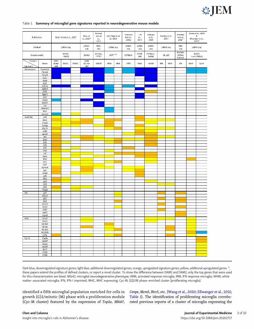
identified a fifth microglial population enriched for cells ingrowth (G)2/mitotic (M) phase with a proliferation module(Cyc-M cluster) featured by the expression of Top2a, Mki67,
Cenpe,Mcm5, Birc5, etc. (Wang et al., 2020; Ellwanger et al., 2021;Table 1). The identification of proliferating microglia corrobo-rated previous reports of a cluster of microglia expressing the
Table 1. Summary of microglial gene signatures reported in neurodegenerative mouse models
Dark blue, downregulated signature genes; light blue, additional downregulated genes; orange, upregulated signature genes; yellow, additional upregulated genes. *,these papers extend the profiles of defined clusters, or report a novel cluster. To show the difference between DAM1 and DAM2, only the top genes that were usedfor this characterization are listed. MGnD, microglial neurodegenerative phenotype; ARM, activated response microglia; IRM, IFN response microglia; WAM, whitematter–associated microglia; IFN, IFN-I imprinted; MHC, MHC expressing; Cyc-M, (G)2/M phase–enriched cluster (proliferating microglia).
Chen and Colonna Journal of Experimental Medicine 3 of 10
Insight into microglia’s role in Alzheimer’s disease https://doi.org/10.1084/jem.20202717

proliferation marker Ki67 around Aβ plaques in 5xFAD mice(Wang et al., 2016) and of a limited increase of mitotic genes inneurodegenerative models (Friedman et al., 2018). In one of thesescRNA-seq studies, a trajectory analysis further addressed therelationships among all microglial populations (Ellwanger et al.,2021), showing that homeostatic microglia differentiate through acontinuum of progressively activated states, which ultimatelybranch into four separate trajectories: DAM, IFN-I, MHC(expressing both MHC-II and MHC-I genes), and proliferating(Cyc-M). Further studies will be necessary to verify whetherany of these trajectories convert into another at some point. Therepresentation of all four terminal fates was reduced in 5xFAD ×Trem2−/− mice, demonstrating a general requirement of TREM2for microglia activation. However, a single injection of anti-TREM2 antibody in adult 5xFAD mice with advanced Aβ ag-gregation selectively expanded proliferating microglia and hadlittle effect on other differentiation fates (Wang et al., 2020;Ellwanger et al., 2021). Since Trem2−/− mice have a constitutivemicroglial defect that precedes the onset of Aβ aggregation, thelimited impact of antibody-mediated TREM2 activation onmicroglia may be dictated by the late and transient timing ofthis intervention. Early and/or sustained TREM2 engagementmay be necessary to induce the DAM, IFN-I, and MHC-II mi-croglial populations.
DAM-like signatures in development and agingMicroglia responses to AD pathology recapitulate, at least inpart, microglial function during CNS development, such asphagocytosis of apoptotic cells and damaged myelin. Thus, it isnot surprising that the “DAM” paradigm has been confirmed byscRNA-seq during CNS development. One example is the axontract-associated microglia, which appear in postnatal day 4 (P4)/P5 mice but are absent in embryonic day 14.5 mouse embryosand P30-P100 adult mice (Hammond et al., 2019; Table 1 andFig. 1). These microglia, which were found along axon tracts thatbecome heavily myelinated and were shown to prune synapsesin a transient time window of brain development (Schafer andStevens, 2015), exhibited a DAM-like expression profile. SimilarDAM-like microglia were reported to be present in P7 mice inassociation with postnatal myelination and scarce in adult mice(Li et al., 2019; Table 1 and Fig. 1). Remarkably, this study showedthat a lack of TREM2 or APOE, both of which affect DAM dif-ferentiation in AD pathology, has no impact on DAM-likemicroglia during development. It is possible that myelin andsynapse debris trigger pathways of activation partially differentfrom those elicited by Aβ aggregates. Moreover, other glial cellsor neuronal cells may produce soluble factors during develop-ment that can compensate for TREM2 and/or APOE deficiency(Li and Barres, 2018). Another report showed that activatedmicroglia with a DAM signature are present in the mouse em-bryonic brain but diminish postnatally, allowing another mi-croglia population expressing Cst3 and Sparc to populate thepostnatal brain (Masuda et al., 2019). Altogether, these studiesconcur that waves of activated microglia with a DAM-like profiledifferentiate during critical stages of CNS development tofacilitate recycling of lipid components resulting from remod-eling of neuronal synapses and myelin (Fig. 1).
Physiological aging is associated with an accumulation ofdegenerative features in the CNS, which also elicit the dif-ferentiation of DAM, although with delayed kinetics com-pared with AD pathology (Fig. 1). Accordingly, a recent studyidentified a microglia population within the white matter thatdevelops with aging, partially overlaps with DAM, and mayeventually progress into DAM with further accumulation oftissue damage (Safaiyan et al., 2021; Table 1). This signaturewas affected by TREM2 deficiency, but not by APOE deficiency,unless the aged mice also carried the APP/PS1 transgene medi-ating Aβ accumulation (Safaiyan et al., 2021), suggesting that theimpact of APOE on the DAM signature during aging is minimalunless AD pathology is present (Krasemann et al., 2017; Wanget al., 2021).
DAM-like signatures in microglia repopulationMicroglia development and maintenance is dependent on en-gagement of the CSF1 receptor by IL-34 secreted by neurons andCSF1 released by astrocytes and microglia themselves (Chituet al., 2016). Administration of small molecules that inhibitCSF1R, such as PLX5622, induce a profound depletion of mi-croglia, which is reversed upon suspension of treatment; re-sidual microglia proliferate and repopulate the CNS (Bruttgeret al., 2015; Huang et al., 2018), although the origin of theproliferating cells is a matter of debate (Elmore et al., 2014;Jakel and Dimou, 2017; Askew et al., 2017). A recent scRNA-seqanalysis of microglia 2 d after removal of PLX5622 divulged adecline in the homeostatic signature, paralleled by the ap-pearance of three distinct signatures: one enriched in mitosismarkers, a second expressing MHC-II, and a third DAM-like sig-nature with elevated expression of chemokines (Zhan et al., 2020).All subsets expressed Galectin-3 (MAC2 or Lgals3), which hasbeen reported to be a ligand for TREM2 (Boza-Serrano et al.,2019). Why repopulating microglia acquire MHC-II and DAM-like characteristics rather than homeostatic features is not clear.Given that CSF1R inhibitors are being tested as potential thera-peutic agents in the treatment of AD and neurodegenerativediseases (Waisman et al., 2015; Shi et al., 2019; Spangenberget al., 2019; Casali et al., 2020; Green et al., 2020), it will beimportant to establish whether and how repopulating microgliamodulate pathology.
Discrepancies between human andmousemicroglia signaturesin neurodegenerationMicroglial signatures obtained from snRNA-seq of human ADbrain specimens evince considerable heterogeneity and seem todiffer from mouse signatures. In an initial study, AD-enrichedmodules mainly included genes for myelination and neuronalsurvival, whereas no DAM signature was observed (Mathyset al., 2019; Table 2). Major gene expression changes in-cluded (1) up-regulation of heat shock protein chaperones,perhaps indicative of a reported unfolded protein response totau aggregation (Ballatore et al., 2007; Ittner and Gotz, 2011); (2)up-regulation of the neuron apoptosis regulator LINGO1; (3) down-regulation of the neuron regeneration genes NEGR1, BEX1, andNTING1; and (4) sex bias for some AD-related changes, including amore drastic reduction of inhibitory neurons in females than in
Chen and Colonna Journal of Experimental Medicine 4 of 10
Insight into microglia’s role in Alzheimer’s disease https://doi.org/10.1084/jem.20202717

males. Only a fewmicroglia genes, including APOE and SPP1, wereupregulated (Table 2). A subsequent study in which more nuclei/specimens were sequenced identified an incomplete DAM patternwith up-regulation of TREM2, APOE, CD68, and HLA-DRA (Zhouet al., 2020; Table 2). Other key DAM genes were either unde-tected (CST7, GPNMB, and LPL) or downregulated (SPP1), whileexpression of some homeostatic signature genes was heightened(TMEM119, P2RY12, and CX3CR1). Enhanced expression of genes notreported in mouse models of AD was also observed. These in-cluded IRF8, a regulator of reactive microglia (Kierdorf et al.,2013); SORL1, an AD risk factor involved in APOE uptake andAPP trafficking (Rogaeva et al., 2007; Li et al., 2008); the chitinase-like protein CHI3L1, a CSF biomarker for preclinical AD; and Alpha-2-macroglobulin. Subsequent -omics analyses of human AD alsoreported an incomplete DAM signature. Few DAM signaturesgenes (ApoE and HLA-DRA), together with other genes such asCD163 and HIF1A, were also captured by another snRNA-seqstudy (Grubman et al., 2019; Table 2). In one study of pre-frontal cortex specimens, a small population of AD-specificmicroglia showed up-regulation of genes for complement(C1QA, C1QB, and C1QC) and cytokine receptors (IL4R and IL1-RAP; Lau et al., 2020; Table 2). In another study of patientscarrying PSEN1 mutations causing familial AD, the top fivedifferentially expressed microglial genes included EEF1A1,GLULL, KIAA1217, LDLRAD3, and SPP1 (Del-Aguila et al., 2019),together with ∼20 DAM genes (Table 2).
A novel snRNA-seq approach allowing for increased numbersof nuclei sequenced/specimen characterized two microglialpopulations; dystrophic microglia expressed genes for ironmetabolism (FTL and FTH1), and amyloid-responsive microgliaexpressed CD163, BIN1,MS4A6A, and CELF1 (Nguyen et al., 2020;Table 2). Although CD163 expression correlated with AD-associated APOE (APOE4) and TREM2 (TREM2R47H) poly-morphisms, the amyloid-responsive microglia evoked no DAMsignature except for a slight up-regulation of APOE, while other
DAM genes were downregulated. Conversely, the highest ex-pression of TREM2, APOC1, APOE, HLA-DRA, and HLA-DRB1 wasnoted in the dystrophic cluster. The relationship between thesemicroglial populations remains to be defined. The limitedoverlap between human and mouse AD microglial signatureswas corroborated by proteomics of human AD specimens,which revealed an AD-associated astrocyte-microglial metabo-lism module characterized by high expression of CD44, PRDX1,and DDAH2, while only few of the proteins in this modulemirrored the mouse DAM phenotype (Johnson et al., 2020).scRNA-seq profiles of surgically dissected human brain speci-mens from patients with multiple sclerosis revealed variousmicroglia populations expressing DAM genes, although mul-tiple sclerosis and AD are quite different diseases (Masuda et al.,2019); this implies that at least some of the DAM genes mayreflect a conserved pattern of microglial responses to differentCNS diseases, despite the heterogeneity of brain pathology.
Biological and technical bases for microglial profilesheterogeneities in ADOne biological reason for the differences between human andmouse microglial signatures is that human specimens representa terminal stage of ADwith amyloid and tau pathology, as well asextensive neuronal cell death (Kumar et al., 2015; Long andHoltzman, 2019); mouse specimens, however, often encapsu-late either earlier stages of the disease characterized mostprominently by Aβ accumulation or frontotemporal dementia–like tauopathy without amyloidosis (Gotz et al., 2018). To de-termine how human microglia respond to progressive stagesof AD with either amyloid pathology or tauopathy, a recentstudy performed snRNA-seq after separating the occipital cortex(Gerrits et al., 2021), which mainly develops amyloid pathology,from the occipitotemporal cortex, which accumulates both am-yloid aggregates and neuronal fibrillary tangles (Jucker andWalker, 2013, 2018). This approach led to the identification of
Figure 1. Temporal appearance of microglial subsets with activated signatures in mouse life span. Signatures of proliferative region–associated mi-croglia (PAM) and axon tract–associated microglia (ATM) transiently appear in early stages of postnatal development. The dashed line above ATM and PAMindicates the possibility that ATM and PAM may belong to the same wave of microglia activation. Moreover, microglia with DAM/MHC/IFN-I activationsignatures slowly increase with aging, a process that is accelerated in models of neurodegeneration.
Chen and Colonna Journal of Experimental Medicine 5 of 10
Insight into microglia’s role in Alzheimer’s disease https://doi.org/10.1084/jem.20202717

Table 2. Summary of human microglial gene signatures
Light blue, downregulated genes; yellow, upregulated genes; gray, not detected; orange, used in signature definition. Genes present in Table 1 but absent inTable 2 were not differentially expressed in human samples. The table includes a partial list of the main differentially expressed genes discovered in thereferenced papers. C1q&Cyto, C1q and cytokines; C, control; NA, not available.
Chen and Colonna Journal of Experimental Medicine 6 of 10
Insight into microglia’s role in Alzheimer’s disease https://doi.org/10.1084/jem.20202717

three populations: (1) homeostatic microglia expressing P2RY12and CX3CR1; (2) amyloid-associated microglia expressing DAMgenes; and (3) tauopathy-associated microglia expressing thegene for the glutamate receptor GRID2, which was highly enrichedaround neuritic plaques but may not be functional (Gerrits et al.,2021; Table 2). Future investigation of microglial genes in mousemodels of tauopathy are warranted to further advance the notionof tauopathy-associated microglia. Other biological reasons thatmay explain the heterogeneous detection of DAM signature genesin human AD are: (1) the different brain regions from which thespecimens originate; (2) the genetic diversity of AD patients; and(3) the disparate ethnic origins of the various cohorts analyzed,which may impact both the genetic background and environ-mental factors regulating AD.
There are also several technical reasons that can contribute tohuman–human and human–mouse discrepancies in the AD pro-files. While DAM were mainly identified by scRNA-seq, almost allmicroglial profiles in human AD were determined by snRNA-seq.The number of microglia nuclei sequenced may be insufficient toidentify a DAMcluster (Table 2); indeed, DAMand IFN-I signatureswere detected in AD patients by bulk RNA-seq, which providesmore depth (Sobue et al., 2021), and SPP1 was detected in twostudies in which large numbers of nuclei were sequenced (Mathyset al., 2019; Del-Aguila et al., 2019). Additionally, mRNAs may beless abundant in the nucleus vs. cytosol; accordingly, mRNA ofkey DAM markers was less abundant in nuclei than in wholecells obtained from the same individuals by neocortical resection(Thrupp et al., 2020). Finally, the overall quality of human RNAsamples may be poorer than mouse samples due to postmortemintervals preceding human sample collection and processing. In-deed, a study showed that human postmortem samples lost theexpression of certain mRNA, including SPP1 and SOCS3 (Dachetet al., 2021).Moreover, this study showed an up-regulation of CD68in microglia with postponed fixation for tissue staining. Studies ofhuman samples promptly collected after neurosurgery or autopsyand analyzed by scRNA-seq have reported microglial profiles moresimilar to those identified in mouse models (Masuda et al., 2019;Olah et al., 2020). Thus, differences in tissue collection and pro-cessing may contribute to heterogeneity of human samples andhuman–mouse discrepancies.
Innovative approaches for investigating humanmicroglia in ADBeyond high-resolution transcriptional profiling, several additionalapproaches have been developed to investigate human microglia inAD and understand the impact of AD risk variants in their func-tions. Microglia have been generated from human induced plurip-otent stem cells (iPSCs) and embryonic stem cells (ESCs; Douvaraset al., 2017; Abud et al., 2017; Haenseler and Rajendran, 2019). DAM-like signatures were observed in microglia from human ESCs inwhich familial AD-associated mutations were introduced in APPand PSEN1 genes (Liu et al., 2020). TREM2 dependency of DAMwas reproduced in TREM2-sufficient and TREM2-deficient humaniPSCs (Reich et al., 2021). Since culture conditions may not fullyreproduce the complexity of CNS microenvironment (Gosselinet al., 2017), iPSC- and ESC-derived microglia have been trans-planted into the mouse CNS (Hasselmann et al., 2019; Mancusoet al., 2019; McQuade et al., 2020). Transplanted human ESC–
derived microglia not only retained a transcriptome profile distinctfrom that of endogenousmousemicroglia for 2mo after intracranialinjection (Hasselmann et al., 2019) but also recapitulated some keymicroglia patterns observed in human AD samples when injectedtogether with oligomeric Aβ (Mancuso et al., 2019). Moreover,microglia from human iPSCs with or without the TREM2 genewere injected into the brain of humanized 5xFAD × MITRGxenotransplantation-compatible mice (CSF1, CSF2, TPO, Rag2knockout, and Il2rg- knockout); comparison of the two revealedTREM2-dependent DAM- and MHC-II–related signatures (Hasselmannet al., 2019; McQuade et al., 2020).
While genetic studies have identified many risk variants forhuman AD potentially affecting microglia, most of the studieshave focused on coding variants of APOE and TREM2. However,many AD risk variants are present in noncoding regions of thehuman genome, affecting enhancers regulating gene expressioninmicroglia (Nott et al., 2019). The introduction of genome-wideanalyses of chromatin-accessible regions and histone mod-ifications coupled with single-cell analyses has significantlyadvanced our understanding of the microglia regulome and howit is affected by AD risk variants (Gosselin et al., 2014, 2017). Forexample, although the bridging integrator 1 (BIN1) gene is ex-pressed in both neurons and microglia, it has been shown that theAD risk variant of BIN1 affects an enhancer active inmicroglia, butnot in neurons, suggesting that this variant predisposes to AD byaffecting microglial rather than neuronal functions (Nott et al.,2019). The precise definition of promoter–enhancer regulomesin humans and mice may also explain some of the discrepanciesbetween human AD and mouse models.
Finally, because requiring disruption of tissues into cell ornuclei suspensions, scRNA-seq and snRNA-seq analyses cannotdefine the precise location of microglia subsets and signatureswithin CNS niches and potential interactions with other cells.However, novel technologies have been developed to overcomethis limitation; for example, spatial transcriptomics has beenapplied to study AppNL-G-F mice, which confirmed the associationbetween DAM and amyloid plaques (Chen et al., 2020b). Strate-gies that combine multiplexed fluorescence in situ hybridizationwith sequencing, known as seqFISH and MERFISH, have alsobeen developed (Moffitt et al., 2018; Eng et al., 2019). For thesetechniques, RNAs are imprinted with oligo-conjugated barcodesthat are measured through sequential rounds of hybridizationand super-resolution imaging. Application of such technologies toneurodegeneration will provide information on individual cellswithin their environment of surrounding cells and AD pathology.
ConclusionsOverall, the studies in mouse models discussed here have shownfundamental and consistent patterns of microglial activation inresponse to AD pathology: DAM, IFN-I microglia, MHC-II mi-croglia, and proliferating (Cyc-M) microglia. We conclude thatall currently characterized microglia subsets correspond to ei-ther one of them or a combination, depending in part on theclustering strategy applied to the analysis, as well as the ADmodel being assessed (Table 1). The DAM, MHC-II, and IFN-Isignatures appear not only in models of neurodegeneration butalso during development and in aged mice, although to a lesser
Chen and Colonna Journal of Experimental Medicine 7 of 10
Insight into microglia’s role in Alzheimer’s disease https://doi.org/10.1084/jem.20202717

extent (Fig. 1). On the contrary, human microglia are moreheterogeneous (Table 2). Beyond a detailed description of dif-ferent microglia subsets, it will be essential to investigate thebiological functions of these populations and determine whetherthey have a beneficial or detrimental impact in AD progression.DAM genes encode TREM2 and phagocytic molecules that mayslow down the progression of disease. However, other molecules,such as inflammatory cytokines and chemokines, have not beenthoroughly studied. MHC-II expressed by microglia in AD maypresent antigens. Studies have shown a strong correlation be-tween expression of MHC-II and ApoE in the cuprizone model ofdemyelination (Olah et al., 2012; Poliani et al., 2015; Masudaet al., 2019). Moreover, microglial secretion of ApoE may con-tribute to capture of lipoprotein antigens that can be processedand presented (Bonacina et al., 2018). Thus, it is possible thatApoE+MHC-II+ microglia present lipoprotein antigens duringaging and neurodegenerative diseases.
Although the studies reviewed here mainly focus on mi-croglia, high-resolution single-cell and single-nuclei analysesin neurodegenerative and neuroinflammatory diseases are cur-rently being extended to other brain macrophages and CCR2+
monocytes, which might infiltrate the CNS and convert intomicroglia-like cells in certain contexts (Mrdjen et al., 2018; Chenet al., 2020a). A wealth of transcriptome single-cell data are alsobecoming available for other glial cells, such as astrocytes andoligodendrocytes, as well as neurons (Mathys et al., 2019; Mundtet al., 2019; Jordão et al., 2019; Zhou et al., 2020; Habib et al.,2020; Leng et al., 2021). Integration of these analyses with mi-croglia data will provide a global view of cellular responses to ADpathology in humans and mice.
AcknowledgmentsWe thank Susan Gilfillan and Yingyue Zhou for helpful suggestions.
This work was supported by the National Institutes of Health(grants RF1 AG051485, R21 AG059176, and RF1 AG059082 to M.Colonna).
Author contributions: Y. Chen and M. Colonna wrote themanuscript.Disclosures: M. Colonna reported grants from NIH during theconduct of the study and received research support from Alec-tor, Amgen, Ono, and Pfizer; in addition, M. Colonna is a sci-entific advisory board member of Vigil and NGMBio, is aconsultant for Cell Signaling Technologies, and has a patent toTREM2 pending. No other disclosures were reported.
Submitted: 18 April 2021Revised: 2 June 2021Accepted: 9 June 2021
ReferencesAbud, E.M., R.N. Ramirez, E.S. Martinez, L.M. Healy, C.H.H. Nguyen, S.A.
Newman, A.V. Yeromin, V.M. Scarfone, S.E.Marsh, C. Fimbres, et al. 2017.iPSC-Derived Human Microglia-like Cells to Study Neurological Diseases.Neuron. 94:278–293.e9. https://doi.org/10.1016/j.neuron.2017.03.042
Ashok, B.S., T.A. Ajith, and S. Sivanesan. 2017. Hypoxia-inducible factors asneuroprotective agent in Alzheimer’s disease. Clin. Exp. Pharmacol.Physiol. 44:327–334. https://doi.org/10.1111/1440-1681.12717
Askew, K., K. Li, A. Olmos-Alonso, F. Garcia-Moreno, Y. Liang, P. Richardson,T. Tipton, M.A. Chapman, K. Riecken, S. Beccari, et al. 2017. CoupledProliferation and Apoptosis Maintain the Rapid Turnover of Microgliain the Adult Brain. Cell Rep. 18:391–405. https://doi.org/10.1016/j.celrep.2016.12.041
Ballatore, C., V.M.-Y. Lee, and J.Q. Trojanowski. 2007. Tau-mediated neu-rodegeneration in Alzheimer’s disease and related disorders. Nat. Rev.Neurosci. 8:663–672. https://doi.org/10.1038/nrn2194
Bertram, L., and R.E. Tanzi. 2009. Genome-wide association studies in Alz-heimer’s disease. Hum. Mol. Genet. 18(R2):R137–R145. https://doi.org/10.1093/hmg/ddp406
Bonacina, F., D. Coe, G. Wang, M.P. Longhi, A. Baragetti, A. Moregola, K.Garlaschelli, P. Uboldi, F. Pellegatta, L. Grigore, et al. 2018. Myeloidapolipoprotein E controls dendritic cell antigen presentation and T cellactivation. Nat. Commun. 9:3083. https://doi.org/10.1038/s41467-018-05322-1
Boza-Serrano, A., R. Ruiz, R. Sanchez-Varo, J. Garcıa-Revilla, Y. Yang, I. Ji-menez-Ferrer, A. Paulus, M. Wennstrom, A. Vilalta, D. Allendorf, et al.2019. Galectin-3, a novel endogenous TREM2 ligand, detrimentallyregulates inflammatory response in Alzheimer’s disease. Acta Neuro-pathol. 138:251–273. https://doi.org/10.1007/s00401-019-02013-z
Bruttger, J., K. Karram, S. Wortge, T. Regen, F. Marini, N. Hoppmann, M.Klein, T. Blank, S. Yona, Y. Wolf, et al. 2015. Genetic Cell Ablation Re-veals Clusters of Local Self-Renewing Microglia in the MammalianCentral Nervous System. Immunity. 43:92–106. https://doi.org/10.1016/j.immuni.2015.06.012
Butovsky, O., M.P. Jedrychowski, C.S. Moore, R. Cialic, A.J. Lanser, G. Ga-briely, T. Koeglsperger, B. Dake, P.M. Wu, C.E. Doykan, et al. 2014.Identification of a unique TGF-β-dependent molecular and functionalsignature in microglia. Nat. Neurosci. 17:131–143. https://doi.org/10.1038/nn.3599
Casali, B.T., K.P. MacPherson, E.G. Reed-Geaghan, and G.E. Landreth. 2020.Microglia depletion rapidly and reversibly alters amyloid pathology bymodification of plaque compaction and morphologies. Neurobiol. Dis.142:104956. https://doi.org/10.1016/j.nbd.2020.104956
Chen, H.-R., Y.-Y. Sun, C.-W. Chen, Y.-M. Kuo, I.S. Kuan, Z.-R. Tiger Li, J.C.Short-Miller, M.R. Smucker, and C.-Y. Kuan. 2020a. Fate mapping viaCCR2-CreER mice reveals monocyte-to-microglia transition in devel-opment and neonatal stroke. Sci. Adv. 6:eabb2119. https://doi.org/10.1126/sciadv.abb2119
Chen, W.-T., A. Lu, K. Craessaerts, B. Pavie, C. Sala Frigerio, N. Corthout, X.Qian, J. Lalakova, M. Kühnemund, I. Voytyuk, et al. 2020b. SpatialTranscriptomics and In Situ Sequencing to Study Alzheimer’s Disease.Cell. 182:976–991.e19. https://doi.org/10.1016/j.cell.2020.06.038
Chen, Y., M.R. Strickland, A. Soranno, and D.M. Holtzman. 2021. Apolipo-protein E: Structural Insights and Links to Alzheimer Disease Patho-genesis. Neuron. 109:205–221. https://doi.org/10.1016/j.neuron.2020.10.008
Chitu, V., S. Gokhan, S. Nandi, M.F. Mehler, and E.R. Stanley. 2016. EmergingRoles for CSF-1 Receptor and its Ligands in the Nervous System. TrendsNeurosci. 39:378–393. https://doi.org/10.1016/j.tins.2016.03.005
Cruz, J.C., H.-C. Tseng, J.A. Goldman, H. Shih, and L.-H. Tsai. 2003. AberrantCdk5 activation by p25 triggers pathological events leading to neuro-degeneration and neurofibrillary tangles. Neuron. 40:471–483. https://doi.org/10.1016/S0896-6273(03)00627-5
Cuyvers, E., and K. Sleegers. 2016. Genetic variations underlying Alz-heimer’s disease: evidence from genome-wide association studiesand beyond. Lancet Neurol. 15:857–868. https://doi.org/10.1016/S1474-4422(16)00127-7
Dachet, F., J.B. Brown, T. Valyi-Nagy, K.D. Narayan, A. Serafini, N. Boley, T.R.Gingeras, S.E. Celniker, G. Mohapatra, and J.A. Loeb. 2021. Selectivetime-dependent changes in activity and cell-specific gene expression inhuman postmortem brain. Sci. Rep. 11:6078. https://doi.org/10.1038/s41598-021-85801-6
Deczkowska, A., H. Keren-Shaul, A. Weiner, M. Colonna, M. Schwartz, and I.Amit. 2018. Disease-Associated Microglia: A Universal Immune Sensorof Neurodegeneration. Cell. 173:1073–1081. https://doi.org/10.1016/j.cell.2018.05.003
Del-Aguila, J.L., Z. Li, U. Dube, K.A. Mihindukulasuriya, J.P. Budde, M.V.Fernandez, L. Ibanez, J. Bradley, F. Wang, K. Bergmann, et al. 2019. Asingle-nuclei RNA sequencing study of Mendelian and sporadic AD inthe human brain. Alzheimers Res. Ther. 11:71. https://doi.org/10.1186/s13195-019-0524-x
Douvaras, P., B. Sun, M. Wang, I. Kruglikov, G. Lallos, M. Zimmer, C.Terrenoire, B. Zhang, S. Gandy, E. Schadt, et al. 2017. Directed
Chen and Colonna Journal of Experimental Medicine 8 of 10
Insight into microglia’s role in Alzheimer’s disease https://doi.org/10.1084/jem.20202717
Differentiation of Human Pluripotent Stem Cells to Microglia. Stem CellReports. 8:1516–1524. https://doi.org/10.1016/j.stemcr.2017.04.023
Ellwanger, D.C., S. Wang, S. Brioschi, Z. Shao, L. Green, R. Case, D. Yoo, D.Weishuhn, P. Rathanaswami, J. Bradley, et al. 2021. Prior activationstate shapes the microglia response to antihuman TREM2 in a mousemodel of Alzheimer’s disease. Proc. Natl. Acad. Sci. USA. 118:e2017742118.https://doi.org/10.1073/pnas.2017742118
Elmore, M.R.P., A.R. Najafi, M.A. Koike, N.N. Dagher, E.E. Spangenberg, R.A.Rice, M. Kitazawa, B. Matusow, H. Nguyen, B.L. West, and K.N. Green.2014. Colony-stimulating factor 1 receptor signaling is necessary formicroglia viability, unmasking a microglia progenitor cell in the adultbrain. Neuron. 82:380–397. https://doi.org/10.1016/j.neuron.2014.02.040
Eng, C.L., M. Lawson, Q. Zhu, R. Dries, N. Koulena, Y. Takei, J. Yun, C. Cronin,C. Karp, G.-C. Yuan, and L. Cai. 2019. Transcriptome-scale super-resolved imaging in tissues by RNA seqFISH. Nature. 568:235–239.https://doi.org/10.1038/s41586-019-1049-y
Friedman, B.A., K. Srinivasan, G. Ayalon,W.J. Meilandt, H. Lin, M.A. Huntley,Y. Cao, S.-H. Lee, P.C.G. Haddick, H. Ngu, et al. 2018. Diverse BrainMyeloid Expression Profiles Reveal Distinct Microglial Activation Statesand Aspects of Alzheimer’s Disease Not Evident in Mouse Models. CellRep. 22:832–847. https://doi.org/10.1016/j.celrep.2017.12.066
Gerrits, E., N. Brouwer, S.M. Kooistra, M.E. Woodbury, Y. Vermeiren, M.Lambourne, J. Mulder, M. Kummer, T. Moller, K. Biber, et al. 2021.Distinct amyloid-β and tau-associated microglia profiles in Alzheimer’sdisease. Acta Neuropathol. 141:681–696. https://doi.org/10.1007/s00401-021-02263-w
Ginhoux, F., M. Greter, M. Leboeuf, S. Nandi, P. See, S. Gokhan, M.F. Mehler,S.J. Conway, L.G. Ng, E.R. Stanley, et al. 2010. Fate mapping analysisreveals that adultmicroglia derive from primitivemacrophages. Science.330:841–845. https://doi.org/10.1126/science.1194637
Gosselin, D., V.M. Link, C.E. Romanoski, G.J. Fonseca, D.Z. Eichenfield, N.J.Spann, J.D. Stender, H.B. Chun, H. Garner, F. Geissmann, and C.K. Glass.2014. Environment drives selection and function of enhancers con-trolling tissue-specific macrophage identities. Cell. 159:1327–1340.https://doi.org/10.1016/j.cell.2014.11.023
Gosselin, D., D. Skola, N.G. Coufal, I.R. Holtman, J.C.M. Schlachetzki, E. Sajti,B.N. Jaeger, C. O’Connor, C. Fitzpatrick, M.P. Pasillas, et al. 2017. Anenvironment-dependent transcriptional network specifies human mi-croglia identity. Science. 356:eaal3222. https://doi.org/10.1126/science.aal3222
Gotz, J., L.-G. Bodea, and M. Goedert. 2018. Rodent models for Alzheimerdisease. Nat. Rev. Neurosci. 19:583–598. https://doi.org/10.1038/s41583-018-0054-8
Green, K.N., J.D. Crapser, and L.A. Hohsfield. 2020. To Kill a Microglia: ACase for CSF1R Inhibitors. Trends Immunol. 41:771–784. https://doi.org/10.1016/j.it.2020.07.001
Grubman, A., G. Chew, J.F. Ouyang, G. Sun, X.Y. Choo, C. McLean, R.K.Simmons, S. Buckberry, D.B. Vargas-Landin, D. Poppe, et al. 2019. Asingle-cell atlas of entorhinal cortex from individuals with Alzheimer’sdisease reveals cell-type-specific gene expression regulation. Nat.Neurosci. 22:2087–2097. https://doi.org/10.1038/s41593-019-0539-4
Habib, N., C. McCabe, S. Medina, M. Varshavsky, D. Kitsberg, R. Dvir-Szternfeld, G. Green, D. Dionne, L. Nguyen, J.L. Marshall, et al. 2020.Disease-associated astrocytes in Alzheimer’s disease and aging.Nat. Neurosci.23:701–706. https://doi.org/10.1038/s41593-020-0624-8
Haenseler, W., and L. Rajendran. 2019. Concise review: Modeling neurode-generative diseases with human pluripotent stem cell-derived mi-croglia: Modeling neurodegenerative diseases with HPSC. Stem Cells. 37:724–730. https://doi.org/10.1002/stem.2995
Hammond, T.R., C. Dufort, L. Dissing-Olesen, S. Giera, A. Young, A. Wysoker,A.J. Walker, F. Gergits, M. Segel, J. Nemesh, et al. 2019. Single-Cell RNASequencing of Microglia throughout the Mouse Lifespan and in theInjured Brain Reveals Complex Cell-State Changes. Immunity. 50:253–271.e6.https://doi.org/10.1016/j.immuni.2018.11.004
Hansen, D.V., J.E. Hanson, and M. Sheng. 2018. Microglia in Alzheimer’sdisease. J. Cell Biol. 217:459–472. https://doi.org/10.1083/jcb.201709069
Hasselmann, J., M.A. Coburn, W. England, D.X. Figueroa Velez, S. KianiShabestari, C.H. Tu, A. McQuade, M. Kolahdouzan, K. Echeverria, C.Claes, et al. 2019. Development of a Chimeric Model to Study and Ma-nipulate Human Microglia In Vivo. Neuron. 103:1016–1033.e10. https://doi.org/10.1016/j.neuron.2019.07.002
Huang, Y., Z. Xu, S. Xiong, F. Sun, G. Qin, G. Hu, J. Wang, L. Zhao, Y.-X. Liang,T. Wu, et al. 2018. Repopulated microglia are solely derived from theproliferation of residual microglia after acute depletion. Nat. Neurosci.21:530–540. https://doi.org/10.1038/s41593-018-0090-8
Ittner, L.M., and J. Gotz. 2011. Amyloid-β and tau--a toxic pas de deux inAlzheimer’s disease. Nat. Rev. Neurosci. 12:65–72. https://doi.org/10.1038/nrn2967
Jakel, S., and L. Dimou. 2017. Glial Cells and Their Function in the Adult Brain:A Journey through the History of Their Ablation. Front. Cell. Neurosci. 11:24. https://doi.org/10.3389/fncel.2017.00024
Johnson, E.C.B., E.B. Dammer, D.M. Duong, L. Ping, M. Zhou, L. Yin, L.A.Higginbotham, A. Guajardo, B. White, J.C. Troncoso, et al. 2020. Large-scale proteomic analysis of Alzheimer’s disease brain and cerebrospinalfluid reveals early changes in energy metabolism associated with mi-croglia and astrocyte activation. Nat. Med. 26:769–780. https://doi.org/10.1038/s41591-020-0815-6
Jordão, M.J.C., R. Sankowski, S.M. Brendecke, G. Sagar, G. Locatelli, Y.H. Tai,T.L. Tay, E. Schramm, S. Armbruster, N. Hagemeyer, et al. 2019. Single-cell profiling identifies myeloid cell subsets with distinct fates dur-ing neuroinflammation. Science. 363:eaat7554. https://doi.org/10.1126/science.aat7554
Jucker, M., and L.C. Walker. 2013. Self-propagation of pathogenic proteinaggregates in neurodegenerative diseases.Nature. 501:45–51. https://doi.org/10.1038/nature12481
Jucker, M., and L.C. Walker. 2018. Propagation and spread of pathogenicprotein assemblies in neurodegenerative diseases. Nat. Neurosci. 21:1341–1349. https://doi.org/10.1038/s41593-018-0238-6
Keren-Shaul, H., A. Spinrad, A. Weiner, O. Matcovitch-Natan, R. Dvir-Szternfeld, T.K. Ulland, E. David, K. Baruch, D. Lara-Astaiso, B. Toth,et al. 2017. A Unique Microglia Type Associated with Restricting De-velopment of Alzheimer’s Disease. Cell. 169:1276–1290.e17. https://doi.org/10.1016/j.cell.2017.05.018
Kierdorf, K., D. Erny, T. Goldmann, V. Sander, C. Schulz, E.G. Perdiguero, P.Wieghofer, A. Heinrich, P. Riemke, C. Holscher, et al. 2013. Microgliaemerge from erythromyeloid precursors via Pu.1- and Irf8-dependentpathways. Nat. Neurosci. 16:273–280. https://doi.org/10.1038/nn.3318
Krasemann, S., C. Madore, R. Cialic, C. Baufeld, N. Calcagno, R. El Fatimy, L.Beckers, E. O’Loughlin, Y. Xu, Z. Fanek, et al. 2017. The TREM2-APOEPathway Drives the Transcriptional Phenotype of Dysfunctional Mi-croglia in Neurodegenerative Diseases. Immunity. 47:566–581.e9. https://doi.org/10.1016/j.immuni.2017.08.008
Kumar, A., A. Singh, and Ekavali. 2015. A review on Alzheimer’s diseasepathophysiology and its management: an update. Pharmacol. Rep. 67:195–203. https://doi.org/10.1016/j.pharep.2014.09.004
Kunkle, B.W., B. Grenier-Boley, R. Sims, J.C. Bis, V. Damotte, A.C. Naj, A.Boland,M. Vronskaya, S.J. van der Lee, A. Amlie-Wolf, et al. Genetic andEnvironmental Risk in AD/Defining Genetic, Polygenic and Environ-mental Risk for Alzheimer’s Disease Consortium (GERAD/PERADES).2019. Genetic meta-analysis of diagnosed Alzheimer’s disease identifiesnew risk loci and implicates Aβ, tau, immunity and lipid processing.Nat. Genet. 51:414–430. https://doi.org/10.1038/s41588-019-0358-2
Lau, S.-F., H. Cao, A.K.Y. Fu, and N.Y. Ip. 2020. Single-nucleus transcriptomeanalysis reveals dysregulation of angiogenic endothelial cells and neu-roprotective glia in Alzheimer’s disease. Proc. Natl. Acad. Sci. USA. 117:25800–25809. https://doi.org/10.1073/pnas.2008762117
Leng, K., E. Li, R. Eser, A. Piergies, R. Sit, M. Tan, N. Neff, S.H. Li, R.D. Ro-driguez, C.K. Suemoto, et al. 2021. Molecular characterization of se-lectively vulnerable neurons in Alzheimer’s disease. Nat. Neurosci. 24:276–287. https://doi.org/10.1038/s41593-020-00764-7
Li, Q., and B.A. Barres. 2018. Microglia andmacrophages in brain homeostasisand disease. Nat. Rev. Immunol. 18:225–242. https://doi.org/10.1038/nri.2017.125
Li, Y., C. Rowland, J. Catanese, J. Morris, S. Lovestone, M.C. O’Donovan, A.Goate, M. Owen, J. Williams, and A. Grupe. 2008. SORL1 variants andrisk of late-onset Alzheimer’s disease.Neurobiol. Dis. 29:293–296. https://doi.org/10.1016/j.nbd.2007.09.001
Li, Q., Z. Cheng, L. Zhou, S. Darmanis,N.F.Neff, J. Okamoto, G.Gulati,M.L. Bennett,L.O. Sun, L.E. Clarke, et al. 2019. Developmental Heterogeneity of Microgliaand Brain Myeloid Cells Revealed by Deep Single-Cell RNA Sequencing.Neuron. 101:207–223.e10. https://doi.org/10.1016/j.neuron.2018.12.006
Liu, T., B. Zhu, Y. Liu, X. Zhang, J. Yin, X. Li, L. Jiang, A.P. Hodges, S.B.Rosenthal, L. Zhou, et al. 2020. Multi-omic comparison of Alzheimer’svariants in human ESC-derived microglia reveals convergence at APOE.J. Exp. Med. 217:e20200474. https://doi.org/10.1084/jem.20200474
Long, J.M., and D.M. Holtzman. 2019. Alzheimer Disease: An Update onPathobiology and Treatment Strategies. Cell. 179:312–339. https://doi.org/10.1016/j.cell.2019.09.001
Mancuso, R., J. Van Den Daele, N. Fattorelli, L. Wolfs, S. Balusu, O. Burton, A.Liston, A. Sierksma, Y. Fourne, S. Poovathingal, et al. 2019. Stem-cell-
Chen and Colonna Journal of Experimental Medicine 9 of 10
Insight into microglia’s role in Alzheimer’s disease https://doi.org/10.1084/jem.20202717

derived human microglia transplanted in mouse brain to study humandisease. Nat. Neurosci. 22:2111–2116. https://doi.org/10.1038/s41593-019-0525-x
Masuda, T., R. Sankowski, O. Staszewski, C. Bottcher, L. Amann, C. Sagar,C. Scheiwe, S. Nessler, P. Kunz, G. van Loo, et al. 2019. Spatial andtemporal heterogeneity of mouse and human microglia at single-cellresolution. Nature. 566:388–392. https://doi.org/10.1038/s41586-019-0924-x
Mathys, H., C. Adaikkan, F. Gao, J.Z. Young, E. Manet, M. Hemberg, P.L. DeJager, R.M. Ransohoff, A. Regev, and L.-H. Tsai. 2017. TemporalTracking of Microglia Activation in Neurodegeneration at Single-CellResolution. Cell Rep. 21:366–380. https://doi.org/10.1016/j.celrep.2017.09.039
Mathys, H., J. Davila-Velderrain, Z. Peng, F. Gao, S. Mohammadi, J.Z. Young,M. Menon, L. He, F. Abdurrob, X. Jiang, et al. 2019. Single-cell tran-scriptomic analysis of Alzheimer’s disease.Nature. 570:332–337. https://doi.org/10.1038/s41586-019-1195-2
McQuade, A., Y.J. Kang, J. Hasselmann, A. Jairaman, A. Sotelo, M. Coburn,S.K. Shabestari, J.P. Chadarevian, G. Fote, C.H. Tu, et al. 2020. Geneexpression and functional deficits underlie TREM2-knockout microgliaresponses in human models of Alzheimer’s disease. Nat. Commun. 11:5370. https://doi.org/10.1038/s41467-020-19227-5
Moffitt, J.R., D. Bambah-Mukku, S.W. Eichhorn, E. Vaughn, K. Shekhar, J.D.Perez, N.D. Rubinstein, J. Hao, A. Regev, C. Dulac, and X. Zhuang. 2018.Molecular, spatial, and functional single-cell profiling of the hypotha-lamic preoptic region. Science. 362:eaau5324. https://doi.org/10.1126/science.aau5324
Mrdjen, D., A. Pavlovic, F.J. Hartmann, B. Schreiner, S.G. Utz, B.P. Leung,I. Lelios, F.L. Heppner, J. Kipnis, D. Merkler, et al. 2018. High-Dimensional Single-Cell Mapping of Central Nervous System ImmuneCells Reveals Distinct Myeloid Subsets in Health, Aging, and Disease.Immunity. 48:599. https://doi.org/10.1016/j.immuni.2018.02.014
Mundt, S., D. Mrdjen, S.G. Utz, M. Greter, B. Schreiner, and B. Becher.2019. Conventional DCs sample and present myelin antigens in thehealthy CNS and allow parenchymal T cell entry to initiate neuro-inflammation. Sci. Immunol. 4:eaau8380. https://doi.org/10.1126/sciimmunol.aau8380
Nguyen, A.T., K. Wang, G. Hu, X. Wang, Z. Miao, J.A. Azevedo, E. Suh, V.M.Van Deerlin, D. Choi, K. Roeder, et al. 2020. APOE and TREM2 regulateamyloid-responsive microglia in Alzheimer’s disease. Acta Neuropathol.140:477–493. https://doi.org/10.1007/s00401-020-02200-3
Nott, A., I.R. Holtman, N.G. Coufal, J.C.M. Schlachetzki, M. Yu, R. Hu, C.Z.Han, M. Pena, J. Xiao, Y. Wu, et al. 2019. Brain cell type-specificenhancer-promoter interactome maps and disease-risk association.Science. 366:1134–1139. https://doi.org/10.1126/science.aay0793
Olah, M., S. Amor, N. Brouwer, J. Vinet, B. Eggen, K. Biber, and H.W.G.M.Boddeke. 2012. Identification of a microglia phenotype supportive ofremyelination. Glia. 60:306–321. https://doi.org/10.1002/glia.21266
Olah, M., V. Menon, N. Habib, M.F. Taga, Y. Ma, C.J. Yung, M. Cimpean, A.Khairallah, G. Coronas-Samano, R. Sankowski, et al. 2020. Single cellRNA sequencing of human microglia uncovers a subset associated withAlzheimer’s disease. Nat. Commun. 11:6129. https://doi.org/10.1038/s41467-020-19737-2
Poliani, P.L., Y. Wang, E. Fontana, M.L. Robinette, Y. Yamanishi, S. Gilfillan,and M. Colonna. 2015. TREM2 sustains microglial expansion duringaging and response to demyelination. J. Clin. Invest. 125:2161–2170. https://doi.org/10.1172/JCI77983
Prinz, M., S. Jung, and J. Priller. 2019. Microglia Biology: One Century ofEvolving Concepts. Cell. 179:292–311. https://doi.org/10.1016/j.cell.2019.08.053
Reich, M., I. Paris, M. Ebeling, N. Dahm, C. Schweitzer, D. Reinhardt, R.Schmucki, M. Prasad, F. Kochl, M. Leist, et al. 2021. Alzheimer’s RiskGene TREM2 Determines Functional Properties of New Type of HumaniPSC-Derived Microglia. Front. Immunol. 11:617860. https://doi.org/10.3389/fimmu.2020.617860
Rogaeva, E., Y. Meng, J.H. Lee, Y. Gu, T. Kawarai, F. Zou, T. Katayama, C.T.Baldwin, R. Cheng, H. Hasegawa, et al. 2007. The neuronal sortilin-related receptor SORL1 is genetically associated with Alzheimer dis-ease. Nat. Genet. 39:168–177. https://doi.org/10.1038/ng1943
Safaiyan, S., S. Besson-Girard, T. Kaya, L. Cantuti-Castelvetri, L. Liu, H. Ji, M.Schifferer, G. Gouna, F. Usifo, N. Kannaiyan, et al. 2021. White matteraging drives microglial diversity. Neuron. 109:1100–1117.e10. https://doi.org/10.1016/j.neuron.2021.01.027
Sala Frigerio, C., L. Wolfs, N. Fattorelli, N. Thrupp, I. Voytyuk, I. Schmidt, R.Mancuso, W.-T. Chen, M.E. Woodbury, G. Srivastava, et al. 2019. The
Major Risk Factors for Alzheimer’s Disease: Age, Sex, and Genes Mod-ulate the Microglia Response to Aβ Plaques. Cell Rep. 27:1293–1306.e6.https://doi.org/10.1016/j.celrep.2019.03.099
Schafer, D.P., and B. Stevens. 2015. Microglia Function in Central NervousSystem Development and Plasticity. Cold Spring Harb. Perspect. Biol. 7:a020545. https://doi.org/10.1101/cshperspect.a020545
Shi, Y., M. Manis, J. Long, K. Wang, P.M. Sullivan, J. Remolina Serrano, R.Hoyle, and D.M. Holtzman. 2019. Microglia drive APOE-dependentneurodegeneration in a tauopathy mouse model. J. Exp. Med. 216:2546–2561. https://doi.org/10.1084/jem.20190980
Shibata, N., T. Kawarai, J.H. Lee, H.-S. Lee, E. Shibata, C. Sato, Y. Liang, R.Duara, R.P. Mayeux, P.H. St George-Hyslop, and E. Rogaeva. 2006.Association studies of cholesterol metabolism genes (CH25H, ABCA1and CH24H) in Alzheimer’s disease. Neurosci. Lett. 391:142–146. https://doi.org/10.1016/j.neulet.2005.08.048
Sierra, A., O. Abiega, A. Shahraz, and H. Neumann. 2013. Janus-facedmicroglia: beneficial and detrimental consequences of microglialphagocytosis. Front. Cell. Neurosci. 7:6. https://doi.org/10.3389/fncel.2013.00006
Sobue, A., O. Komine, Y. Hara, F. Endo, H. Mizoguchi, S. Watanabe, S.Murayama, T. Saito, T.C. Saido, N. Sahara, et al. 2021. Microglial genesignature reveals loss of homeostatic microglia associated with neuro-degeneration of Alzheimer’s disease. Acta Neuropathol. Commun. 9:1.https://doi.org/10.1186/s40478-020-01099-x
Song, W.M., and M. Colonna. 2018. The identity and function of microglia inneurodegeneration. Nat. Immunol. 19:1048–1058. https://doi.org/10.1038/s41590-018-0212-1
Spangenberg, E., P.L. Severson, L.A. Hohsfield, J. Crapser, J. Zhang, E.A.Burton, Y. Zhang, W. Spevak, J. Lin, N.Y. Phan, et al. 2019. Sustainedmicroglial depletion with CSF1R inhibitor impairs parenchymal plaquedevelopment in an Alzheimer’s disease model. Nat. Commun. 10:3758.https://doi.org/10.1038/s41467-019-11674-z
Srinivasan, K., B.A. Friedman, A. Etxeberria, M.A. Huntley, M.P. van derBrug, O. Foreman, J.S. Paw, Z. Modrusan, T.G. Beach, G.E. Serrano, andD.V. Hansen. 2020. Alzheimer’s Patient Microglia Exhibit EnhancedAging and Unique Transcriptional Activation. Cell Rep. 31:107843.https://doi.org/10.1016/j.celrep.2020.107843
Thrupp, N., C. Sala Frigerio, L. Wolfs, N.G. Skene, N. Fattorelli, S. Poova-thingal, Y. Fourne, P.M. Matthews, T. Theys, R. Mancuso, et al. 2020.Single-Nucleus RNA-Seq Is Not Suitable for Detection of MicroglialActivation Genes in Humans. Cell Rep. 32:108189. https://doi.org/10.1016/j.celrep.2020.108189
Vainchtein, I.D., and A.V. Molofsky. 2020. Astrocytes and Microglia: InSickness and in Health. Trends Neurosci. 43:144–154. https://doi.org/10.1016/j.tins.2020.01.003
Villacampa, N., and M.T. Heneka. 2020. Microglia in Alzheimer’s disease:Local heroes! J. Exp. Med. 217:e20192311. https://doi.org/10.1084/jem.20192311
Waisman, A., F. Ginhoux, M. Greter, and J. Bruttger. 2015. Homeostasis ofMicroglia in the Adult Brain: Review of Novel Microglia DepletionSystems. Trends Immunol. 36:625–636. https://doi.org/10.1016/j.it.2015.08.005
Wang, Y., T.K. Ulland, J.D. Ulrich, W. Song, J.A. Tzaferis, J.T. Hole, P. Yuan,T.E. Mahan, Y. Shi, S. Gilfillan, et al. 2016. TREM2-mediated earlymicroglial response limits diffusion and toxicity of amyloid plaques.J. Exp. Med. 213:667–675. https://doi.org/10.1084/jem.20151948
Wang, S., M. Mustafa, C.M. Yuede, S.V. Salazar, P. Kong, H. Long, M.Ward, O. Siddiqui, R. Paul, S. Gilfillan, et al. 2020. Anti-humanTREM2 induces microglia proliferation and reduces pathology inan Alzheimer’s disease model. J. Exp. Med. 217:e20200785. https://doi.org/10.1084/jem.20200785
Wang, C., M. Xiong, M. Gratuze, X. Bao, Y. Shi, P.S. Andhey, M. Manis, C.Schroeder, Z. Yin, C. Madore, et al. 2021. Selective removal of astrocyticAPOE4 strongly protects against tau-mediated neurodegeneration anddecreases synaptic phagocytosis by microglia. Neuron. 109:1657–1674.e7.https://doi.org/10.1016/j.neuron.2021.03.024
Zhan, L., L. Fan, L. Kodama, P.D. Sohn, M.Y. Wong, G.A. Mousa, Y. Zhou, Y.Li, and L. Gan. 2020. A MAC2-positive progenitor-like microglial pop-ulation is resistant to CSF1R inhibition in adult mouse brain. eLife. 9:e51796. https://doi.org/10.7554/eLife.51796
Zhou, Y., W.M. Song, P.S. Andhey, A. Swain, T. Levy, K.R. Miller, P.L. Poliani,M. Cominelli, S. Grover, S. Gilfillan, et al. 2020. Human and mousesingle-nucleus transcriptomics reveal TREM2-dependent and TREM2-independent cellular responses in Alzheimer’s disease. Nat. Med. 26:131–142. https://doi.org/10.1038/s41591-019-0695-9
Chen and Colonna Journal of Experimental Medicine 10 of 10
Insight into microglia’s role in Alzheimer’s disease https://doi.org/10.1084/jem.20202717