Non-Destructive Testing of Aircraft Structures Using Microwire ...
Upload
independentCategory
view
1download
0
Article
USP18 lack in microglia causes destructiveinterferonopathy of the mouse brainTobias Goldmann1, Nicolas Zeller1, Jenni Raasch1, Katrin Kierdorf1, Kathrin Frenzel1, Lars Ketscher1,
Anja Basters1, Ori Staszewski1, Stefanie M Brendecke1, Alena Spiess1, Tuan Leng Tay1, Clemens Kreutz2,
Jens Timmer2,3, Grazia MS Mancini4, Thomas Blank1, Günter Fritz1, Knut Biber5,6, Roland Lang7,
Danielle Malo8, Doron Merkler9, Mathias Heikenwälder10, Klaus-Peter Knobeloch1,† & Marco Prinz1,3,*,†
Abstract
Microglia are tissue macrophages of the central nervous system(CNS) that control tissue homeostasis. Microglia dysregulation isthought to be causal for a group of neuropsychiatric, neurodegen-erative and neuroinflammatory diseases, called “microgliopathies”.However, how the intracellular stimulation machinery in microgliais controlled is poorly understood. Here, we identified the ubiquitin-specific protease (Usp) 18 in white matter microglia thatessentially contributes to microglial quiescence. We further foundthat microglial Usp18 negatively regulates the activation of Stat1and concomitant induction of interferon-induced genes, therebyterminating IFN signaling. The Usp18-mediated control was inde-pendent from its catalytic activity but instead required the inter-action with Ifnar2. Additionally, the absence of Ifnar1 restoredmicroglial activation, indicating a tonic IFN signal which needs tobe negatively controlled by Usp18 under non-diseased conditions.These results identify Usp18 as a critical negative regulator ofmicroglia activation and demonstrate a protective role of Usp18for microglia function by regulating the Ifnar pathway. The find-ings establish Usp18 as a new molecule preventing destructivemicrogliopathy.
Keywords EAE; microglia; multiple sclerosis; type I interferon; Usp18
Subject Categories Immunology; Neuroscience
DOI 10.15252/embj.201490791 | Received 12 December 2014 | Revised 3 March
2015 | Accepted 17 March 2015
Introduction
Microglia are the tissue macrophages of the brain, crucially involved
in the scavenging of dying cells, pathogens and molecules through
phagocytosis/endocytosis and the use of pathogen-associated
molecular pattern (PAMPs) receptors (Hanisch & Kettenmann, 2007;
Ransohoff & Perry, 2009). Moreover, dysregulation of microglia acti-
vation is nowadays considered the pathogenetic basis for a group of
neurodegenerative and neuroinflammatory conditions, called
“microgliopathies” (Prinz & Priller, 2014). These include roles for
several microglia molecules such as Csf1r in hereditary diffuse
leukoencephalopathy with spheroids (Rademakers et al, 2012),
CD33 in Alzheimer’s disease (Hollingworth et al, 2011; Naj et al,
2011), Trem2 in frontotemporal dementia (Guerreiro et al, 2013),
and Tnfrsf1a and Irf8 in multiple sclerosis (De Jager et al, 2009).
In general, engagement of recognition receptors initiates a
complex machinery of various signaling pathways that lead to the
induction of inflammatory cytokines and type I interferons such as
interferon-a (IFN-a) and IFN-b, which are critical for inhibiting early
viral replication in the host (Gonzalez-Navajas et al, 2012). The
induction of such inflammatory mediators is controlled in a multi-
faceted fashion at the transcriptional level (Gonzalez-Navajas et al,
2012). These activation mechanisms have to be tightly regulated to
prevent harmful tissue damage caused by hyperinflammatory reac-
tions. Type I interferons signal through a common heterodimeric
receptor known as the IFN-a/b receptor (Ifnar), which is expressed
by nearly all cell types (Gonzalez-Navajas et al, 2012). This receptor
consists of two subunits—Ifnar1 and Ifnar2—that are associated
with Janus kinase 1 (Jak1) (Honda et al, 2006). Upon Jak1 activa-
tion, several signal transducer and activator of transcription (Stat)
1 Institute of Neuropathology, University of Freiburg, Freiburg, Germany2 Institute of Physics & Center for Systems Biology (ZBSA), University of Freiburg, Freiburg, Germany3 BIOSS Centre for Biological Signalling Studies, University of Freiburg, Freiburg, Germany4 Department of Clinical Genetics, Erasmus University Medical Center, Rotterdam, The Netherlands5 Department of Psychiatry, University of Freiburg, Freiburg, Germany6 Department of Neuroscience, University Medical Center Groningen, Groningen, The Netherlands7 Institute of Clinical Microbiology, Immunology and Hygiene, University Hospital Erlangen, Erlangen, Germany8 Department of Human Genetics, McGill University, Montreal, QC, Canada9 Department of Pathology and Immunology, University of Geneva, Geneva, Switzerland10 Institute of Virology, Technische Universität München/Helmholtz-Zentrum Munich, München, Germany
*Corresponding author. Tel: +49 761 270 51050; Fax: +49 761 270 50500; E-mail: [email protected]†These authors contributed equally to this work
ª 2015 The Authors The EMBO Journal 1
Published online: April 20, 2015

family members, such as Stat1, are activated that finally induce the
induction of a plethora of interferon-induced genes (ISGs) such as
Isg15, 2050Oas, Mx1 and many more (Honda et al, 2006). Recent
data have also uncovered potentially harmful sides of type I IFNs,
including roles in inflammatory diseases such as autoimmunity and
diabetes (Gonzalez-Navajas et al, 2012; Gough et al, 2012). For
example, mutations in the human 30 repair exonuclease 1 (Trex1)
gene cause Aicardi-Goutieres syndrome (AGS), an IFN-associated
inflammatory disorder found in the brains of infants that suffer from
epileptic seizures, intracerebral calcifications and leukodystrophy
(Gall et al, 2012; Prinz & Knobeloch, 2012; Crow, 2015). On the
other side, constitutive type I IFN levels are important for the main-
tenance, maturation and mobilization of the innate immune system
in the body (Gough et al, 2012). Taking into account these highly
divergent effects of type I IFNs, their tight regulation is imperative
for ensuring immune homeostasis.
However, it is not known yet how microglia under homeostatic
conditions are kept in a quiescent state, but intracellular proteases
are potential candidates for such regulatory functions. Among them,
ubiquitin-specific proteases (Usps) form the largest family of
deubiquitinating enzymes (Dubs) that have key functions in
immune responses and many other biological processes (Hershko &
Ciechanover, 1998; Liu et al, 2005). Cylindromatosis (Cyld) has
been extensively studied and shown to regulate various immune
functions (Sun, 2008). It is now clear that Usps like Cyld target
multiple signaling molecules, such as members of the Traf (tumor
necrosis factor (Tnf) receptor (Tnfr)-associated factor) family, the
Ikk (inhibitor of the NF-jB (ΙjB) kinase) regulatory subunit Ikkc(also known as Nemo) (Brummelkamp et al, 2003; Kovalenko et al,
2003; Trompouki et al, 2003), the Src protein tyrosine kinase Lck
(Reiley et al, 2006), the transforming growth factor-b (Tgfb)-activated kinase 1 (Tak1) (Reiley et al, 2007) and many more. Not
surprisingly, Usps regulate diverse biological functions, including
host defense against infections, immune-cell development, activa-
tion and inflammation, cell survival, cell proliferation and tumori-
genesis, microtubule assembly and cell migration, mitotic cell entry,
calcium-channel function, spermatogenesis, osteoclastogenesis and
many more (Sun, 2008). Furthermore, individual Dubs such as A20
or Uspl1 were shown to possess additional functions besides their
protease activity further extending potential functions of this class
of proteins (Schulz et al, 2012; De et al, 2014).
Although the role of some Usps for the peripheral immune
system is starting to emerge, the Usp family members that shape the
innate immune system in the CNS, especially in microglia, have
been less well studied. Here, we identified Usp18 as a novel micro-
glia protein that is essential to prevent aberrant activation and that
is required for the termination of the Ifnar2 activation signal medi-
ated by the Ifnar1 subunit of the Ifnar heterodimer complex upon
stimulation. Our data further indicate that Usp18-mediated control
of the type I IFN system is critical for microglia quiescence prevent-
ing uncontrolled tissue damage. Our data additionally suggest that
microglia heterogeneity in cortical and subcortical regions is deter-
mined by diverse endogenous functional programs.
Results
Usp18 silences white matter microglia underhomeostatic conditions
It is not yet known how microglia are kept in a quiescent state under
homeostatic conditions, but intracellular proteases are potential
candidates for regulatory functions. Among them, ubiquitin-specific
proteases (Usps) form the largest family of deubiquitinating enzymes
(Dubs) that have key functions in immune responses and other
biological processes (Hershko & Ciechanover, 1998; Liu et al, 2005).
We first performed whole-genome gene expression analysis of
CD11b+CD45lo microglia from the white and gray matter and exam-
ined the expression of ubiquitin-specific proteases (Usps) as impor-
tant regulators of the immune response (Sun, 2008). We found
several Usps expressed in both white and gray matter microglia with
only few differently expressed proteases in the gray and white
matter, most prominently Usp18 (Fig 1A and B). Usp18 transcripts
were found to be highly expressed in unstimulated microglia with
only background expression levels in other CNS cells (Fig 1C). We
next confirmed microglia specificity of this protease in the white
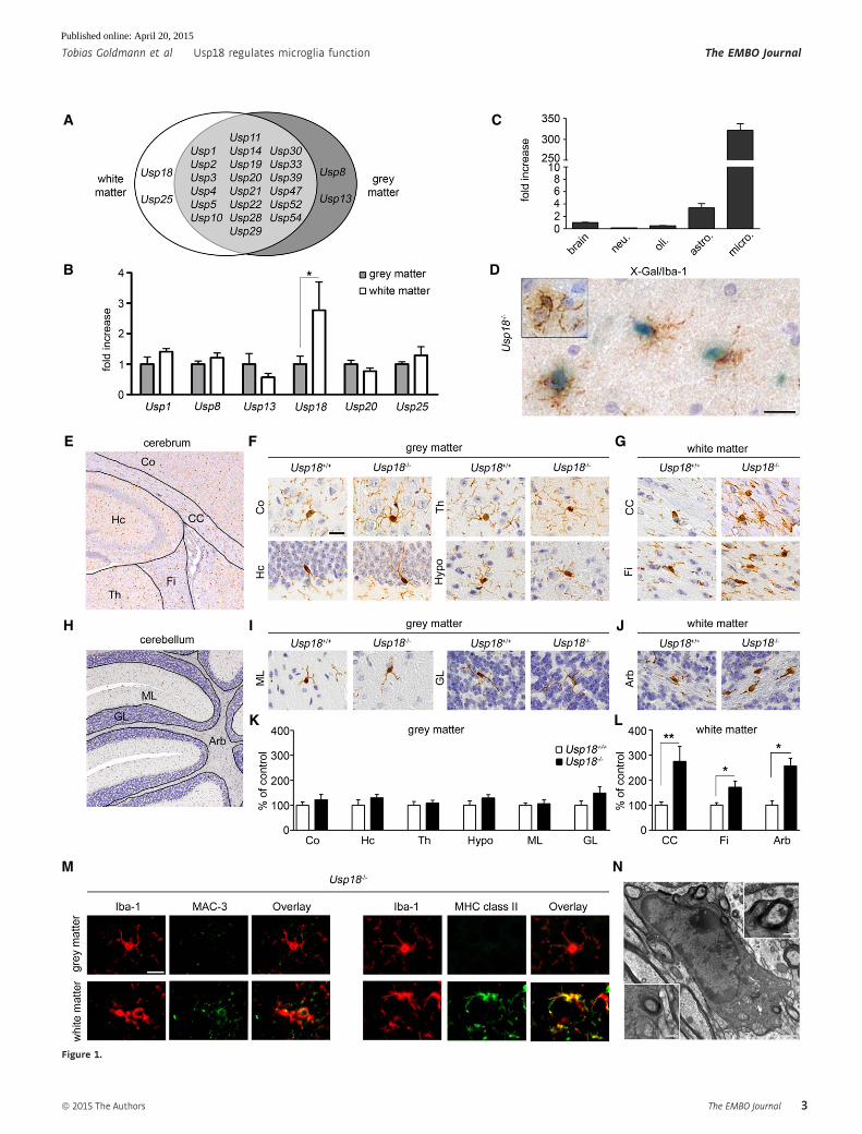
▸Figure 1. Usp18 is a distinct feature of white matter microglia and essentially regulates microglia quiescence.
A Spatial distribution of ubiquitin-specific protease (USP) transcripts based on FACS-sorted adult microglia isolated from the white or gray matter that weresubsequently examined by MouseRef-8 v2.0 Expression Bead Chip (Illumina) array analysis (Olah et al, 2012). Each USP shown exceeds a median expression valueof two resulting from five mice compared to the mean expression value of the same gene in the other brain region.
B Quantitative RT–PCR of indicated genes in FACS-isolated adult microglia. Bars represent means � s.e.m. with three mice in each group (*P < 0.05). Significantdifferences are determined by an unpaired t-test.
C Expression of Usp18 mRNA measured by qRT–PCR in primary microglia (micro.), astrocytes (astro.), neurons (neu.) and oligodendrocytes (oli.). Bars representmeans � s.e.m with at least three samples in each group normalized to the mean expression value of Usp18 transcripts in the whole brain.
D Cell-specific expression of Usp18. Light microscopic analysis of X-gal-stained (blue) white matter brain tissue of adult Usp18LacZ/LacZ mice. Iba-1 staining (brown)reveals microglia. Inserts show microglia from the cortex. Scale bar, 20 lm.
E–G Histology of different brain areas in the cerebrum of adult Usp18+/+ and Usp18LacZ/LacZ (Usp18�/�) mice. Cortex (Co), hippocampus (Hc), thalamus (Th) andhypothalamus (Hypo) represent areas of the gray matter, whereas corpus callosum (CC) and fimbria (Fi) are defined as white matter. Scale bar, 10 lm.
H–J Histological pictures of different cerebellar regions of adult Usp18+/+ and Usp18LacZ/LacZ (Usp18�/�) mice. Molecular layer (ML) and granular layer (GL) representareas of the gray matter, whereas arbor vitae (Arb) is part of the white matter.
K, L Quantification of Iba-1+ microglia in the different areas of Usp18LacZ/LacZ (Usp18�/�) mice. Microglia numbers are normalized to that found in Usp18+/+ littermatesand are displayed as % of control. At least five mice per genotype were counted. Significant differences are determined by an unpaired t-test or Mann–WhitneyU-test and marked with asterisks (*P < 0.05, **P < 0.01). Bars represent means � s.e.m.
M Immunofluorescence of white matter and gray matter microglia (Iba-1, red) in adult Usp18�/� animals (green, scale bar, 10 lm). Three animals per genotype wereexamined. One characteristic picture is shown.
N Transmission electron microscopy of myelin-phagocytosing microglia in adult Usp18LacZ/LacZ (Usp18�/�) mice. Scale bars, 1 lm (overview) and 250 nm (zoom).
The EMBO Journal ª 2015 The Authors
The EMBO Journal Usp18 regulates microglia function Tobias Goldmann et al
2
Published online: April 20, 2015
A C
B
E
H
M N
LK
I J
GF
D
Figure 1.
ª 2015 The Authors The EMBO Journal
Tobias Goldmann et al Usp18 regulates microglia function The EMBO Journal
3
Published online: April 20, 2015
matter of Usp18LacZ/LacZ (designated Usp18�/�) mice by X-gal stain-
ing (Fig 1D).
To further investigate the physiological role of Usp18 for the
CNS, we performed a thorough histopathological analysis of differ-
ent brain areas of adult Usp18LacZ/LacZ mice. While there were no
obvious histopathological abnormalities in the gray matter, a signifi-
cant increase of Iba-1+ microglia numbers was detectable in several
white matter regions in Usp18-deficient mice (Fig 1E–L). Further-
more, only subcortical white matter microglia but not cortical
microglia exhibited strong expression of the activation marker MHC
class II and of MAC-3 (LAMP2, CD107b, Fig 1M). Transmission
electron microscopy was used to confirm that myelin debris was
visible in phagocytotically active microglia (Fig 1N). Importantly,
Usp18�/� brains did not show any infiltrating lymphocytes or
monocytes (Supplementary Fig S1), indicating a sole microglia acti-
vation phenotype that we defined as “white matter microglia activa-
tion” (WMMA).
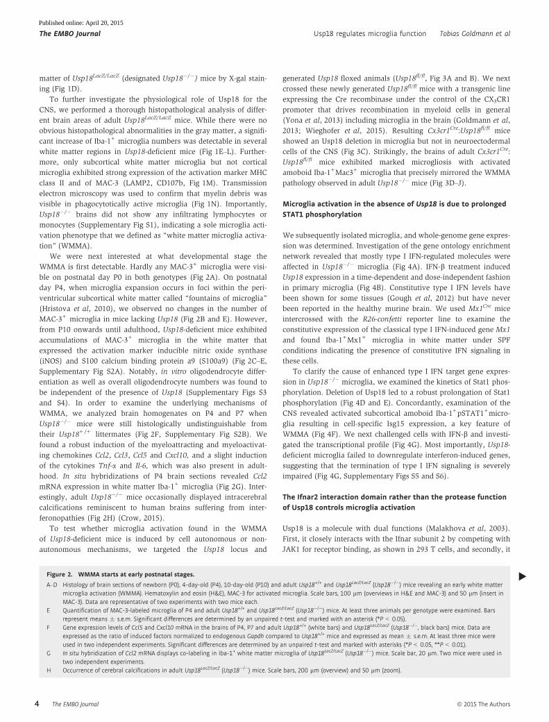
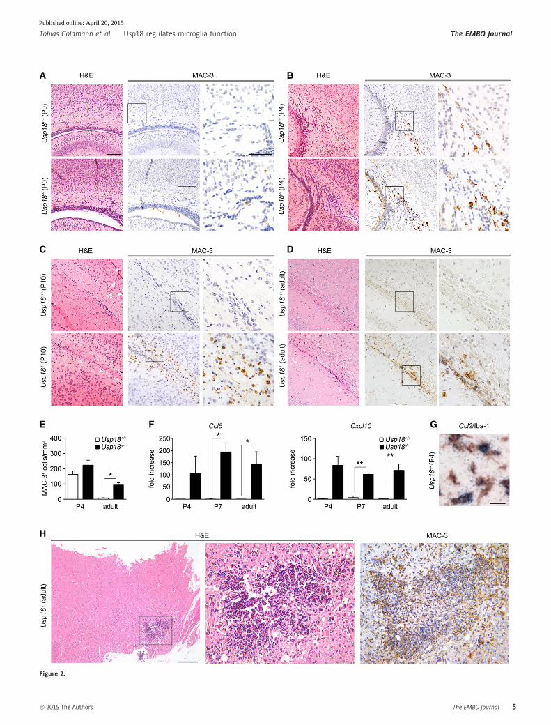
We were next interested at what developmental stage the
WMMA is first detectable. Hardly any MAC-3+ microglia were visi-
ble on postnatal day P0 in both genotypes (Fig 2A). On postnatal
day P4, when microglia expansion occurs in foci within the peri-
ventricular subcortical white matter called “fountains of microglia”
(Hristova et al, 2010), we observed no changes in the number of
MAC-3+ microglia in mice lacking Usp18 (Fig 2B and E). However,
from P10 onwards until adulthood, Usp18-deficient mice exhibited
accumulations of MAC-3+ microglia in the white matter that
expressed the activation marker inducible nitric oxide synthase
(iNOS) and S100 calcium binding protein a9 (S100a9) (Fig 2C–E,
Supplementary Fig S2A). Notably, in vitro oligodendrocyte differ-
entiation as well as overall oligodendrocyte numbers was found to
be independent of the presence of Usp18 (Supplementary Figs S3
and S4). In order to examine the underlying mechanisms of
WMMA, we analyzed brain homogenates on P4 and P7 when
Usp18�/� mice were still histologically undistinguishable from
their Usp18+/+ littermates (Fig 2F, Supplementary Fig S2B). We
found a robust induction of the myeloattracting and myeloactivat-
ing chemokines Ccl2, Ccl3, Ccl5 and Cxcl10, and a slight induction
of the cytokines Tnf-a and Il-6, which was also present in adult-
hood. In situ hybridizations of P4 brain sections revealed Ccl2
mRNA expression in white matter Iba-1+ microglia (Fig 2G). Inter-
estingly, adult Usp18�/� mice occasionally displayed intracerebral
calcifications reminiscent to human brains suffering from inter-
feronopathies (Fig 2H) (Crow, 2015).
To test whether microglia activation found in the WMMA
of Usp18-deficient mice is induced by cell autonomous or non-
autonomous mechanisms, we targeted the Usp18 locus and
generated Usp18 floxed animals (Usp18fl/fl, Fig 3A and B). We next
crossed these newly generated Usp18fl/fl mice with a transgenic line
expressing the Cre recombinase under the control of the CX3CR1
promoter that drives recombination in myeloid cells in general
(Yona et al, 2013) including microglia in the brain (Goldmann et al,
2013; Wieghofer et al, 2015). Resulting Cx3cr1Cre:Usp18fl/fl mice
showed an Usp18 deletion in microglia but not in neuroectodermal
cells of the CNS (Fig 3C). Strikingly, the brains of adult Cx3cr1Cre:
Usp18fl/fl mice exhibited marked microgliosis with activated
amoboid Iba-1+Mac3+ microglia that precisely mirrored the WMMA
pathology observed in adult Usp18�/� mice (Fig 3D–J).
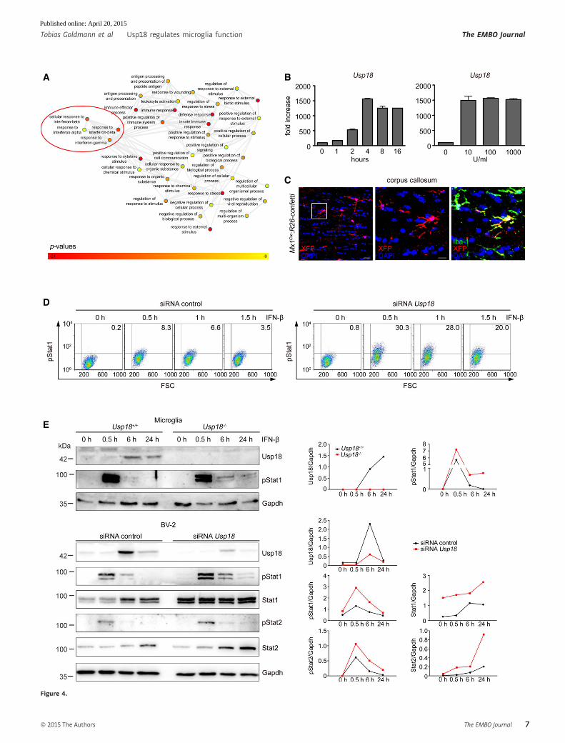
Microglia activation in the absence of Usp18 is due to prolongedSTAT1 phosphorylation
We subsequently isolated microglia, and whole-genome gene expres-
sion was determined. Investigation of the gene ontology enrichment
network revealed that mostly type I IFN-regulated molecules were
affected in Usp18�/� microglia (Fig 4A). IFN-b treatment induced
Usp18 expression in a time-dependent and dose-independent fashion
in primary microglia (Fig 4B). Constitutive type I IFN levels have
been shown for some tissues (Gough et al, 2012) but have never
been reported in the healthy murine brain. We used Mx1Cre mice
intercrossed with the R26-confetti reporter line to examine the
constitutive expression of the classical type I IFN-induced gene Mx1
and found Iba-1+Mx1+ microglia in white matter under SPF
conditions indicating the presence of constitutive IFN signaling in
these cells.
To clarify the cause of enhanced type I IFN target gene expres-
sion in Usp18�/� microglia, we examined the kinetics of Stat1 phos-
phorylation. Deletion of Usp18 led to a robust prolongation of Stat1
phosphorylation (Fig 4D and E). Concordantly, examination of the
CNS revealed activated subcortical amoboid Iba-1+pSTAT1+micro-
glia resulting in cell-specific Isg15 expression, a key feature of
WMMA (Fig 4F). We next challenged cells with IFN-b and investi-
gated the transcriptional profile (Fig 4G). Most importantly, Usp18-
deficient microglia failed to downregulate interferon-induced genes,
suggesting that the termination of type I IFN signaling is severely
impaired (Fig 4G, Supplementary Figs S5 and S6).
The Ifnar2 interaction domain rather than the protease functionof Usp18 controls microglia activation
Usp18 is a molecule with dual functions (Malakhova et al, 2003).
First, it closely interacts with the Ifnar subunit 2 by competing with
JAK1 for receptor binding, as shown in 293 T cells, and secondly, it
▸Figure 2. WMMA starts at early postnatal stages.
A–D Histology of brain sections of newborn (P0), 4-day-old (P4), 10-day-old (P10) and adult Usp18+/+ and Usp18LacZ/LacZ (Usp18�/�) mice revealing an early white mattermicroglia activation (WMMA). Hematoxylin and eosin (H&E), MAC-3 for activated microglia. Scale bars, 100 lm (overviews in H&E and MAC-3) and 50 lm (insert inMAC-3). Data are representative of two experiments with two mice each.
E Quantification of MAC-3-labeled microglia of P4 and adult Usp18+/+ and Usp18LacZ/LacZ (Usp18�/�) mice. At least three animals per genotype were examined. Barsrepresent means � s.e.m. Significant differences are determined by an unpaired t-test and marked with an asterisk (*P < 0.05).
F Gene expression levels of Ccl5 and Cxcl10 mRNA in the brains of P4, P7 and adult Usp18+/+ (white bars) and Usp18LacZ/LacZ (Usp18�/�, black bars) mice. Data areexpressed as the ratio of induced factors normalized to endogenous Gapdh compared to Usp18+/+ mice and expressed as mean � s.e.m. At least three mice wereused in two independent experiments. Significant differences are determined by an unpaired t-test and marked with asterisks (*P < 0.05, **P < 0.01).
G In situ hybridization of Ccl2 mRNA displays co-labeling in Iba-1+ white matter microglia of Usp18LacZ/LacZ (Usp18�/�) mice. Scale bar, 20 lm. Two mice were used intwo independent experiments.
H Occurrence of cerebral calcifications in adult Usp18LacZ/LacZ (Usp18�/�) mice. Scale bars, 200 lm (overview) and 50 lm (zoom).
The EMBO Journal ª 2015 The Authors
The EMBO Journal Usp18 regulates microglia function Tobias Goldmann et al
4
Published online: April 20, 2015
A B
C
E
H
F G
D
Figure 2.
ª 2015 The Authors The EMBO Journal
Tobias Goldmann et al Usp18 regulates microglia function The EMBO Journal
5
Published online: April 20, 2015

A B C
D E J
F G
H I
Figure 3. WMMA due to Usp18 deletion occurs in a cell-autonomous manner.
A Targeting strategy for the conditional mutagenesis of the Usp18 gene. A targeting vector was used to modify the Usp18 gene locus. Upon homologousrecombination and elimination of the frt-flanked selection marker (neo), the third exon of the gene was flanked by loxP sites allowing Cre-mediated deletion ofUsp18.
B Homologous recombination in ES cells was detected by genomic Southern blot analysis. As depicted in (A), probe A detects a diagnostic 3.5-kb band upon KpnIrestriction digest diagnostic for the mutated Usp18 allele.
C PCR analysis of the Usp18 deletion in primary microglia, astrocytes, oligodendrocytes or neurons of Usp18fl/fl, Cx3cr1Cre:Usp18fl/fl and wild-type mice. Recombinationis only taking place in microglia but not in astrocytes, oligodendrocytes or neurons. One representative experiment out of two performed is shown.
D, E Histology of different brain areas in the cerebrum of adult Usp18fl/fl and Cx3cr1Cre:Usp18fl/fl mice. Cortex (Co), hippocampus (Hc), thalamus (Th) and hypothalamus(Hypo) represent areas of the gray matter, whereas corpus callosum (CC) and fimbria (Fi) are parts of the white matter. Scale bar = 10 lm.
F, G Histological pictures of different cerebellar regions of adult Usp18fl/fl and Cx3cr1Cre:Usp18fl/fl mice. Molecular layer (ML) and granular layer (GL) represent areas ofthe gray matter, whereas arbor vitae (Arb) is part of white matter.
H, I Quantification of Iba-1+ cells in Cx3cr1Cre:Usp18fl/fl mice. Microglia numbers are normalized to that found in Usp18+/+ littermates and are displayed as % of control.At least five mice per genotype were counted. Significant differences are determined by an unpaired t-test or Mann–Whitney U-test and marked with asterisks(*P < 0.05, **P < 0.01, ***P < 0.0001). Bars represent means � s.e.m.
J Immunofluorescence of white and gray matter microglia (Iba-1, red) in adult Usp18�/� animals (green, scale bar, 10 lm). Three animals per genotype wereexamined. One characteristic sample is shown.
The EMBO Journal ª 2015 The Authors
The EMBO Journal Usp18 regulates microglia function Tobias Goldmann et al
6
Published online: April 20, 2015
A B
C
D
E
Figure 4.
ª 2015 The Authors The EMBO Journal
Tobias Goldmann et al Usp18 regulates microglia function The EMBO Journal
7
Published online: April 20, 2015

F
G
Figure 4. Lack of Usp18 enhances type I IFN gene expression in microglia due to prolonged Stat1 phosphorylation.
A Gene ontology enrichment network on differentially expressed genes in microglia from unstimulated Usp18+/+ and Usp18LacZ/LacZ (Usp18�/�) microglia on the basis ofan Affymetrix DNA microarray analysis. Diagram depicts results of GO clustering through GORilla. Only very highly significantly overrepresented GO terms areincluded with P-values ranging from P < 10�9 (yellow) to P < 10�24 (red).
B Quantitative RT–PCR for Usp18 transcripts in primary microglia stimulated for the designated time points with 100 U/ml of IFN-b (left panel) or with 10, 100 or1,000 U/ml of IFN-b and measured after 4 h (right panel). Bars represent means � s.e.m with three to four samples in each group. Data are representative of twoindependently performed experiments.
C Fluorescence microscopy of the white matter (corpus callosum) of Mx1Cre:R26-confetti mice raised under specific pathogen-free conditions. Recombination of GFP,RFP, CFP or YFP (combined into one channel to XFP and displayed in red) was found in Iba-1+ microglia of the white matter. Scale bars: 20 lm (overview) and 10 lm(zoom).
D Flow cytometric quantification of Stat1 phosphorylation in BV-2 microglial cells transfected with control siRNA (siRNA co) or siRNA against Usp18. Representative dotblots of IFN-b-treated cells at indicated time points are shown that were obtained from two independent experiments. FSC: forward scatter.
E Immunoblot analysis of type I IFN signaling in microglia lacking Usp18. Upper panel: Absence of Usp18 protein leads to prolonged Stat1 activation upon IFN-bchallenge (500 U/ml) of microglia from Usp18+/+ and Usp18LacZ/LacZ (Usp18�/�) mice. Gapdh is shown as a loading control. Lower panel: Altered IFN signaling in themicroglia cell line BV-2 transfected with control siRNA (siRNA co) or siRNA against Usp18. Quantification of band intensities is depicted next to the blots.Representative Western blots of three to four independently performed experiments are shown.
F Brain histology of the white matter reveals increased pStat1 and interferon-induced gene (ISG) 15 levels in white matter microglia in adult Cx3cr1Cre:Usp18fl/fl andUsp18LacZ/LacZ (Usp18�/�) mice but not in Usp18+/+ individuals. Quantification of Isg15+ cells in the gray (GM) and white matter (WM) is presented next to therespective histological images. Scale bars: 200 lm (overview) and 10 lm (insert). Each symbol indicates the mean of one mouse. Error bars represent s.e.m.Significant differences are determined by an unpaired t-test and marked with an asterisk (*P < 0.05).
G Heat map (standardized and scaled to log2 expression) of non-stimulated conditions (0 h) or after IFN-b (500 U/ml for 6 h and 24 h) treatment in primary microgliafrom Usp18LacZ/LacZ (Usp18�/�) and Usp18+/+ mice or the microglia cell line BV-2 (transfected with control siRNA [siRNA co] or siRNA against Usp18). Expression profileof top 50 induced genes in Usp18+/+ or siRNA co upon 6 and 24 h IFN-b is shown.
The EMBO Journal ª 2015 The Authors
The EMBO Journal Usp18 regulates microglia function Tobias Goldmann et al
8
Published online: April 20, 2015

is the major protease for Isg15 to deconjugate Isg15 (deISGylate)
from substrate proteins (Malakhova et al, 2006) (Fig 5A). In the
murine Usp18, the cysteine at position 61 (Cys61) is essential for
protease activity (Malakhov et al, 2002). To test whether the cata-
lytic domain of Usp18 regulates microglia activation in vivo, we
employed a novel mouse strain recently generated in our lab, in
which the endogenous Usp18 gene locus was mutated to express an
Usp18 protein selectively lacking its isopeptidase activity due to a
single amino acid exchange from cysteine to alanine at position 61
(Usp18-C61A) (Ketscher et al, 2015). Usp18-C61A microglia neither
changed the kinetics of phosphorylated Stat1 protein (Fig 5B) nor
altered the gene expression profile upon stimulation with IFN-b(Fig 5C). Gene profiles of Usp18-C61A microglia were indistinguish-
able from those in Usp18+/+ microglia and showed no alteration in
A
C
D
B
Figure 5. Usp18-mediated microglia activation is independent of its catalytic activity.
A Scheme of Usp18 interactions with either cytoplasmatic substrate/interacting proteins or trans-membrane anchored type I interferon receptor (Ifnar) and its subunits1 and 2. The red cross indicates the genetically inactivated motif in Usp18-C61A mutant mice.
B Immunoblot of primary microglia from Usp18+/+ and catalytically inactive Usp18-C61A mutant mice reveals normal pStat1 kinetics at different time points after IFN-b(500 U/ml) challenge. Gapdh is shown as a loading control. Quantification of band intensities is depicted next to the blots. One representative data set out of threeindependent experiments is illustrated.
C Gene expression scatter plot of Affymetrix oligo-array data depicting different gene expression patterns in microglia from Usp18 mutants. The relative mRNA levelsfrom wild-type microglia (Usp18+/+, x-axis) are normalized to Usp18LacZ/LacZ (Usp18�/�) microglia (y-axis, red dots) and catalytically inactive Usp18-C61A (y-axis, blackdots) microglia under non-stimulated conditions and after IFN-b (500 U/ml) exposure. Pooled data from two independent experiments are shown.
D CNS histology of adult Usp18-C61A mice discloses unchanged microglial cells with normal morphological appearance (Iba-1) but no MAC-3+ amoboid microglia.Quantification of Isg15+ cells in the GM and WM is shown next to the respective histological images. Three mice per genotype were examined. Scale bars: 200 lm(overview), 10 lm (zoom).
ª 2015 The Authors The EMBO Journal
Tobias Goldmann et al Usp18 regulates microglia function The EMBO Journal
9
Published online: April 20, 2015

termination of IFN signaling. Importantly, WMMA was absent in
brain sections of adult Usp18-C61A mice, clearly showing that
USP18-mediated microglia activation is mediated in an isopeptidase-
independent manner (Fig 5D).
As deISGylation does not play a role for Usp18-induced Stat1 acti-
vation in microglia, it is conceivable that expression of Usp18 may
affect signaling via the Ifnar. We therefore tested whether Usp18
interacts with the subunit 2 of Ifnar by immunoprecipitating Ifnar2
and found physical interaction of both partners (Fig 6A). Close prox-
imity of Usp18 with Ifnar2 could further be confirmed by proximity
ligation (Duolink) (Fig 6B). To prove the in vivo relevance of
the Usp18 interaction with Ifnar2, we next took advantage of a
A
C
E
G
F
D
B
Figure 6.
The EMBO Journal ª 2015 The Authors
The EMBO Journal Usp18 regulates microglia function Tobias Goldmann et al
10
Published online: April 20, 2015
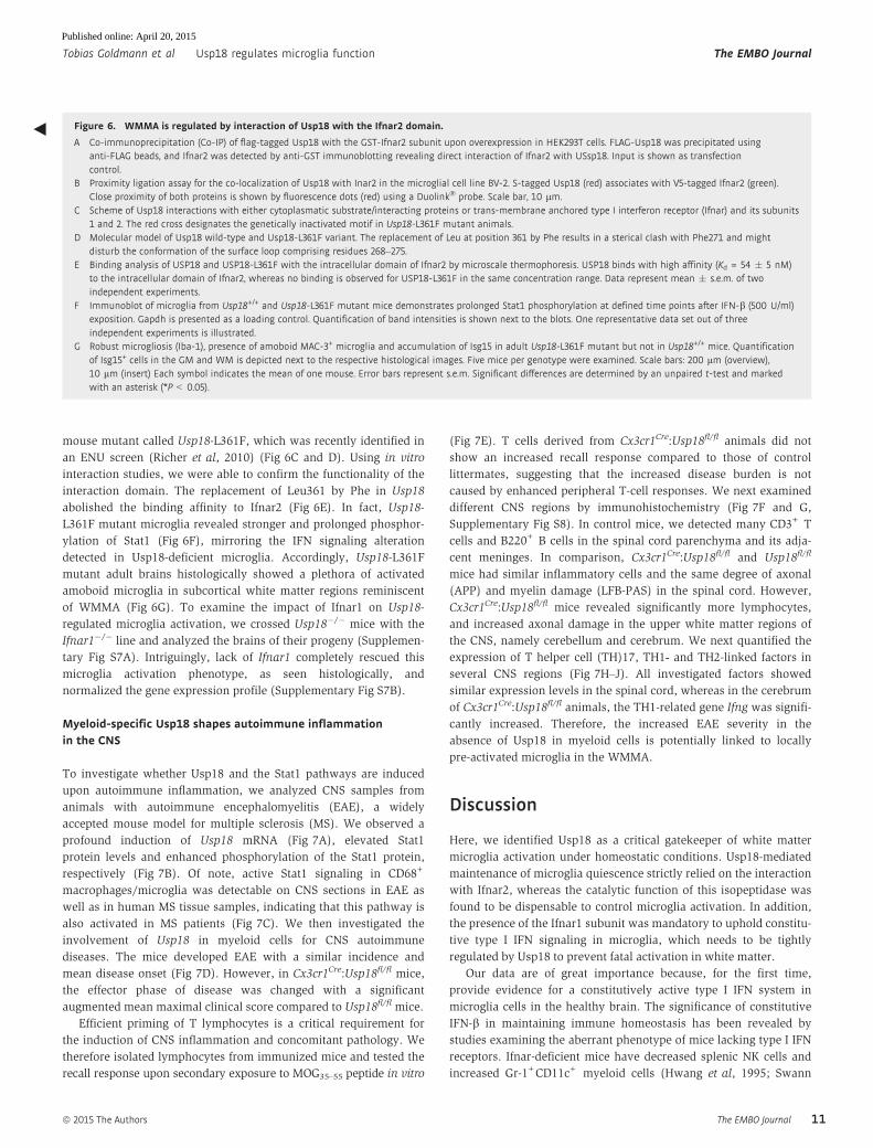
mouse mutant called Usp18-L361F, which was recently identified in
an ENU screen (Richer et al, 2010) (Fig 6C and D). Using in vitro
interaction studies, we were able to confirm the functionality of the
interaction domain. The replacement of Leu361 by Phe in Usp18
abolished the binding affinity to Ifnar2 (Fig 6E). In fact, Usp18-
L361F mutant microglia revealed stronger and prolonged phosphor-
ylation of Stat1 (Fig 6F), mirroring the IFN signaling alteration
detected in Usp18-deficient microglia. Accordingly, Usp18-L361F
mutant adult brains histologically showed a plethora of activated
amoboid microglia in subcortical white matter regions reminiscent
of WMMA (Fig 6G). To examine the impact of Ifnar1 on Usp18-
regulated microglia activation, we crossed Usp18�/� mice with the
Ifnar1�/� line and analyzed the brains of their progeny (Supplemen-
tary Fig S7A). Intriguingly, lack of Ifnar1 completely rescued this
microglia activation phenotype, as seen histologically, and
normalized the gene expression profile (Supplementary Fig S7B).
Myeloid-specific Usp18 shapes autoimmune inflammationin the CNS
To investigate whether Usp18 and the Stat1 pathways are induced
upon autoimmune inflammation, we analyzed CNS samples from
animals with autoimmune encephalomyelitis (EAE), a widely
accepted mouse model for multiple sclerosis (MS). We observed a
profound induction of Usp18 mRNA (Fig 7A), elevated Stat1
protein levels and enhanced phosphorylation of the Stat1 protein,
respectively (Fig 7B). Of note, active Stat1 signaling in CD68+
macrophages/microglia was detectable on CNS sections in EAE as
well as in human MS tissue samples, indicating that this pathway is
also activated in MS patients (Fig 7C). We then investigated the
involvement of Usp18 in myeloid cells for CNS autoimmune
diseases. The mice developed EAE with a similar incidence and
mean disease onset (Fig 7D). However, in Cx3cr1Cre:Usp18fl/fl mice,
the effector phase of disease was changed with a significant
augmented mean maximal clinical score compared to Usp18fl/fl mice.
Efficient priming of T lymphocytes is a critical requirement for
the induction of CNS inflammation and concomitant pathology. We
therefore isolated lymphocytes from immunized mice and tested the
recall response upon secondary exposure to MOG35–55 peptide in vitro
(Fig 7E). T cells derived from Cx3cr1Cre:Usp18fl/fl animals did not
show an increased recall response compared to those of control
littermates, suggesting that the increased disease burden is not
caused by enhanced peripheral T-cell responses. We next examined
different CNS regions by immunohistochemistry (Fig 7F and G,
Supplementary Fig S8). In control mice, we detected many CD3+ T
cells and B220+ B cells in the spinal cord parenchyma and its adja-
cent meninges. In comparison, Cx3cr1Cre:Usp18fl/fl and Usp18fl/fl
mice had similar inflammatory cells and the same degree of axonal
(APP) and myelin damage (LFB-PAS) in the spinal cord. However,
Cx3cr1Cre:Usp18fl/fl mice revealed significantly more lymphocytes,
and increased axonal damage in the upper white matter regions of
the CNS, namely cerebellum and cerebrum. We next quantified the
expression of T helper cell (TH)17, TH1- and TH2-linked factors in
several CNS regions (Fig 7H–J). All investigated factors showed
similar expression levels in the spinal cord, whereas in the cerebrum
of Cx3cr1Cre:Usp18fl/fl animals, the TH1-related gene Ifng was signifi-
cantly increased. Therefore, the increased EAE severity in the
absence of Usp18 in myeloid cells is potentially linked to locally
pre-activated microglia in the WMMA.
Discussion
Here, we identified Usp18 as a critical gatekeeper of white matter
microglia activation under homeostatic conditions. Usp18-mediated
maintenance of microglia quiescence strictly relied on the interaction
with Ifnar2, whereas the catalytic function of this isopeptidase was
found to be dispensable to control microglia activation. In addition,
the presence of the Ifnar1 subunit was mandatory to uphold constitu-
tive type I IFN signaling in microglia, which needs to be tightly
regulated by Usp18 to prevent fatal activation in white matter.
Our data are of great importance because, for the first time,
provide evidence for a constitutively active type I IFN system in
microglia cells in the healthy brain. The significance of constitutive
IFN-b in maintaining immune homeostasis has been revealed by
studies examining the aberrant phenotype of mice lacking type I IFN
receptors. Ifnar-deficient mice have decreased splenic NK cells and
increased Gr-1+CD11c+ myeloid cells (Hwang et al, 1995; Swann
◀ Figure 6. WMMA is regulated by interaction of Usp18 with the Ifnar2 domain.
A Co-immunoprecipitation (Co-IP) of flag-tagged Usp18 with the GST-Ifnar2 subunit upon overexpression in HEK293T cells. FLAG-Usp18 was precipitated usinganti-FLAG beads, and Ifnar2 was detected by anti-GST immunoblotting revealing direct interaction of Ifnar2 with USsp18. Input is shown as transfectioncontrol.
B Proximity ligation assay for the co-localization of Usp18 with Inar2 in the microglial cell line BV-2. S-tagged Usp18 (red) associates with V5-tagged Ifnar2 (green).Close proximity of both proteins is shown by fluorescence dots (red) using a Duolink� probe. Scale bar, 10 lm.
C Scheme of Usp18 interactions with either cytoplasmatic substrate/interacting proteins or trans-membrane anchored type I interferon receptor (Ifnar) and its subunits1 and 2. The red cross designates the genetically inactivated motif in Usp18-L361F mutant animals.
D Molecular model of Usp18 wild-type and Usp18-L361F variant. The replacement of Leu at position 361 by Phe results in a sterical clash with Phe271 and mightdisturb the conformation of the surface loop comprising residues 268–275.
E Binding analysis of USP18 and USP18-L361F with the intracellular domain of Ifnar2 by microscale thermophoresis. USP18 binds with high affinity (Kd = 54 � 5 nM)to the intracellular domain of Ifnar2, whereas no binding is observed for USP18-L361F in the same concentration range. Data represent mean � s.e.m. of twoindependent experiments.
F Immunoblot of microglia from Usp18+/+ and Usp18-L361F mutant mice demonstrates prolonged Stat1 phosphorylation at defined time points after IFN-b (500 U/ml)exposition. Gapdh is presented as a loading control. Quantification of band intensities is shown next to the blots. One representative data set out of threeindependent experiments is illustrated.
G Robust microgliosis (Iba-1), presence of amoboid MAC-3+ microglia and accumulation of Isg15 in adult Usp18-L361F mutant but not in Usp18+/+ mice. Quantificationof Isg15+ cells in the GM and WM is depicted next to the respective histological images. Five mice per genotype were examined. Scale bars: 200 lm (overview),10 lm (insert) Each symbol indicates the mean of one mouse. Error bars represent s.e.m. Significant differences are determined by an unpaired t-test and markedwith an asterisk (*P < 0.05).
ª 2015 The Authors The EMBO Journal
Tobias Goldmann et al Usp18 regulates microglia function The EMBO Journal
11
Published online: April 20, 2015

A
D E
F G
H I J
B C
Figure 7.
The EMBO Journal ª 2015 The Authors
The EMBO Journal Usp18 regulates microglia function Tobias Goldmann et al
12
Published online: April 20, 2015

et al, 2007). Constitutive IFN-b signaling augments myeloid cell
function and macrophage homeostasis, as shown by analysis of
macrophages from C3H-HeJ mice, which are incapable of inducing
IFN in response to LPS because of a defect in the Tlr4 gene (Poltorak
et al, 1998). Culturing C3H-HeJ macrophages with supernatants
from wild-type C3H-HeN macrophages that express constitutive
IFN-b enhanced their phagocytic potential. A similar effect was
obtained by adding low “priming” concentrations of IFN to C3H-HeJ
macrophages (Vogel & Fertsch, 1984). Conversely, phagocytic
capacity was attenuated when C3H-HeN macrophages were incu-
bated with IFN-a- and IFN-b-neutralizing antibodies (Vogel &
Fertsch, 1984), documenting the requirement for constitutive IFN in
preserving macrophage function. The physiologic consequence of
the importance of constitutive IFN for macrophage function may be
reflected by the influence of the gut microbiota on hematopoietic
homeostasis through basal TLR signaling (Musso et al, 2011).
Whether intestinal colonization is also able to tune constitutive IFN
responses in myeloid cells inside the brain remains to be deter-
mined. Although the constitutive activation of type I IFNs in micro-
glia has not been addressed so far under germ-free conditions to
assess a potential role for commensal microorganisms, we clearly
detected Mx1-positive microglia in brains from healthy mice kept in
a specific pathogen-free environment. However, it also becomes
clear from our experiments that a strict control of constitutive IFN
signaling in white matter microglia is necessary to keep these
myeloid cells under resting conditions to avoid cellular hyperactiva-
tion with detrimental consequences for the tissue environment. On
the other hand, constitutive IFN signaling might be important for
priming microglia to maintain a rapid and robust innate and adap-
tive immune response to subsequent challenge. Our data provide
evidence that this tight balance is secured by Usp18.
Our data on the specific expression of Usp18 in white matter
rather than in cortical microglia highlight the regional heterogeneity
of these glial cells in the CNS. It is unknown so far whether micro-
glia heterogeneity is a result of irreversible subtype-specific differen-
tiation or a consequence of continuous but reversible induction of
diverse functional programs (Hanisch, 2013). Local tissue-derived
signals are thought to control functional polarization of microglia,
but their identity remains largely unknown. Our data on the region-
specific upregulation of Usp18 in microglia, however, suggest that
cell autonomous programs exist that may correlate with their
regional-specific function. Microglia seem to assume also particular
function in the normal turnover of myelin (Fitzner et al, 2011).
Oligodendrocytes create well-organized and also tremendously
elaborate myelin structures that require constant upkeep. Appar-
ently, they thereby rely on the assistance of microglia. Oligodendro-
cytes can wrap myelin material from their turnover process in
exosomes and deliver them to the microglia in their surrounding
(Fitzner et al, 2011), indicating that subpopulations of microglia for
myelin clearance do exist.
One may speculate that a disturbance of physiological myelin
processing by microglia could be detrimental, and there might be
functional links to the triggering of myelin-afflicting autoimmune
diseases such as MS. Just recently, vulnerability of microglia has
been connected to several neurological and psychiatric disorders
that are characterized by primary or secondary “microgliopathies”
(Prinz & Priller, 2014). In fact, recent genomewide association stud-
ies with thousands of MS patients revealed the existence of some
microglia-related factors for disease pathogenesis such as interferon
regulatory factor (Irf-) 8 and Tnfrsf1a (De Jager et al, 2009). In a
recent study, two Usp18-related polymorphisms, one intronic and
another located in the promoter region of the Usp18 gene, were
found to be associated with MS susceptibility (Malhotra et al, 2013).
Importantly, haplotypic analysis revealed one haplotype, which
correlated with lower Usp18 gene expression in peripheral blood
mononuclear cells and higher clinical disease activity (Malhotra
et al, 2011). These clinical observations fit to our experimental find-
ings with an augmented EAE course in myeloid-specific Usp18
knockout animals. However, this clinical study included only a
limited number of MS patients. Therefore, additional functional
studies will be needed to delineate the role of microglia-specific
Usp18 in MS. Our current study, however, suggests a potential role
of microglia dysregulation in the pathogenesis of white matter
diseases.
◀ Figure 7. Myeloid-specific Usp18 deficiency shapes clinical course and pathology of autoimmune CNS inflammation.
A Quantitative RT–PCR for Usp18 mRNA in the spinal cord of healthy control and EAE-diseased mice. Data are expressed as ratio of Usp18 expression versusendogenous Gapdh relative to control and shown as mean � s.e.m. Each symbol represents one mouse. (**P < 0.01). Significant differences are determined by aMann-Whitney U-test and marked with a asterisk (**P < 0.01).
B Immunoblot for pStat1 and Stat1 in spinal cord lysates of diseased or control mice. Gapdh is shown as a loading control. Data are representative of threeindependent experiments performed.
C Left panel: immunohistochemistry for phosphorylated Stat1 (pStat1) in the border region of demyelinating lesions in spinal cord EAE samples (above) and brainsamples from a patient with multiple sclerosis (MS). Scale bars: 100 lm (overview) and 25 lm (zoom). Four EAE-diseased animals and three biopsies of MSpatients were examined, and one representative picture is shown. Right panel: immunofluorescence of pSTAT1 (red) and CD68 (green) in demyelinating lesions ofMS patients. Quantification of pSTAT1+CD68+ and pSTAT1+CD68� cells are shown next to the respective histological images. Each symbol represents on patientsample. Error bars represent s.e.m. Significant differences are determined by an unpaired t-test and marked with an asterisk (*P < 0.05).
D EAE was induced by active immunization of Cx3cr1Cre:Usp18fl/fl (n = 9, filled squares) and Usp18fl/fl (n = 15, open squares) mice, and disease was scored. Each data
point represents the mean � s.e.m. Significant differences are determined by a Mann–Whitney U-test and marked with asterisks (*P < 0.05, **P < 0.01). The datashown are the mean from two independent experiments.
E Normal recall assay in Cx3cr1Cre:Usp18fl/fl mice. Lymph node T cells were collected and cultured for 48 h at the indicated MOG35–55 concentrations. Proliferation was
measured by BrdU incorporation for 16 h (left). IL-17 (middle) and IFN-c (right) release were measured by ELISA. Data represent mean � s.e.m. of at least threeanimals per group. Results are representative of two independent experiments.
F, G Histology of cerebral, cerebellar and spinal cord sections from diseased mice using CD3 for T lymphocytes (F) and amyloid precursor protein (APP, arrowhead) for axonaldamage (G). Scale bars, 100 lm. Quantification of T-cell infiltrates and axonal damages are depicted below the respective histological images. Each symbol indicatesthe mean of one mouse. Significant differences are determined by an unpaired t-test and marked with asterisks (*P < 0.05, **P < 0.01). Error bars represent s.e.m.
H–J Gene expression levels of TH1- (H), TH17- (I) and TH2-linked factors (J) in isolated mononuclear cells from the cerebrum (cere.) or the spinal cord (sc) of Cx3cr1Cre:
Usp18fl/fl (black bars; n = 7) or Usp18fl/fl (white bars; n = 7) mice. Data are normalized to endogenous Gapdh, expressed as fold increase of diseased Usp18fl/fl miceand displayed as mean � s.e.m. Significant differences are determined by an unpaired t-test and marked with an asterisk (*P < 0.05).
ª 2015 The Authors The EMBO Journal
Tobias Goldmann et al Usp18 regulates microglia function The EMBO Journal
13
Published online: April 20, 2015

Further studies indicated broader effects of Usp18 that may
be targeted to treat other autoimmune diseases as well. It was
previously reported that Usp18 regulates T-cell activation and
Th17 cell differentiation. Usp18-deficient T cells were defective
in Th17 differentiation, and Usp18�/� mice consecutively showed
alterations in CNS inflammation (Liu et al, 2013). Usp18 has
further been identified as a candidate gene for type I diabetes (Santin
& Eizirik, 2013; Altorok et al, 2014), and active hypomethylated CpG
sites of Usp18 were found to be expressed in patients with primary
Sjogren’s syndrome (Altorok et al, 2014). Furthermore, Usp18 may
also be involved in viral infections that are often associated with
a break of immunological tolerance and subsequent induction of
autoimmune diseases. In fact, it was recently shown that in a mouse
model of autoimmune diabetes, Usp18-enforced viral replication
evoked a break of immunological tolerance (Honke et al, 2013).
Interestingly, the presence of intracranial calcifications as well as an
increased type IFN-signature due to reduced protein stability of
Usp18 was described recently in Isg15-deficient patients (Zhang
et al, 2015).
In sum, we report here a crucial function of Usp18 in microglia
activation during health and disease and provide evidence for
Usp18-related regulation of constitutive IFN activity in the CNS.
These results may potentially assist our understanding and treat-
ment of microglia-associated brain diseases in general and inflam-
matory diseases in particular.
Materials and Methods
Human samples
Active demyelinating MS samples that were histologically classified
as described before (Lucchinetti et al, 2000). Samples were collected
according to the approval by the Ethics Committee of the Albert-
Ludwigs University Freiburg, Germany.
Mice
All animal experiments were approved by the Federal Ministry for
Nature, Environment and Consumers’ Protection of the state of
Baden-Wurttemberg (35-9185.81/G12/71) and were performed in
accordance with the respective national, federal and institutional
regulations. We bred Usp18+/� heterozygous mice on a mixed
background (C57/Bl6 × 129/S) to obtain Usp18�/� and Usp18+/+
mice (Ritchie et al, 2002). To generate mice allowing conditional
Usp18 depletion, a target vector was constructed as depicted in
detail in Fig 2A. After transfection of ES cells and upon homologous
recombination, Exon 3 was flanked by loxP sites. ES cells carrying
the desired mutant gene locus were detected by Southern analysis
of genomic DNA digested with KpnI due to the appearance of a
diagnostic 3.5- and 7.5-kb band recognized by 50probe A and
30probe B, respectively. Germline chimeras were generated from
these ES cells by morula injection. Resulting mutant offspring was
crossed to a flp-deleter strain to excise the frt-flanked pgk-neo selec-
tion marker from the genome yielding mice with the loxP-flanked
Usp18 gene (Usp18fl/+). Usp18fl/+ mice were backcrossed for > 9
generations onto the C57Bl/7 background and finally crossed to
Cx3cr1Cre (Yona et al, 2013). Usp18fl/fl mice were genotyped by PCR
using the following primers 50-cacctccatttggtttcagg-30 and 50-aactccttcctctggcttcc-30 that amplify a 250-bp fragment from the trans-
genic region. Usp18-C61A mice were generated by us and bred on a
C57/Bl6 background (Ketscher et al, 2015). Usp18-L361F mice were
kept under 129/S1 background (Richer et al, 2010). Usp18+/� were
crossed to Ifnar1�/� (Prinz et al, 2008) in order to generate Usp18�/�:Ifnar1�/� double mutants. Mx1Cre were crossed to R26-confetti mice
(Snippert et al, 2010). In all experiments, littermates carrying the
respective loxP-flanked alleles but lacking expression of Cre recombin-
ase were used as controls.
Cell culture
Primary cultures from microglia, astrocytes and oligodendrocytes
were prepared from newborn mice; neurons were prepared from
E16 embryos and cultured as described previously (Raasch et al,
2011). HEK293T and BV-2 cells were grown in DMEM GlutaMAX
(Invitrogen) supplemented with 10% fetal calf serum (PAA) and 1%
penicillin/streptomycin (PAA) at 37°C and 5% CO2. Transfections of
plasmid DNA and siRNA in HEK 293T and BV-2 cells were
performed using X-tremeGENE HP (Roche) FuGENE (Promega) and
Lipofectamine RNAiMAX (Invitrogen) according to the manufac-
turer’s protocol.
Induction of EAE
Female mice from each group, aged 6–8 weeks, were immunized
subcutaneously with 200 lg of MOG35–55 peptide emulsified in
CFA containing 1 mg of Mycobacterium tuberculosis (H37RA; Difco
Laboratories, Detroit, Michigan, USA). The mice received intraperi-
toneal injections with 250 ng pertussis toxin (Sigma-Aldrich,
Deisenhofen, Germany) at the time of immunization and 48 h
later.
Clinical evaluation
Mice were scored daily as follows: 0, no detectable signs of EAE; 0.5
distal limb tail; 1.0, complete limp tail; 1.5, limp tail and hind limb
weakness; 2, unilateral partial hind limb paralysis; 2.5, bilateral
partial hind limb paralysis; 3, complete bilateral hind limb paralysis;
3.5, complete hind limb paralysis and unilateral forelimb paralysis;
and 4, total paralysis of fore and hind limbs.
Recall assay
On day 7 after immunization, the draining axillary and inguinal
lymph nodes (LN) were removed from MOG35–55-immunized mice
and single-cell suspensions were prepared. 6 × 10⁵ LN cells were
placed as triplicates in a 96-well plate and pulsed with the indicated
dosages of MOG peptide. BrdU uptake was measured for 16 h to
determine proliferation (Cell Proliferation ELISA colorimetric, Roche
Applied Science) according to the manufacturer’s protocol. RPMI
1,640 (Invitrogen) and Dulbecco’s modified Eagle’s medium (Invi-
trogen) each supplemented with 10% (v/v) fetal calf serum (FCS),
3 mM L-glutamine, 100 U/ml penicillin and 100 lg/ml streptomycin
(all from Sigma-Aldrich) were used. For cytokine analysis, sister
cultures supernatants were analyzed by ELISA for IFN-c and IL-17
(R&D Systems, Wiesbaden, Germany).
The EMBO Journal ª 2015 The Authors
The EMBO Journal Usp18 regulates microglia function Tobias Goldmann et al
14
Published online: April 20, 2015

Isolation of mononuclear cells
For gene expression, samples of the cerebrum and spinal cord were
prepared by density gradient separation as described previously
(Mildner et al, 2009).
Histology
Histology was performed as described recently (Goldmann et al,
2013). Cerebrum, cerebellum and spinal cord were removed on day
26 after immunization and fixed in 4% buffered formalin. Then,
tissue samples were embedded in paraffin before staining with H&E,
luxol fast blue (LFB-PAS) to assess the degree of demyelination,
Mac-3 for activated macrophages/microglia, CD3 for T cells, B220
for B cells, APP for indication of axonal damage, Iba-1 for microglia,
Nogo for oligodendrocytes, iNOS and S100a9 for microglia activa-
tion, pStat1 and Isg15 as representative IFN-induced genes. For the
b-galactosidase staining, tissue was directly frozen unfixed in tissue
tec on dry ice. Cryosections were postfixed using 0.2% glutaralde-
hyde and stained by X-gal staining solution. Tissue sections were
evaluated on Olympus BX-61 microscope using the cell-P software
(Olympus).
Fluorescence microscopy
After transcardial perfusion with phosphate-buffered saline (PBS),
brains were fixed in 4% PFA and embedded. 14-lm cryosections
were obtained as described previously (Goldmann et al, 2013).
Sections were then blocked with PBS containing 5% bovine serum
albumin and permeabilized with 0.1% Triton-X 100 in blocking
solution. Primary antibodies were added overnight at a dilution of
1:500 for Iba-1(019-19741, WACO, Japan), 1:50 for pStat1 (# 9167,
Cell Signaling, Danvers, USA), 1:250 for MAC-3 (ab13524, Abcam,
Cambridge, UK) and 1:100 for Mhc class II (ab23990, Abcam), at
4°C. Secondary antibodies were added as follows: Alexa Flour� 488
1:500, Alexa Flour� 555 1:500 and Alexa Fluor� 568 1:500 for 2 h at
RT. Nuclei were counterstained with DAPI. The examined area was
determined microscopically using a conventional fluorescence
microscope (Olympus BX-61), and the confocal pictures were taken
with Fluoview FV 1000 (Olympus).
Electron microscopy
Samples for electron microscopy were prepared as previously
described (Kierdorf et al, 2013). Briefly, tissue was fixed in 3%
glutaraldehyde in Sorensen phosphate buffer and embedded in aral-
dite, and subsequently, ultra-thin sections were cut. Ultrastructural
analysis was performed using CM100 transmission electron micro-
scope (Philips).
Flow cytometry
Cells were stained with primary antibodies directed against CD11b
and CD45 (eBioscience, San Diego, USA) at 4°C for 15 min. Cells
were washed and sorted for microarray analyses. pStat1 (612596, BD
Bioscience) staining was performed according to the manufacturer’s
protocol and analyzed using a FACSCanto II (Becton Dickinson).
Viable cells were gated by forward and side scatter pattern. Data were
acquired with FACSdiva software (Becton Dickinson). Postacquisi-
tion analysis was performed using FlowJo software (Tree Star, Inc.).
Microarray & qRT–PCR
Tissues were dissected and flushed with ice-cold HBSS. Cells were
washed with PBS. RNA was isolated using the RNeasy Mini Kit
(Qiagen, Hilden, Germany) following the manufacturer0s instruc-
tions. Before microarray analyses were performed, and RNA quality
was assessed using Agilent 2100 bioanalyzer. Affymetrix GeneChips
were used for genomewide expression analysis. Total RNA was
processed using the GeneChip Expression 30 Amplification One-
Cycle Target Labeling Kit according to the manufacturer’s instruc-
tion. Biotinylated cRNA was hybridized on MOE430A 2.0 GeneChips
that were stained, washed and scanned following standard proce-
dures. Cel files were normalized using robust multiarray analysis
(RMA) implemented in the R affy package (www.bioconductor.org).
The normalized expression data were then analyzed for differential
gene expression using the software package LIMMA for R. For
qRT–PCR, RNA-samples were treated with DNaseI (Roche, Mannheim,
Germany) and 1.5 lg of RNA was transcribed into cDNA using
either oligo-dT primers and the SuperScript II RT kit (Invitrogen,
Carlsbad, CA) or High-Capacity RNA-to-cDNATM kit (Life Technolo-
gies). A total of 1 ll cDNA was transferred into a 96-well Multiply�
PCR plate (Sarstedt, Germany) and 11.5 ll ABsoluteTM QPCR� SYBR
Green master mix (Thermo Fisher). RT–PCRs were performed as
described recently (Goldmann et al, 2013).
Cloning, expression and purification of recombinant proteins
Murine Usp18 was expressed in Sf21 insect cells (Basters et al,
2014). The cDNA encoding amino acid residues 264–374 of mouse
Ifnar2 were amplified from a mouse cDNA clone (Source BioSci-
ence) using primers EcoRI-Ifnar2-for (AAAAAGAATTCAAACGGA
TTGGTTATATATGCC) and XhoI-Ifnar2-rev (TTTTTCTCGAGTC
AAGCTTCATCAGATTCCTCAGC). EcoRI and XhoI restriction sites
were introduced during amplification. The PCR product was
digested with restriction enzymes EcoRI and XhoI and ligated into
pGEX6p1 vector digested with the same enzymes. The resulting
vector pGEX6p1-Ifnar2 (264–374) encoded Ifnar2 amino acid resi-
dues 264–374 fused in frame to a N-terminal GST-tag. E. coli TB1
cells were transformed with pGEX6p1-Ifnar2 (264–374) vector.
E. coli cells were grown in DYT medium supplemented with 0.2%
(w/v) glucose and 100 lg/ml ampicillin. At an OD600 nm = 0.6,
protein expression was induced by addition of 0.5 mM IPTG to a
final concentration of 0.5 mM and cells were grown at 19°C for
20 h. Cells were harvested by centrifugation, suspended in PBS
supplemented with complete protease inhibitor (Roche) and
disrupted by three passages through a French pressure cell at
137 Mpa. The crude extract was cleared by centrifugation at
40,000 g for 1 h, and the supernatant was applied to a 30-ml gluta-
thione column. The bound protein was eluted with 50 mM Tris–
HCl, 10 mM reduced glutathione, pH 8.0.
Microscale thermophoresis
Recombinant USP18 was labeled with fluorescence dye using the
L001 Monolith NT.115 Protein Labeling Kit RED-NHS according to
ª 2015 The Authors The EMBO Journal
Tobias Goldmann et al Usp18 regulates microglia function The EMBO Journal
15
Published online: April 20, 2015

the instructions of the manufacturer (NanoTemper). For microscale
thermophoresis analysis, the labeled protein was used at a concen-
tration of 200 nM. GST-Ifnar2 (264–374) was added in a concentra-
tion range from 3 nM to 10 lM and equilibrated for 30 min prior
analysis. The thermophoresis measurement was performed in
20 mM sodium phosphate, pH 7.4, 300 mM NaCl, 5 mM DTT and
0.05% (v/v) Tween-20 at 25°C in standard treated glass capillaries
using a Monolith NT.115 Red/Blue instrument (NanoTemper).
Protein interaction
Co-immunoprecipitation was performed using anti-FLAG M2
Magnetic Bead (M8823, Sigma-Aldrich) according to the manufac-
turer’s protocol. Briefly, transfected cells were harvested and
extracts were incubated with the beads overnight. After washing in
PBS, samples were separated by SDS–PAGE. Duolink in situ kit was
utilized in transfected BV-2 cells using antibodies against S-tag
(1:100, Novagen) and V5 (1:50, sc-81594, Santa Cruz) in compliance
with the manufacturer’s instructions.
Western blot analysis
Tissues or cells were harvested and extracted in RIPA buffer (25 mM
Tris–HCl, 150 mM NaCl, 1% Nonidet P-40, 0.5% sodium deoxycho-
late, 0.1% SDS, pH 7.5). Samples were separated by SDS–PAGE and
immunoblotted using antibodies to pStat1 (1:1,000, #9171, Cell
signaling), Stat1 (1:1,000, #9176, Cell signaling), pStat2 (1:1,000,
#4441, Cell signaling), Stat2 (1:1,000, #4597, Cell signaling), Usp18
[1:1,000 (Ketscher et al, 2015)], Gapdh (1:2,500, Mab374, Millipore,
Billerica, USA), anti-GST (1:1,000, GE healthcare) and anti-FLAG
(1:2,000, F1804, Sigma-Aldrich). Band intensities were quantified
with ImageJ software.
Statistical analysis
No statistical methods were used to predetermine sample sizes,
and exact group numbers were determined by animal availability.
However, we did ensure that our sample sizes were similar to
those generally employed in the field. For the sample size in EAE
experiments, power analysis was performed. A sample size of at
least n = 15 per group was determined by 80% power to reach
statistical significance of 0.05 to detect an effect size of at least
1.06. To obtain unbiased data, experimental mice of all relevant
genotypes were all processed together by technicians and cell
quantifications were performed blinded to the genotype by two
scientists independently and separately. Only after finalization of
all quantitative measurements were the samples allocated to their
genotypes.
If not otherwise stated, data were tested for normality applying
the Kolmogorov–Smirnov test. If normality was given, an unpaired
t-test was applied. If the data did not meet the criteria of normality,
the Mann–Whitney U-test was applied. Differences were considered
significant when P-value < 0.05.
Data availability
The datasets of all microarray analyses are deposited at Gene
Expression Omnibus (GEO) (GSE61501).
Supplementary information for this article is available online:
http://emboj.embopress.org
AcknowledgementsThis work is dedicated to our former friend and teacher Ivan Horak, past
director of the Department of Molecular Genetics at the Leibnitz-Institute
for Molecular Pharmacology, Berlin, who devoted his whole life to the
exploration of the genetic basis of the immune system. We thank Maria
Oberle, Margarethe Ditter, Dr. Alexandra Müller, Tina El-Gaz, Anika Lückoff,
Jana Dautzenberg and Christopher Fix for technical assistance. MP was
supported by the BMBF-funded competence network of multiple sclerosis
(KKNMS), the DFG (SFB 992, FOR1336, ZE 894/1-1, PR 577/8-1), the
Fritz-Thyssen Foundation, the Sobek Foundation, the Gemeinnützige Hertie
Foundation (GHST) and ERA-Net NEURON initiative “NEURO-IFN”. This
work was further supported by DFG Grants KN590/3-2 (SPP1365) and
KN590/1-3 to KPK. DM was supported by the Canadian Institutes of Health
Research (CTP-87520). Doron Merkler is supported by the Swiss National
Science Foundation (PP00P3_152928), the Klaus-Tschira Foundation and
the Gebert-Rüf Foundation.
Author contributionsTG, NZ, JR, KK, KF, LK, AB, OS, SMB, AS, TLT, TB, GF, KB, DMa, MH and KPK
conducted experiments. RL, CK, JT and GMSM analyzed gene expression data.
DMe provided mice. NZ and KPK generated mice. MP and KPK supervised the
project and wrote the manuscript.
Conflict of interestThe authors declare that they have no conflict of interest.
References
Altorok N, Coit P, Hughes T, Koelsch KA, Stone DU, Rasmussen A, Radfar L,
Scofield RH, Sivils KL, Farris AD, Sawalha AH (2014) Genome-wide DNA
methylation patterns in naive CD4+ T cells from patients with primary
Sjogren’s syndrome. Arthritis Rheumatol 66: 731 – 739
Basters A, Geurink PP, El OF, Ketscher L, Casutt MS, Krause E, Ovaa H,
Knobeloch KP, Fritz G (2014) Molecular characterization of ubiquitin-
specific protease 18 reveals substrate specificity for interferon-stimulated
gene 15. FEBS J 281: 1918 – 1928
Brummelkamp TR, Nijman SM, Dirac AM, Bernards R (2003) Loss of the
cylindromatosis tumour suppressor inhibits apoptosis by activating NF-
kappaB. Nature 424: 797 – 801
Crow YJ (2015) Type I interferonopathies: Mendelian type I interferon up-
regulation. Curr Opin Immunol 32C: 7 – 12
De A, Dainichi T, Rathinam CV, Ghosh S (2014) The deubiquitinase
activity of A20 is dispensable for NF-kappaB signaling. EMBO Rep 15:
775 – 783
De Jager PL, Jia X, Wang J, de Bakker PI, Ottoboni L, Aggarwal NT, Piccio L,
Raychaudhuri S, Tran D, Aubin C, Briskin R, Romano S, Baranzini SE,
McCauley JL, Pericak-Vance MA, Haines JL, Gibson RA, Naeglin Y,
Uitdehaag B, Matthews PM et al (2009) Meta-analysis of genome scans
and replication identify CD6, IRF8 and TNFRSF1A as new multiple sclerosis
susceptibility loci. Nat Genet 41: 776 – 782
Fitzner D, Schnaars M, van RD, Krishnamoorthy G, Dibaj P, Bakhti M, Regen
T, Hanisch UK, Simons M (2011) Selective transfer of exosomes from
oligodendrocytes to microglia by macropinocytosis. J Cell Sci 124:
447 – 458
The EMBO Journal ª 2015 The Authors
The EMBO Journal Usp18 regulates microglia function Tobias Goldmann et al
16
Published online: April 20, 2015

Gall A, Treuting P, Elkon KB, Loo YM, Gale M Jr, Barber GN, Stetson DB (2012)
Autoimmunity initiates in nonhematopoietic cells and progresses via
lymphocytes in an interferon-dependent autoimmune disease. Immunity
36: 120 – 131
Goldmann T, Wieghofer P, Muller PF, Wolf Y, Varol D, Yona S, Brendecke
SM, Kierdorf K, Staszewski O, Datta M, Luedde T, Heikenwalder M, Jung
S, Prinz M (2013) A new type of microglia gene targeting shows TAK1
to be pivotal in CNS autoimmune inflammation. Nat Neurosci 16:
1618 – 1626
Gonzalez-Navajas JM, Lee J, David M, Raz E (2012) Immunomodulatory
functions of type I interferons. Nat Rev Immunol 12: 125 – 135
Gough DJ, Messina NL, Clarke CJ, Johnstone RW, Levy DE (2012) Constitutive
type I interferon modulates homeostatic balance through tonic signaling.
Immunity 36: 166 – 174
Guerreiro RJ, Lohmann E, Bras JM, Gibbs JR, Rohrer JD, Gurunlian N, Dursun
B, Bilgic B, Hanagasi H, Gurvit H, Emre M, Singleton A, Hardy J (2013)
Using exome sequencing to reveal mutations in TREM2 presenting as a
frontotemporal dementia-like syndrome without bone involvement. JAMA
Neurol 70: 78 – 84
Hanisch UK, Kettenmann H (2007) Microglia: active sensor and versatile
effector cells in the normal and pathologic brain. Nat Neurosci 10:
1387 – 1394
Hanisch UK (2013) Functional diversity of microglia – how heterogeneous are
they to begin with? Front Cell Neurosci 7: 65
Hershko A, Ciechanover A (1998) The ubiquitin system. Annu Rev Biochem 67:
425 – 479
Hollingworth P, Harold D, Sims R, Gerrish A, Lambert JC, Carrasquillo MM,
Abraham R, Hamshere ML, Pahwa JS, Moskvina V, Dowzell K, Jones N,
Stretton A, Thomas C, Richards A, Ivanov D, Widdowson C, Chapman J,
Lovestone S, Powell J et al (2011) Common variants at ABCA7, MS4A6A/
MS4A4E, EPHA1, CD33 and CD2AP are associated with Alzheimer’s disease.
Nat Genet 43: 429 – 435
Honda K, Takaoka A, Taniguchi T (2006) Type I interferon [corrected] gene
induction by the interferon regulatory factor family of transcription
factors. Immunity 25: 349 – 360
Honke N, Shaabani N, Zhang DE, Iliakis G, Xu HC, Haussinger D, Recher M,
Lohning M, Lang PA, Lang KS (2013) Usp18 driven enforced viral
replication in dendritic cells contributes to break of immunological
tolerance in autoimmune diabetes. PLoS Pathog 9: e1003650
Hristova M, Cuthill D, Zbarsky V, Acosta-Saltos A, Wallace A, Blight K, Buckley
SM, Peebles D, Heuer H, Waddington SN, Raivich G (2010) Activation and
deactivation of periventricular white matter phagocytes during postnatal
mouse development. Glia 58: 11 – 28
Hwang SY, Hertzog PJ, Holland KA, Sumarsono SH, Tymms MJ, Hamilton JA,
Whitty G, Bertoncello I, Kola I (1995) A null mutation in the gene
encoding a type I interferon receptor component eliminates
antiproliferative and antiviral responses to interferons alpha and beta
and alters macrophage responses. Proc Natl Acad Sci USA 92:
11284 – 11288
Ketscher L, Hannss R, Morales DJ, Basters A, Guerra S, Goldmann T,
Hausmann A, Prinz M, Naumann R, Pekosz A, Utermohlen O, Lenschow DJ,
Knobeloch KP (2015) Selective inactivation of USP18 isopeptidase activity
in vivo enhances ISG15 conjugation and viral resistance. Proc Natl Acad Sci
USA 112: 1577 – 1582
Kierdorf K, Erny D, Goldmann T, Sander V, Schulz C, Perdiguero EG, Wieghofer
P, Heinrich A, Riemke P, Holscher C, Muller DN, Luckow B, Brocker T,
Debowski K, Fritz G, Opdenakker G, Diefenbach A, Biber K, Heikenwalder
M, Geissmann F et al (2013) Microglia emerge from erythromyeloid
precursors via Pu.1- and Irf8-dependent pathways. Nat Neurosci 16:
273 – 280
Kovalenko A, Chable-Bessia C, Cantarella G, Israel A, Wallach D, Courtois G
(2003) The tumour suppressor CYLD negatively regulates NF-kappaB
signalling by deubiquitination. Nature 424: 801 – 805
Liu YC, Penninger J, Karin M (2005) Immunity by ubiquitylation: a reversible
process of modification. Nat Rev Immunol 5: 941 – 952
Liu X, Li H, Zhong B, Blonska M, Gorjestani S, Yan M, Tian Q, Zhang DE, Lin X,
Dong C (2013) USP18 inhibits NF-kappaB and NFAT activation during
Th17 differentiation by deubiquitinating the TAK1-TAB 1 complex. J Exp
Med 210: 1575 – 1590
Lucchinetti C, Bruck W, Parisi J, Scheithauer B, Rodriguez M, Lassmann H
(2000) Heterogeneity of multiple sclerosis lesions: implications for the
pathogenesis of demyelination. Ann Neurol 47: 707 – 717
Malakhov MP, Malakhova OA, Kim KI, Ritchie KJ, Zhang DE (2002) UBP43
(USP18) specifically removes ISG15 from conjugated proteins. J Biol Chem
277: 9976 – 9981
Malakhova OA, Yan M, Malakhov MP, Yuan Y, Ritchie KJ, Kim KI, Peterson LF,
Shuai K, Zhang DE (2003) Protein ISGylation modulates the JAK-STAT
signaling pathway. Genes Dev 17: 455 – 460
Malakhova OA, Kim KI, Luo JK, Zou W, Kumar KG, Fuchs SY, Shuai K, Zhang
DE (2006) UBP43 is a novel regulator of interferon signaling independent
of its ISG15 isopeptidase activity. EMBO J 25: 2358 – 2367
Malhotra S, Bustamante MF, Perez-Miralles F, Rio J, Ruiz de Villa MC, Vegas
E, Nonell L, Deisenhammer F, Fissolo N, Nurtdinov RN, Montalban X,
Comabella M (2011) Search for specific biomarkers of IFNbeta bioactivity
in patients with multiple sclerosis. PLoS ONE 6: e23634
Malhotra S, Morcillo-Suarez C, Nurtdinov R, Rio J, Sarro E, Moreno M, Castillo
J, Navarro A, Montalban X, Comabella M (2013) Roles of the ubiquitin
peptidase USP18 in multiple sclerosis and the response to interferon-beta
treatment. Eur J Neurol 20: 1390 – 1397
Mildner A, Mack M, Schmidt H, Bruck W, Djukic M, Zabel MD, Hille A,
Priller J, Prinz M (2009) CCR2+Ly-6Chi monocytes are crucial for the
effector phase of autoimmunity in the central nervous system. Brain
132: 2487 – 2500
Musso G, Gambino R, Cassader M (2011) Interactions between gut
microbiota and host metabolism predisposing to obesity and diabetes.
Annu Rev Med 62: 361 – 380
Naj AC, Jun G, Beecham GW, Wang LS, Vardarajan BN, Buros J, Gallins PJ,
Buxbaum JD, Jarvik GP, Crane PK, Larson EB, Bird TD, Boeve BF, Graff-
Radford NR, De Jager PL, Evans D, Schneider JA, Carrasquillo MM, Ertekin-
Taner N, Younkin SG et al (2011) Common variants at MS4A4/MS4A6E,
CD2AP, CD33 and EPHA1 are associated with late-onset Alzheimer’s
disease. Nat Genet 43: 436 – 441
Olah M, Amor S, Brouwer N, Vinet J, Eggen B, Biber K, Boddeke HW (2012)
Identification of a microglia phenotype supportive of remyelination. Glia
60: 306 – 321
Poltorak A, He X, Smirnova I, Liu MY, Van HC, Du X, Birdwell D, Alejos E, Silva
M, Galanos C, Freudenberg M, Ricciardi-Castagnoli P, Layton B, Beutler B
(1998) Defective LPS signaling in C3H/HeJ and C57BL/10ScCr mice:
mutations in Tlr4 gene. Science 282: 2085 – 2088
Prinz M, Schmidt H, Mildner A, Knobeloch KP, Hanisch UK, Raasch J, Merkler
D, Detje C, Gutcher I, Mages J, Lang R, Martin R, Gold R, Becher B, Bruck
W, Kalinke U (2008) Distinct and nonredundant in vivo functions of IFNAR
on myeloid cells limit autoimmunity in the central nervous system.
Immunity 28: 675 – 686
Prinz M, Knobeloch KP (2012) Type I interferons as ambiguous modulators of
chronic inflammation in the central nervous system. Front Immunol 3: 67
ª 2015 The Authors The EMBO Journal
Tobias Goldmann et al Usp18 regulates microglia function The EMBO Journal
17
Published online: April 20, 2015

Prinz M, Priller J (2014) Microglia and brain macrophages in the molecular
age: from origin to neuropsychiatric disease. Nat Rev Neurosci 15:
300 – 312
Raasch J, Zeller N, van Loo G, Merkler D, Mildner A, Erny D, Knobeloch KP,
Bethea JR, Waisman A, Knust M, Del Turco D, Deller T, Blank T, Priller J,
Bruck W, Pasparakis M, Prinz M (2011) I{kappa}B kinase 2 determines
oligodendrocyte loss by non-cell-autonomous activation of NF-{kappa}B
in the central nervous system. Brain 134: 1184 – 1198
Rademakers R, Baker M, Nicholson AM, Rutherford NJ, Finch N, Soto-Ortolaza
A, Lash J, Wider C, Wojtas A, DeJesus-Hernandez M, Adamson J, Kouri N,
Sundal C, Shuster EA, Aasly J, MacKenzie J, Roeber S, Kretzschmar HA,
Boeve BF, Knopman DS et al (2012) Mutations in the colony stimulating
factor 1 receptor (CSF1R) gene cause hereditary diffuse
leukoencephalopathy with spheroids. Nat Genet 44: 200 – 205
Ransohoff RM, Perry VH (2009) Microglial physiology: unique stimuli,
specialized responses. Annu Rev Immunol 27: 119 – 145
Reiley WW, Zhang M, Jin W, Losiewicz M, Donohue KB, Norbury CC, Sun SC
(2006) Regulation of T cell development by the deubiquitinating enzyme
CYLD. Nat Immunol 7: 411 – 417
Reiley WW, Jin W, Lee AJ, Wright A, Wu X, Tewalt EF, Leonard TO, Norbury
CC, Fitzpatrick L, Zhang M, Sun SC (2007) Deubiquitinating enzyme CYLD
negatively regulates the ubiquitin-dependent kinase Tak1 and prevents
abnormal T cell responses. J Exp Med 204: 1475 – 1485
Richer E, Prendergast C, Zhang DE, Qureshi ST, Vidal SM, Malo D (2010)
N-ethyl-N-nitrosourea-induced mutation in ubiquitin-specific peptidase
18 causes hyperactivation of IFN-alpha signaling and suppresses STAT4-
induced IFN-gamma production, resulting in increased susceptibility to
Salmonella typhimurium. J Immunol 185: 3593 – 3601
Ritchie KJ, Malakhov MP, Hetherington CJ, Zhou L, Little MT, Malakhova OA,
Sipe JC, Orkin SH, Zhang DE (2002) Dysregulation of protein modification
by ISG15 results in brain cell injury. Genes Dev 16: 2207 – 2212
Santin I, Eizirik DL (2013) Candidate genes for type 1 diabetes modulate
pancreatic islet inflammation and beta-cell apoptosis. Diabetes Obes
Metab 15(Suppl. 3): 71 – 81
Schulz S, Chachami G, Kozaczkiewicz L, Winter U, Stankovic-Valentin N,
Haas P, Hofmann K, Urlaub H, Ovaa H, Wittbrodt J, Meulmeester E,
Melchior F (2012) Ubiquitin-specific protease-like 1 (USPL1) is a SUMO
isopeptidase with essential, non-catalytic functions. EMBO Rep 13:
930 – 938
Snippert HJ, Haegebarth A, Kasper M, Jaks V, van Es JH, Barker N, van de
Wetering M, van den Born M, Begthel H, Vries RG, Stange DE, Toftgard R,
Clevers H (2010) Lgr6 marks stem cells in the hair follicle that generate all
cell lineages of the skin. Science 327: 1385 – 1389
Sun SC (2008) Deubiquitylation and regulation of the immune response. Nat
Rev Immunol 8: 501 – 511
Swann JB, Hayakawa Y, Zerafa N, Sheehan KC, Scott B, Schreiber RD, Hertzog
P, Smyth MJ (2007) Type I IFN contributes to NK cell homeostasis,
activation, and antitumor function. J Immunol 178: 7540 – 7549
Trompouki E, Hatzivassiliou E, Tsichritzis T, Farmer H, Ashworth A,
Mosialos G (2003) CYLD is a deubiquitinating enzyme that negatively
regulates NF-kappaB activation by TNFR family members. Nature 424:
793 – 796
Vogel SN, Fertsch D (1984) Endogenous interferon production by endotoxin-
responsive macrophages provides an autostimulatory differentiation
signal. Infect Immun 45: 417 – 423
Wieghofer P, Knobeloch KP, Prinz M (2015) Genetic targeting of microglia.
Glia 63: 1 – 22
Yona S, Kim KW, Wolf Y, Mildner A, Varol D, Breker M, Strauss-Ayali D,
Viukov S, Guilliams M, Misharin A, Hume DA, Perlman H, Malissen B,
Zelzer E, Jung S (2013) Fate mapping reveals origins and dynamics of
monocytes and tissue macrophages under homeostasis. Immunity 38:
79 – 91
Zhang X, Bogunovic D, Payelle-Brogard B, Francois-Newton V, Speer SD,
Yuan C, Volpi S, Li Z, Sanal O, Mansouri D, Tezcan I, Rice GI, Chen C,
Mansouri N, Mahdaviani SA, Itan Y, Boisson B, Okada S, Zeng L, Wang
X et al (2015) Human intracellular ISG15 prevents interferon-alpha/
beta over-amplification and auto-inflammation. Nature 517:
89 – 93
The EMBO Journal ª 2015 The Authors
The EMBO Journal Usp18 regulates microglia function Tobias Goldmann et al
18
Published online: April 20, 2015

Published online: April 20, 2015
Copyright © 2022 FDOKUMEN