Fate Mapping Analysis Reveals That Adult Microglia Derive from Primitive Macrophages
9
/ www.sciencexpress.org / 21 October 2010 / Page 1 / 10.1126/science.1194637 Microglia are the resident macrophages of the central nervous system and are associated with the pathogenesis of many neurodegenerative and brain inflammatory diseases; however, the origin of adult microglia remains controversial. We show that post-natal hematopoietic progenitors do not significantly contribute to microglia homeostasis in the adult brain. In contrast to many macrophage populations, we demonstrate that microglia develop in mice that lack colony stimulating factor-1 (CSF-1), but are absent in CSF-1 receptor-deficient mice. In vivo lineage tracing studies established that adult microglia derive from primitive myeloid progenitors that arise before embryonic day 8. These results identify microglia as an ontogenically distinct population in the mononuclear phagocyte system and have implications for the use of embryonically-derived microglial progenitors for the treatment of various brain disorders. Although microglial ontogeny is an extensive area of research, much controversy remains regarding the nature of microglial progenitors (1, 2). The most consensual hypothesis to date is that embryonic and peri-natal hematopoietic waves of microglial recruitment and differentiation occur in the central nervous system (CNS) (1, 2). The exact contribution of embryonic and post-natal hematopoietic progenitors to the adult microglial pool in the steady state, however, remains unclear. Here we examined the contribution of primitive and definitive hematopoiesis to the adult microglial population that populates the CNS during normal development. Our results provide direct evidence that adult microglia derive from primitive myeloid progenitors that arise before embryonic age E8.0 and for the predominant contribution of primitive myeloid progenitors to an adult hematopoietic compartment. To address the contribution of peri-natal circulating hematopoietic precursors to microglial homeostasis, we reconsttuted sub-lethally -irradiated C57BL/6 CD45.2 + newborns with hematopoietic cells isolated from CD45.1 + congenic mice. Although more than 30% circulating leukocytes and tissue macrophages were of donor origin 3 months after transplant (fig. S1A), 95% of adult microglia remained of host origin at this time-point (fig. S1, A and B). These results suggest that in contrast to previous reports (3, 4), peri-natal circulating hematopoietic precursors, including monocytes, do not substantially contribute to adult microglial homeostasis. Using adult congenic bone marrow chimera models, evidence in favour of (5–7) and against (8, 9) the contribution of circulating hematopoietic cells to microglial homeostasis has been proposed. We found consistently that 10 to 20% microglia in the brain parenchyma are of donor origin at 10, 15 and 21 months post-transplant (fig. S1C). Parabiotic mice, which share the same blood circulation, provide a means to follow the turnover of adult circulating hematopoietic precursors without the need for exposure to radiation injuries. Although the mixing of the myeloid lineage is less efficient than the mixing of the lymphoid lineage (10), an average of 30% of monocytes and tissue macrophages were donor-derived at 1 month and 12 months post-parabiosis Fate Mapping Analysis Reveals That Adult Microglia Derive from Primitive Macrophages Florent Ginhoux, 1,2,3 Melanie Greter, 1,2 Marylene Leboeuf, 1,2 Sayan Nandi, 4 Peter See, 3 Solen Gokhan, 5,6,7 Mark F. Mehler, 5,6,7,8,9 Simon J. Conway, 10 Lai Guan Ng, 3 E. Richard Stanley, 4 Igor M. Samokhvalov, 11 Miriam Merad 1,2 1 Department of Gene and Cell Medicine, Mount Sinai School of Medicine, 1425 Madison Ave., New York, NY 10029, USA. 2 The Immunology Institute, Mount Sinai School of Medicine, 1425 Madison Ave., New York, NY 10029, USA. 3 Singapore Immunology Network (SIgN), 8A Biomedical Grove, IMMUNOS Building #3-4, BIOPOLIS, 138648, Singapore. 4 Department of Developmental and Molecular Biology, Albert Einstein College of Medicine, 1300 Morris Park Ave., Bronx, NY 10461, USA. 5 Institute for Brain Disorders and Neural Regeneration, , Albert Einstein College of Medicine, 1410 Pelham Parkway South, Bronx, NY 10461, USA. 6 The Rose F. Kennedy Center for Research on Intellectual and Developmental Disabilities, , Albert Einstein College of Medicine, 1410 Pelham Parkway South, Bronx, NY 10461, USA. 7 Department of Neurology, Albert Einstein College of Medicine, 1410 Pelham Parkway South, Bronx, NY 10461, USA. 8 Department of Neuroscience, Albert Einstein College of Medicine, 1410 Pelham Parkway South, Bronx, NY 10461, USA. 9 Department of Psychiatry and Behavioral Sciences, Albert Einstein College of Medicine, 1410 Pelham Parkway South, Bronx, NY 10461, USA. 10 Herman B Wells Center for Pediatric Research, Indiana University School of Medicine, 1044 West Walnut Street, Indianapolis, IN 46202, USA. 11 Laboratory for Stem Cell Biology, Center for Developmental Biology, RIKEN Kobe, Kobe 6500047, Japan. *To whom correspondence should be addressed. E-mail: [email protected] (M.M.); [email protected] star.edu.sg (F.G.)
-
Upload
independent -
Category
Documents
-
view
2 -
download
0
Transcript of Fate Mapping Analysis Reveals That Adult Microglia Derive from Primitive Macrophages

/ www.sciencexpress.org / 21 October 2010 / Page 1 / 10.1126/science.1194637
Microglia are the resident macrophages of the central nervous system and are associated with the pathogenesis of many neurodegenerative and brain inflammatory diseases; however, the origin of adult microglia remains controversial. We show that post-natal hematopoietic progenitors do not significantly contribute to microglia homeostasis in the adult brain. In contrast to many macrophage populations, we demonstrate that microglia develop in mice that lack colony stimulating factor-1 (CSF-1), but are absent in CSF-1 receptor-deficient mice. In vivo lineage tracing studies established that adult microglia derive from primitive myeloid progenitors that arise before embryonic day 8. These results identify microglia as an ontogenically distinct population in the mononuclear phagocyte system and have implications for the use of embryonically-derived microglial progenitors for the treatment of various brain disorders.
Although microglial ontogeny is an extensive area of research, much controversy remains regarding the nature of microglial progenitors (1, 2). The most consensual hypothesis to date is that embryonic and peri-natal hematopoietic waves of microglial recruitment and differentiation occur in the central nervous system (CNS) (1, 2). The exact contribution of embryonic and post-natal hematopoietic progenitors to the adult microglial pool in the steady state, however, remains unclear. Here we examined the contribution of primitive and definitive hematopoiesis to the adult microglial population that populates the CNS during normal development. Our
results provide direct evidence that adult microglia derive from primitive myeloid progenitors that arise before embryonic age E8.0 and for the predominant contribution of primitive myeloid progenitors to an adult hematopoietic compartment.
To address the contribution of peri-natal circulating hematopoietic precursors to microglial homeostasis, we reconsttuted sub-lethally -irradiated C57BL/6 CD45.2+ newborns with hematopoietic cells isolated from CD45.1+ congenic mice. Although more than 30% circulating leukocytes and tissue macrophages were of donor origin 3 months after transplant (fig. S1A), 95% of adult microglia remained of host origin at this time-point (fig. S1, A and B). These results suggest that in contrast to previous reports (3, 4), peri-natal circulating hematopoietic precursors, including monocytes, do not substantially contribute to adult microglial homeostasis. Using adult congenic bone marrow chimera models, evidence in favour of (5–7) and against (8, 9) the contribution of circulating hematopoietic cells to microglial homeostasis has been proposed. We found consistently that 10 to 20% microglia in the brain parenchyma are of donor origin at 10, 15 and 21 months post-transplant (fig. S1C). Parabiotic mice, which share the same blood circulation, provide a means to follow the turnover of adult circulating hematopoietic precursors without the need for exposure to radiation injuries. Although the mixing of the myeloid lineage is less efficient than the mixing of the lymphoid lineage (10), an average of 30% of monocytes and tissue macrophages were donor-derived at 1 month and 12 months post-parabiosis
Fate Mapping Analysis Reveals That Adult Microglia Derive from Primitive Macrophages Florent Ginhoux,1,2,3 Melanie Greter,1,2 Marylene Leboeuf,1,2 Sayan Nandi,4 Peter See,3 Solen Gokhan,5,6,7 Mark F. Mehler,5,6,7,8,9
Simon J. Conway,10 Lai Guan Ng,3 E. Richard Stanley,4 Igor M. Samokhvalov,11 Miriam Merad1,2 1Department of Gene and Cell Medicine, Mount Sinai School of Medicine, 1425 Madison Ave., New York, NY 10029, USA. 2The Immunology Institute, Mount Sinai School of Medicine, 1425 Madison Ave., New York, NY 10029, USA. 3Singapore Immunology Network (SIgN), 8A Biomedical Grove, IMMUNOS Building #3-4, BIOPOLIS, 138648, Singapore. 4Department of Developmental and Molecular Biology, Albert Einstein College of Medicine, 1300 Morris Park Ave., Bronx, NY 10461, USA. 5Institute for Brain Disorders and Neural Regeneration, , Albert Einstein College of Medicine, 1410 Pelham Parkway South, Bronx, NY 10461, USA. 6The Rose F. Kennedy Center for Research on Intellectual and Developmental Disabilities, , Albert Einstein College of Medicine, 1410 Pelham Parkway South, Bronx, NY 10461, USA. 7Department of Neurology, Albert Einstein College of Medicine, 1410 Pelham Parkway South, Bronx, NY 10461, USA. 8Department of Neuroscience, Albert Einstein College of Medicine, 1410 Pelham Parkway South, Bronx, NY 10461, USA. 9Department of Psychiatry and Behavioral Sciences, Albert Einstein College of Medicine, 1410 Pelham Parkway South, Bronx, NY 10461, USA. 10Herman B Wells Center for Pediatric Research, Indiana University School of Medicine, 1044 West Walnut Street, Indianapolis, IN 46202, USA. 11Laboratory for Stem Cell Biology, Center for Developmental Biology, RIKEN Kobe, Kobe 6500047, Japan.
*To whom correspondence should be addressed. E-mail: [email protected] (M.M.); [email protected] (F.G.)
/ www.sciencexpress.org / 21 October 2010 / Page 2 / 10.1126/science.1194637
(fig. S1D and (11)). In contrast, less than 5% of microglia were donor-derived at these time-points (fig. S1D), in agreement with a previous report in 5 month-old parabionts (8). Consistent with previous reports (8, 9, 12), these results suggest that the recruitment of bone marrow-derived cells to the brain of chimeric animals is dependent on radiation-induced brain injuries that followed the transplantation regimen. These results also suggest that post-natal microglia are maintained independently of circulating monocytes throughout life and are maintained by local radio-resistant precursors that colonize the brain prior to birth.
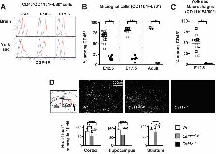
Next we examined the origin of microglia during development. In mouse embryos, the first wave of hematopoietic progenitors appears in the extra-embryonic yolk sac and leads to the production of primitive hematopoiesis, which takes place between E7.0 and E9.0 (13, 14). An independent wave of hematopoiesis termed “definitive hematopoiesis” is initiated within the embryo proper in the aorta, gonads and mesonephros (AGM) region (13, 14). Around E10.5, hematopoietic progenitors start to colonize the fetal liver, which serves as a major hematopoietic organ after E11.5, while later during development, hematopoiesis takes place in the spleen and bone marrow (14). Tissue macrophages in the adult are thought to derive from bone marrow monocytes and the contribution of primitive hematopoiesis to adult tissue macrophages remains unclear (15). To determine the developmental stage at which the seeding of myeloid cells occurs in the brain, we used Cx3cr1gfp/+ knock-in mice (16) as the fractalkine receptor (CX3CR1) is a marker of early myeloid progenitors (17) and microglia (18). Consistent with previous results (19), myeloid cells expressing the hematopoietic marker CD45 and the adult macrophage/microglia markers CD11b, F4/80 and CX3CR1 were detectable in the developing brain starting from E9.5 (Fig. 1, A and B and fig S2). At E10.5, microglia cells were present in both the cephalic mesenchyme and the neuroepithelium, although at a lower density in the latter (Fig. 1A, fig. S2 and movies S1 and S2). The phenotype of microglial cells resembled that of yolk sac macrophages throughout embryonic development (Fig. 1, B and C and fig. S3). Analysis of the DNA content of microglia and in vivo live imaging indicated that they were highly proliferative throughout embryonic life (fig. S4 and movie S3).
The differentiation of most macrophage populations in adult mice is controlled by colony stimulating factor-1 (CSF-1) and its receptor (CSF-1R) (20) but the role of the CSF-1 and CSF-1R in the development of yolk sac primitive macrophages and microglia is unknown. We found that CSF-1R was expressed in similar amounts on yolk sac macrophages and microglia at E9.5 (Fig. 2A). CSF-1R expression on microglia was maintained throughout
development (Fig. 2A and fig. S5). Consistent with a requirement for CSF-1R expression, absence of CSF-1R greatly reduced the development of microglia (Fig. 2B and fig. S6A) and yolk sac macrophages (Fig. 2C and fig. S6B), whereas circulating monocytes were present in these mice (fig. S6, C and D). Furthermore, microglia remained largely absent throughout life in CSF-1R–deficient mice (Fig. 2B and fig. S6A). Altogether, these results suggest that the development of yolk sac macrophages and microglia, but not monocytes, is strongly dependent on CSF-1R. Importantly and in contrast to many tissue macrophages, adult microglia can still form, albeit at reduced levels in Csf1op/op mice, which carry a natural null mutation of the Csf1 gene (21) but are absent in mice that lack the CSF-1R (Fig. 2D). A second CSF-1R ligand, interleukin 34 (IL-34), has recently been identified in mice, humans (22) and birds (23). The expression of IL-34 mRNA in the brain is much higher than the expression of CSF-1 mRNA during early post-natal development and in the adult (24), consistent with an important role for IL-34 in the regulation of microglial homeostasis and with the increased severity of the microglial phenotype in Csf1r-/- compared with Csf1op/op mice.
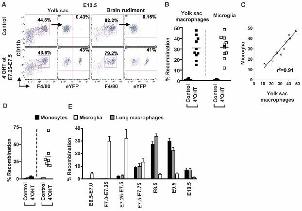
To examine the potential contribution of primitive myeloid precursors to adult microglia in vivo, we performed lineage tracing studies using mice expressing the tamoxifen-inducible MER-Cre-MER recombinase gene under the control of one of the endogenous promoters of the runt-related transcription factor 1 (Runx1) locus. Runx1 expression is first seen at E6.5 and is strongly up-regulated around E7.5 in the proximal visceral yolk sac region and until E8.0, Runx1+ cells are restricted to the extra-embryonic yolk sac and are absent from the embryo proper or allantois (25). We crossed the Runx1-MER-Cre-MER mice (Runx1Cre/wt) with the Cre-reporter mouse strain Rosa26R26R-eYF/R26R-eYFP and induced recombination by a single injection of 4-Hydroxytamoxifen (4’OHT) into pregnant females at different days of gestation. Active recombination in these knock-in mice occurs in a small time window that does not exceed 24 hrs post-injection (25) and leads to the irreversible expression of the enhanced yellow fluorescent protein (eYFP) reporter gene in Runx1+ cells and their progeny. At E10.5, embryos activated at E7.25-E7.5 exhibited a similar proportion of labeled yolk sac macrophages and microglia (31% +/- 10.8 for yolk sac macrophages and 32% +/- 10.7 for brain progenitors, n=10) (Fig. 3, A and B). The proportion of brain-infiltrating hematopoietic cells and microglia was similar in control littermate and heterozygote Runx1Cre/wt embryos, suggesting that microglia homeostasis was not perturbed in Runx1Cre/wt embryos (fig. S7, A and B). Furthermore, the proportion of eYFP+ microglia was similar in E10.5 and E13.5 embryos activated at E7.25-E7.5 (fig. S7C) and within individual embryos, the proportions of eYFP+ yolk sac macrophages and
/ www.sciencexpress.org / 21 October 2010 / Page 3 / 10.1126/science.1194637
eYFP+ microglia were highly correlated (Fig. 3C). The partial labeling of Runx1+ yolk sac cells is inherent to in vivo labeling techniques and likely results from the insufficient expression of MER-Cre-MER and limited availability of the ligand in target cells (25). Thus our model likely underestimates the contribution of Runx1+ precursors to adult microglia homeostasis, although we cannot exclude the potential contribution of non-labeled precursors to this process. Nevertheless, these results strongly suggest that yolk sac macrophages and microglia have the same origin and that the first wave of microglia is specified prior the end of E8.0. Strikingly, in adult mice activated at E7.25¬-E7.5 (n=10 from 3 litters), 32.11% +/- 18.0 of microglia were also eYFP+ (Fig. 3D and fig. S8), a proportion similar to that of yolk sac macrophages and microglia found in E10.5 and E13.5 embryos activated at E7.25-E7.5 (Fig. 3C and fig. S7). In contrast, less than 3% of blood circulating and tissue macrophages including dermal and lung macrophages, as well as circulating T cells, B cells and granulocytes were eYFP+ in these mice (Fig. 3E, fig. S8 and fig. S9). We also examined the earliest stage at which Runx1+ progenitors contributed to the adult microglial pool. In adult mice activated at E6.5-E7.0, less than 3.77% +/- 3.54 microglia were eYFP+, whereas 29.6% +/- 10 microglia were eYFP+ in adult mice activated at E7.0-E7.25 (Fig. 3E). In contrast 0.19% +/- 0.26 and 0.09% +/- 0.05 circulating leukocytes were eYFP+ in adult mice activated at E6.5-E7.0 and E7.0-E7.25, respectively (fig. S9). These results establish that primitive myeloid progenitors that arise prior to E7.5 contribute significantly to adult microglial homeostasis in the healthy brain, but have limited potential to give rise to adult blood leukocytes.
To determine at which stage, Runx1+ precursors or their progeny seed the brain during development, we injected Runx1Cre/wt : Rosa26R26R-LacZ pregnant females with a single dose of 4’OHT at E7.25-E7.5 and traced the apparition of labeled cells into the brain rudiment at different time-points after injection. A large number of Lac-Z+ cells populated the yolk sac of E8.5 embryos, whereas no labeled cells were detectable in the brain rudiment at this time-point (Fig. 4A and fig. S10A). Brain-infiltrating cells appeared only when blood circulation developed and a significant proportion of Lac-Z+ cells appeared associated with blood vessels and infiltrated the brain rudiment in E9.5 conceptus (Fig. 4B and fig. S10, B and C). These results are consistent with prior findings showing that CSF-1R+ cells first accumulate in the yolk sac around E8.0 and infiltrate the embryo proper when blood vessels develop around E9.0 (26). To address whether the development of functional blood vessels was required for the recruitment of myeloid precursors into the brain rudiment, we used Ncx-1–/– animals that lack a heartbeat and functional blood circulation due to a defect in the sodium calcium
exchanger 1 (27). We found that E9.5-E10.5 Ncx-1–/– embryos have yolk sac macrophages levels comparable or higher than control littermates (Fig. 4, C and E). In contrast, Ncx-1–/–embryos have no detectable microglia in the brain, whereas Ncx-1+/+ control littermates already have a substantial number of microglia in the brain at this time-point (Fig. 4, D and E). Altogether, these results suggest that Runx1+ progenitors migrate from the yolk sac into the brain through blood vessels between E8.5 and E9.5.
To examine the contribution of definitive hematopoiesis to microglial homeostasis, we injected 4’OHT at E8.5, E9.5 and E10.5. The proportion of eYFP+ leukocytes known to derive from definitive hematopoiesis was much higher in mice activated at E8.5 and E9.5 compared to mice activated at E7.25-E7.5 (up to 40% versus less than 3%, n=10) (Fig. 3E and fig. S9). In contrast, few eYFP+ microglia were detected in the brains of adult mice activated after E8.5 and onwards (Fig. 3E). The sharp descending contribution levels between E7.5 and E8.5 argue against the contribution of post-E7.5 Runx1+ anatomic locations to the labeling of the adult microglia lineage. Altogether, these data suggest minimal, if any, contribution of definitive hematopoiesis to the development of adult microglia.
Our results provide evidence that primitive myeloid precursors give rise to microglia residing in the adult CNS in the steady state. Primitive macrophages differentiate in the yolk sac of mammals, birds and zebrafish prior to the onset of blood circulation (13). Studies in zebrafish revealed that yolk sac-derived macrophages spread in the cephalic mesenchyme before invading the brain through the pial surfaces and the fourth ventricle (28–30); findings consistent with the observation that microglia in humans are formed in the pia mater (31). The conservation of primitive macrophages throughout evolution implies that they serve an important role in the early embryo (13), most likely related to the clearance of apoptotic bodies and the normal remodeling of brain tissues. In contrast to most adult tissue macrophages, microglia are maintained throughout life independently of any blood input and can resist high dose γ-ray irradiation. Whether these primitive macrophages are uniquely suited to reducing the risk of inflammation-induced injuries and maintaining the CNS integrity throughout adult life will be important to determine.
The results of this study should help unravel the regulatory program that controls microglial differentiation and function in vivo and identify new means to manipulate microglia for the treatment of neural diseases.
References and Notes 1. W. Y. Chan, S. Kohsaka, P. Rezaie, Brain Res Rev 53, 344
(Feb, 2007). 2. R. M. Ransohoff, V. H. Perry, Annu Rev Immunol 27, 119
(2009).

/ www.sciencexpress.org / 21 October 2010 / Page 4 / 10.1126/science.1194637
3. E. A. Ling, J Anat 128, 847 (Jun, 1979). 4. S. K. Leong, E. A. Ling, Glia 6, 39 (1992). 5. L. J. Lawson, V. H. Perry, S. Gordon, Neuroscience 48,
405 (1992). 6. J. Priller et al., Nat Med 7, 1356 (Dec, 2001). 7. A. R. Simard, D. Soulet, G. Gowing, J. P. Julien, S. Rivest,
Neuron 49, 489 (Feb 16, 2006). 8. B. Ajami, J. L. Bennett, C. Krieger, W. Tetzlaff, F. M.
Rossi, Nat Neurosci 10, 1538 (Dec, 2007). 9. A. Mildner et al., Nat Neurosci 10, 1544 (Dec, 2007). 10. K. Liu et al., Nat Immunol 8, 578 (Jun, 2007). 11. M. Bogunovic et al., J Exp Med 203, 2627 (Nov 27,
2006). 12. L. Vallieres, P. E. Sawchenko, J Neurosci 23, 5197 (Jun
15, 2003). 13. A. M. Lichanska, D. A. Hume, Exp Hematol 28, 601 (Jun,
2000). 14. S. H. Orkin, L. I. Zon, Cell 132, 631 (Feb 22, 2008). 15. R. van Furth, Z. A. Cohn, J Exp Med 128, 415 (1968). 16. S. Jung et al., Mol Cell Biol 20, 4106 (Jun, 2000). 17. K. Liu et al., Science, (Mar 12, 2009). 18. J. K. Harrison et al., Proc Natl Acad Sci U S A 95, 10896
(Sep 1, 1998). 19. F. Alliot, I. Godin, B. Pessac, Brain Res Dev Brain Res
117, 145 (Nov 18, 1999). 20. V. Chitu, E. R. Stanley, Curr Opin Immunol 18, 39 (Feb,
2006). 21. G. Blevins, S. Fedoroff, J Neurosci Res 40, 535 (Mar 1,
1995). 22. H. Lin et al., Science 320, 807 (May 9, 2008). 23. V. Garceau et al., J Leukoc Biol 87, 753 (May, 2010). 24. S. Wei et al., J Leukoc Biol, (May 26, 2010). 25. I. M. Samokhvalov, N. I. Samokhvalova, S. Nishikawa,
Nature 446, 1056 (Apr 26, 2007). 26. D. A. Ovchinnikov et al., J Leukoc Biol 83, 430 (Feb,
2008). 27. S. V. Koushik et al., FASEB J 15, 1209 (May, 2001). 28. S. P. Sorokin, R. F. Hoyt, Jr., D. G. Blunt, N. A. McNelly,
Anat Rec 232, 527 (Apr, 1992). 29. P. Herbomel, B. Thisse, C. Thisse, Dev Biol 238, 274
(Oct 15, 2001). 30. P. Herbomel, B. Thisse, C. Thisse, Development 126,
3735 (Sep, 1999). 31. M. A. Cuadros, J. Navascues, Prog Neurobiol 56, 173
(Oct, 1998). 32. We thank Dr S. Nishikawa, Dr. H. Snoeck and Dr. P.S.
Frenette for intellectual input in the study, the RIKEN CDB Laboratory for Animal Resources and Genetic Engineering for providing the Runx1-MER-Cre-MER mice, Dr. G. Hoeffel, Dr. X.H. Zong, R. Basu and H. Ketchum for technical assistance; Dr. L. Robinson for critical review and editing of the manuscript. This work
was supported by National Institutes of Health grants CA112100, HL086899 and AI080884 to M.M, CA32551 and CA26504 to E.R.S and MH66290 and NS38902 to M.F.M. I.M.S. is supported by a grant from RIKEN Strategic Programs for Research and Development (President’s Fund). M.G. is supported by the National Science Foundation of Switzerland. Runx1-MER-Cre-MER mice have CDB Acc. No. CDB0524K: http://www.cdb.riken.jp/arg/mutant%20mice%20list.html
Supporting Online Material www.sciencemag.org/cgi/content/full/science.1194637/DC1 Materials and Methods Figs. S1 to S10 Movies S1 to S3 References
06 July 2010; accepted 07 October 2010 Published online 21 October 2010; 10.1126/science.1194637
Fig. 1. Microglia arise during early embryonic life. (A) Left panel, schematic of the imaging field. Right panel, three-dimensional rendering of E10.5 brain rudiment from Cx3cr1gfp/+ mice. DAPI (blue) stains the ectoderm. Representative data of 2 experiments (n=2). (B-C) Flow cytometric analysis of the expression of CD11b and GFP (CX3CR1) on gated DAPI-CD45+ brain (B) and yolk sac (C) cells isolated from Cx3cr1gfp/+ mice at different stages during development. Histograms show F4/80 (red) or isotype control (blue) on gated cells. Representative data of 3 experiments (n=3).
Fig. 2. Microglia and yolk sac macrophages are absent in Csf1r–/– mice. (A) Flow cytometric analysis of CSF-1R expression (red) on microglia and yolk sac macrophages (blue: isotype control). Representative data of 3 experiments (n=3). (B-C) Percentage of microglia (B) and yolk sac macrophages (C) in Csf1r–/– (black squares) or control littermate (Wt) (white squares) FVB/NJ mice. Pooled data from 3 separate experiments. **, P<0.001; ***, P<0.0001. (D) Coronal sections of 3 week-old Wt, Csf1op/op and Csf1r-/- brains of region boxed in the schematic stained for the microglial marker Iba (DG, Dentate Gyrus; Cx, Cerebral cortex; CA3, CA3 region of the hippocampus). Mean number of Iba1+ cells/field, from three different brain regions is shown. Average of six fields (0.5 mm2) per region per genotype. Error bars represent mean ± SD of data from 2 pooled experiments. *, P<0.05; ****, P<0.00001 .
Fig. 3. Microglia arise from primitive myeloid progenitors. Runx1Cre/wt : Rosa26R26R-eYFP mice were treated with 4’OHT to induce Cre-mediated recombination at E7.25-E7.5 and analyzed at E10.5 (A-C) or at 8 weeks post-birth (D-E). Controls are non-treated mice. (A) Flow cytometric analysis
/ www.sciencexpress.org / 21 October 2010 / Page 5 / 10.1126/science.1194637
from one representative embryo showing the % recombination among yolk sac macrophages and microglial progenitors. (B and D) Pooled data from 2 experiments showing the % recombination among yolk sac macrophages and microglial progenitors cells in embryos (B), and among monocytes and microglia in adult mice (n=10) (D). (C) Correlation and regression analysis between the % recombination in microglial progenitors and yolk sac macrophages. r2, coefficient of regression. (E) % recombination among monocytes, lung macrophages and microglia in adult mice activated at different embryonic age. Error bars represent mean ± SEM of pooled data from 2 experiments (n= 8-16). Gating strategy for each leukocyte population is detailed in fig. S8.
Fig. 4. Runx1+ yolk sac progenitors seed the brain between E8.5 and E9.5 through blood circulation (A-B) Runx1Cre/wt : Rosa26R26R-LacZ embryos activated at E7.25-E7.5 were isolated at E8.25-E8.5 (A) or E9.25-E9.5 (B) and processed for whole mount LacZ staining as described in the material and method section. At E8.25-E8.5, labeled cells are detected in the yolk sac but not in the brain rudiment or in the neural tube (A) whereas labeled cells infiltrate the brain rudiment of E9.25-E9.5 embryos (B). (C-E) Yolk sac and brain rudiment tissues were isolated from E10.0-E10.5 Ncx1–/– embryos or control littermates and processed for flow cytometry analysis as described in the material and method section. Dot plots show the presence of yolk sac macrophages in Ncx1–/– embryos and control littermates (C), whereas microglia were present in control but not in Ncx1–/– embryos (D). (E) Graph shows the percentage +/- SEM of hematopoietic cells (CD45+) in control littermates (white bars, n=4) and Ncx1–/– embryos (back bars, n=3).