NPY in rat retina is present in neurons, in endothelial cells and also in microglial and Müller...
7
NPY in rat retina is present in neurons, in endothelial cells and also in microglial and Mu ¨ller cells Ana Rita A ´ lvaro a,b , Joana Rosmaninho-Salgado a , Ana Raquel Santiago a,b , Joa ˜o Martins a , Ce ´lia Aveleira b , Paulo F. Santos a , Tiago Pereira a , Denisa Gouveia a , Ana Luı ´sa Carvalho a , Eric Grouzmann c , Anto ´nio Francisco Ambro ´sio a,b , Cla ´udia Cavadas a,d, * a Center for Neuroscience and Cell Biology, University of Coimbra, Coimbra, Portugal b Center of Ophthalmology of Coimbra, IBILI, Faculty of Medicine, University of Coimbra, Coimbra, Portugal c Division of Clinical Pharmacology and Toxicology, CHUV, Lausanne, Switzerland d Faculty of Pharmacy, University of Coimbra, Coimbra, Portugal Received 10 October 2006; received in revised form 12 December 2006; accepted 22 January 2007 Available online 8 February 2007 Abstract NPY is present in the retina of different species but its role is not elucidated yet. In this work, using different rat retina in vitro models (whole retina, retinal cells in culture, microglial cell cultures, rat Mu ¨ller cell line and retina endothelial cell line), we demonstrated that NPY staining is present in the retina in different cell types: neurons, macroglial, microglial and endothelial cells. Retinal cells in culture express NPY Y 1 ,Y 2 ,Y 4 and Y 5 receptors. Retina endothelial cells express all NPY receptors except NPY Y 5 receptor. Moreover, NPY is released from retinal cells in culture upon depolarization. In this study we showed for the first time that NPY is present in rat retina microglial cells and also in rat Mu ¨ ller cells. These in vitro models may open new perspectives to study the physiology and the potential pathophysiological role of NPY in the retina. # 2007 Elsevier Ltd. All rights reserved. Keywords: Neuropeptide Y (NPY); Rat retina; Microglial cells; Mu ¨ller cells; Endothelial cells; NPY receptors; NPY release 1. Introduction The retina is a very organized structure of the nervous tissue that contains two main classes of cells: neurons and glial cells. Retinal neurons include photoreceptors, bipolar, ganglion, horizontal and amacrine cells that are strategically localized in various layers through. Glial cells comprise macroglial and microglial cells. Two main macroglial elements are present in the retina (Newman, 2004): Mu ¨ller cells that span the entire thickness of the retina, give the support to the retina by regulating retinal metabolism and modulate the function of neurons and blood vessels; astrocytes are associated with optic nerve axons and retinal blood vessels. Microglia is normally quiescent but very sensitive to the retina homeostatic state. When the homeostasis is disturbed, microglia rapidly acts as phagocytes, or release inflammatory mediators (Chen et al., 2002). Neuropeptide Y (NPY) is a highly conserved, 36 amino acid peptide, widely distributed in the central nervous system (Silva et al., 2005b) being also present in the retina of different species (Bruun et al., 1986; Hutsler and Chalupa, 1995; Jen et al., 1994; Oh et al., 2002). NPY-ir is mainly localized in amacrine cells but it is also present in ganglion cells (Ammar et al., 1998; Hutsler and Chalupa, 1994; Sinclair and Nirenberg, 2001). In rat retina, NPY-ir is present in two populations of amacrine cells, one located in the inner nuclear layer and the other in the ganglion cell layer. Little is known about the function of NPY system in the retina but some observations propose that NPY-ir cells participate in multiple circuits mediating visual informa- tion processing in the inner retina. It was suggested that these cells are involved in spatial tuning, specifically, in tuning ganglion cells to low spatial frequencies (Sinclair et al., 2004). In addition, exogenous application of NPY stimulates the release of glycine, dopamine, and 5-hydroxytryptamine from the rabbit retina, and gamma-aminobutyric acid (GABA) and www.elsevier.com/locate/neuint Neurochemistry International 50 (2007) 757–763 * Corresponding author at: Faculty of Pharmacy, University of Coimbra, 3020-123 Coimbra, Portugal. Tel.: +351 96 3928766. E-mail address: [email protected] (C. Cavadas). 0197-0186/$ – see front matter # 2007 Elsevier Ltd. All rights reserved. doi:10.1016/j.neuint.2007.01.010
-
Upload
independent -
Category
Documents
-
view
3 -
download
0
Transcript of NPY in rat retina is present in neurons, in endothelial cells and also in microglial and Müller...
www.elsevier.com/locate/neuint
Neurochemistry International 50 (2007) 757–763
NPY in rat retina is present in neurons, in endothelial cells and
also in microglial and Muller cells
Ana Rita Alvaro a,b, Joana Rosmaninho-Salgado a, Ana Raquel Santiago a,b, Joao Martins a,Celia Aveleira b, Paulo F. Santos a, Tiago Pereira a, Denisa Gouveia a, Ana Luısa Carvalho a,
Eric Grouzmann c, Antonio Francisco Ambrosio a,b, Claudia Cavadas a,d,*a Center for Neuroscience and Cell Biology, University of Coimbra, Coimbra, Portugal
b Center of Ophthalmology of Coimbra, IBILI, Faculty of Medicine, University of Coimbra, Coimbra, Portugalc Division of Clinical Pharmacology and Toxicology, CHUV, Lausanne, Switzerland
d Faculty of Pharmacy, University of Coimbra, Coimbra, Portugal
Received 10 October 2006; received in revised form 12 December 2006; accepted 22 January 2007
Available online 8 February 2007
Abstract
NPY is present in the retina of different species but its role is not elucidated yet. In this work, using different rat retina in vitro models (whole
retina, retinal cells in culture, microglial cell cultures, rat Muller cell line and retina endothelial cell line), we demonstrated that NPY staining is
present in the retina in different cell types: neurons, macroglial, microglial and endothelial cells. Retinal cells in culture express NPY Y1, Y2, Y4
and Y5 receptors. Retina endothelial cells express all NPY receptors except NPY Y5 receptor. Moreover, NPY is released from retinal cells in
culture upon depolarization. In this study we showed for the first time that NPY is present in rat retina microglial cells and also in rat Muller cells.
These in vitro models may open new perspectives to study the physiology and the potential pathophysiological role of NPY in the retina.
# 2007 Elsevier Ltd. All rights reserved.
Keywords: Neuropeptide Y (NPY); Rat retina; Microglial cells; Muller cells; Endothelial cells; NPY receptors; NPY release
1. Introduction
The retina is a very organized structure of the nervous tissue
that contains two main classes of cells: neurons and glial cells.
Retinal neurons include photoreceptors, bipolar, ganglion,
horizontal and amacrine cells that are strategically localized in
various layers through. Glial cells comprise macroglial and
microglial cells. Two main macroglial elements are present in
the retina (Newman, 2004): Muller cells that span the entire
thickness of the retina, give the support to the retina by
regulating retinal metabolism and modulate the function of
neurons and blood vessels; astrocytes are associated with optic
nerve axons and retinal blood vessels. Microglia is normally
quiescent but very sensitive to the retina homeostatic state.
When the homeostasis is disturbed, microglia rapidly acts as
* Corresponding author at: Faculty of Pharmacy, University of Coimbra,
3020-123 Coimbra, Portugal. Tel.: +351 96 3928766.
E-mail address: [email protected] (C. Cavadas).
0197-0186/$ – see front matter # 2007 Elsevier Ltd. All rights reserved.
doi:10.1016/j.neuint.2007.01.010
phagocytes, or release inflammatory mediators (Chen et al.,
2002).
Neuropeptide Y (NPY) is a highly conserved, 36 amino acid
peptide, widely distributed in the central nervous system (Silva
et al., 2005b) being also present in the retina of different species
(Bruun et al., 1986; Hutsler and Chalupa, 1995; Jen et al., 1994;
Oh et al., 2002). NPY-ir is mainly localized in amacrine cells
but it is also present in ganglion cells (Ammar et al., 1998;
Hutsler and Chalupa, 1994; Sinclair and Nirenberg, 2001). In
rat retina, NPY-ir is present in two populations of amacrine
cells, one located in the inner nuclear layer and the other in the
ganglion cell layer. Little is known about the function of NPY
system in the retina but some observations propose that NPY-ir
cells participate in multiple circuits mediating visual informa-
tion processing in the inner retina. It was suggested that these
cells are involved in spatial tuning, specifically, in tuning
ganglion cells to low spatial frequencies (Sinclair et al., 2004).
In addition, exogenous application of NPY stimulates the
release of glycine, dopamine, and 5-hydroxytryptamine from
the rabbit retina, and gamma-aminobutyric acid (GABA) and

A.R. Alvaro et al. / Neurochemistry International 50 (2007) 757–763758
choline from the chicken retina, suggesting that NPY
modulates neurotransmitter release in the retina (Bruun and
Ehinger, 1993). All these observations suggest that NPY is
probably released in response to visual stimulus. However, the
conditions required for NPY release must be investigated. The
fact that NPY is released by high frequency light stimulation or
depolarization was only demonstrated only in the frog retina
(Bruun et al., 1991). The release of NPY in the retina of other
vertebrates was not yet demonstrated.
NPY mediates its effects through the activation of six G-
protein-coupled receptor subtypes named Y1, Y2, Y3, Y4, Y5, and
y6. Five distinct NPY receptors have been cloned, Y1, Y2, Y4, Y5
and y6 (Silva et al., 2002). The Y3 receptor has not yet been cloned
while y6 receptor is not functional (Michel et al., 1998). The
presence of mRNA for the Y1, Y2, Y4 and Y5 receptors was
detected in intact rat retinas (D’Angelo and Brecha, 2004).
Endothelial cells are another cell type that forms an
important structure of the retina: blood-retinal-barrier (BRB).
This structure separates the retina from the blood circulation by
a barrier located both at the retinal vessels, the inner BRB, and
at the retinal pigment epithelium, the outer BRB. NPY has been
reported to directly stimulate endothelial cell proliferation and
migration (Ekstrand et al., 2003; Ghersi et al., 2001; Marion-
Audibert et al., 2000; Zukowska-Grojec et al., 1998a,b). In the
retina, some evidences have shown that NPY and Y2 receptors
are involved in development of diabetic retinopathy and retinal
neovascularization (Koulu et al., 2004).
Several lines of evidence suggest that the expression of NPY
and NPY receptors, in central nervous system, may not be
restricted to neurons, but could also extend to glial cells,
namely in astrocytes (Barnea et al., 1998; Gimpl et al., 1993; St-
Pierre et al., 2000). The presence of NPY in retina glial cells,
such as in microglial cells and in Muller cells, was not yet
investigated.
The aim of the present study was to better characterize the
presence of NPY on different in vitro rat retina models.
Therefore, whole rat retinas, primary cell cultures of rat retinal
cells, primary cell cultures of retina microglial cells, a cell line
of rat Muller cells, and a cell line of rat retina endothelial cells
were used. The mRNA for NPY and NPY receptors were
detected in whole rat retinas, primary cell cultures of rat retinal
cells and in a cell line of rat retina endothelial cells. Moreover,
the NPY release was evaluated using the primary culture of rat
retinal cells. The development and characterization of new in
vitro models of the retina will allow defining the role of NPY in
retinal physiology and pathophysiology.
2. Materials and methods
2.1. Materials
Minimum essential medium (MEM) was purchased from Sigma Chemical
Co., St. Louis, MO, USA. Dulbecco’s modified Eagle medium (DMEM) and
trypsin were purchased from GIBCO BRL, Life Technologies, Scotland, UK.
Taq DNA polymerase was purchased from Amersham Biosciences Europe
GmbH, Freiburg, Germany. First strand cDNA Synthesis Kit for RT-PCR
(AMV) was purchased from Roche Applied Science, PO, Indianapolis,
USA. Fetal bovine serum was obtained from Biochrom, Berlin, Germany.
2.2. Cell cultures
2.2.1. Primary rat retina neural cell culture
Three to 5 days old Wistar rats were used to prepare primary rat retina cell
culture as previously described (Santiago et al., 2006). All procedures involving
animals were in agreement with the Association for Research in Vision and
Ophthalmology (ARVO) statement in vision and ophthalmic research. Briefly,
rat retinas were digested with 0.1% trypsin (w/v) for 15 min at 37 8C. Cells were
plated on poly-D-lysine (0.1 mg/ml) coated wells with MEM, supplemented
with 25 mM HEPES, 26 mM NaHCO3, 10% FCS and penicillin (100 U/ml)–
streptomycin (100 mg/ml) for 9 days (37 8C, 5% CO2), at a density of
2 � 106 cells/cm2. For immunocytochemistry experiments, cells were plated
on glass coverslips and for RNA extraction cells were plated on 35 mm Petri
dishes at the same density. For NPY assays retina neural cell cultures were
plated on 60 mm Petri dishes at density of 3 � 106 cells/cm2.
2.2.2. Rat retina microglial cell culture
Primary rat microglial cell cultures were prepared from 3 to 5 days old
Wistar rats retinas as described with some modifications (Wang et al., 2005).
Briefly, retinas were dissected under sterile conditions using a light microscope
in Ca2+- and Mg2+-free Hank’s balanced salt solution (HBSS, in mM: 137 NaCl,
5.4 KCl, 0.45 KH2PO4, 0.34 Na2HPO4, 4 NaHCO3, 5 glucose; pH 7.4). The
retinas were digested in HBSS supplemented with 0.1% trypsin (w/v) and
0.001% DNase (w/v), for 15 min at 37 8C. After dissociation, trypsin was
inactivated by 10% heat-inactivated fetal bovine serum (FBS) and the cells were
pelleted by centrifugation. The cells were resuspended in DMEM/F-12 HAM
supplemented with 10% heat-inactivated FBS, 100 U/ml penicillin and 100 mg/
ml streptomycin, and plated at a density of 1.5 � 106 cells/cm2 on 75 cm2
growth surface area plastic flasks coated with poly-D-lysine (0.1 mg/ml). The
cells were kept in culture at 37 8C under a 95% air/5% CO2 atmosphere and
the medium was changed every 3 days. After 2 weeks, microglia cells
were harvested in culture medium containing serum by shaking the flasks at
150 rpm for 30 min. The detached cells were plated at a density of
25 � 104 cells/cm2 on 16 mm glass coverslips coated with poly-D-lysine
(0.1 mg/ml). The cells were used for immunocytochemistry experiments after
3 days in culture.
2.2.3. Culture of rat retina endothelial cell line—TR-iBRB
Conditionally immortalized retinal capillary endothelial cell line (TR-
iBRB—transgenic rat-inner blood-retinal barrier) was obtained from Dr.
Ken-ichi Hosoya, and cultured as previously described (Hosoya et al.,
2001). These cells were plated on glass coverslips and cultured in DMEM
at 33 8C/5% CO2 for 3–4 days until 80–90% of confluence.
2.2.4. Culture of rat retina Muller cell line
Conditionally immortalized rat Muller cell line (TR-MUL—transgenic rat
Muller cells) were cultured as previously described (Tomi et al., 2003). Muller
cells were plated on glass coverslips and cultured in DMEM at 33 8C/5% CO2
for 3–4 days until 80–90% of confluence.
2.3. Determination of NPY content and release in rat retina cell
cultures
Retina cells in culture were washed three times with Krebs buffer (132 mM
NaCl, 4 mM KCl, 1.4 mM MgCl2, 10 mM glucose, 10 mM HEPES, pH 7.4)
containing Tween 20 (0.0001%) in order to prevent NPY sticking to plastic.
After 20 min of pre-incubation, at room temperature in Krebs buffer, the cells
were incubated with Krebs buffer for 10 min at room temperature with or
without 50 mM KCl. NPY released without KCl (constitutive release) and with
KCl was measured by an ELISA assay (described below). NPY intracellular
content in whole retina or in cell cultures were determined in Krebs buffer with
50 mM EDTA and 0.0001% Tween 20 and after freeze–defreeze, and sonication
for 12 s. The cell suspension was centrifuged (14,000 rpm/5 min/4 8C). NPY
(released and content) was measured using a NPY sandwich-ELISA assay as
previously described (Grouzmann et al., 1992a). Each well from 96 wells
microplate (PolySorp, Nunc) was coated with 200 ng (100 ml) of the mono-
clonal antibody NPY02 diluted in Tris buffer (50 mM, pH 7.5) for 16 h at 4 8C.

A.R. Alvaro et al. / Neurochemistry International 50 (2007) 757–763 759
The plates were washed three times with Tris buffer containing 0.08% Tween 20
(Sigma) (Tris–Tween buffer) and incubated for 30 min with 200 ml of Tris–
Tween 0.08% buffer containing 50 g/l low-fat dry milk. After washing four
times with Tris–Tween 0.08% buffer, wells were filled with standards (standards
ranging from 0.5 to 100 pM) and samples. The samples and standards were
incubated overnight at RT. After washing four times with Tris–Tween 0.08%
buffer a second antibody, NPY05 conjugated with alkaline phosphatase, diluted
(1:4000) in 5% low-fat milk in Tris–Tween 0.08% buffer, was added (7 h at
room temperature). The plates were then washed twice with Tris buffer
containing 0.25% Tween 20 followed by washing two times with Tris buffer
with 150 mM NaCl. Bound alkaline phosphatase was revealed by the addition
of 50 ml of the substrate (NADPH solution from ELISA amplification kit,
Invitrogen Life Technologies, Carlsbad, CA, USA). The amplifier (50 ml)
(ethanol dehydrogenase mixed with diaforase from ELISA amplification kit,
Invitrogen Life Technologies, Carlsbad, CA, USA) was added 45 min later,
according to the manufacturer recommendations. The optic density was read at
492 nm (ELISA reader, SLT, Spectra II) for 30 min. The detection limit was
0.5 pM. This NPY assay measures biologically active forms of NPY (NPY1-36,
NPY2-36 and NPY3-36) (Grouzmann et al., 1992a). The protein content was
measured by Bradford assay (Biorad) (Bradford, 1976).
2.4. Immunocytochemistry
Cells on glass coverslips were washed twice with PBS and fixed in 4%
paraformaldehyde (30 min; room temperature). Then, the cells were incubated
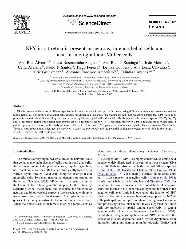
Fig. 1. NPY staining in different types of rat retinal cells in culture (A–I): double sta
(green) with a monoclonal mouse anti-NPYantibody (A, D, G). Retinal cells (A–I) we
Neurons were labelled (red) with an anti-MAP-2 antibody (B and C); astrocytes a
microglial cells were labelled (red) with an anti-Mac-1 antibody (H and I). The imag
references to colour in this figure legend, the reader is referred to the web version
for 5 min with 1% Triton, and for 1 h with 3% (w/v) fatty acid-free bovine
serum albumin (BSA), supplemented with 0.2% Tween 20, to prevent non-
specific binding, at room temperature. Cells were incubated with primary
antibodies for 90 min at room temperature: rabbit anti-Microtubule Asso-
ciated Protein 2 (MAP-2; 1:500; Chemicon International, Temecula, CA,
USA); mouse anti-NPY (NPY02; 1:1500; Dr. Eric Grouzmann, CHUV,
Lausanne, Switzerland; Grouzmann et al., 1992b); rabbit anti-glial-fibrilliary
acid protein (GFAP; 1:500; Dako, Denmark) or rat anti-macrophage antigen-1
(Mac-1, 1:500; Serotec, Oxfordshire, UK). After washing, the cells were
incubated for 1 h at room temperature with secondary antibodies (1:200
AlexaTM 488 anti-mouse IgG, 1:150 AlexaTM 594 anti-rat IgG or 1:200
AlexaTM 594 anti-rabbit IgG; Invitrogen, Eugene, Oregon, USA). Cells were
mounted using a Prolong Antifade Kit and visualized using a fluorescent
microscopic (Zeiss Axioshop 2 Plus) coupled to a digital camera (Axiocam
HRc).
2.5. RNA extraction and reverse transcriptase-PCR
Total RNA was isolated from the intact retina, neural and endothelial cell
cultures using TRI Reagent (Sigma). The total amount of RNA was quantified
by optical density (OD) measurements at 260 nm, and the purity was evaluated
by measuring the ratio of OD at 260 and 280 nm (RNA/DNA calculator
GeneQuantII, Pharmacia Biotech Amersham Biosciences AB, Uppsala, Swe-
den). First strand cDNAs were synthesized by incubating 2 mg of the total RNA
from the neural and endothelial cell culture, or from intact retina with random
ining with neuronal, macroglial and microglial cell markers. NPY was detected
re double stained with NPYand neuronal, macroglial or microglial cell markers.
nd Muller cells were identified (red) with an anti-GFAP antibody (E and F);
es are representative of three independent cell cultures. (For interpretation of the
of the article.)

Fig. 2. NPY staining in rat retina endothelial cells (A) and in rat Muller cells (B). Rat retina endothelial cells (TR-iBRB cell line) and rat Muller cells (TR-mull cell
line) were labelled with anti-NPY antibody. The images are representative of three independent cell cultures. 600�.
A.R. Alvaro et al. / Neurochemistry International 50 (2007) 757–763760
oligo (dT) primers, RNase inhibitor, and Avian Microblastosis Virus (AMV)
reverse transcriptase (RT) for 1 h at 42 8C.
Reverse transcriptase-PCR (RT-PCR) was performed using specific primer
pairs for rat NPY and NPY receptors. The primers were (forward and reverse
and product size: NPY, 50-AGAGATCCAGCCCTGAGACA-30, 50-AACGA-
CAACAAGGGAAATGG-30 (236 bp); Y1, 50-AACCTCTCCTTCTCAGA-
CTTGC-30, 50-CACAGTGTTGAAGATGGTAAGG-30 (616 bp); Y2, 50-CTCCAAGCAAATCAGCTTCC-30, 50-GTTTTGTGCCTTCGCTGATGG-30
(318 bp); Y4, 50-AACCTACTCATTGCCAACCTG-30, 50-ATGTAGCAGAC-
CAGGATGAAG-30 (476 bp); Y5, 50-GTGTTCCCGAGGTGCTTCTA-30, 50-ATTCCGAGCAGCAGCTGTAT-30 (248 bp) (MWG-Biotech AG, Ebersberg,
Germany). Positive controls were plasmids encoding Y1, Y2, and Y5 and rat
brain cDNA (for NPYand Y4). Negative controls were performed without RNA
sample, which was substituted by water. All amplifications were performed in
PCR buffer containing 1.5 mM MgCl2 and 200 mM random oligo (dT) primers,
0.2 mM of each primer, and 2.5 units/ml Taq DNA polymerase (Amersham
Biosciences). The reactions were done as follows: 1 min at 95 8C, 35 cycles at
95 8C for 1 min, 55 8C for 1 min, and final extension at 72 8C for 1 min. PCR
products were analyzed by electrophoresis on a 2% agarose gel containing
ethidium bromide. Densitometrical analysis was performed on Versa-Doc
Imaging System (Model 3000, Bio-Rad Laboratories, Hercules, CA, USA).
3. Results
3.1. NPY content in rat retina and retina cell cultures
Retinas isolated from 9-week-old rats contained 15.1 �1.4 fmol NPY/mg protein (n = 4). The intracellular content of
Fig. 3. NPY staining in rat retina microglial cells in culture. NPY was detected (gre
cells were double stained with the anti-NPYantibody (A) and with an anti-Mac-1 ant
interpretation of the references to colour in this figure legend, the reader is referre
NPYof retinal cell cultures was 1.4� 0.1 fmol NPY/mg protein
(n = 3, duplicates of three independent cultures). The intracel-
lular content of NPYof rat retina endothelial cell line was below
the limit of detection of the NPY assay used (<0.5 pM).
3.2. NPY staining in different retina cell types
Labelling NPY and pro-NPY with a highly specific
monoclonal antibody, NPY02 (Grouzmann et al., 1992b),
revealed the presence of NPYand pro-NPY in rat retinal cells in
culture (Fig. 1). Some NPY-positive cells were neurons, since
NPY-positive cells were also labelled with a neuronal marker
(MAP-2). NPY staining in MAP-2-positive cells was observed
mainly in the cell bodies, but some immunoreactivity was also
observed in the cell processes (Fig. 1A–C). Some NPY-positive
cells were also immunoreactive to GFAP, a marker of astrocytes
and Muller cells (Fig. 1D–F). In addition, some NPY-positive
cells were microglial cells (Fig. 1G–I). A similar pattern of
immunoreactivity was also observed when NPY labelling was
assessed with NPY05 antibody (results not shown), another
monoclonal antibody (Grouzmann et al., 1992b), which binds
the amidated C-terminal part of NPY.
The presence of NPY in Muller cells was confirmed by NPY
staining in a cell line of rat Muller cells (Fig. 2B). Retina
endothelial cells were also labelled with the NPY02 antibody,
en) with a monoclonal mouse anti-NPY antibody (NPY02; 1:1500). Microglial
ibody (B). The images are representative of three independent cell cultures. (For
d to the web version of the article.)

A.R. Alvaro et al. / Neurochemistry International 50 (2007) 757–763 761
and a punctuate labelling throughout the cell was evident, but
mostly in the cytosol and in the perinuclear region (Fig. 2A). To
corroborate the presence of NPY in microglial cells in retina
cells in culture, a primary cell culture of retina microglia cells
was used, and as shown in Fig. 3, NPY staining is observed in
retina microglial cells in culture.
3.3. Detection of mRNA for NPY and NPY receptors by RT-
PCR
NPY mRNA was detected in the whole rat retina. The
mRNA for NPY was also detected in the retina cells in cultures
and in the retina endothelial cell line (Fig. 4A–C). The mRNAs
of NPY receptors Y1, Y2, Y4 and Y5 were present in whole rat
retina and in the retina cells in culture (Fig. 4A and B). In retina
endothelial cells, the mRNAs for NPY Y1, Y2 and Y4 receptors
were detected, but the mRNA for Y5 receptor was not detected
(Fig. 4C).
3.4. NPY release from rat retinal cell cultures
The amount of constitutive amidated-NPY release (10 min in
Krebs buffer) from rat retinal cell in culturewas below the limit of
Fig. 4. NPY and NPY receptors mRNAs in whole rat retina (A), rat retina cells
in culture (B) and rat retina endothelial cells (C). The mRNA expression of NPY
and NPY receptors was assessed by reverse transcriptase-PCR followed by
agarose gel electrophoresis. PCR products used (base pairs (bp): NPY, 236 bp;
NPY Y1, 616 bp; NPY Y2, 318 bp; NPY Y4, 476 bp; NPY Y5, 248 bp; actin,
427 bp. Each figure is representative of three different retinas or three inde-
pendent cell cultures.
detection of the NPYassay used. Incubating the cells 10 min with
50 mM KCl the NPY release was 0.5–3.0 pM, which represents a
7.8� 4.2% of the intracellular content of NPY (n = 3).
4. Discussion
NPY is present in both mammalian and non-mammalian
retinas (Bruun et al., 1986; Hutsler and Chalupa, 1995; Jen
et al., 1994; Oh et al., 2002; Tornqvist and Ehinger, 1988).
However, its role in retinal physiology and visual processing is
not yet elucidated, in part due to lack of information about the
retinal cell types that express and release NPY, as well as the
expression and localization of NPY receptors. In the present
study, using different retina cell cultures, we aimed to identify
retinal cell types that express NPY.
Using an ELISA-sandwich assay to quantify the biologically
active NPY, amidated-NPY, we verified that the peptide is
present in the whole rat retina and in primary retina cells in
culture. The presence of NPY in retina cells was demonstrated
and confirmed by immunocytochemistry and mRNA expres-
sion. The presence of NPY-ir was also observed in rat retina by
others (Ferriero and Sagar, 1989; Oh et al., 2002), and also in
the retinas of other mammalian species, including human (Jen
et al., 1994; Tornqvist and Ehinger, 1988), mouse (Sinclair and
Nirenberg, 2001), monkey (Marshak, 1989).
In retina cells in culture, using double immunolabelling, we
found that NPY is present in retinal neurons. This observation is
in accordance with other studies that described that NPY-ir in
the retina is mainly localized in neurons, specially in amacrine
cells, but also in ganglion cells (Ammar et al., 1998; Hutsler
et al., 1993; Sinclair and Nirenberg, 2001). In rat retina, it was
demonstrated that NPY-ir is present in two populations of
amacrine cells, one located in the internal nuclear layer and the
other in the ganglion cell layer (Oh et al., 2002). These
observations suggest that NPY-ir cells participate in multiple
circuits, mediating visual information processing, mainly in the
inner retina.
Several lines of evidence suggest that the expression of NPY
and NPY receptors, in central nervous system, may not be
restricted to neurons, but could also extend to glial cells, namely
in astrocytes (Barnea et al., 1998; Gimpl et al., 1993; St-Pierre
et al., 2000). In fact, we also observed NPY-ir in retina macroglial
cells and in microglial cells. These results demonstrate, for the
first time that NPY is present not only in retinal neurons, but also
in glial cells in the retina. Some studies strongly suggest that
retinal macroglia and microglia may be associated with retinal
dysfunctions such as vitreoretinopathy, diabetic retinopathy,
retinitis pigmentosa and age-related macular degeneration
(Gupta et al., 2003; McGillem and Dacheux, 1998; Rungger-
Brandle et al., 2000; Soler et al., 2002). Therefore, the presence
of NPY in retinal microglia may suggest a putative role of this
peptide on inflammatory states observed in those retinal
dysfunctions and should be further investigated (Leal et al.,
2005; Meleth et al., 2005; Zhou et al., 2005).
In this study, NPY-ir was also detected in cultured rat retina
endothelial cells line by immunocytochemistry and mRNA
expression. The punctuate labelling was particularly evident in

A.R. Alvaro et al. / Neurochemistry International 50 (2007) 757–763762
the cytosol and around the nucleus. NPY-ir was detected in
other endothelial cells: in human umbilical-vein endothelial
cells (Cai et al., 1993; Silva et al., 2005a) and in human
endocardial endothelial cells (Jacques et al., 2003). In this study
we detected mRNAs for NPY Y1, Y2 and Y4 receptors but not
for Y5 receptor in retina endothelial cells. The presence of
mRNA for Y1 and Y2 receptors is consistent with previous
studies using HUVECs (Silva et al., 2003; Zukowska-Grojec
et al., 1998b). On the contrary, the Y4 receptor mRNA was not
detected in HUVECs and the Y5 receptor mRNA was detected
only after cytokine treatment (Silva et al., 2003). These
differences in NPY receptors mRNA expression between the
two types of endothelial cells may influence endothelial cell
physiology (Rymaszewski et al., 1992).
Regarding whole retinas and retina cells in culture, in the
present work we show that the mRNAs of all known NPY
receptors (NPY Y1, Y2, Y4 and Y5) were detected. These results
are in accordance to a previous study in whole rat retina
(D’Angelo and Brecha, 2004). Also, mRNAs for Y1 receptor
and for Y2 receptors were detected in the mouse retina (Yoon
et al., 2002). Immunohistochemical studies in rat retina showed
that Y1 receptor is present in amacrine cells in the distal inner
nuclear layer and also in horizontal cell bodies and their
processes in the outer plexiform layer (D’Angelo et al., 2002).
Furthermore, in the rat retina there is a difference between the
distribution of NPY-ir and Y1 receptor-ir, suggesting that NPY
acts in a paracrine manner on cells located far from the site of
their release (D’Angelo et al., 2002). The Y4 and Y5 receptors
presence in retinal tissue and their cellular localization and
physiological role remain to be determined. The presence of
functional Y2 receptors in the rat retina, specifically in rod
bipolar cells in culture, was indirectly demonstrated by others,
who have shown that NPY and two Y2 receptor agonists
inhibited the depolarization-induced Ca2+ influx into rod
bipolar cell axon terminals (D’Angelo and Brecha, 2004).
In the present study, we measure for the first time the release
of the biologically active forms of NPY (NPY1-36, NPY2-36 and
NPY3-36), evoked by high potassium, in rat retina cells in
culture. Other authors, using whole frog retina, also showed that
NPY release was evoked by high potassium or by light flashes
in a calcium-dependent manner (Bruun et al., 1991).
In conclusion, in the present work, using different rat retina
in vitro models (whole retina, rat retinal cell cultures, rat retina
microglial cell culture, cell line of rat Muller cells, and cell line
of rat retina endothelial cells), we demonstrated that NPY is
synthesized and expressed in rat retina in different cells
population. A main finding of this study is the fact that NPY
staining was detected in retina macroglial (Muller cells) and
microglial cells. The role of NPY in retina glial cells should be
further investigated and may open new perspectives to study the
physiology and the potential pathophysiological function of
NPY in the retina.
Acknowledgements
This work was supported by Fundacao para a Ciencia e a
Tecnologia, Portugal (POCTI/CBO/38545/01, SFRH/BD/
10394/2002, SFRH/BD9690/2002, SFRH/BD/12900/2003,
SFRH/BD/18827/2004) and FEDER. The endothelial and the
Muller cell lines were kindly given by Dr. Ken-ichi Hosoya
(Faculty of Pharmacological Sciences, Toyama Medical and
Pharmaceutical University, Toyama, Japan).
References
Ammar, D.A., Hughes, B.A., Thompson, D.A., 1998. Neuropeptide Y and the
retinal pigment epithelium: receptor subtypes, signaling, and bioelectrical
responses. Invest. Ophthalmol. Vis. Sci. 39, 1870–1878.
Barnea, A., Aguila-Mansilla, N., Bigio, E.H., Worby, C., Roberts, J., 1998.
Evidence for regulated expression of neuropeptide Y gene by rat and human
cultured astrocytes. Regul. Pept. 75/76, 293–300.
Bradford, M.M., 1976. A rapid and sensitive method for the quantitation of
microgram quantities of protein utilizing the principle of protein–dye
binding. Anal. Biochem. 72, 248–254.
Bruun, A., Ehinger, B., 1993. NPY-induced neurotransmitter release from the
rabbit and chicken retina. Acta Ophthalmol. (Copenhagen) 71, 590–596.
Bruun, A., Ehinger, B., Ekman, R., 1991. Characterization of neuropeptide Y-
like immunoreactivity in vertebrate retina. Exp. Eye Res. 53, 539–543.
Bruun, A., Tornqvist, K., Ehinger, B., 1986. Neuropeptide Y (NPY) immunor-
eactive neurons in the retina of different species. Histochemistry 86, 135–140.
Cai, W.Q., Bodin, P., Sexton, A., Loesch, A., Burnstock, G., 1993. Localization
of neuropeptide Y and atrial natriuretic peptide in the endothelial cells of
human umbilical blood vessels. Cell Tissue Res. 272, 175–181.
Chen, L., Yang, P., Kijlstra, A., 2002. Distribution, markers, and functions of
retinal microglia. Ocul. Immunol. Inflamm. 10, 27–39.
D’Angelo, I., Brecha, N.C., 2004. Y2 receptor expression and inhibition of
voltage-dependent ca(2+) influx into rod bipolar cell terminals. Neu-
roscience 125, 1039–1049.
D’Angelo, I., Oh, S.J., Chun, M.H., Brecha, N.C., 2002. Localization of
neuropeptide Y1 receptor immunoreactivity in the rat retina and the synaptic
connectivity of Y1 immunoreactive cells. J. Comp. Neurol. 454, 373–382.
Ekstrand, A.J., Cao, R., Bjorndahl, M., Nystrom, S., Jonsson-Rylander, A.C.,
Hassani, H., Hallberg, B., Nordlander, M., Cao, Y., 2003. Deletion of
neuropeptide Y (NPY) 2 receptor in mice results in blockage of NPY-
induced angiogenesis and delayed wound healing. Proc. Natl. Acad. Sci.
U.S.A. 100, 6033–6038.
Ferriero, D.M., Sagar, S.M., 1989. Development of neuropeptide Y-immunor-
eactive neurons in the rat retina. Brain Res. 48, 19–26.
Ghersi, G., Chen, W., Lee, E.W., Zukowska, Z., 2001. Critical role of dipeptidyl
peptidase IV in neuropeptide Y-mediated endothelial cell migration in
response to wounding. Peptides 22, 453–458.
Gimpl, G., Kirchhoff, F., Lang, R.E., Kettenmann, H., 1993. Identification of
neuropeptide Y receptors in cultured astrocytes from neonatal rat brain. J.
Neurosci. Res. 34, 198–205.
Grouzmann, E., Aubert, J.F., Waeber, B., Brunner, H.R., 1992a. A sensitive and
specific two-site, sandwich-amplified enzyme immunoassay for neuropep-
tide Y. Peptides 13, 1049–1054.
Grouzmann, E., Comoy, E., Walker, P., Burnier, M., Bohuon, C., Waeber, B.,
Brunner, H., 1992b. Production and characterization of four anti-neuropep-
tide Y monoclonal antibodies. Hybridoma 11, 409–424.
Gupta, N., Brown, K.E., Milam, A.H., 2003. Activated microglia in human
retinitis pigmentosa, late-onset retinal degeneration, and age-related macu-
lar degeneration. Exp. Eye Res. 76, 463–471.
Hosoya, K., Tomi, M., Ohtsuki, S., Takanaga, H., Ueda, M., Yanai, N., Obinata,
M., Terasaki, T., 2001. Conditionally immortalized retinal capillary
endothelial cell lines (TR-iBRB) expressing differentiated endothelial cell
functions derived from a transgenic rat. Exp. Eye Res. 72, 163–172.
Hutsler, J.J., Chalupa, L.M., 1994. Neuropeptide Y immunoreactivity identifies
a regularly arrayed group of amacrine cells within the cat retina. J. Comp.
Neurol. 346, 481–489.
Hutsler, J.J., Chalupa, L.M., 1995. Development of neuropeptide Y immunor-
eactive amacrine and ganglion cells in the pre- and postnatal cat retina. J.
Comp. Neurol. 361, 152–164.

A.R. Alvaro et al. / Neurochemistry International 50 (2007) 757–763 763
Hutsler, J.J., White, C.A., Chalupa, L.M., 1993. Neuropeptide Y immunor-
eactivity identifies a group of gamma-type retinal ganglion cells in the cat. J.
Comp. Neurol. 336, 468–480.
Jacques, D., Sader, S., Perreault, C., Fournier, A., Pelletier, G., Beck-Sickinger,
A.G., Descorbeth, M., 2003. Presence of neuropeptide Yand the Y1 receptor
in the plasma membrane and nuclear envelope of human endocardial
endothelial cells: modulation of intracellular calcium. Can. J. Physiol.
Pharmacol. 81, 288–300.
Jen, P.Y., Li, W.W., Yew, D.T., 1994. Immunohistochemical localization of
neuropeptide Y and somatostatin in human fetal retina. Neuroscience 60,
727–735.
Koulu, M., Movafagh, S., Tuohimaa, J., Jaakkola, U., Kallio, J., Pesonen, U.,
Geng, Y., Karvonen, M.K., Vainio-Jylha, E., Pollonen, M., Kaipio-Salmi,
K., Seppala, H., Lee, E.W., Higgins, R.D., Zukowska, Z., 2004. Neuropep-
tide Y and Y2-receptor are involved in development of diabetic retinopathy
and retinal neovascularization. Ann Med. 36, 232–240.
Leal, E.C., Santiago, A.R., Ambrosio, A.F., 2005. Old and new drug targets in
diabetic retinopathy: from biochemical changes to inflammation and neu-
rodegeneration. Curr. Drug Targets CNS Neurol. Disord. 4, 421–434.
Marion-Audibert, A.M., Nejjari, M., Pourreyron, C., Anderson, W., Gouysse,
G., Jacquier, M.F., Dumortier, J., Scoazec, J.Y., 2000. Effects of endocrine
peptides on proliferation, migration and differentiation of human endothe-
lial cells. Gastroenterol. Clin. Biol. 24, 644–648.
Marshak, D.W., 1989. Peptidergic neurons of the macaque monkey retina.
Neurosci. Res. Suppl. 10, 117–130.
McGillem, G.S., Dacheux, R.F., 1998. Migration of retinal microglia in
experimental proliferative vitreoretinopathy. Exp. Eye Res. 67, 371–375.
Meleth, A.D., Agron, E., Chan, C.C., Reed, G.F., Arora, K., Byrnes, G., Csaky,
K.G., Ferris III, F.L., Chew, E.Y., 2005. Serum inflammatory markers in
diabetic retinopathy. Invest. Ophthalmol. Vis. Sci. 46, 4295–4301.
Michel, M.C., Beck-Sickinger, A., Cox, H., Doods, H.N., Herzog, H., Lar-
hammar, D., Quirion, R., Schwartz, T., Westfall, T., 1998. XVI. Interna-
tional Union of Pharmacology recommendations for the nomenclature of
neuropeptide Y, peptide YY, and pancreatic polypeptide receptors. Phar-
macol. Rev. 50, 143–150.
Newman, E.A., 2004. Glial modulation of synaptic transmission in the retina.
Glia 47, 268–274.
Oh, S.J., D’Angelo, I., Lee, E.J., Chun, M.H., Brecha, N.C., 2002. Distribution
and synaptic connectivity of neuropeptide Y-immunoreactive amacrine cells
in the rat retina. J. Comp. Neurol. 446, 219–234.
Rungger-Brandle, E., Dosso, A.A., Leuenberger, P.M., 2000. Glial reactivity, an
early feature of diabetic retinopathy. Invest. Ophthalmol. Vis. Sci. 41, 1971–
1980.
Rymaszewski, Z., Szymanski, P.T., Abplanalp, W.A., Myatt, L., Di Salvo, J.,
Cohen, R.M., 1992. Human retinal vascular cells differ from umbilical cells
in synthetic functions and their response to glucose. Proc. Soc. Exp. Biol.
Med. 199, 183–191.
Santiago, A.R., Pereira, T.S., Garrido, M.J., Cristovao, A.J., Santos, P.F.,
Ambrosio, A.F., 2006. High glucose and diabetes increase the release of
[3H]-D-aspartate in retinal cell cultures and in rat retinas. Neurochem. Int.
48, 453–458.
Silva, A.P., Cavadas, C., Baisse-Agushi, B., Spertini, O., Brunner, H.R.,
Grouzmann, E., 2003. NPY, NPY receptors, and DPP IV activity are
modulated by LPS, TNF-alpha and IFN-gamma in HUVEC. Regul. Pept.
116, 71–79.
Silva, A.P., Cavadas, C., Grouzmann, E., 2002. Neuropeptide Yand its receptors
as potential therapeutic drug targets. Clin. Chim. Acta 326, 3–25.
Silva, A.P., Kaufmann, J.E., Vivancos, C., Fakan, S., Cavadas, C., Shaw, P.,
Brunner, H.R., Vischer, U., Grouzmann, E., 2005a. Neuropeptide Y expres-
sion, localization and cellular transducing effects in HUVEC. Biol. Cell 97,
457–467.
Silva, A.P., Xapelli, S., Grouzmann, E., Cavadas, C., 2005b. The putative
neuroprotective role of neuropeptide Y in the central nervous system. Curr.
Drug Targets CNS Neurol. Disord. 4, 331–347.
Sinclair, J.R., Jacobs, A.L., Nirenberg, S., 2004. Selective ablation of a class of
amacrine cells alters spatial processing in the retina. J. Neurosci. 24, 1459–
1467.
Sinclair, J.R., Nirenberg, S., 2001. Characterization of neuropeptide Y-expres-
sing cells in the mouse retina using immunohistochemical and transgenic
techniques. J. Comp. Neurol. 432, 296–306.
Soler, M.V., Gallo, J.E., Dodds, R.A., Hokfelt, T., Villar, M.J., Suburo, A.M.,
2002. Y1 receptor of neuropeptide Y as a glial marker in proliferative
vitreoretinopathy and diseased human retina. Glia 39, 320–324.
St-Pierre, J.A., Nouel, D., Dumont, Y., Beaudet, A., Quirion, R., 2000. Sub-
population of cultured hippocampal astrocytes expresses neuropeptide Y
Y(1) receptors. Glia 30, 82–91.
Tomi, M., Funaki, T., Abukawa, H., Katayama, K., Kondo, T., Ohtsuki, S.,
Ueda, M., Obinata, M., Terasaki, T., Hosoya, K., 2003. Expression and
regulation of L-cystine transporter, system xc-, in the newly developed rat
retinal Muller cell line (TR-MUL). Glia 43, 208–217.
Tornqvist, K., Ehinger, B., 1988. Peptide immunoreactive neurons in the human
retina. Invest. Ophthalmol. Vis. Sci. 29, 680–686.
Wang, A.L., Yu, A.C., Lau, L.T., Lee, C., Wu le, M., Zhu, X., Tso, M.O., 2005.
Minocycline inhibits LPS-induced retinal microglia activation. Neurochem.
Int. 47, 152–158.
Yoon, H.Z., Yan, Y., Geng, Y., Higgins, R.D., 2002. Neuropeptide Y expression
in a mouse model of oxygen-induced retinopathy. Clin. Exp. Ophthalmol.
30, 424–429.
Zhou, X., Li, F., Kong, L., Tomita, H., Li, C., Cao, W., 2005. Involvement of
inflammation, degradation, and apoptosis in a mouse model of glaucoma. J.
Biol. Chem. 280, 31240–31248.
Zukowska-Grojec, Z., Karwatowska-Prokopczuk, E., Fisher, T.A., Ji, H., 1998a.
Mechanisms of vascular growth-promoting effects of neuropeptide Y: role
of its inducible receptors. Regul. Pept. 75-76, 231–238.
Zukowska-Grojec, Z., Karwatowska-Prokopczuk, E., Rose, W., Rone, J.,
Movafagh, S., Ji, H., Yeh, Y., Chen, W.T., Kleinman, H.K., Grouzmann,
E., Grant, D.S., 1998b. Neuropeptide Y: a novel angiogenic factor from the
sympathetic nerves and endothelium. Circ. Res. 83, 187–195.