β-adaptin: Key molecule for microglial scavenger receptor function under oxidative stress

Microglial Cells Are Involved in the Susceptibility ofNADPH Oxidase Knockout Mice to 6-Hydroxy-Dopamine-Induced NeurodegenerationMarina S. Hernandes1*, Graziella D. R. Santos1, Cecília C. Café-Mendes1, Larissa S. Lima2, CristoforoScavone2, Carolina D. Munhoz2, Luiz R. G. Britto1
1 Department of Physiology and Biophysics, Institute of Biomedical Sciences, University of São Paulo, São Paulo, Brazil, 2 Department of Pharmacology,Institute of Biomedical Sciences, University of São Paulo, São Paulo, Brazil
Abstract
We explored the impact of Nox-2 in modulating inflammatory-mediated microglial responses in the 6-hydroxydopamine (6-OHDA)-induced Parkinson’s disease (PD) model. Nox1 and Nox2 gene expression were foundto increase in striatum, whereas a marked increase of Nox2 expression was observed in substantia nigra (SN) ofwild-type (wt) mice after PD induction. Gp91phox-/- 6-OHDA-lesioned mice exhibited a significant reduction in theapomorphine-induced rotational behavior, when compared to wt mice. Immunolabeling assays indicated that striatal6-OHDA injections reduced the number of dopaminergic (DA) neurons in the SN of wt mice. In gp91phox-/- 6-OHDA-lesioned mice the DA degeneration was negligible, suggesting an involvement of Nox in 6-OHDA-mediated SNdegeneration. Gp91phox-/- 6-OHDA-lesioned mice treated with minocycline, a tetracycline derivative that exerts multipleanti-inflammatory effects, including microglial inhibition, exhibited increased apomorphine-induced rotational behaviorand degeneration of DA neurons after 6-OHDA injections. The same treatment also increased TNF-α release andpotentiated NF-κB activation in the SN of gp91phox-/--lesioned mice. Our results demonstrate for the first time thatinhibition of microglial cells increases the susceptibility of gp91phox-/- 6-OHDA lesioned mice to develop PD. Blockadeof microglia leads to NF-κB activation and TNF-α release into the SN of gp91phox-/- 6-OHDA lesioned mice, a likelymechanism whereby gp91phox-/- 6-OHDA lesioned mice may be more susceptible to develop PD after microglial cellinhibition. Nox2 adds an essential level of regulation to signaling pathways underlying the inflammatory responseafter PD induction.
Citation: Hernandes MS, Santos GDR, Café-Mendes CC, Lima LS, Scavone C, et al. (2013) Microglial Cells Are Involved in the Susceptibility of NADPHOxidase Knockout Mice to 6-Hydroxy-Dopamine-Induced Neurodegeneration. PLoS ONE 8(9): e75532. doi:10.1371/journal.pone.0075532
Editor: Thomas Langmann, University of Cologne, Germany
Received May 30, 2013; Accepted August 15, 2013; Published September 23, 2013
Copyright: © 2013 Hernandes et al. This is an open-access article distributed under the terms of the Creative Commons Attribution License, which permitsunrestricted use, distribution, and reproduction in any medium, provided the original author and source are credited.
Funding: This study was funded by the official brazilian agencies FAPESP and CNPq, in addition to the Applied Neuroscience Nucleus (NAPNA,University of São Paulo). The funders had no role in study design, data collection and analysis, decision to publish, or preparation of the manuscript.
Competing interests: The authors have declared that no competing interests exist.
* E-mail: [email protected]
Introduction
NADPH oxidases (Noxes) are multi-subunit enzymes able totransfer electrons across biological membranes, catalyzing thereduction of oxygen to O2
·− (superoxide) at the expense ofNADPH. Superoxide is the primary product of the electrontransfer, but other downstream reactive oxygen species (ROS),such as hydrogen peroxide (H2O2), can also be generated [1,2].Considering the high susceptibility of the nervous tissue tooxidative damage, the expression of a specialized enzymaticsystem able to produce ROS in the brain not as a byproduct,but rather as the primary function of the enzymatic complex,was considered unlikely for a long time [3]. However, Noxfamily members and the ROS they generate have beenidentified as important contributors to the regulation of
physiological and pathological events in the nervous system[3-5]. Seven Nox isoforms have been identified so far: Nox1,gp91phox (Nox2), Nox3, Nox4, Nox5, and Dual Oxidases 1 and 2(Duox1 and Duox2). Expression of each of them variesaccording to different tissues and species. In the centralnervous system, the presence of Nox1, Nox2, Nox3, and Nox4isoforms has been identified in several brain structures [6]. Themisregulation of Nox isoforms has been linked to a variety ofneurodegenerative conditions such as Alzheimer’s andParkinson’s diseases (PD) [7-10] and, as a consequence,these enzymes have been proposed as a potentialpharmacological target for slowing disease progression [11].However, the mechanisms involved are complex and stillincompletely understood. In relation to Parkinson’s disease(PD), a neurodegenerative disorder characterized by the
PLOS ONE | www.plosone.org 1 September 2013 | Volume 8 | Issue 9 | e75532

progressive loss of dopaminergic (DA) neurons of thenigrostriatal pathway of the brain, increasing evidence hassuggested the involvement of oxidative stress caused byoveractivation of this enzymatic system on its pathogenesis[12,13]. Recently, Nox1expression was found to be increasedin the substantia nigra (SN) of PD patients, suggesting that theNox complex plays a role in the degeneration of those neurons.In addition, the genetic intervention on Nox1 and its chemicalinhibition protected nuclear DNA from oxidative stress damage[14]. In rat primary mesencephalic cultures a significantlyincreased ROS production and Nox subunit protein expressionwere observed and as early as 24 h after administration of 6-hydroxydopamine (6-OHDA), a classical toxin-inducing PDmodel. Furthermore, the Nox subunities gp91phox and p47phox
were intensely expressed in microglial cells [15,16]. In line withthese findings, degeneration of DA neurons induced by 1-methyl-4-phenyl-1,2,3,6-tetrahydropyridine (MPTP) wasattenuated in gp91phox-/- mice in comparison to the Wt mice [17].
In the present study, we explored the impact of Nox-2 inmodulating inflammatory-mediated microglial responses in the6-OHDA-induced PD model. Hereby we present criticalevidence that inhibition of microglial cells with minocycline ingp91phox-/- mice increases the susceptibility of these mice todevelop PD through nuclear factor kappa B (NF-κB) activationand tumor necrosis factor alpha (TNF-α) release into SN.
Materials and Methods
AnimalsTen week-old male gp91phox-/- mice (Jackson Laboratories,
Maine, USA) (n = 60), along with wild type (Wt) mice (C57BL/6)(n = 70) were used throughout this study. The animals had freeaccess to food and water and were maintained on a 12: 12 hlight–dark cycle. Experiments were performed with age- andweight-matched (25-30g) animals. All procedures wereapproved by the Institutional Animal Care Committee of theInstitute of Biomedical Sciences, University of São Paulo.
Surgical proceduresIn order to lesion the nigrostriatal system, 6-OHDA was
unilaterally injected into the right striatum of both gp91phox-/- andWt mice. The animals were anaesthetized using 2-2-2tribromoethanol (2%, Sigma-Aldrich Co., St. Louis, MO, USA)and placed into a stereotaxic frame with nose and ear barsspecially adapted for mice. 6-OHDA (Sigma Chemical Co., St.Louis, MO, USA) was dissolved at a concentration of 10 µg/µlin saline (NaCl 0.9%) with 0.1% ascorbic acid [18]. Theinjection was performed using a Hamilton syringe (model 701)at the following coordinates: AP: −0.4 mm; ML: ±2.0 mm; DV:3.0 mm relative to the bregma [19]. The total volume injectedwas 1 µl. The injection was conducted at a rate of 0.5 µl/minand the needle was left in place for additional 3 min before itwas slowly removed. The left striatum received 1µl of vehicle(saline in 0.1% ascorbic acid) in the same coordinates and wasused as a control. Additionally, sham-operated mice wereinfused with 1 µl of vehicle into both right and left striatum andserved as controls in the apomorphine-induced rotation test.Clinical signs were also monitored daily after the surgery,
including general body condition and dehydration. Behavioralanalyses were typically conducted during the morning hours.The animals were euthanized for analysis 15 days after thesurgery.
RNA isolation, cDNA synthesis and real-time PCRTissue from the SN and striatum from Wt mice were directly
homogenized in 1 ml TRIzol (Invitrogen, Carlsbad, CA, USA)and total RNA was isolated following the manufacturer’ssuggested protocol. Following two chloroform extraction steps,RNA was precipitated with isopropanol and the pellet washedtwice in 70% ethanol. After air-drying, RNA was resuspendedin DEPC-treated water and the concentration of each sampleobtained from A260 measurements. Residual DNA was removedusing DNase I (Amersham, Piscataway, NJ, USA) by followingthe manufacturer’s protocol. For each 20 µl reversetranscription reaction, 2 µg total RNA was mixed with 1 µloligodT primer (0.5 µg; Invitrogen) and incubated for 10 min at65 °C. After cooling on ice the solution was mixed with 4 µl 5×first strand buffer, 2 µl of 0.1 M DTT, 1 µl of dATP, dTTP, dCTPand dGTP (10 mM each), and 1 µl SuperScript II reversetranscriptase (200 U; Invitrogen) and incubated for 60 min at42 °C. Reaction was inactivated by heating at 70 °C for 15 min.
PCR reactions were performed, recorded, and analyzedusing the Corbett Research system (Corbett Life Sciences,Sydney, Australia). The conditions for PCR were as follows: 50°C for 2 min, 95 °C for 2 min, then 30 cycles of 95 °C for 15 s,60 °C for 1 min, and 72 °C for 15 s. The specificity of theSYBR® green assay was confirmed by melting-point analysis.Expression data were calculated from the cycle threshold (Ct)value using the ΔCt method for quantification [20]. Geneexpression of HPRT was used for normalization. Results wereexpressed as percent increases. All oligonucleotides andreagents utilized in this protocol were purchased fromInvitrogen, Carlsbad, CA. Sequences used were: Nox 1 (sense5’-CCATGAGTTTTCAATGTGGGACA -3’; antisense 5’-AACCCCCACCGCAGACTT -3’), Nox 2 (sense 5’-TCAAGACCATTGCAAGTGAACAC -3’; antisense 5’-TCAGGGCCACACAGGAAAA-3’) and Nox 4 (sense 5’-TGGGCGTCCTCGGTGGAAACT -3’; antisense 5’-CAGGCGCCCAATACGGCCAA-3’). Statistical analyses wereperformed using Graphpad Prism (3.02).
Western BlotsTotal, cytosolic, and plasma membrane proteins were
prepared as described [21]. The membranes were incubatedwith the following antibodies: monoclonal anti-rabbit p67phox
(1:1000; Chemicon), monoclonal anti-mouse Rac-1 (1:2000,ABCAM) and monoclonal anti-β-actin (1:5000; Sigma). Theprobed proteins were developed by using a chemiluminescentkit (ECL, Amersham Biosciences, NJ, EUA). Films werequantified by using the NIH ImageJ analysis system.
ImmunohistochemistryMice were deeply anesthetized with ketamine hydrochloride
(100 mg/kg of body weight, i.m.) and xylazine (16 mg/kg ofbody weight, i.m.) and subjected to transcardiac perfusion witha buffered saline solution, followed by a fixative solution
NADPH Oxidase and Parkinson’s Disease
PLOS ONE | www.plosone.org 2 September 2013 | Volume 8 | Issue 9 | e75532

comprised of 4% paraformaldehyde (PFA) dissolved in 0.1 Mphosphate buffer (PB, pH 7.4). The brains were collected, post-fixed in PFA for 4 h, and transferred to a 30% sucrose solutionin PB to ensure cryoprotection, which lasted for 48h. Brainsections (30 µm) were obtained on a sliding microtomeadapted for cryosectioning. The sections were incubated free-floating for 12-16 h with anti-OX42 (CD11b/c, Biosciences, CA,USA) to detect microglial cells and anti-tyrosine hydroxylase(TH, Chemicon, Temecula, CA, USA) to detect DA neurons,both diluted 1:1000 in 0.3% of Triton X-100, containing 0.05%normal goat serum. Following 3 washes of 10 min each withPB, sections were incubated for 2 h with a biotinylatedsecondary antibody (donkey anti-mouse IgG, JacksonImmunoResearch, PA, USA, 1:200), then with the avidin-biotincomplex (1:100; ABC Elite kit, Vector Labs, Burlingame, CA,USA). After washing, the sections were reacted with 0.05%3,3-diaminobenzidine and 0.01% hydrogen peroxide in PB.Intensification was conducted with 0.05% osmium tetroxide inwater. The sections were mounted on gelatinized slides,dehydrated, cleared and coverslipped. Controls forimmunostaining included the omission of the primary antibody,and its substitution for normal goat serum, which completelyeliminated staining. The material was analyzed on a lightmicroscope and digital images were collected. Semi-quantitative image analysis was performed using ImageJsoftware (National Institutes of Health/USA). OX42immunostaining was evaluated in terms of optical density within0.4 mm2 areas for SNpc. The mean optical density of striatumand SNpc labeled areas was compared with the mean densityof neighboring, non-labeled areas in the same sections, toobtain a labeling index reflecting the mean signal-to noise ratio,as previously described [22]. The resulting indexes for the Wtand gp91phox-/- groups were then compared and subjected tostatistical analysis using Graphpad Prism 3.02 (GraphPadSoftware Inc., San Diego, CA, USA).
The numbers of TH-labeled cells in SNpc were determinedby serial section analysis of micrographs obtained from stainedbrain sections using ImageJ. Measurements were taken from 5different SNpc-containing sections through rostro-caudal axisof the structure (Bregma coordinates: -3.08, -3.16, -3.20, -3.40and -3.52) [19] for each animal and the results were averagedand subjected to statistical analysis using the softwareGraphpad Prism 3.02 (GraphPad Software Inc., San Diego,CA, USA).
2.6 Apomorphine-induced rotation testApomorphine (Tocris Bioscience, Ellisville, MO, USA) was
injected i.p. at a dose of 0.1 mg/kg [23]. Mice were placed in anautomated rotometer (Rota-count, Columbus Instruments,Columbus, OH, USA) and allowed to adapt to theirenvironment for 5 min before the rotations were recorded over10 min. Results were expressed as number of rotations to theside contralateral to the lesion per minute.
Minocycline treatmentTo evaluate the impact of microglial cells inhibition in the 6-
OHDA-induced PD, Wt and gp91phox-/- mice received an i.p.injection of PBS or minocycline (40 mg/kg) [24], a broad-
spectrum tetracycline antibiotic that exerts multiple anti-inflammatory effects, including microglial inhibition, 7 daysbefore PD induction and for the following 14 consecutive daysafter the lesion. Minocycline (Sigma, St. Louis, MO) wasdissolved in sterile water and sonicated to ensure completesolubilization.
Determination of cytokines and chemokines in thebrain tissue
Concentrations of IL-4, IL-1β, IL-2, IL-10, IFN-γ, TNF-α andRANTES were quantified in brain tissue samples using amouse multiplexed bead-based immunoassay Milliplex MapKit, MCYTOMAG-70K-PX32 (Millipore, Billerica, MA, USA)[25]. The concentration of TGF-β1 was quantified separatelyusing a TGF-β1 Single Plex with a filter plate (non-magneticbeads) Milliplex Map Kit, TGFB-64K-01 (Millipore, Billerica, MA,USA). Briefly, SN and striatum from lesioned (6-OHDA) andanatomically matching tissue from the contralateral controlhemisphere (saline) were collected from treated and non-treated Wt and gp91phox-/- minocycline mice groups. The flash-frozen brain tissue was homogenized in a buffer containing 20mmol/L Tris-HCl (pH 7.5), 150 mmol/L NaCl, 1 mmol/L PMSF,0.05% Tween-20, and a cocktail of protease inhibitors (Roche).The protein concentration was measured in each sample. Eachassay plate layout consisted of six standards in duplicate, twoblank wells and up to 78 tissue samples. At the time of theassay, samples were thawed on ice and centrifuged at20,000×g for 2 min at 4°C and the supernatant used for theanalysis.
Cytokine and chemokine concentrations were determinedusing antibodies for each analyte covalently immobilized to aset of microspheres according to protocol developed andvalidated at LINCO Research, Inc [26]. The analytes on thesurface of microspheres were then detected by a cocktail ofbiotinylated antibodies. Following binding of streptavidin–phycoerythrin conjugate, the reporter fluorescent signal wasmeasured with a Luminex100 reader (Luminex Corp., Austin,TX 78727, USA). Data were calculated using a calibrationcurve obtained in each experiment using the respectiverecombinant proteins diluted in a lysis buffer for tissuesamples. Concentration of cytokines were calculated usingStatLIA® software (Brendan Scientific Corp., Calrsbad, CA92008, USA) with a five-parameter logistic curve-fitting method,and normalized to the amount of protein in each sample. Theconcentration of IL-4 was below detectable levels.
Electrophoretic mobility shift assay (EMSA) to NF-κBconsensus oligonucleotide
Fifteen days after PD induction, mice were decapitated, theirbrains were removed and the striatum and SN samples wereimmediately collected. The samples were frozen in liquidnitrogen and stored at -70°C until use. Each sample washomogenized using a Dounce homogenizer in cold PBSsupplemented with 0.5 mM DTT, 0.5mM PMSF, 2 lg/mlleupeptin, 2 lg/ml antipain, and 3 mM sodium ortovanadate andcentrifuged at 48C for 30 sec at 12,000g. Pellets wereresuspended in lysis buffer (10 mM HEPES pH 7.9, 1.5 mMMgCl2, 10 mM KCl, 0.1 mM EDTA, 0.5 mM PMSF, 2 µg/mL
NADPH Oxidase and Parkinson’s Disease
PLOS ONE | www.plosone.org 3 September 2013 | Volume 8 | Issue 9 | e75532

leupeptin, 2 µg/mL antipain, 3 mM sodium ortovanadate, 30mM sodium fluoride, 20 mM sodium pyrophosphate) andincubated on ice for 10 min. After the addition of NP-40 (0.5%),samples were mixed and centrifuged for 30 s at 13,000 g.Nuclei were resuspended in extraction buffer (20 mM HEPES,pH 7.9, 25% glycerol, 1.5 mM MgCl2, 300 mM NaCl, 0.25 mMEDTA, 0.5 mM PMSF, 2 µg/mL leupeptin, 2 µg/mL antipain),incubated for 20 min on ice and centrifuged for 20 min at13,000 g at 4°C. The remaining supernatants containingnuclear proteins were stored at -80°C. Protein concentrationwas determined using the BioRad protein reagent. EMSA toNF-κB was performed using a gel shift assay kit from Promega[27-29]. NF-κB double-stranded consensus oligonucleotide (5'-AGTTGAGGGGACTTTCCCAGGC- 3') was end-labeled withγ-32P-ATP using T4 polynucleotide kinase. Unincorporatednucleotides were removed by running the reaction mixturethrough a Sephadex G-25 spin column (Amersham-Pharmacia,Uppsala, Sweden). Purified 32P-labeled probe (30,000 cpm)was incubated in 20 µL with 5 µg nuclear extracts in a bindingreaction mixture containing 50 mM NaCl, 0.2 mM EDTA, 0.5mM DTT, 4% glycerol, 10 mM Tris-HCl (pH 7.5) and 0.05 µgpoly (dI-dC) for 30 min at room temperature. DNA-proteincomplexes were separated by electrophoresis through a 6%non-denaturing acrylamide:bis-acrylamide gel in 0.5×Tris-borate/EDTA (TBE) for 2h at 150V. Gels were vacuum-driedand analyzed by autoradiography. Autoradiographquantification was performed with ImageJ (National Institutes ofHealth/USA). Previous studies of our laboratory rancompetition experiments in brain control samples by using NF-κB and TFIID (5´-GCAGAGCATATAAGGTGAGGTAGGA-3`)unlabeled double-stranded consensus oligonucleotide in five-to 20- fold molar excess over the amount of 32P- NF-κB probein order to detect specific (NF-κB) and non-specific DNA-protein interactions (NS), respectively. To the specificity of theassay, unlabeled oligonucleotides were added to the reactionmixture 20 min before the radioactive probe [30,31].
Data analysisData were expressed as means ± standard error of the mean
(SEM) and were analyzed using two-way analysis of variance(ANOVA), followed by pairwise comparisons (Tukey’s HSDtest). For individual comparisons, statistical analysis wasperformed using unpaired Student’s t-test. In all cases, p ≤ 0.05was considered to be statistically significant.
Results
NADPH oxidase activation in the 6-OHDA-induced PDmice model
In order to test whether Nox isoform gene expression can beinduced by 6-OHDA, the mRNAs encoding each Nox isoformswere evaluated by RT-PCR in striatum and SN after the lesion.Nox1, Nox2 and Nox4 isoforms were detected in bothstructures analyzed. Nox1 and Nox2 gene expression werefound to be increased in striatum (Figure 1 A-C), whereasenhanced Nox2 gene expression was observed into SN (Figure1F-H). Classically, activation of Nox2 requires the translocationof its cytosolic subunits to plasma membrane. In 6-OHDA
injected Wt mice, Western blotting assays revealed noalterations of p67phox membrane and cytosolic protein content ineither striatum or SN (Figure 1D and 1I). However, our Westernblotting assays clearly showed increased Rac-1 (a small GTPbinding protein required for the assembly of the Nox complex)total protein content in both structures analyzed (Figure 1E and1J).
The NADPH oxidase complex is involved in 6-OHDA-mediated dopaminergic degeneration
Apomorphine-induced rotation tests were performed at day14th post-lesion. Administration of apomorphine was found tostimulate contralateral rotational behavior in Wt mice (p<0.001),when compared to the Sham group. Gp91phox-/- 6-OHDA-lesioned mice exhibited a significant reduction in theapomorphine-induced rotational response (80% vs Wt 6-OHDA-lesioned mice, p<0.001) (Figure 2A). TH-immunolabeling indicated that unilateral striatal 6-OHDAinjections in wt mice reduced the number of DA neurons in theSN, in comparison to the saline group. In contrast, in gp91phox-/-
6-OHDA-lesioned mice the DA degeneration was negligible(Figure 2B), further supporting our hypothesis that Nox2 play arole in the degeneration of DA neurons in PD.
Minocycline treatment alters the rotational behaviorand TH immunostaining in gp91phox-/--lesioned mice
By using standard immunostaining procedures, wecompared OX42 immunoreactivity in the SN of Wt andgp91phox-/- mice in order to investigate whether the deletion ofgp91phox subunit would affect microglial cells in normalconditions. The intensity of OX42 immunoreactivity and numberof OX42- positive cells were higher in the gp91phox-/- mice(Figure 3A and B), suggesting that deletion of gp91phox subunitelevates the population of microglial cells within the SN. Thisobservation led us to treat Wt and gp91phox-/- mice with themicroglia inhibitor minocycline to determine whether microglialcells play detrimental or beneficial roles in gp91phox-/- mice afterPD induction. After the treatment, apomorphine-inducedrotation tests were performed to characterize the impact oflesion induced by 6-OHDA and the minocycline effects. Asexpected, the treatment with minocycline was able tosignificantly decrease the number of contralateral rotations perminute in Wt-6-OHDA-lesioned mice (40%, p<0.05). However,the quantification of rotational behavior in gp91phox-/- lesionedmice showed increased susceptibility of gp91phox-/- lesionedmice treated with minocycline to develop apomorphine-inducedrotational behavior (p<0.01 - gp91phox-/- 6-OHDA lesioned vsgp91phox-/- 6-OHDA lesioned treated with minocycline). Wt 6-OHDA lesioned mice and gp91phox-/- lesioned mice treated withminocycline showed similar rotation scores over repeated tests,suggesting that microglial cells and Nox2 are involved in thesignaling pathways underlying the inflammatory response(Figure 3C). Corroborating with these data, we also observedthat minocycline treatment reduced the degeneration of TH-positive neurons induced by 6-OHDA in Wt mice. Nevertheless,the same treatment was able to significantly decrease thenumber of TH-positive neurons in gp91phox-/- 6-OHDA-lesioned
NADPH Oxidase and Parkinson’s Disease
PLOS ONE | www.plosone.org 4 September 2013 | Volume 8 | Issue 9 | e75532
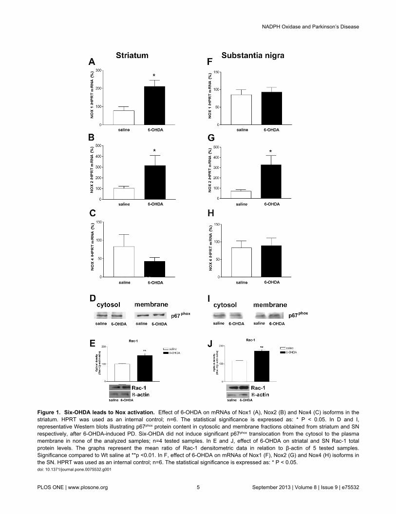
Figure 1. Six-OHDA leads to Nox activation. Effect of 6-OHDA on mRNAs of Nox1 (A), Nox2 (B) and Nox4 (C) isoforms in thestriatum. HPRT was used as an internal control; n=6. The statistical significance is expressed as: * P < 0.05. In D and I,representative Western blots illustrating p67phox protein content in cytosolic and membrane fractions obtained from striatum and SNrespectively, after 6-OHDA-induced PD. Six-OHDA did not induce significant p67phox translocation from the cytosol to the plasmamembrane in none of the analyzed samples; n=4 tested samples. In E and J, effect of 6-OHDA on striatal and SN Rac-1 totalprotein levels. The graphs represent the mean ratio of Rac-1 densitometric data in relation to β-actin of 5 tested samples.Significance compared to Wt saline at **p <0.01. In F, effect of 6-OHDA on mRNAs of Nox1 (F), Nox2 (G) and Nox4 (H) isoforms inthe SN. HPRT was used as an internal control; n=6. The statistical significance is expressed as: * P < 0.05.doi: 10.1371/journal.pone.0075532.g001
NADPH Oxidase and Parkinson’s Disease
PLOS ONE | www.plosone.org 5 September 2013 | Volume 8 | Issue 9 | e75532

Figure 2. NADPH oxidase is involved in the 6-OHDA-mediated degeneration of DA neurons in SN. A, apomorphine-inducedcontralateral rotations evaluated at 14 days post-PD induction in Sham, Wt 6-OHDA-lesioned mice and gp91phox-/- 6-OHDA lesionedmice groups; n=12. Values are expressed as percent of induced rotations/min in relation to the Sham group. Representative digitalimages demonstrating TH-immunoreactive cell bodies in the SN of Wt saline, Wt 6-OHDA and gp91phox-/- 6-OHDA-lesioned micegroups (B). Images are representative of at least six independent experiments. The statistical significance is expressed as: ***P<0.001 versus Wt Sham group and *** p<0.001 gp91phox-/- 6-OHDA versus Wt 6-OHDA.doi: 10.1371/journal.pone.0075532.g002
NADPH Oxidase and Parkinson’s Disease
PLOS ONE | www.plosone.org 6 September 2013 | Volume 8 | Issue 9 | e75532

mice (Figure 3D and E), suggesting an enhancement ofneurodegeneration.
Minocycline treatment increases TNF-α release in thesubstantia nigra of gp91phox-/--lesioned mice
The next series of experiments investigated the mechanismby which the minocycline treatment increases the susceptibilityof gp91phox-/- 6-OHDA lesioned mice to develop PD. Weanalyzed the concentration of multiple cytokines and onechemokine in SN and striatum samples from control Wt andgp91phox-/- mice and in 6-OHDA-lesioned mice treated withminocycline or PBS. All the molecules measured have beenpreviously implicated in the pathogenesis of PD and includedproinflammatory cytokines (IL-1β, TNF-α, IFN-γ, IL-2), anti-inflammatory cytokines (IL-10 and TGF-β1) and the chemokineRANTES.
In striatum, 6-OHDA was able to significantly increase theproduction of IFN-γ and TNF-α, which was not observed ingp91phox-/- mice. Striatal concentration of IL-10, RANTES, IL-1β,IL-2 and TGF-β1 remained the same after the lesion in both Wtand gp91phox-/- mice. The basal concentration of IL-1β wasfound significantly diminished in gp91phox-/- mice, whencompared to Wt mice. Minocycline treatment significantlydecreased the production of IFN-γ and TNF-α stimulated by 6-OHDA. A trend towards a decreased production of all thecytokines analyzed was observed after the minocyclinetreatment, although, the treatment was only able to significantlydecrease the basal production of IL-10, RANTES and IL-2 ingp91phox-/- mice and of IL-1β in Wt mice (Figure 4).
In the SN, 6-OHDA did not stimulate the production of IL-10,RANTES, IL-1β, IL-2, TGF-β1 and IFN-γ. Unlike what wasfound in striatum samples, minocycline did not change thebasal production of any of the molecules tested. Theproduction of TNF-α was found to be significantly increasedafter 6-OHDA injection in Wt but not in gp91phox-/- mice.However, the treatment with minocycline increased TNF-αproduction in gp91phox-/- 6-OHDA injected mice, furthersupporting our hypothesis that Nox-2 is modulatinginflammatory-mediated microglial responses (Figure 5).
Minocycline treatment potentiates NF-κB activation ingp91phox-/- lesioned mice
Since NF-κB activation induces the production and release ofTNF-α, we investigate whether inhibition of microglial cells ingp91phox-/- lesioned mice could modulate NF-κB activation. Asevaluated by EMSA, 6-OHDA did not change basal NF-κBactivity in the striatum of Wt mice (Figure 6A and B). However,6-OHDA significantly increased NF-κB/DNA binding activity inSN nuclear extracts, which was clearly attenuated by theminocycline treatment. In gp91phox-/- lesioned mice, 6-OHDA didnot change basal NF-κB binding activity in either brain region,although a trend towards an increase in SN samples has beenobserved. However, minocycline treatment significantlypotentiates 6-OHDA-induced NF-κB activation in the SN ofgp91phox-/- lesioned mice (Figure 6C and D), suggesting that NF-κB activation may be related to the increased release of TNF-α.
Discussion
The present study investigated the impact of Nox-2 in themodulation of inflammatory-mediated microglial responses inthe 6-OHDA- induced PD. Our major findings demonstrate that(1) The Nox2 isoform is involved in 6-OHDA-mediated DAdegeneration in SN; (2) inhibition of microglial cells by thetreatment with minocycline increases the susceptibility ofgp91phox-/- 6-OHDA lesioned mice to develop PD, as evaluatedby apomorphine-induced rotational behavior and THimmunolabeling; (3) minocycline treatment leads to NF-κBactivation and TNF-α release into the SNpc of gp91phox-/- 6-OHDA lesioned mice.
Minocycline, a tetracycline derivative, is a versatile drug witha broad spectrum of action including multiple anti-inflammatoryeffects, besides its anti-microbial properties. In the centralnervous system, it has been demonstrated that minocyclineinhibits apoptosis, proteolysis and the activation andproliferation of microglial cells, triggered by several differentstimuli [32]. However, the molecular mechanisms underlying itsanti-inflammatory actions and its influence on cytokine releaseare not fully understood [33]. Minocycline has been reported toreduce dopaminergic degeneration, due to an attenuation ofmicroglial cell activation following unilateral 6-OHDA injectioninto striatum of mice [34]. Here we investigated minocyclineability (through microglial cell inhibition) to change thesusceptibility of gp91phox-/- 6-OHDA lesioned mice to developPD.
Overactivation of Noxes leads to excessive ROS production,which disrupts redox signaling and results in oxidative stress[35]. Independently of the ROS source, oxidative stress isincreasingly recognized as one of the most relevantcontributors to neurodegeneration [36,37].
Although the exact etiology of PD still remains largelyunknown, some studies have implicated increased ROSproduction through Noxes in the pathophysiology of thisdisease [38-40]. Current research also suggests that DAneurons are inherently more vulnerable to oxidative stress,when compared to other cell types [41]. Consistent withprevious reports, we observed that 6-OHDA induces increasedgene expression of Nox1 and Nox2 isoforms [14,15],suggesting that Nox activation is a relevant component of the6-OHDA-induced DA degeneration. Corroborating with thisview, a strong argument for a role of Nox2 activation inneuronal death is the fact that gp91phox-/- exhibited significantlyreduced apomorphine-induced rotational behavior and also anegligible degeneration of DA neurons after 6-OHDA-injections.
Over the last decades the question of whether microglialcells play detrimental or beneficial roles in neurodegenerativeconditions has been widely debated [42]. Similar to peripheralmacrophages, microglial cells are functionally polarized intodifferent activation phenotypes during neuroinflammation. Ithas been recently demonstrated that Nox plays a critical role inthe modulation of the microglial phenotype. In fact,hippocampal levels of IL-4, an anti-inflammatory cytokine, andthe expression of IL-4 receptor α mRNA were significantlyincreased 24 h after intracerebroventricular injection of LPS in
NADPH Oxidase and Parkinson’s Disease
PLOS ONE | www.plosone.org 7 September 2013 | Volume 8 | Issue 9 | e75532
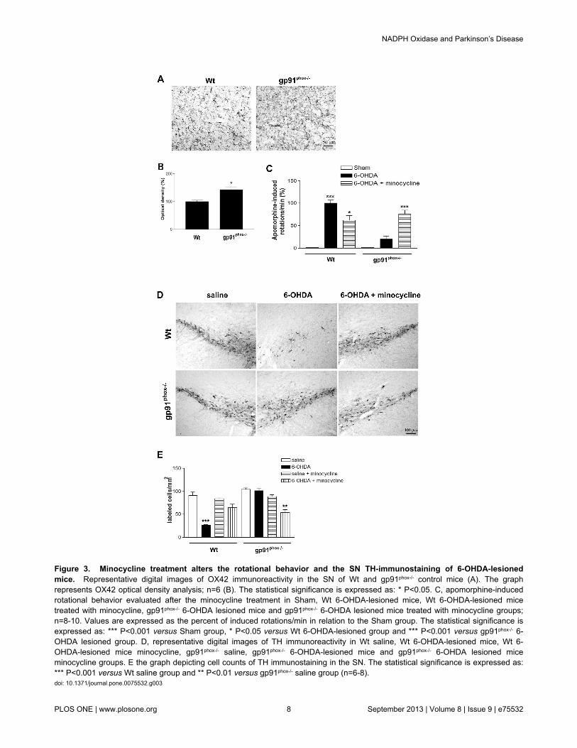
Figure 3. Minocycline treatment alters the rotational behavior and the SN TH-immunostaining of 6-OHDA-lesionedmice. Representative digital images of OX42 immunoreactivity in the SN of Wt and gp91phox-/- control mice (A). The graphrepresents OX42 optical density analysis; n=6 (B). The statistical significance is expressed as: * P<0.05. C, apomorphine-inducedrotational behavior evaluated after the minocycline treatment in Sham, Wt 6-OHDA-lesioned mice, Wt 6-OHDA-lesioned micetreated with minocycline, gp91phox-/- 6-OHDA lesioned mice and gp91phox-/- 6-OHDA lesioned mice treated with minocycline groups;n=8-10. Values are expressed as the percent of induced rotations/min in relation to the Sham group. The statistical significance isexpressed as: *** P<0.001 versus Sham group, * P<0.05 versus Wt 6-OHDA-lesioned group and *** P<0.001 versus gp91phox-/- 6-OHDA lesioned group. D, representative digital images of TH immunoreactivity in Wt saline, Wt 6-OHDA-lesioned mice, Wt 6-OHDA-lesioned mice minocycline, gp91phox-/- saline, gp91phox-/- 6-OHDA-lesioned mice and gp91phox-/- 6-OHDA lesioned miceminocycline groups. E the graph depicting cell counts of TH immunostaining in the SN. The statistical significance is expressed as:*** P<0.001 versus Wt saline group and ** P<0.01 versus gp91phox-/- saline group (n=6-8).doi: 10.1371/journal.pone.0075532.g003
NADPH Oxidase and Parkinson’s Disease
PLOS ONE | www.plosone.org 8 September 2013 | Volume 8 | Issue 9 | e75532

p47phox-/- mice when compared to wt mice, indicating thatdeletion of p47phox subunit alters the IL-4-dependent signalingpathway and attenuates the inflammatory response [14].
Despite the fact that the deletion and the pharmacologicalinhibition of the Nox complex promotes an anti-inflammatorymicroglial activation after LPS injection, we present here strong
evidence that Nox2 plays an important role in modulating theinflammatory response induced by 6-OHDA through a TNF-α/NF-κB mediated signaling pathway, a likely mechanismwhereby gp91phox-/- 6-OHDA lesioned mice may be moresusceptible to develop PD after microglial cell inhibition. Thecooperative relationship between TNF-α production, a classic
Figure 4. Effect of minocycline treatment on striatal cytokine concentration in Wt and gp91phox-/- 6-OHDA-lesionedmice. Striatal concentration of pro-inflammatory (IL-1β, TNF-α, IFN-γ, IL-2) and anti-inflammatory cytokines (IL-10 and TGF-β1)and the chemokine RANTES following 6-OHDA-induced PD in Wt and gp91phox-/- treated with minocycline or PBS. All concentrationswere expressed in pg/ml. The statistical significance is expressed as: * P<0.05; ** P<0.01 and *** P<0.001; n=6 for eachexperimental group tested.doi: 10.1371/journal.pone.0075532.g004
NADPH Oxidase and Parkinson’s Disease
PLOS ONE | www.plosone.org 9 September 2013 | Volume 8 | Issue 9 | e75532

inflammatory cytokine, and NF-κB activation has beenpreviously demonstrated as an important molecular eventoccurring in the 6-OHDA-induced neurodegeneration [43,44].
TNF-α release has been identified as a critical mechanisminvolved in DA neuroinflammation and in neuron damage.
Continuous production of this cytokine induced by anadenoviral vector leads to chronic microglia and macrophageactivation in the SN, progressive neurodegeneration, anddelayed motor symptoms [45]. Among all the cytokinesevaluated in the present study, we observed that microglial cell
Figure 5. Effect of minocycline treatment on substantia nigra cytokine concentration in Wt and gp91phox-/- 6-OHDA-lesionedmice. Concentration of pro-inflammatory (IL-1β, TNF-α, IFN-γ, IL-2) and anti-inflammatory cytokines (IL-10 and TGF-β1) and thechemokine RANTES in the substantia nigra following 6-OHDA-induced PD in Wt and gp91phox-/- treated with minocycline or PBS. Allconcentrations were expressed in pg/ml. The statistical significance is expressed as: * P<0.05; ** P<0.01 and *** P<0.001; n=6 foreach experimental group tested.doi: 10.1371/journal.pone.0075532.g005
NADPH Oxidase and Parkinson’s Disease
PLOS ONE | www.plosone.org 10 September 2013 | Volume 8 | Issue 9 | e75532

Figure 6. Minocycline treatment potentiates NF-κB activation in gp91phox-/- 6-OHDA lesioned mice. In A, representative panelof NF-κB activation in striatal nuclear extracts. In B, densitometric analysis of NF-κB bands obtained from nuclear extracts ofstriatum. In C, representative panel of NF-κB activation in SN nuclear extracts. In D, densitometric analysis of the NF-κB bandsobtained from nuclear extracts of SN. The statistical significance is expressed as: * P<0.05; ** P<0.01 and *** P<0.001; n=4.Competition studies were performed using brain extract (15 µg) in the absence or presence of unlabeled specific (NF-κB consensussequence, 5-, 10- and 20- fold molar excess) or non-specific oligonucleotide (TFIID consensus sequence, 20- fold molar excess), asindicated. The position of specific NF-κB/DNA binding complexes is indicated (NF-κB). NS represents no specific binding (NS). Thelocalization of the probe is also indicated. Results are representative of three experiments.doi: 10.1371/journal.pone.0075532.g006
NADPH Oxidase and Parkinson’s Disease
PLOS ONE | www.plosone.org 11 September 2013 | Volume 8 | Issue 9 | e75532

inhibition increased TNF-α production into the SN ofgp91phox-/--6-OHDA lesioned mice. It is noteworthy thatastrocytes are able to release TNF-α [46], which could be onepossible explanation for the increased TNF-α concentration inthe SN of gp91phox-/--6-OHDA lesioned mice after microglial cellinhibition observed in the present study. Increased LPS-induced TNF-α release from astrocyte cultures has beendemonstrated [47], and increased astrocytic immunoreactivityfor TNF-α was observed in the SN of parkinsonian patients[48]. In addition, in parkinsonian monkeys TNF-αimmunoreactivity was observed very close to GFAPimmunoreactivity in activated astrocytes, which suggests thatTNF-α synthesis and/or release may be intimately linked withthe astrocyte cytoskeleton [49].
We also observed in gp91phox-/- 6-OHDA lesioned mice thatminocycline treatment potentiated the activation of NF-κB,which can induce TNF-α production [50], suggesting theparticipation of that transcription factor in the process.However, considering that TNF-α release may also trigger asignaling cascade that can converge on the activation of thetranscription factor NF-κB [51], whether NF-κB activationprecedes TNF-α production is an issue that remains to bedetermined.
Since inhibition of microglial cells by minocycline treatmentincreased the susceptibility of gp91phox-/- 6-OHDA lesioned miceto develop PD and exacerbated the pro-inflammatory responseinduced by the neurotoxin, our results further strengthen thehypothesis that Nox2 adds an essential level of regulation tosignaling pathways underlying the inflammatory response afterPD induction. The Nox complex may play a role not only in thesignaling events leading to microglia activation, but may also
act as an important modulator of signal transduction in othercell types. Our results are also likely to provide further insightsin the direction of a better understanding of the mechanisms ofNox-dependent oxidative stress involvement inpathophysiological conditions.
Conclusions
We demonstrated here that inhibition of microglial cells ingp91phox-/- 6-OHDA lesioned mice triggers DA degeneration andexacerbated the pro-inflammatory response induced by theneurotoxin through a TNF-α/NF-κB mediated signalingpathway. Our results suggest that Nox2 adds an essential levelof regulation to signaling pathways underlying the inflammatoryresponse after PD induction, and indicated that both Nox2 andmicroglial cells could represent therapeutics targets in PD.
Acknowledgements
We thank Professor Rui Curi for supplying gp91phox-/- mice.Thanks are also due to Adilson S. Alves for technicalassistance. Thanks are also due to Prof. Francisco Laurindo forthe kind donation of the Rac-1 antibody.
Author Contributions
Conceived and designed the experiments: MSH LRGB CDM.Performed the experiments: MSH GDRS LSM CCCM.Analyzed the data: MSH LRGB. Contributed reagents/materials/analysis tools: LSM CS CDM. Wrote the manuscript:MSH LRGB.
References
1. Babior BM (2002) The activity of leukocyte NADPH oxidase: regulationby p47PHOX cysteine and serine residues. Antioxid Redox Signal 4:35-38. doi:10.1089/152308602753625834. PubMed: 11970841.
2. Babior BM, Lambeth JD, Nauseef W (2002) The neutrophil NADPHoxidase. Arch Biochem Biophys 397: 342-344. doi:10.1006/abbi.2001.2642. PubMed: 11795892.
3. Bedard K, Krause KH (2007) The NOX family of ROS-generatingNADPH oxidases: physiology and pathophysiology. Physiol Rev 87:245-313. doi:10.1152/physrev.00044.2005. PubMed: 17237347.
4. Ibi M, Katsuyama M, Fan C, Iwata K, Nishinaka T et al. (2006) NOX1/NADPH oxidase negatively regulates nerve growth factor-inducedneurite outgrowth. Free Radic Biol Med 40: 1785-1795. doi:10.1016/j.freeradbiomed.2006.01.009. PubMed: 16678016.
5. Vallet P, Charnay Y, Steger K, Ogier-Denis E, Kovari E et al. (2005)Neuronal expression of the NADPH oxidase NOX4, and its regulation inmouse experimental brain ischemia. Neuroscience 132: 233-238. doi:10.1016/j.neuroscience.2004.12.038. PubMed: 15802177.
6. Hernandes MS, Britto LR (2012) NADPH oxidase andneurodegeneration. Curr Neuropharmacol 10: 321-327. doi:10.2174/157015912804499483. PubMed: 23730256.
7. Abramov AY, Canevari L, Duchen MR (2004) Calcium signals inducedby amyloid beta peptide and their consequences in neurons andastrocytes in culture. Biochim Biophys Acta 1742: 81-87. doi:10.1016/j.bbamcr.2004.09.006. PubMed: 15590058.
8. Gao HM, Jiang J, Wilson B, Zhang W, Hong JS et al. (2002) Microglialactivation-mediated delayed and progressive degeneration of rat nigraldopaminergic neurons: relevance to Parkinson’s disease. J Neurochem81: 1285-1297. doi:10.1046/j.1471-4159.2002.00928.x. PubMed:12068076.
9. Kumar A, Singh BK, Ahmad I, Shukla S, Patel DK et al. (2012)Involvement of NADPH oxidase and glutathione in zinc-induceddopaminergic neurodegeneration in rats: similarity with paraquat
neurotoxicity. Brain Res 1438: 48-64. doi:10.1016/j.brainres.2011.12.028. PubMed: 22244881.
10. Mangano EN, Litteljohn D, So R, Nelson E, Peters S et al. (2011)Interferon-gamma plays a role in paraquat-induced neurodegenerationinvolving oxidative and proinflammatory pathways. Neurobiol Aging.33(7): 1411-26.
11. Choi SH, Aid S, Kim HW, Jackson SH, Bosetti F (2012) Inhibition ofNADPH oxidase promotes alternative and anti-inflammatory microglialactivation during neuroinflammation. J Neurochem 120: 292-301. doi:10.1111/j.1471-4159.2011.07572.x. PubMed: 22050439.
12. Gao HM, Liu B, Hong JS (2003) Critical role for microglial NADPHoxidase in rotenone-induced degeneration of dopaminergic neurons. JNeurosci 23: 6181-6187. PubMed: 12867501.
13. Rodriguez-Pallares J, Rey P, Parga JA, Muñoz A, Guerra MJ et al.(2008) Brain angiotensin enhances dopaminergic cell death viamicroglial activation and NADPH-derived ROS. Neurobiol Dis 31:58-73. doi:10.1016/j.nbd.2008.03.003. PubMed: 18499466.
14. Choi DH, Cristóvão AC, Guhathakurta S, Lee J, Joh TH et al. (2012)NADPH oxidase 1-mediated oxidative stress leads to dopamine neurondeath in Parkinson’s disease. Antioxid Redox Signal 16: 1033-1045.doi:10.1089/ars.2011.3960. PubMed: 22098189.
15. Rodriguez-Pallares J, Parga JA, Muñoz A, Rey P, Guerra MJ et al.(2007) Mechanism of 6-hydroxydopamine neurotoxicity: the role ofNADPH oxidase and microglial activation in 6-hydroxydopamine-induced degeneration of dopaminergic neurons. J Neurochem 103:145-156. PubMed: 17573824.
16. Schober A (2004) Classic toxin-induced animal models of Parkinson’sdisease: 6-OHDA and MPTP. Cell Tissue Res 318: 215-224. doi:10.1007/s00441-004-0938-y. PubMed: 15503155.
17. Zhang W, Wang T, Qin L, Gao HM, Wilson B et al. (2004)Neuroprotective effect of dextromethorphan in the MPTP Parkinson’sdisease model: role of NADPH oxidase. FASEB J 18: 589-591.PubMed: 14734632.
NADPH Oxidase and Parkinson’s Disease
PLOS ONE | www.plosone.org 12 September 2013 | Volume 8 | Issue 9 | e75532

18. Molina-Luna K, Pekanovic A, Röhrich S, Hertler B, Schubring-Giese Met al. (2009) Dopamine in motor cortex is necessary for skill learningand synaptic plasticity. PLOS ONE 4: e7082. doi:10.1371/journal.pone.0007082. PubMed: 19759902.
19. Paxinos G, Franklin K (2007) The Mouse Brain in StereotaxicCoordinates. New York: Academic Press. 360pp
20. Dussault AA, Pouliot M (2006) Rapid and simple comparison ofmessenger RNA levels using real-time PCR. Biol Proced Online 8:1-10. doi:10.1251/bpo114. PubMed: 16446781.
21. Wu DC, Jackson-Lewis V, Vila M, Tieu K, Teismann P et al. (2002)Blockade of microglial activation is neuroprotective in the 1-methyl-4-phenyl-1,2,3,6-tetrahydropyridine mouse model of Parkinson disease. JNeurosci 22: 1763-1771. PubMed: 11880505.
22. Hernandes MS, Britto LR, Real CC, Martins DO, Lopes LR (2010)Reactive oxygen species and the structural remodeling of the visualsystem after ocular enucleation. Neuroscience 170: 1249-1260. doi:10.1016/j.neuroscience.2010.07.065. PubMed: 20728508.
23. Iancu R, Mohapel P, Brundin P, Paul G (2005) Behavioralcharacterization of a unilateral 6-OHDA-lesion model of Parkinson’sdisease in mice. Behav Brain Res 162: 1-10. doi:10.1016/j.bbr.2005.02.023. PubMed: 15922062.
24. Nazemi S, Manaheji H, Zaringhalam J, Sadeghi M, Haghparast A(2012) Post-injury repeated administrations of minocycline improve theantinociceptive effect of morphine in chronic constriction injury model ofneuropathic pain in rat. Pharmacol Biochem Behav 102: 520-525. doi:10.1016/j.pbb.2012.07.001. PubMed: 22789876.
25. Moncunill G, Aponte JJ, Nhabomba AJ, Dobaño C (2013) Performanceof multiplex commercial kits to quantify cytokine and chemokineresponses in culture supernatants from Plasmodium falciparumstimulations. PLOS ONE 8: e52587. doi:10.1371/journal.pone.0052587.PubMed: 23300981.
26. Fox C, Dingman A, Derugin N, Wendland MF, Manabat C et al. (2005)Minocycline confers early but transient protection in the immature brainfollowing focal cerebral ischemia-reperfusion. J Cereb Blood FlowMetab 25: 1138-1149. doi:10.1038/sj.jcbfm.9600121. PubMed:15874975.
27. Hussain AR, Ahmed SO, Ahmed M, Khan OS, Al Abdulmohsen S et al.(2012) Cross-talk between NFkB and the PI3-kinase/AKT pathway canbe targeted in primary effusion lymphoma (PEL) cell lines for efficientapoptosis. PLOS ONE 7: e39945. doi:10.1371/journal.pone.0039945.PubMed: 22768179.
28. Hernandes MS, Lima LS, Scavone C, Lopes LR, Britto LR (2012) Eyeenucleation activates the transcription nuclear factor kappa-B in the ratsuperior colliculus. Neurosci Lett 521: 104-108. doi:10.1016/j.neulet.2012.05.051. PubMed: 22634628.
29. Kawamoto EM, Lima LS, Munhoz CD, Yshii LM, Kinoshita PF et al.(2012) Influence of N-methyl-D-aspartate receptors on ouabainactivation of nuclear factor-κB in the rat hippocampus. J Neurosci Res90: 213-228. doi:10.1002/jnr.22745. PubMed: 22006678.
30. Munhoz CD, Lepsch LB, Kawamoto EM, Malta MB, Lima Lde S et al.(2006) Chronic unpredictable stress exacerbates lipopolysaccharide-induced activation of nuclear factor-kappaB in the frontal cortex andhippocampus via glucocorticoid secretion. J Neurosci 26: 3813-3820.doi:10.1523/JNEUROSCI.4398-05.2006. PubMed: 16597735.
31. Glezer I, Munhoz CD, Kawamoto EM, Marcourakis T, Avellar MC et al.(2003) MK-801 and 7-Ni attenuate the activation of brain NF-kappa Binduced by LPS. Neuropharmacology 45: 1120-1129. doi:10.1016/S0028-3908(03)00279-X. PubMed: 14614955.
32. Henry CJ, Huang Y, Wynne A, Hanke M, Himler J et al. (2008)Minocycline attenuates lipopolysaccharide (LPS)-inducedneuroinflammation, sickness behavior, and anhedonia. JNeuroinflammation 5: 15. doi:10.1186/1742-2094-5-15. PubMed:18477398.
33. Zemke D, Majid A (2004) The potential of minocycline forneuroprotection in human neurologic disease. Clin Neuropharmacol 27:293-298. doi:10.1097/01.wnf.0000150867.98887.3e. PubMed:15613934.
34. He Y, Appel S, Le W (2001) Minocycline inhibits microglial activationand protects nigral cells after 6-hydroxydopamine injection into mousestriatum. Brain Res 909: 187-193. doi:10.1016/S0006-8993(01)02681-6. PubMed: 11478935.
35. Laurindo FR, Fernandes DC, Amanso AM, Lopes LR, Santos CX(2008) Novel role of protein disulfide isomerase in the regulation of
NADPH oxidase activity: pathophysiological implications in vasculardiseases. Antioxid Redox Signal 10: 1101-1113. doi:10.1089/ars.2007.2011. PubMed: 18373437.
36. Pestana RR, Kinjo ER, Hernandes MS, Britto LR (2010) Reactiveoxygen species generated by NADPH oxidase are involved inneurodegeneration in the pilocarpine model of temporal lobe epilepsy.Neurosci Lett 484: 187-191. doi:10.1016/j.neulet.2010.08.049.PubMed: 20732386.
37. Zeevalk GD, Bernard LP, Song C, Gluck M, Ehrhart J (2005)Mitochondrial inhibition and oxidative stress: reciprocating players inneurodegeneration. Antioxid Redox Signal 7: 1117-1139. doi:10.1089/ars.2005.7.1117. PubMed: 16115016.
38. Block ML, Li G, Qin L, Wu X, Pei Z et al. (2006) Potent regulation ofmicroglia-derived oxidative stress and dopaminergic neuron survival:substance P vs. dynorphin. FASEB J 20: 251-258. doi:10.1096/fj.05-4553com. PubMed: 16449797.
39. Wu DC, Teismann P, Tieu K, Vila M, Jackson-Lewis V et al. (2003)NADPH oxidase mediates oxidative stress in the 1-methyl-4-phenyl-1,2,3,6-tetrahydropyridine model of Parkinson’s disease. ProcNatl Acad Sci U S A 100: 6145-6150. doi:10.1073/pnas.0937239100.PubMed: 12721370.
40. Zawada WM, Banninger GP, Thornton J, Marriott B, Cantu D et al.(2011) Generation of reactive oxygen species in 1-methyl-4-phenylpyridinium (MPP+) treated dopaminergic neurons occurs as anNADPH oxidase-dependent two-wave cascade. J Neuroinflammation 8:129. doi:10.1186/1742-2094-8-129. PubMed: 21975039.
41. Loeffler DA, DeMaggio AJ, Juneau PL, Havaich MK, LeWitt PA (1994)Effects of enhanced striatal dopamine turnover in vivo on glutathioneoxidation. Clin Neuropharmacol 17: 370-379. doi:10.1097/00002826-199408000-00009. PubMed: 9316685.
42. Lull ME, Block ML (2010) Microglial activation and chronicneurodegeneration. Neurotherapeutics 7: 354-365. doi:10.1016/j.nurt.2010.05.014. PubMed: 20880500.
43. Pranski EL, Dalal NV, Sanford CV, Herskowitz JH, Gearing M et al.(2013) RING finger protein 11 (RNF11) modulates susceptibility to 6-OHDA-induced nigral degeneration and behavioral deficits through NF-kappaB signaling in dopaminergic cells. Neurobiol Dis. 54: 264-79.
44. Tarabin V, Schwaninger M (2004) The role of NF-kappaB in 6-hydroxydopamine- and TNFalpha-induced apoptosis of PC12 cells.Naunyn Schmiedebergs Arch Pharmacol 369: 563-569. doi:10.1007/s00210-004-0938-1. PubMed: 15141332.
45. De Lella Ezcurra AL, Chertoff M, Ferrari C, Graciarena M, Pitossi F(2010) Chronic expression of low levels of tumor necrosis factor-alphain the substantia nigra elicits progressive neurodegeneration, delayedmotor symptoms and microglia/macrophage activation. Neurobiol Dis37: 630-640. doi:10.1016/j.nbd.2009.11.018. PubMed: 19969084.
46. Soliman ML, Combs CK, Rosenberger TA (2013) Modulation ofinflammatory cytokines and mitogen-activated protein kinases byacetate in primary astrocytes. J Neuroimmune Pharmacol 8: 287-300.doi:10.1007/s11481-012-9426-4. PubMed: 23233245.
47. Guerra MC, Tortorelli LS, Galland F, Da Ré C, Negri E et al. (2011)Lipopolysaccharide modulates astrocytic S100B secretion: a study incerebrospinal fluid and astrocyte cultures from rats. JNeuroinflammation 8: 128. doi:10.1186/1742-2094-8-128. PubMed:21970823.
48. Boka G, Anglade P, Wallach D, Javoy-Agid F, Agid Y et al. (1994)Immunocytochemical analysis of tumor necrosis factor and its receptorsin Parkinson’s disease. Neurosci Lett 172: 151-154. doi:10.1016/0304-3940(94)90684-X. PubMed: 8084523.
49. Barcia C, Ros CM, Annese V, Gomez A, Ros-Bernal F et al. (2011)IFN-gamma signaling, with the synergistic contribution of TNF-alpha,mediates cell specific microglial and astroglial activation inexperimental models of Parkinson’s disease. Cell Death. Drosophila InfServ 2: e142.
50. Kim HJ, Park KG, Yoo EK, Kim YH, Kim YN et al. (2007) Effects ofPGC-1alpha on TNF-alpha-induced MCP-1 and VCAM-1 expressionand NF-kappaB activation in human aortic smooth muscle andendothelial cells. Antioxid Redox Signal 9: 301-307. doi:10.1089/ars.2006.1456. PubMed: 17184171.
51. Bouwmeester T, Bauch A, Ruffner H, Angrand PO, Bergamini G et al.(2004) A physical and functional map of the human TNF-alpha/NF-kappa B signal transduction pathway. Nat Cell Biol 6: 97-105. doi:10.1038/ncb1086. PubMed: 14743216.
NADPH Oxidase and Parkinson’s Disease
PLOS ONE | www.plosone.org 13 September 2013 | Volume 8 | Issue 9 | e75532
Copyright © 2022 FDOKUMEN