Inhibition of human vascular NADPH oxidase by apocynin derived oligophenols
17
Inhibition of Human Vascular NADPH Oxidase by Apocynin Derived Oligophenols Mauricio Mora-Pale 1 , Michel Weïwer 2 , Jingjing Yu 3 , Robert J. Linhardt 1,2,3 , and Jonathan S. Dordick 1,3 1 Department of Chemical and Biological Engineering, Center for Biotechnology and Interdisciplinary Studies, Rensselaer Polytechnic Institute, Troy NY, 12180, USA 2 Department of Chemistry and Chemical Biology, Center for Biotechnology and Interdisciplinary Studies, Rensselaer Polytechnic Institute, Troy NY, 12180, USA 3 Department of Biology, Center for Biotechnology and Interdisciplinary Studies, Rensselaer Polytechnic Institute, Troy NY, 12180, USA Abstract Enzymatic oxidation of apocynin, which may mimic in vivo metabolism, affords a large number of oligomers (apocynin oxidation products, AOP) that inhibit vascular NADPH oxidase. In vitro studies of NADPH oxidase activity were performed to identify active inhibitors, resulting in a trimer hydroxylated quinone (IIIHyQ) that inhibited NADPH oxidase with an IC 50 = 31 nM. Apocynin itself possessed minimal inhibitory activity. NADPH oxidase is believed to be inhibited through prevention of the interaction between two NADPH oxidase subunits, p47 phox and p22 phox . To that end, while apocynin was unable to block the interaction of his-tagged p47 phox with a surface immobilized biotinalyted p22 phox peptide, the IIIHyQ product strongly interfered with this interaction (apparent IC 50 = 1.6 μM). These results provide evidence that peroxidase-catalyzed AOP, which consist of oligomeric phenols and quinones, inhibit critical interactions that are involved in the assembly and activation of human vascular NADPH oxidase. 1. Introduction Recent years have seen substantial improvement in our understanding of the role of superoxide anion (•O 2 − ) in eliciting oxidative stress and vascular diseases [1–5]. The production of •O 2 − is catalyzed by a variety of enzymes, including xanthine oxidase, cytochromes P450, lipoxygenase, enzymes in the mitochondrial respiratory chain, and NADPH oxidases [6]. The latter, in particular, have been identified as the major source of •O 2 − in vascular endothelial cells (VECs). Excessive production of •O 2 − in VECs leads to increased oxidative stress and endothelial dysfunction. This in turn can result in a diverse array of cardiovascular diseases, including atherosclerosis, hypertension, diabetes, heart failure, stroke, and restenosis [1,7– 11]. The most well-studied NADPH oxidase in humans is found in neutrophils. In the resting state, the neutrophil enzyme consists of at least six partially dissociated components [12]. Tight Correspondence authors: [email protected], [email protected]. Publisher's Disclaimer: This is a PDF file of an unedited manuscript that has been accepted for publication. As a service to our customers we are providing this early version of the manuscript. The manuscript will undergo copyediting, typesetting, and review of the resulting proof before it is published in its final citable form. Please note that during the production process errors may be discovered which could affect the content, and all legal disclaimers that apply to the journal pertain. NIH Public Access Author Manuscript Bioorg Med Chem. Author manuscript; available in PMC 2010 July 15. Published in final edited form as: Bioorg Med Chem. 2009 July 15; 17(14): 5146–5152. doi:10.1016/j.bmc.2009.05.061. NIH-PA Author Manuscript NIH-PA Author Manuscript NIH-PA Author Manuscript
-
Upload
independent -
Category
Documents
-
view
1 -
download
0
Transcript of Inhibition of human vascular NADPH oxidase by apocynin derived oligophenols

Inhibition of Human Vascular NADPH Oxidase by ApocyninDerived Oligophenols
Mauricio Mora-Pale1, Michel Weïwer2, Jingjing Yu3, Robert J. Linhardt1,2,3, and Jonathan S.Dordick1,31 Department of Chemical and Biological Engineering, Center for Biotechnology andInterdisciplinary Studies, Rensselaer Polytechnic Institute, Troy NY, 12180, USA2 Department of Chemistry and Chemical Biology, Center for Biotechnology and InterdisciplinaryStudies, Rensselaer Polytechnic Institute, Troy NY, 12180, USA3 Department of Biology, Center for Biotechnology and Interdisciplinary Studies, RensselaerPolytechnic Institute, Troy NY, 12180, USA
AbstractEnzymatic oxidation of apocynin, which may mimic in vivo metabolism, affords a large number ofoligomers (apocynin oxidation products, AOP) that inhibit vascular NADPH oxidase. In vitro studiesof NADPH oxidase activity were performed to identify active inhibitors, resulting in a trimerhydroxylated quinone (IIIHyQ) that inhibited NADPH oxidase with an IC50 = 31 nM. Apocyninitself possessed minimal inhibitory activity. NADPH oxidase is believed to be inhibited throughprevention of the interaction between two NADPH oxidase subunits, p47phox and p22phox. To thatend, while apocynin was unable to block the interaction of his-tagged p47phox with a surfaceimmobilized biotinalyted p22phox peptide, the IIIHyQ product strongly interfered with thisinteraction (apparent IC50 = 1.6 μM). These results provide evidence that peroxidase-catalyzed AOP,which consist of oligomeric phenols and quinones, inhibit critical interactions that are involved inthe assembly and activation of human vascular NADPH oxidase.
1. IntroductionRecent years have seen substantial improvement in our understanding of the role of superoxideanion (•O2
−) in eliciting oxidative stress and vascular diseases [1–5]. The production of •O2−
is catalyzed by a variety of enzymes, including xanthine oxidase, cytochromes P450,lipoxygenase, enzymes in the mitochondrial respiratory chain, and NADPH oxidases [6]. Thelatter, in particular, have been identified as the major source of •O2
− in vascular endothelialcells (VECs). Excessive production of •O2
− in VECs leads to increased oxidative stress andendothelial dysfunction. This in turn can result in a diverse array of cardiovascular diseases,including atherosclerosis, hypertension, diabetes, heart failure, stroke, and restenosis [1,7–11].
The most well-studied NADPH oxidase in humans is found in neutrophils. In the resting state,the neutrophil enzyme consists of at least six partially dissociated components [12]. Tight
Correspondence authors: [email protected], [email protected]'s Disclaimer: This is a PDF file of an unedited manuscript that has been accepted for publication. As a service to our customerswe are providing this early version of the manuscript. The manuscript will undergo copyediting, typesetting, and review of the resultingproof before it is published in its final citable form. Please note that during the production process errors may be discovered which couldaffect the content, and all legal disclaimers that apply to the journal pertain.
NIH Public AccessAuthor ManuscriptBioorg Med Chem. Author manuscript; available in PMC 2010 July 15.
Published in final edited form as:Bioorg Med Chem. 2009 July 15; 17(14): 5146–5152. doi:10.1016/j.bmc.2009.05.061.
NIH
-PA Author Manuscript
NIH
-PA Author Manuscript
NIH
-PA Author Manuscript
regulation of NADPH oxidase activity is achieved by at least two mechanisms; the associationof the cytosolic subunits and the modulation of reversible protein-protein and protein-membrane interactions [13]. Despite near 100% homology with the neutrophil NADPHoxidase [14,15], the exact assembly and activation of VEC NADPH oxidase is poorlyunderstood [16]. Nevertheless, current evidence suggests that phosphorylation of key serineresidues in p47phox facilitates interaction of a Src homology 3 (SH3) domain of this proteinwith a proline-rich region (PRR) of p22phox, thereby forming the active membrane-associatedNADPH oxidase complex [17,18] (Scheme 1).
The catalytic subunit of NADPH oxidase (Nox) has several isoforms and, at least three of themare expressed in VEC (Nox1, Nox2, and Nox4). Their precise activation mechanisms andcellular regulation remain unclear [19,20]. Nox1 appears to be of only minor importance in thegeneration of VEC reactive oxygen species (ROS). Nox4, however, is abundantly expressedin endothelial cells, more than Nox2 [21]. Nevertheless, Li et. al. [21] showed that, despite lowexpression relative to Nox4, under starvation conditions Nox2 was upregulated ~ 8-fold and,as a consequence of this nutrient deprivation-induced oxidative stress, the production of•O2
− increased ~2.3-fold. Importantly, Nox2 requires assembly of p47phox with other cytosolicsubunits prior to translocation to the membrane to form the active NADPH oxidase complex[22,23].
Due to the key role VEC NADPH oxidase appears to play in vascular diseases, identificationof selective inhibitors is of great interest. Along these lines, several inhibitors have beenidentified, including nitrovasodilators [24], the flavonoid derivative 6,8-diallyl-5,7-dihydroxy-2-(2-allyl-3-hydroxy-4-methoxyphenyl)-1-H-benzo-[b]-pyran-4-one [25], andpeptides such as the antibiotic PR-39 [26]. Interestingly, PRR regions are also known to bindpolyphenols (such as flavonoids) [27]. It is not surprising, therefore, that phenolics have beenfound to have NADPH oxidase inhibitory activity.
Apocynin (4′-hydroxy-3′-methoxyacetophenone) [28,29] is a particularly interesting phenolthat has been used as inhibitor of NADPH oxidase. While apocynin itself was found to havelow activity in vitro [29,30] metabolism in vivo converts the phenol into active metabolites thatinhibit the enzyme [30–34]. This may be due to peroxidase catalysis [29,30,35,36] leading todisruption of the p47phox-p22phox interaction, which is required for translocation of thecytosolic enzyme components to the membrane leading to activation of the enzyme complex.In the current work, we demonstrate that several oligomeric apocynin oxidation productsgenerated by peroxidases are extremely potent inhibitors of VEC NADPH oxidase in vitro.Moreover, a strong correlation exists between the inhibition of VEC NADPH oxidase inendothelial cell-based assays and disruption of the interaction of EC p47phox-p22phox in cell-free assays. These results provide additional mechanistic insight into the nature and functionof active metabolites of apocynin.
2. Results and discussionUnder conditions of oxidative stress, overactive NADPH oxidase in the vasculature generates•O2
−, thereby leading to increased levels of H2O2. In the presence of peroxidases in the blood,e.g., myeloperoxidase, and reducing substrates of peroxidases, such as phenols, peroxidaticreactions can occur. To mimic this scenario, we used a simple commercially availableperoxidase from soybean (SBP) to catalyze the oxidation of apocynin in the presence ofH2O2 following our earlier published procedure [37]. Such reaction would then be expectedto mimic peroxidatic metabolism in the vasculature.
Following the enzymatic oxidation of apocynin, a water-soluble fraction was extracted intoethyl acetate to give fraction AOP-1 and a chloroform soluble precipitate was fractionated by
Mora-Pale et al. Page 2
Bioorg Med Chem. Author manuscript; available in PMC 2010 July 15.
NIH
-PA Author Manuscript
NIH
-PA Author Manuscript
NIH
-PA Author Manuscript

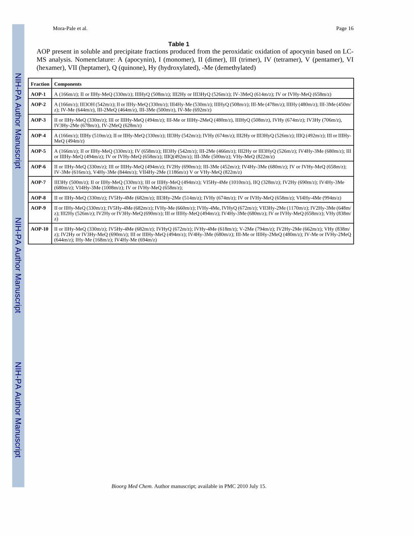
silica chromatography to yield nine fractions (AOP-2 to AOP-10). Each fraction was analyzedby LC-MS to qualitatively identify the AOP in each mixture, giving rise to the identificationof oligomers in their demethylated, hydroxylated, or quinone forms (Table 1). The totalconversion of the enzymatic reaction was ~50%; the precipitate representing 87% of the totalproducts while 13% remained in the aqueous phase. The ability of each AOP fraction to inhibitVEC NADPH oxidase was then assessed at a dose range from 0 to 1000 μM (based on apocyninmonomer unit mass), as determined by cytochrome c reduction for extracellular superoxidedetection and by dihydroethidium (DHE) staining for intracellular superoxide detection.
NADPH oxidase activity - Cytochrome c reductionThe NADPH oxidase inhibitory activity of apocynin and AOP fractions was assessed in anendothelial cell-based assay by measuring the generation of •O2
− via cytochrome c reduction.Apocynin itself possesses minimal inhibitory activity (IC50 > 1 mM; Figure 1), which isconsistent with reports in the literature [29,30]. However, the extracted water-soluble phase(AOP-1) exhibited an apparent IC50 value of 155 nM (Figure 1), despite analysis of this mixtureby NMR, TLC, and LC-MS, which indicated that the major component was unreacted apocynin(~90%). Thus, the inhibition of NADPH oxidase must result from the presence of at least onevery strong NADPH oxidase inhibitor present in the remaining 10% of the water-solublefraction. Following purification via silica chromatography, a trimer hydroxylated quinone(IIIHyQ, 508 m/z, Figure 1) was identified as the major active compound in AOP-1, with stronginhibitory activity against VEC NADPH oxidase (IC50 = 31 nM).
A similar study was performed for the chloroform-soluble enzyme reaction precipitate, whichfollowing chromatographic separation, resulted in nine distinct fractions (AOP-2 throughAOP-10, IC50 values summarized in Table 2). Fractions AOP-2 – AOP-4 showed substantialNADPH oxidase inhibitory activity (< 1.0 μM). Fractions AOP-1 and AOP-2 consisted ofIIIHyQ along with other oligomeric species. AOP-4, however, consisted of other trimericcompounds, including trimeric quinones (IIIQ, IIIHy-MeQ, and III3HyQ) that also inhibitedNADPH oxidase. Higher oligomeric species (e.g., tetrameric to heptameric) showed relativelylow inhibitory activity. These results demonstrate that some of the AOP possess strong VECNADPH oxidase inhibitory activity, as reflected in the cell-based enzyme assay.
Intracellular superoxide detection – DHE stainingTo establish the ability of AOP to suppress intracellular •O2
− formation by NADPH oxidases,we used DHE staining of whole EC following incubation with apocynin, IIIHyQ, andphenylarsine oxide (PAO, an inhibitor of NADPH oxidase [39] and serving as a positivecontrol). DHE is a cell-permeable reagent that reacts with •O2
− to form oxyethidium [38],which in turn interacts with nucleic acids to emit a bright red color detectable qualitatively byfluorescence microscopy. Figure 2 shows images of DHE stained EC after incubation with theaforementioned compounds. In the absence of compound, the EC fluoresce as a result of strongand expected •O2
− production. Apocynin, even up to 1 mM, did not appreciably reduce thisfluorescence (Figure 2A). In contrast, IIIHyQ strongly reduced the •O2
− induced fluorescence(Figure 2B), which was consistent with the effect of the positive control compound (PAO,Figure 2C). These results suggest that the IIIHyQ inhibits NADPH oxidase intracellularly. Inaddition, the inability of very high concentrations of apocynin to inhibit NADPH oxidase (asreflected in the lack of inhibition of •O2
− production) also indicates that apocynin is unable toscavenge •O2
−.
Interaction between his-p47phox protein and biotin-p22 peptideThe p47phox and p22phox protein subunits play a significant role in the activation of NADPHoxidase [13]. Translocation of p47phox from the cytosol to bind to membrane-associatedp22phox is a key event in the mechanism of NADPH oxidase activation and is presumably
Mora-Pale et al. Page 3
Bioorg Med Chem. Author manuscript; available in PMC 2010 July 15.
NIH
-PA Author Manuscript
NIH
-PA Author Manuscript
NIH
-PA Author Manuscript

driven by the interaction of an SH3 domain on the p47phox with a proline-rich region on thep22phox [17,18]. To ascertain whether the mechanism of AOP inhibition of NADPH oxidaseis due to disruption of the interaction of p47phox with p22phox, we performed a well-plate ELISAassay. Isolated IIIHyQ and the AOP-1 mixture were potent inhibitors of this interaction (Figure3; Table 2) with IC50 values of 1.60 μM and 0.546 μM, respectively. Apocynin had no effecton protein-peptide interaction.
We then evaluated the remaining AOP fractions for their ability to disrupt the interaction ofp47phox with p22phox (Table 2). ELISA results followed a similar pattern to that observed inthe cell based studies, with low activity in fractions consisting of higher oligomeric species. Aplot of the log(IC50) values from the cell-based assay and from ELISA (Figure 4) shows goodlinear correlation (R2 = 0.87), suggesting that VEC NADPH oxidase inhibition is likelyexplained by the ability of the AOP to disrupt the interaction of p47phox with p22phox.
Proposed mechanism of AOP-mediated inhibition of NADPH oxidaseApocynin was a poor inhibitor of NADPH oxidase in VEC-based assays, suggesting thatprevious results describing the effectiveness of apocynin as an NADPH oxidase inhibitor werea consequence of its conversion into active metabolites, likely catalyzed by peroxidases. In thepresent study, we produced the oligomers in vitro, which allowed the structural characterizationof AOP. As a result, a trimer hydroxylated quinone (IIIHyQ) was identified as a strong inhibitorfrom AOP-1 and its structure was characterized by high resolution mass spectrometry (HRMS)and nuclear magnetic resonance (1H-NMR and 13C-NMR); detailed information on HRMSand NMR is provided in the Supporting Information. However, not all the oligomers werestrong inhibitors. Dimeric and trimeric AOP were more effective inhibitors of VEC NADPHoxidase than higher order oligomers identified in fractions AOP-6 to AOP-10. A possibleexplanation is that large oligomers are poorly soluble in aqueous media and are, therefore lesscapable of interacting with key subunits of VEC NADPH to inhibit assembly.
One possible mechanism of inhibition is the ability of reactive metabolites (e.g., quinones suchas IIIHyQ) to form Michael adducts with the cysteine residues of p47phox [40,41]. This wouldbe consistent with the known neutrophils NADPH oxidase inhibitor, phenylarsine oxide, whichbinds covalently with thiol groups in the enzyme, thereby preventing assembly of proteinsubunits [42]. Human p47phox contains four cysteine residues at positions 98, 111, 196 and378. Indeed, Cys-196 is located in one of the two SH3 domains of p47phox (SH3-N, aminoacids 156–215). As indicated above, the interaction of the p47phox (in complex with the othercytosolic NADPH oxidase subunits) with the membrane-associated p22phox occurs throughthis SH3 site on p47phox with a PRR on p22phox [43]. Modification of Cys-196 by quinone-containing may disrupt this critical interaction. However, one cannot rule out the modificationof the other three cysteine residues, which may lead to additional structural changes ofp47phox that could disrupt binding of the p47phox to the membrane or interaction of thep47phox with PRR of the p67phox subunit. This is a key part of the formation of the cytosoliccomplex [13,44,45] and is required for translocation of p67phox to the membrane [46,47].
To determine the importance of Cys on the interaction between p22phox and p47phox weexamined the interaction of immobilized biotin-p22 (2 μM) with his-p47phox (0 – 1.0 μM) inthe presence of iodoacetamide (IAA) (Figure 5), which reacts with thiol groups on cysteineresidues. The presence of 1 μM IAA results in a > 50% drop in the amount of bound p47 tothe immobilized biotin-p22 phox; hence, thiol reactivity of quinone-based AOPs may be acritical mechanism of inhibition of VEC NADPH oxidase assembly.
In summary, we have demonstrated that peroxidase-generated apocynin metabolites serve asstrong inhibitors of VEC NADPH oxidase. The IIIHyQ was a particularly strong (nanomolar)inhibitor of the enzyme as determined through EC-based assays. This compound was also
Mora-Pale et al. Page 4
Bioorg Med Chem. Author manuscript; available in PMC 2010 July 15.
NIH
-PA Author Manuscript
NIH
-PA Author Manuscript
NIH
-PA Author Manuscript

effective at disrupting the p47phox- p22phox interaction in vitro. This suggests that themechanism of apocynin inhibition of NADPH oxidase is a result of peroxidase metabolism toyield reactive quinones that bind to Cys residues in p47phox and disrupt translocation to themembrane through SH3-PRR association with the p22phox. Since peroxidases are commonenzymes in the vasculature (e.g., myeloperoxidases) one may anticipate that similarmetabolites are generated in vivo. Further work is needed to determine whether thesemetabolites provide a means to the generation of potential therapeutic molecules.
3. Experimental proceduresApocynin, SBP, solvents, H2O2, LDL, superoxide dismutase (SOD), low density lipoprotein(LDL), cytochrome c, Tween 20, 3,3′,5,5′-tetramethyl-benzidine (TMB), sodium caseinate,fetal bovine serum, heparin, and endothelial growth supplement were purchased from Sigma-Aldrich. Endothelial cells and medium were purchased from ATCC. E. coli BL21 (DE3), IPTGand Ni-affinity column (Probond system) were purchased from Invitrogen. Antibodies werepurchased from Upstate. High-affinity streptavidin-coated-96 well plates were purchased fromPierce. LC-MS analyses were performed in a Shimadzu LCMS-2010A. Samples for LC-MSwere separated in an Agilent Zorbax 300SB-C18 column (5 μm, 2.1 × 150 mm). Silica gel 230–400 mesh was purchased from Natland International Corporation. Thin layer chromatography(TLC) plates were purchased from Merck. Microplate reader analyses were performed in aPerkin-Elmer, HTS 7000, Bio Assay Reader.
Enzymatic production of Apocynin Oxidation Products (AOP)AOP were synthesized by soybean peroxidase (SBP)-catalyzed oxidation of apocynin, asdescribed by Antoniotti et. al. [37]. Briefly, apocynin (6 mmol) was dissolved in 5 mL ofdimethylformamide (DMF) and transferred to 490 mL phosphate buffer (50 mM, pH 7). SBP(5 mL of a 1mg/mL solution) was added and the reaction was initiated by using a syringe pumpto introduce H2O2 (30% w/v) at 0.1 mL/min for 12 min to afford 12 mmol H2O2. Finally, thereaction was stopped after 2 h. Soluble and precipitated phases were separated by centrifugationand ethyl acetate was added to supernatant to extract organic compounds (AOP-1). Bothprecipitated and extracted supernatant fractions were dried and stored at −20°C under argon.
Fractionation of non-soluble phaseThe precipitated fraction (60 mg) was dissolved in the minimum amount of chloroform andloaded onto a silica gel column (flash chromatography, 4 g silica gel 230–400 mesh, NatlandInternational Corporation) and eluted with a gradient of petroleum ether:ethyl acetate 2:1 to0:1. Nine fractions (AOP-2 to AOP-10) were collected and analyzed by LC-MS (ShimadzuLCMS-2010A) using an Agilent Zorbax 300SB-C18 column, 5 μm, 2.1 × 150 mm with isocraticelution (MeCN:H2O, 3:7; 0.2 mL/min).
Purification of the trimer hydroxylated quinone (IIIHyQ)AOP-1 (290 mg) was dissolved in chloroform and loaded on a silica gel column (15 g) andeluted with a gradient of petroleum ether:ethyl acetate (2:1 to 0:1). Unmodified apocynin wasrecovered in the early fractions (210 mg, Rf 0.62 with petroleum ether:ethyl acetate, 1:1) andfurther elution with pure ethyl acetate furnished the IIIHyQ as a white powder (14 mg, Rf 0.34with petroleum ether:ethyl acetate, 1:1). HRMS m/z, calculated for C27H25O10 [M+H]+
509.1442, found 509.1442. Calculated for C27H24O10Na [M+Na]+ 531.1258, found 531.1261.Calculated for C27H24O10K [M+K]+ 547.1001, found 547.0997. 1H-NMR (500 MHZ,CDCl3) 7.65 (1H, d, J = 1.5 Hz), 7.57 (1H, d, J = 1.8 Hz), 7.40 (1H, d, J = 1.5 Hz), 7.20 (1H,d, J = 1.5 Hz), 6.07 (1H, s), 4.05 (1H, s), 3.99 (3H, s), 3.98 (3H, s), 3.69 (3H, s), 2.57 (3H, s),2.44 (3H, s), 2.18 (3H, s). 13C-NMR (125 MHZ, CDCl3) 201.22, 196.02, 195.63, 195.46,153.08, 148.88, 145.82, 144.94, 133.14, 132.85, 123.70, 121.53, 120.41, 119.30, 113.34,
Mora-Pale et al. Page 5
Bioorg Med Chem. Author manuscript; available in PMC 2010 July 15.
NIH
-PA Author Manuscript
NIH
-PA Author Manuscript
NIH
-PA Author Manuscript
111.62, 98.31, 90.18, 63.13, 62.59, 62.16, 56.40, 56.23, 53.21, 26.48, 26.29, 23.65. 1H-NMRand 13C-NMR spectra were recorded at room temperature, in CDCl3 (Varian 500 MHz orBruker 600 MHz and 800 MHz). Chemical shifts (δ) are indicated in ppm and couplingconstants (J) in Hz. Flash chromatography was performed using silica gel 230–400 mesh(Natland International Corporation). Thin-layer chromatography (TLC) was carried out usingMerck plates of silica gel 60 with fluorescent indicator and revealed with UV light (254 nm)and 5% H2SO4 in EtOH.
Endothelial cell cultureHuman umbilical vascular endothelial cells (HUVEC; ATCC) were cultured in F-12K mediumsupplemented with 10% (v/v) fetal bovine serum, heparin (0.1 mg/mL), and endothelial growthsupplement (0.04 mg/mL). The medium was replenished every two days until confluence wasachieved. The cells were propagated by detaching them with 0.25% (w/v) trypsin - 0.53 mMEDTA solution, adding 8 mL of F-12K medium, and centrifuging, and then the cells were sub-cultured in new culture vessels until the desired number of cells was obtained.
NADPH oxidase activity - Cytochrome c reductionThe inhibitory effect of AOP on NADPH oxidase was assessed by the inhibition of •O2
−
generated by VEC and measured by reduction of cytochrome c [16]. Cells were resuspendedin DMEM without phenol red and incubated in 96-well flat bottom culture plates (105 cells/mL) for 10 min at 37°C in a humidified CO2 incubator. Low density lipoprotein (100 μg/mL)was used to induce activation of NADPH oxidase. AOP were incubated at concentrationsranging from 0 to 1000 μM in the presence of 100 μM NADPH, with or without superoxidedismutase (SOD, 200 μg/mL), and in the presence of cytochrome c (250 μM) for 30 min atroom temperature. Cytochrome c reduction was measured by reading absorbance at 550 nm ina microplate reader. Inhibition of NADPH oxidase was calculated from the difference betweenthe absorbance of sample with or without SOD and the extinction coefficient for the changeof oxidized cytochrome c to reduced cytochrome c (18.7 cm−1mM−1); experiments wereperformed in triplicate.
Intracellular superoxide production – DHE stainingEndothelial cells were incubated in black, clear-bottom 96-well cell binding surface plates andincubated with apocynin, IIIHyQ, or PAO at concentration ranging from 0–1 mM for 2 h inDMEM in the absence of phenol red. DMEM was removed and the cells were washed twicewith PBS and then resuspended in fresh DMEM. NADPH oxidase was activated with phorbolmyristate acetate (PMA, 1 μM) for 30 min and the cells were then incubated with DHE (3μM) for 30 min and then NADPH (100 μM) to generate •O2
−; the experiment was performedin the dark. Cell images were captured after 30 min with a Zeiss LSM 510 laser scanningconfocal microscope at excitation and emission wavelengths of 520 and 610 nm, respectively.
Production and purification of his-p47phox and biotin-p22A proline-rich p22phox peptide N′-151PPSNPPPRPPAEARK165-C′, which was biotinalytedat the N-terminus and amidated at the C-terminus was obtained from Genemed Synthesis Inc.(South San Francisco, CA). The biotin group was attached through a 4-residue spacerconsisting of SGSG. The purity of the peptide was 99.99%. Endothelial cell derived p47phox
DNA (6 His-tagged) was obtained from SUNY Albany and Stratton VA Medical Center andconfirmed by DNA sequence analysis (U. of Maine). His-p47phox protein was expressed inBL21 (DE3) cells for 9 h using 0.5 mM isopropyl-β-D-thiogalactopyranoside (IPTG) at 35°C.The protein was purified using a Ni-affinity column (ProBond System) and confirmed bywestern blot analysis with anti p47phox antibody and the purity (80%) was calculated with the
Mora-Pale et al. Page 6
Bioorg Med Chem. Author manuscript; available in PMC 2010 July 15.
NIH
-PA Author Manuscript
NIH
-PA Author Manuscript
NIH
-PA Author Manuscript

Image J software (NIH, USA, public domain) based on the intensity of each protein band onthe electrophoresis gel.
Biotin-p22 and his-p47phox interactionInteraction of p47phox with the p22phox peptide was studied using ELISA, which was modifiedfrom the technique reported by Dahan et al. [48]. Experiments were performed in high-affinitystreptavidin-coated-96 well plates. To block non-specific binding sites, each well was re-blocked with 300 μL of PBS supplemented with 0.1% (v/v) Tween 20 and 1% sodium caseinate.To each well, 100 μL of 2 μM biotin-p22phox peptide solution were added and incubated atroom temperature for 1 h. After washing each well four-times with 300 μL PBS-Tweensolution, 100 μL of 0.30 μM his-p47phox (in PBS-Tween solution containing 1% sodiumcaseinate) and AOP (0 – 1000 μM) were added to each well and incubated at room temperaturefor 1 h. Unbound components were removed by washing four times with 300 μL/well PBS-Tween solution. The amounts of bound his-p47phox were quantified by adding 100 μL/well ofpolyclonal goat anti-p47phox (diluted 1:2000 in PBS-Tween solution containing 1% sodiumcaseinate) and incubating at room temperature for 1 h. Each well was washed four times with300 μL PBS-Tween solution and incubated with 100 μL/well of HRP-conjugated rabbit anti-goat IgG secondary antibody (diluted 1:10000 in PBS-Tween solution containing 1% sodiumcaseinate) at room temperature for 1 h. The plate was finally washed four times with 300 μL/well of PBS-Tween solution and two additional washes with 300 μL/well of PBS. Detectionof peroxidase activity was performed with a ready-to-use TMB liquid substrate by adding 200μL/well and incubating at room temperature for 30 min. The reaction was terminated by adding100 μL/well of 0.5 M H2SO4 solution and the absorbance was read at 450 nm in the microplatereader. All experiments were performed in triplicate and results were quantified from a standardcurve of the interaction between biotin-p22phox (2 μM) and his-p47phox (0 to 0.40 μM).
Supplementary MaterialRefer to Web version on PubMed Central for supplementary material.
AcknowledgmentsWe are grateful to NIH (AT002115) for the financial support of this project and to Dr. Christopher Bjornsson, Directorof Microscopy and Imaging Core Facility (Center for Biotechnology and Interdisciplinary Studies, RensselaerPolytechnic Institute), for his help with the confocal fluorescent microscope analysis.
References1. Li J, Mullen A, Shah A. J Mol Cell Cardiol 2001;33:1119–1131. [PubMed: 11444917]2. Touyz RM. Hypertension 2004;44:248–252. [PubMed: 15262903]3. Li Q, Subbulakshmi V, Fields AP, Murray NR, Cathcart MK. J Biol Chem 1999;274(6):3764–3771.
[PubMed: 9920929]4. Walch L, Massade L, Dufilho M, Brunet A, Rendu F. Atherosclerosis 2006;187:285–291. [PubMed:
16249002]5. Holland JA, Ziegler LM, Meyer JW. J Cell Physiol 1996;166:144–151. [PubMed: 8557764]6. Lassegue B, Clempus RE. Am J Physiol Regul Integr Comp Physiol 2003;285:277–297.7. Babior BM. Blood 1999;93(5):1464–1476. [PubMed: 10029572]8. Finkel T. Curr Op Cell Biol 2003;15:247–254. [PubMed: 12648682]9. Meng TC, Fukada T, Tonks NK. Mol Cell 2002;9:387–399. [PubMed: 11864611]10. Taniyama Y, Griendling KK. Hypertension 2003;42:1075–1081. [PubMed: 14581295]11. Meyer JW, Schmitt ME. FEBS Lett 2000;472:1–4. [PubMed: 10781793]12. Seguchi H, Kobayashi T. J Electron Microsc 2002;51(2):87–91.
Mora-Pale et al. Page 7
Bioorg Med Chem. Author manuscript; available in PMC 2010 July 15.
NIH
-PA Author Manuscript
NIH
-PA Author Manuscript
NIH
-PA Author Manuscript

13. Groemping Y, Rittinger K. Biochem J 2005;386:401–416. [PubMed: 15588255]14. Gorlach A, Brandes RP, Nguyen K, Amidi M, Dehghani F, Busse R. Circ Res 2000;87:26–32.
[PubMed: 10884368]15. Li J, Shah AM. Cardiovasc Res 2001;52:477–486. [PubMed: 11738065]16. Li JM, Shah AM. J Biol Chem 2002;277(22):19952–19960. [PubMed: 11893732]17. Hiroaki H, Ago T, Ito T, Sumimoto H, Kohda D. Nature Struct Biol 2001;8(6):526–530. [PubMed:
11373621]18. Sumimoto H, Kage Y, Nunoi H, Sasaki H, Nose T, Fukumari Y, Ohno M, Minakami S, Takeshige
K. Proc Natl Acad Sci 1994;91:5345–5349. [PubMed: 8202490]19. Martyn KD, Frederick LM, von Loehneysen K, Dinauer MC, Knaus UG. Cell Signalling 2006;18:69–
82. [PubMed: 15927447]20. Kuroda J, Nakagawa K, Yamasaki T, Nakamura K, Takeya R, Kuribayashi F, Imajoh-Ohmi S,
Igarashi K, Shibata Y, Sueishi K, Sumimoto H. Genes to Cells 2005;10:1139–1151. [PubMed:16324151]
21. Li J, Fan LM, George VT, Brooks G. Free Radical Biol 2007;43:976–986.22. Taura M, Miyano K, Miakaru R, Kamabura S, Takeya R, Sumimoto H. Biochem J. 2009Epub ahead
of print23. Frey RS, Ushio-Fukai M, Malik A. Antioxid Redox Signal. 2008Ahead of print24. Selemidis S, Dusting GJ, Peshavariya H, Kemp-Harper BK, Drummond GR. Cardiovasc Res
2007;75:349–358. [PubMed: 17568572]25. Cayatte AJ, Rupin A, Oliver-Krasinski J, Maitland K, Sansilvestri-Morel P, Boussard M, Wierzbicki
M, Verbeuren TJ, Cohen RA. Arterioescler Thromb Vasc Biol 2001;21:1577–1584.26. Shi J, Ross CR, Leto TL, Blecha F. Proc Natl Acad Sci 1996;93:6014–6018. [PubMed: 8650211]27. Kanegae MPP, da Fonseca LM, Brunetti IL, de Oliveira Silva S, Ximenes VF. Biochem Pharmacol
2007;74:457–464. [PubMed: 17544376]28. Yu J, Weiwer M, Linhardt R, Dordick JS. Curr Vasc Pharmacol 2008;6:1–14. [PubMed: 18220934]29. Johnson DK, Schillinger KJ, Kwait DM, Hughes CV, McNamara EJ, Ishmael F, O’Donnell RW,
Chang M, Hogg MG, Dordick JS, Santhanam L, Ziegler LM, Holland JA. Endothelium 2002;9:191–203. [PubMed: 12380644]
30. Heumüller S, Wind S, Barbosa-Sicard E, Schmidt HHHW, Busse R, Schröeder K, Brandes RP.Hypertension 2008;51:211–217. [PubMed: 18086956]
31. Vejraska M, Micek R, Stipek S. Biochim Biophys Acta 2005;1722:143–147. [PubMed: 15716123]32. Dodd OJM, Pearse DB. Am J Physiol Heart Circ Physiol 2000;279:303–312.33. Muijers RBR, van den Worm E, Folkerts G, Beukelman CJ, Koster AS, Postma DS, Nijkamp FP.
Brit J Pharmacol 2000;130:932–936. [PubMed: 10864902]34. Chan EC, Datla SR, Dilley R, Hickey H, Drummond GR, Dusting GJ. Cardiovasc Res 2007;75:710–
718. [PubMed: 17659266]35. Ximenes VF, Kanegae MPP, Rissato SR, Galhiane MS. Arch Biochem Biophys 2007;457:134–141.
[PubMed: 17166480]36. Stolk J, Hiltermann TJ, Dijkman JH, Verhoeven AJ. Am J Respir Cell Mol Biol 1994;11(1):95–102.
[PubMed: 8018341]37. Antoniotti S, Santhanam L, Ahuja D, Hogg M, Dordick JS. Org Lett 2004;6(12):1975–1978.
[PubMed: 15176797]38. Bendall JK, Rinze R, Adlam D, Tatham AL, de Bono J, Channon KM. Circ Res 2007;100:1016–
1025. [PubMed: 17363703]39. Doussiere J, Poinas A, Blais C, Vignais P. Eur J Biochem 1998;251:649–658. [PubMed: 9490037]40. ‘t Hart BA, Simons JM, Rijkers GT, Hoogvliet JC, Van Dijt H, Labadie RP. Free Radical Bio Med
1990;8:241–249. [PubMed: 2341054]41. Simons JM, t’ Hart BA, Vai Ching Tvan Dijk H, Labadie RP. Free Rad Biol Med 1990;8(3):251–
258. [PubMed: 2160411]42. Le Cabec V, Maridonneau-Parini I. J Biol Chem 1995;270(5):2067–2073. [PubMed: 7530716]43. Leto TL, Adams AG, De Mendez I. Proc Natl Acad Sci 1994;91:10650–10654. [PubMed: 7938008]
Mora-Pale et al. Page 8
Bioorg Med Chem. Author manuscript; available in PMC 2010 July 15.
NIH
-PA Author Manuscript
NIH
-PA Author Manuscript
NIH
-PA Author Manuscript

44. Takeya R, Ueno N, Kami K, Taura M, Kohjima M, Izaki T, Nunoi H, Sumimoto H. J Biol Chem2003;278(27):25234–25246. [PubMed: 12716910]
45. Lapouge K, Smith SJM, Groemping Y, Rittinger K. J Biol Chem 2002;277(12):10121–10128.[PubMed: 11796733]
46. Morozov I, Lotan O, Joseph G, Gorzalczany Y, Pick E. J Biol Chem 1998;273(25):15435–15444.[PubMed: 9624128]
47. Park J. Biochem Biophys Res Commun 1996;220:31–35. [PubMed: 8602852]48. Dahan I, Issaeva I, Gorzalczany Y, Sigal N, Hirshberg M. J Biol Chem 2002;227(10):8421–8432.
[PubMed: 11733522]
Mora-Pale et al. Page 9
Bioorg Med Chem. Author manuscript; available in PMC 2010 July 15.
NIH
-PA Author Manuscript
NIH
-PA Author Manuscript
NIH
-PA Author Manuscript

Figure 1.Effect of apocynin, IIIHyQ, and AOP-1 on NADPH oxidase activity. Experiments wereperformed by reduction of cytochrome c using EC in a 96-well plate with AOP concentrationsof 0 – 1 mM.
Mora-Pale et al. Page 10
Bioorg Med Chem. Author manuscript; available in PMC 2010 July 15.
NIH
-PA Author Manuscript
NIH
-PA Author Manuscript
NIH
-PA Author Manuscript
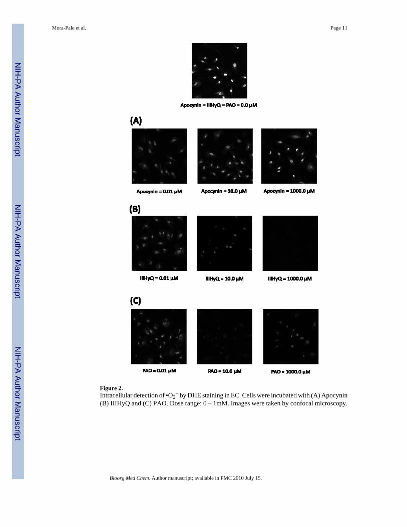
Figure 2.Intracellular detection of •O2
− by DHE staining in EC. Cells were incubated with (A) Apocynin(B) IIIHyQ and (C) PAO. Dose range: 0 – 1mM. Images were taken by confocal microscopy.
Mora-Pale et al. Page 11
Bioorg Med Chem. Author manuscript; available in PMC 2010 July 15.
NIH
-PA Author Manuscript
NIH
-PA Author Manuscript
NIH
-PA Author Manuscript

Figure 3.Effect of (A) isolated IIIHyQ, (B) AOP-2 and (C) apocynin, on biotin-p22phox-his-p47phox
interaction by ELISA. Biotin- p22phox = 2.0 μM, his- p47phox = 0.3 μM.
Mora-Pale et al. Page 12
Bioorg Med Chem. Author manuscript; available in PMC 2010 July 15.
NIH
-PA Author Manuscript
NIH
-PA Author Manuscript
NIH
-PA Author Manuscript

Figure 4.Correlation of experimental log (IC50) values of cell-based and ELISA assays.
Mora-Pale et al. Page 13
Bioorg Med Chem. Author manuscript; available in PMC 2010 July 15.
NIH
-PA Author Manuscript
NIH
-PA Author Manuscript
NIH
-PA Author Manuscript

Figure 5.Interaction between biotin-p22phox (2.0 μM) and his-p47phox (0 – 1.0 μM) in the absence andpresence of IAA (1.0 μM).
Mora-Pale et al. Page 14
Bioorg Med Chem. Author manuscript; available in PMC 2010 July 15.
NIH
-PA Author Manuscript
NIH
-PA Author Manuscript
NIH
-PA Author Manuscript
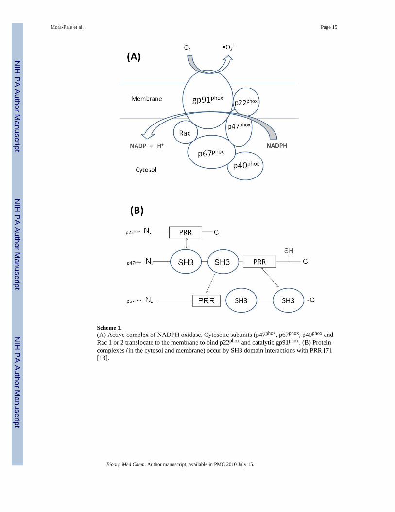
Scheme 1.(A) Active complex of NADPH oxidase. Cytosolic subunits (p47phox, p67phox, p40phox andRac 1 or 2 translocate to the membrane to bind p22phox and catalytic gp91phox. (B) Proteincomplexes (in the cytosol and membrane) occur by SH3 domain interactions with PRR [7],[13].
Mora-Pale et al. Page 15
Bioorg Med Chem. Author manuscript; available in PMC 2010 July 15.
NIH
-PA Author Manuscript
NIH
-PA Author Manuscript
NIH
-PA Author Manuscript
NIH
-PA Author Manuscript
NIH
-PA Author Manuscript
NIH
-PA Author Manuscript
Mora-Pale et al. Page 16
Table 1AOP present in soluble and precipitate fractions produced from the peroxidatic oxidation of apocynin based on LC-MS analysis. Nomenclature: A (apocynin), I (monomer), II (dimer), III (trimer), IV (tetramer), V (pentamer), VI(hexamer), VII (heptamer), Q (quinone), Hy (hydroxylated), -Me (demethylated)
Fraction Components
AOP-1 A (166m/z); II or IIHy-MeQ (330m/z); IIIHyQ (508m/z); III2Hy or III3HyQ (526m/z); IV-3MeQ (614m/z); IV or IVHy-MeQ (658m/z)
AOP-2 A (166m/z); III3OH (542m/z); II or IIHy-MeQ (330m/z); III4Hy-Me (530m/z); IIIHyQ (508m/z); III-Me (478m/z); IIIHy (480m/z); III-3Me (450m/z); IV-Me (644m/z), III-2MeQ (464m/z), III-3Me (500m/z), IV-Me (692m/z)
AOP-3 II or IIHy-MeQ (330m/z); III or IIIHy-MeQ (494m/z); III-Me or IIIHy-2MeQ (480m/z), IIIHyQ (508m/z), IVHy (674m/z); IV3Hy (706m/z),IV3Hy-2Me (678m/z), IV-2MeQ (628m/z)
AOP-4 A (166m/z); IIIHy (510m/z); II or IIHy-MeQ (330m/z); III3Hy (542m/z); IVHy (674m/z); III2Hy or III3HyQ (526m/z); IIIQ (492m/z); III or IIIHy-MeQ (494m/z)
AOP-5 A (166m/z); II or IIHy-MeQ (330m/z); IV (658m/z); III3Hy (542m/z); III-2Me (466m/z); III2Hy or III3HyQ (526m/z); IV4Hy-3Me (680m/z); IIIor IIIHy-MeQ (494m/z); IV or IVHy-MeQ (658m/z); IIIQ(492m/z); III-3Me (500m/z); VHy-MeQ (822m/z)
AOP-6 II or IIHy-MeQ (330m/z); III or IIIHy-MeQ (494m/z); IV2Hy (690m/z); III-3Me (452m/z); IV4Hy-3Me (680m/z); IV or IVHy-MeQ (658m/z);IV-3Me (616m/z), V4Hy-3Me (844m/z); VII4Hy-2Me (1186m/z) V or VHy-MeQ (822m/z)
AOP-7 III3Hy (500m/z); II or IIHy-MeQ (330m/z); III or IIIHy-MeQ (494m/z); VI5Hy-4Me (1010m/z), IIQ (328m/z); IV2Hy (690m/z); IV4Hy-3Me(680m/z); VI4Hy-3Me (1008m/z); IV or IVHy-MeQ (658m/z);
AOP-8 II or IIHy-MeQ (330m/z); IV5Hy-4Me (682m/z); III3Hy-2Me (514m/z); IVHy (674m/z); IV or IVHy-MeQ (658m/z); VI4Hy-4Me (994m/z)
AOP-9 II or IIHy-MeQ (330m/z); IV5Hy-4Me (682m/z); IVHy-Me (660m/z); IVHy-4Me, IVHyQ (672m/z); VII3Hy-2Me (1170m/z); IV2Hy-3Me (648m/z); III2Hy (526m/z); IV2Hy or IV3Hy-MeQ (690m/z); III or IIIHy-MeQ (494m/z); IV4Hy-3Me (680m/z); IV or IVHy-MeQ (658m/z); VHy (838m/z)
AOP-10 II or IIHy-MeQ (330m/z); IV5Hy-4Me (682m/z); IVHyQ (672m/z); IVHy-4Me (618m/z); V-2Me (794m/z); IV2Hy-2Me (662m/z); VHy (838m/z); IV2Hy or IV3Hy-MeQ (690m/z); III or IIIHy-MeQ (494m/z); IV4Hy-3Me (680m/z); III-Me or IIIHy-2MeQ (480m/z); IV-Me or IVHy-2MeQ(644m/z); IHy-Me (168m/z); IV4Hy-Me (694m/z)
Bioorg Med Chem. Author manuscript; available in PMC 2010 July 15.

NIH
-PA Author Manuscript
NIH
-PA Author Manuscript
NIH
-PA Author Manuscript
Mora-Pale et al. Page 17
Table 2Dose response IC50 values of AOP fractions following peroxidase-catalyzed oxidation of apocynin and silica gelchromatography.
Fraction Log (IC50) (IC50 in parentheses, μM) Cell-based assay Log (IC50) (IC50 in parentheses, μM) ELISA assay
Apocynin 3.04 ± 0.49 (1090) 4.78 ± 6.65 (61,000)
IIIHyQ −1.51 ± 0.05 (0.03) 0.20 ± 0.15 (1.60)
AOP-2 −1.40 ± 0.05 (0.04) −0.27 ± 0.15 (0.55)
AOP-3 −0.61 ± 0.08 (0.25) 1.70 ± 0.15 (50.0)
AOP-4 −0.67 ± 0.10 (0.22) 0.47 ± 0.18 (2.96)
AOP-5 0.34 ± 0.11 (2.20) 0.44 ± 0.90 (7.96)
AOP-6 −0.09 ± 0.09 (0.82) 2.01 ± 0.16 (103)
AOP-7 0.001 ± 0.06 (1.00) 2.66 ± 456 (453)
AOP-8 0.03 ± 0.22 (1.08) 2.38 ± 0.08 (239)
AOP-9 −0.04 ± 0.13 (0.92) 1.54 ± 0.08 (34.8)
AOP-10 −0.11 ± 0.12 (0.78) 2.03 ± 0.08 (107)
Bioorg Med Chem. Author manuscript; available in PMC 2010 July 15.