Urotensin II Inhibits Skeletal Muscle Glucose Transport Signaling Pathways via the NADPH Oxidase...
10
Urotensin II Inhibits Skeletal Muscle Glucose Transport Signaling Pathways via the NADPH Oxidase Pathway Hong-Xia Wang 1 , Xin-Rui Wu 1 , Hui Yang 1 , Chun-Lin Yin 2 , Li-Jin Shi 3 , Xue-Jiang Wang 1* 1 Department of Physiology and Pathophysiology, School of Basic Medical Sciences, Capital Medical University, Beijing, China, 2 Department of Cardiology, Xuan Wu Hospital, Capital Medical University, Beijing, China, 3 Department of Neurology, the First Affiliated Hospital of Xinxiang Medical College, Xinxiang, China Abstract Our previous studies have demonstrated that the urotensin (UII) and its receptor are up-regulated in the skeletal muscle of mice with type II diabetes mellitus (T2DM), but the significance of UII in skeletal muscle insulin resistance remains unknown. The purpose of this study was to investigate the effect of UII on NADPH oxidase and glucose transport signaling pathways in the skeletal muscle of mice with T2DM and in C2C12 mouse myotube cells. KK/upj- AY/J mice (KK) mice were divided into the following groups: KK group, with saline treatment for 2 weeks; KK+ urantide group, with daily 30 µg/kg body weight injections over the same time period of urantide, a potent urotensin II antagonist peptide; Non-diabetic C57BL/6J mice were used as normal controls. After urantide treatment, mice were subjected to an intraperitoneal glucose tolerance test, in addition to measurements of the levels of ROS, NADPH oxidase and the phosphorylated AKT, PKC and ERK. C2C12 cells were incubated with serum-free DMEM for 24 hours before conducting the experiments, and then administrated with 100 nM UII for 2 hours or 24 hours. Urantide treatment improved glucose tolerance, decreased the translocation of the NADPH subunits p40-phox and p47-phox, and increased levels of the phosphorylated PKC, AKT and ERK. In contrast, UII treatment increased ROS production and p47-phox and p67-phox translocation, and decreased the phosphorylated AKT, ERK1/2 and p38MAPK; Apocynin abrogated this effect. In conclusion, UII increased ROS production by NADPH oxidase, leading to the inhibition of signaling pathways involving glucose transport, such as AKT/PKC/ERK. Our data imply a role for UII at the molecular level in glucose homeostasis, and possibly in skeletal muscle insulin resistance in T2DM. Citation: Wang H-X, Wu X-R, Yang H, Yin C-L, Shi L-J, et al. (2013) Urotensin II Inhibits Skeletal Muscle Glucose Transport Signaling Pathways via the NADPH Oxidase Pathway. PLoS ONE 8(10): e76796. doi:10.1371/journal.pone.0076796 Editor: Rebecca Berdeaux, University of Texas Health Science Center at Houston, United States of America Received April 16, 2013; Accepted August 27, 2013; Published October 8, 2013 Copyright: © 2013 Wang et al. This is an open-access article distributed under the terms of the Creative Commons Attribution License, which permits unrestricted use, distribution, and reproduction in any medium, provided the original author and source are credited. Funding: This work was supported by National Natural Science Foundation of China (no.81000343, http://www.nsfc.gov.cn/Portal0/default152.htm) and Beijing Natural Science Foundation (no.7112013, http://www.bjnsf.org/). The funders had no role in study design, data collection and analysis, decision to publish, or preparation of the manuscript. Competing interests: The authors have declared that no competing interests exist. * E-mail: [email protected] Introduction Urotensin II (UII) is a vasoactive peptide that was first discovered in teleost fishes, and later in mammals and humans [1,2]. UII acts by binding to the G protein coupled receptor GPR14 (now known as UT) [3], and have been detected in cardiac and vascular tissues, and the spinal cord, central nervous system, kidney, liver and pancreas [4]. Importantly, UII and UT are abundant in the skeletal muscle of mouse and monkey, and radio-ligand binding assay has shown that UT binds [ 125 I]UII with high affinity in skeletal muscle [5]. Besides its important role in the cardiovascular system, UII also participates in metabolic regulation and plays a significant role in diabetes and its complications [6,7]. Our previous studies demonstrated that the UII/UT system is up-regulated in the skeletal muscle of mice with type II diabetes mellitus (T2DM), and UII inhibited insulin-stimulated 2-DG uptake in skeletal muscle [8]. We speculated that skeletal muscle-derived UII might be involved as an autocrine/paracrine factor in the pathogenesis of skeletal muscle insulin resistance (IR), although the mechanism remains unclear. IR, the major defect of T2DM, is a common pathophysiological state in which higher than normal concentrations of insulin are required to exert its biological effect in target tissues such as the skeletal muscle, adipose tissue and liver [9]. Considering the skeletal muscle accounts for the majority of insulin-mediated glucose disposal in the post-prandial state, skeletal muscle IR contributes significantly to the metabolic derangements seen in T2DM patients. The precise molecular mechanisms responsible for insulin resistance remain incompletely understood, however, particularly in skeletal muscle. Emerging data indicated that oxidative stress due to increased reactive oxygen species (ROS) generation and/or compromised antioxidant systems PLOS ONE | www.plosone.org 1 October 2013 | Volume 8 | Issue 10 | e76796
Transcript of Urotensin II Inhibits Skeletal Muscle Glucose Transport Signaling Pathways via the NADPH Oxidase...
Urotensin II Inhibits Skeletal Muscle Glucose TransportSignaling Pathways via the NADPH Oxidase PathwayHong-Xia Wang1, Xin-Rui Wu1, Hui Yang1, Chun-Lin Yin2, Li-Jin Shi3, Xue-Jiang Wang1*
1 Department of Physiology and Pathophysiology, School of Basic Medical Sciences, Capital Medical University, Beijing, China, 2 Department of Cardiology,Xuan Wu Hospital, Capital Medical University, Beijing, China, 3 Department of Neurology, the First Affiliated Hospital of Xinxiang Medical College, Xinxiang,China
Abstract
Our previous studies have demonstrated that the urotensin (UII) and its receptor are up-regulated in the skeletalmuscle of mice with type II diabetes mellitus (T2DM), but the significance of UII in skeletal muscle insulin resistanceremains unknown. The purpose of this study was to investigate the effect of UII on NADPH oxidase and glucosetransport signaling pathways in the skeletal muscle of mice with T2DM and in C2C12 mouse myotube cells. KK/upj-AY/J mice (KK) mice were divided into the following groups: KK group, with saline treatment for 2 weeks; KK+urantide group, with daily 30 µg/kg body weight injections over the same time period of urantide, a potent urotensin IIantagonist peptide; Non-diabetic C57BL/6J mice were used as normal controls. After urantide treatment, mice weresubjected to an intraperitoneal glucose tolerance test, in addition to measurements of the levels of ROS, NADPHoxidase and the phosphorylated AKT, PKC and ERK. C2C12 cells were incubated with serum-free DMEM for 24hours before conducting the experiments, and then administrated with 100 nM UII for 2 hours or 24 hours. Urantidetreatment improved glucose tolerance, decreased the translocation of the NADPH subunits p40-phox and p47-phox,and increased levels of the phosphorylated PKC, AKT and ERK. In contrast, UII treatment increased ROS productionand p47-phox and p67-phox translocation, and decreased the phosphorylated AKT, ERK1/2 and p38MAPK;Apocynin abrogated this effect. In conclusion, UII increased ROS production by NADPH oxidase, leading to theinhibition of signaling pathways involving glucose transport, such as AKT/PKC/ERK. Our data imply a role for UII atthe molecular level in glucose homeostasis, and possibly in skeletal muscle insulin resistance in T2DM.
Citation: Wang H-X, Wu X-R, Yang H, Yin C-L, Shi L-J, et al. (2013) Urotensin II Inhibits Skeletal Muscle Glucose Transport Signaling Pathways via theNADPH Oxidase Pathway. PLoS ONE 8(10): e76796. doi:10.1371/journal.pone.0076796
Editor: Rebecca Berdeaux, University of Texas Health Science Center at Houston, United States of America
Received April 16, 2013; Accepted August 27, 2013; Published October 8, 2013
Copyright: © 2013 Wang et al. This is an open-access article distributed under the terms of the Creative Commons Attribution License, which permitsunrestricted use, distribution, and reproduction in any medium, provided the original author and source are credited.
Funding: This work was supported by National Natural Science Foundation of China (no.81000343, http://www.nsfc.gov.cn/Portal0/default152.htm) andBeijing Natural Science Foundation (no.7112013, http://www.bjnsf.org/). The funders had no role in study design, data collection and analysis, decision topublish, or preparation of the manuscript.
Competing interests: The authors have declared that no competing interests exist.
* E-mail: [email protected]
Introduction
Urotensin II (UII) is a vasoactive peptide that was firstdiscovered in teleost fishes, and later in mammals and humans[1,2]. UII acts by binding to the G protein coupled receptorGPR14 (now known as UT) [3], and have been detected incardiac and vascular tissues, and the spinal cord, centralnervous system, kidney, liver and pancreas [4]. Importantly, UIIand UT are abundant in the skeletal muscle of mouse andmonkey, and radio-ligand binding assay has shown that UTbinds [125I]UII with high affinity in skeletal muscle [5]. Besidesits important role in the cardiovascular system, UII alsoparticipates in metabolic regulation and plays a significant rolein diabetes and its complications [6,7]. Our previous studiesdemonstrated that the UII/UT system is up-regulated in theskeletal muscle of mice with type II diabetes mellitus (T2DM),and UII inhibited insulin-stimulated 2-DG uptake in skeletal
muscle [8]. We speculated that skeletal muscle-derived UIImight be involved as an autocrine/paracrine factor in thepathogenesis of skeletal muscle insulin resistance (IR),although the mechanism remains unclear.
IR, the major defect of T2DM, is a commonpathophysiological state in which higher than normalconcentrations of insulin are required to exert its biologicaleffect in target tissues such as the skeletal muscle, adiposetissue and liver [9]. Considering the skeletal muscle accountsfor the majority of insulin-mediated glucose disposal in thepost-prandial state, skeletal muscle IR contributes significantlyto the metabolic derangements seen in T2DM patients. Theprecise molecular mechanisms responsible for insulinresistance remain incompletely understood, however,particularly in skeletal muscle. Emerging data indicated thatoxidative stress due to increased reactive oxygen species(ROS) generation and/or compromised antioxidant systems
PLOS ONE | www.plosone.org 1 October 2013 | Volume 8 | Issue 10 | e76796
represents an important factor in the progression of insulinresistance [10]. One of the main sources of ROS is NADPHoxidase (NOX), a multi-protein enzyme complex that usesNADPH as a substrate to convert molecular oxygen to ROS.Components of NADPH oxidase complex of phagocytesinclude the membrane-bound cytochrome b558, composed of 2subunits, p22-phox and gp91-phox, and 4 cytosolic subunits,p47-phox, p67-phox, p40-phox, and the small GTP-bindingprotein, Rac. Moreover, expression of gp91phox, p22phox, p40phox, p47phox, and p67phox have been documented inskeletal muscle [11]. Wei et al found NADPH oxidase activationand ROS generation play an important role in Ang II-inducedinhibition of insulin signaling in skeletal muscle cells [12]. Giventhese data, studies are warranted to ascertain whether UIImediates skeletal muscle IR by increasing ROS production viaNADPH oxidase.
In the present study, we sought to determine whether UIIantagonism improved glucose tolerance by decreasing theoxidative state in KK mice, and to investigate the effect of UIIon ROS production and on glucose transport signaling inC2C12 mouse myotube cells. We study the effects of UII onROS production and NADPH oxidase levels, and itsinvolvement in the regulation of the AKT/PKC/ERK signalingpathway.
Results
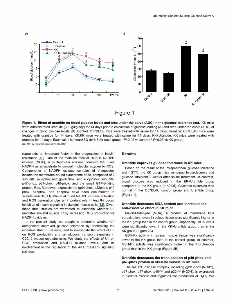
Urantide improves glucose tolerance in KK miceBased on the result of the intraperitoneal glucose tolerance
test (IGTT), the KK group mice remained hyperglycemic andglucose intolerant 2 weeks after saline treatment. In contrast,blood glucose was reduced in the KK+Urantide groupcompared to the KK group (p <0.05). Glycemic excursion wasnormal in the C57BL/6J control group and Urantide group(Figure 1).
Urantide decreases MDA content and increases theanti-oxidative effect in KK mice
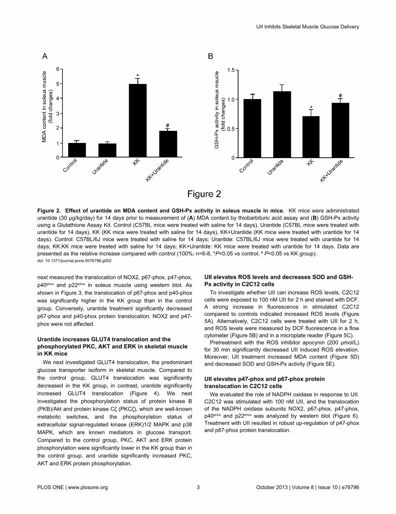
Malondialdehyde (MDA), a product of membrane lipidperoxidation, levels in soleus tissue were significantly higher inthe KK group than in the control group. Importantly, MDA levelswere significantly lower in the KK+Urantide group than in theKK group (Figure 2A).
GSH-Px activity in soleus muscle tissue was significantlylower in the KK group than in the control group. In contrast,GSH-Px activity was significantly higher in the KK+Urantidegroup than in the KK group (Figure 2B).
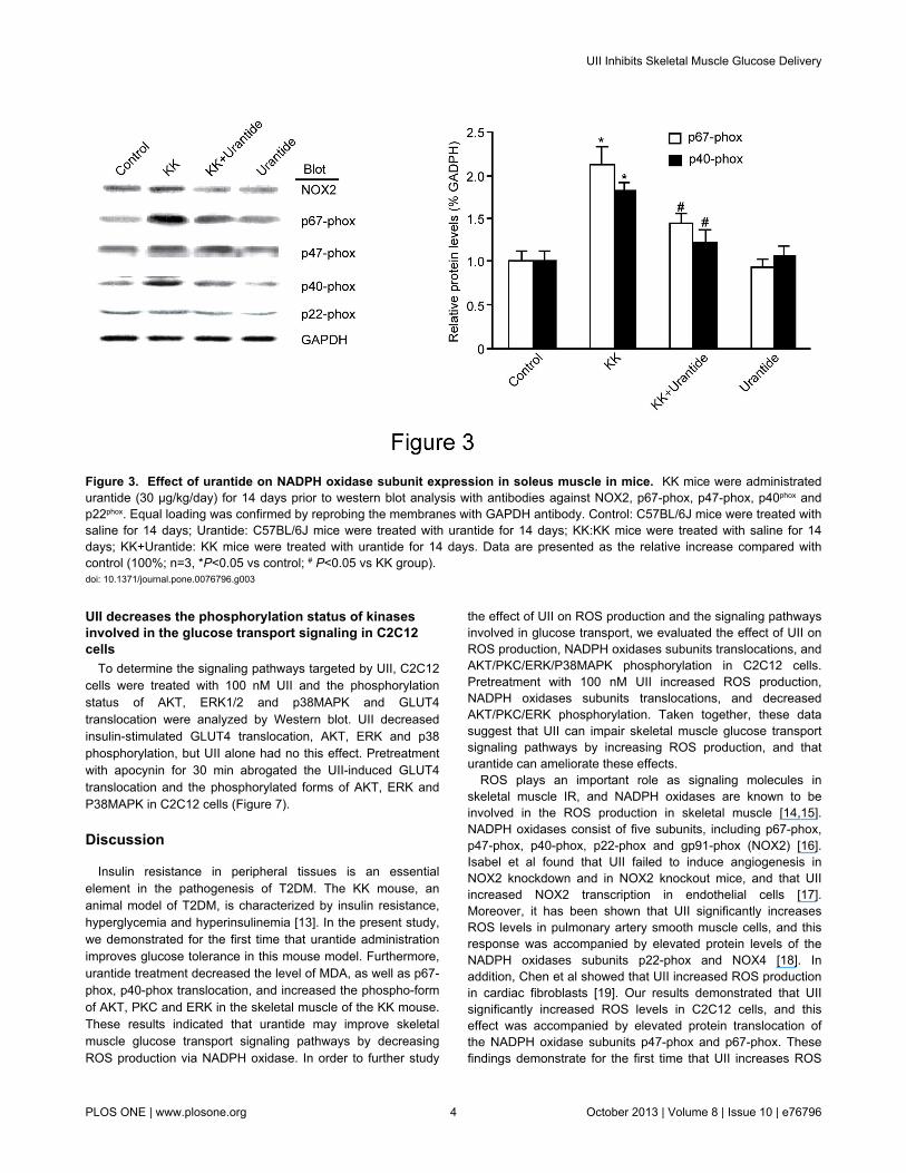
Urantide decreases the translocation of p40-phox andp67-phox protein in skeletal muscle in KK mice
The NADPH oxidase complex, including gp91-phox (NOX2),p67-phox, p47-phox, p40phox and p22phox (NOX4), is expressedin skeletal muscle and regulates the production of H2O2. We
Figure 1. Effect of urantide on blood glucose levels and area under the curve (AUC) in the glucose tolerance test. KK micewere administrated urantide (30 µg/kg/day) for 14 days prior to calculation of glucose loading (A) and area under the curve (AUC) ofchanges in blood glucose levels (B). Control: C57BL/6J mice were treated with saline for 14 days; Urantide: C57BL/6J mice weretreated with urantide for 14 days; KK:KK mice were treated with saline for 14 days; KK+Urantide: KK mice were treated withurantide for 14 days. Each value is mean±SD (n=6-8 for each group, *P<0.05 vs control; # P<0.05 vs KK group).doi: 10.1371/journal.pone.0076796.g001
UII Inhibits Skeletal Muscle Glucose Delivery
PLOS ONE | www.plosone.org 2 October 2013 | Volume 8 | Issue 10 | e76796
next measured the translocation of NOX2, p67-phox, p47-phox,p40phox and p22phox in soleus muscle using western blot. Asshown in Figure 3, the translocation of p67-phox and p40-phoxwas significantly higher in the KK group than in the controlgroup. Conversely, urantide treatment significantly decreasedp67-phox and p40-phox protein translocation. NOX2 and p47-phox were not affected.
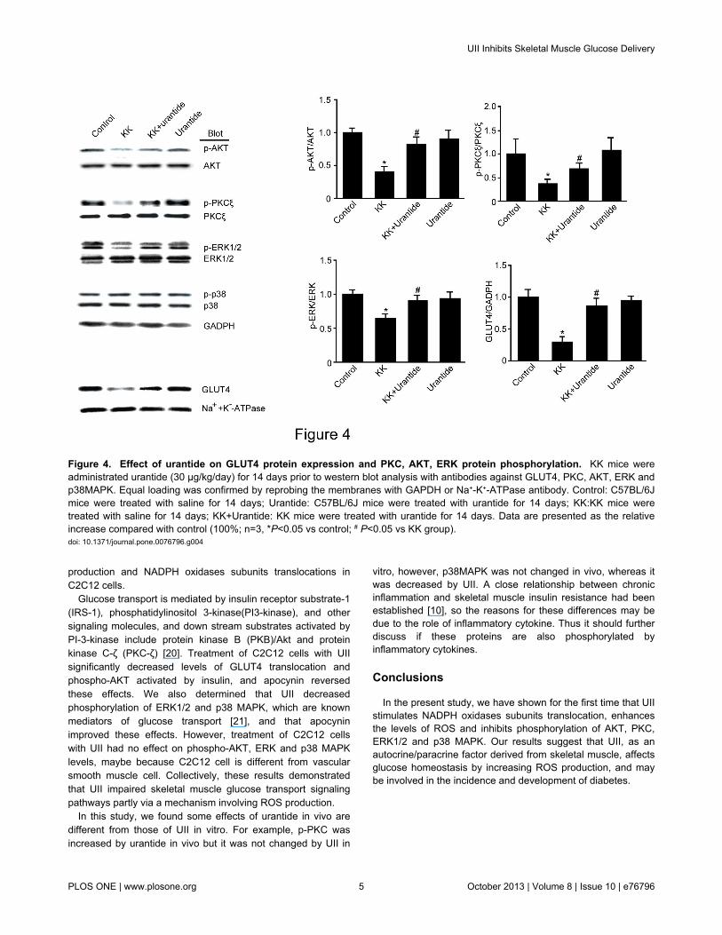
Urantide increases GLUT4 translocation and thephosphorylated PKC, AKT and ERK in skeletal musclein KK mice
We next investigated GLUT4 translocation, the predominantglucose transporter isoform in skeletal muscle. Compared tothe control group, GLUT4 translocation was significantlydecreased in the KK group, in contrast, urantide significantlyincreased GLUT4 translocation (Figure 4). We nextinvestigated the phosphorylation status of protein kinase B(PKB)/Akt and protein kinase Cζ (PKCζ), which are well-knownmetabolic switches, and the phosphorylation status ofextracellular signal-regulated kinase (ERK)1/2 MAPK and p38MAPK, which are known mediators in glucose transport.Compared to the control group, PKC, AKT and ERK proteinphosphorylation were significantly lower in the KK group than inthe control group, and urantide significantly increased PKC,AKT and ERK protein phosphorylation.
UII elevates ROS levels and decreases SOD and GSH-Px activity in C2C12 cells
To investigate whether UII can increase ROS levels, C2C12cells were exposed to 100 nM UII for 2 h and stained with DCF.A strong increase in fluorescence in stimulated C2C12compared to controls indicated increased ROS levels (Figure5A). Alternatively, C2C12 cells were treated with UII for 2 h,and ROS levels were measured by DCF fluorescence in a flowcytometer (Figure 5B) and in a microplate reader (Figure 5C).
Pretreatment with the ROS inhibitor apocynin (200 µmol/L)for 30 min significantly decreased UII induced ROS elevation.Moreover, UII treatment increased MDA content (Figure 5D)and decreased SOD and GSH-Px activity (Figure 5E).
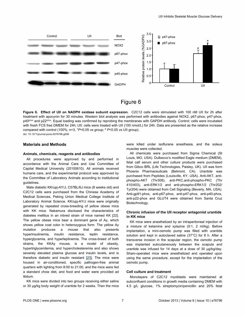
UII elevates p47-phox and p67-phox proteintranslocation in C2C12 cells
We evaluated the role of NADPH oxidase in response to UII.C2C12 was stimulated with 100 nM UII, and the translocationof the NADPH oxidase subunits NOX2, p67-phox, p47-phox,p40phox and p22phox was analyzed by western blot (Figure 6).Treatment with UII resulted in robust up-regulation of p47-phoxand p67-phox protein translocation.
Figure 2. Effect of urantide on MDA content and GSH-Px activity in soleus muscle in mice. KK mice were administratedurantide (30 µg/kg/day) for 14 days prior to measurement of (A) MDA content by thiobarbituric acid assay and (B) GSH-Px activityusing a Glutathione Assay Kit. Control (C57BL mice were treated with saline for 14 days), Urantide (C57BL mice were treated withurantide for 14 days), KK (KK mice were treated with saline for 14 days), KK+Urantide (KK mice were treated with urantide for 14days). Control: C57BL/6J mice were treated with saline for 14 days; Urantide: C57BL/6J mice were treated with urantide for 14days; KK:KK mice were treated with saline for 14 days; KK+Urantide: KK mice were treated with urantide for 14 days. Data arepresented as the relative increase compared with control (100%; n=6-8, *P<0.05 vs control; # P<0.05 vs KK group).doi: 10.1371/journal.pone.0076796.g002
UII Inhibits Skeletal Muscle Glucose Delivery
PLOS ONE | www.plosone.org 3 October 2013 | Volume 8 | Issue 10 | e76796
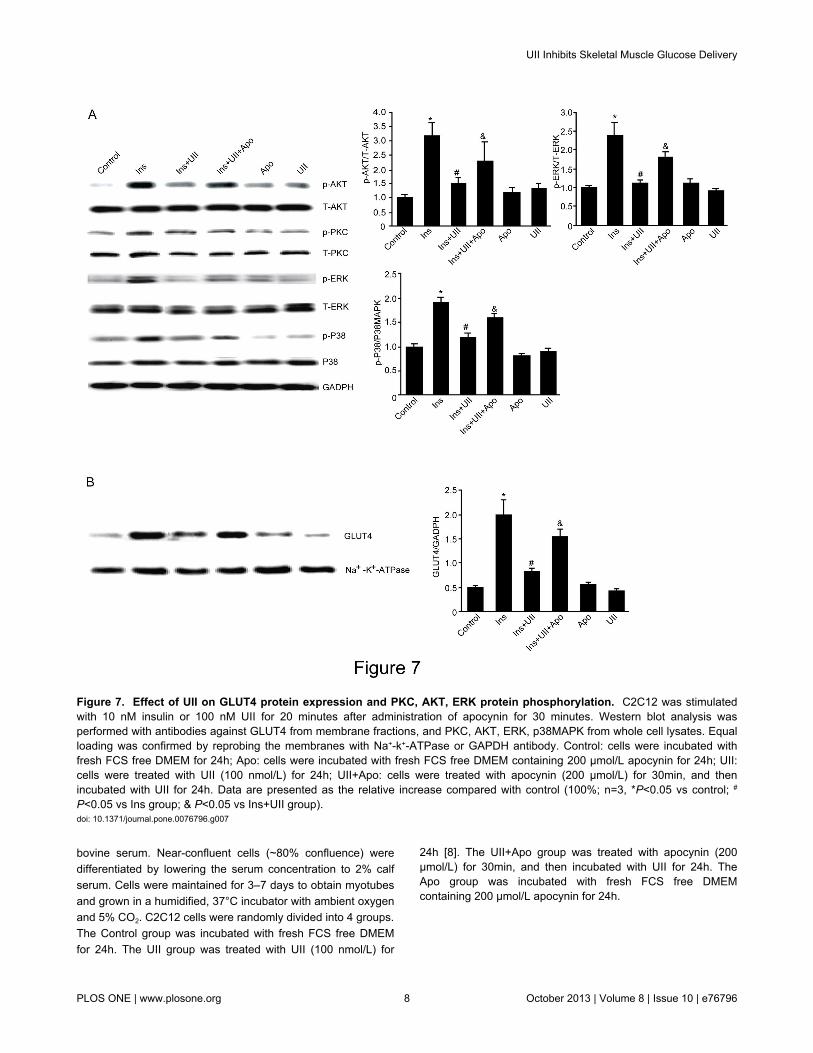
UII decreases the phosphorylation status of kinasesinvolved in the glucose transport signaling in C2C12cells
To determine the signaling pathways targeted by UII, C2C12cells were treated with 100 nM UII and the phosphorylationstatus of AKT, ERK1/2 and p38MAPK and GLUT4translocation were analyzed by Western blot. UII decreasedinsulin-stimulated GLUT4 translocation, AKT, ERK and p38phosphorylation, but UII alone had no this effect. Pretreatmentwith apocynin for 30 min abrogated the UII-induced GLUT4translocation and the phosphorylated forms of AKT, ERK andP38MAPK in C2C12 cells (Figure 7).
Discussion
Insulin resistance in peripheral tissues is an essentialelement in the pathogenesis of T2DM. The KK mouse, ananimal model of T2DM, is characterized by insulin resistance,hyperglycemia and hyperinsulinemia [13]. In the present study,we demonstrated for the first time that urantide administrationimproves glucose tolerance in this mouse model. Furthermore,urantide treatment decreased the level of MDA, as well as p67-phox, p40-phox translocation, and increased the phospho-formof AKT, PKC and ERK in the skeletal muscle of the KK mouse.These results indicated that urantide may improve skeletalmuscle glucose transport signaling pathways by decreasingROS production via NADPH oxidase. In order to further study
the effect of UII on ROS production and the signaling pathwaysinvolved in glucose transport, we evaluated the effect of UII onROS production, NADPH oxidases subunits translocations, andAKT/PKC/ERK/P38MAPK phosphorylation in C2C12 cells.Pretreatment with 100 nM UII increased ROS production,NADPH oxidases subunits translocations, and decreasedAKT/PKC/ERK phosphorylation. Taken together, these datasuggest that UII can impair skeletal muscle glucose transportsignaling pathways by increasing ROS production, and thaturantide can ameliorate these effects.
ROS plays an important role as signaling molecules inskeletal muscle IR, and NADPH oxidases are known to beinvolved in the ROS production in skeletal muscle [14,15].NADPH oxidases consist of five subunits, including p67-phox,p47-phox, p40-phox, p22-phox and gp91-phox (NOX2) [16].Isabel et al found that UII failed to induce angiogenesis inNOX2 knockdown and in NOX2 knockout mice, and that UIIincreased NOX2 transcription in endothelial cells [17].Moreover, it has been shown that UII significantly increasesROS levels in pulmonary artery smooth muscle cells, and thisresponse was accompanied by elevated protein levels of theNADPH oxidases subunits p22-phox and NOX4 [18]. Inaddition, Chen et al showed that UII increased ROS productionin cardiac fibroblasts [19]. Our results demonstrated that UIIsignificantly increased ROS levels in C2C12 cells, and thiseffect was accompanied by elevated protein translocation ofthe NADPH oxidase subunits p47-phox and p67-phox. Thesefindings demonstrate for the first time that UII increases ROS
Figure 3. Effect of urantide on NADPH oxidase subunit expression in soleus muscle in mice. KK mice were administratedurantide (30 µg/kg/day) for 14 days prior to western blot analysis with antibodies against NOX2, p67-phox, p47-phox, p40phox andp22phox. Equal loading was confirmed by reprobing the membranes with GAPDH antibody. Control: C57BL/6J mice were treated withsaline for 14 days; Urantide: C57BL/6J mice were treated with urantide for 14 days; KK:KK mice were treated with saline for 14days; KK+Urantide: KK mice were treated with urantide for 14 days. Data are presented as the relative increase compared withcontrol (100%; n=3, *P<0.05 vs control; # P<0.05 vs KK group).doi: 10.1371/journal.pone.0076796.g003
UII Inhibits Skeletal Muscle Glucose Delivery
PLOS ONE | www.plosone.org 4 October 2013 | Volume 8 | Issue 10 | e76796
production and NADPH oxidases subunits translocations inC2C12 cells.
Glucose transport is mediated by insulin receptor substrate-1(IRS-1), phosphatidylinositol 3-kinase(PI3-kinase), and othersignaling molecules, and down stream substrates activated byPI-3-kinase include protein kinase B (PKB)/Akt and proteinkinase C-ζ (PKC-ζ) [20]. Treatment of C2C12 cells with UIIsignificantly decreased levels of GLUT4 translocation andphospho-AKT activated by insulin, and apocynin reversedthese effects. We also determined that UII decreasedphosphorylation of ERK1/2 and p38 MAPK, which are knownmediators of glucose transport [21], and that apocyninimproved these effects. However, treatment of C2C12 cellswith UII had no effect on phospho-AKT, ERK and p38 MAPKlevels, maybe because C2C12 cell is different from vascularsmooth muscle cell. Collectively, these results demonstratedthat UII impaired skeletal muscle glucose transport signalingpathways partly via a mechanism involving ROS production.
In this study, we found some effects of urantide in vivo aredifferent from those of UII in vitro. For example, p-PKC wasincreased by urantide in vivo but it was not changed by UII in
vitro, however, p38MAPK was not changed in vivo, whereas itwas decreased by UII. A close relationship between chronicinflammation and skeletal muscle insulin resistance had beenestablished [10], so the reasons for these differences may bedue to the role of inflammatory cytokine. Thus it should furtherdiscuss if these proteins are also phosphorylated byinflammatory cytokines.
Conclusions
In the present study, we have shown for the first time that UIIstimulates NADPH oxidases subunits translocation, enhancesthe levels of ROS and inhibits phosphorylation of AKT, PKC,ERK1/2 and p38 MAPK. Our results suggest that UII, as anautocrine/paracrine factor derived from skeletal muscle, affectsglucose homeostasis by increasing ROS production, and maybe involved in the incidence and development of diabetes.
Figure 4. Effect of urantide on GLUT4 protein expression and PKC, AKT, ERK protein phosphorylation. KK mice wereadministrated urantide (30 µg/kg/day) for 14 days prior to western blot analysis with antibodies against GLUT4, PKC, AKT, ERK andp38MAPK. Equal loading was confirmed by reprobing the membranes with GAPDH or Na+-K+-ATPase antibody. Control: C57BL/6Jmice were treated with saline for 14 days; Urantide: C57BL/6J mice were treated with urantide for 14 days; KK:KK mice weretreated with saline for 14 days; KK+Urantide: KK mice were treated with urantide for 14 days. Data are presented as the relativeincrease compared with control (100%; n=3, *P<0.05 vs control; # P<0.05 vs KK group).doi: 10.1371/journal.pone.0076796.g004
UII Inhibits Skeletal Muscle Glucose Delivery
PLOS ONE | www.plosone.org 5 October 2013 | Volume 8 | Issue 10 | e76796
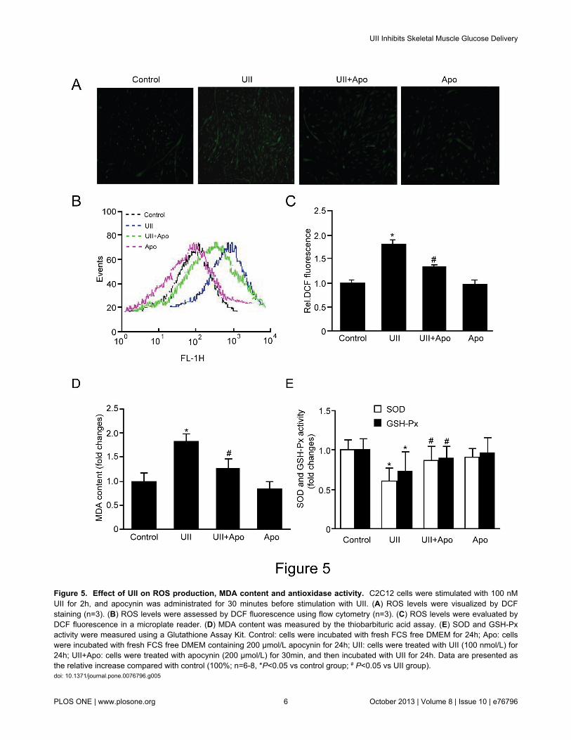
Figure 5. Effect of UII on ROS production, MDA content and antioxidase activity. C2C12 cells were stimulated with 100 nMUII for 2h, and apocynin was administrated for 30 minutes before stimulation with UII. (A) ROS levels were visualized by DCFstaining (n=3). (B) ROS levels were assessed by DCF fluorescence using flow cytometry (n=3). (C) ROS levels were evaluated byDCF fluorescence in a microplate reader. (D) MDA content was measured by the thiobarbituric acid assay. (E) SOD and GSH-Pxactivity were measured using a Glutathione Assay Kit. Control: cells were incubated with fresh FCS free DMEM for 24h; Apo: cellswere incubated with fresh FCS free DMEM containing 200 µmol/L apocynin for 24h; UII: cells were treated with UII (100 nmol/L) for24h; UII+Apo: cells were treated with apocynin (200 µmol/L) for 30min, and then incubated with UII for 24h. Data are presented asthe relative increase compared with control (100%; n=6-8, *P<0.05 vs control group; # P<0.05 vs UII group).doi: 10.1371/journal.pone.0076796.g005
UII Inhibits Skeletal Muscle Glucose Delivery
PLOS ONE | www.plosone.org 6 October 2013 | Volume 8 | Issue 10 | e76796
Materials and Methods
Animals, chemicals, reagents and antibodiesAll procedures were approved by and performed in
accordance with the Animal Care and Use Committee ofCapital Medical University (20100610). All animals receivedhumane care, and the experimental protocol was approved bythe Committee of Laboratory Animals according to institutionalguidelines.
Male diabetic KK/upj-AY/J, C57BL/6J mice (8 weeks old) andC2C12 cells were purchased from the Chinese Academy ofMedical Sciences, Peking Union Medical College Institute ofLaboratory Animal Science. KK/upj-AY/J mice were originallygenerated by repeated cross-breeding of yellow obese micewith KK mice. Nakamura disclosed the characteristics ofdiabetes mellitus in an inbred strain of mice named KK [22].The yellow obese mice bear a dominant gene of Ay, whichshows yellow coat color in heterozygous form. The yellow Aymutation produces a mouse that also presentshyperinsulinemia, insulin resistance, leptin resistance,hyperglycemia, and hyperleptinemia. The cross-breed of bothstrains, the KKAy mouse, is a model of obesity,hypertriglyceridemia, and hypercholesteremia and also showsseverely elevated plasma glucose and insulin levels, and istherefore diabetic and insulin resistant [23]. The mice werehoused in air-conditioned, specific pathogen-free animalquarters with lighting from 8:00 to 21:00, and the mice were feda standard chow diet, and food and water were provided adlibitum.
KK mice were divided into two groups receiving either salineor 30 µg/kg body weight of urantide for 2 weeks. Then the mice
were killed under isoflurane anesthesia, and the soleusmuscles were collected.
All chemicals were purchased from Sigma Chemical (StLouis, MO, USA). Dulbecco’s modified Eagle medium (DMEM),fetal calf serum and other culture products were purchasedfrom Gibco BRL (Life Technologies, Paisley, UK). UII was formPhoenix Pharmaceuticals (Belmont, CA). Urantide waspurchased from Peptides (Louisville, KY, USA). Anti-AKT, anti-phospho-AKT (Thr308), anti-PKC,anti-phospho-PKC (Thr410/403), anti-ERK1/2 and anti-phospho-ERK1/2 (Thr202/Tyr204) were obtained from Cell Signaling (Beverly, MA, USA).Anti-gp91-phox, anti-p67-phox, anti-p47-phox, anti-p40-phox,anti-p22-phox and GLUT4 were obtained from Santa CruzBiotechnology.
Chronic infusion of the UII receptor antagonist urantidein KK mice
KK mice were anesthetized by an intraperitoneal injection ofa mixture of ketamine and xylazine (9:1, 2 ml/kg). Beforeimplantation, a mini-osmotic pump was filled with urantidesolution and kept in autoclaved saline (37°C) for 8 h. After atransverse incision in the scapular region, the osmotic pumpwas implanted subcutaneously between the scapula andurantide was infused for 14 days at a dose of 30 µg/kg/day.Sham-operated mice were anesthetized and operated uponusing the same procedure, except for the implantation of theosmotic pump.
Cell culture and treatmentMonolayers of C2C12 myoblasts were maintained at
subconfluent conditions in growth media containing DMEM with4.5 g/L glucose, 1% streptomycin/penicillin and 20% fetal
Figure 6. Effect of UII on NADPH oxidase subunit expression. C2C12 cells were stimulated with 100 nM UII for 2h aftertreatment with apocynin for 30 minutes. Western blot analysis was performed with antibodies against NOX2, p67-phox, p47-phox,p40phox and p22phox. Equal loading was confirmed by reprobing the membranes with GAPDH antibody. Control: cells were incubatedwith fresh FCS free DMEM for 24h; UII: cells were treated with UII (100 nmol/L) for 24h. Data are presented as the relative increasecompared with control (100%; n=3, *P<0.05 vs group; # P<0.05 vs UII group).doi: 10.1371/journal.pone.0076796.g006
UII Inhibits Skeletal Muscle Glucose Delivery
PLOS ONE | www.plosone.org 7 October 2013 | Volume 8 | Issue 10 | e76796
bovine serum. Near-confluent cells (~80% confluence) weredifferentiated by lowering the serum concentration to 2% calfserum. Cells were maintained for 3–7 days to obtain myotubesand grown in a humidified, 37°C incubator with ambient oxygenand 5% CO2. C2C12 cells were randomly divided into 4 groups.The Control group was incubated with fresh FCS free DMEMfor 24h. The UII group was treated with UII (100 nmol/L) for
24h [8]. The UII+Apo group was treated with apocynin (200µmol/L) for 30min, and then incubated with UII for 24h. TheApo group was incubated with fresh FCS free DMEMcontaining 200 µmol/L apocynin for 24h.
Figure 7. Effect of UII on GLUT4 protein expression and PKC, AKT, ERK protein phosphorylation. C2C12 was stimulatedwith 10 nM insulin or 100 nM UII for 20 minutes after administration of apocynin for 30 minutes. Western blot analysis wasperformed with antibodies against GLUT4 from membrane fractions, and PKC, AKT, ERK, p38MAPK from whole cell lysates. Equalloading was confirmed by reprobing the membranes with Na+-k+-ATPase or GAPDH antibody. Control: cells were incubated withfresh FCS free DMEM for 24h; Apo: cells were incubated with fresh FCS free DMEM containing 200 µmol/L apocynin for 24h; UII:cells were treated with UII (100 nmol/L) for 24h; UII+Apo: cells were treated with apocynin (200 µmol/L) for 30min, and thenincubated with UII for 24h. Data are presented as the relative increase compared with control (100%; n=3, *P<0.05 vs control; #
P<0.05 vs Ins group; & P<0.05 vs Ins+UII group).doi: 10.1371/journal.pone.0076796.g007
UII Inhibits Skeletal Muscle Glucose Delivery
PLOS ONE | www.plosone.org 8 October 2013 | Volume 8 | Issue 10 | e76796
Intraperitoneal glucose tolerance test (IGTT)All mice in the study underwent an IGTT after a 6h fast. Each
mouse was treated by intraperitoneal gavage with 2 g/kg bodyweight of glucose diluted in distilled water (120 mg/ml). Bloodsamples from the tail vein were collected at 0 (before glucoseinjection), 30, 60 and 120 min after glucose challenge. Theblood glucose concentration was determined using an Accu-Chek Advantage glucometer (Roche, Germany).
Determination of ROS, T-SOD, GSH-PX andmalondialdehyde (MDA) activities
The dye 2’, 7’-dichlorofluorescein diacetate (DCF-DA,Molecular Probes, Cat NO: E004, Nanjing JianchengBiotechnology Institute, China) was used to detect changes incellular ROS levels. Briefly, C2C12 cells were incorporated withDCF-DA for 30 min and washed with PBS three times.Fluorescent images in 24 well plates were immediatelyvisualized with a microscope (Olympus DX-63, Japan). ROSlevels were determined in 96 well plates by measuring thefluorescence strength with a BioTek Synergy 2 microplatereader (Winooski, VT, USA). The intensity of the fluorescencewas expressed as arbitrary units per microgram of proteins.The intensity of fluorescence in 6 wells plate was analyzed byflow cytometry.
Soleus muscle tissue homogenates were obtained fromfrozen tissue in cold physiologic saline. Homogenates werethen centrifuged at 3,000 g for 15 min at 4°C, and the proteinconcentrations of the supernatants were determined using aBCA Protein Assay Kit (BioTeke Corporation, China). The totalactivities of SOD (T-SOD) and GSH-Px, as well as themalondialdehyde (MDA) levels, were determined usingcolorimetric assays with commercial kits (Cat NO: A001-1,A005, A003-1, Nanjing Jiancheng Biotechnology Institute,China). The T-SOD activity was measured using a colorimetricassay at 560 nm, and the results are presented as U permilligram of protein in the tissue. The GSH-Px activity wasdetermined as a function of the rate of GSH consumption in areaction in which GSH is converted to GSSG at the same timeas H2O2 is reduced to H2O. MDA levels were determined usinga spectrophotometer at 532 nm to monitor the reaction with 2-thiobarbituric acid (TBA). The results are presented as nmolper milligram of tissue protein.
ImmunoblottingProtein samples were prepared from C2C12 cells and soleus
muscle tissue, and western blot analysis was performed asdescribed [24]. In total, 50µg protein was loaded on SDS-PAGE gels, transferred to PVDF membrane and incubated withprimary antibodies Anti-AKT (1:1000), anti-phospho-AKT(1:800), anti-PKC (1:1000), anti-phospho-PKC (1:800), anti-ERK1/2(1:1000), anti-phospho-ERK1/2 (1:800), anti-gp91-phox(1:500), anti-p67-phox (1:500), anti-p47-phox (1:500), anti-p40-phox (1:500), anti-p22-phox (1:500) or GLUT4 (1:200). Blotswere developed by use of a chemiluminescent system, anddensitometry analysis was carried out using a Gel-pro 4.5Analyzer (Media Cybernetics).
NADPH oxidase and GLUT4 translocationThe plasma Membrance-enriched fractions isolated by
differential centrifugation and further centrifuged at 100,000×g.Pellets containing the plasma membrane (PM) fractions wereresuspended in homogenization buffer and analyzed byWestern blot analysis using antibodies to NADPH oxidase andGLUT4. We have verified PM enriched fractions by notingrobust Na+-K+-ATPase α-subunit expression by Western blotand by confirming negligible citrate synthase (mitochondriaactivities) (data not shown).
Statistical analysisAll values are expressed as the mean ± SD and were
analyzed using SPSS 11.5 (SPSS Inc., Chicago, IL, USA).Differences between groups were assessed by one-wayANOVA or two-way ANOVA, and Bonferroni’s test was used formultiple comparisons. A p-value of < 0.05 was consideredsignificant.
Author Contributions
Conceived and designed the experiments: HXW XJW.Performed the experiments: HXW XRW HY XJW CLY LJS.Analyzed the data: HXW CLY LJS. Contributed reagents/materials/analysis tools: XJW HXW. Wrote the manuscript:HXW XJW.
References
1. Bern HA, Pearson D, Larson BA, Nishioka RS (1985) Neurohormonesfrom fish tails: the caudal neurosecretory system. I. "Urophysiology"and the caudal neurosecretory system of fishes. Recent Prog HormRes 41: 533-552.
2. Coulouarn Y, Lihrmann I, Jegou S, Anouar Y, Tostivint H et al. (1998)Cloning of the cDNA encoding the urotensin II precursor in frog andhuman reveals intense expression of the urotensin II gene inmotoneurons of the spinal cord. Proc Natl Acad Sci U S A 95:15803-15808. doi:10.1073/pnas.95.26.15803. PubMed: 9861051.
3. Ames RS, Sarau HM, Chambers JK, Willette RN, Aiyar NV et al. (1999)Human urotensin-II is a potent vasoconstrictor and agonist for theorphan receptor GPR14. Nature 401: 282-286. doi:10.1038/45809.PubMed: 10499587.
4. Vaudry H, Do Rego JC, Le Mevel JC, Chatenet D, Tostivint H et al.(2010) Urotensin II, from fish to human. Ann N Y Acad Sci 1200: 53-66.doi:10.1111/j.1749-6632.2010.05514.x. PubMed: 20633133.
5. Elshourbagy NA, Douglas SA, Shabon U, Harrison S, Duddy G et al.(2002) Molecular and pharmacological characterization of genes
encoding urotensin-II peptides and their cognate G-protein-coupledreceptors from the mouse and monkey. Br J Pharmacol 136: 9-22. doi:10.1038/sj.bjp.0704671. PubMed: 11976263.
6. Watson AM, Olukman M, Koulis C, Tu Y, Samijono D et al. (2013)Urotensin II receptor antagonism confers vasoprotective effects indiabetes associated atherosclerosis: studies in humans and in a mousemodel of diabetes. Diabetologia 56: 1155-1165. doi:10.1007/s00125-013-2837-9. PubMed: 23344731.
7. Barrette PO, Schwertani AG (2012) A closer look at the role ofurotensin II in the metabolic syndrome. Front Endocrinol (Lausanne) 3.p. 165.
8. Wang HX, Zeng XJ, Liu Y, Wang J, Lu LQ et al. (2009) Elevatedexpression of urotensin II and its receptor in skeletal muscle of diabeticmouse. Regul Pept 154: 85-90. doi:10.1016/j.regpep.2009.01.004.PubMed: 19323985.
9. Woods YL, Petrie JR, Sutherland C (2009) Dissecting insulin signalingpathways: individualised therapeutic targets for diagnosis andtreatment of insulin resistant states. Endocrinol Metab Immune Disord
UII Inhibits Skeletal Muscle Glucose Delivery
PLOS ONE | www.plosone.org 9 October 2013 | Volume 8 | Issue 10 | e76796
Drug Targets 9: 187-198. doi:10.2174/187153009788452408. PubMed:19519467.
10. Wei Y, Chen K, Whaley-Connell AT, Stump CS, Ibidah Ja et al. (2008)Skeletal muscle insulin resistance: role of inflammatory cytokines andreactive oxygen species. Am J Physiol Regul Integr Comp Physiol 294:R673-R680. doi:10.1152/ajpregu.00561.2007. PubMed: 18094066. doi:10.1152/ajpregu.00561.2007 PubMed: 18094066
11. Sullivan-Gunn MJ, Campbell-O’Sullivan SP, Tisdale MJ, LewandowskiPA (2011) Decreased NADPH oxidase expression and antioxidantactivity in cachectic skeletal muscle. J Cachexia Sarcopenia Muscle 2:181-188. doi:10.1007/s13539-011-0037-3. PubMed: 21966644.
12. Wei Y, Sowers JR, Clark SE, Li W, Ferrario CM et al. (2008)Angiotensin II-induced skeletal muscle insulin resistance mediated byNF-kappaB activation via NADPH oxidase. Am J Physiol EndocrinolMetab 294: E345-E351. PubMed: 18073321.
13. Ishikawa Y, Gohda T, Tanimoto M, Omote K, Furukawa M et al. (2012)Effect of exercise on kidney function, oxidative stress, and inflammationin type 2 diabetic KK-A(y) mice. Exp. Diabetes Res 2012: 702948.
14. Samocha-Bonet D, Heilbronn LK, Lichtenberg D, Campbell LV (2010)Does skeletal muscle oxidative stress initiate insulin resistance ingenetically predisposed individuals? Trends Endocrinol Metab 21:83-88. doi:10.1016/j.tem.2009.09.008. PubMed: 19854062.
15. Henriksen EJ, Diamond-Stanic MK, Marchionne EM (2011) Oxidativestress and the etiology of insulin resistance and type 2 diabetes. FreeRadic Biol Med 51: 993-999. doi:10.1016/j.freeradbiomed.2010.12.005.PubMed: 21163347.
16. Wang X, Lu J, Khaidakov M, Mitra S, Ding Z et al. (2012) Aspirinsuppresses cardiac fibroblast proliferation and collagen formationthrough downregulation of angiotensin type 1 receptor transcription.Toxicol Appl Pharmacol 259: 346-354. doi:10.1016/j.taap.2012.01.013.PubMed: 22306536.
17. Diebold I, Petry A, Burger M, Hess J, Görlach A (2011) NOX4 mediatesactivation of FoxO3a and matrix metalloproteinase-2 expression by
urotensin-II. Mol Cell Biol 22: 4424-4434. doi:10.1091/mbc.E10-12-0971. PubMed: 21965295.
18. Djordjevic T, BelAiba RS, Bonello S, Pfeilschifter J, Hess J et al. (2005)Human urotensin II is a novel activator of NADPH oxidase in humanpulmonary artery smooth muscle cells. Arterioscler Thromb Vasc Biol25: 519-525. doi:10.1161/01.ATV.0000154279.98244.eb. PubMed:15618545.
19. Chen YL, Liu JC, Loh SH, Chen CH, Hong CY et al. (2008)Involvement of reactive oxygen species in urotensin II-inducedproliferation of cardiac fibroblasts. Eur J Pharmacol 593: 24-29. doi:10.1016/j.ejphar.2008.07.025. PubMed: 18671962.
20. Wang ZQ, Ribnicky D, Zhang XH, Zuberi A, Raskin I et al. (2011) Anextract of Artemisia Dracunculus L. enhances insulin receptor signalingand modulates gene expression in skeletal muscle in KK-A(y) mice. JNutr Biochem 22: 71-78. doi:10.1016/j.jnutbio.2009.11.015. PubMed:20447816.
21. Ha E, Yim SV, Chung JH, Yoon KS, Kang I et al. (2006) Melatoninstimulates glucose transport via insulin receptor substrate-1/phosphatidylinositol 3-kinase pathway in C2C12 murine skeletal musclecells. J Pineal Res 41: 67-72. doi:10.1111/j.1600-079X.2006.00334.x.PubMed: 16842543.
22. Nakamura M, Yamada K (1967) Studies on a diabetic (KK) strain of themouse. Diabetologia 3: 212-221. doi:10.1007/BF01222198. PubMed:4907141.
23. Castle CK, Colca JR, Melchior GW (1993) Lipoprotein profilecharacterization of the KKA(y) mouse, a rodent model of type IIdiabetes, before and after treatment with the insulin-sensitizing agentpioglitazone. Arterioscler Thromb 13: 302-309. doi:10.1161/01.ATV.13.2.302. PubMed: 8427865.
24. Wang HX, Zhang DM, Zeng XJ, Mu J, Yang H et al. (2012)Upregulation of cytochrome P450 2J3/11,12-epoxyeicosatrienoic acidinhibits apoptosis in neonatal rat cardiomyocytes by a caspase-dependent pathway. Cytokine 60: 360-368. doi:10.1016/j.cyto.2012.04.029. PubMed: 22717287.
UII Inhibits Skeletal Muscle Glucose Delivery
PLOS ONE | www.plosone.org 10 October 2013 | Volume 8 | Issue 10 | e76796