ATP-mediated Activation of the NADPH Oxidase DUOX1 Mediates Airway Epithelial Responses to Bacterial...
11
ATP-mediated Activation of the NADPH Oxidase DUOX1 Mediates Airway Epithelial Responses to Bacterial Stimuli * Received for publication, December 29, 2008, and in revised form, March 24, 2009 Published, JBC Papers in Press, April 21, 2009, DOI 10.1074/jbc.M809761200 Agnes W. Boots ‡§ , Milena Hristova ‡ , David I. Kasahara ‡ , Guido R. M. M. Haenen § , Aalt Bast § , and Albert van der Vliet ‡1 From the ‡ Department of Pathology, University of Vermont, Burlington, Vermont 05405 and the § Department of Pharmacology and Toxicology, Faculty of Medicine, Maastricht University, 6200 Maastricht, The Netherlands Activation of the NADPH oxidase homolog dual oxidase 1 (DUOX1) within the airway epithelium represents a key mech- anism of innate airway host defense, through enhanced produc- tion of H 2 O 2 , which mediates cellular signaling pathways that regulate the production of various inflammatory mediators. Production of the CXC chemokine interleukin (IL)-8/CXCL8 forms a common epithelial response to many diverse stimuli, including bacterial and viral triggers, environmental oxidants, and other biological mediators, suggesting the potential involvement of a common signaling pathway that may involve DUOX1-dependent H 2 O 2 production. Following previous reports showing that DUOX1 is activated by extracellular ATP and purinergic receptor stimulation, this study demonstrates that airway epithelial IL-8 production in response to several bac- terial stimuli involves ATP release and DUOX1 activation. ATP- mediated DUOX1 activation resulted in the activation of ERK1/2 and NF-B pathways, which was associated with epider- mal growth factor receptor (EGFR) ligand shedding by ADAM17 (a disintegrin and metalloproteinase-17). Although ATP-mediated ADAM17 activation and IL-8 release were not prevented by extracellular H 2 O 2 scavenging by catalase, these responses were attenuated by intracellular scavengers of H 2 O 2 or related oxidants, suggesting an intracellular redox signaling mechanism. Both ADAM17 activation and IL-8 release were suppressed by inhibitors of EGFR/ERK1/2 signaling, which can regulate ADAM17 activity by serine/threonine phosphoryla- tion. Collectively, our results indicate that ATP-mediated DUOX1 activation represents a common response mechanism to several environmental stimuli, involving H 2 O 2 -dependent EGFR/ERK activation, ADAM17 activation, and EGFR ligand shedding, leading to amplified epithelial EGFR activation and IL-8 production. The respiratory tract is continuously subjected to inhaled microorganisms and environmental pathogens that can cause airway injury and impact on lung function, and the airway epi- thelium that lines the respiratory tract therefore provides a first-line defense, by constituting a physical barrier and as a source for various innate host defense mechanisms, including inflammatory cytokines/chemokines and mucus proteins (1, 2). Among the various components that include innate epithelial host defense, tracheobronchial and alveolar epithelial cells are capable of producing hydrogen peroxide (H 2 O 2 ) in response to cell activation, which largely originates from the NADPH oxi- dase homolog dual oxidase 1 (DUOX1), 2 located primarily at the apical surface (2–5). DUOX1-derived H 2 O 2 forms a sub- strate for lactoperoxidase within airway secretions to facilitate oxidation of thiocyanate (SCN ) to the highly bactericidal hypothiocyanate (OSCN ) (3, 6). In addition to this direct host defense mechanism, recent studies have also linked airway epi- thelial DUOX1 to intracellular signaling pathways that are involved in the production of various mucins, matrix metallo- proteinase-9, and other inflammatory mediators, in response to environmental or bacterial stimuli (7–10). Therefore, epithelial DUOX1 appears to participate in additional host defense actions, such as mucociliary clearance, and may contribute to inflammatory responses and epithelial repair processes during exogenous stress. The respiratory epithelium is a major source of the CXC chemokine interleukin (IL)-8/CXCL8, which plays a pivotal role in controlling neutrophil and monocyte chemotaxis toward sites of infection, and it may also be involved in epithe- lial regeneration in response to injury (11). Epithelial produc- tion of IL-8/CXCL8 is enhanced in response to many diverse stimuli, including bacterial and viral triggers that activate Toll- like receptors (TLR), various environmental oxidants, and other biological mediators such as neutrophil elastase or lipid mediators (12–18). In many cases, IL-8 production by these diverse stimuli was found to involve cellular oxidant produc- tion (19 –21), and extracellular oxidants such as H 2 O 2 are capa- ble of directly promoting IL-8 production (19, 22). A central role for cellular H 2 O 2 in airway epithelial IL-8 production in response to various TLR ligands was recently linked to activa- tion of DUOX1 (7, 12), although it is not yet understood how * This work was supported, in whole or in part, by National Institutes of Health Grants HL068865, HL074295, and HL089646 (to A. v. d. V.). This work was also supported by a Kootstra fellowship and a Niels Stensen Stipendium (to A. W. B.). 1 To whom correspondence should be addressed: Dept. of Pathology, D205 Given Bldg., University of Vermont, 89 Beaumont Ave., Burlington, VT 05405. Tel.: 802-656-8638; Fax: 802-656-8892; E-mail: Albert.van-der- [email protected]. 2 The abbreviations used are: DUOX1, dual oxidase 1; ASGM1, asialo-GM1; ADAM, a disintegrin and metalloproteinase; EGFR, epidermal growth fac- tor receptor; ERK, extracellular signal-regulated kinase; LPS, lipopolysac- charide; TGF-, transforming growth factor-; IL, interleukin; HPLC, high pressure liquid chromatography; mAb, monoclonal antibody; PBS, phos- phate-buffered saline; siRNA, short interfering RNA; DPI, diphenylene iodo- nium; ELISA, enzyme-linked immunosorbent assay; GAPDH, glyceralde- hyde-3-phosphate dehydrogenase; NS, nonspecific; TLR, Toll-like receptor; DCFH 2 -DA, 2,7-dichlorofluorescein diacetate. THE JOURNAL OF BIOLOGICAL CHEMISTRY VOL. 284, NO. 26, pp. 17858 –17867, June 26, 2009 © 2009 by The American Society for Biochemistry and Molecular Biology, Inc. Printed in the U.S.A. 17858 JOURNAL OF BIOLOGICAL CHEMISTRY VOLUME 284 • NUMBER 26 • JUNE 26, 2009 by guest on October 10, 2016 http://www.jbc.org/ Downloaded from
-
Upload
independent -
Category
Documents
-
view
0 -
download
0
Transcript of ATP-mediated Activation of the NADPH Oxidase DUOX1 Mediates Airway Epithelial Responses to Bacterial...

ATP-mediated Activation of the NADPH Oxidase DUOX1Mediates Airway Epithelial Responses to Bacterial Stimuli*
Received for publication, December 29, 2008, and in revised form, March 24, 2009 Published, JBC Papers in Press, April 21, 2009, DOI 10.1074/jbc.M809761200
Agnes W. Boots‡§, Milena Hristova‡, David I. Kasahara‡, Guido R. M. M. Haenen§, Aalt Bast§,and Albert van der Vliet‡1
From the ‡Department of Pathology, University of Vermont, Burlington, Vermont 05405 and the §Department of Pharmacologyand Toxicology, Faculty of Medicine, Maastricht University, 6200 Maastricht, The Netherlands
Activation of the NADPH oxidase homolog dual oxidase 1(DUOX1) within the airway epithelium represents a key mech-anism of innate airway host defense, through enhanced produc-tion of H2O2, which mediates cellular signaling pathways thatregulate the production of various inflammatory mediators.Production of the CXC chemokine interleukin (IL)-8/CXCL8forms a common epithelial response to many diverse stimuli,including bacterial and viral triggers, environmental oxidants,and other biological mediators, suggesting the potentialinvolvement of a common signaling pathway that may involveDUOX1-dependent H2O2 production. Following previousreports showing that DUOX1 is activated by extracellular ATPand purinergic receptor stimulation, this study demonstratesthat airway epithelial IL-8production in response to several bac-terial stimuli involvesATPrelease andDUOX1activation.ATP-mediated DUOX1 activation resulted in the activation ofERK1/2 andNF-�Bpathways,whichwas associatedwith epider-mal growth factor receptor (EGFR) ligand shedding byADAM17 (a disintegrin and metalloproteinase-17). AlthoughATP-mediated ADAM17 activation and IL-8 release were notprevented by extracellular H2O2 scavenging by catalase, theseresponses were attenuated by intracellular scavengers of H2O2or related oxidants, suggesting an intracellular redox signalingmechanism. Both ADAM17 activation and IL-8 release weresuppressed by inhibitors of EGFR/ERK1/2 signaling, which canregulate ADAM17 activity by serine/threonine phosphoryla-tion. Collectively, our results indicate that ATP-mediatedDUOX1 activation represents a common response mechanismto several environmental stimuli, involving H2O2-dependentEGFR/ERK activation, ADAM17 activation, and EGFR ligandshedding, leading to amplified epithelial EGFR activation andIL-8 production.
The respiratory tract is continuously subjected to inhaledmicroorganisms and environmental pathogens that can causeairway injury and impact on lung function, and the airway epi-thelium that lines the respiratory tract therefore provides a
first-line defense, by constituting a physical barrier and as asource for various innate host defense mechanisms, includinginflammatory cytokines/chemokines andmucus proteins (1, 2).Among the various components that include innate epithelialhost defense, tracheobronchial and alveolar epithelial cells arecapable of producing hydrogen peroxide (H2O2) in response tocell activation, which largely originates from the NADPH oxi-dase homolog dual oxidase 1 (DUOX1),2 located primarily atthe apical surface (2–5). DUOX1-derived H2O2 forms a sub-strate for lactoperoxidase within airway secretions to facilitateoxidation of thiocyanate (SCN�) to the highly bactericidalhypothiocyanate (OSCN�) (3, 6). In addition to this direct hostdefense mechanism, recent studies have also linked airway epi-thelial DUOX1 to intracellular signaling pathways that areinvolved in the production of various mucins, matrix metallo-proteinase-9, and other inflammatorymediators, in response toenvironmental or bacterial stimuli (7–10). Therefore, epithelialDUOX1 appears to participate in additional host defenseactions, such as mucociliary clearance, and may contribute toinflammatory responses and epithelial repair processes duringexogenous stress.The respiratory epithelium is a major source of the CXC
chemokine interleukin (IL)-8/CXCL8, which plays a pivotalrole in controlling neutrophil and monocyte chemotaxistoward sites of infection, and it may also be involved in epithe-lial regeneration in response to injury (11). Epithelial produc-tion of IL-8/CXCL8 is enhanced in response to many diversestimuli, including bacterial and viral triggers that activate Toll-like receptors (TLR), various environmental oxidants, andother biological mediators such as neutrophil elastase or lipidmediators (12–18). In many cases, IL-8 production by thesediverse stimuli was found to involve cellular oxidant produc-tion (19–21), and extracellular oxidants such asH2O2 are capa-ble of directly promoting IL-8 production (19, 22). A centralrole for cellular H2O2 in airway epithelial IL-8 production inresponse to various TLR ligands was recently linked to activa-tion of DUOX1 (7, 12), although it is not yet understood how
* This work was supported, in whole or in part, by National Institutes of HealthGrants HL068865, HL074295, and HL089646 (to A. v. d. V.). This work wasalso supported by a Kootstra fellowship and a Niels Stensen Stipendium (toA. W. B.).
1 To whom correspondence should be addressed: Dept. of Pathology, D205Given Bldg., University of Vermont, 89 Beaumont Ave., Burlington, VT05405. Tel.: 802-656-8638; Fax: 802-656-8892; E-mail: [email protected].
2 The abbreviations used are: DUOX1, dual oxidase 1; ASGM1, asialo-GM1;ADAM, a disintegrin and metalloproteinase; EGFR, epidermal growth fac-tor receptor; ERK, extracellular signal-regulated kinase; LPS, lipopolysac-charide; TGF-�, transforming growth factor-�; IL, interleukin; HPLC, highpressure liquid chromatography; mAb, monoclonal antibody; PBS, phos-phate-buffered saline; siRNA, short interfering RNA; DPI, diphenylene iodo-nium; ELISA, enzyme-linked immunosorbent assay; GAPDH, glyceralde-hyde-3-phosphate dehydrogenase; NS, nonspecific; TLR, Toll-like receptor;DCFH2-DA, 2�,7�-dichlorofluorescein diacetate.
THE JOURNAL OF BIOLOGICAL CHEMISTRY VOL. 284, NO. 26, pp. 17858 –17867, June 26, 2009© 2009 by The American Society for Biochemistry and Molecular Biology, Inc. Printed in the U.S.A.
17858 JOURNAL OF BIOLOGICAL CHEMISTRY VOLUME 284 • NUMBER 26 • JUNE 26, 2009
by guest on October 10, 2016
http://ww
w.jbc.org/
Dow
nloaded from

these various diverse stimuli activate DUOX1. Activation ofDUOX1 is known to depend on intracellular Ca2� mobiliza-tion, and one of the main biological mechanisms of epithelialCa2� mobilization and DUOX1 activation is the activation ofP2Y purinergic receptor by ATP (6, 10). Many diverse mechan-ical or biological stimuli, including bacterial stimuli such asflagellin (14, 15), are known to be capable of provoking therelease of cellular ATP and activating purinergic receptor-me-diated intracellular Ca2� signaling (2, 6, 10). This may suggestthat cellular ATP release and activation of purinergic receptorspresent a central and commonpathway leading toDUOX1acti-vation and downstream pro-inflammatory cell signaling cas-cades in response to various environmental and bacterialtriggers.Epithelial production of IL-8 production is regulated by both
transcriptional and post-transcriptional mechanisms, and itinvolves the activation of mitogen-activated protein kinasesand activation of the transcription factors, nuclear factor (NF)-�B, activator protein (AP)-1, and CCAAT/enhancer-bindingprotein (13, 23–26). A critical event in airway epithelial IL-8production in response to many stimuli is the activation ofERK1/2, associated with increased activation of the epidermalgrowth factor receptor (EGFR) (12, 16, 27). In addition, EGFRstimulation can also promote the activation of NF-�B (28). Sus-tained EGFR activation by production of autocrine ligands suchas transforming growth factor (TGF)-�, because of the activa-tion of sheddases such as a disintegrin andmetalloproteinase 17(ADAM17, also known as TNF-� converting enzyme) (29),contributes to enhanced epithelial production of IL-8 and othermediators (30, 31). Indeed, recent studies have indicated thatDUOX1 activation promotes epithelial EGFR signaling by acti-vating ADAM17 (8, 9). It has been suggested that DUOX1-derived H2O2 directly stimulates a precursor form of ADAM17by oxidative activation of its cysteine switch at the cell surface(8, 9), although this is inconsistent with known mechanisms ofintracellular proteolytic ADAM17 processing and maturation(32), which may be independent of the cysteine switch (33, 34).Moreover, activation of mature ADAM17 at the cell surface isalso subject to regulation by phosphorylation and may dependon initial EGFR/ERK activation (35–37).In this study, we sought to determine whether ATP release
andDUOX1 activation contribute to airway epithelial IL-8 pro-duction in response to several bacterial stimuli acting ondiverse TLR receptors. Moreover, we wished to further clarifythemechanisms bywhichDUOX1activationmay be associatedwith ADAM17/EGFR activation in response to these diversestimuli. Overall, our results indicate that various TLR ligandscan promote ATP release to activate DUOX1, which contrib-utes to IL-8 production by these stimuli. Moreover, DUOX1participates in ATP-mediated activation of ERK1/2 and NF-�Bsignaling pathways, by intracellular activation of an ADAM17-EGFR amplification loop.
EXPERIMENTAL PROCEDURES
Cell Culture and Treatments—Immortalized human bron-chial epithelial (HBE1) cells were kindly provided byDrs. R.Wuand J. Yankaskas and cultured in Ham’s F-12/Dulbecco’s mod-ified Eagle’smedium (1:1) supplementedwith insulin (5�g/ml),
transferrin (5 �g/ml), epidermal growth factor (10 ng/ml), dex-amethasone (0.1 �M), cholera toxin (10 ng/ml), bovine hypo-thalamus extract (15 �g/ml), and bovine serum albumin (0.5mg/ml) at 37 °C and 5%CO2 (38). Unless otherwisementioned,cells were grown to confluence in 24-well plates and starved for24 h by reducing the concentrations of supplement to 10% or byomitting epidermal growth factor from the growthmedia, priorto experimentation. Following starvation, medium wasreplaced with fresh starvation medium for 2 h, and variouspharmacological inhibitors were added 30min before cell stim-ulation with lipopolysaccharide (LPS from Escherichia coli; 10�g/ml; Sigma), flagellin (from Salmonella typhimurium; 10�g/ml; InvivoGen, SanDiego), or an activating antibody againstasialo-GM1 (�-ASGM1, 1:100; Wako Chemicals, Richmond,VA), the glycolipid receptor that mediates cell responses toflagellin (15). For comparison, cells were stimulated with exog-enous ATP (100 �M). At various time points after stimulation,cells or conditionedmedia were collected for the various assaysdescribed below.RNA Interference Silencing of DUOX1 Expression—After
reaching �80% confluence, cells were transfected with pre-de-signed DUOX1 siRNA or nonspecific control siRNA, lackingany significant sequence similarity to mouse, rat, or humangene sequences (100 nM; Ambion, Austin, TX), using Lipo-fectamine 2000 reagent (Invitrogen) (10). After 48 h, mediumwas replaced by starvation medium for an additional 24 h priorto experimentation. Efficiency of DUOX1 silencing was con-firmed by reverse transcription-PCR analysis of DUOX1mRNA using the primer set 5�-GCA GGA CAT CAA CCCTGC ACT CTC-3� and 5�-CTG CCA TCT ACC ACA CGGAGC TGC-3�.Measurement of IL-8 Protein Levels and Gene Expression—
Upon 24 h of stimulation, medium was collected to measureIL-8 protein levels, and cellular RNA was extracted for analysisof IL-8 mRNA expression. IL-8 protein levels were measuredusing an IL-8 ELISA kit (BDBiosciences) according to theman-ufacturer’s manual. Total RNA was isolated using RNeasyextraction kit (Qiagen, Valencia, CA) and reverse-transcribedusing superscript reverse transcriptase (Invitrogen). Subse-quently, real time quantitative PCR was performed using Taq-Man Universal PCR Master Mix (Applied Biosystems) andprimers specifically designed and validated for human IL-8 andthe housekeeping gene GAPDH. The primer set used for IL-8amplification was 5�-CGA TGT CAT GCA TAA AGA CA-3�and 5�-TGA ATT CTC AGC CCT CTT CAA AAA-3�. Thehousekeeping gene GAPDHwas amplified using the primer set5�-TTC ATT GAC CTC AAC TAC AT-3� and 5�-GAG GGGCCA TCC ACA GTC TT-3�. Using a Prism 7900HT sequencedetection system (Applied Biosystems), 40 cycles of PCR wereperformed using the following cycling conditions: denaturationat 95 °C for 15 s and annealing/extension at 60 °C for 1min. Thelevel of IL-8 expression was normalized to GAPDH levels, andrelative IL-8 mRNA levels were determined according to thecomparative threshold cycle (CT) method as described previ-ously (39). In short, CT values were determined for all IL-8 andGAPDH samples, after which the �CT was calculated for eachtreatment by subtracting the CT of GAPDH from that of IL-8.Next, the ��CT values were calculated by subtracting the �CT
ATP-mediated DUOX1 Activation in Epithelial IL-8 Production
JUNE 26, 2009 • VOLUME 284 • NUMBER 26 JOURNAL OF BIOLOGICAL CHEMISTRY 17859
by guest on October 10, 2016
http://ww
w.jbc.org/
Dow
nloaded from
of the treated samples from the control. Finally, the ��CT val-ues were transformed into absolute values with the equation2���CT.Cellular H2O2 Production—Production of extracellular
H2O2wasmonitored as described previously (10). In short, cellswere stimulated with ATP, LPS, or �-ASGM1 in Hanks’ bal-anced salt solution for 1 h in the presence of L-tyrosine (1 mM)and lactoperoxidase (10 �g/ml), and formation of o,o�-dity-rosine was determined using HPLC with fluorescence detec-tion. Production of intracellular oxidants was monitored aftercell loading with the oxidant-sensitive fluorescent probeDCFH2-DA (Invitrogen). Cells were seeded at confluence in96-well plates and preincubated with DCFH2-DA for 15 min,after which media was replaced with Hanks’ buffered salinesolution, and cells were stimulated, and fluorescence was mon-itored in a Synergy HT fluorescence plate reader (BioTek,Winooski, VT) for up to 60 min.Measurement of ATP Release—Cellular release of ATP
release into the medium was examined using a luciferase/lu-ciferin bioluminescence ATP determination kit (MolecularProbes, Eugene, OR). Upon addition of the bacterial stimuli,medium aliquots (100 �l) were taken at different time pointsand analyzed using luciferase/luciferin in a Lumat LB 9507luminometer (Oak Ridge, TN). To confirm the presence ofATP, cells were stimulated in the presence of apyrase (10 units/ml). The amount of ATP released was calculated using externalATP standards (1–500 nM).Western Blot Analysis—For analyses of protein tyrosine
phosphorylation, or phosphorylation of ERK1/2 or I�B�, cellswere washed twice with cold PBS after stimulation and col-lected in 100 �l of lysis buffer (50 mM Hepes, pH 7.4, 250 mMNaCl, 10% glycerol, 1% Triton, 1.5 mM MgCl2, 1 mM phenyl-methylsulfonyl fluoride, 1 mM EGTA, 2 mMNa3VO4, 10 �g/mlaprotinin, and 10 �g/ml leupeptin) per well. Cell lysates werekept on ice for 15 min and then collected by scraping and cen-trifuging (14,500 rpm, 5 min) to remove cell debris. Total pro-tein content within cell lysates was measured using the BCAprotein assay kit (Pierce), and samples containing equivalentamounts of protein (10–15 �g) were analyzed by SDS-PAGEand Western blotting using polyclonal antibodies (Cell Signal-ing, Beverly, MA) against �-phosphotyrosine (4G10; 1:1000),phosphorylated ERK (ppERK; 1:1000), total ERK1/2 (1:500),and pI�B-� (1:500). Primary antibody binding was visualizedusing horseradish-conjugated secondary antibody (Cell Signal-ing Technology) and enhanced chemiluminescence (Pierce).Measurement of TGF-�—HBE1 cells were starved for 24 h
and stimulated with either ATP or �-ASGM1 for 2 h, and con-ditioned media were collected for analysis of TGF-� proteinlevels using ELISA (BD Biosciences). To avoid TGF-� bindingto EGFR, cells were in some cases pretreated with an �-EGFRmAb (225; 4 �g/ml; Calbiochem) for 30 min prior tostimulation.Analysis of ADAM17 Activity—Upon starvation, cell mono-
layers were stimulated in the presence of 10 �M of fluorogenicADAM17 Substrate II (Calbiochem) for 2 h, after which mediawere collected. ADAM17 activity was analyzed using a fluores-cence plate reader (excitation, 310 nm; emission, 400 nm) andexpressed relative to basal activity of untreated cells.
Immunohistochemical DUOX Analysis—HBE1 cells wereseeded on glass chamber slides at subconfluence, fixed with 4%paraformaldehyde in PBS, and permeabilized with 0.2% TritonX-100 in 1% bovine serum albumin/PBS for 20 min. Afterblocking with 1% bovine serum albumin/PBS for 1 h, slideswere incubated with �-DUOX antibody (1:100 dilution; kindlyprovided by Dr. Miot) for 2 h, and washed 4–5 times with PBS.Slides were subsequently incubated with AlexaFluor�568 goatanti-rabbit IgG (Invitrogen; 5 �g/ml in 1% bovine serum albu-min/PBS) for 45 min and washed 4–5 times with PBS, andnuclei were stained with 4�,6-diamidino-2-phenylindole (10�g/ml for 15 min), washed, andmounted for analysis by confo-cal microscopy.Data Expression and Statistical Analysis—All experiments
were performed at least in triplicate, and data are presented asmeans� S.E. Statistical analysis to compare the different treat-ment and pro-treatment groups was performed using one-wayanalysis of variance, anddifferenceswere considered significantif p � 0.05.
RESULTS
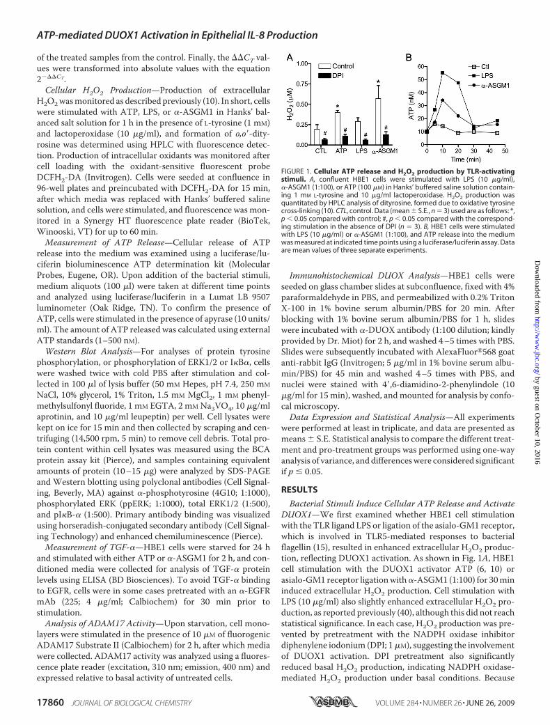
Bacterial Stimuli Induce Cellular ATP Release and ActivateDUOX1—We first examined whether HBE1 cell stimulationwith the TLR ligand LPS or ligation of the asialo-GM1 receptor,which is involved in TLR5-mediated responses to bacterialflagellin (15), resulted in enhanced extracellular H2O2 produc-tion, reflecting DUOX1 activation. As shown in Fig. 1A, HBE1cell stimulation with the DUOX1 activator ATP (6, 10) orasialo-GM1 receptor ligationwith�-ASGM1 (1:100) for 30mininduced extracellular H2O2 production. Cell stimulation withLPS (10 �g/ml) also slightly enhanced extracellular H2O2 pro-duction, as reported previously (40), although this did not reachstatistical significance. In each case, H2O2 production was pre-vented by pretreatment with the NADPH oxidase inhibitordiphenylene iodonium (DPI; 1�M), suggesting the involvementof DUOX1 activation. DPI pretreatment also significantlyreduced basal H2O2 production, indicating NADPH oxidase-mediated H2O2 production under basal conditions. Because
FIGURE 1. Cellular ATP release and H2O2 production by TLR-activatingstimuli. A, confluent HBE1 cells were stimulated with LPS (10 �g/ml),�-ASGM1 (1:100), or ATP (100 �M) in Hanks’ buffered saline solution contain-ing 1 mM L-tyrosine and 10 �g/ml lactoperoxidase. H2O2 production wasquantitated by HPLC analysis of dityrosine, formed due to oxidative tyrosinecross-linking (10). CTL, control. Data (mean � S.E., n � 3) used are as follows: *,p � 0.05 compared with control; #, p � 0.05 compared with the correspond-ing stimulation in the absence of DPI (n � 3). B, HBE1 cells were stimulatedwith LPS (10 �g/ml) or �-ASGM1 (1:100), and ATP release into the mediumwas measured at indicated time points using a luciferase/luciferin assay. Dataare mean values of three separate experiments.
ATP-mediated DUOX1 Activation in Epithelial IL-8 Production
17860 JOURNAL OF BIOLOGICAL CHEMISTRY VOLUME 284 • NUMBER 26 • JUNE 26, 2009
by guest on October 10, 2016
http://ww
w.jbc.org/
Dow
nloaded from
some bacterial stimuli are capable of inducing ATP releasefrom cells (15), we hypothesized that ATP release and puriner-gic receptor activation might present a commonmechanism ofDUOX1 activation and cell responses to diverse stimuli such asLPS and �-ASGM1. Indeed, stimulation of the HBE1 cells witheither LPS or �-ASGM1 resulted in a rapid and transientincrease of extracellular ATP (Fig. 1B), which was abolished inthe presence of the ATPase/ADPase apyrase (10 units/ml; notshown). Therefore, analogous to our recent studies demon-strating ATP release and DUOX1 activation in HBE1 cells inresponse to mechanical injury (10), these findings indicate thatbacterial stimuli can also promote cellular ATP release, whichsubsequentlymay contribute to DUOX1 activation and cellularH2O2 production.Bacterial Stimuli Induce IL-8 Expression by ATP-mediated
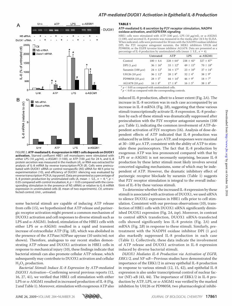
DUOX1 Activation—Confirming several previous reports (12,16, 27, 41), we verified that HBE1 cell stimulation with eitherLPS or �-ASGM1 resulted in increased production of IL-8 (Fig.2 andTable 1).Moreover, stimulationwith exogenousATPalso
induced IL-8 production, albeit to a lesser extent (Fig. 2A). Theincrease in IL-8 secretion was in each case accompanied by anincrease in IL-8 mRNA (Fig. 2B), suggesting that these variousstimuli transcriptionally activate IL-8 expression. IL-8 produc-tion by each of these stimuli was dramatically suppressed afterpreincubation with the P2Y receptor antagonist suramin (100�M; Table 1), indicating the common involvement of ATP-de-pendent activation of P2Y receptors (16). Analysis of dose-de-pendent effects of ATP indicated that IL-8 production wasenhanced by as little as 3�MATP, and responses weremaximalat 30–100 �M ATP, consistent with the ability of ATP to stim-ulate these purinoceptors. The fact that IL-8 production byexogenous ATP was less pronounced compared with that byLPS or �-ASGM1 is not necessarily surprising, because IL-8production by these latter stimuli most likely involves severalconcerted signaling mechanisms, some of which may be inde-pendent of ATP. However, the dramatic inhibitory effect ofpurinergic receptor blockade by suramin (Table 1) suggeststhat P2Y receptor stimulation is integral to the overall induc-tion of IL-8 by these various stimuli.To determinewhether the increased IL-8 expression by these
stimuli is associated with activation of DUOX1, we used siRNAto silence DUOX1 expression in HBE1 cells prior to cell stim-ulation. Consistent with our previous observations (10), trans-fection of HBE1 cells with DUOX1 siRNA significantly dimin-ished DUOX1 expression (Fig. 2A, top). Moreover, in contrastto control siRNA transfection, DUOX1 siRNA-transfectedcells showed significantly less IL-8 protein (Fig. 2A) andmRNA (Fig. 2B) in response to these stimuli. Similarly, pre-treatment with the NADPH oxidase inhibitor DPI (1 �M)also markedly suppressed IL-8 production in each case(Table 1). Collectively, these data indicate the involvementof ATP release and DUOX1 activation in IL-8 expressioninduced by diverse bacterial stimuli.DUOX1 Mediates IL-8 Production via Activation of EGFR,
ERK1/2, and NF-�B—Previous studies have demonstrated theimportance of the ERK1/2 in airway epithelial IL-8 productionin response to various stimuli (12, 15, 42), and epithelial IL-8expression is also under transcriptional control of nuclear fac-tor (NF)-�B (43, 44). The importance of ERK1/2 in IL-8 pro-duction by ATP, LPS, or �-ASGM1 was verified by the markedinhibition by U0126 or PD98058, two pharmacological inhibi-
FIGURE 2. ATP-mediated IL-8 expression in HBE1 cells depends on DUOX1activation. Starved confluent HBE1 cell monolayers were stimulated witheither LPS (10 �g/ml), �-ASGM1 (1:100), or ATP (100 �M) for 24 h, and IL-8protein secretion was measured in the medium (A), or RNA was extracted foranalysis of IL-8 mRNA by reverse transcription-PCR (B). Cells were preincu-bated with DUOX1 siRNA or control nonspecific (NS) siRNA for 48 h prior toexperimentation (10), and efficiency of DUOX1 silencing was evaluated byreverse transcription-PCR (A, top panel). Data are presented as a percentage ofIL-8 protein production by unstimulated cells (A, mean � S.E., n � 4; *, p �0.05 compared with control incubation; #, p � 0.05 compared with the corre-sponding stimulation in the presence of NS siRNA) or relative to IL-8 mRNAexpression in unstimulated cells (B, mean of two experiments). Ctl, untrans-fected control; Untr., untreated.
TABLE 1ATP-mediated IL-8 secretion by P2Y receptor stimulation, NADPHoxidase activation, and EGFR/ERK signalingHBE1 cells were stimulated with ATP (100 �M), LPS (10 �g/ml), or �-ASGM1(1:100), and secreted IL-8 protein was measured in the media after 24 h by ELISA.When indicated, cells were pretreated for 30minwith theNADPHoxidase inhibitorDPI, the P2Y receptor antagonist suramin, the MEK1 inhibitors U0126 andPD98058, or the EGFR tyrosine kinase inhibitor AG1478. Data are presented as apercentage of IL-8 production by unstimulated cells (mean � S.E., n � 4).
Untreated ATP LPS �-ASGM1Control 100 � 4.4 228 � 64a 238 � 45a 327 � 87a
DPI (1 �M) 36 � 16b 33 � 12b 49 � 15b 70 � 18b
Suramin (100 �M) 24 � 12b 34 � 17b 23 � 10b 37 � 14b
U0126 (10 �M) 36 � 12b 28 � 8b 32 � 4b 38 � 9b
PD98058 (10 �M) 28 � 5b 44 � 14b 46 � 9b 18 � 7b
AG1478 (10 �M) 16 � 8b 17 � 8b 16 � 5b 38 � 8ba p � 0.05 as compared with unstimulated cells.b p � 0.05 as compared with the corresponding controls.
ATP-mediated DUOX1 Activation in Epithelial IL-8 Production
JUNE 26, 2009 • VOLUME 284 • NUMBER 26 JOURNAL OF BIOLOGICAL CHEMISTRY 17861
by guest on October 10, 2016
http://ww
w.jbc.org/
Dow
nloaded from

tors of the upstream kinaseMEK1 that activates ERK1/2 (Table1). Following our recent studies showing that ATP-mediatedDUOX1 stimulation activates ERK1/2 in HBE1 cells (10), we
evaluated whether ATP as well asLPS or �-ASGM1 promote the acti-vation of ERK1/2 and NF-�B, andwhether this was the result ofDUOX1 activation. As shown in Fig.3A, Western blot analysis demon-strated significant activation ofERK1/2 by each stimulus, whichwas suppressed after cell transfec-tion with DUOX1 siRNA but not bycontrol NS siRNA (Fig. 3A). Simi-larly, each of these stimuli alsoenhanced the phosphorylation oftheNF-�B inhibitor I�B-�, illustrat-ing their ability to activate NF-�B,and this was again suppressed afterDUOX1 siRNA silencing (Fig. 3B).Activation of both ERK1/2 and
NF-�B in response to severaldiverse stimuli has been linked toactivation of EGFR (13, 45, 46), andrecent studies have implicatedDUOX1 in epithelial EGFR activa-tion and IL-8 production (12, 21).Accordingly, inhibition of EGFRtyrosine kinase activity usingAG1478 (1 �M) markedly decreasedIL-8 production induced by ATP,LPS, or �-ASGM1 (Table 1). Simi-larly, addition of AG1478 also sup-pressedATP-mediated phosphoryl-ation of ERK1/2 and I-�B� (Fig. 4A),indicating the involvement of EGFRactivation in these signaling events.Activation of EGFR or related tyro-sine kinases by these stimuli was
demonstrated more directly by Western blot analysis of tyro-sine-phosphorylated proteins. As shown in Fig. 4B, HBE1 cellstimulation with either ATP, LPS, or �-ASGM1 resulted inincreased phosphorylation of several proteins, including a pro-tein of�180 kDa,most likely representing EGFR autophospho-rylation on tyrosine residues. These increases in tyrosine phos-phorylation were in each case attenuated after DUOX1 siRNAsilencing, but not by NS siRNA, indicating the involvement ofDUOX1 in overall protein tyrosine phosphorylation and EGFRautophosphorylation and activation by these stimuli. Takentogether, these results indicate that ATP-mediated DUOX1activation promotes EGFR-dependent ERK and NF-�B activa-tion, which subsequently enhances IL-8 expression andsecretion.ATP/DUOX1 Activation Promotes TGF-� Production—
Upon demonstrating an important role for EGFR in the ATP/DUOX1-mediated IL-8 production, we assessed the potentialinvolvement of an EGFR ligand. Based on previous studiesimplicating TGF-� in IL-8 production (12, 30), we consideredthat ATP stimulation might promote TGF-� activation.Indeed, as shown in Fig. 5A, HBE1 cell stimulation with eitherATP or �-ASGM1 led to increased accumulation of TGF-� in
FIGURE 3. DUOX1 mediates ATP-mediated phosphorylation of ERK1/2 and I�B. Confluent starved HBE1cells were stimulated for 10 min with either ATP (100 �M), LPS (10 �g/ml), or �-ASGM1 (1:100), and harvestedfor Western blot analysis of phosphorylated and total ERK1/2 (A) or phospho-I�B (B). Where indicated, cellswere preincubated for 48 h with pre-designed DUOX1 siRNA and control siRNA (NS-siRNA) (10). Representativeblots and quantitative analysis by densitometry (n � 3) are shown. Ctr, untransfected control; Untr., untreated.
FIGURE 4. Role of EGFR in ATP-mediated signaling. A, HBE1 were stimulated for 10 min with ATP (100 �M) inthe absence or presence of the EGFR inhibitor AG1478 (10 �M), and cell lysates were collected for Western blotanalysis of phosphorylated and total ERK1/2 and I�B. B, HBE1 cells were preincubated with DUOX1 siRNA andNS siRNA and stimulated for 10 min with ATP, LPS, or �-ASGM1, as in Fig. 3, and cell lysates were analyzed fortyrosine-phosphorylated proteins, using �-PY antibody. A representative blot of three experiments is shown.Arrow indicates a major phosphorylated protein of �180 kDa, presumably representing EGFR. Untr., untreated.
FIGURE 5. ATP-mediated DUOX1 activation promotes TGF-� shedding inHBE1 cells. A, HBE1 cells were stimulated with either ATP or �-ASGM1 for 2 h,in the absence or presence of �-EGFR mAb (225; 4 �g/ml), and conditionedmedia were collected for analysis of TGF-� by ELISA. B, HBE1 cells werepreincubated with NS siRNA (white bars) or DUOX1 siRNA (black bars) for48 h and subsequently stimulated with ATP or �-ASGM1 in the presence of�-EGFR mAb for analysis of TGF-�. *, p � 0.05 compared with untreatedcontrol; #, p � 0.05 compared with the corresponding stimulation in theabsence of �-EGFR mAb (A) or in the presence of NS siRNA (B, n � 4). Ctl,untreated control.
ATP-mediated DUOX1 Activation in Epithelial IL-8 Production
17862 JOURNAL OF BIOLOGICAL CHEMISTRY VOLUME 284 • NUMBER 26 • JUNE 26, 2009
by guest on October 10, 2016
http://ww
w.jbc.org/
Dow
nloaded from

the media, although this was observed only when cells werepreincubated with a neutralizing EGFR antibody. This suggeststhat production of TGF-� by ATP or �-ASGM1 normallyengages in EGFR binding and activation, thereby preventing itsaccumulation in the media to detectable levels. Silencing ofDUOX1 attenuated the ATP- and �-ASGM1-induced TGF-�production observed in the presence of EGFR mAb (Fig. 5B),indicating the involvement of DUOX1 activation in ATP-in-duced TGF-� production and EGFR activation.DUOX1 Mediates ATP-dependent Activation of ADAM17—
Activation of EGFR ligands such as TGF-� is commonly medi-ated by activation of sheddases such as ADAM17, which cleavemembrane-bound forms of these ligands to promote EGFRactivation (47). We therefore evaluated whether HBE1 cellstimulation with ATP or �-ASGM1 results in activation ofADAM17, and whether this is mediated by DUOX1 activation.As shown in Fig. 6A, both ATP and �-ASGM1 indeed resultedin enhancedADAM17 activity,measured by increased cleavageof a fluorogenic ADAM17 substrate, and this was attenuatedafter siRNA silencing of DUOX1 (Fig. 6A) and in the presenceof the NADPH oxidase inhibitor DPI (data not shown). Thus,our findings are consistent with previous studies indicating arole for DUOX1 in epithelial ADAM17 activation (12, 13).The importance of P2Y purinergic receptor stimulation in
ADAM17 activation could be demonstrated by the ability ofsuramin to abolish the effects of both stimuli (Fig. 6B). Interest-ingly, ATP-mediated ADAM17 activation was also inhibited byblocking ERK signaling with U0126 (Fig. 6B), consistent with arole of ERK-mediated phosphorylation in ADAM17 activation(47, 48). Moreover, ATP-mediated ADAM17 activation wasalso suppressed by the EGFR kinase inhibitor AG1478 and, to alesser extent, by preincubationwith�-EGFR antibody (Fig. 6B),indicating a role for EGFR in ADAM17 activation.Role of H2O2 in DUOX1-mediated ADAM17 Activation and
IL-8 Production—Based on the presence of a common “cysteineswitch” consensus motif within the prodomain of matrix met-alloproteinases/ADAM proteases, it was recently suggestedthat DUOX1 activation promotes ADAM17 activation by
direct oxidation of the pro-ADAM17 cysteine at the cell surface(12, 13). However, the importance of the pro-domain cysteineswitch in regulation of ADAM17 activation has recently beenquestioned (34), and ADAM17 activity is also regulated byintracellular signaling mechanisms such as EGFR/ERK-medi-ated pathways (as indicated above) (47), which could be regu-lated by H2O2-mediated signaling. To address the involvementof either extracellular or intracellular H2O2 in DUOX1-dependent activation of ADAM17, we performed experimentsin the presence of catalase, which would rapidly detoxify extra-cellular H2O2. Alternatively, cells were preincubated with twostructurally unrelated cell-permeable peroxidase/catalasemimetics, ebselen and EUK134, to quench intracellular H2O2.As shown in Fig. 7, addition of catalase (up to 2,000 units/ml) toHBE1 cells failed to prevent ATP-mediated activation ofADAM17 (Fig. 7A) and did not affect production of IL-8 (Fig.7B). In contrast, addition of either ebselen or EUK134 signif-icantly attenuated ATP-induced ADAM17 activation as wellas IL-8 production (Fig. 7, A and B). Similar effects of cata-lase, ebselen, or EUK134 were observed on ADAM17 activa-tion and/or IL-8 production in response to LPS or �-ASGM1(data not shown). Hence, our results indicate that DUOX1-mediated activation of ADAM17 and EGFR/ERK signalingand production of IL-8 is primarily mediated by intracellularH2O2. Accordingly, analysis of DUOX protein within HBE1cells by confocal imaging revealed that DUOX is largely pres-ent intracellularly (Fig. 7C), presumably in association with
FIGURE 6. ATP-mediated activation of ADAM17 by activation of DUOX1-EGFR-ERK signaling. Confluent starved HBE1 cells were preincubated witheither NS siRNA or DUOX1 siRNA (A) or with the P2Y antagonist suramin (100�M), the MEK1 inhibitor U0126 (10 �M), or inhibitors EGFR (AG1478, 10 �M;�-EGFR mAb, 4 �g/ml) for 30 min each (B), and stimulated with ATP in thepresence of a fluorogenic ADAM17 substrate (Substrate II, Calbiochem; 10�M) for up to 2 h, and media were transferred to a fluorescence microplate foranalysis of fluorescence (excitation, 310 nm; emission, 400 nm). *, p � 0.05compared with control incubation; #, p � 0.05 compared with the corre-sponding incubation with NS siRNA or without inhibitor (n � 4 – 6). Ctr, con-trol; Untr., untreated.
FIGURE 7. Effects of H2O2 scavengers on ATP-mediated signaling. A andB, confluent HBE1 cells were preincubated with catalase (2000 units/ml) orthe peroxidase mimics ebselen (10 �M) or EUK134 (50 �M). B, for 30 min beforestimulation with ATP for 2 h in the presence of fluorogenic ADAM17 SubstrateII for analysis of ADAM17 activity (A) or 24 h for analysis of IL-8 production byELISA (B). Ctl, control; Untr., untreated. *, p � 0.05 compared with controlincubation; #, p � 0.05 compared with corresponding stimulation withoutantioxidant (n � 4). C, subconfluent HBE1 cells were fixed and stained with�-DUOX (red) and 4�,6-diamidino-2-phenylindole (blue) for confocal imaging.D, HBE1 cells in 24-well plates were preloaded with DCFH2-DA (10 �M; 15 min)and washed and stimulated with ATP, and fluorescence was monitored usinga fluorescence plate reader. A typical result of three experiments is shown.
ATP-mediated DUOX1 Activation in Epithelial IL-8 Production
JUNE 26, 2009 • VOLUME 284 • NUMBER 26 JOURNAL OF BIOLOGICAL CHEMISTRY 17863
by guest on October 10, 2016
http://ww
w.jbc.org/
Dow
nloaded from
intracellular membrane structures of the endoplasmic retic-ulum or Golgi. Moreover, ATP stimulation also resulted inintracellular oxidant production, increase 2�,7�-dichlo-rofluorescein of HBE1 preloaded with DCFH2-DA, suggest-ing intracellular production of H2O2 and related oxidants(Fig. 7D). Thus, ATP-mediated activation of intracellularDUOX1 appears to participate in cellular signaling eventsthat promote ADAM17 activation and production of IL-8.
DISCUSSION
The results described in this study offer several new insightsinto the functional role of the NADPH oxidase DUOX1 in epi-thelial host defense responses. First, this study highlights a gen-eral role ofATP release andP2Y receptor activation inDUOX1-dependent H2O2 production in response to diverse bacterialtriggers that act on various TLR receptors (41). As demon-strated, epithelial production of the neutrophil chemokine IL-8in response to the TLR4 ligand LPS or activation of the glyco-lipid receptor for bacterial flagellin, ASGM1, which associateswith TLR5 (16), involves the release of ATP and activation ofDUOX1. Thus, in addition to recent studies indicating directassociation of TLR4 with other NADPH oxidase homologs orMyD88-mediated phosphorylation of NOX activator proteinsin other cell types (49, 50), our studies indicate that TLR-medi-ated activation of epithelial DUOX1 is related to release of cel-lular ATP and P2Y receptor activation, which stimulatesDUOX1 by Ca2�-mediated signaling (6). However, the mecha-nisms by which these triggers provoke ATP release are stillunknown (16, 51).Second, our studies further address the mechanisms by
which P2Y receptor stimulation may lead to activation ofADAM17-EGFR signaling cascades that mediate the activationof various host defensemechanisms, including IL-8 production(52, 53), and establish a role for DUOX1 in this signaling path-way. Thus, in addition to participating in innate host defense byproviding H2O2 to produce antimicrobial oxidants (3), epithe-lial DUOX1 also appears to participate in additional hostresponses to infection, by mediating intracellular signalingpathways that result in production of neutrophil chemokinesand other inflammatory mediators (7, 10).Release of epithelial ATP in response to cell stimulation or
injury can promote a variety of epithelial responses throughactivation of P2X and P2Y receptor types that are expressed onthe epithelial surface (54, 55). Although we used extracellularATP at up to 100 �M to mimic these responses, which con-siderably exceeds levels of released ATP in response to cellstimulation with either LPS or �-ASGM1 (Fig. 1B), it isimportant to note that such measurements in the bulk mediatypically underestimate local concentrations of releasedATP at the cell surface that is often sufficient to activatepurinoceptors (10). Moreover, even though these concentra-tions of exogenous ATP can activate both P2X and P2Yreceptor subtypes (54), the inhibitory effects of the P2Y-selective antagonist suramin suggest that these responses aredue to stimulation of this receptor type. Moreover, studies inmice deficient in P2Y1 and P2Y2 purinergic receptors havedemonstrated the importance of these receptors in airwayhost defense against bacterial infection (56). Furthermore,
our previous studies (10) and recent studies by others (3, 6)indicate that epithelial H2O2 production by exogenous ATPis primarily mediated by P2Y receptor stimulation ofDUOX1, although we cannot fully rule out the potential con-tribution of other purinoceptors and alternative cellularsources of H2O2. The P2Y receptors belong to the family ofG-protein-coupled receptors that mediate signaling throughintracellular Ca2� and protein kinase C signaling (57).Although it is not precisely understood how P2Y stimulationactivates DUOX1, both Ca2� mobilization and proteinkinase C-activating stimuli have been shown to activateDUOX1-dependent H2O2 production in airway epithelialcells (6, 9). Recent studies indicated that basal DUOX1 activ-ity is suppressed by interaction with NOXA1 and that Ca2�-mobilizing stimuli promote NOXA1 dissociation fromDUOX1, and thereby enhance its NADPH oxidase activity(58).In agreementwith previous observations (9, 12, 13), our stud-
ies indicate that DUOX1 activation promotes the activation ofEGFR and subsequent ERK1/2 and NF-�B signaling pathways,both critical in mediating IL-8 expression (43, 44). Moreover,our studies also demonstrate that DUOX1 activation contrib-utes to the activation of ADAM17 and shedding of EGFRligands such as TGF-�. Although the activation of ADAM17and TGF-� shedding may contribute to EGFR activation andsubsequent IL-8 production (12, 13), it is still unclear howDUOX1 activation leads to ADAM17 activation, and the over-all regulation of ADAM17 activity is still incompletely under-stood (47). It was suggested that DUOX1 might directly medi-ate the activation of ADAM17 at the cell surface by oxidativedisruption of the cysteine switch motif within the ADAM17prodomain (13, 46), although direct evidence for such an oxi-dative activation mechanisms is lacking. Moreover, thishypothesis is also inconsistent with findings that ADAM17maturation andproteolytic processing occur intracellularly anddo not critically depend on the prodomain cysteine switch (34).To address the role of oxidative mechanisms in ATP-medi-
ated ADAM17 activation and subsequent IL-8 production, weused various extracellular or cellular antioxidant strategies.First, addition of the antioxidant enzyme catalase (up to 3,000units/ml) completely failed to prevent DUOX1-mediatedADAM17 activation and subsequent IL-8 production, arguingagainst a role for extracellular H2O2. However, pretreatmentwith the cell-permeable peroxidase mimics ebselen, andEUK134 partially suppressed these responses, indicating thecontribution of intracellular H2O2 or related oxidants in thissignaling cascade.In accordance with recent studies indicating the critical
importance of subcellular compartmentalization of NOX-de-rived oxidant production, and its role in EGFR activation orNF-�B signaling (59, 60), our results suggest that DUOX1 acti-vation not only generates extracellular H2O2 as amechanism ofepithelial antimicrobial defense (3, 6), but it also producesintracellular H2O2 or related oxidants to mediate intracellularsignaling mechanisms involved in EGFR activation and expres-sion of IL-8. In line with this notion, immunohistochemicalanalysis of HBE1 cells revealed that DUOX1 protein is largelydetected intracellularly (Fig. 7C), consistent with studies of thy-
ATP-mediated DUOX1 Activation in Epithelial IL-8 Production
17864 JOURNAL OF BIOLOGICAL CHEMISTRY VOLUME 284 • NUMBER 26 • JUNE 26, 2009
by guest on October 10, 2016
http://ww
w.jbc.org/
Dow
nloaded from

rocytes indicating that the bulk of DUOX1 protein is localizedto intracellular compartments (61). Although intracellularDUOX proteins are usually considered immature and inactive(61), intracellular DUOX has been reported to possess someintrinsic oxidant-producing activity (62). Accordingly, ATPstimulation of HBE1 cells enhanced intracellular oxidant pro-duction, as determined by 2�,7�-dichlorofluorescein fluores-cence (Fig. 7D), indicative of intracellular DUOX1 activation.Thus, although the innate host defense properties of airwayepithelial DUOX1 are commonly believed to depend on matu-rated DUOX1 protein at the cell surface (3, 6), further studieswill be required to determine the relative contribution of intra-cellular DUOX1 in this respect.Whereas activation of ADAM17 and EGFR ligand shedding
contribute importantly to autocrine EGFR activation and sub-sequent IL-8 production (12, 18), several reports suggest thatligand-independent EGFR activation by various stimuli pro-motes ADAM17-mediated shedding by ERK1/2-mediated Ser/Thr phosphorylation of ADAM17 (36, 48, 63). Accordingly, weobserved that ATP-mediated ADAM17 activation was dramat-ically blocked by inhibitors of ERK1/2 or EGFR tyrosine kinaseactivity, but not by a blocking �-EGFR antibody, thus suggest-ing a role for ligand-independent EGFR/ERK1/2 activation inADAM17 activation and EGFR ligand shedding. In this regard,various lines of evidence suggest that H2O2 can enhance EGFRactivation by ligand-independent mechanisms, including oxi-dative inactivation of protein-tyrosine phosphatases to sup-press EGFR dephosphorylation (59) and activation of the non-receptor tyrosine kinase c-Src, which interacts with andphosphorylates EGFR (64–66). Indeed, c-Src is involved inligand-independent activation of EGFR by several G-protein-coupled receptors, including P2Y2 purinergic receptors (67),and mediates epithelial IL-8 production in response to pro-tease-activated receptor stimulation (24). Therefore, we pro-pose that DUOX1-derived intracellular H2O2 mediates ATP-dependent EGFR activation by activating c-Src, analogous to itsactivation by other NADPH oxidases within endosomal com-partments, a prominent site of EGFR activation (64, 68). Inaddition, observations that ADAM17 activity may also bedirectly regulated by the protein-tyrosine phosphatase PTPH1(69) may suggest that intracellularly produced H2O2 could alsomore directly regulate ADAM17 activity and EGFR ligandshedding by inactivating PTPH1.Our collective results indicate that EGFR activation in
response to DUOX1 activation involves both ligand-dependentand -independent mechanisms, which may be required foramplified or prolonged EGFR activation to allow for appropri-ate activation of transcription factors and induction of IL-8 orother mediators (Fig. 8). Moreover, although some of these sig-naling events may be regulated by DUOX1-derived H2O2, itwas recently reported that H2O2-mediated, ligand-independ-ent activation of EGFR in airway epithelial cells may alsodepend on intracellular oxidant generation because ofincreased expression of NOX4 (70). This would tempt specula-tion that DUOX1-mediated activation of cell signaling path-ways might also involve indirect oxidative signaling eventsbecause of induction and activation of NOX4. Future studieswill be necessary to specifically address the importance of these
various oxidative signaling events in DUOX1-mediated EGFRactivation.Because epithelial production of IL-8 forms an integral
response in innate host defense as well as repair mechanismsin response to infection or injury, dysregulated productionof IL-8 or other mediators by inappropriate DUOX activa-tion may adversely affect these processes and contribute tolung disease pathology. For example, DUOX1 expression isdecreased in large airways of smokers and patients sufferingfrom mild or moderate chronic obstructive pulmonary dis-ease (71), and it may be suppressed in lung cancers (72).Conversely, DUOX1 expression can be enhanced by Th2cytokines such as IL-13 (10, 38), which can promoteADAM17 activation, TGF-� shedding, and subsequent epi-thelial cell proliferation (73), and may thereby contribute toepithelial hypertrophy and other features associated withairway remodeling in allergic asthma.In summary, our present results demonstrate that ATP-me-
diated activation of epithelial DUOX1 represents a generalresponse mechanism to diverse bacterial triggers that activatedifferent TLR receptors, as illustrated by enhanced IL-8 pro-duction. Activation of epithelial DUOX1 augments pro-inflam-matory ADAM17-TGF-�-ERK-EGFR cascades, which appearsto be initiated by H2O2-mediated, ligand-independent EGFRactivation. As such, epithelial DUOX1 is involved in innateimmune responses in addition to direct antimicrobial extracel-lular oxidant production.
Acknowledgments—We thank Derek Sham for technical assistance.
REFERENCES1. Knight, D. A., and Holgate, S. T. (2003) Respirology 8, 432–4462. van der Vliet, A. (2008) Free Radic. Biol. Med. 44, 938–9553. Moskwa, P., Lorentzen, D., Excoffon, K. J., Zabner, J., McCray, P. B., Jr.,
Nauseef, W. M., Dupuy, C., and Banfi, B. (2007) Am. J. Respir. Crit. Care
FIGURE 8. Schematic representation of DUOX1 involvement in TLR-medi-ated IL-8 production in response to bacterial stimuli. Cell stimulation withLPS or �-ASGM1 mediates release of ATP, which activates EGFR-ERK1/2-NF-�B signaling cascades which is amplified by ADAM17-TGF-� activationcascade. Intracellular production of H2O2 by DUOX1 promotes this signalingpathway by ligand-independent EGFR activation or activation of upstreamtyrosine kinases such as Src.
ATP-mediated DUOX1 Activation in Epithelial IL-8 Production
JUNE 26, 2009 • VOLUME 284 • NUMBER 26 JOURNAL OF BIOLOGICAL CHEMISTRY 17865
by guest on October 10, 2016
http://ww
w.jbc.org/
Dow
nloaded from

Med. 175, 174–1834. Fischer, H., Gonzales, L. K., Kolla, V., Schwarzer, C., Miot, F., Illek, B., and
Ballard, P. L. (2007) Am. J. Physiol. Lung Cell Mol. Physiol. 292,L1506–1514
5. Geiszt, M., Witta, J., Baffi, J., Lekstrom, K., and Leto, T. L. (2003) FASEB J.17, 1502–1504
6. Forteza, R., Salathe, M., Miot, F., Forteza, R., and Conner, G. E. (2005)Am. J. Respir. Cell Mol. Biol. 32, 462–469
7. Koff, J. L., Shao, M. X., Ueki, I. F., and Nadel, J. A. (2008) Am. J. Physiol.Lung Cell Mol. Physiol. 294, L1068–1075
8. Kuwahara, I., Lillehoj, E. P., Koga, T., Isohama, Y., Miyata, T., and Kim,K. C. (2007) Am. J. Respir. Cell Mol. Biol. 37, 691–698
9. Shao, M. X., and Nadel, J. A. (2005) Proc. Natl. Acad. Sci. U.S.A. 102,767–772
10. Wesley, U. V., Bove, P. F., Hristova,M.,McCarthy, S., and van der Vliet, A.(2007) J. Biol. Chem. 282, 3213–3220
11. Coraux, C.,Martinella-Catusse, C., Nawrocki-Raby, B., Hajj, R., Burlet, H.,Escotte, S., Laplace, V., Birembaut, P., and Puchelle, E. (2005) J. Pathol.206, 160–169
12. Nakanaga, T., Nadel, J. A., Ueki, I. F., Koff, J. L., and Shao, M. X. (2007)Am. J. Physiol. Lung Cell Mol. Physiol. 292, L1289–1296
13. Kuwahara, I., Lillehoj, E. P., Lu,W., Singh, I. S., Isohama, Y.,Miyata, T., andKim, K. C. (2006) Am. J. Physiol. Lung Cell Mol. Physiol. 291, L407–416
14. Fu, Z., Bettega, K., Carroll, S., Buchholz, K. R., and Machen, T. E. (2007)Am. J. Physiol. Lung Cell Mol. Physiol. 292, L353–364
15. McNamara, N., Khong, A., McKemy, D., Caterina, M., Boyer, J., Julius, D.,and Basbaum, C. (2001) Proc. Natl. Acad. Sci. U.S.A. 98, 9086–9091
16. McNamara, N., Gallup, M., Sucher, A., Maltseva, I., McKemy, D., andBasbaum, C. (2006) Am. J. Respir. Cell Mol. Biol. 34, 653–660
17. Gomez, M. I., and Prince, A. (2008) Pediatr. Pulmonol. 43, 11–1918. Zhao, Y., He,D., Saatian, B.,Watkins, T., Spannhake, E.W., Pyne,N. J., and
Natarajan, V. (2006) J. Biol. Chem. 281, 19501–1951119. Biagioli, M. C., Kaul, P., Singh, I., and Turner, R. B. (1999) Free Radic. Biol.
Med. 26, 454–46220. Look, D. C., Stoll, L. L., Romig, S. A., Humlicek, A., Britigan, B. E., and
Denning, G. M. (2005) J. Immunol. 175, 4017–402321. Koff, J. L., Shao, M. X., Ueki, I. F., and Nadel, J. A. (2008) Am. J. Physiol.
Lung. Cell Mol. Physiol. 294, L1068–107522. Pelaia, G., Cuda, G., Vatrella, A., Gallelli, L., Fratto, D., Gioffre, V.,
D’Agostino, B., Caputi, M., Maselli, R., Rossi, F., Costanzo, F. S., andMar-sico, S. A. (2004) J. Cell. Biochem. 93, 142–152
23. Li, J., Kartha, S., Iasvovskaia, S., Tan, A., Bhat, R. K.,Manaligod, J.M., Page,K., Brasier, A. R., and Hershenson, M. B. (2002) Am. J. Physiol. Lung CellMol. Physiol. 283, L690–699
24. Lin, C.H., Cheng,H.W.,Hsu,M. J., Chen,M.C., Lin, C. C., andChen, B. C.(2006) J. Immunol. 177, 3427–3438
25. Strieter, R. M. (2002) Am. J. Physiol. Lung Cell Mol. Physiol. 283,L688–689
26. Yu, Y., Nagai, S.,Wu, H., Neish, A. S., Koyasu, S., andGewirtz, A. T. (2006)J. Immunol. 176, 6194–6201
27. Li, J., Johnson, X. D., Iazvovskaia, S., Tan, A., Lin, A., and Hershenson,M. B. (2003) Am. J. Physiol. Lung Cell Mol. Physiol. 284, L307–315
28. Sethi, G., Ahn, K. S., Chaturvedi, M. M., and Aggarwal, B. B. (2007)Onco-gene 26, 7324–7332
29. Sahin, U., Weskamp, G., Kelly, K., Zhou, H.M., Higashiyama, S., Peschon,J., Hartmann, D., Saftig, P., and Blobel, C. P. (2004) J. Cell Biol. 164,769–779
30. Richter, A., O’Donnell, R. A., Powell, R.M., Sanders,M.W., Holgate, S. T.,Djukanovic, R., and Davies, D. E. (2002) Am. J. Respir. Cell Mol. Biol. 27,85–90
31. Hamilton, L. M., Puddicombe, S. M., Dearman, R. J., Kimber, I., Sand-strom, T., Wallin, A., Howarth, P. H., Holgate, S. T., Wilson, S. J., andDavies, D. E. (2005) Eur. Respir. J. 25, 978–985
32. Schlondorff, J., Becherer, J. D., and Blobel, C. P. (2000) Biochem. J. 347,131–138
33. Leonard, J. D., Lin, F., and Milla, M. E. (2005) Biochem. J. 387, 797–80534. Milla, M. E., Gonzales, P. E., and Leonard, J. D. (2006) Cell Biochem. Bio-
phys. 44, 342–348
35. Fan, H., and Derynck, R. (1999) EMBO J. 18, 6962–697236. Gomez, M. I., Seaghdha, M. O., and Prince, A. S. (2007) EMBO J. 26,
701–70937. Soond, S. M., Everson, B., Riches, D.W., andMurphy, G. (2005) J. Cell Sci.
118, 2371–238038. Harper, R. W., Xu, C., Eiserich, J. P., Chen, Y., Kao, C. Y., Thai, P., Setiadi,
H., and Wu, R. (2005) FEBS Lett. 579, 4911–491739. Bevelander, M., Mayette, J., Whittaker, L. A., Paveglio, S. A., Jones, C. C.,
Robbins, J., Hemenway, D., Akira, S., Uematsu, S., and Poynter, M. E.(2007) J. Immunol. 179, 3680–3688
40. Koff, J. L., Shao, M. X., Kim, S., Ueki, I. F., and Nadel, J. A. (2006) J. Immu-nol. 177, 8693–8700
41. Sha, Q., Truong-Tran, A. Q., Plitt, J. R., Beck, L. A., and Schleimer, R. P.(2004) Am. J. Respir. Cell Mol. Biol. 31, 358–364
42. Tephly, L. A., and Carter, A. B. (2007) Am. J. Physiol. Lung Cell Mol.Physiol. 293, L1143–1155
43. Joseph, T., Look, D., and Ferkol, T. (2005) Am. J. Physiol. Lung Cell Mol.Physiol. 288, L471–479
44. Roger, T., Out, T., Mukaida, N., Matsushima, K., Jansen, H., and Lutter, R.(1998) Biochem. J. 330, 429–435
45. Liu, K., Anderson, G. P., and Bozinovski, S. (2008) Am. J. Respir. Cell Mol.Biol. 39, 305–311
46. Shao, M. X., Ueki, I. F., and Nadel, J. A. (2003) Proc. Natl. Acad. Sci. U.S.A.100, 11618–11623
47. Blobel, C. P. (2005) Nat. Rev. Mol. Cell Biol. 6, 32–4348. Fan, H., Turck, C. W., and Derynck, R. (2003) J. Biol. Chem. 278,
18617–1862749. Park, H. S., Jung, H. Y., Park, E. Y., Kim, J., Lee, W. J., and Bae, Y. S. (2004)
J. Immunol. 173, 3589–359350. Laroux, F. S., Romero, X., Wetzler, L., Engel, P., and Terhorst, C. (2005)
J. Immunol. 175, 5596–560051. Lazarowski, E. R., Boucher, R. C., and Harden, T. K. (2003)Mol. Pharma-
col. 64, 785–79552. Buvinic, S., Bravo-Zehnder, M., Boyer, J. L., Huidobro-Toro, J. P., and
Gonzalez, A. (2007) J. Cell Sci. 120, 4289–430153. Boucher, I., Yang, L., Mayo, C., Klepeis, V., and Trinkaus-Randall, V.
(2007) Exp. Eye Res. 85, 130–14154. Schwiebert, E. M., and Zsembery, A. (2003) Biochim. Biophys. Acta 1615,
7–3255. Communi, D., Paindavoine, P., Place, G. A., Parmentier, M., and Boey-
naems, J. M. (1999) Br. J. Pharmacol. 127, 562–56856. Geary, C., Akinbi, H., Korfhagen, T., Fabre, J. E., Boucher, R., and Rice,W.
(2005) Am. J. Physiol. Lung Cell Mol. Physiol. 289, L890–89557. Ahmad, S., Ahmad, A., andWhite, C.W. (2006) Free Radic. Biol. Med. 41,
29–4058. Pacquelet, S., Lehmann, M., Luxen, S., Regazzoni, K., Frausto, M., Noack,
D., and Knaus, U. G. (2008) J. Biol. Chem. 283, 24649–2465859. Chen, K., Kirber, M. T., Xiao, H., Yang, Y., and Keaney, J. F., Jr. (2008)
J. Cell Biol. 181, 1129–113960. Li, Q., Harraz, M. M., Zhou, W., Zhang, L. N., Ding, W., Zhang, Y., Egg-
leston, T., Yeaman, C., Banfi, B., and Engelhardt, J. F. (2006)Mol. Cell. Biol.26, 140–154
61. Song, Y., Driessens, N., Costa,M., DeDeken, X., Detours, V., Corvilain, B.,Maenhaut, C., Miot, F., Van Sande, J., Many, M. C., and Dumont, J. E.(2007) J. Clin. Endocrinol. Metab. 92, 3764–3773
62. Ameziane-El-Hassani, R., Morand, S., Boucher, J. L., Frapart, Y.M., Apos-tolou, D., Agnandji, D., Gnidehou, S., Ohayon, R., Noel-Hudson, M. S.,Francon, J., Lalaoui, K., Virion, A., andDupuy, C. (2005) J. Biol. Chem. 280,30046–30054
63. Zhang, Q., Thomas, S. M., Lui, V. W., Xi, S., Siegfried, J. M., Fan, H.,Smithgall, T. E., Mills, G. B., and Grandis, J. R. (2006) Proc. Natl. Acad. Sci.U.S.A. 103, 6901–6906
64. Donepudi, M., and Resh, M. D. (2008) Cell. Signal. 20, 1359–136765. Zhuang, S., Schnellmann, R. G., and Zhougang, S. (2004) Am. J. Physiol.
Renal Physiol. 286, F858–86566. Khan, E. M., Heidinger, J. M., Levy, M., Lisanti, M. P., Ravid, T., and
Goldkorn, T. (2006) J. Biol. Chem. 281, 14486–1449367. Liu, J., Liao, Z., Camden, J., Griffin, K. D., Garrad, R. C., Santiago-Perez,
ATP-mediated DUOX1 Activation in Epithelial IL-8 Production
17866 JOURNAL OF BIOLOGICAL CHEMISTRY VOLUME 284 • NUMBER 26 • JUNE 26, 2009
by guest on October 10, 2016
http://ww
w.jbc.org/
Dow
nloaded from
L. I., Gonzalez, F. A., Seye, C. I., Weisman, G. A., and Erb, L. (2004) J. Biol.Chem. 279, 8212–8218
68. Li, Q., Zhang, Y., Marden, J. J., Banfi, B., and Engelhardt, J. F. (2008) Bio-chem. J. 411, 531–541
69. Zheng, Y., Schlondorff, J., and Blobel, C. P. (2002) J. Biol. Chem. 277,42463–42470
70. Kim, H. J., Park, Y. D., Moon, U. Y., Kim, J. H., Jeon, J. H., Lee, J. G., Bae,
Y. S., and Yoon, J. H. (2008) Am. J. Respir. Cell Mol. Biol. 39, 598–60971. Nagai, K., Betsuyaku, T., Suzuki,M., Nasuhara, Y., Kaga, K., Kondo, S., and
Nishimura, M. (2008) Antioxid. Redox. Signal. 10, 705–71472. Luxen, S., Belinsky, S. A., and Knaus, U. G. (2008) Cancer Res. 68,
1037–104573. Booth, B. W., Sandifer, T., Martin, E. L., and Martin, L. D. (2007) Respir.
Res. 8, 51
ATP-mediated DUOX1 Activation in Epithelial IL-8 Production
JUNE 26, 2009 • VOLUME 284 • NUMBER 26 JOURNAL OF BIOLOGICAL CHEMISTRY 17867
by guest on October 10, 2016
http://ww
w.jbc.org/
Dow
nloaded from

Bast and Albert van der VlietAgnes W. Boots, Milena Hristova, David I. Kasahara, Guido R. M. M. Haenen, Aalt
Epithelial Responses to Bacterial StimuliATP-mediated Activation of the NADPH Oxidase DUOX1 Mediates Airway
doi: 10.1074/jbc.M809761200 originally published online April 21, 20092009, 284:17858-17867.J. Biol. Chem.
10.1074/jbc.M809761200Access the most updated version of this article at doi:
Alerts:
When a correction for this article is posted•
When this article is cited•
to choose from all of JBC's e-mail alertsClick here
http://www.jbc.org/content/284/26/17858.full.html#ref-list-1
This article cites 73 references, 45 of which can be accessed free at
by guest on October 10, 2016
http://ww
w.jbc.org/
Dow
nloaded from