Fibrotic response induced by angiotensin-II requires NAD(P)H oxidase-induced reactive oxygen species...
6
Fibrotic response induced by angiotensin-II requires NAD(P)H oxidase-induced reactive oxygen species (ROS) in skeletal muscle cells Claudio Cabello-Verrugio a,b,⇑ , María José Acuña b , María Gabriela Morales b , Alvaro Becerra c , Felipe Simon c , Enrique Brandan b a Centro de Genética Humana (CGH), Facultad de Medicina-Clínica Alemana de Santiago, Universidad del Desarrollo, Santiago, Chile b Centro de Regulación Celular y Patología (CRCP), Centro de Regeneración y Envejecimiento (CARE), Departamento de Biología Celular y Molecular, Facultad de Ciencias Biológicas, Pontificia Universidad Católica de Chile, Santiago, Chile c Laboratorio de Fisiopatología Celular y Molecular, Departamento de Ciencias Biológicas, Facultad de Ciencias Biológicas and Facultad de Medicina, Universidad Andres Bello, Santiago, Chile article info Article history: Received 3 June 2011 Available online 12 June 2011 Keywords: Angiotensin-II Fibrosis NAD(P)H oxidase Skeletal muscle Reactive oxygen species (ROS) AT-1 receptor abstract Fibrotic disorders are typified by excessive connective tissue and extracellular matrix (ECM) deposition that precludes normal healing processes in different tissues. Angiotensin-II (Ang-II) is involved in the fibrotic response. Several muscular dystrophies are characterized by extensive fibrosis. However, the exact role of Ang-II in skeletal muscle fibrosis is unknown. Here we show that myoblasts responded to Ang-II by increasing protein levels of connective tissue growth factor (CTGF/CCN2), collagen-III and fibronectin. These Ang-II-induced pro-fibrotic effects were mediated by AT-1 receptors. Remark- ably, Ang-II induced reactive oxygen species (ROS) via a NAD(P)H oxidase-dependent mechanism, as shown by inhibition of ROS production via the NAD(P)H oxidase inhibitors diphenylene iodonium (DPI) and apocynin. This increase in ROS is critical for Ang-II-induced fibrotic effects, as indicated by the decrease in Ang-II-induced CTGF and fibronectin levels by DPI and apocynin. We also show that Ang-II-induced ROS production and fibrosis require PKC activity as indicated by the generic PKC inhib- itor chelerythrine. These results strongly suggest that the fibrotic response induced by Ang-II is mediated by AT-1 receptor and requires NAD(P)H-induced ROS in skeletal muscle cells. Ó 2011 Elsevier Inc. All rights reserved. 1. Introduction Fibrotic disorders are typically characterized by excessive con- nective tissue and extracellular matrix (ECM) deposition that pre- cludes normal healing in different tissues. A marked increase in angiotensin-II (Ang-II) levels has been linked to the pathogenesis of fibrotic disorders in tissues such as liver, cardiac muscle and kid- ney [1–3]. Ang-II is a potent inducer of ECM proteins such as colla- gen-III and fibronectin, and of CTGF expression through the AT-1 receptor [4–6]. Reactive oxygen species (ROS) have been implicated in the sig- nal transduction of Ang-II-dependent cellular responses via activa- tion of redox-sensitive signaling cascades [1,7]. One of the main sources of ROS is NAD(P)H oxidase (NOX), a multi-protein enzyme complex that uses NAD(P)H as a substrate to convert molecular oxygen to ROS [8]. One of the main activation mechanisms of NOX is the phosphorylation of the regulatory subunit p47 phox by the Ca 2+ -dependent protein kinase C (PKC) [9]. Different experimental evidence suggests the involvement of NAD(P)H oxidase-1 and NAD(P)H oxidase-2 in fibrotic disease in several tissues among them skeletal muscle [10]. Ang-II activates NAD(P)H oxidase in several cell types [11]. Furthermore, NAD(P)H oxidase activation and increased ROS production are implicated in Ang-II-induced effects such as vascular smooth muscle hypertro- phy, hypertension and cardiac and liver fibrosis [12,13]. Several muscular dystrophies, among them Duchenne muscular dystrophy (DMD) and its murine model, mdx mice, are characterized by development fibrosis [14,15]. Among the cell types that contrib- ute to fibrotic response in skeletal muscle are the myoblasts [16,17]. Indeed, ROS were postulated to contribute to the pathogenesis of DMD [18,19]. In addition, it has recently been reported that NAD(P)H oxidase protein levels and activity are increased in mdx muscle, being a major source of ROS production in dystrophic muscle [19]. 0006-291X/$ - see front matter Ó 2011 Elsevier Inc. All rights reserved. doi:10.1016/j.bbrc.2011.06.051 Abbreviations: Ang-II, angiotensin-II; AT-1, angiotensin-II receptor type 1; ARB, angiotensin-II receptor type I blocker; CTGF/CCN2, connective tissue growth factor; DTT, dithiothreitol; DMD, Duchenne muscular dystrophy; ECM, extracellular matrix; NOX, NAD(P)H oxidase; ROS, reactive oxygen species; TGF-b, transforming growth factor type-b. ⇑ Corresponding author at: Centro de Genética Humana (CGH), Facultad de Medicina-Clínica Alemana de Santiago, Universidad del Desarrollo, Código Postal 7710162, Santiago, Chile. Fax: +56 2 3279306. E-mail address: [email protected] (C. Cabello-Verrugio). Biochemical and Biophysical Research Communications 410 (2011) 665–670 Contents lists available at ScienceDirect Biochemical and Biophysical Research Communications journal homepage: www.elsevier.com/locate/ybbrc
Transcript of Fibrotic response induced by angiotensin-II requires NAD(P)H oxidase-induced reactive oxygen species...

Biochemical and Biophysical Research Communications 410 (2011) 665–670
Contents lists available at ScienceDirect
Biochemical and Biophysical Research Communications
journal homepage: www.elsevier .com/locate /ybbrc
Fibrotic response induced by angiotensin-II requires NAD(P)H oxidase-inducedreactive oxygen species (ROS) in skeletal muscle cells
Claudio Cabello-Verrugio a,b,⇑, María José Acuña b, María Gabriela Morales b, Alvaro Becerra c, Felipe Simon c,Enrique Brandan b
a Centro de Genética Humana (CGH), Facultad de Medicina-Clínica Alemana de Santiago, Universidad del Desarrollo, Santiago, Chileb Centro de Regulación Celular y Patología (CRCP), Centro de Regeneración y Envejecimiento (CARE), Departamento de Biología Celular y Molecular, Facultad de Ciencias Biológicas,Pontificia Universidad Católica de Chile, Santiago, Chilec Laboratorio de Fisiopatología Celular y Molecular, Departamento de Ciencias Biológicas, Facultad de Ciencias Biológicas and Facultad de Medicina, Universidad Andres Bello,Santiago, Chile
a r t i c l e i n f o
Article history:Received 3 June 2011Available online 12 June 2011
Keywords:Angiotensin-IIFibrosisNAD(P)H oxidaseSkeletal muscleReactive oxygen species (ROS)AT-1 receptor
0006-291X/$ - see front matter � 2011 Elsevier Inc. Adoi:10.1016/j.bbrc.2011.06.051
Abbreviations: Ang-II, angiotensin-II; AT-1, angioteangiotensin-II receptor type I blocker; CTGF/CCN2, conDTT, dithiothreitol; DMD, Duchenne muscular dysmatrix; NOX, NAD(P)H oxidase; ROS, reactive oxygengrowth factor type-b.⇑ Corresponding author at: Centro de Genética H
Medicina-Clínica Alemana de Santiago, Universidad7710162, Santiago, Chile. Fax: +56 2 3279306.
E-mail address: [email protected] (C. Cabello-Verrug
a b s t r a c t
Fibrotic disorders are typified by excessive connective tissue and extracellular matrix (ECM) depositionthat precludes normal healing processes in different tissues. Angiotensin-II (Ang-II) is involved in thefibrotic response. Several muscular dystrophies are characterized by extensive fibrosis. However, theexact role of Ang-II in skeletal muscle fibrosis is unknown. Here we show that myoblasts respondedto Ang-II by increasing protein levels of connective tissue growth factor (CTGF/CCN2), collagen-IIIand fibronectin. These Ang-II-induced pro-fibrotic effects were mediated by AT-1 receptors. Remark-ably, Ang-II induced reactive oxygen species (ROS) via a NAD(P)H oxidase-dependent mechanism, asshown by inhibition of ROS production via the NAD(P)H oxidase inhibitors diphenylene iodonium(DPI) and apocynin. This increase in ROS is critical for Ang-II-induced fibrotic effects, as indicated bythe decrease in Ang-II-induced CTGF and fibronectin levels by DPI and apocynin. We also show thatAng-II-induced ROS production and fibrosis require PKC activity as indicated by the generic PKC inhib-itor chelerythrine.
These results strongly suggest that the fibrotic response induced by Ang-II is mediated by AT-1receptor and requires NAD(P)H-induced ROS in skeletal muscle cells.
� 2011 Elsevier Inc. All rights reserved.
1. Introduction tion of redox-sensitive signaling cascades [1,7]. One of the main
Fibrotic disorders are typically characterized by excessive con-nective tissue and extracellular matrix (ECM) deposition that pre-cludes normal healing in different tissues. A marked increase inangiotensin-II (Ang-II) levels has been linked to the pathogenesisof fibrotic disorders in tissues such as liver, cardiac muscle and kid-ney [1–3]. Ang-II is a potent inducer of ECM proteins such as colla-gen-III and fibronectin, and of CTGF expression through the AT-1receptor [4–6].
Reactive oxygen species (ROS) have been implicated in the sig-nal transduction of Ang-II-dependent cellular responses via activa-
ll rights reserved.
nsin-II receptor type 1; ARB,nective tissue growth factor;trophy; ECM, extracellularspecies; TGF-b, transforming
umana (CGH), Facultad dedel Desarrollo, Código Postal
io).
sources of ROS is NAD(P)H oxidase (NOX), a multi-protein enzymecomplex that uses NAD(P)H as a substrate to convert molecularoxygen to ROS [8]. One of the main activation mechanisms ofNOX is the phosphorylation of the regulatory subunit p47phox bythe Ca2+-dependent protein kinase C (PKC) [9].
Different experimental evidence suggests the involvement ofNAD(P)H oxidase-1 and NAD(P)H oxidase-2 in fibrotic disease inseveral tissues among them skeletal muscle [10]. Ang-II activatesNAD(P)H oxidase in several cell types [11]. Furthermore, NAD(P)Hoxidase activation and increased ROS production are implicated inAng-II-induced effects such as vascular smooth muscle hypertro-phy, hypertension and cardiac and liver fibrosis [12,13].
Several muscular dystrophies, among them Duchenne musculardystrophy (DMD) and its murine model, mdx mice, are characterizedby development fibrosis [14,15]. Among the cell types that contrib-ute to fibrotic response in skeletal muscle are the myoblasts [16,17].Indeed, ROS were postulated to contribute to the pathogenesis ofDMD [18,19]. In addition, it has recently been reported that NAD(P)Hoxidase protein levels and activity are increased in mdx muscle,being a major source of ROS production in dystrophic muscle [19].

Fig. 1. Angiotensin-II induces fibrosis via an AT-1-dependent mechanism in skeletal muscle cells. (A) C2C12 myoblasts were incubated with Ang-II (500 nM), TGF-b1 (10 ng/ml) or CTGF (80 ng/ml) for 48 h. The extracts obtained were separated by SDS–PAGE. The presence of collagen-III (Col-III) was evaluated by Western blot using anti-Col-IIIantibody. Levels of GAPDH are shown as a loading control. (B) C2C12 cells were pre-incubated with ARB ZD-7155 (10 lM) and then incubated with Ang-II (500 nM) as in A.Levels of collagen-III (Col-III) and fibronectin (FN) were determined by Western blot. Levels of GAPDH are shown as a loading control. (C) Myoblasts were incubated with Ang-II (500 nM) or TGF-b1 (5 ng/ml) for 1 and 6 h respectively. Total RNA was isolated, blotted and probed with cDNA for mouse CTGF and tubulin as a loading control. The sizes ofCTGF and tubulin mRNAs are shown in kilobases (kb). (D) C2C12 cells were pre-treated with ZD-7155 (10 lM) and incubated with Ang-II as in (A). Levels of CTGF protein weredetermined by Western blot. Levels of GAPDH are shown as a loading control. (E) Quantitative analysis of experiments shown in (B and D). The graph shows the fold ofinduction relative to control treatment. Values correspond to the mean ± standard deviation from three independent experiments (⁄, #, �P < 0.05). In A, B and D the molecularweight standards are indicated in kilodaltons (kDa).
666 C. Cabello-Verrugio et al. / Biochemical and Biophysical Research Communications 410 (2011) 665–670
In the present study, we therefore investigated the role of Ang-IIin skeletal muscle fibrosis using C2C12 myoblasts and the participa-tion of ROS as a key mediator. We demonstrated that Ang-IIinduced an increase in ECM and CTGF protein levels through itsAT-1 receptor. These pro-fibrotic effects of Ang-II were dependenton NAD(P)H oxidase-induced ROS generation. Indeed, the activa-tion of NAD(P)H oxidase was a Ca2+-dependent PKC-induced event.Thus, our results provide an insight into the fundamental mecha-nisms of Ang-II-induced fibrosis in skeletal muscle cells.
2. Materials and methods
2.1. Cell cultures
The skeletal muscle cell line C2C12, obtained from adult mouseleg (American Type Culture Collection), was grown as describedpreviously [20]. Cells were serum-starved for 18 h and then sub-jected to different treatments. For pro-fibrotic cytokine treatmentmyoblasts were incubated for 48 h with TGF-b1 (10 ng/ml) (R&DSystem, USA), recombinant CTGF (80 ng/ml) [16], H2O2 (Merck,
USA) or Ang-II (500 nM) (Sigma, USA). The following inhibitorswere pre-incubated for 1 h prior to exposure to Ang-II: AT-1 andAT-2 receptor blockers ZD-7155 (5 lM) and PD-123319 (10 lM)(both from Tocris, USA) respectively, dithiothreitol (DTT)(0.5 mM, Invitrogen, USA), NAD(P)H oxidase inhibitors diphenyl-ene iodonium (DPI) (0.1, 1 and 5 lM, Sigma, USA) or apocynin(1 mM, Sigma, USA), or PKC inhibitor chelerythrine (10 lV, Sigma,USA).
2.2. Measurement of intracellular ROS production and [Ca2+] by FACS
Treated C2C12 cells were harvested, resuspended, and loadedwith the following cell permeant dyes: dichlorodihydrofluorescein(DCF, 5 lM) or dihydroethdium (DHE, 10 lM) for ROS determina-tion, and Fura-3 (5 lM) for Ca2+ measurements (all from Invitro-gen, USA) for 15–30 min at room temperature in the absence oflight. They were then analyzed immediately by Fluorescence Acti-vated Cell Sorting (FACS) using a flow cytometry system (FACSCan-to, BD Biosciences, USA). A minimum of 10,000 cells were analyzedper sample. The cellular intensity of the dyes was analyzed usingFACSDiva software v4.1.1 (BD Biosciences, USA).

C. Cabello-Verrugio et al. / Biochemical and Biophysical Research Communications 410 (2011) 665–670 667
2.3. RNA isolation and Northern blot analysis
Myoblasts were serum-starved for 18 h and then incubated for1 h with Ang-II (500 nM) or for 6 h with TGF-b1 (10 ng/ml). TotalRNA was isolated from cell cultures at the indicated times usingTrizol (Invitrogen, USA). RNA samples (20 lg/lane) were electro-phoresed, transferred and hybridized with probes for mouse tubu-lin and CTGF as described previously [21].
2.4. Immunoblot analysis
For cell immunoblot analyses, protein extracts from myoblastswere prepared as previously described [16,17] and subjected toSDS–PAGE, electrophoretically transferred onto PDVF membranes(Schleicher & Schuell, USA) and probed with goat anti-CTGF (SantaCruz Biotechnology, USA), rabbit anti-fibronectin and mouse anti-tubulin (Sigma–Aldrich, USA), rabbit anti-collagen-III (Rockland,USA), and mouse anti-GAPDH (Chemicon, USA). All immunoreac-tions were visualized by enhanced chemiluminescence (Pierce,USA).
2.5. Protein determination
Proteins were determined in aliquots of cell extracts using thebicinchoninic acid protein assay kit (Pierce, USA) with BSA as thestandard.
2.6. Statistics
The statistical significance of the differences between themeans of the experimental groups was evaluated using one-way
Fig. 2. Reactive oxygen species (ROS) are induced by angiotensin-II via the AT-1 receptorexperiments of C2C12 cells exposed to vehicle alone (left upper panel), ZD-7155 alone (mAng-II + ZD-7155 (middle lower panel), or Ang-II + PD-123319 (right lower panel). Datahistograms above are summarized in (B and C) for DCF and DHE respectively. Bars represDCF (B) or DHE (C) fluorescence. (⁄, #, �P < 0.05).
analysis of variance (ANOVA) with a post hoc Bonferroni multi-ple-comparison test (Sigma Stat 3.5 Software). A difference wasconsidered statistically significant at a P value <0.05.
3. Results
3.1. Angiotensin-II-induced fibrotic response is AT-1 receptor-dependent in skeletal muscle cells
We studied the effect of Ang-II as a possible inducer of fibrosis inskeletal muscle cells. Fig. 1A and B show an increase in the amount ofcollagen-III and fibronectin of C2C12 cells in response to Ang-II. TGF-b1 and CTGF are showed as fibrotic factors increasing the levels ofcollagen-III and fibronectin [16,17]. Fig. 1B also shows that the in-crease in collagen-III and fibronectin induced by Ang-II in C2C12
myoblasts was prevented in presence of the Ang-II receptor type 1blocker (ARB) ZD-7155 suggesting that is mediated by AT-1 recep-tor. Fig. 1C and D show that Ang-II induces increased levels of CTGFmRNA and protein respectively. Fig. 1D also shows that Ang-II-in-duced CTGF levels are dependent on the AT-1 receptor. Fig. 1E showsthe quantitative analysis of the ARB ZD-7155 effect on Ang-II-in-duced collagen-III, fibronectin and CTGF levels.
Together, these results suggest that Ang-II induces pro-fibroticeffects in skeletal muscle cells.
3.2. Angiotensin-II-induced intracellular production of reactive oxygenspecies (ROS) is AT-1 receptor-dependent and participates in thefibrosis of skeletal muscle cells
We tested whether skeletal muscle cells increase their intracel-lular ROS production in response to Ang-II. By means of flow
in skeletal muscle cells. (A) Representative DCF fluorescence histograms from FACSiddle upper panel), PD-123319 alone (right upper panel), Ang-II (left lower panel),
obtained from three to four independent experiments such as those depicted in theent the percentage (mean ± standard deviation) of cells within the P1 population of
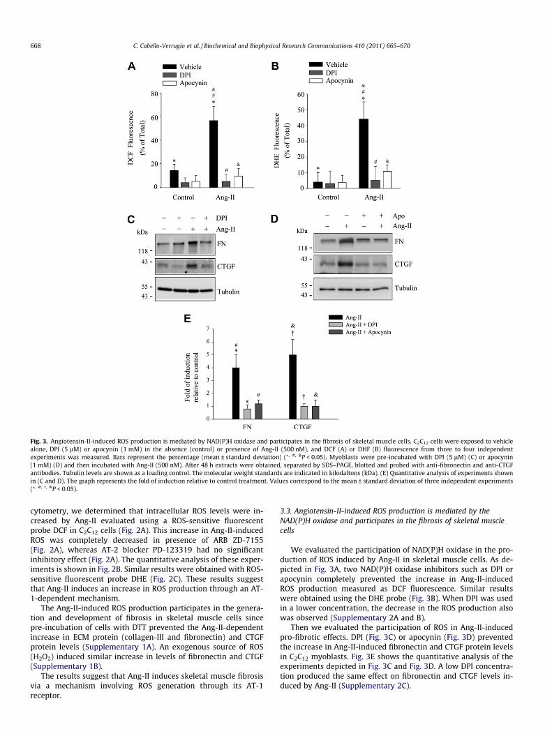
Fig. 3. Angiotensin-II-induced ROS production is mediated by NAD(P)H oxidase and participates in the fibrosis of skeletal muscle cells. C2C12 cells were exposed to vehiclealone, DPI (5 lM) or apocynin (1 mM) in the absence (control) or presence of Ang-II (500 nM), and DCF (A) or DHF (B) fluorescence from three to four independentexperiments was measured. Bars represent the percentage (mean ± standard deviation) (⁄, #, &P < 0.05). Myoblasts were pre-incubated with DPI (5 lM) (C) or apocynin(1 mM) (D) and then incubated with Ang-II (500 nM). After 48 h extracts were obtained, separated by SDS–PAGE, blotted and probed with anti-fibronectin and anti-CTGFantibodies. Tubulin levels are shown as a loading control. The molecular weight standards are indicated in kilodaltons (kDa). (E) Quantitative analysis of experiments shownin (C and D). The graph represents the fold of induction relative to control treatment. Values correspond to the mean ± standard deviation of three independent experiments(⁄, #, �, &P < 0.05).
668 C. Cabello-Verrugio et al. / Biochemical and Biophysical Research Communications 410 (2011) 665–670
cytometry, we determined that intracellular ROS levels were in-creased by Ang-II evaluated using a ROS-sensitive fluorescentprobe DCF in C2C12 cells (Fig. 2A). This increase in Ang-II-inducedROS was completely decreased in presence of ARB ZD-7155(Fig. 2A), whereas AT-2 blocker PD-123319 had no significantinhibitory effect (Fig. 2A). The quantitative analysis of these exper-iments is shown in Fig. 2B. Similar results were obtained with ROS-sensitive fluorescent probe DHE (Fig. 2C). These results suggestthat Ang-II induces an increase in ROS production through an AT-1-dependent mechanism.
The Ang-II-induced ROS production participates in the genera-tion and development of fibrosis in skeletal muscle cells sincepre-incubation of cells with DTT prevented the Ang-II-dependentincrease in ECM protein (collagen-III and fibronectin) and CTGFprotein levels (Supplementary 1A). An exogenous source of ROS(H2O2) induced similar increase in levels of fibronectin and CTGF(Supplementary 1B).
The results suggest that Ang-II induces skeletal muscle fibrosisvia a mechanism involving ROS generation through its AT-1receptor.
3.3. Angiotensin-II-induced ROS production is mediated by theNAD(P)H oxidase and participates in the fibrosis of skeletal musclecells
We evaluated the participation of NAD(P)H oxidase in the pro-duction of ROS induced by Ang-II in skeletal muscle cells. As de-picted in Fig. 3A, two NAD(P)H oxidase inhibitors such as DPI orapocynin completely prevented the increase in Ang-II-inducedROS production measured as DCF fluorescence. Similar resultswere obtained using the DHE probe (Fig. 3B). When DPI was usedin a lower concentration, the decrease in the ROS production alsowas observed (Supplementary 2A and B).
Then we evaluated the participation of ROS in Ang-II-inducedpro-fibrotic effects. DPI (Fig. 3C) or apocynin (Fig. 3D) preventedthe increase in Ang-II-induced fibronectin and CTGF protein levelsin C2C12 myoblasts. Fig. 3E shows the quantitative analysis of theexperiments depicted in Fig. 3C and Fig. 3D. A low DPI concentra-tion produced the same effect on fibronectin and CTGF levels in-duced by Ang-II (Supplementary 2C).

Fig. 4. Angiotensin-II-induced ROS production and fibrosis require PKC activity inskeletal muscle cells. C2C12 cells were exposed to vehicle alone or the generic PKCinhibitor chelerythrine (10 lM) in the absence (control) or presence of Ang-II, andDCF (A) or DHE (B) fluorescence from three to four independent experiments wasmeasured. Bars represent the percentage (mean ± standard deviation) (⁄, #P < 0.05).(C) Myoblasts were pre-incubated with chelerythrine (10 lM) and then incubatedwith Ang-II (500 nM). After 48 h extracts were obtained and fibronectin (FN) andCTGF protein levels were determined by Western blot analysis. Tubulin levels areshown as a loading control. The molecular weight standards are indicated inkilodaltons (kDa). The images are representative of two independent experiments.
C. Cabello-Verrugio et al. / Biochemical and Biophysical Research Communications 410 (2011) 665–670 669
These results strongly suggest that NAD(P)H oxidase-inducedROS is involved in the pro-fibrotic effects induced by Ang-II in skel-etal muscle cells.
3.4. Protein kinase C is required for angiotensin-II-inducedintracellular ROS production and fibrosis of skeletal muscle cells
We determined that inhibition of PKC activity by chelerythrineprevented Ang-II-induced ROS production in C2C12 cells, as evalu-ated using DCF (Fig. 4A) and DHE (Fig. 4B) probes. Also, we showthat inhibition of PKC reduced Ang-II-induced fibronectin andCTGF protein levels to basal levels in C2C12 myoblast (Fig. 4C).These results suggest that PKC activity is essential for mediatingAng-II-induced ROS production and fibrosis in skeletal musclecells.
Ang-II also increased levels of intracellular Ca2+ mediated byAT-1 receptor in C2C12 cells (Supplementary 3A) which was not
affected by DPI or apocynin, suggesting that this calcium increaseoccurs upstream of the NAD(P)H oxidase activity and ROS genera-tion induced by Ang-II (Supplementary 3B).
Together, these results suggest that PKC activity is required forinduction of fibrosis by Ang-II and required to activate NAD(P)Hoxidase.
4. Discussion
In this paper we show that Ang-II produces pro-fibrotic effectsin skeletal muscle cells through an AT-1-dependent mechanisminvolving the participation of ROS generated by NAD(P)H oxidase,which is activated by Ca2+-dependent PKC.
Skeletal muscles from DMD or its murine model mdx mice de-velop fibrosis [22,23]. These dystrophic skeletal muscles have in-creased levels of angiotensin-II converting enzyme (ACE) and AT-1 receptor compared to normal muscle [24]. According our resultsin vitro, the RAS axis would be more active in DMD, producingmore intramuscular Ang-II which could augment signaling throughits fibrotic receptor AT-1, thus contributing to the fibrosis found indystrophic skeletal muscle. Experimental evidence from Cohn et al.and our laboratory (data not published) suggests that AT-1 recep-tor blockade decreases skeletal muscle fibrosis and improves tissueregeneration [25]. However, the direct involvement of Ang-II inskeletal muscle fibrosis in vivo must be analyzed further.
Our study is the first evidence of the participation of Ang-II-in-duced ROS in the skeletal muscle fibrosis. Inhibition of the Ang-II-induced fibrotic effect using DTT demonstrates a redox effect ofROS. Moreover the participation of ROS in fibrotic skeletal muscleassociated with DMD or mdx mice is strongly supported by theuse of antioxidants as a therapeutic tool to decrease fibrosis[26,27]. However, the precise mechanism by which ROS producesits fibrotic effect is unknown. One possibility is that gene expres-sion of the pro-fibrotic factor CTGF and ECM proteins can be in-duced in a ROS-dependent manner by Ang-II such as have beendescribed in other models [28,29].
Our results show that Ang-II-induced ROS production appearedto be generated by NAD(P)H oxidase activation. The expression andactivity of NAD(P)H oxidase in skeletal muscle have been reportedpreviously [30–32]. The reduction in Ang-II-induced ROS genera-tion produced by DPI and apocynin NAD(P)H oxidase inhibitorsindicates that activated NAD(P)H oxidase is the source of intracel-lular ROS. A similar pharmacological strategy to demonstrate thatNAD(P)H oxidase is responsible for ROS generation in myotubesand T-tubules have been previously described [32,33]. Moreover,the inhibition of NAD(P)H oxidase by apocynin suggests that aphagocytic-like oxidase (NAD(P)H oxidase type 2 is involved inthe Ang-II-induced ROS generation in skeletal muscle cells.
Inhibition of PKC activity significantly diminished ROS produc-tion, fibronectin and CTGF levels induced by Ang-II. These effectswere probably produced by inhibition of NAD(P)H oxidase sincecalcium-dependent PKC-mediated serine phosphorylation is themain mechanism for activating NAD(P)H oxidase [34,35]. ThePI3-K and PKB/Akt pathways have also been reported to activateNAD(P)H oxidase in the presence or absence of PKC activity [36].However, further experiments are needed to determine whetherthese pathways are involved.
Our results indicate that Ang-II induced a significant increase inintracellular calcium levels which was not inhibited by NAD(P)Hoxidase blockers, suggesting that Ang-II-induced ROS are not in-volved in the increase in calcium. Thus, Ang-II-induced ROS signalsact differently to previously observed in myotubes and isolated tri-ads (containing T-tubules), in which NAD(P)H oxidase-generatedROS induced an increase in calcium levels [32,33]. Further experi-ments must be performed to determine the mechanism by which

670 C. Cabello-Verrugio et al. / Biochemical and Biophysical Research Communications 410 (2011) 665–670
Ang-II increases calcium levels. In our model, it is possible tohypothesize that the Ang-II-induced calcium increment was re-quired to activate PKC and thus to promote NAD(P)H oxidase acti-vation, ROS generation, and the subsequent Ang-II-induced fibroticeffect. However, the direct participation of PKC and which PKC iso-form could be involved in the NAD(P)H oxidase-induced ROS andAng-II-induced fibrosis has not yet been demonstrated.
To summarize, in this report we show that Ang-II induces fibro-sis in skeletal muscle through its AT-1 receptor, inducing ROS pro-duction via a PKC-dependent NAD(P)H oxidase mechanism. Theseresults strongly show the importance of Ang-II in the fibrotic phe-notype in skeletal muscle and support the use of ARB or ACE inhib-itors as possible therapeutic drugs to decrease skeletal musclefibrosis associated with dystrophies such as DMD.
Conflict of interest
The authors confirm that there are no conflicts of interest.
Acknowledgments
This study was supported by research grants from FONDECYT11080212, CARE PFB12/2007, Conicyt AT-24100061, Conicyt AT-24100047, MDA 89419, FONDECYT 11080119 and UNAB DI 33-11/R.
Appendix A. Supplementary data
Supplementary data associated with this article can be found, inthe online version, at doi:10.1016/j.bbrc.2011.06.051.
References
[1] R. Bataller, E. Gabele, C.J. Parsons, et al., Systemic infusion of angiotensin IIexacerbates liver fibrosis in bile duct-ligated rats, Hepatology 41 (2005) 1046–1055.
[2] P. Brecher, Angiotensin II and cardiac fibrosis, Trends Cardiovasc. Med. 6(1996) 193–198.
[3] G. Guo, J. Morrissey, R. McCracken, et al., Contributions of angiotensin II andtumor necrosis factor-alpha to the development of renal fibrosis, Am. J. Physiol.Renal Physiol. 280 (2001) F777–F785.
[4] T.M. Seccia, A.S. Belloni, R. Kreutz, et al., Cardiac fibrosis occurs early andinvolves endothelin and AT-1 receptors in hypertension due to endogenousangiotensin II, J. Am. Coll. Cardiol. 41 (2003) 666–673.
[5] N. Tarif, G.L. Bakris, Angiotensin II receptor blockade and progression ofnondiabetic-mediated renal disease, Kidney Int. Suppl. 63 (1997) S67–S70.
[6] A.P. Lakshmanan, K. Watanabe, R.A. Thandavarayan, et al., Telmisartanattenuates oxidative stress and renal fibrosis in streptozotocin induceddiabetic mice with the alteration of angiotensin-(1-7) mas receptorexpression associated with its PPAR-gamma agonist action, Free Radic. Res.45 (2011) 575–584.
[7] A.M. Briones, N. Rodriguez-Criado, R. Hernanz, et al., Atorvastatin preventsangiotensin II-induced vascular remodeling and oxidative stress, Hypertension54 (2009) 142–149.
[8] B. Lassegue, R.E. Clempus, Vascular NAD(P)H oxidases: specific features,expression, and regulation, Am. J. Physiol. Regul. Integr. Comp. Physiol. 285(2003) R277–R297.
[9] B.M. Babior, The activity of leukocyte NADPH oxidase: regulation by p47PHOXcysteine and serine residues, Antioxid. Redox Signal. 4 (2002) 35–38.
[10] C. Doerries, K. Grote, D. Hilfiker-Kleiner, et al., Critical role of the NAD(P)Hoxidase subunit p47phox for left ventricular remodeling/dysfunction andsurvival after myocardial infarction, Circ. Res. 100 (2007) 894–903.
[11] K. Block, A. Eid, K.K. Griendling, et al., Nox4 NAD(P)H oxidase mediates Src-dependent tyrosine phosphorylation of PDK-1 in response to angiotensin II:
role in mesangial cell hypertrophy and fibronectin expression, J. Biol. Chem.283 (2008) 24061–24076.
[12] I.R. Hanna, Y. Taniyama, K. Szocs, et al., NAD(P)H oxidase-derived reactiveoxygen species as mediators of angiotensin II signaling, Antioxid. Redox Signal.4 (2002) 899–914.
[13] T. Hannken, R. Schroeder, R.A. Stahl, et al., Angiotensin II-mediated expressionof p27Kip1 and induction of cellular hypertrophy in renal tubular cells dependon the generation of oxygen radicals, Kidney Int. 54 (1998) 1923–1933.
[14] V. Mezzano, D. Cabrera, C. Vial, et al., Constitutively activated dystrophicmuscle fibroblasts show a paradoxical response to TGF-beta and CTGF/CCN2, J.Cell. Commun. Signal. 1 (2007) 205–217.
[15] R. Fadic, V. Mezzano, K. Alvarez, et al., Increase in decorin and biglycan inDuchenne Muscular Dystrophy: role of fibroblasts as cell source of theseproteoglycans in the disease, J. Cell. Mol. Med. 10 (2006) 758–769.
[16] C. Vial, L.M. Zuniga, C. Cabello-Verrugio, et al., Skeletal muscle cells express theprofibrotic cytokine connective tissue growth factor (CTGF/CCN2) whichinduces their dedifferentiation, J. Cell. Physiol. 215 (2008) 410–421.
[17] C. Cabello-Verrugio, G. Cordova, C. Vial, et al., Connective tissue growth factorinduction by lysophosphatidic acid requires transactivation of transforminggrowth factor type beta receptors and the JNK pathway, Cell. Signal. 23 (2011)449–457.
[18] V.M. Shkryl, A.S. Martins, N.D. Ullrich, et al., Reciprocal amplification of ROSand Ca(2+) signals in stressed mdx dystrophic skeletal muscle fibers, PflugersArch. 458 (2009) 915–928.
[19] N.P. Whitehead, E.W. Yeung, S.C. Froehner, et al., Skeletal muscle NADPHoxidase is increased and triggers stretch-induced damage in the mdx mouse,PLoS One 5 (2010) e15354.
[20] C. Cabello-Verrugio, E. Brandan, A novel modulatory mechanism oftransforming growth factor-beta signaling through decorin and LRP-1, J. Biol.Chem. 282 (2007) 18842–18850.
[21] C. Riquelme, J. Larrain, E. Schonherr, et al., Antisense inhibition of decorinexpression in myoblasts decreases cell responsiveness to transforming growthfactor beta and accelerates skeletal muscle differentiation, J. Biol. Chem. 276(2001) 3589–3596.
[22] S. Caceres, C. Cuellar, J.C. Casar, et al., Synthesis of proteoglycans is augmentedin dystrophic mdx mouse skeletal muscle, Eur. J. Cell Biol. 79 (2000) 173–181.
[23] K. Alvarez, R. Fadic, E. Brandan, Augmented synthesis and differentiallocalization of heparan sulfate proteoglycans in Duchenne musculardystrophy, J. Cell. Biochem. 85 (2002) 703–713.
[24] G. Sun, K. Haginoya, H. Dai, et al., Intramuscular renin-angiotensin system isactivated in human muscular dystrophy, J. Neurol. Sci. 280 (2009) 40–48.
[25] R.D. Cohn, C. van Erp, J.P. Habashi, et al., Angiotensin II type 1 receptorblockade attenuates TGF-beta-induced failure of muscle regeneration inmultiple myopathic states, Nat. Med. 13 (2007) 204–210.
[26] N.P. Whitehead, C. Pham, O.L. Gervasio, et al., N-Acetylcysteine amelioratesskeletal muscle pathophysiology in mdx mice, J. Physiol. 586 (2008) 2003–2014.
[27] L. Austin, M. de Niese, A. McGregor, et al., Potential oxyradical damage andenergy status in individual muscle fibres from degenerating muscle diseases,Neuromuscul. Disord. 2 (1992) 27–33.
[28] H.D. Yan, X.Z. Li, J.M. Xie, et al., Effects of advanced glycation end products onrenal fibrosis, oxidative stress in cultured NRK-49F cells, Chin. Med. J. (Engl.)120 (2007) 787–793.
[29] M. Wang, J. Zhang, S.J. Walker, et al., Involvement of NADPH oxidase in age-associated cardiacremodeling, J. Mol. Cell. Cardiol. 48 (2010) 765–772.
[30] D. Javesghani, S.A. Magder, E. Barreiro, et al., Molecular characterization of asuperoxide-generating NAD(P)H oxidase in the ventilatory muscles, Am. J.Respir. Crit. Care Med. 165 (2002) 412–418.
[31] G. Cheng, Z. Cao, X. Xu, et al., Homologs of gp91phox: cloning and tissueexpression of Nox3, Nox4, and Nox5, Gene 269 (2001) 131–140.
[32] C. Hidalgo, G. Sanchez, G. Barrientos, et al., A transverse tubule NADPH oxidaseactivity stimulates calcium release from isolated triads via ryanodine receptortype 1 S-glutathionylation, J. Biol. Chem. 281 (2006) 26473–26482.
[33] A. Espinosa, A. Leiva, M. Pena, et al., Myotube depolarization generates reactiveoxygen species through NAD(P)H oxidase; ROS-elicited Ca2+ stimulates ERK,CREB, early genes, J. Cell. Physiol. 209 (2006) 379–388.
[34] B.M. Babior, NADPH oxidase, Curr. Opin. Immunol. 16 (2004) 42–47.[35] L.R. Lopes, C.R. Hoyal, U.G. Knaus, et al., Activation of the leukocyte NADPH
oxidase by protein kinase C in a partially recombinant cell-free system, J. Biol.Chem. 274 (1999) 15533–15537.
[36] C.R. Hoyal, A. Gutierrez, B.M. Young, et al., Modulation of p47PHOX activity bysite-specific phosphorylation: Akt-dependent activation of the NADPHoxidase, Proc. Natl. Acad. Sci. USA 100 (2003) 5130–5135.