Gemcitabine/cannabinoid combination triggers autophagy in pancreatic cancer cells through a...
12
Gemcitabine/cannabinoid combination triggers autophagy in pancreatic cancer cells through a ROS-mediated mechanism M Donadelli 1 , I Dando 1,6 , T Zaniboni 1,6 , C Costanzo 1 , E Dalla Pozza 1 , MT Scupoli 2 , A Scarpa 3 , S Zappavigna 4 , M Marra 4 , A Abbruzzese 4 , M Bifulco 5 , M Caraglia 4 and M Palmieri* ,1 Gemcitabine (GEM, 2 0 ,2 0 -difluorodeoxycytidine) is currently used in advanced pancreatic adenocarcinoma, with a response rate of o 20%. The purpose of our work was to improve GEM activity by addition of cannabinoids. Here, we show that GEM induces both cannabinoid receptor-1 (CB1) and cannabinoid receptor-2 (CB2) receptors by an NF-jB-dependent mechanism and that its association with cannabinoids synergistically inhibits pancreatic adenocarcinoma cell growth and increases reactive oxygen species (ROS) induced by single treatments. The antiproliferative synergism is prevented by the radical scavenger N-acetyl- L-cysteine and by the specific NF-jB inhibitor BAY 11-7085, demonstrating that the induction of ROS by GEM/cannabinoids and of NF-jB by GEM is required for this effect. In addition, we report that neither apoptotic nor cytostatic mechanisms are responsible for the synergistic cell growth inhibition, which is strictly associated with the enhancement of endoplasmic reticulum stress and autophagic cell death. Noteworthy, the antiproliferative synergism is stronger in GEM-resistant pancreatic cancer cell lines compared with GEM-sensitive pancreatic cancer cell lines. The combined treatment strongly inhibits growth of human pancreatic tumor cells xenografted in nude mice without apparent toxic effects. These findings support a key role of the ROS-dependent activation of an autophagic program in the synergistic growth inhibition induced by GEM/cannabinoid combination in human pancreatic cancer cells. Cell Death and Disease (2011) 2, e152; doi:10.1038/cddis.2011.36; published online 28 April 2011 Subject Category: Cancer Pancreatic adenocarcinoma is one of the most aggressive and devastating human malignancies with a death-to- incidence ratio of 0.99. Although it represents only 2–3% of all cancers, pancreatic adenocarcinoma is the fourth cause of death by tumors. 1 At diagnosis, o20% of patients are candidates for surgery with curative intent. 2 Monotherapy with gemcitabine (GEM, 2 0 ,2 0 -difluorodeoxycytidine) has been the standard treatment during the last decade for its modest improvement in the quality of life of patients, although it has a response rate of o20%. 3 Many clinical trials have failed to demonstrate an improvement in overall survival with the addition of different drugs to GEM, including cetuximab and bevacizumab. 1 Nevertheless, some modest but interesting advances have been provided by drug combination therapies, such as GEM–erlotinib, GEM–capecitabine, and GEM– platinum salt. 2 Nowadays, research is focused on the identification of novel potential targets to efficiently enhance GEM antitumor activity. Reactive oxygen species (ROS) have recently emerged as promising targets for anticancer drug discovery. Indeed, constitutively elevated levels of ROS represent a specific vulnerability of malignant cells that can be selectively targeted by pro-oxidant drugs. 4 Accordingly, we have recently demonstrated that the induction of ROS is one of the mechanisms of GEM antitumor action and that pancreatic adenocarcinoma cell lines with lower basal levels of ROS are more resistant to GEM compared with cells with higher ROS levels. 5 In recent years, there has been increasing interest in cannabinoids as therapeutic drugs for their antineoplastic, anticachectic, and analgesic potential. Growth inhibitory activities of cannabinoids have been demonstrated for various malignancies, including brain, breast, prostate, colorectal, skin and, recently, pancreatic cancer. 6,7 Cannabinoid effects are mediated through the activation of G-protein-coupled receptors, namely cannabinoid receptor-1 (CB1) 8 and canna- binoid receptor-2 (CB2). 9 Noteworthy, these receptors are overexpressed in several tumors, including pancreatic adeno- carcinoma, whereas they are undetectable or expressed at low level in normal tissues, 10 suggesting that a cannabinoid-based therapy may activate cell death pathways predominantly in Received 03.2.11; revised 22.3.11; accepted 29.3.11; Edited by G Melino 1 Department of Life and Reproduction Sciences, Biochemistry Section, University of Verona, Verona, Italy; 2 Interdepartmental Laboratory for Medical Research, University of Verona, Verona, Italy; 3 Department of Pathology and Diagnostics, University of Verona, Verona, Italy; 4 Department of Biochemistry and Biophysics, II University of Naples, Naples, Italy and 5 Department of Pharmaceutical and Biomedical Sciences, University of Salerno, Fisciano, Italy *Corresponding author: M Palmieri, Department of Life and Reproduction Sciences, Biochemistry Section, University of Verona, Strada Le Grazie 8, Verona 37134, Italy. Tel: þ 39 045 802 7169; Fax: þ 39 045 802 7170; E-mail: [email protected] 6 These authors equally contributed to this work. Keywords: pancreatic cancer; reactive oxygen species; gemcitabine; cannabinoid; ER stress; autophagy Abbreviations: IRE1, inositol-requiring ER to nucleus signal kinase 1; XBP-1, X-box-binding protein-1; PERK, RNA-dependent protein kinase-like ER kinase; ATF-6, activating transcription factor-6; CHOP, CCAAT/enhancer-binding protein (C/EBP) homologous protein; GRP78, glucose-regulated protein 78 kDa; LC3, microtubule- associated protein 1 light-chain 3a; ATG5, autophagy-related 5 homolog; BIM, Bcl-2-like protein 11; Bcl-2, B-cell lymphoma gene-2 Citation: Cell Death and Disease (2011) 2, e152; doi:10.1038/cddis.2011.36 & 2011 Macmillan Publishers Limited All rights reserved 2041-4889/11 www.nature.com/cddis
-
Upload
independent -
Category
Documents
-
view
0 -
download
0
Transcript of Gemcitabine/cannabinoid combination triggers autophagy in pancreatic cancer cells through a...

Gemcitabine/cannabinoid combination triggersautophagy in pancreatic cancer cells througha ROS-mediated mechanism
M Donadelli1, I Dando1,6, T Zaniboni1,6, C Costanzo1, E Dalla Pozza1, MT Scupoli2, A Scarpa3, S Zappavigna4, M Marra4,
A Abbruzzese4, M Bifulco5, M Caraglia4 and M Palmieri*,1
Gemcitabine (GEM, 20,20-difluorodeoxycytidine) is currently used in advanced pancreatic adenocarcinoma, with a response rateof o 20%. The purpose of our work was to improve GEM activity by addition of cannabinoids. Here, we show that GEM inducesboth cannabinoid receptor-1 (CB1) and cannabinoid receptor-2 (CB2) receptors by an NF-jB-dependent mechanism and that itsassociation with cannabinoids synergistically inhibits pancreatic adenocarcinoma cell growth and increases reactive oxygenspecies (ROS) induced by single treatments. The antiproliferative synergism is prevented by the radical scavenger N-acetyl-L-cysteine and by the specific NF-jB inhibitor BAY 11-7085, demonstrating that the induction of ROS by GEM/cannabinoids andof NF-jB by GEM is required for this effect. In addition, we report that neither apoptotic nor cytostatic mechanisms areresponsible for the synergistic cell growth inhibition, which is strictly associated with the enhancement of endoplasmicreticulum stress and autophagic cell death. Noteworthy, the antiproliferative synergism is stronger in GEM-resistant pancreaticcancer cell lines compared with GEM-sensitive pancreatic cancer cell lines. The combined treatment strongly inhibits growth ofhuman pancreatic tumor cells xenografted in nude mice without apparent toxic effects. These findings support a key role of theROS-dependent activation of an autophagic program in the synergistic growth inhibition induced by GEM/cannabinoidcombination in human pancreatic cancer cells.Cell Death and Disease (2011) 2, e152; doi:10.1038/cddis.2011.36; published online 28 April 2011Subject Category: Cancer
Pancreatic adenocarcinoma is one of the most aggressiveand devastating human malignancies with a death-to-incidence ratio of 0.99. Although it represents only 2–3% ofall cancers, pancreatic adenocarcinoma is the fourth causeof death by tumors.1 At diagnosis, o20% of patients arecandidates for surgery with curative intent.2 Monotherapy withgemcitabine (GEM, 20,20-difluorodeoxycytidine) has been thestandard treatment during the last decade for its modestimprovement in the quality of life of patients, although it has aresponse rate of o20%.3 Many clinical trials have failed todemonstrate an improvement in overall survival with theaddition of different drugs to GEM, including cetuximab andbevacizumab.1 Nevertheless, some modest but interestingadvances have been provided by drug combination therapies,such as GEM–erlotinib, GEM–capecitabine, and GEM–platinum salt.2 Nowadays, research is focused on theidentification of novel potential targets to efficiently enhanceGEM antitumor activity. Reactive oxygen species (ROS) haverecently emerged as promising targets for anticancer drugdiscovery. Indeed, constitutively elevated levels of ROS
represent a specific vulnerability of malignant cells that canbe selectively targeted by pro-oxidant drugs.4 Accordingly, wehave recently demonstrated that the induction of ROS is oneof the mechanisms of GEM antitumor action and thatpancreatic adenocarcinoma cell lines with lower basal levelsof ROS are more resistant to GEM compared with cells withhigher ROS levels.5
In recent years, there has been increasing interest incannabinoids as therapeutic drugs for their antineoplastic,anticachectic, and analgesic potential. Growth inhibitoryactivities of cannabinoids have been demonstrated for variousmalignancies, including brain, breast, prostate, colorectal,skin and, recently, pancreatic cancer.6,7 Cannabinoid effectsare mediated through the activation of G-protein-coupledreceptors, namely cannabinoid receptor-1 (CB1)8 and canna-binoid receptor-2 (CB2).9 Noteworthy, these receptors areoverexpressed in several tumors, including pancreatic adeno-carcinoma, whereas they are undetectable or expressed at lowlevel in normal tissues,10 suggesting that a cannabinoid-basedtherapy may activate cell death pathways predominantly in
Received 03.2.11; revised 22.3.11; accepted 29.3.11; Edited by G Melino
1Department of Life and Reproduction Sciences, Biochemistry Section, University of Verona, Verona, Italy; 2Interdepartmental Laboratory for Medical Research,University of Verona, Verona, Italy; 3Department of Pathology and Diagnostics, University of Verona, Verona, Italy; 4Department of Biochemistry and Biophysics,II University of Naples, Naples, Italy and 5Department of Pharmaceutical and Biomedical Sciences, University of Salerno, Fisciano, Italy*Corresponding author: M Palmieri, Department of Life and Reproduction Sciences, Biochemistry Section, University of Verona, Strada Le Grazie 8, Verona 37134, Italy.Tel: þ 39 045 802 7169; Fax: þ 39 045 802 7170; E-mail: [email protected] authors equally contributed to this work.Keywords: pancreatic cancer; reactive oxygen species; gemcitabine; cannabinoid; ER stress; autophagyAbbreviations: IRE1, inositol-requiring ER to nucleus signal kinase 1; XBP-1, X-box-binding protein-1; PERK, RNA-dependent protein kinase-like ER kinase; ATF-6,activating transcription factor-6; CHOP, CCAAT/enhancer-binding protein (C/EBP) homologous protein; GRP78, glucose-regulated protein 78 kDa; LC3, microtubule-associated protein 1 light-chain 3a; ATG5, autophagy-related 5 homolog; BIM, Bcl-2-like protein 11; Bcl-2, B-cell lymphoma gene-2
Citation: Cell Death and Disease (2011) 2, e152; doi:10.1038/cddis.2011.36& 2011 Macmillan Publishers Limited All rights reserved 2041-4889/11
www.nature.com/cddis

tumor cells. A causal relationship between ROS productionand cell death has also been demonstrated in cannabinoid-induced cell growth inhibition. In this regard, Sarker et al.11
have shown that the cannabinoid anandamide inducesapoptosis of PC-12 cells by increasing superoxide levels.Furthermore, it has been observed that cannabinoid admin-istration in rats induces cerebral lipoperoxidation underex vivo conditions.12 Recent findings have shown that thetumor growth-inhibiting activity of cannabinoids relies on theupregulation of the endoplasmic reticulum (ER) stress path-way13 and that ROS enhancement can determine perturba-tions of ER homeostasis affecting protein folding, thuscausing ER stress.14 ER stress response involves transla-tional attenuation, upregulation of ER chaperone genes andrelated proteins, and degradation of unfolded proteins by aquality-control system.15 The ER resident transmembranekinase/endoribonuclease IRE1 (inositol-requiring ER tonucleus signal kinase 1) is a primary ER sensor for unfoldedproteins.16 It transmits this information to the cytosol byactivating its endoribonuclease domain, which initiates anunconventional mRNA-splicing reaction of the transcriptionalactivator XBP-1 (X-box-binding protein-1).15 The other sen-sors of ER stress are the transmembrane proteins PERK(RNA-dependent protein kinase-like ER kinase) and ATF-6(activating transcription factor-6).16 In concert, these threepathways stimulate the expression of a set of proteinsinvolved in ER stress response including the luminal ERchaperone Grp78 (glucose-regulated protein 78 kDa; BiP).17
However, when the ER function is severely impaired, theorganelle elicits cell death signals through activation of CHOP(CCAAT/enhancer-binding protein (C/EBP) homologous pro-tein; GADD153),17 which in turn is described to promoteapoptosis by B-cell lymphoma gene-2 (Bcl-2)-like protein 11(BIM) induction and Bcl-2 inhibition,18 and/or autophagy byLC3 (microtubule-associated protein 1 light-chain 3a) andATG5 (autophagy-related 5 homolog) induction.19
Autophagy is a highly conserved cellular process in whichcytoplasmic materials, including organelles, are sequesteredinto double-membrane vesicles called autophagosomes anddelivered to lysosomes for degradation or recycling. Besidesits cytoprotective role in cellular homeostasis, for examplein situations of nutrient starvation, autophagy can be a formof programmed cell death, designated ‘type II programmedcell death’.20
In the present study, we have investigated the effect of thecombination between GEM and three different CB ligands,arachidonoyl cyclopropamide (ACPA) and SR141716 (SR1)for CB1, and GW405833 (GW) for CB2 on pancreaticadenocarcinoma cell growth. Our results show that GEMinduces both CB1 and CB2 receptors by an NF-kB-dependentmechanism. Moreover, we demonstrate that all the threecannabinoids determine ROS production, ER stress, andautophagic cell death, and that these effects are potentiated byGEM. GEM/cannabinoid treatment produces a strong syner-gistic inhibition of pancreatic cancer cell growth in vitro, andsignificantly enhances the antitumoral effect of GEM in vivo.The same treatment is ineffective on normal fibroblasts anddoes not determine apparent toxicity in nude mice. Altogether,these data strongly support the development of GEM/canna-binoid treatment in the management of pancreatic cancer.
Results
GEM/cannabinoid combined treatments synergisticallyinhibited pancreatic adenocarcinoma cell proliferation. Theantiproliferative effect of the synthetic cannabinoids GW, ACPA,and SR1 in combination with GEM was examined on sixpancreatic adenocarcinoma cell lines characterized by differentsensitivities to GEM. As reported in our previous papers,5
CFPAC1, T3M4, and MiaPaCa2 cells are more sensitive toGEM treatment than PaCa3, PaCa44, and Panc1 cells.Figure 1a shows that the combined treatments significantlyenhanced Panc1 cell growth inhibition compared with singletreatments. Similar results were obtained with all the pancreaticadenocarcinoma cell lines assessed in this study (data notshown). On the contrary, normal fibroblasts were significantlyless sensitive to cannabinoids and their growth was not furtherinhibited by GEM addition. To analyze the trend of the inhibitoryeffect over the time, we performed a time-dependent analysis ofthe antiproliferative activity following a 24-h single-step treatmentwith low concentrations of GEM and/or cannabinoids. Figure 1bshows that, starting from the fourth day, only the combinedtreatments were able to significantly inhibit Panc1 cellproliferation, with a growth ratio inhibition at the sixth day intreated versus untreated cells of 48, 36, and 57% for GEM/GW,GEM/ACPA, and GEM/SR1, respectively.
To evaluate whether cell growth inhibition by GEM/cannabinoids was synergistic, we analyzed cell growthinhibition curves by using the dedicated software CalcuSyn(Biosoft, Ferguson, MO, USA; see ‘Materials and Methods’).Figure 1c reports the percentages of the combination index(CI) values encompassed between 1 and 0.3 (synergism) orlower than 0.3 (strong synergism) for all combinations.Although GEM-resistant cell lines showed percentages ofthe overall synergism (CIo1) similar to those of GEM-sensitive cell lines, they had a level of strong synergism(CIo0.3) significantly higher than that of the latter cell lines(Supplementary Figure 1). This result suggests that canna-binoids sensitize cancer cells to the antiproliferative effectscaused by GEM. Supplementary Table 1 shows that in theresistant Panc1 cells, cannabinoids potentiated the effects ofGEM from 5- to 10-fold (PF). Similar results were obtainedwith the other GEM-resistant cells (data not shown). On theother hand, in agreement with data shown in Figure 1a, GEM/cannabinoid combinations did not determine any synergism innormal fibroblasts.
GEM/cannabinoid combined treatments enhancedintracellular ROS production. As it was previouslyreported that the antiproliferative effect of GEM orcannabinoids is mediated by oxidative stress,5,21 wemeasured ROS levels in Panc1 cells treated withincreasing concentrations of the single compounds or theircombinations. Figure 2 shows that GEM/cannabinoids wereable to significantly enhance ROS production, induced bysingle treatments, at 4 h. Similar enhancement was obtainedat 16 h (data not shown).
GEM induced cannabinoid receptor expression byNF-jB-mediated mechanism. To investigate whetherGEM was able to regulate cannabinoid receptor
Pancreas cancer inhibition by GEM/cannabinoidsM Donadelli et al
2
Cell Death and Disease

expression, we performed a time-dependent analysis of themRNA for CB1 and CB2 receptors on Panc1 cells. As shownin Figure 3a and Supplementary Figure 2, GEM determinedan approximately fourfold and sevenfold induction of CB1and CB2 mRNAs, respectively. CB mRNAs induction byGEM was transcriptionally regulated, as their increase wascompletely antagonized by the addition of actinomycin D(ActD). Similarly, both CB1 and CB2 mRNA GEM-mediatedincrease was completely blocked by three NF-kB inhibitors(BAY, pyrrolidine dithiocarbamate (PDTC), and MG132),but not by the free radical scavenger N-acetyl-L-cysteine(NAC; Figure 3b). IL-1, a known NF-kB inducer, stimulatedboth CB1 and CB2 expression, which was antagonized byMG132 addition. Moreover, the antiproliferative synergismwas significantly reduced by MG132 and completelyabrogated by BAY (Figure 3c). Altogether, these dataindicate that NF-kB is involved in both ROS-independentcannabinoid receptor induction by GEM and in the
antiproliferative synergism by GEM/cannabinoid combi-nations, suggesting a role for CB1 and CB2 activation byGEM in the latter effect.
GEM enhanced cannabinoid-induced ER stress. It waspreviously reported that ER stress is a molecular mechanisminvolved in cannabinoid antiproliferative effect.13 Toinvestigate whether GEM was able to enhance thecannabinoid-induced ER stress, we analyzed mRNAexpression of the ER stress sensors XBP-1, Grp78, andCHOP following single or combined treatments. Figure 4shows that all the three mRNAs were induced by GW, ACPA,or SR1, and their levels were significantly enhanced by theaddition of GEM, even if GEM alone was ineffective.
GEM enhanced cannabinoid-induced autophagy by aROS-mediated mechanism. To determine whetherapoptosis and/or cell cycle arrest were involved in the
100
80
60
Panc1
40
fibroblasts
% c
ell g
row
th
20
0
GE
M
GW
SR
1
GW
SR
1
AC
PA
AC
PA
+ GEM
GEM+ACPA GEM+SR1GEM+GW
25CTRL
25 25*** ** ***
20 20 20GEM
15 15 15cannabinoid**
10 * 10 *10 GEM +cannabinoidg
row
th r
atio
5 5 5
0 0 00 1 2 3 4 5 6 0 1 2 3 4 5 6 0 1 2 3 4 5 6
treatmenttreatment treatmentdays
100c
synergism0.3<CI<1
80
60strong synergismCI<0.340
% G
EM
/can
nab
ino
idan
tip
rolif
erat
ive
syn
erg
ism
20
0
GE
M+G
W
GE
M+S
R1
GE
M+A
CP
A
GE
M+G
W
GE
M+S
R1
GE
M+A
CP
A
GE
M+G
W
GE
M+S
R1
GE
M+A
CP
A
GE
M+G
W
GE
M+S
R1
GE
M+A
CP
A
GE
M+G
W
GE
M+S
R1
GE
M+A
CP
A
GE
M+G
W
GE
M+S
R1
GE
M+A
CP
A
GE
M+G
W
GE
M+S
R1
GE
M+A
CP
AfibroblastsCFPAC1 MiaPaCa2T3M4PaCa44 PaCa3 Panc1
GEM-sensitive cell linesGEM-resistant cell lines
Figure 1 Effect of GEM and/or GW, ACPA, or SR1 on growth of pancreatic adenocarcinoma cell lines and normal fibroblasts. (a) Cells were seeded in 96-well plates andincubated overnight. The compounds were added at the concentrations of 200 nM GEM, 16mM SR1 and GW, and 90mM ACPA; cells were incubated for additional 48 h.Values are reported as percentage of treated versus untreated cells, and are the means of triplicate samples from three independent experiments (±S.D.). Statistical analysis:Po0.001, GEM versus each combination; Po0.01, each cannabinoid versus its combination with GEM in Panc1 cells, no significance between the various treatments infibroblasts. (b) Panc1 cells were seeded in 96-well plates and incubated overnight. The compounds were added at the following concentrations for 24 h: 25 nM GEM, 2 mMSR1 and GW, and 11.25mM ACPA. The growth ratio on the y axis was obtained by dividing the absorbance of untreated or treated cell lines by the mean absorbance of eachcell line measured at time 0. Values are the means of triplicate samples from three independent experiments (±S.D.). The statistical analysis was performed for eachcombined treatment versus control. (c) Antiproliferative synergism by GEM/cannabinoids. The percentage values were obtained by analyzing CI/effect curves, as described inMaterials and Methods. Statistical analysis: Po0.001, % CIo1 in all cancer cell lines versus normal fibroblasts
Pancreas cancer inhibition by GEM/cannabinoidsM Donadelli et al
3
Cell Death and Disease

antiproliferative synergism between GEM and cannabinoids,we performed both annexin V-FITC/propidium iodide (PI)assay and cell cycle analysis by flow cytometry after singleor combined treatments. Figure 5a shows that GEM, but notcannabinoids, significantly induced apoptosis at 48 h. Thiseffect was partially reduced by the addition of cannabinoids.Similar results were obtained at 24 h, and with PaCa44 andT3M4 cell lines (data not shown). Figure 5b shows that GEMincreased the percentages of cells in G1 and S phases andcannabinoids in G1 phase, and the combined treatments didnot induce a potentiation of the accumulation of cells in aparticular phase of cell cycle when compared with the changesinduced by GEM or cannabinoids alone. Altogether, these dataindicate that GEM/cannabinoid synergism is likely mediated byneither apoptotic nor cell cycle modulation.
Recently, Salazar et al.13 reported that tetrahydrocannabi-nol (THC) is able to induce autophagy-mediated cell death inhuman glioma cells. Therefore, we investigated whether GW,ACPA, or SR1 also were able to induce autophagy andwhether GEM could additionally enhance this effect.
Figure 5c shows that at 24 h LC3-II protein, the phospho-ethanolaminated form of the autophagosome protein LC3-I,was increased by single treatments with GEM or cannabi-noids and additionally enhanced by GEM/cannabinoid com-binations. Similar results were obtained at 48 h (data notshown). A late step in the autophagic cell death process is thefusion of lysosomes with autophagosomes into autolyso-somes, which can be detected by measuring their acidificationwith acridine orange staining. Interestingly, we found acridineorange staining (characterized by a punctuation, suggestingvacuole formation) slightly increased in GEM or cannabinoid-treated cells and significantly potentiated in cells treated withGEM/cannabinoid combinations (Figure 6a). These effectswere almost completely antagonized by the addition of eitherthe scavenger NAC or the autophagy inhibitors CQ or 3-methyladenine (3-MA). This observation was confirmed byFACS analysis, which shows a significant difference in theacidification of the acidic vesicular organelles (AVOs) inpancreatic cancer cells treated with GEM or cannabinoidscompared with control, and with GEM/cannabinoid combina-tion compared with single treatments (Figure 6b). Blockingautophagy with 3-MA or CQ resulted in a decrease inautolysosomal acidification, demonstrating that acidificationinduced by the different combinations was linked to theautophagic pathway.
To ascertain whether the presence of numerous cyto-plasmic vacuoles in GEM/combination-treated cells was reallydue to the induction of autophagy, the autofluorescent drugmonodansylcadaverine (MDC), a selective marker for AVOs,such as autophagic vacuoles and especially autolysosomes,was used.22 Figure 6c shows the quantitative evaluation ofMDC staining performed by FACS. We again found that GEM,SR-1, and ACPA induced a similar increase in AVO formation,which was more prominent in GW-treated cells. This effectwas strongly potentiated by the GEM/cannabinoid combina-tions and antagonized by the autophagy inhibitors CQ or 3-MAor the scavenger NAC.
Figure 7a shows that either NAC, CQ, or 3-MA stronglyreduced the percentages of the antiproliferative synergism byGEM/cannabinoids. Similar results were obtained withPaCa44 and PaCa3 cell lines (data not shown). It is worth tonote that the NAC concentration used in these experiments(20 mM) was non-toxic by itself and was able to completelyabolish ROS induction by GEM/cannabinoids (data notshown). Altogether, these data demonstrate that GEMenhances cannabinoid-induced autophagy by a ROS-mediated mechanism and that this event is required forGEM/cannabinoid synergism. Figures 7b and c show a kineticanalysis of the events involved in GEM/cannabinoid anti-proliferative synergism, that is, oxidative stress, ER stress, andautophagy. Although ROS were induced within 4 h, the peak ofGrp78 and LC3-II protein expression appeared at 8 and 12/16 h,respectively, suggesting that ER stress could be a mechanismconnecting ROS induction with autophagic cell death.
GEM and cannabinoids strongly inhibited growth ofhuman pancreatic adenocarcinoma cells in vivo. Theeffect of GEM and/or SR1 on growth inhibition of PaCa44cells subcutaneously xenografted in nude mice was alsoinvestigated. Figure 8a shows that the volume of tumor in
5
4
3
2
1
0
5 * * * *4
3
2
1
0
GEM
GW
RO
S (
fold
ind
uct
ion
)
5
4
3
2
1
0
GEM cannabinoid GEM + cannabinoid
500 (nM)
GE
M+G
WG
EM
+AC
PA
GE
M+S
R1
(�M)
200100
8 16 40
0
0
GEM
ACPA
500 (nM)
(�M)
200100
45 90 225
0
0
GEM
SR1
500 (nM)
(�M)
200100
8 16 40
0
0
* * * * *
* * ** *
Figure 2 Effect of GEM and/or cannabinoids on intracellular ROS production.Panc1 cells were treated with increasing concentrations of the compounds for 4 h atconstant dose ratios, as reported in Materials and Methods. The DCF fluorescenceintensity, corresponding to the level of ROS production, was measured by amultimode plate reader. Values are the means of triplicate samples from threeindependent experiments. The statistical analysis was performed for each combinedtreatment versus single treatments
Pancreas cancer inhibition by GEM/cannabinoidsM Donadelli et al
4
Cell Death and Disease

mice treated with the combination GEMþSR1 remainedessentially unchanged during the observation time, whereasit increased considerably in the control group and, at a lowerextent, in either GEM- or SR1-treated groups. Figure 8bshows that mice body masses did not change during theexperiment, suggesting that the treatments did not produceany apparent toxicity. At the end of the treatment time, thepercentages of mean tumor mass reduction relative tocontrol were 65, 34, or 92% in mice treated with GEM,SR1, or GEMþSR1, respectively (Figure 8c).
Discussion
In the present study, we have demonstrated that thecombination between the standard chemotherapy agentGEM and cannabinoids synergistically inhibited pancreaticadenocarcinoma cell growth by a ROS-dependent autophagic
cell death. We used highly specific cannabinoid ligands ofCB1 (ACPA) and CB2 (GW), and the clinically relevant CB1ligand SR1. The latter has been described as a CB1antagonist or inverse agonist;23 however, at high concentra-tion it possesses an agonist activity.24 Our results were inagreement with the last observation and additionally con-firmed the dual and concentration-dependent effect of SR1 oncell response (data not shown). SR1 counteracts obesity andits metabolic complications regulating food intake at centraland peripheral level25 and also exerts antitumoral activity insome cancer types and in thyroid tumor xenografts.26 Incontrast to SR1, to our knowledge, the antitumor activity ofACPA and GW has never been reported before. Thus, ourresults show for the first time that GW, ACPA, or SR1, inaddition to GEM, were able to synergistically inhibit pancreaticadenocarcinoma cell growth. Our data also demonstrated thatlow concentrations of GEM/cannabinoids added to the cells
*** 9 ***5a
b c
874
63
5423
******1 2
CB
1 m
RN
A (
fold
ind
uct
ion
)
CB
1 m
RN
A (
fold
ind
uct
ion
)
CB
2 m
RN
A (
fold
ind
uct
ion
)
CB
2 m
RN
A (
fold
ind
uct
ion
)
10 0
- GEM-
-+
+-
++ actD
- GEM-
-+
+-
++ actD
958
4 7
63 5
42
3
211
0 0
NA
C
BA
Y
PD
TC
MG
132
MG
132
NA
C
BA
Y
PD
TC
MG
132
MG
132
+ GEM + IL-1 + GEM + IL-1
100
80
synergism0.3<CI<1
60
strong synergismCI<0.3
40
20
0% a
nti
pro
lifer
ativ
e sy
ner
gis
m
+BA
Y
+ M
G13
2
+BA
Y
+ M
G13
2
+BA
Y
+ M
G13
2
GEM+ACPA GEM+SR1GEM+GW
Figure 3 Role of NF-kB in CB1 and CB2 activation by GEM and in the antiproliferative synergism by GEM/cannabinoids. (a) Panc1 cells were seeded in 60-mm plates andincubated overnight. Cells were pretreated with 5mg/ml ActD for 1 h, then 2 mM GEM was added, and the treatments prolonged up to 16 h. Total RNA extraction and real-timePCR were performed, as described in Materials and Methods. Values are the means of triplicate samples from four independent experiments (±S.D.). (b) qPCR analysis ofCB1 and CB2 mRNAs from cells treated with 2 mM GEM or 10mg/ml IL-1. In all, 10 mM NAC, 10 mM BAY, 100mM PDTC, or 100mM MG132, where indicated, were added 1 hbefore treatments. CB1 and CB2 mRNAs were analyzed at 16 h. Values are the means of triplicate samples from three independent experiments (±S.D.). Statistical analysis:Po0.001, control versus GEM or GEMþNAC and Po0.001, GEM versus GEMþMG132, GEMþ BAY, or GEMþPDTC. Po0.001, IL-1 versus IL-1þMG132 (for bothCB1 and CB2). (c) Analysis of the antiproliferative synergism by 2mM GEM and 40mM GW, 225mM ACPA, or 40mM SR1 in the absence or presence of 100 nM MG132 or1mM BAY. Values are the means of three independent experiments (±S.D.). Statistical analysis for total synergism (0.3oCIo1): Po0.001, GEMþGW versusGEMþGWþBAY; Po0.001, GEMþACPA versus GEMþACPAþ BAY; Po0.001, GEMþSR1 versus GEMþSR1þMG or GEMþSR1þ BAY; Po0.05,GEMþGW versus GEMþGWþMG; and Po0.05, GEMþ ACPA versus GEMþ ACPAþMG. Statistical analysis for high synergism (CIo0.3): Po0.001, GEMþGWversus GEMþGWþMG or GEMþGWþ BAY; Po0.001, GEMþSR1 versus GEMþ SR1þBAY; Po0.01, GEMþACPA versus GEMþACPAþ BAY; Po0.01,GEMþSR1 versus GEMþ SR1þ BAY; and Po0.05, GEMþACPA versus GEMþACPAþMG
Pancreas cancer inhibition by GEM/cannabinoidsM Donadelli et al
5
Cell Death and Disease

for 24 h were able to significantly reduce pancreatic adeno-carcinoma cell growth at least for 6 days from the beginning ofthe treatment. This result may be clinically relevant, suggest-ing the possibility to set up therapeutic protocols for pancreaticcancer with low concentrations of GEM/cannabinoids thatmay give reduced side effects. Our data also show thatcannabinoids were quite ineffective in normal fibroblasts, andcombined treatments with GEM does not further increase cellgrowth inhibition. These results are in agreement with theobservation that cannabinoid receptors are overexpressed incancer cells, whereas they are undetectable or expressed atlow levels in normal cells10 and that GEM is selectively activein cancer cells, which generally show a higher growth rate ascompared with the normal counterpart. Recently, ourresearch group has reported that p53�/� pancreatic adeno-carcinoma cell growth is strongly inhibited by ROS-inducingcompounds.27,28 Moreover, we have demonstrated thatpancreatic adenocarcinoma cell growth inhibition by GEM isdue, at least in part, to ROS induction and that cell lines withlower basal levels of ROS are more resistant to GEMcompared with cells with higher ROS levels.5 Here, we reportthat GEM-resistant cell lines showed a significantly highersynergism (CIo0.3) of cell growth inhibition by GEM/cannabinoids compared with GEM-sensitive cell lines andthat the synergism was dependent on the increase inintracellular ROS induced by the combinations. This mechan-ism is supported by the observation that the radical scavengerNAC totally inhibited the synergistic antiproliferative effectinduced by GEM/cannabinoids. These findings strongly
support the idea that the increase in ROS production maybe a good strategy to overcome GEM resistance in thetherapeutic management of pancreatic cancer.
It has previously been described that cannabinoid receptoroverexpression can potentiate cannabinoid antitumoreffects.29 Here, we report for the first time that GEM treatmentdetermined CB1 and CB2 transcriptional induction, suggest-ing its involvement in GEM/cannabinoid-induced synergismon cell growth inhibition. The regulation of CB1 and CB2 geneexpression is currently poorly studied. Recently, Borneret al.30 demonstrated that STAT6 mediates the induction ofCB1 gene by IL-4 in T lymphocytes. However, it has not beenpreviously described that GEM treatment can activate STAT6,strongly suggesting that CB1 and CB2 gene induction by GEMoccurs by a different mechanism. AS NF-kB is one of the mostimportant transcription factors induced by GEM,31 weanalyzed the activation of CB gene by GEM in the presenceof the NF-kB inhibitors MG132, BAY, and PDTC. Both CB1and CB2 gene induction by GEM was totally prevented bythese inhibitors, and IL-1, a known inducer of NF-kB, was ableto activate CB genes. Similar results were also obtained usingTNF-a (data not shown). As NF-kB induction is described tobe mediated by oxidative stress, we analyzed CB geneinduction by GEM in the presence of NAC. Our data show thatNAC failed to prevent CB induction by GEM, indicating thatGEM induces CB gene expression by a ROS-independentmechanism. The molecular mechanism that is the basis ofNF-kB induction by GEM is still unknown and its clarificationneeds additional investigations.
20
15XBP-1(U)
XBP-1(S)10
�-actin5
XB
P-1
(S)
mR
NA
(fo
ld in
du
ctio
n)
0
+ GEM
358
30
6 25
204 15
Grp
78 m
RN
A(f
old
ind
uct
ion
)
CH
OP
mR
NA
(fo
ld in
du
ctio
n)
1025
0
+ GEM + GEM
GE
M
GW
GW
CT
RL
SR
1
SR
1
AC
PA
AC
PA
GE
M
GW
GW
CT
RL
SR
1
SR
1
AC
PA
AC
PA
GE
M
GW
GW
CT
RL
SR
1
SR
1
AC
PA
AC
PA
GE
M
GW
GW
CT
RL
SR
1
SR
1
AC
PA
AC
PA
+ GEM
Figure 4 Effect of GEM and/or cannabinoids on ER stress-related gene expression. Panc1 cells were treated with 500 nM GEM and/or 40 mM GW, 225mM ACPA, or40mM SR1 for 8 h. RT-PCR for XBP-1(S) and qPCR for Grp78 and CHOP were performed, as described in Materials and Methods. Densitometric analysis of XBP-1(S) wasnormalized to b-actin and performed, as described in Materials and Methods. Values are the means of triplicate samples from three independent experiments (±S.D.).Statistical analysis: Po0.001, GEM versus each cannabinoid and each cannabinoid versus its combination with GEM (for all the three genes)
Pancreas cancer inhibition by GEM/cannabinoidsM Donadelli et al
6
Cell Death and Disease

The involvement of ER stress induction in cannabinoidantiproliferative effect has been already described.10 Accord-ingly, we report that the cannabinoids ACPA, GW, and SR1activated the splicing of XBP-1 and inducted both Grp78 andCHOP genes, which are the molecular switch inducingapoptotic or autophagic cell death signals.17,18 Interestingly,we show that all the three ER stress-related genes induced bycannabinoids, including CHOP, are additionally enhanced byGEM, supporting their involvement in GEM/cannabinoid-mediated antiproliferative synergism.
It has been reported that THC induces caspase activationin pancreatic tumor cells.10 On the other hand, our datademonstrate that SR1, ACPA, or GW did not indeed induceapoptotic cell death, even if they induced cell cycle arrest atthe G1 phase. The discrepancy between our results and thoseof Carracedo et al.10 may rely on the nature of THC, which is anonspecific cannabinoid receptor agonist, and requiresadditional investigations to be explained. Moreover, ourresults demonstrate that GEM-induced apoptosis was par-tially, but significantly, prevented by cannabinoids. As it has
been reported that autophagy generally precedes apopto-sis,32 one possible explanation may be that the stimulation ofautophagy by SR1, ACPA, or GW or by their combinationswith GEM is so elevated to inhibit the development of theapoptotic cell death program. In agreement with this hypo-thesis, the inhibition of autophagosome degradation by CQ isreported to promote apoptosis in cancer cells.33 On the otherhand, in line with the recent discovery that THC action inducesautophagy-mediated cell death in human glioma cells,13 wedescribe that cannabinoids, as well as GEM, were able toinduce autophagy in pancreatic cancer cells and that theircombination strongly potentiated this effect. Our data alsodemonstrate that CQ or 3-MA addition to GEM significantlyreduces the antiproliferative effects of the latter, supportingthe concept that autophagy has a role in cancer cell growthinhibition in the present experimental model. These findingsare in agreement with a recent demonstration that GEM isable to induce autophagy and LC3-II upregulation in pancrea-tic cancer cell lines.34 Autophagy induction by GEM may relyon the ability of GEM to stimulate sphingomyelinase or inhibit
G1 S G270
50 60
5040
4030
30
20
% a
po
pto
sis
20
10 % c
ell c
ycle
dis
trib
uti
on
10
0 0
C
GW
GW
SR
1
SR
1
AC
PA
AC
PA
GE
M
+ GEM + GEM
12
10
8LC3-ILC3-II
16 KD
614 KD�-tubulin 460 KD
2L
C3-
II (f
old
ind
uct
ion
)
0
+ GEM
+ GEM
GE
M
GW
GW
CT
RL
SR
1
SR
1
AC
PA
AC
PA
GE
M
GW
GW
CT
RL
SR
1
SR
1
AC
PA
AC
PA
GE
MG
W
GW
CT
RL
SR
1
SR
1
AC
PA
AC
PA
Figure 5 Analysis of apoptosis, cell cycle, and autophagy by GEM and/or cannabinoids. (a) Panc1 cells were treated with 200 nM GEM and/or 16 mM GW, 90mM ACPA,or 16mM SR1 for 48 h and analyzed by flow cytometry to determine the percentages of apoptotic cells. Values are the means of three independent experiments (±S.D.).Statistical analysis: Po0.001, control versus GEM; Po0.05, control versus each combination; Po0.01, GEM versus each combination, no significance between control andeach cannabinoid. (b) Cells were treated with 200 nM GEM and/or 16 mM GW, 90mM ACPA, or 16mM SR1 for 48 h. Cell cycle distribution was analyzed by a flow cytometerafter DNA staining with PI. Values are the means of three independent experiments (±S.D.). Statistical analysis: no significance of GEM versus each combination.(c) Western blot analysis of LC3 was performed using total protein extracts from Panc1 cells treated with 500nM GEM and/or 40 mM GW, 225mM ACPA, or 40mM SR1 for 24 hin the presence of acid lysosomal protease inhibitors E64d (10 mM) and pepstatin A (10 mg/ml). Densitometric quantification of LC3-II bands was normalized to a-tubulin andperformed, as described in Materials and Methods. Values are the means of triplicate samples from three independent experiments (±S.D.). Statistical analysis: Po0.01,control versus GW or ACPA; Po0.05, control versus SR1; and Po0.01, each cannabinoid versus its combination
Pancreas cancer inhibition by GEM/cannabinoidsM Donadelli et al
7
Cell Death and Disease
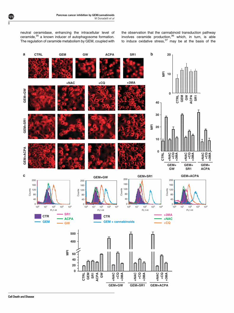
neutral ceramidase, enhancing the intracellular level ofceramide,35 a known inducer of autophagosome formation.The regulation of ceramide metabolism by GEM, coupled with
the observation that the cannabinoid transduction pathwayinvolves ceramide production,36 which, in turn, is ableto induce oxidative stress,37 may be at the basis of the
20CTRL GEM SR1ACPAGW
MF
I
10
+NAC +CQ +3MA
0
SR
1
GW
CT
RL
GE
M
AC
PA
40GE
M+G
W
30
MF
I
20
GE
M+S
R1
10
0
+CQ
CT
RL
+NA
C
+3M
A
+CQ
+NA
C
+3M
A
+CQ
+NA
C
+3M
A
GEM+SR1
GEM+GW
GEM+ACPA
GE
M+A
CP
A
GEM+GW GEM+SR1 GEM+ACPA
+3MACTRSR1
CTRACPA
GEM+NAC
GEM + cannabinoids +CQGW
500
400
MF
I
60
40
20
0
GE
M
GW
CT
RL
+NA
C+C
Q+3
MA
+NA
C+C
Q+3
MA
+NA
C+C
Q+3
MA
SR
1A
CP
A
GEM+GW GEM+SR1 GEM+ACPA
200
160
120
80
40
0100 101 102 103 104
FL1-H100 101 102 103 104
FL1-H100 101 102 103 104
FL1-H100 101 102 103 104
FL1-H
Cou
nts
200
160
120
80
40
0
Cou
nts
200
160
120
80
40
0
Cou
nts
200
160
120
80
40
0
Cou
nts
Pancreas cancer inhibition by GEM/cannabinoidsM Donadelli et al
8
Cell Death and Disease

ROS-dependent synergistic autophagic cell death by GEM/cannabinoid combinations.
Our kinetic studies revealed that ROS induction by drugcombinations preceded the activation of the ER stress markerGrp78, which, in turn, preceded the autophagy marker LC3-IIinduction. The observation that LC3-II increase was pre-vented by NAC and that ROS could induce ER stress14
strongly supports the hypothesis that oxidative stress, ERstress, and autophagic cell death were sequential events inour experimental conditions.
For in vivo studies, we chose SR1, in addition to GEM, onthe basis of its clinical relevance.25 Our in vivo experimentsshow that intraperitoneal injections of GEMþSR1 into nudemice bearing a subcutaneous mass of human pancreatic
Figure 6 Involvement of ROS in GEM/cannabinoid-induced autophagy. (a) Fluorescence microscopy analysis of autophagosome formation in Panc1 cells after acridineorange staining treated with 500 nM GEM and/or 40 mM GW, 225mM ACPA, or 40mM SR1 in the absence or presence of 20 mM NAC or 10 mM CQ or 1 mM 3-MA for 24 h.(b) The MFIs were calculated, as described in ‘Materials and Methods’, at FACS after trypsinization of the acridine orange-labeled cells. Values are the means of triplicatesamples from three independent experiments (±S.D.). Statistical analysis: Po0.05, control versus GEM, GW, ACPA, or SR1; Po0.001, each cannabinoid or GEM versustheir combination; and Po0.001, GEM/cannabinoids versus GEM/cannabinoidsþNAC, GEM/cannabinoidsþCQ, or GEM/cannabinoidþ 3-MA. (c) Flow cytometricanalyses of autophagosomes formation (MDC incorporation) in Panc1 cells treated with 500 nM GEM and/or 40 mM GW, 225mM ACPA, or 40mM SR1 in the absence orpresence of 20 mM NAC or 10mM CQ or 1 mM 3-MA for 24 h. The MFIs were calculated, as described in ‘Materials and Methods’. Values are the means of three independentexperiments (±S.D.). Statistical analysis: Po0.01, control versus GEM, SR1, or ACPA; Po0.001, control versus GW; Po0.001, each cannabinoid or GEM versus theircombination; and Po0.001, GEM/cannabinoids versus GEM/cannabinoidsþNAC, GEM/cannabinoidsþCQ, or GEM/cannabinoidþ 3-MA
12
10
GE
M+G
WG
EM
+AC
PA
GE
M+S
R1
8
6
4
2100
0
12
10
8
6
4
2
0
12
10
8
6
4
2
0
900 4 8 12 16 20 2480
70
60
50fo
ld in
du
ctio
n
40
30
0 4 8 12 16 20 24
20
% a
nti
pro
lifer
ativ
e sy
ner
gis
m
10
0
0 4 8 12 16 20 24
hours
ROS Grp78
LC3-II
Grp78ROS
0
+CQ
+NA
C
+3-M
A
+CQ
+NA
C
+3-M
A
+CQ
+NA
C
+3-M
A
GEM+SR1
GEM+GW
GEM+ACPA
LC3-II
(h)2420161284
Figure 7 Involvement of ROS and autophagy in the antiproliferative synergism by GEM/cannabinoids and kinetic analysis of ROS, Grp78, and LC3-II induction by GEM/cannabinoids. (a) Analysis of the antiproliferative synergism by 500 nM GEM and 40 mM GW, 225mM ACPA, or 40mM SR1 in the absence or presence of 20 mM NAC or10mM CQ or 2.5 mM 3-MA. Values are the means of three independent experiments (±S.D.). Statistical analysis: Po0.001, GEM/cannabinoids versus GEM/cannabinoidsþNAC, GEM/cannabinoidsþCQ, or GEM/cannabinoidþ 3-MA. (b) Panc1 cells were treated with 500 nM GEM and 40mM GW, 225mM ACPA, or 40mMSR1 for the indicated time points. ROS, Grp78, and LC3-II were analyzed, as described in Materials and Methods. Values are the means of three independent experiments.(c) Schematic representation of the kinetic analysis of oxidative stress (ROS), ER stress (Grp78), and autophagy (LC3-II) marker induction by GEM/cannabinoids
Pancreas cancer inhibition by GEM/cannabinoidsM Donadelli et al
9
Cell Death and Disease

adenocarcinoma cells quite completely inhibited tumorgrowth. No apparent form of toxicity in vivo, such as micedeath, body mass changes, or other apparent toxicity-relatedfeatures, was observed in mice treated with GEMþSR1.
In conclusion, the results presented here provide the firstevidence that the GEM/cannabinoid combinations exerted astrong synergistic antiproliferative effect on pancreaticadenocarcinoma GEM-resistant cell lines by ROS-dependentmechanisms, whereas it is scarcely toxic toward normal cells.Moreover, in vivo studies strongly boost the addition ofcannabinoids to GEM in designing new therapeutic strategiesfor pancreatic cancer treatment.
Materials and MethodsChemicals. GEM (Gemzar) was provided by Eli Lilly (Florence, Italy). SR141716 (N-(piperidino-1-yl)-5-(4-chlorophenyl)-1-(2,4dichlorophenyl)-4-methyl-pyrazole-3-carboxamide);SR1 (rimonabant; Acomplia, Sanofi-Aventis, Milan, Italy) was kindly provided by Dr. MaurizioBifulco (University of Salerno, Italy); ACPA was obtained from Cayman Chemicals (Inalco,Milan, Italy); and GW405833 hydrochloride (1-(2,3-dichlorobenzoyl)-2-methyl-3-(2-(1-morpholine)ethyl)-5-methoxyindole; GW) from Sigma (Milan, Italy). ActD, PDTC, NAC,E64d, chloroquine diphosphate (CQ; N4-(7-chloro-4-quinolinyl)-N1,N1-dimethyl-1,4-pentanediamine), and 3-MA were obtained from Sigma. MG132 and BAY 11-7085 (2E)-
3-94-(1,1-dimethylethyl)-phenyl-sulfonyl-2-propenenitrile) (BAY) were obtained fromEnzo Life Sciences (VinciBiochem, Florence, Italy), pepstatin A from AppliChem(Darmstadt, Germany), and the recombinant human IL-l from PeproTech (Inalco,Milan, Italy).
Cell culture. PaCa44, PaCa3, Panc1, CFPAC1, T3M4, and MiaPaCa2 celllines38 were grown in RPMI 1640 supplemented with 2 mM glutamine, 10% FBS,and 50mg/ml gentamicin sulfate (BioWhittaker, Lonza, Bergamo, Italy), andincubated at 371C with 5% CO2. Normal primary fibroblasts (PromoCell, PBI, Milan,Italy) were grown in DMEM supplemented with 2 mM glutamine, 10% FBS, and50mg/ml gentamicin sulfate, and were incubated at 371C with 5% CO2.
Cell proliferation assay. Cells were plated in 96-well cell culture plates(5� 103 cells per well) and treated with various compounds, as indicated in thelegends to figures. At the end of the treatments, cell proliferation was evaluated byCrystal Violet (Sigma) staining. The dye was solubilized in 1% SDS in PBS andmeasured photometrically (A595 nm) to determine cell viability. Three or fourindependent experiments were performed for each assay condition.
Drug combination studies. The compounds were added for 48 h at thefollowing concentration ranges: 1 nM–1mM for GEM, 80 nM–80mM for SR1 andGW, and 450 nM–450mM for ACPA in GEM-resistant cell lines; 0.2 nM–200 nMfor GEM, 100 nM–100mM for SR1 and GW, and 560 nM–560mM for ACPA inGEM-sensitive cell lines. The CI was calculated by the Chou–Talalay equation,which takes into account both the potency (IC50) and the shape of the dose–effect
4.5 40
4.0
303.5
3.0
202.5
2.0
1.5101.0
Mic
e b
od
y m
ass
(g)
Tu
mo
ur
volu
me
(cm
3 )
0.5
0.0 00 1 2 3 4 5 6 7 8 1 2 3 4 5 6 7 80
InjectionsInjections
CTRL GEM SR1 GEM+SR1
* * ** *
2.5* *
2.0
1.5
1.0
Tu
mo
ur
mas
s (g
)
0.5
0.0
SR
1
GE
M
CT
RL
GE
M+S
R1
Figure 8 Effect of GEMþSR1 treatment on xenografts of PaCa44 cells in nude mice. Cells were subcutaneously injected into female nude mice. After 1 week, i.p.injections with PBS (solution vehicle), GEM, or/and SR1 were administered twice a week for 4 weeks, as described in Materials and Methods. (a) Values are the means of micetumor volume measured at 3 days after each injection. (b) Values are the means of mice body mass measured at 3 days after each injection. (c) Values are the means of micetumor mass (±S.D.) measured after 8 injections
Pancreas cancer inhibition by GEM/cannabinoidsM Donadelli et al
10
Cell Death and Disease

curve,39 by using the CalcuSyn software. The general equation for the classicisobologram is given by CI¼ (D)1/(Dx)1þ (D)2/(Dx)2þ ((D)1 (D)2)/((Dx)1.(Dx)2),where (Dx)1 and (Dx)2 in the denominators are the doses (or concentrations) for D1(drug 1) and D2 (drug 2) alone that give x% growth inhibition, whereas (D)1 and (D)2in the numerators are the doses of drug 1 and drug 2 in combination that alsoinhibited x% cell viability (i.e., isoeffective). CIo1 and CIo0.3 indicated synergismand strong synergism, whereas CI¼ 1 and CI41 indicated additive effect andantagonism, respectively. CI/effect curves represent the CI versus the fraction (0–1)of cells killed by drug combination. The synergism percentage was obtainedanalyzing CI/effect curve and measuring the CI values at each 0.05 fraction, that is,5% growth inhibition, of the antiproliferative effect. Throughout all the experiments,we obtained a linear correlation coefficient (r)40.90. Drug combination studieswere performed using the following concentration ratios: [GEM]:[SR1] and[GEM]:[GW]¼ 1 : 500 and [GEM]:[ACPA]¼ 1 : 2800 in GEM-sensitive cell lines,whereas [GEM]:[SR1] and [GEM]:[GW]¼ 1 : 80 and [GEM]:[ACPA]¼ 1 : 450 inGEM-resistant cell lines. These molar ratios were calculated on the basis of GEMand cannabinoid IC50 mean values at 48 h. For GEM, they correspond to 36 nM forthe GEM-sensitive cell lines and 220 nM for the GEM-resistant cell lines, as reportedin our previous works.5 The three cannabinoids were all effective in the six cell lines,with IC50 mean values corresponding to 18 mM for SR1 and GW and 100mM forACPA. By inverting the molar ratios of the compounds between the two groups ofcell lines, no significant alteration of the results was observed (data not shown),indicating that the data obtained were not influenced by the specific experimentalcondition tested.
Analysis of ROS. The non-fluorescent diacetylated 20,70-dichlorofluorescein(DCF-DA) probe (Sigma), which becomes highly fluorescent upon oxidation, wasused to evaluate intracellular ROS production. Briefly, cells were plated in 96-wellplates (5� 103 cells per well) and, 24 h later, treated with various compounds, asindicated in the legends to figures. Then, cells were incubated with 10 mM DCF-DAfor 15 min at 371C, and the DCF fluorescence was measured by using a multimodeplate reader (Ex 485 nm and Em 535 nm). Three independent experiments wereperformed for each assay condition.
RNA extraction qPCR, RT-PCR, and image analysis. Total RNA wasextracted from 9� 105 cells using TRIzol Reagent (Invitrogen, Milan, Italy), and1mg of RNA was reverse transcribed using first-strand cDNA synthesis. Real-timequantification was performed in triplicate samples by SYBR Green detectionchemistry with SYBR Green PCR Master Mix (Applied Biosystems, Invitrogen) on a7000 Sequence Detection System. The primers used were: CB1For 50-GAGAGGTGCCAAGGGAGCTT-30 and CB1Rev 50-GGTGCGGAAGGTGGTATCTG-30;CB2For 50-CACAGCCTCTGTGGGTAGCC-30 and CB2Rev 50-ACGGGTGAGCAGAGCTTTGT-30; Grp78For 50-GACGGGCAAAGATGTCAGGAA-30 and Grp78Rev50-TCATAGTAGACCGGAACAGATCCA-30; CHOPFor 50-GCAGCCCATGAAGGAGAAAG-30 and CHOPRev 50-CGGTCGATTTCCTGCTTGAG-30; and GAPDHFor50-TGTGTCCGTCGTGGATCTGA-30 and GAPDHRev 50-R-GATGCCTGCTTCACCACCTT-30. The following cycling conditions were used: 951C for 10 min, 40 cyclesat 951C for 15 s, 601C for 1 min, and 721C for 30 s. The average of cycle thresholdof each triplicate was analyzed according to the 2(�DDCt) method. For RT-PCR,one-tenth of the cDNA was used as a template for PCR amplification using the followingprimers and cycling conditions: b-actinFor 50-ACCAACTGGGACGACATGGAGAA-30
and b-actinRev 50-GTGGTGGTGAAGCTGTAGCC-30, 25 cycles of 941C for 60 s, 551Cfor 60 s, and 721C for 60 s; XBP-1For 50-CCTTGTAGTTGAGAACCAGG-30 and XBP-1Rev 50-GGGGCTTGGTATATATGTGG-30, 40 cycles of 931C for 60 s, 551C for60 s, and 721C for 60 s. PCR products were separated by electrophoresis throughethidium bromide-stained 3.5% agarose gel and visualized by ultraviolet light. To quantifyXBP-1 splicing, XBP-1(S) bands were scanned as digital peaks and the areas ofthe peaks were calculated using the public domain NIH Image software(http://imagej.nih.gov), normalized with b-actin mRNA expression, and reported asfold induction relative to controls.
For ActD experiments, 2.5� 105 Panc1 cells were seeded in 60-mm plates andincubated overnight. Cells were pretreated with 5mg/ml ActD for 1 h, then 2mMGEM was added, and the treatments prolonged up to 16 h. Total RNA extractionand real-time PCR were performed, as above described.
Apoptosis. The percentage of apoptotic cells was evaluated by staining 3� 105
cells with annexin V-FITC (Bender Med System, Prodotti Gianni, Milan, Italy) and5mg/ml PI in binding buffer (10 mM HEPES/NaOH (pH 7.4), 140 mM NaOH, and2.5 mM CaCl2) for 10 min at room temperature in the dark. The samples were
analyzed by flow cytometry (FACScalibur, Becton Dickinson, San Jose, CA, USA) todetermine the percentage of cells displaying annexin Vþ /PI� (early apoptosis) orannexin Vþ /PIþ staining (late apoptosis). For each sample, we report thepercentage values corresponding to the addition of early and late apoptosis. Threeindependent experiments were performed for each assay condition.
Cell cycle analysis. Cell cycle distribution was analyzed by staining 3� 105
cells with PI. After the indicated treatments, cells were washed with PBS, incubatedwith 0.1% sodium citrate dihydrate, 0.1% Triton X-100, 200mg/ml RNase A, 50mg/ml PI(Roche Diagnostics, Milan, Italy), and analyzed using a flow cytometer(FACScalibur, Becton Dickinson). The percentage of cells in the various stagesof the cell cycle was determined using the ModFitLT software (Verity SoftwareHouse, Topsham, ME, USA). Three independent experiments were performed foreach assay condition.
Immunoblot analysis. In all, 1.2� 106 cells were treated with the variouscompounds, as indicated in the legends to figures, collected, washed in PBS, andresuspended in RIPA buffer, pH 8 (150 mM NaCl, pH 8; 50 mM Tris-HCl; 1% NP-40;0.5% Na-Doc; and 0.1% SDS), 1 mM PMSF, 1 mM Na3VO4, 1 mM NaF, 2.5 mMEDTA, and 1� protease inhibitor cocktail (Roche Diagnostics) for 30 min on ice.The lysate was centrifuged at 14 000� g for 10 min at 41C, and the supernatantwas used for western blot. Protein concentration was measured with the Bradfordprotein assay reagent (Pierce, Celbio, Milan, Italy) using bovine serum albumin asstandard. Fifty (for LC3-II) or thirty (for Grp78) micrograms of protein extracts wereelectrophoresed through a 15% SDS-polyacrylamide gel and electroblotted ontoPVDF membranes (Millipore, Milan, Italy). Membranes were then incubated for 2 hat room temperature with blocking solution (5% low-fat milk in TBST (100 mM Tris,pH 7.5, 0.9% NaCl, and 0.1% Tween 20)) and probed overnight at 41C with theappropriate primary antibody (1 : 1000 in blocking solution of LC3B (Cell Signaling,Milan, Italy), Grp78 (Santa Cruz Biotechnology, DBA, Milan, Italy), or a-tubulin(oncogene) antibodies). Horseradish peroxidase-conjugated IgG (1 : 2000 inblocking solution, Upstate Biotechnology, Milan, Italy) was used to detect specificproteins. Immunodetection was carried out using chemiluminescent substrates(Amersham Pharmacia Biotech, Milan, Italy) and recorded using a Hyperfilm ECL(Amersham Pharmacia Biotech). To quantify LC3-II and Grp78 expression, bandswere scanned as digital peaks and the areas of the peaks were calculated inarbitrary units using the public domain NIH Image software (http://rsb.info.nih.gov/nihimage/), normalized with a-tubulin expression, and reported as fold inductionrelative to controls.
Labeling of autophagic vacuoles with MDC. To quantify the inductionof the autophagic process in Panc-1 cells treated with the various compounds asindicated in the legend, MDC staining was performed, as previously described.40
Following the treatments, cells were incubated with 50 mM MDC in PBS at 371C for15 min. After incubation, cells were washed with PBS, trypsinized, and immediatelyanalyzed by flow cytometry. All fluorescences were analyzed with a FACScaliburflow cytometer (Becton Dickinson). The fluorescent emissions were collectedthrough a 530 nm band pass filter (FL1 channel). At least 10 000 events wereacquired in log mode. For the quantitative evaluation of MDC, CellQuest software(Becton Dickinson) was used to calculate mean fluorescence intensities (MFIs). TheMFIs were calculated by the formula (MFItreated/MFIcontrol), where MFItreated isthe fluorescence intensity of cells treated with the various compounds andMFIcontrol is the fluorescence intensity of untreated and unstained cells. Valuesreported in the figures are the means±S.D.s from three independent experiments.
Quantification of AVOs with acridine orange. In acridine orange-stained cells, the cytoplasm and nucleus are bright green and dim red, whereasacidic compartments are bright red. The intensity of the red fluorescence isproportional to the degree of acidity. Following the treatments, as specified in thelegend, cells were incubated with acridine orange solution (1 mg/ml) at 371C; after15 min in drug-free medium at 371C, they were washed with PBS, immediatelystained with 1 mg/ml of acridine orange for 15 min, and observed with a NikonEclipse TE300 Inverted microscope (Nikon Instruments, Inc., Melville, NY, USA).Then, cells were trypsinized and analyzed by flow cytometry using FACScancytometer and CellQuest software, as previously described. Statistical analyseswere performed, as described above.
In vivo studies. On the basis of our previous experience, we could not make useof Panc1 cells for in vivo studies because of their low growth rate in nude mice.
Pancreas cancer inhibition by GEM/cannabinoidsM Donadelli et al
11
Cell Death and Disease

Thus, we chose PaCa44 cells because of their similar behavior to Panc1 cells inin vitro growth inhibition studies. PaCa44 cells (2� 106 cells/mice) were s.c.injected into female nude mice (4 weeks of age, Harlan laboratories, Udine, Italy).One week after cell inoculation, five randomized animals for each experimentalgroup received solution vehicle (PBS), 25 mg/kg GEM, or/and 0.28 mg/kg SR1 byintraperitoneal injection biweekly for 4 weeks. Drug doses were chosen on the basisof their respective clinical use following US Food and Drug Administration directives.Tumor volume and body mass were biweekly recorded for each animal. Animalswere killed at the end of the 4-week study period, and the tumors were resected andweighted. Animal studies were approved by the Verona University Review Board.
Statistical analysis. ANOVA (post hoc Bonferroni) analysis was performed byGraphPad Prism 5 (GraphPad Software, Inc., La Jolla, CA, USA). P-values o0.05,0.01, or 0.001 were indicated as *, **, or ***, respectively.
Conflict of Interest
The authors declare no conflict of interest.
Acknowledgements. This work was supported by Associazione ItalianaRicerca Cancro, Milan, Italy; Fondazione CariPaRo, Padova, Italy; Joint Project ofUniversity of Verona, Verona, Italy; and MIUR, PRIN 2008 (Alberto Abbruzzese).SR141716 (Rimonabant) was provided by Sanofi-Aventis (grant to M Bifulco, no.051309). We thank Dr. Federica Cioffi (University of Verona) for FACS analysissupport, and Dr. Giorgio Malpeli (University of Verona) for qPCR technical support.
1. Li J, Merl MY, Chabot J, Saif MW. Updates of adjuvant therapy in pancreatic cancer: whereare we and where are we going? Highlights from the ‘2010 ASCO Annual Meeting’.Chicago, IL, USA. JOP 2010; 11: 310–312.
2. Neoptolemos JP, Cunningham D, Friess H, Bassi C, Stocken DD, Tait DM et al. Adjuvanttherapy in pancreatic cancer: historical and current perspectives. Ann Oncol 2003; 14:675–692.
3. Burris III HA, Moore MJ, Andersen J, Green MR, Rothenberg ML, Modiano MR et al.Improvements in survival and clinical benefit with gemcitabine as first-line therapyfor patients with advanced pancreas cancer: a randomized trial. J Clin Oncol 1997; 15:2403–2413.
4. Laurent A, Nicco C, Chereau C, Goulvestre C, Alexandre J, Alves A et al. Controlling tumorgrowth by modulating endogenous production of reactive oxygen species. Cancer Res2005; 65: 948–956.
5. Donadelli M, Costanzo C, Beghelli S, Scupoli MT, Dandrea M, Bonora A et al. Synergisticinhibition of pancreatic adenocarcinoma cell growth by trichostatin A and gemcitabine.Biochim Biophys Acta 2007; 1773: 1095–1106.
6. Casanova ML, Blazquez C, Martinez-Palacio J, Villanueva C, Fernandez-Acenero MJ,Huffman JW et al. Inhibition of skin tumor growth and angiogenesis in vivo by activation ofcannabinoid receptors. J Clin Invest 2003; 111: 43–50.
7. De Petrocellis L, Melck D, Palmisano A, Bisogno T, Laezza C, Bifulco M et al. Theendogenous cannabinoid anandamide inhibits human breast cancer cell proliferation. ProcNatl Acad Sci USA 1998; 95: 8375–8380.
8. Matsuda LA, Lolait SJ, Brownstein MJ, Young AC, Bonner TI. Structure of a cannabinoidreceptor and functional expression of the cloned cDNA. Nature 1990; 346: 561–564.
9. Munro S, Thomas KL, Abu-Shaar M. Molecular characterization of a peripheral receptor forcannabinoids. Nature 1993; 365: 61–65.
10. Carracedo A, Gironella M, Lorente M, Garcia S, Guzman M, Velasco G et al. Cannabinoidsinduce apoptosis of pancreatic tumor cells via endoplasmic reticulum stress-related genes.Cancer Res 2006; 66: 6748–6755.
11. Sarker KP, Obara S, Nakata M, Kitajima I, Maruyama I. Anandamide induces apoptosis ofPC-12 cells: involvement of superoxide and caspase-3. FEBS Lett 2000; 472: 39–44.
12. Costa B, Colleoni M. Changes in rat brain energetic metabolism after exposure toanandamide or Delta(9)-tetrahydrocannabinol. Eur J Pharmacol 2000; 395: 1–7.
13. Salazar M, Carracedo A, Salanueva IJ, Hernandez-Tiedra S, Lorente M, Egia A et al.Cannabinoid action induces autophagy-mediated cell death through stimulation of ERstress in human glioma cells. J Clin Invest 2009; 119: 1359–1372.
14. Yokouchi M, Hiramatsu N, Hayakawa K, Okamura M, Du S, Kasai A et al. Involvement ofselective reactive oxygen species upstream of proapoptotic branches of unfolded proteinresponse. J Biol Chem 2008; 283: 4252–4260.
15. Rutkowski DT, Kaufman RJ. That which does not kill me makes me stronger: adapting tochronic ER stress. Trends Biochem Sci 2007; 32: 469–476.
16. Kaufman RJ. Orchestrating the unfolded protein response in health and disease. J ClinInvest 2002; 110: 1389–1398.
17. Schonthal AH. Endoplasmic reticulum stress and autophagy as targets for cancer therapy.Cancer Lett 2009; 275: 163–169.
18. Puthalakath H, O’Reilly LA, Gunn P, Lee L, Kelly PN, Huntington ND et al. ER stresstriggers apoptosis by activating BH3-only protein Bim. Cell 2007; 129: 1337–1349.
19. Rouschop KM, van den Beucken T, Dubois L, Niessen H, Bussink J, Savelkouls K et al.The unfolded protein response protects human tumor cells during hypoxiathrough regulation of the autophagy genes MAP1LC3B and ATG5. J Clin Invest 2010;120: 127–141.
20. Kondo Y, Kondo S. Autophagy and cancer therapy. Autophagy 2006; 2: 85–90.21. Massi P, Vaccani A, Bianchessi S, Costa B, Macchi P, Parolaro D. The non-psychoactive
cannabidiol triggers caspase activation and oxidative stress in human glioma cells. Cell MolLife Sci 2006; 63: 2057–2066.
22. Biederbick A, Kern HF, Elsasser HP. Monodansylcadaverine (MDC) is a specific in vivomarker for autophagic vacuoles. Eur J Cell Biol 1995; 66: 3–14.
23. Xie S, Furjanic MA, Ferrara JJ, McAndrew NR, Ardino EL, Ngondara A et al. Theendocannabinoid system and rimonabant: a new drug with a novel mechanism of actioninvolving cannabinoid CB1 receptor antagonism – or inverse agonism–as potential obesitytreatment and other therapeutic use. J Clin Pharm Ther 2007; 32: 209–231.
24. Krylatov AV, Maslov LN, Lasukova OV, Pertwee RG. Cannabinoid receptor antagonistsSR141716 and SR144528 exhibit properties of partial agonists in experiments on isolatedperfused rat heart. Bull Exp Biol Med 2005; 139: 558–561.
25. Bifulco M, Grimaldi C, Gazzerro P, Pisanti S, Santoro A. Rimonabant: just an antiobesitydrug? Current evidence on its pleiotropic effects. Mol Pharmacol 2007; 71: 1445–1456.
26. Bifulco M, Laezza C, Valenti M, Ligresti A, Portella G, V DIM. A new strategy to block tumorgrowth by inhibiting endocannabinoid inactivation. Faseb J 2004; 18: 1606–1608.
27. Donadelli M, Dalla Pozza E, Costanzo C, Scupoli MT, Piacentini P, Scarpa A et al.Increased stability of P21(WAF1/CIP1) mRNA is required for ROS/ERK-dependentpancreatic adenocarcinoma cell growth inhibition by pyrrolidine dithiocarbamate. BiochimBiophys Acta 2006; 1763: 917–926.
28. Donadelli M, Dalla Pozza E, Scupoli MT, Costanzo C, Scarpa A, Palmieri M. Intracellularzinc increase inhibits p53(�/�) pancreatic adenocarcinoma cell growth by ROS/AIF-mediated apoptosis. Biochim Biophys Acta 2009; 1793: 273–280.
29. Shi Y, Zou M, Baitei EY, Alzahrani AS, Parhar RS, Al-Makhalafi Z et al. Cannabinoid 2receptor induction by IL-12 and its potential as a therapeutic target for the treatment ofanaplastic thyroid carcinoma. Cancer Gene Ther 2008; 15: 101–107.
30. Borner C, Bedini A, Hollt V, Kraus J. Analysis of promoter regions regulating basal andinterleukin-4-inducible expression of the human CB1 receptor gene in T lymphocytes. MolPharmacol 2008; 73: 1013–1019.
31. Arlt A, Gehrz A, Muerkoster S, Vorndamm J, Kruse ML, Folsch UR et al. Role of NF-kappaB and Akt/PI3K in the resistance of pancreatic carcinoma cell lines againstgemcitabine-induced cell death. Oncogene 2003; 22: 3243–3251.
32. Maiuri MC, Zalckvar E, Kimchi A, Kroemer G. Self-eating and self-killing: crosstalk betweenautophagy and apoptosis. Nat Rev Mol Cell Biol 2007; 8: 741–752.
33. Boya P, Gonzalez-Polo RA, Casares N, Perfettini JL, Dessen P, Larochette N et al.Inhibition of macroautophagy triggers apoptosis. Mol Cell Biol 2005; 25: 1025–1040.
34. Mukubou H, Tsujimura T, Sasaki R, Ku Y. The role of autophagy in the treatment ofpancreatic cancer with gemcitabine and ionizing radiation. Int J Oncol 2010; 37: 821–828.
35. Wu BX, Zeidan YH, Hannun YA. Downregulation of neutral ceramidase by gemcitabine:implications for cell cycle regulation. Biochim Biophys Acta 2009; 1791: 730–739.
36. Galve-Roperh I, Sanchez C, Cortes ML, Gomez del Pulgar T, Izquierdo M, Guzman M.Anti-tumoral action of cannabinoids: involvement of sustained ceramide accumulation andextracellular signal-regulated kinase activation. Nat Med 2000; 6: 313–319.
37. Garcia-Ruiz C, Colell A, Mari M, Morales A, Fernandez-Checa JC. Direct effect of ceramideon the mitochondrial electron transport chain leads to generation of reactive oxygenspecies. Role of mitochondrial glutathione. J Biol Chem 1997; 272: 11369–11377.
38. Moore PS, Sipos B, Orlandini S, Sorio C, Real FX, Lemoine NR et al. Genetic profile of 22pancreatic carcinoma cell lines. Analysis of K-ras, p53, p16 and DPC4/Smad4. VirchowsArch 2001; 439: 798–802.
39. Chou TC, Talalay P. Quantitative analysis of dose-effect relationships: the combinedeffects of multiple drugs or enzyme inhibitors. Adv Enzyme Regul 1984; 22: 27–55.
40. Meschini S, Condello M, Calcabrini A, Marra M, Formisano G, Lista P et al. The plantalkaloid voacamine induces apoptosis-independent autophagic cell death on both sensitiveand multidrug resistant human osteosarcoma cells. Autophagy 2008; 4: 1020–1033.
Cell Death and Disease is an open-access journalpublished by Nature Publishing Group. This work is
licensed under the Creative Commons Attribution-Noncommercial-NoDerivative Works 3.0 Unported License. To view a copy of this license,visit http://creativecommons.org/licenses/by-nc-nd/3.0/
Supplementary Information accompanies the paper on Cell Death and Disease website (http://www.nature.com/cddis)
Pancreas cancer inhibition by GEM/cannabinoidsM Donadelli et al
12
Cell Death and Disease