Synthesis of PEGylated lactose analogs for inhibition studies on T.cruzi trans-sialidase
Upload
independentCategory
view
4download
0
ORIGINAL ARTICLE
In vivo activity of gemcitabine-loaded PEGylatedsmall unilamellar liposomes against pancreatic cancer
Donato Cosco Æ Alessandra Bulotta Æ Monica Ventura Æ Christian Celia ÆTeresa Calimeri Æ Gino Perri Æ Donatella Paolino Æ Nicola Costa Æ Paola Neri ÆPierosandro Tagliaferri Æ Pierfrancesco Tassone Æ Massimo Fresta
Received: 17 December 2008 / Accepted: 4 February 2009 / Published online: 5 March 2009
� Springer-Verlag 2009
Abstract Gemcitabine (GEM) is presently the standard
option for the treatment of advanced pancreatic cancer
(PC). We investigated the in vitro and in vivo antitumor
potential of GEM-loaded PEGylated liposomes (L-GEM)
as a novel agent for the treatment of PC. In vitro analysis of
antitumor activity against human PC cell lines, BXPC-3
and PSN-1, showed a significant time- and dose-dependent
reduction of cell viability following exposure to L-GEM as
compared to free GEM [at 72 h, IC50: 0.009 vs. 0.027 lM
(P = 0.003) for BXPC-3 and 0.003 vs. 0.009 lM
(P \ 0.001) for PSN1, respectively]. Confocal laser
scanning microscopy demonstrated an effective liposome/
cell interaction and internalization process following 3-h
cell exposure to L-GEM. The in vivo antitumor activity of
L-GEM was investigated in a cohort of SCID mice bearing
BxPC-3 or PSN-1 xenografts. Animals were i.p. treated
with L-GEM (5 mg/kg), or a threefold increased dose of
free GEM (15 mg/kg), or empty liposomes or vehicle,
twice a week for 35 days. A significant higher inhibition of
tumor growth in mice treated with L-GEM versus free
GEM (P = 0.006 and P = 0.004 for BXPC-3 and PSN-1,
respectively) or control groups (P = 0.0001), translated in
a survival advantage of L-GEM treated animals versus
other groups. Pharmacokinetic studies showed enhance-
ment of systemic bioavailability of L-GEM (t1/2 = 8 h)
versus to GEM (t1/2 = 1.5 h). Our findings demonstrate
that L-GEM is an effective agent against PC and exerts
higher antitumor activity as compared to free GEM with no
appreciable increase in toxicity. These results provide the
pre-clinical rational for L-GEM clinical development for
the treatment of PC patients.
Keywords Pancreatic cancer � Liposomes �Gemcitabine � Mouse models � BXPC-3 � PSN-1
List of abbreviations
AUC Area under the curve
Chol Cholesterol
CLSM Confocal laser scanning microscopy
Cmax Maximum plasmatic concentration
DPPC 1, 2-Dipalmitoyl-sn-glycero-3-
phospocholine monohydrate
DSPE-MPEG 2000 N-(Carbonyl-methoxypolyethylene
glycol-2000)-1, 2-distearoyl-sn-
glycero-3-phosphoethanolamine
D. Cosco and A. Bulotta contributed equally to this work.
In memory of Prof. Salvatore Venuta, MD, a victim of pancreatic
cancer, an oncologist and a scientist who lived future frontiers of
cancer therapeutics by strongly trusting in biomedical
nanotechnologies.
D. Cosco � C. Celia � N. Costa � M. Fresta
Department of Pharmacobiological Sciences,
University ‘‘Magna Græcia’’, Campus Salvatore Venuta,
Catanzaro, Italy
A. Bulotta � M. Ventura � T. Calimeri � G. Perri � P. Neri �P. Tagliaferri � P. Tassone (&)
Medical Oncology Unit and Referal Unit for Genetic
Counselling and Innovative Treatments,
Tommaso Campanella Cancer Center,
University ‘‘Magna Græcia’’, Campus Salvatore Venuta,
Viale Europa, 88100 Catanzaro, Italy
e-mail: [email protected]
D. Paolino � P. Neri � P. Tagliaferri � P. Tassone
Department of Experimental and Clinical Medicine G. Salvatore,
University ‘‘Magna Græcia’’, Campus Salvatore Venuta,
Catanzaro, Italy
123
Cancer Chemother Pharmacol (2009) 64:1009–1020
DOI 10.1007/s00280-009-0957-1

EPR Enhanced permeation and retention
Fluorescein-DHPE N-(Fluorescein-5-tiocarbamoyl)-1,
2-dihexadecanoyl-sn-glycero-3-
phosphoethanolamine
triethylammonium salt
GEM Gemcitabine—2I,
2I-difluorodeoxycytidine
HPLC High performance liquid
chromatography
L-GEM Gemcitabine-loaded pegylated small
unilamellar liposomes
PBS Phosphate buffer saline solution
PC Human pancreatic adenocarcinoma
cancer
PEG Poly-ethylene glycol
t1/2 Plasma half-life
Vd Volume of distribution
Introduction
Pancreatic cancer (PC) is one of the most aggressive
tumors with only\4% of all patients surviving longer than
5 years after diagnosis [1]. Even if the surgical resection of
the primary tumor may offer a chance of cure, most of
patients recurs or are not resectable at time of diagnosis,
due to the very early local aggressiveness. Locally
advanced or metastatic PC patients have a very poor
prognosis. To date, systemic chemotherapy, based on the
nucleoside analog gemcitabine (2I,2I-difluorodeoxycyti-
dine) (GEM), is the mainstay of palliative treatment,
improving the disease-related symptoms and producing an
objective response rate of \20%.
The mechanism of action of GEM is based on its pro-
gressive intracellular phosphorylation up to the formation
of the triphosphate derivative, which acts as a false
nucleotide being incorporated in the DNA chain. In addi-
tion, both diphosphate and triphosphate GEM nucleotides
are able to inhibit the ribonucleotide reductase, thus elic-
iting a reduction of the concentration of deoxynucleotides
necessary for DNA synthesis. These combined actions lead
to the inhibition of DNA synthesis and the arrest of cell
proliferation in S-phase [2]. Under the biopharmaceutical
point of view, GEM is rapidly converted into the inactive
metabolite 2I-deoxy-2I,2I-difluorouridine by cytidine
deaminase following systemic infusion and the inactive
metabolite is excreted in the urine [3]. The very short
plasma half-life (8–17 min) partially explains the low
systemic activity of GEM [4].
Accumulating body of evidence supports the idea that
drug entrapment in liposomes is a successful strategy to
overcome limitations of drug pharmacokinetics and to
avoid rapid metabolic inactivation [5, 6]. Liposomes,
which are the archetypal and the most biocompatible and
bio-mimetic form of drug nanovectors, may significantly
improve the drug activity taking benefit from the slow
vascular and extravascular flow in the tumor and from the
leaky cancer neovasculature [7]. In fact, the presence of
large fenestrations in tumor vessels significantly enhances
permeability and allows local retention of liposomal
nanovectors [enhanced permeation and retention (EPR)
effect] with the final goal of an efficient passive targeting
and increased therapeutic effects. In particular, the reduc-
tion of liposomal size (\200 nm) and the decrease of the
surface charge by means of liposome coating with
uncharged highly hydrophilic polymers, i.e., poly-ethylene
glycol (PEG) (PEGylated liposomes), improve the sys-
temic carrier half-life with the advantage of a better
bioavailability at tumor site and the reduction of side
effects, as in the case of Doxorubicin-loaded PEGylated
liposomes (Doxil�) [8–10]. Even though more sophisti-
cated active targeting strategies have been proposed for
cancer therapy [11, 12], the passive targeting, based on the
EPR effect, and the PEGylation of colloidal carrier, is
already a reliable and clinically effective strategy [13, 14].
However, an extensive investigation on the passive tar-
geting of vesicular colloidal carriers by pegylation is still
needed to study the potential therapeutic role of this
approach in cancer treatment.
Aim of this study is the investigation of in vitro and in
vivo effects induced by gemcitabine-loaded PEGylated
small unilamellar liposomes (L-GEM) against human PC
cell lines and human xenografts as compared to free GEM.
The interaction of liposomal vectors with the human PC
cells was also investigated by means of confocal laser
scanning microscopy (CLSM) and the in vivo pharmaco-
kinetic profiles of both free GEM and L-GEM was
assessed.
Materials and methods
Chemicals and biochemicals
Cholesterol (Chol), phosphate buffer saline solution (PBS),
N-(fluorescein-5-tiocarbamoyl)-1,2-dihexadecanoyl-sn-gly-
cero-3-phosphoethanolamine triethylammonium salt
(fluorescein-DHPE), 3-[4,5-dimethylthiazol-2-yl]-3,5-diphe-
nyltetrazolium bromide salt (tetrazolium salt), RPMI 1640
media, fetal bovine serum, 2 mM L-glutamine, 100 U/ml
penicillin, 100 lg/ml streptomycin were obtained from
Sigma Chemicals Co. (St. Louis, USA). 1,2-dipalmitoyl-
sn-glycero-3-phospocholine monohydrate (DPPC) and
N-(carbonyl-methoxypolyethylene glycol-2000)-1,2-dis-
tearoyl-sn-glycero-3-phosphoethanolamine (DSPE-MPEG
1010 Cancer Chemother Pharmacol (2009) 64:1009–1020
123

2000) were purchased from Genzyme (Suffolk, UK). Sterile
saline was a product of Frekenius Kabi Potenza S.r.l.
(Verona, Italy). The human pancreatic adenocarcinoma
cancer cell lines BxPC-3 and PSN-1 were purchased from the
Cell Bank of the National Institute for Cancer Research
(Genova, Italy). Gemcitabine (20,20-difluorodeoxycytidine)
hydrochloride (HPLC purity [99%) was obtained from
Eli-Lilly Italia S.p.A. (Sesto Fiorentino, Florence, Italy) and
it was used without further purification. All other chemical
reagents used in this investigation were of analytical grade
(Carlo Erba, Milan, Italy).
GEM stability in the culture medium was assessed by
repeated HPLC analysis.
Liposome preparation
Liposomes were made up of DPPC/Chol/DSPE-
MPEG2000 (6:3:1 molar ratio) and were prepared and
characterized as reported elsewhere [15]. Briefly, lipid
mixture (20 mg) was dissolved in a round-bottomed flask
by using a chloroform/methanol (3:1 v/v) solvent mixture,
which was removed by means of a rotary evaporator
(Buchi R-210 Switzerland) and by an overnight storage at
room temperature in a Buchi T51 glass drying oven con-
nected to a vacuum pump, thus allowing the formation of a
thin layer lipid film. When required, fluorescent labeled
liposomes were prepared by co-dissolving fluorescein-
DHPE (0.1% molar) with the lipids. Lipid films were
hydrated with a 250 mM ammonium sulfate solution
(1 ml) and then submitted to ten cycles of freezing (with
liquid nitrogen) and thawing (with a water bath at 40�C),
thus achieving a pH gradient with a homogenous acid
environment in the intra-liposomal aqueous compartments.
Multilamellar vesicles were submitted to extrusion through
400, 200 and 100 nm pore size two stacked polycarbonate
filters (Costar, Corning Incorporated, NY, USA) by using a
stainless steel extrusion device (Lipex Biomembranes,
Vancouver, BC, USA) and un-entrapped ammonium sul-
fate solution was removed by centrifugation. Small
unilamellar colloidal vesicles were suspended in an iso-
tonic solution (1 ml) of GEM-hydrochloride (1 mM) and
kept at room temperature for 3 h. The unentrapped drug
was removed by gel permeation chromatography thus
obtaining L-GEM. The liposome encapsulation efficiency
was calculated as a percentage with respect to the amount
added during preparation. Liposomes were physico-chem-
ically characterized by light scattering.
Physicochemical characterization of liposomes
Mean size and size distribution (polydispersity index)
of L-GEM were evaluated by dynamic light-scattering
experiments. Zetamaster (Malvern Instruments Ltd.,
Spring Lane South, Worcs, UK), a photo-correlation
spectroscopy apparatus, was used for the dimensional
analysis. Zetamaster is equipped with a 4.5-mW laser
diode operating at 670 nm. Experiments were carried out
at a scattering angle of 90�. A third-order cumulant fit-
ting correlation function was performed by a Malvern
PCS sub-micron particle analyzer to obtain mean size
and polydispersity index of L-GEM. A medium refrac-
tive index of 1.330, a medium viscosity of 1.0 mPa s
and a dielectric constant of 80.4 were set as instrumental
parameters for light-scattering experiments. Samples
were suitably diluted with a filtered (Sartorius membrane
filters 0.22 lm) saline to avoid multiscattering phenom-
ena and placed in a quartz cuvette. Experiments were
carried out at room temperature.
Tissue culture
PC cells BxPC-3 and PSN-1 were grown in RPMI 1640
media (Sigma Aldrich, Italy) supplemented with 10% fetal
bovine serum (Sigma), 2 mM L-glutamine (Sigma),
100 U/ml penicillin (Sigma), and 100 lg/ml streptomycin
(Sigma). The two PC cell lines were cultured in plastic
culture dishes (100 mm 9 20 mm) at 37�C in a humidified
atmosphere with 5% CO2. Cultures were free of Myco-
plasma and pathogenic murine viruses and were
maintained for no longer than 8 weeks after recovery from
frozen stocks (liquid nitrogen). When a *80% confluence
was reached, cells were trypsinized (2 ml) and collected
into a centrifuge tube containing 4 ml of the culture
medium. The dishes were further washed with 2 ml of PBS
to remove the remaining cells and then the washing buffer
was transferred into the centrifuge tube, which was cen-
trifuged at 1,000 rpm at room temperature for 10 min
(Megafuge 1.0, Heraeus Sepatech, Osterode/Harz, Ger-
many). The pellet was resuspended in a suitable volume of
culture medium and seeded in culture dishes before in vitro
investigations.
In vitro studies
Cell proliferation assays
To assess cellular cytotoxicity the MTT test was carried
out. PSN-1 (5 9 103/well) and BxPC-3 (7.5 9 103/well)
cells were seeded in 96-well tissue culture plates for
24 h at 37�C, thus allowing cell adhesion to culture
plates. Culture medium was removed and replaced with
fresh medium containing free or liposomally entrapped
GEM at different concentrations (evaluation of the dose-
dependent activity) and cells were then incubated for 48
or 72 h. Every plate had 8 wells with untreated cells as
Cancer Chemother Pharmacol (2009) 64:1009–1020 1011
123

the control and 8 wells with cells treated with empty
liposomes as the blank. In this case, empty liposomes
underwent to the same dilution of L-GEM. After each
incubation period, 10 ll of tetrazolium salt solubilized in
PBS solution (5 mg/ml) was added to each well and the
plates were incubated at 37�C for 3 h. The medium was
then removed and the formazan salts were dissolved with
200 ll of a solution of ethanol/DMSO (1:1 v/v) by
shaking plates for 20 min at 230 rpm (IKA� KS 130
Control, IKA� WERKE GMBH & Co., Staufen, Ger-
many). The sample absorbance was measured by means
of a microplate reader (LabSystems Multiskan MS M-
Medical, Italy) at a wavelength of 540 nm with reference
at a wavelength of 690 nm. The percent cell viability
was calculated according to the following equation:
cell viability ¼ AbsT=AbsC � 100 ð1Þ
where AbsT is the absorbance of treated cells and AbsC
is the absorbance of control (untreated) cells. The for-
mazan concentration is directly proportional to the cell
viability. Data on cell viability are reported as the mean
of six different experiments ± standard deviation.
Confocal laser scanning microscopy (CLSM) experiments
The interaction between PC cells and liposomes was
evaluated by CLSM. Cells were placed in 6-well culture
plates (4 9 105 cells/ml) with culture medium. In each
well a sterile glass slide was previously positioned.
Plates were incubated for 24 h and then cells were
treated with liposomes labeled with fluorescein-DHPE
for different incubation times, from 3 h up to 24 h. After
incubation, each well was washed with PBS (3 times) to
remove the excess of vesicles and cells were fixed on the
sterile glass slides by using 1 ml of an ethanolic solution
(70% v/v). Each slide glass was washed again with PBS
three times and PBS (2 ml) was added to each well.
Plates were stored at 4�C up to the confocal microscopy
analysis. Before analysis, slide glasses were positioned
on cover-glass by using a glycerol solution (70% v/v) to
remove enclosed air and they were fixed by a transparent
glue. The analysis was carried out using a Leika TCS
SP2 MP laser scanning confocal microscope at a
kexc = 496 nm and a kem = 519 nm. A scan resolution
up to 4,096 9 4,096 pixels with an Ar/Kr laser beam of
75 mW, equipped with a fluorescein analyzer filter, was
used for experimental investigations. Samples were
recorded by a macro developer software package having
multi-dimensional series acquisition and direct-access
digital control knobs. An immersion oil lens 1009 was
used.
In vivo studies
Human PC xenograft models
CB-17 SCID-mice were purchased from Charles River
(France), maintained and monitored in our animal research
facility. Care and handling of animals were in accordance
with the Italian low.
Mice were subcutaneously (s.c.) inoculated in the inter-
scapular area with 3 9 106 and 2.5 9 106 PSN-1 and BxPC-
3 cells (100 ll in RPMI-1640 medium), respectively. When
the tumor was measurable, approximately 2 and 3 weeks
after PSN-1 or BxPC-3 cell injection, respectively, mice
were i.p. treated with L-GEM (5 mg/kg, n = 5 mice), a
threefold increased dose of free GEM (15 mg/kg, n = 5),
empty liposomes (n = 5) or isotonic saline solution (NaCl
0.9% w/v, n = 5), twice a week for 35 days. GEM dose and
schedule used here have been chosen according to other
reports where GEM as been used at doses in a range between
7.5 and 15 mg/kg weekly or twice weekly [16, 17]. GEM and
L-GEM concentrations used in our study are below MTD
which has been found at significant higher dose (approxi-
mately 200 mg/kg). Tumor size was measured every 4 days
in two dimensions using an electronic caliper, and the tumor
volume was calculated using the following formula:
V ¼ 0:5ab2 ð2Þ
where a and b are the long and short diameter of the tumor,
respectively. To avoid unnecessary sufferance, animals
were killed when their tumors reached 2 cm in diameter.
Survival was calculated from the first day of treatment until
the day of killing. Detailed procedure of in vivo studies has
been previously reported [18, 19].
Pharmacokinetic studies
CB-17 SCID-mice (1 month old, 25 g) were used for phar-
macokinetic studies. Free GEM or L-GEM (1 mg/ml) were
injected (100 ll) through the tail vein (groups of five ani-
mals). Blood samples (200 ll) were taken from the tail vein
at various times and refrigerated. Then, defrost samples were
immediately centrifuged (10 min, 12,000 rpm) at room
temperature and acetic acid (50 ll) was added to plasma
samples to decrease hydrogen bonding between nucleosides
and proteins. Acetonitrile (1 ml) (HPLC grade) was added to
plasma samples, that were vortex-mixed and then centri-
fuged at 800g for 15 min at 4�C. The supernatant was
removed and collected in a glass tube and acetonitrile (1 ml)
was added to the pellet. Three cycles of vortex-mixing and
centrifugation procedure were carried out. Supernatants
were combined and evaporated to dryness under nitrogen
flux at 42�C (thermostated water bath) and stored at -20�C.
1012 Cancer Chemother Pharmacol (2009) 64:1009–1020
123

Before the HPLC analysis, the residue was resuspended in
water (1 ml) (HPLC grade), incubated for 5 min at 37�C and
then centrifuged at 12,000g for 10 min at 20�C. The super-
natant was removed, filtered through a 0.22 lm pore size
Anotop 10 syringe filter (Whatman, Springfield Mill, UK)
and placed in 4-ml HPLC glass vials for the analytical
determination. Analysis was performed by using an HPLC
system (Varian Inc., Palo Alto, USA) consisted of a 200-
2031 Metachem online degasser, a M210 binary pump, a
ProStar 410 autosampler, a G1316A thermostated column
compartment, a 25 ll CSL20 Cheminert Sample Loop
injector. Data were acquired and processed with a Galaxie�
chromatography manager software (Varian Inc., Palo Alto,
USA). Chromatographic separation was carried out at room
temperature by using a GraceSmart RP C18 column
(4.6 9 250 mm, 5 lm particle size, Alltech Grom GmbH,
Rottenburg-Hailfingen, Germany). The mobile phase con-
sisted of deionized water (HPLC grade) and acetonitrile
(HPLC grade) 95:5 v/v. The flow rate was 1 ml/min and UV
detection was performed at 269 nm.
No interference was observed for GEM and its metab-
olite, 20,20-difluorodeoxyuridine, HPLC peaks due to any
plasma component. The chromatographic method provided
a suitable separation of the peaks of GEM and its metab-
olite (20,20-difluorodeoxyuridine), which showed a
retention time of 6 and 8.70 min, respectively. The GEM
quantification was carried out by using an external standard
curve in the linear concentration range between 0.1 and
10 lg/ml. A standard solution of GEM (1 mg/ml) was used
for the construction of the standard curve. Plasma amount
of GEM were determined using the standard curve
according to the following equation:
AUC ¼ 0:60112xþ 0:02840 ð3Þ
where x is the drug concentration (lg/ml) and AUC the
area under the curve (mAu 9 min).
GEM plasma amount were expressed as lg/ml. Exper-
imental data are the mean of three different experiments.
Statistical analysis
One-way ANOVA and Student t-test was used for statis-
tical analysis of the experimental date. A posteriori
Bonferroni t-test was carried out to check the ANOVA test.
A P-value \0.05 was considered statistically significant.
Values are reported as the mean ± standard deviation.
Results
The pH-gradient method used for L-GEM preparation
allowed an effective drug entrapment as high as 88.5% with
respect to the amount added during the preparation
procedure, in agreement with our previous results [15].
These findings demonstrated the suitability of liposomes as
potential drug carrier for GEM. Taking into account the great
relevance in terms of biopharmaceutical features of the mean
size and size distribution of a colloidal carrier as a suitable
drug delivery system for anticancer treatment, PEGylated
liposomes underwent to an extrusion procedure thus pro-
ducing a mean vesicular size of 136 ± 14 nm with a
polydispersity index of 0.12 (highly homogenous size
distribution).
In vitro activity of L-GEM against PC cell lines
We first investigated the in vitro antitumor activity of
L-GEM against BxPC-3 and PSN-1 PC cells by MTT assay.
As shown in Fig. 1, L-GEM induced a significant dose-
dependent reduction of the cell viability as compared to free
GEM. A lower IC50 value of L-GEM versus the free drug was
achieved: at 72 h, the IC50 value L-GEM was significantly
(P \ 0.001) lower as compared to the free GEM for both cell
lines (0.009 vs. 0.027 lM for BXPC-3 and 0.003 vs.
0.009 lM for PSN1, respectively) (Table 1). A physical
mixture of free GEM and empty liposomes was also tested to
evaluate the real advantage of the drug encapsulation. This
mixture did not show any improvement in terms of antitumor
activity as compared to the free drug (data not shown).
Vehicle or empty liposomes did not affect cell survival of
both BxPC-3 and PSN-1 lines (data not shown).
Liposome–cell interaction
An important step of our research was the study of the
interaction between the liposomal carrier and the biological
substrate. A liposome formulation labeled with fluorescein-
DHPE was used for CLSM studies. Liposome/cell interac-
tion was evaluated after 3, 6, 12 and 24 h of incubation. To
evaluate the occurrence of artefacts due to the basal fluo-
rescence of the cellular components, control samples were
observed both in transmission mode (Figs. 2a, 3a) and
after exposition at the fluorescein excitation wavelength
(Figs. 2b, 3b). No significant fluorescent emission was
observed, suggesting the absence of fluorescence interfer-
ence phenomena. Following 3 h incubation (Figs. 2c, 3c),
cell membranes were stained by vesicular carriers. The
photomicrograph in Fig. 3c shows the presence of liposomes
on the cell surface of BxPC-3 cells. Following 24 h, intra-
cytoplasmic localization of vesicular carriers (Figs. 2, 3f)
was detected.
In vivo antitumor activity
We next evaluated the in vivo antitumor activity of L-GEM
by two xenograft murine models of human PC. A cohort of
Cancer Chemother Pharmacol (2009) 64:1009–1020 1013
123
40 SCID mice bearing s.c. PSN-1 (20 mice) or BxPC-3 (20
mice) human xenografts were respectively i.p. treated with
L-GEM (5 mg/kg, n = 5 mice), with a the threefold
increased dose of free GEM (15 mg/kg, n = 5 mice), with
empty liposomes (n = 5) or vehicle (n = 5), twice a week
for 35 days. It is important to consider that a wide range of
GEM concentrations has been previously reported [16, 17,
20, 21]. These differences can be explained by (1) intrinsic
sensitivity of different tumor cell lines to the drug, (2)
different in vivo models, (3) different drug bioavailability
in different mouse strains. As shown in Fig. 4, a significant
tumor growth inhibition was detected in animals treated
with L-GEM or GEM versus control groups. Specifically,
L-GEM induced higher growth inhibition as compared to
free GEM (P = 0.006) and a significant survival advantage
(20 days; P = 0.03) of mice bearing BXPC-3 xenografts
(Fig. 4b, d). In parallel, L-GEM showed a significantly
higher tumor growth inhibition versus free GEM in PSN-1
tumor xenografts (P = 0.004). Also in this case, the sur-
vival of L-GEM-treated animals was longer than free drug-
treated mice (77 vs. 61 days; P = 0.02) (Fig. 4a, c).
These findings clearly indicate that L-GEM exerts more
effective antitumor activity than the free drug, even if L-
GEM has been used at a threefold lower concentration.
Table 1 IC50 values (lM) of
free GEM and L-GEM after 48
and 72 h treatment of PC cells
48 h 72 h
PSN-1 BXPC-3 PSN-1 BXPC-3
GEM 0.009 ± 0.0008 0.048 ± 0.006 0.009 ± 0.001 0.027 ± 0.005
L-GEM 0.006 ± 0.0004 0.021 ± 0.004 0.003 ± 0.0008 0.009 ± 0.0006
P-value 0.005 0.002 0.001 0.003
Fig. 1 In vitro dose and time-
dependent antitumor effects
induced by GEM (filled circles)
and L-GEM (filled triangles) on
PSN-1 and BxPc-3 cells.
Analysis was performed by
MTT assay. Data are reported as
percentage of control (untreated
cells). Results are the mean of
six different
experiments ± standard
deviation. Statistical analysis
was performed by one-way
ANOVA and a posteriori
Bonferroni t-test: *P \ 0.05;
**P \ 0.001
1014 Cancer Chemother Pharmacol (2009) 64:1009–1020
123

Importantly, by both formulations, we did not observe any
toxicity including weight loss or other signs related to
compromise of quality of life nor sudden death at used
concentrations.
Pharmacokinetic studies
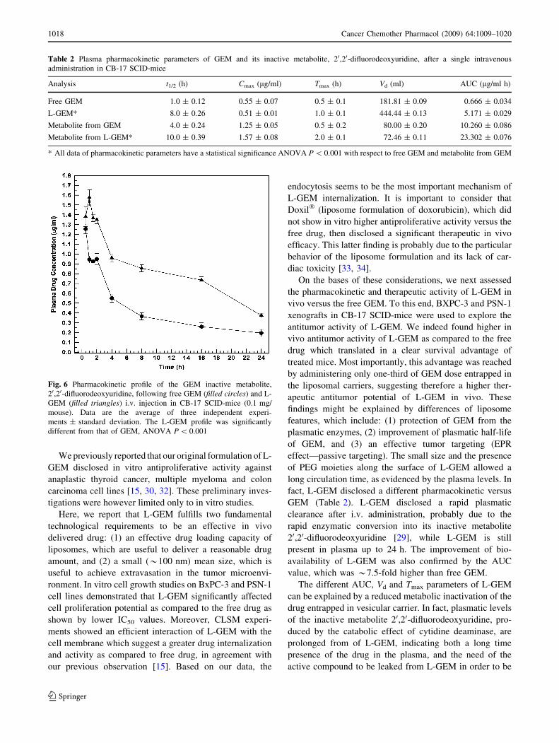
Plasma concentration profiles of free GEM and L-GEM
and a summary of the major pharmacokinetic parameters
were reported in Fig. 5 and Table 2, respectively. Differ-
ences in pharmacokinetic properties of GEM and its
metabolite, 20,20-difluorodeoxyuridine, were observed.
GEM disappeared rapidly from plasma (Fig. 5) due to
the metabolic conversion into 20,20-difluorodeoxyuridine
metabolite [22–24]. Pharmacokinetic profiles showed a
biphasic pattern of the drug both in the free and liposomal
formulation. The maximum plasmatic concentration (Cmax)
of GEM was 0.55 lg/ml at 0.5 h following the intravenous
administration. The plasma concentration of GEM declined
rapidly after 1 h of infusion. The GEM half-life (t1/2) was
1.5 h with a measured plasma drug concentration of
0.26 lg/ml (Table 2). A different behavior was observed
for the GEM metabolite, 20,20-difluorodeoxyuridine. In
fact, the Cmax of 20,20-difluorodeoxyuridine was 1.25 lg/ml
at 1 h after the administration and the half-life was reached
after 4 h. The pharmacokinetic profile of 20,20-difluorode-
oxyuridine declined by a similar shape observed for the
native drug. However, a constant 20,20-difluorodeoxyuri-
dine plasma concentration was followed by a gradual
decrease at 1–2 h. These findings evidenced that GEM and
its inactive metabolite had a similar plasmatic distribution,
but the metabolite was still present in the plasma after 20 h
(Fig. 6).
Significant differences in the pharmacokinetic profile
and parameters were observed when GEM was entrapped
in liposomes. In fact, L-GEM was slowly removed from
Fig. 2 Confocal laser scanning
micrographs showing the
interaction between L-GEM and
PSN-1 cells. The time-
dependent intracellular
localization of fluorescein-
DHPE labeled liposomes is
shown. a and b are controls
(untreated cells): in a cells are
observed in transmission mode,
in b no significant cellular
fluorescence was observed from
the autofluorescent phenomena
caused by the cellular
components. c, d, e and f(treated cells) after 3, 6, 12 and
24 h, respectively
Cancer Chemother Pharmacol (2009) 64:1009–1020 1015
123

blood circulation following intravenous administration
(Fig. 5). A Cmax of *0.52 lg/ml was obtained at 2 h
after the infusion and the plasma drug concentration
remained elevated (*0.2 lg/ml) up to 16 h with respect
to the un-entrapped GEM, which was no more detectable
after 2 h from the infusion. The t1/2 of L-GEM was 8 h
(Table 2). These findings were also supported by the
evaluation of the volume of distribution (Vd) and area
under the curve (AUC) parameters (Table 2). In fact, Vd
value of L-GEM (444.44 ml) was 2.5-fold higher than
that of GEM (181.81 ml), probably due to the rapid
enzymatic inactivation of the drug, while the AUC value
of L-GEM (5.171 lg/ml) showed an improvement of
7.5-fold with respect to free GEM (0.666 lg/ml).
A different pharmacokinetic profile of the 20,20-difluoro-
deoxyuridine was demonstrated for L-GEM (Fig. 6). The
metabolite pharmacokinetic parameters were also different
for free GEM, i.e., a Cmax and t1/2 of 1.57 lg/ml and *10 h
were achieved for L-GEM, respectively (Table 2). In
particular, the pharmacokinetic profile of 20,20-difluorode-
oxyuridine following L-GEM administration showed a
plasma concentration increase up to 2 h, when the maximum
metabolite concentration was measured, and a gradual
decrease up to 24 h. The 20,20-difluorodeoxyuridine plasma
concentration remained elevated (C0.3 lg/ml) up to 16 h
and a significant concentration was still determined in the
plasma after 24 h (Fig. 6). Also in this case, these results
were confirmed by the analysis of the Vd and AUC values.
The metabolite derived from L-GEM showed an AUC value
(23.302 lg/ml) *2.2-fold higher than that obtained for the
free GEM (10.26 lg/ml), while the Vd values of the two
formulations were similar.
Fig. 3 Confocal laser scanning
micrographs showing the
interaction between L-GEM and
BXPC-3 cells. The time-
dependent intracellular
localization of fluorescein-
DHPE labeled liposomes is
shown. a and b are controls
(untreated cells): in a cells are
observed in transmission mode,
in b no significant cellular
fluorescence was observed from
the autofluorescent phenomena
caused by the cellular
components. c, d, e and f(treated cells) after 3, 6, 12 and
24 h, respectively
1016 Cancer Chemother Pharmacol (2009) 64:1009–1020
123

Discussion
PC is a challenging disease for the modern oncology.
While, localized and the locally advanced disease may
benefit from surgery, radiation therapy and chemotherapy,
the systemic treatment has only a palliative role in the
advanced disease [25]. At present the conventional che-
motherapy is based on GEM, which has a low toxicity
profile, a short plasma half-life and a rapid inactivation
by plasmatic enzymes, which may significantly affect the
therapeutic potential of the drug.
A variety of approaches have been previously proposed
to overcome the short half-life and increase bioavailability
of GEM. Specifically, the conjugation with biocompatible
materials [26–28] or the encapsulation within innovative
colloidal drug delivery systems [29–31] have been inves-
tigated with the aim to modulate the biopharmaceutical
properties of L-GEM. However, liposomes, which are
commonly used in cancer treatment, still appear the
most reliable drug delivery system for rapid translational
purposes.
Fig. 4 In vivo antitumor effects
of GEM (15 mg/kg) and L-
GEM (5 mg/kg) against PC
xenografts (a, b). CB-17 SCID-
mice bearing PSN-1 and BxPC-
3 xenografts cancer were i.p.
treated twice a week for
5 weeks. Mouse survival
following treatments are shown
in c and d. Symbols: filledcircles control; filled trianglestreatment with unloaded
liposomes; filled squarestreatment with GEM; filleddiamonds treatment with
L-GEM. *P value refers to L-
GEM versus GEM
Fig. 5 Pharmacokinetic profile of free GEM (filled circles) and
L-GEM (filled triangles) in CB-17 SCID-mice following i.v. admin-
istration (0.1 mg/mouse). Data are the average of three independent
experiments ± standard deviation. The L-GEM profile was signifi-
cantly different from that of GEM, ANOVA P \ 0.001
Cancer Chemother Pharmacol (2009) 64:1009–1020 1017
123
We previously reported that our original formulation of L-
GEM disclosed in vitro antiproliferative activity against
anaplastic thyroid cancer, multiple myeloma and colon
carcinoma cell lines [15, 30, 32]. These preliminary inves-
tigations were however limited only to in vitro studies.
Here, we report that L-GEM fulfills two fundamental
technological requirements to be an effective in vivo
delivered drug: (1) an effective drug loading capacity of
liposomes, which are useful to deliver a reasonable drug
amount, and (2) a small (*100 nm) mean size, which is
useful to achieve extravasation in the tumor microenvi-
ronment. In vitro cell growth studies on BxPC-3 and PSN-1
cell lines demonstrated that L-GEM significantly affected
cell proliferation potential as compared to the free drug as
shown by lower IC50 values. Moreover, CLSM experi-
ments showed an efficient interaction of L-GEM with the
cell membrane which suggest a greater drug internalization
and activity as compared to free drug, in agreement with
our previous observation [15]. Based on our data, the
endocytosis seems to be the most important mechanism of
L-GEM internalization. It is important to consider that
Doxil� (liposome formulation of doxorubicin), which did
not show in vitro higher antiproliferative activity versus the
free drug, then disclosed a significant therapeutic in vivo
efficacy. This latter finding is probably due to the particular
behavior of the liposome formulation and its lack of car-
diac toxicity [33, 34].
On the bases of these considerations, we next assessed
the pharmacokinetic and therapeutic activity of L-GEM in
vivo versus the free GEM. To this end, BXPC-3 and PSN-1
xenografts in CB-17 SCID-mice were used to explore the
antitumor activity of L-GEM. We indeed found higher in
vivo antitumor activity of L-GEM as compared to the free
drug which translated in a clear survival advantage of
treated mice. Most importantly, this advantage was reached
by administering only one-third of GEM dose entrapped in
the liposomal carriers, suggesting therefore a higher ther-
apeutic antitumor potential of L-GEM in vivo. These
findings might be explained by differences of liposome
features, which include: (1) protection of GEM from the
plasmatic enzymes, (2) improvement of plasmatic half-life
of GEM, and (3) an effective tumor targeting (EPR
effect—passive targeting). The small size and the presence
of PEG moieties along the surface of L-GEM allowed a
long circulation time, as evidenced by the plasma levels. In
fact, L-GEM disclosed a different pharmacokinetic versus
GEM (Table 2). L-GEM disclosed a rapid plasmatic
clearance after i.v. administration, probably due to the
rapid enzymatic conversion into its inactive metabolite
20,20-difluorodeoxyuridine [29], while L-GEM is still
present in plasma up to 24 h. The improvement of bio-
availability of L-GEM was also confirmed by the AUC
value, which was *7.5-fold higher than free GEM.
The different AUC, Vd and Tmax parameters of L-GEM
can be explained by a reduced metabolic inactivation of the
drug entrapped in vesicular carrier. In fact, plasmatic levels
of the inactive metabolite 20,20-difluorodeoxyuridine, pro-
duced by the catabolic effect of cytidine deaminase, are
prolonged from of L-GEM, indicating both a long time
presence of the drug in the plasma, and the need of the
active compound to be leaked from L-GEM in order to be
Table 2 Plasma pharmacokinetic parameters of GEM and its inactive metabolite, 20,20-difluorodeoxyuridine, after a single intravenous
administration in CB-17 SCID-mice
Analysis t1/2 (h) Cmax (lg/ml) Tmax (h) Vd (ml) AUC (lg/ml h)
Free GEM 1.0 ± 0.12 0.55 ± 0.07 0.5 ± 0.1 181.81 ± 0.09 0.666 ± 0.034
L-GEM* 8.0 ± 0.26 0.51 ± 0.01 1.0 ± 0.1 444.44 ± 0.13 5.171 ± 0.029
Metabolite from GEM 4.0 ± 0.24 1.25 ± 0.05 0.5 ± 0.2 80.00 ± 0.20 10.260 ± 0.086
Metabolite from L-GEM* 10.0 ± 0.39 1.57 ± 0.08 2.0 ± 0.1 72.46 ± 0.11 23.302 ± 0.076
* All data of pharmacokinetic parameters have a statistical significance ANOVA P \ 0.001 with respect to free GEM and metabolite from GEM
Fig. 6 Pharmacokinetic profile of the GEM inactive metabolite,
20,20-difluorodeoxyuridine, following free GEM (filled circles) and L-
GEM (filled triangles) i.v. injection in CB-17 SCID-mice (0.1 mg/
mouse). Data are the average of three independent experi-
ments ± standard deviation. The L-GEM profile was significantly
different from that of GEM, ANOVA P \ 0.001
1018 Cancer Chemother Pharmacol (2009) 64:1009–1020
123

metabolized. Therefore, L-GEM has an increased plasma
stability and this can explain the improved therapeutic
activity of the drug.
Taken together, our results strongly suggest that L-GEM
has a promising antitumor activity in vivo against PC. Our
work provides a rationale for further development of
L-GEM as an investigational new drug for clinical use.
Acknowledgments This investigation was supported by a grant from
the Italian Ministry of University and Research (PRIN 2006, P.I.:M.F.,
from the Italian Ministry of Health—Regione Calabria Dipartimento
Tutela della Salute Politiche Sanitarie e Sociali), from the Italian
Ministry of University and Research (PRIN 2007, P.I.:P.T.), and from
Associazione Italiana Ricerca sul Cancro (AIRC, P.I.:P.T.).
References
1. Jemal A, Siegel R, Ward E, Hao Y, Xu J, Murray T, Thun MJ
(2008) Cancer statistics, 2008. CA Cancer J Clin 58:71–96
2. Manegold C (2004) Gemcitabine (Gemzar) in non-small cell lung
cancer. Expert Rev Anticancer Ther 4:345–360
3. Abbruzzese JL, Grunewald R, Weeks EA, Gravel D, Adams T,
Nowak B, Mineishi S, Tarassoff P, Satterlee W, Raber MN et al
(1991) A phase I clinical, plasma, and cellular pharmacology
study of gemcitabine. J Clin Oncol 9:491–498
4. Moog R, Burger AM, Brandl M, Schuler J, Schubert R, Unger C,
Fiebig HH, Massing U (2002) Change in pharmacokinetic and
pharmacodynamic behavior of gemcitabine in human tumor xe-
nografts upon entrapment in vesicular phospholipid gels. Cancer
Chemother Pharmacol 49:356–366
5. Soloman R, Gabizon AA (2008) Clinical pharmacology of lipo-
somal anthracyclines: focus on pegylated liposomal Doxorubicin.
Clin Lymphoma Myeloma 8:21–32
6. Celano M, Schenone S, Cosco D, Navarra M, Puxeddu E, Ra-
canicchi L, Brullo C, Varano E, Alcaro S, Ferretti E, Botta G,
Filetti S, Fresta M, Botta M, Russo D (2008) Cytotoxic effects of
a novel pyrazolopyrimidine derivative entrapped in liposomes in
anaplastic thyroid cancer cells in vitro and in xenograft tumors in
vivo. Endocr Relat Cancer 15:499–510
7. Harasym TO, Cullis PR, Bally MB (1997) Intratumor distribution
of doxorubicin following i.v. administration of drug encapsulated
in egg phosphatidylcholine/cholesterol liposomes. Cancer Che-
mother Pharmacol 40:309–317
8. Drummond DC, Meyer O, Hong K, Kirpotin DB, Papahadjopoulos
D (1999) Optimizing liposomes for delivery of chemotherapeutic
agents to solid tumors. Pharmacol Rev 51:691–743
9. Nagayasu A, Uchiyama K, Kiwada H (1999) The size of lipo-
somes: a factor which affects their targeting efficiency to tumors
and therapeutic activity of liposomal antitumor drugs. Adv Drug
Deliv Rev 40:75–87
10. Gabizon AA, Shmeeda H, Zalipsky S (2006) Pros and cons of the
liposome platform in cancer drug targeting. J Liposome Res
16:175–183
11. Hatakeyama H, Akita H, Ishida E, Hashimoto K, Kobayashi H,
Aoki T, Yasuda J, Obata K, Kikuchi H, Ishida T, Kiwada H,
Harashima H (2007) Tumor targeting of doxorubicin by anti-
MT1-MMP antibody-modified PEG liposomes. Int J Pharm
342:194–200
12. Beduneau A, Saulnier P, Benoit JP (2007) Active targeting of
brain tumors using nanocarriers. Biomaterials 28:4947–4967
13. Tassone P, Tagliaferri P, Cucinotto I, Lavecchia AM, Leone F,
Pietragalla A, Salvino A, Barbieri V, Venuta S (2007) Pegylated
liposomal doxorubicin is active in Stewart-Treves syndrome. Ann
Oncol 18:959–960
14. Minisini AM, Andreetta C, Fasola G, Puglisi F (2008) Pegylated
liposomal doxorubicin in elderly patients with metastatic breast
cancer. Expert Rev Anticancer Ther 8:331–342
15. Celia C, Calvagno MG, Paolino D, Bulotta S, Ventura CA, Russo
D, Fresta M (2008) Improved in vitro anti-tumoral activity,
intracellular uptake and apoptotic induction of gemcitabine-loa-
ded pegylated unilamellar liposomes. J Nanosci Nanotechnol
8:2102–2113
16. Hwang RF, Yokoi K, Bucana CD, Tsan R, Killion JJ, Evans DB,
Fidler IJ (2003) Inhibition of platelet-derived growth factor
receptor phosphorylation by STI571 (Gleevec) reduces growth
and metastasis of human pancreatic carcinoma in an orthotopic
nude mouse model. Clin Cancer Res 9:6534–6544
17. Hylander BL, Pitoniak R, Penetrante RB, Gibbs JF, Oktay D,
Cheng J, Repasky EA (2005) The anti-tumor effect of Apo2L/
TRAIL on patient pancreatic adenocarcinomas grown as xeno-
grafts in SCID mice. J Transl Med 3:22
18. Tassone P, Gozzini A, Goldmacher V, Shammas MA, Whiteman
KR, Carrasco DR, Li C, Allam CK, Venuta S, Anderson KC, Munshi
NC (2004) In vitro and in vivo activity of the maytansinoid immu-
noconjugate huN901–N20-deacetyl-N20-(3-mercapto-1-oxopropyl)-
maytansine against CD56? multiple myeloma cells. Cancer Res
64:4629–4636
19. Neri P, Tagliaferri P, Di Martino MT, Calimeri T, Amodio N,
Bulotta A, Ventura M, Eramo PO, Viscomi C, Arbitrio M, Rossi
M, Caraglia M, Munshi NC, Anderson KC, Tassone P (2008) In
vivo anti-myeloma activity and modulation of gene expression
profile induced by valproic acid, a histone deacetylase inhibitor.
Br J Haematol 143:520–531
20. Shimamura T, Royal RE, Kioi M, Nakajima A, Husain SR, Puri
RK (2007) Interleukin-4 cytotoxin therapy synergizes with
gemcitabine in a mouse model of pancreatic ductal adenocarci-
noma. Cancer Res 67:9903–9912
21. Damaraju VL, Bouffard DY, Wong CK, Clarke ML, Mackey JR,
Leblond L, Cass CE, Grey M, Gourdeau H (2007) Synergistic
activity of troxacitabine (Troxatyl) and gemcitabine in pancreatic
cancer. BMC Cancer 7:121
22. Heinemann V, Xu YZ, Chubb S, Sen A, Hertel LW, Grindey GB,
Plunkett W (1992) Cellular elimination of 20, 20-difluorodeoxycyti-
dine 50-triphosphate: a mechanism of self-potentiation. Cancer Res
52:533–539
23. Bouffard DY, Laliberte J, Momparler RL (1993) Kinetic studies
on 20, 20-difluorodeoxycytidine (Gemcitabine) with purified
human deoxycytidine kinase and cytidine deaminase. Biochem
Pharmacol 45:1857–1861
24. Matsuda A, Sasaki T (2004) Antitumor activity of sugar-modified
cytosine nucleosides. Cancer Sci 95:105–111
25. Morgan MA, Parsels LA, Kollar LE, Normolle DP, Maybaum J,
Lawrence TS (2008) The combination of epidermal growth factor
receptor inhibitors with gemcitabine and radiation in pancreatic
cancer. Clin Cancer Res 14:5142–5149
26. Wu W, Sigmond J, Peters GJ, Borch RF (2007) Synthesis and
biological activity of a gemcitabine phosphoramidate prodrug.
J Med Chem 50:3743–3746
27. Pasut G, Canal F, Dalla Via L, Arpicco S, Veronese FM, Schi-
avon O (2008) Antitumoral activity of PEG-gemcitabine
prodrugs targeted by folic acid. J Control Release 127:239–248
28. Reddy LH, Couvreur P (2008) Novel approaches to deliver
gemcitabine to cancers. Curr Pharm Des 14:1124–1137
29. Stella B, Arpicco S, Rocco F, Marsaud V, Renoir JM, Cattel L,
Couvreur P (2007) Encapsulation of gemcitabine lipophilic
derivatives into polycyanoacrylate nanospheres and nanocap-
sules. Int J Pharm 344:71–77
Cancer Chemother Pharmacol (2009) 64:1009–1020 1019
123

30. Celia C, Malara N, Terracciano R, Cosco D, Paolino D, Fresta M,
Savino R (2008) Liposomal delivery improves the growth-
inhibitory and apoptotic activity of low doses of gemcitabine in
multiple myeloma cancer cells. Nanomedicine 4:155–166
31. Paolino D, Cosco D, Licciardi M, Giammona G, Fresta M,
Cavallaro G (2008) Polyaspartylhydrazide copolymer-based
supramolecular vesicular aggregates as delivery devices for
anticancer drugs. Biomacromolecules 9:1117–1130
32. Calvagno MG, Celia C, Paolino D, Cosco D, Iannone M, Castelli
F, Doldo P, Frest M (2007) Effects of lipid composition and
preparation conditions on physical-chemical properties, techno-
logical parameters and in vitro biological activity of gemcitabine-
loaded liposomes. Curr Drug Deliv 4:89–101
33. Rahman AM, Yusuf SW, Ewer MS (2007) Anthracycline-
induced cardiotoxicity and the cardiac-sparing effect of liposomal
formulation. Int J Nanomedicine 2:567–583
34. Verma S, Dent S, Chow BJ, Rayson D, Safra T (2008) Metastatic
breast cancer: the role of pegylated liposomal doxorubicin after
conventional anthracyclines. Cancer Treat Rev 34:391–406
1020 Cancer Chemother Pharmacol (2009) 64:1009–1020
123
Copyright © 2022 FDOKUMEN