Polycyclic aromatic hydrocarbons-induced ROS accumulation enhances mutagenic potential of T-antigen...
12
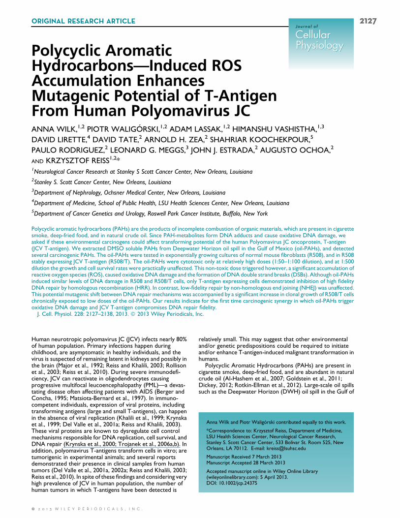
Polycyclic Aromatic Hydrocarbons—Induced ROS Accumulation Enhances Mutagenic Potential of T-Antigen From Human Polyomavirus JC ANNA WILK, 1,2 PIOTR WALIGO ´ RSKI, 1,2 ADAM LASSAK, 1,2 HIMANSHU VASHISTHA, 1,3 DAVID LIRETTE, 4 DAVID TATE, 2 ARNOLD H. ZEA, 2 SHAHRIAR KOOCHEKPOUR, 5 PAULO RODRIGUEZ, 2 LEONARD G. MEGGS, 3 JOHN J. ESTRADA, 2 AUGUSTO OCHOA, 2 AND KRZYSZTOF REISS 1,2 * 1 Neurological Cancer Research at Stanley S Scott Cancer Center, New Orleans, Louisiana 2 Stanley S. Scott Cancer Center, New Orleans, Louisiana 3 Department of Nephrology, Ochsner Medical Center, New Orleans, Louisiana 4 Department of Medicine, School of Public Health, LSU Health Sciences Center, New Orleans, Louisiana 5 Department of Cancer Genetics and Urology, Roswell Park Cancer Institute, Buffalo, New York Polycyclic aromatic hydrocarbons (PAHs) are the products of incomplete combustion of organic materials, which are present in cigarette smoke, deep-fried food, and in natural crude oil. Since PAH-metabolites form DNA adducts and cause oxidative DNA damage, we asked if these environmental carcinogens could affect transforming potential of the human Polyomavirus JC oncoprotein, T-antigen (JCV T-antigen). We extracted DMSO soluble PAHs from Deepwater Horizon oil spill in the Gulf of Mexico (oil-PAHs), and detected several carcinogenic PAHs. The oil-PAHs were tested in exponentially growing cultures of normal mouse fibroblasts (R508), and in R508 stably expressing JCV T-antigen (R508/T). The oil-PAHs were cytotoxic only at relatively high doses (1:50–1:100 dilution), and at 1:500 dilution the growth and cell survival rates were practically unaffected. This non-toxic dose triggered however, a significant accumulation of reactive oxygen species (ROS), caused oxidative DNA damage and the formation of DNA double strand breaks (DSBs). Although oil-PAHs induced similar levels of DNA damage in R508 and R508/T cells, only T-antigen expressing cells demonstrated inhibition of high fidelity DNA repair by homologous recombination (HRR). In contrast, low-fidelity repair by non-homologous end joining (NHEJ) was unaffected. This potential mutagenic shift between DNA repair mechanisms was accompanied by a significant increase in clonal growth of R508/T cells chronically exposed to low doses of the oil-PAHs. Our results indicate for the first time carcinogenic synergy in which oil-PAHs trigger oxidative DNA damage and JCV T-antigen compromises DNA repair fidelity. J. Cell. Physiol. 228: 2127–2138, 2013. 2013 Wiley Periodicals, Inc. Human neurotropic polyomavirus JC (JCV) infects nearly 80% of human population. Primary infections happen during childhood, are asymptomatic in healthy individuals, and the virus is suspected of remaining latent in kidneys and possibly in the brain (Major et al., 1992; Reiss and Khalili, 2003; Rollison et al., 2003; Reiss et al., 2010). During severe immunodefi- ciency, JCV can reactivate in oligodendrocytes causing progressive multifocal leucoencephalopathy (PML)—a devas- tating disease often affecting patients with AIDS (Berger and Concha, 1995; Matsiota-Bernard et al., 1997). In immuno- competent individuals, expression of viral proteins, including transforming antigens (large and small T-antigens), can happen in the absence of viral replication (Khalili et al., 1999; Krynska et al., 1999; Del Valle et al., 2001a; Reiss and Khalili, 2003). These viral proteins are known to dysregulate cell control mechanisms responsible for DNA replication, cell survival, and DNA repair (Krynska et al., 2000; Trojanek et al., 2006a,b). In addition, polyomavirus T-antigens transform cells in vitro; are tumorigenic in experimental animals; and several reports demonstrated their presence in clinical samples from human tumors (Del Valle et al., 2001a, 2002a; Reiss and Khalili, 2003; Reiss et al., 2010). In spite of these findings and considering very high prevalence of JCV in human population, the number of human tumors in which T-antigens have been detected is relatively small. This may suggest that other environmental and/or genetic predispositions could be required to initiate and/or enhance T-antigen-induced malignant transformation in humans. Polycyclic Aromatic Hydrocarbons (PAHs) are present in cigarette smoke, deep-fried food, and are abundant in natural crude oil (Al-Hashem et al., 2007; Goldstein et al., 2011; Dickey, 2012; Rotkin-Ellman et al., 2012). Large-scale oil spills such as the Deepwater Horizon (DWH) oil spill in the Gulf of Anna Wilk and Piotr Waligo ´ rski contributed equally to this work. *Correspondence to: Krzysztof Reiss, Department of Medicine, LSU Health Sciences Center, Neurological Cancer Research, Stanley S. Scott Cancer Center, 533 Bolivar St. Room 525, New Orleans, LA 70112. E-mail: [email protected] Manuscript Received 7 March 2013 Manuscript Accepted 28 March 2013 Accepted manuscript online in Wiley Online Library (wileyonlinelibrary.com): 5 April 2013. DOI: 10.1002/jcp.24375 ORIGINAL RESEARCH ARTICLE 2127 Journal of Journal of Cellular Physiology Cellular Physiology 2013 WILEY PERIODICALS, INC.
-
Upload
independent -
Category
Documents
-
view
1 -
download
0
Transcript of Polycyclic aromatic hydrocarbons-induced ROS accumulation enhances mutagenic potential of T-antigen...
Polycyclic AromaticHydrocarbons—Induced ROSAccumulation EnhancesMutagenic Potential of T-AntigenFrom Human Polyomavirus JCANNA WILK,1,2 PIOTR WALIGORSKI,1,2 ADAM LASSAK,1,2 HIMANSHU VASHISTHA,1,3
DAVID LIRETTE,4 DAVID TATE,2 ARNOLD H. ZEA,2 SHAHRIAR KOOCHEKPOUR,5
PAULO RODRIGUEZ,2 LEONARD G. MEGGS,3 JOHN J. ESTRADA,2 AUGUSTO OCHOA,2
AND KRZYSZTOF REISS1,2*1Neurological Cancer Research at Stanley S Scott Cancer Center, New Orleans, Louisiana2Stanley S. Scott Cancer Center, New Orleans, Louisiana3Department of Nephrology, Ochsner Medical Center, New Orleans, Louisiana4Department of Medicine, School of Public Health, LSU Health Sciences Center, New Orleans, Louisiana5Department of Cancer Genetics and Urology, Roswell Park Cancer Institute, Buffalo, New York
Polycyclic aromatic hydrocarbons (PAHs) are the products of incomplete combustion of organic materials, which are present in cigarettesmoke, deep-fried food, and in natural crude oil. Since PAH-metabolites form DNA adducts and cause oxidative DNA damage, weasked if these environmental carcinogens could affect transforming potential of the human Polyomavirus JC oncoprotein, T-antigen(JCV T-antigen). We extracted DMSO soluble PAHs from Deepwater Horizon oil spill in the Gulf of Mexico (oil-PAHs), and detectedseveral carcinogenic PAHs. The oil-PAHs were tested in exponentially growing cultures of normal mouse fibroblasts (R508), and in R508stably expressing JCV T-antigen (R508/T). The oil-PAHs were cytotoxic only at relatively high doses (1:50–1:100 dilution), and at 1:500dilution the growth and cell survival rates were practically unaffected. This non-toxic dose triggered however, a significant accumulation ofreactive oxygen species (ROS), caused oxidative DNA damage and the formation of DNA double strand breaks (DSBs). Although oil-PAHsinduced similar levels of DNA damage in R508 and R508/T cells, only T-antigen expressing cells demonstrated inhibition of high fidelityDNA repair by homologous recombination (HRR). In contrast, low-fidelity repair by non-homologous end joining (NHEJ) was unaffected.This potential mutagenic shift between DNA repair mechanisms was accompanied by a significant increase in clonal growth of R508/T cellschronically exposed to low doses of the oil-PAHs. Our results indicate for the first time carcinogenic synergy in which oil-PAHs triggeroxidative DNA damage and JCV T-antigen compromises DNA repair fidelity.J. Cell. Physiol. 228: 2127–2138, 2013. 2013 Wiley Periodicals, Inc.
Human neurotropic polyomavirus JC (JCV) infects nearly 80%of human population. Primary infections happen duringchildhood, are asymptomatic in healthy individuals, and thevirus is suspected of remaining latent in kidneys and possibly inthe brain (Major et al., 1992; Reiss and Khalili, 2003; Rollisonet al., 2003; Reiss et al., 2010). During severe immunodefi-ciency, JCV can reactivate in oligodendrocytes causingprogressive multifocal leucoencephalopathy (PML)—a devas-tating disease often affecting patients with AIDS (Berger andConcha, 1995; Matsiota-Bernard et al., 1997). In immuno-competent individuals, expression of viral proteins, includingtransforming antigens (large and small T-antigens), can happenin the absence of viral replication (Khalili et al., 1999; Krynskaet al., 1999; Del Valle et al., 2001a; Reiss and Khalili, 2003).These viral proteins are known to dysregulate cell controlmechanisms responsible for DNA replication, cell survival, andDNA repair (Krynska et al., 2000; Trojanek et al., 2006a,b). Inaddition, polyomavirus T-antigens transform cells in vitro; aretumorigenic in experimental animals; and several reportsdemonstrated their presence in clinical samples from humantumors (Del Valle et al., 2001a, 2002a; Reiss and Khalili, 2003;Reiss et al., 2010). In spite of these findings and considering veryhigh prevalence of JCV in human population, the number ofhuman tumors in which T-antigens have been detected is
relatively small. This may suggest that other environmentaland/or genetic predispositions could be required to initiateand/or enhance T-antigen-induced malignant transformation inhumans.
Polycyclic Aromatic Hydrocarbons (PAHs) are present incigarette smoke, deep-fried food, and are abundant in naturalcrude oil (Al-Hashem et al., 2007; Goldstein et al., 2011;Dickey, 2012; Rotkin-Ellman et al., 2012). Large-scale oil spillssuch as the Deepwater Horizon (DWH) oil spill in the Gulf of
Anna Wilk and Piotr Waligorski contributed equally to this work.
*Correspondence to: Krzysztof Reiss, Department of Medicine,LSU Health Sciences Center, Neurological Cancer Research,Stanley S. Scott Cancer Center, 533 Bolivar St. Room 525, NewOrleans, LA 70112. E-mail: [email protected]
Manuscript Received 7 March 2013Manuscript Accepted 28 March 2013
Accepted manuscript online in Wiley Online Library(wileyonlinelibrary.com): 5 April 2013.DOI: 10.1002/jcp.24375
ORIGINAL RESEARCH ARTICLE 2127J o u r n a l o f
J o u r n a l o f
Cellular
Physiology
Cellular
Physiology
2 0 1 3 W I L E Y P E R I O D I C A L S , I N C .

Mexico in 2010, contribute to the overall increase in PAHsconcentration in water, soil, and air. Although chronic effectsof low doses of PAHs on human health are not well defined,several PAHs are classified as potent carcinogens, which canform covalent DNA adducts and cause oxidative DNA damage(Seike et al., 2003; Xue and Warshawsky, 2005; Ji et al., 2010;Liu et al., 2010). These primary DNA lesions may result in theaccumulation of random mutations when cellular mechanismsresponsible for DNA repair fidelity are compromised. In thisregard, our previous studies demonstrate that JCV largeT-antigen (JCV T-antigen), in addition to its well documentedrole in reactivating cell proliferation (Brodsky and Pipas, 1998;Sullivan et al., 2000), can compromise DNA repair fidelity byinhibiting homologous recombination directed DNA repair(HRR) (Reiss et al., 2006; Trojanek et al., 2006a,b; Reisset al., 2012).
In the present study we ask if PAHs extracted from thenatural crude oil affect transforming potential of the JCVT-antigen. We demonstrate that DMSO soluble PAHsobtained from the 2010 DWH oil spill (oil-PAHs) contain amixture of at least five carcinogenic PAHs. Chronic cellexposure to the oil-PAHs resulted in excessive ROSaccumulation, which resulted in oxidative DNA damage andtriggered DNA double strand breaks (DSBs) in cells replicatingDNA. Importantly, ROS scavengers, N-acetyl cysteine (NAC)and Trolox, attenuated both ROS accumulation and DNAdamage. In addition, JCV T-antigen positive cells exposed tolow doses of the oil-PAHs, demonstrated a shift from HRR toless faithful NHEJ, which correlated with a significant increasein their clonogenic growth. Considering that oil-PAH-inducedoxidative DNA damage potentiates carcinogenic effects of JCvirus, and that polyomaviruses are ubiquitous in humans,vaccination against the virus and the use of antioxidants shouldbe considered at least for populations at high risk of long-termexposure.
Materials and MethodsPAHs samples
Natural crude oil was obtained from 2010 Deep Water Horizon(DWH) oil spill in the Gulf of Mexico. The sample was collectedfrom BP-MS Canyon 252 (Discover Enterprise sample #011obtained in May 20, 2010 at 11:15 am) and transported to the lab inair- and light-tight bottle at 4˚C. For DMSO extraction of aromaticcomponents, crude oil was diluted in DMSO (1:100, v/v). Theobtained mixture was stirred at room temperature for 4 h withoccasional gentle mixing every 30min. Next, the surface oilfraction was removed and the oil-free solution was diluted 10times in double distillated water. After mixing, the solution wasfiltered and frozen at 20˚C in 5ml aliquots (oil-PAHs).
High performance liquid chromatography (HPLC)
The PAH-DMSO extracts were analyzed by HPLC (DionexUltimate 3000; Sunnyvale, CA) with a photodiode array UVdetector. Chromatographic separation was achieved using a 150-by 4.6-mm (inner diameter) C18 reverse-phase column (particlesize, 3mm; ACCLAIM 120 C-18). The column was equilibrated atflow rate 0.5ml/min with mobile-phase consisting of 60% waterand 40% acetonitrile followed by a linear gradient to 100%acetonitrile. Detection was monitored with UV at an absorbanceof 254 nm. The obtained peaks of oil-PAHs were identified andquantified by comparing themwith corresponding retention times,spectra, and concentrations of the standard PAHs (Fig. 1C).
Cell culture
To analyze potential mutagenic interaction between oil-PAHs andviral oncoprotein, JCV T-antigen, we used JCV T-antigen positive(R508/T) and negative (R508) mouse embryo fibroblasts, as well as
R508 cells stably expressing DRGFP reporter (R508/DRGFP)—toevaluate HRR. All R508 cells, including R508/T, although areimmortalized, do not grow well in semi-solid culture conditions(soft agar; Del Valle et al., 2002b). These cells were generated andcharacterized in our previous studies (Del Valle et al., 2002b) andare well suited to evaluate changes in cell proliferation, survival,transformation and DNA repair (Trojanek et al., 2003). The cellsare routinely cultured in DMEM (GIBCO BRL, Grand Island, NY)supplemented with 10% FBS (Sigma, St. Louis, MO), 50U/mlpenicillin, 50 ng/ml streptomycin, and 250mg/ml hygromycin at 37˚C in a 5% CO2 atmosphere. In some experiments oil-PAHs wereadded to culture media in dilutions ranging from 1:50 to 1:1,000 inthe presence or absence of ROS scavengers, N-acetyl cysteine(NAC, 1mM) and Trolox (100mM).
ROS measurements
Accumulation of reactive oxygen species (ROS) was evaluatedfollowing cell exposure to oil-PAHs for 72 h. The oil-PAHs weretested in two dilutions (1:100 and 1:500 in DMEM 10% FBS). Thecells were also treated with the mixture of PAHs (mix-PAHs)prepared accordingly to the content of PAHs detected in DMSOfraction fromDWHnatural crude oil (Fig. 1). Intracellular ROSwasmeasured in Synergy 2 micro-plate reader by utilizing 20,70-Dichlorofluorescin diacetate (H2DCFDA), which is cellpermeable, non-fluorescent compound. When H2DCFDA isoxidized it changes into the green fluorescent dye, fluorescein.After exposure to oil-PAHs, for 72 h, the cells were washed withPBS and incubated in 1ml of phenol red-free culture mediumcontaining 10ml of H2DCFDA (stock solution: 50mg H2DCFDAin 86.5ml DMSO) in the dark at 37˚C for 30min. The cells werewashed with PBS again, and 250 ul phenol red-free culture media(without H2DCFDA) was placed into each well. The fluorescencewas measured (excitation/494 nm; emission/521 nm) every 5minfor 30min. ROS accumulation was expressed as fluorescenceintensity unit per time and cell number. In some experiments, thecells preloaded with H2DCFDA were analyzed using flowcytometry (GUAVA easyCyte 8HT).
Cell cycle distribution and cell survival
Briefly, the aliquots of 1 106 cells/ml were fixed in 70% ethanol at20˚C, overnight. The cells were centrifuged at 1,600 rpm and theresulting pellets suspended in 1ml of freshly prepared PropidiumIodide/RNaseA solution. Cell cycle distribution was evaluated inGUAVA easyCyte 8HT using GuavaSoft 1.1. Cell death wasevaluated by the assay based on cell membrane integrity usingViaCount reagents (Millipore, Billerica, MA) and Guava/ViaCountsoftware was used for data analysis.
Neutral comet assay
Neutral comet assay was utilized to detect DNA strand breaks inexponentially growing JCV T-antigen positive and negative R508 cellsin the presence and absence of oil-PAHs. The cells treatedwithH2O2(oxidative DNA damage) or neocarzinostatin (NCS; Sigma) wereused as positive controls for secondary and primary DNA doublestrand breaks (DSBs), respectively. The cells were subjected to singlecell electrophoresis in neutral conditions (Collins et al., 2008) andprocessed by utilizing SYBR green based kit from Trevigen andAutomated Comet Assay System from Loats Associates, Inc. The tailmoment was calculated from 100 cells collected per singlemeasurement in triplicate using Automated Comet Assay Systemsoftware (Loats Associates, Inc., Westminster, MD).
Immunofluorescence
All cells were cultured on glass culture slides (BD Falcon, FranklinLakes, NJ). For immunostaining the cells were fixed andpermeablized with the buffer containing 0.02% Triton X-100 and
JOURNAL OF CELLULAR PHYSIOLOGY
2128 W I L K E T A L .

4% formaldehyde in PBS. 8-hydroxy-2-deoxyguanosine(8-oxo-dG) was detected by mouse anti-8-oxo-dG monoclonalantibody (Millipore) followed by AlexaFluor-conjugated anti-mouse secondary antibody (Invitrogen, Carlsbad, CA).Phosphorylated form of histone H2AX (S139; gH2AX) wasdetected by rabbit polyclonal antibody (Millipore), and rhodamine-conjugated goat anti-rabbit secondary antibody (Molecular Probes,Eugen, OR). The images were visualized with the Nikon EclipseE400 upright fluorescence microscope equipped with EXI aquacamera (Qimaging, Surrey, BC), motorized Z-axis, and SlideBook5acquisition/deconvolution software (Intelligent ImagingInnovations, Inc., Denver, CO). Quantification of 8-oxo-dG andgH2AX labeling was performed by utilizing Mask analysis includedin SlideBook5 software according to manufacturerrecommendation (Intelligent Imaging Innovations, Inc.).
Oxidative DNA damage
8-oxo-dG immunolabeling was performed on formaldehyde-fixedmonolayer cultures of R508 and R508/T cells treated with
oil-PAHs (1:500 dilution) for 14 days. The content of 8-oxo-dG inurine was detected by ELISA kit provided by ACE, measured bySynergy-2 microplate-reader (BioTek, Winooski, VT), andcalculated by the Gen5 software. Parallel urine samples wereanalyzed for urinary creatinine (ELISA kit; ExoCell, Inc.,Philadelphia, PA) to normalize 8-oxo-dG content. The urinesamples were obtained from SV129mice (3 months old) fed once aday with vehicle (50ml of DMSO) or with oil-PAHs (50ml ofundiluted DMSO-PAHs fraction) administered by oral gavage.Every seventh day, mice were transferred to metabolic cages(TechniPlast, Buguhhiate, VA) and urine samples were collectedfor 24 h. The oil-PAHs dilution was calculated per body weight ofthe animal, and was approximated as 1:800.
Lipid peroxidatoion
Accumulation of Malondialdehyde (MAD) as a natural product oflipid peroxidationwas evaluated using MDAAssay Kit according tothe manufacturer recommendations (BioVision, Milpitas, CA) incells exposed to the oil-PAHs (1:100) for 72 h.
Fig. 1. High performance liquid chromatography (HPLC) of DMSO extracts from 2010 Deepwater Horizon oil spill in the Gulf of Mexico.Part A: The histogram of DMSO extract of natural crude oil generated by HPLC. The obtained peaks of the oil-PAHs were identified andquantified by comparing their retention times, spectra, and concentrations with a collection of 13 standard PAHs (Part C), which wereseparated at the same HPLC conditions. Part B: Quantification of the oil-PAHs using three independent DMSO extractions of the samesample of natural crude oil. Each DMSO fraction contained five major peaks, which correspond to the indicated carcinogenic PAHs. Datarepresent average concentrations standard deviation (SD) from three separate extractions in triplicate (n 9).
JOURNAL OF CELLULAR PHYSIOLOGY
P A H s - I N D U C E D R O S E N H A N C E S J C V I R U S M U T A G E N E S I S 2129
DNA repair of double strand breaks (DSBs)
To evaluate NHEJ, we have used previously described cell-freeassay with some modifications (Trojanek et al., 2006b). NHEJreaction mix contained: 50mg of nuclear lysate, 1mM ATP,0.25mM deoxynucleoside triphosphates, 25mM Tris acetate (pH7.5), 100mM potassium acetate, 10mM magnesium acetate, and1mM dithiothreitol. After 5min of preincubation at 37˚C, it wassupplemented with the substrate [500 ng of XhoI-linearizedpBluescript KS()]. The reaction mixture was incubated for 1 h at37˚C to ligate the plasmid and was treated with proteinase K (1mgper reaction at 62˚C for 60min) to digest DNA-bound proteins.NHEJ products were resolved in a 0.6% agarose gel containingethidium bromide (0.5mg/ml).
HRR was assessed using the reporter plasmid pDR-GFP (Pierceet al., 1999) generously provided by M. Jasin (Sloan-KetteringCancer Center). A stable clone of R508 cells expressing theconstruct (R508/DRGFP) was previously characterized (Trojaneket al., 2003). Briefly, the pDR-GFP reporter contains inactive greenfluorescent protein (GFP) gene (SceGFP) as a recombinationreporter and a fragment of the GFP gene as a donor forhomologous repair. The SceGFP cassette has an inactivatinginsertion, which consists of two stop codons and a restriction sitefor the rare-cutting endonuclease I-SceI. When I-SceI is expressedin DR-GFP-expressing clones, it inflicts DSBs within the SceGFPfragment, providing a signal for homologous recombination andthe reconstruction of functional GFP (Pierce et al., 1999).
Clonogenic growth
Exponentially growing R508 and R508/T cells (10% FBS) wereeither treated with DMSO (control) or with oil-PAHs 1:500 for4 weeks. Next the cells were washed with fresh serum-freemedium, trypsinized and transferred to 35mm culture dishes atclonal-density (1 103 cells/35mm dish). Clonogenic growth wasevaluated after additional 2 weeks of a continuous cell growth inmedia containing 10% FBSDMSO or 10% FBS PAHs 1:500.The resulting clones were fixed and stained with 0.25% cristalviolet in methanol.
Statistical analysis
The data were analyzed with homoscedastic Student’s t-test. Thedifferences between the control and experimental groups wereconsidered significant and marked with asterisk () for P valueslower than 0.05.
ResultsPAH content in DMSO crude oil fraction
We have characterized DMSO soluble PAHs from 2010Deepwater Horizon (DWH) oil spill in the Gulf of Mexico(oil-PAHs). Figure 1 demonstrates results from HPLC analysisof this fraction (Parts A and B). The HPLC parameters were setfor detection and quantification of oil-PAHs using a mixture of13 standard PAHs (Part C). The peaks, which demonstratedidentical retention times and identical spectral profiles(excitation, emission, and absorption maxima) in comparisonwith standard PAHs, were selected. This approach allowedidentification and quantification of the following PAHs:fluoranthene (26.6mg/ml), pyrene (11.1mg/ml), benzo-a-pyrene (8.1mg/ml), phenanthrene (3.9mg/ml), and chrysene(3.8mg/ml), which according to NCI Chemical CarcinogenReference Standards Repository, Catalog 2010, arecarcinogenic. We have tested the obtained oil-PAHs in cellculture and in mice using subsequent dilutions: 1:50, 1:100,1:500, 1:800, and 1:1,000. The last column in Figure 1Bdemonstrates final concentrations of the five oil-PAHs at 1:500dilution. Some experiments were repeated with purified PAHs
(Fig. 1C, highlighted yellow), which were pre-mixed and dilutedin DMSO according to the PAH-content found in DWH crudeoil DMSO extract, and are labeled as mix-PAHs.
Cytostatic and cytotoxic effects of oil-PAHs
We evaluated first how different dilutions of oil-PAHs affectcell proliferation and cell survival. This was critically importantin view of a possible contribution of PAHs in supportingmutagenic action of JCV T-antigen. If PAHs could indeedpotentiate malignant transformation by JCV T-antigen, affectedcells should be able to proliferate during the treatment. Only insuch condition acquired mutations could be passed to the nextgeneration contributing to the selection of clones with growthand/or survival advantage. The results in Figure 2 show cellcycle analysis (Part A), cell survival (Part B), and cell growth(Part C) of R508 and R508/T cells exposed to the oil-PAHs.Interestingly, cell cycle distribution (Part A) and cell growth(Part C) were practically the same in DMSO control culturesand in cultures exposed to the oil-PAHs at 1:1,000 and 1:500dilutions. Only at higher doses (oil-PAHs diluted 1:50 and1:100), both R508 and R508/T were characterized by G1arrest. In respect to cell viability, we did not observe anymeasurable cell death at 1:1,000 and 1:500 dilutions. At 1:100,oil-PAHs induced cell death in 12% and 7% of R508 and R508/T,respectively (Part B), which was accompanied by aproportional decline in the growth rate (Part C). Finally, at 1:50dilution oil-PAHs were highly cytotoxic to both cell lines, andcell death levels were comparable to those detected in thepresence of DNA-damaging agent, cisplatin (1mg/ml). Theseresults allowed us to select 1:500 dilution in which both R508and R508/T cells were able to proliferate for many generations.
PAH-induced ROS accumulation and oxidative DNAdamage
In spite of the low cytotoxicity, oil-PAHs at 1:500 dilutiontriggered accumulation of the reactive oxygen species(ROS; Fig. 3A,B). The kinetics of ROS accumulation wereevaluated in living cells pre-loaded with ROS sensitivefluorescent dye, 20,70-dichlorofluorescin-diacetate(H2DCFDA). The results in Figure 3A show that in comparisonto DMSO-treated controls, R508 cells treated with oil-PAHs,1:500 and 1:100 dilution, demonstrated over fivefold andsevenfold increase in ROS-dependent fluorescence,respectively. This PAH-induced ROS accumulation waseffectively quenched in the presence of ROS scavenger, NAC.We have confirmed these findings by flow cytometry-basedmeasurement of ROS (Fig. 3B). In addition, we have comparedROS levels in R508 cells, which were either exposed to the oil-PAHs or to the individual PAHs premixed in DMSO in theproportion corresponding to the content of PAHs in thenatural crude oil (Fig. 1). The results in Figure 3B demonstratedsixfold and threefold increase in ROS accumulation when oil-PAHs (1:500), or the mixture of synthetic PAHs (mix-PAHs;1:500) were used, respectively. Although both oil-PAHs andmix-PAHs triggered ROS accumulation, oil-PAHs aresignificantly more potent (Fig. 3B; compare second and fourthpart in the first row). This discrepancy could indicate that othercomponents extracted from the natural crude oil alsocontribute to the observed ROS accumulation. We have alsoevaluated effects of the oil-PAHs on ROS accumulation in cellsexpressing JCV T-antigen. In comparison to R508, R508/Tcells demonstrated significantly higher accumulation of ROSin both control conditions (DMSO) and following PAHtreatment (Fig. 3B; compare date in the first [R508] and second[R508/T] row). Also here ROS scavengers, NAC, and lesseffectively, Trolox, attenuated PAH-induced ROSaccumulation.
JOURNAL OF CELLULAR PHYSIOLOGY
2130 W I L K E T A L .

This intracellular ROS accumulation observed in thepresence of the oil-PAHs was accompanied by elevatedoxidative DNA damage, detected here by 8-Oxo-20-deoxyguanosine (8-oxo-dG) immunolabeling. 8-oxo-dG, is astable product of DNA repair, which can be detected incytoplasm, extracellular matrix, blood, saliva, and urine (Loftet al., 1999; Poulsen et al., 1999; Park et al., 2005). The results inFigure 3C show a strong cytosolic immunolabeling with anti-8-oxo-dG antibody (green fluorescence) following 14 days of acontinuous cell exposure to oil-PAHs (1:500). Surprisingly,
detected oxidative DNA damage, both basal (DMSO) andPAH-induced was similar in R508 and R508/T cells, despite of asignificantly higher level of ROS in T-antigen expressing cells(compare Fig. 3B,C).
At this point we assumed that PAH-induced ROS isgenerated mostly inside the cell. This is because intracellularaldo-keto reductase activity is necessary to convert PAH-metabolites to PAH-o-quinones, which are strong electronacceptors, and can cause extensive oxidative damage(Burczynski and Penning, 2000; Park et al., 2008). To determine
Fig. 2. Evaluation of cytostatic and cytotoxic effects of the oil-PAHs. Evaluation of cell cycle distribution (Part A), cell viability (Part B), andcell number (Part C) during chronic cell exposure to the oil-PAHs. Exponentially growing R508 and R508/T cells (DMEM 10% FBS) wereincubated with different dilutions of the oil-PAHs ranging from 1:50 to 1:1,000. The cells incubated in DMSO (vehicle) were used as negativecontrol and cells incubated with cisplatin (1mg/ml) were used as positive control for the evaluation of a potential cytostatic and cytotoxiceffects of the oil-PAHs. In all parts data represent average valuesSD from at least three independent experiments in triplicates. In part A,indicates values which are significantly different from the corresponding values of cell cycle distribution (G1, S, G2/M, and sub G1) in cellcultures treated with DMSO. In parts B and C, indicates values which are statistically different from the values of cell survival (B) and cellnumber (C) obtained in control conditions (paired Student’s t-test P 0.05).
JOURNAL OF CELLULAR PHYSIOLOGY
P A H s - I N D U C E D R O S E N H A N C E S J C V I R U S M U T A G E N E S I S 2131

Fig. 3.
JOURNAL OF CELLULAR PHYSIOLOGY
2132 W I L K E T A L .
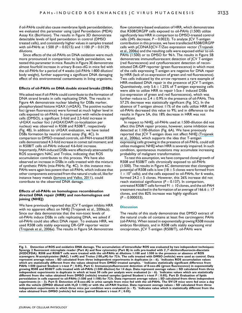
if oil-PAHs could also cause membrane lipids peroxidatidation,we evaluated this parameter using Lipid Peroxidation (MDA)Assay Kit (BioVision). The results in Figure 3D demonstratedetectable levels of lipid peroxidation in control (DMSOtreated cells), and consistently higher values in samples treatedwith oil-PAHs at 1:500 (P 0.0215) and 1:100 (P 0.0129)dilutions.
Since effects of the oil-PAHs on DNA oxidation were muchmore pronounced in comparision to lipids peroxidation, wetested this parameter in mice. Results in Figure 3E demonstratealmost fourfold increase in urinary 8-oxo-dG in mice fed withthe oil-PAHs for a period of 4 weeks (oil-PAHs 1:800 dilution/body weight), further supporting a significant DNA damagingeffect of this environmental contaminants in living organisms.
Effects of oil-PAHs on DNA double strand breaks (DSBs)
Weasked next if oil-PAHs could contribute to the formation ofDNA strand breaks in cells replicating DNA. The results inFigure 4A demonstrate nuclear labeling for DSBs marker,phosphorylated histone H2AX (gH2AX). The positive nuclearfoci (green fluorescence) were formed at much higher level incells exposed to oil-PAHs. In comparison with vehicle-treatedcells (DMSO), a significant 3-fold and 2.5-fold increase ingH2AX nuclear foci (gH2AX/DAPI co-localization pernucleus) was observed in R508 and R508/T, respectively(Fig. 4B). In addition to gH2AX evaluation, we have testedDSBs formation by neutral comet assay (Fig. 4C). Incomparison to DMSO-treated controls, oil-PAH-treated R508cells demonstrated 4.3-fold increase in comet tail moment; andin R508/T cells oil-PAHs induced 4.6-fold increase.Importantly, PAH-inducedDSBswere effectively attenuated byROS scavengers NAC and Trolox, indicating that ROSaccumulation contributes to this process. We have alsoobserved an increase in DSBs in cells treated with the mixtureof synthetic PAHs (mix-PAHs; Fig. 4C). In this instance mix-PAHs were again less potent than oil-PAHs, which indicate thatother components extracted from the natural crude oil, like forinstance heavy metals (Jomova and Valko, 2011), couldcontribute to the observed DNA damage.
Effects of oil-PAHs on homologous recombinationdirected DNA repair (HRR) and non-homologous endjoining (NHEJ)
We have previously reported that JCV T-antigen inhibits HRRwith no apparent effect on NHEJ (Trojanek et al., 2006a,b).Since our data demonstrate that the non-toxic levels ofoil-PAHs induce DSBs in cells replicating DNA, we asked ifoil-PAHs could also affect DNA repair. To evaluate HRR, weused R508 cells stably expressing DR-GFP reporter vector(Trojanek et al., 2006a). The results in Figure 5A demonstrate
flow cytometry-based evaluation of HRR, which demonstratesthat R508/DRGFP cells exposed to oil-PAHs (1:500) utilizesignificantly less HRR in comparison to DMSO-treated controlcells (24% decrease; P 0.0023). To analyze JCV T-antigencontribution in this process, we have transfected R508/DRGFPcells with pCDNA3/JCV-T/Zeo expression vector (Trojaneket al., 2006a) and the resulting cells were exposed either to oil-PAHs (1:500) or to DMSO for 96 h. The results in Figure 5Bdemonstrate immunofluorescent detection of JCV T-antigen(red fluorescence) and cytofluorescent detection of recon-stituted DR-GFP reporter (green fluorescence) in which mostof the cells expressing T-antigen are negative for DNA repairby HRR (lack of co-expression of green and red fluorescence).Two cells indicated by the arrow represent a rare example ofHRR-mediated DNA repair in the presence of JCV T-antigen.Quantitatively, only 5.6 1.25% of T-antigen expressing cellswere able to utilize HRR to repair I-Sce-1 induced DSBs(co-expression of green and red fluorescence), which wasfurther reduce to 2.4 0.9% in the presence of oil-PAHs. This57.2% decrease was statistically significant (Fig. 5C). In theabsence of T-antigen almost 11% of the cells utilize HRR andoil-PAHs decreased this value to 9%; and in difference to theresults in Figure 5A, this 18% decrease in HRR was notsignificant.
In respect to NHEJ, oil-PAHs used at 1:500-dilution did notaffect this DNA repair process, however, some inhibition wasdetected at 1:100-dilution (Fig. 6A). We have previouslyreported that JCV T-antigen does not affect NHEJ (Trojaneket al., 2006a), which supports the idea that T-antigenexpressing cells growing in the presence of oil-PAHs, could stillutilize mutagenic NHEJ when HRR is severely impaired. In suchcondition, spontaneous mutations may accumulate increasingprobability of malignant transformation.
To test this assumption, we have compared clonal growth ofR508 and R508/T cells chronically exposed to oil-PAHs(1:500). The results in Figure 6C demonstrate that clonogenicpotential of R508 cells is low (18 5 clones were formed from1 103 cells); and the cells exposed to oil-PAHs, for 6 weeks,formed 24.2 5 clones. However, this 26% increase did notreach statistical significance (P 0.137). In comparison,untreated R508/T cells formed 91 10 clones, and the oil-PAHtreatment resulted in the formation of an average of 166.6 17clones, and this 82% increase was highly significant(P 0.000033).
Discussion
The results of this study demonstrate that DMSO extract ofthe natural crude oil contains at least five carcinogenic PAHs(oil-PAHs).When tested in exponentially growing R508mouseembryo fibroblasts, and in R508 cells stably expressing viraloncoprotien, JCV T-antigen (R508/T), oil-PAHs were
Fig. 3. Detection of ROS and oxidative DNA damage. The accumulation of intracellular ROS was evaluated by two independent techniques:Synergy 2 fluorescent microplate reader (Part A) and flow cytometry (Part B) in cells pre-loaded with 20,70-dichlorofluorescin-diacetate(H2DCFDA). R508 and R508/T cells were treated with the oil-PAHs at dilutions 1:100 and 1:500 in the presence or absence of ROSscavengers: N-acetylcysteine (NAC; 1mM) and Trolox (100mM) for 72h. The cells treated with DMSO (vehicle) were used as control. Datarepresent average valuesSD calculated from three independent experiments in duplicates (n 6). Indicates ROS accumulation valueswhich are statistically different from the values obtained from DMSO treated samples. Indicates statistically significant difference fromPAHs 1:500 (paired Student’s t-test P 0.05). Part C: Immunocytofluorescent detection of 8-oxo-dG (green fluorescence) in exponentiallygrowing R508 and R508/T cells treated with oil-PAHs (1:500 dilution) for 14 days. Data represent average valuesSD calculated from threeindependent experiments in duplicate in which at least 50 cells per analysis were evaluated (n 6). Indicates values which are statisticallydifferent from the value obtained from DMSO (vehicle) treated samples (paired Student’s t-test P 0.05). Part D: Evaluation of lipidsperoxidation in cells exposed to oil-PAHs (1:500 and 1:100) for 72 h. Data represent average valuesSD calculated from three independentexperiments (n 3). Part E: Detection of 8-oxo-dG in urine sample from mice fed with the oil-PAHs. The SV129 mice were fed once a daywith the vehicle (DMSO diluted with H2O 1:100) or with the oil-PAH fraction. Data represent average valuesSD calculated from threeindependent experiments in which three mice per condition were evaluated (n 9). Indicates value which is statistically different from thevalue obtained from DMSO (vehicle) fed mice (paired Student’s t-test P 0.05).
JOURNAL OF CELLULAR PHYSIOLOGY
P A H s - I N D U C E D R O S E N H A N C E S J C V I R U S M U T A G E N E S I S 2133

Fig. 4. PAHs-induced gH2AX nuclear foci and DSBs. Part A: Immunofluorescent detection of the phosphorylated form of histone H2AX(gH2AX, green fluorescence) in exponentially growing R508 and R508/T cells treated with oil-PAHs (1:500) for 3 days. Part B: Quantificationof nuclear gH2AX foci (DAPI/gH2AX co-localization). Data represent average valuesSD calculated from three independent experiments intriplicate in which at least 100 cells per analysis were evaluated (n 9). Part C: Neutral comet assay was utilized to detect DNA strand breaksin exponentially growing R508 and R508/T cells, which were exposed to the oil-PAHs (1:500-dilution) in the presence and absence of ROSscavengers NAC (1mM) and Trolox (100mM) for 14 days. The cells were subjected to single cell electrophoresis (comet assay) in neutralconditions. Data represent average comet tail momentSD calculated from three independent experiments (n 3); 100 cells per eachexperimental point were evaluated. Indicates values which are statistically different from DMSO treated samples. Indicates values whichare significantly different from oil-PAH treated samples. Indicates values significantly different from mix-PAHs (paired Student’s t-testP 0.05).
JOURNAL OF CELLULAR PHYSIOLOGY
2134 W I L K E T A L .

Fig. 5. Effects of oil-PAHs and JCV T-antigen on HRR. Part A: R508 cells expressing HRR reporter plasmid (R508/DRGFP) were transfectedin transient with DNA damaging pCbA-Sce expression vector and the cells were cultured in the presence of 10%FBSoil-PAHs (1:500) for96h. The cells capable of reconstituting GFP function were analyzed by flowcytomentry. The results were collected from three separateexperiments, in duplicate (n 6). Part B: Cytofluorescent detection of HRR in R508/DRGFP cells expressing JCV T-antigen. The cells weretransfected (nucleoporation; Amaxa) with pCDNA3/JCV-T/Zeo (Trojanek et al., 2006a) and with pCbA-Sce (Pierce et al., 1999) expressionvector. Following 24h of recovery after transfection, the cells were treated either with oil-PAHs (1:500) or with DMSO (control). JCV T-antigen was detected using anti-SV40 T-antigen mouse monoclonal antibody (Calbiochem; red fluorescence). DRGFP fluorescence isindicated green, and total nuclei are labeled with DAPI (white fluorescence). Arrow indicates two nuclei in which T-antigen expressing cellsare positive for HRR-mediated reconstitution of DR-GFP otherwise the majority of T-antigen expressing cells do not express green. Part C:Quantification of the results depicted in part B. The results were collected from three separate experiments, in duplicate in which 1,000 cellsper experiment were counted in at least 50 randomly selected microscopic fields. P-values are indicated above the compared samples (pairedStudent’s t-test P 0.05).
JOURNAL OF CELLULAR PHYSIOLOGY
P A H s - I N D U C E D R O S E N H A N C E S J C V I R U S M U T A G E N E S I S 2135

cytotoxic only at relatively high dose (oil-PAHs diluted 1:50),and at 1:500 dilution the exposed cells were able to proliferatefor many generations. However, at this non-toxic level oil-PAHs triggered intracellular ROS accumulation, and causedoxidative DNA damage, and DNA double strand breaks(DSBs). Although oil-PAHs induced similar DNA damage inR508 and R508/T cells, the impairment of high fidelity HRRwasmuch more evident in cells expressing JCV T-antigen. Incontrast, low-fidelity NHEJ was practically unaffected.Importantly, this potentially mutagenic shift fromHRR toNHEJwas accompanied by a significant increase in clonal growth ofR508/T cells chronically exposed to the oil-PAHs.
Multiple reports including our own studies indicate that JCvirus and PAHs are independent risk factors for tumordevelopment. For example, the viral oncoprotein, JCVT-antigen, is transforming in vitro (Sullivan et al., 2000; DelValle et al., 2002b), is tumorigenic in experimental animals (DelValle et al., 2001b), and has been detected in a variety of tumorclinical samples including lung (Abdel-Aziz et al., 2007) andcolon cancer (Del Valle and Khalili, 2010), and CNSmalignances (Khalili et al., 1999; Del Valle et al., 2001a,b). In
respect to PAHs, these common environmental contaminantshave been linked to skin, bladder, and lung cancer (Boffettaet al., 1997; Gammon and Santella, 2008; Sankpal et al., 2012).An elevated risk for brain tumors has been documented amongpetroleum refinery workers (Alexander et al., 1980; Demerset al., 1991; Carozza et al., 2000). In addition, one of the best-studied PAHs, benzo(a)pyrene, has been shown to inflictextensive DNA damage in different tissues (Demerset al., 1991).
Genotoxic effects of lipophilic PAHs require metabolicprocessing towards more hydrophilic intermediatemetabolites (Stowers and Anderson, 1985). In particular, PAHscan be metabolized to diol-epoxide derivatives by cytochromeP450-dependent monooxygenase and epoxide hydrolase,which are capable of forming covalent DNA adducts (Stowersand Anderson, 1985; Curfs et al., 2003). In addition, aldo-ketoreductase can further convert PAH-metabolites to PAH-o-quinones, which are strong electron acceptors, and can causeextensive oxidative damage (Burczynski and Penning, 2000;Park et al., 2008). Importantly, both oxidative DNA damageand DNA adducts can initiate DNA double strand breaks
Fig. 6. Effects of oil-PAHs on NHEJ. Part A: NHEJ was evaluated using cell-free assay based on the ability of nuclear extracts to ligatelinearized plasmid DNA (Trojanek et al., 2006b). Nuclear extracts were isolated from exponentially growing JCV T-antigen negative (R508)and positive (R508/T) cells, in the presence or absence of the oil-PAHs at 1:100 and 1:500 dilution. Levels of linear plasmid, as well as severalmultimers formed by NHEJ reaction are indicated. Part B: The histogram illustrating an average of densitometric measurements of NHEJproducts obtained from two separate NHEJ experiments. Part C: Clonogenic growth of R508 and R508/T cells exposed to the oil-PAH.Exponentially growing monolayer cultures of R508 and R508/T cells (10% FBS) were either treated with DMSO (control) or with oil-PAHs1:500 for 4 weeks. The clonogenic growth was evaluated 2 weeks after a continuous cell growth in media containing 10% FBSDMSO or 10%FBSoil-PAHs 1:500. Data represent average number of clonesSD calculated from three independent experiments in duplicate (n 6). Pvalues are indicated above the compared samples (paired Student’s t-test P 0.05).
JOURNAL OF CELLULAR PHYSIOLOGY
2136 W I L K E T A L .
(DSBs) when left unrepaired in cells replicating DNA(Hoeijmakers, 2001; Nakanishi et al., 2011). Indeed, our resultsin Figure 4 confirm such a possibility in our experimentalsetting. In addition, PAH-induced DSBs diminished significantlyin the presence of ROS scavengers, NAC and Trolox (Fig. 4),suggesting that oxidative DNA damage may indeed contributeto this potentially mutagenic process.
DSBs activate DNA-dependent kinases such as DNA PK,ATM, and ATR (Hoeijmakers, 2001; Wilk et al., 2012), whichphosphorylate histone H2AX at the sites surrounding DSBs,leading to the recruitment of two major DSBs repairmechanisms: HRR, which is considered faithful; and less faithfulNHEJ (Reiss et al., 2006). It has been also proposed that aproper balance between HRR and NHEJ may play a role in themaintenance of genomic integrity (Hoeijmakers, 2001; Reisset al., 2006; Kass and Jasin, 2010). However, if HRR is impairedand unrepaired DSBs are processed by NHEJ, it may lead to theaccumulation of randommutations especially in cells replicatingDNA. In this respect, our previous studies demonstrated thatJCV T-antigen, in addition to its well-documented role intriggering abnormal cell proliferation, inhibited HRR andcompromised DNA repair fidelity (Reiss et al., 2006; Trojaneket al., 2006a,b). Therefore, mutagenic conditions may occur ifthe cells attempt to repair PAH-inflicted DSBs in the presenceof JCV T-antigen. Although accumulation of random mutationsdoes not necessary meanmalignant transformation, our resultsin Figure 6C demonstrate a significant increase in clonal growthof T-antigen expressing cells chronically exposed to oil-PAHs,which could provide the first step in the process of malignanttransformation. Since large-scale oil spills increase overall PAHconcentrations in the air, soil, and water, the long-term healthimpact could be significant, especially in the populations fromthe affected coastal areas. It should be emphasized, however,that despite of this apparent in vitro correlation betweenexpression of JCV T-antigen, cell exposure to oil-PAHs, andelevated clonogenicity, it is difficult to approximate how theoil-PAH dilutions we are using in this study translate to the realuptake and accumulation of these carcinogens in the humanbody.
In conclusion, our in vitro study indicates the presence of asynergistic action between viral (latent infection with JC virus)and environmental (PAH contamination) factors, whichdysregulate natural control mechanisms responsible for themaintenance of genomic integrity and uncontrolled DNAreplication. Importantly, the abundance of oxidative DNAdamage detected in cells and experimental animals chronicallyexposed to oil-PAHs may suggest the use of antioxidants tocounteract this potential carcinogenic action. Finally, JC virusproteins in association with elevated urinary 8-oxo-dG mayhelp in identifying high-risk populations in danger of tumordevelopment in the aftermath of this industrial disaster andfuture insults.
Acknowledgments
This work is supported by 2RO1 CA095518 (K.R.) and P20RR021970 (A.O., K.R.).
Literature Cited
Abdel-Aziz HO, Murai Y, Hong M, Kutsuna T, Takahashi H, Nomoto K, Murata S,Tsuneyama K, Takano Y. 2007. Detection of the JC virus genome in lung cancers: Possiblerole of the T-antigen in lung oncogenesis. Appl Immunohistochem Mol Morphol 15:394–400.
Alexander V, Leffingwell SS, Lloyd JW, Waxweiler RJ, Miller RL. 1980. Brain cancer inpetrochemical workers: A case series report. Am J Ind Med 1:115–123.
Al-Hashem MA, Brain PF, Omar SA. 2007. Effects of oil pollution at Kuwait’s greater Al-Burgan oil field on polycyclic aromatic hydrocarbon concentrations in the tissues of thedesert lizard Acanthodactylus scutellatus and their ant prey. Ecotoxicology 16:551–555.
Berger JR, Concha M. 1995. Progressive multifocal leukoencephalopathy: The evolution of adisease once considered rare. J Neurovirol 1:5–18.
Boffetta P, Jourenkova N, Gustavsson P. 1997. Cancer risk from occupational andenvironmental exposure to polycyclic aromatic hydrocarbons. Cancer Causes Control8:444–472.
Brodsky JL, Pipas JM. 1998. Polyomavirus T antigens: Molecular chaperones for multiproteincomplexes. J Virol 72:5329–5334.
Burczynski ME, Penning TM. 2000. Genotoxic polycyclic aromatic hydrocarbon ortho-quinones generated by aldo-keto reductases induce CYP1A1 via nuclear translocation ofthe aryl hydrocarbon receptor. Cancer Res 60:908–915.
Carozza SE, Wrensch M, Miike R, Newman B, Olshan AF, Savitz DA, Yost M, Lee M. 2000.Occupation and adult gliomas. Am J Epidemiol 152:838–846.
Collins AR,Oscoz AA, BrunborgG, Gaivao I, Giovannelli L, Kruszewski M, Smith CC, StetinaR. 2008. The comet assay: Topical issues. Mutagenesis 23:143–151.
Curfs DM, Beckers L, Godschalk RW, Gijbels MJ, van Schooten FJ. 2003. Modulation ofplasma lipid levels affects benzo[a]pyrene-induced DNA damage in tissues of twohyperlipidemic mouse models. Environ Mol Mutagen 42:243–249.
Del Valle L, Khalili K. 2010. Detection of human polyomavirus proteins, T-antigen andagnoprotein, in human tumor tissue arrays. J Med Virol 82:806–811.
Del Valle L, Gordon J, Assimakopoulou M, Enam S, Geddes JF, Varakis JN, Katsetos CD,Croul S, Khalili K. 2001a. Detection of JC virus DNA sequences and expression of the viralregulatory protein T-antigen in tumors of the central nervous system. Cancer Res61:4287–4293.
Del Valle L, Gordon J, Ferrante P, Khalili K. 2001b. JC virus in experimental and clinical braintumors. Human polyoviruses molecular and clinical perspectives. 1 ed. New York:Wiley-Liss, Inc. pp 409–430.
Del Valle L, Gordon J, Enam S, Delbue S, Croul S, Abraham S, Radhakrishnan S,Assimakopoulou M, Katsetos CD, Khalili K. 2002a. Expression of human neurotropicpolyomavirus JCV late gene product agnoprotein in human medulloblastoma. J NatlCancer Inst 94:267–273.
Del Valle L, Wang JY, Lassak A, Peruzzi F, Croul S, Khalili K, Reiss K. 2002b. Insulin-likegrowth factor I receptor signaling system in JC virus T antigen-induced primitiveneuroectodermal tumors-medulloblastomas. J Neurovirol 8:138–147.
Demers PA, Vaughan TL, Schommer RR. 1991. Occupation, socioeconomic status, andbrain tumor mortality: A death certificate-based case-control study. J Occup Med33:1001–1006.
Dickey RW. 2012. FDA risk assessment of seafood contamination after the BP oil spill.Environ Health Perspect 120:a54–a55.
Gammon MD, Santella RM. 2008. PAH, genetic susceptibility and breast cancer risk:An update from the Long Island Breast Cancer Study Project. Eur J Cancer 44:636–640.
Goldstein BD, Osofsky HJ, Lichtveld MY. 2011. The Gulf oil spill. N Engl J Med 364:1334–1348.
Hoeijmakers JH. 2001. Genome maintenance mechanisms for preventing cancer. Nature411:366–374.
Ji G, Gu A, Zhou Y, Shi X, Xia Y, Long Y, Song L, Wang S, Wang X. 2010. Interactionsbetween exposure to environmental polycyclic aromatic hydrocarbons and DNA repairgene polymorphisms on bulky DNA adducts in human sperm. PLoS ONE 5:1–9.
Jomova K, Valko M. 2011. Advances in metal-induced oxidative stress and human disease.Toxicology 283:65–87.
Kass EM, Jasin M. 2010. Collaboration and competition between DNA double-strand breakrepair pathways. FEBS Lett 584:3703–3708.
Khalili K, Krynska B, Del Valle L, Katsetos CD, Croul S. 1999. Medulloblastomas and thehuman neurotropic polyomavirus JC virus. Lancet 353:1152–1153.
Krynska B, Del Valle L, Croul S, Gordon J, Katsetos CD, Carbone M, Giordano A, Khalili K.1999. Detection of human neurotropic JC virus DNA sequence and expression of the viraloncogenic protein in pediatric medulloblastomas. Proc Natl Acad Sci USA 96:11519–11524.
Krynska B, Del Valle L, Gordon J, Otte J, Croul S, Khalili K. 2000. Identification of a novel p53mutation in JCV-induced mouse medulloblastoma. Virology 274:65–74.
Liu HH, Lin MH, Chan CI, Chen HL. 2010. Oxidative damage in foundry workersoccupationally co-exposed to PAHs and metals. Int J Hyg Environ Health 213:93–98.
Loft S, Poulsen HE, Vistisen K, Knudsen LE. 1999. Increased urinary excretion of 8-oxo-2’-deoxyguanosine, a biomarker of oxidative DNA damage, in urban bus drivers. Mutat Res441:11–19.
Major EO, Amemiya K, Tornatore CS, Houff SA, Berger JR. 1992. Pathogenesis andmolecular biology of progressive multifocal leukoencephalopathy, the JC virus-induceddemyelinating disease of the human brain. Clin Microbiol Rev 5:49–73.
Matsiota-Bernard P, De Truchis P, Gray F, Flament-SaillourM, Voyatzakis E, Nauciel C. 1997.JC virus detection in the cerebrospinal fluid of AIDS patients with progressive multifocalleucoencephalopathy and monitoring of the antiviral treatment by a PCR method. J MedMicrobiol 46:256–259.
Nakanishi K, Cavallo F, Brunet E, Jasin M. 2011. Homologous recombination assay forinterstrand cross-link repair. Methods Mol Biol 745:283–291.
Park JH, Gopishetty S, Szewczuk LM, Troxel AB, Harvey RG, Penning TM. 2005. Formationof 8-oxo-7,8-dihydro-20-deoxyguanosine (8-oxo-dGuo) by PAH o-quinones: Involvementof reactive oxygen species and copper(II)/copper(I) redox cycling. Chem Res Toxicol18:1026–1037.
Park JH, Gelhaus S, Vedantam S, Oliva AL, Batra A, Blair IA, Troxel AB, Field J, Penning TM.2008. The pattern of p53 mutations caused by PAH o-quinones is driven by 8-oxo-dGuoformation while the spectrum of mutations is determined by biological selection fordominance. Chem Res Toxicol 21:1039–1049.
Pierce AJ, Johnson RD, Thompson LH, Jasin M. 1999. XRCC3 promotes homology-directedrepair of DNA damage in mammalian cells. Genes Dev 13:2633–2638.
Poulsen HE, Weimann A, Loft S. 1999. Methods to detect DNA damage by free radicals:Relation to exercise. Proc Nutr Soc 58:1007–1014.
Reiss K, Khalili K. 2003. Viruses and cancer: Lessons from the human polyomavirus, JCV.Oncogene 22:6517–6523.
Reiss K, Khalili K, Giordano A, Trojanek J. 2006. JC virus large T-antigen and IGF-I signalingsystem merge to affect DNA repair and genomic integrity. J Cell Physiol 206:295–300.
Reiss K, Khalili K, DelValle L. 2010. JC virus association with brain tumors: The role ofT-antigen and Insulin-like growth factor I in DNA repair fidelity. In: Khalili K, Jeang K,editors. Viral oncology basic science and clinical applications. Hoboken, NJ: John Wiley &Sons and Blackwell Publishing.
Reiss K, Del Valle L, Lassak A, Trojanek J. 2012. Nuclear IRS-1 and cancer. J Cell Physiol227:2992–3000.
Rollison DE, Helzlsouer KJ, Alberg AJ, Hoffman S, Hou J, Daniel R, Shah KV, Major EO. 2003.Serum antibodies to JC virus, BK virus, simian virus 40, and the risk of incident adultastrocytic brain tumors. Cancer Epidemiol Biomarkers Prev 12:460–463.
JOURNAL OF CELLULAR PHYSIOLOGY
P A H s - I N D U C E D R O S E N H A N C E S J C V I R U S M U T A G E N E S I S 2137

Rotkin-EllmanM,Wong KK, SolomonGM. 2012. Seafood contamination after the BPGulf oilspill and risks to vulnerable populations: A critique of the FDA risk assessment. EnvironHealth Perspect 120:157–161.
Sankpal UT, Pius H, Khan M, Shukoor MI, Maliakal P, Lee CM, Abdelrahim M, Connelly SF,Basha R. 2012. Environmental factors in causing human cancers: Emphasis ontumorigenesis. Tumour Biol 33:1255–1274.
Seike K, Murata M, Oikawa S, Hiraku Y, Hirakawa K, Kawanishi S. 2003. Oxidative DNAdamage induced by benz[a]anthracenemetabolites via redox cycles of quinone and uniquenon-quinone. Chem Res Toxicol 16:1470–1476.
Stowers SJ, Anderson MW. 1985. Formation and persistence of benzo(a)pyrene metabolite-DNA adducts. Environ Health Perspect 62:31–39.
Sullivan CS, Tremblay JD, Fewell SW, Lewis JA, Brodsky JL, Pipas JM. 2000. Species-specificelements in the large T-antigen J domain are required for cellular transformation andDNAreplication by simian virus 40. Mol Cell Biol 20:5749–5757.
Trojanek J,HoT,Del Valle L,NowickiM,Wang JY, LassakA, Peruzzi F,Khalili K, SkorskiT, ReissK. 2003. Role of the insulin-like growth factor I/insulin receptor substrate 1 axis in Rad51trafficking and DNA repair by homologous recombination. Mol Cell Biol 23:7510–7524.
Trojanek J, Croul S, Ho T,Wang JY, DarbinyanA,Nowicki M,Del Valle L, Skorski T, Khalili K,Reiss K. 2006a. T-antigen of the human polyomavirus JC attenuates faithful DNA repair byforcing nuclear interaction between IRS-1 and Rad51. J Cell Physiol 206:35–46.
Trojanek J, Ho T, Croul S, Wang JY, Chintapalli J, Koptyra M, Giordano A, Khalili K, Reiss K.2006b. IRS-1-Rad51 nuclear interaction sensitizes JCV T-antigen positivemedulloblastoma cells to genotoxic treatment. Int J Cancer 119:539–548.
Wilk A, Waligorska A, Waligorski P, Ochoa A, Reiss K. 2012. Inhibition of ERbeta inducesresistance to cisplatin by enhancing Rad51-mediated DNA repair in humanmedulloblastoma cell lines. PLoS ONE 7:e33867.
Xue W, Warshawsky D. 2005. Metabolic activation of polycyclic and heterocyclic aromatichydrocarbons and DNA damage: A review. Toxicol Appl Pharmacol 206:73–93.
JOURNAL OF CELLULAR PHYSIOLOGY
2138 W I L K E T A L .