Understanding the Renal Fibrotic Process in Leptospirosis
21
International Journal of Molecular Sciences Review Understanding the Renal Fibrotic Process in Leptospirosis Luan Gavião Prado 1,2 and Angela Silva Barbosa 1, * Citation: Prado, L.G.; Barbosa, A.S. Understanding the Renal Fibrotic Process in Leptospirosis. Int. J. Mol. Sci. 2021, 22, 10779. https://doi.org/ 10.3390/ijms221910779 Academic Editor: Frank Zaucke Received: 27 August 2021 Accepted: 2 October 2021 Published: 5 October 2021 Publisher’s Note: MDPI stays neutral with regard to jurisdictional claims in published maps and institutional affil- iations. Copyright: © 2021 by the authors. Licensee MDPI, Basel, Switzerland. This article is an open access article distributed under the terms and conditions of the Creative Commons Attribution (CC BY) license (https:// creativecommons.org/licenses/by/ 4.0/). 1 Laboratório de Bacteriologia, Instituto Butantan, Avenida Vital Brasil, 1500, São Paulo 05503-900, Brazil; [email protected] 2 Departamento de Microbiologia, Instituto de Ciências Biomédicas, Universidade de São Paulo, Avenida Lineu Prestes 1374, São Paulo 05508-000, Brazil * Correspondence: [email protected] Abstract: Leptospirosis is a neglected infectious disease caused by pathogenic species of the genus Leptospira. The acute disease is well-described, and, although it resembles other tropical diseases, it can be diagnosed through the use of serological and molecular methods. While the chronic renal disease, carrier state, and kidney fibrosis due to Leptospira infection in humans have been the subject of discussion by researchers, the mechanisms involved in these processes are still overlooked, and relatively little is known about the establishment and maintenance of the chronic status underlying this infectious disease. In this review, we highlight recent findings regarding the cellular communica- tion pathways involved in the renal fibrotic process, as well as the relationship between renal fibrosis due to leptospirosis and CKD/CKDu. Keywords: fibrosis; kidney fibrosis; CKD/CKDu; Leptospira; leptospirosis 1. Introduction Leptospirosis is an infectious and zoonotic disease caused by pathogenic bacteria of the genus Leptospira. These highly motile spirochetes, characterized by a helicoidal and thin shape, have two endoflagella that never cover the entire bacterial length, which is one of the most important and peculiar characteristics of these bacteria [1–5]. Worldwide, it has been estimated that 1.03 million cases and 58,000 deaths from leptospirosis are reported annually [6–9]. The disease has become an issue of concern in countries in Europe and other developed countries, either as an emerging or re-emerging condition. In developing countries, such as those in East Asia, South and Central America, and Sub-Saharan Africa, the disease has been considered to be neglected [6,10–13]. Natural disasters, rapid urbanization, and a lack of basic sanitization (e.g., water and sewage treatment or final garbage disposal) are considered risk factors for the occurrence of the disease, both in high- and low-income countries—but mostly the latter [8,14,15]. Professional occupation, gender, and age are also considered risk factors and are good predictors for leptospirosis. For example, living in a favela in Brazil or working in a rice plantation in an Asian country are high-risk factors for developing the disease and, as has been shown recently, chronic kidney disease [13,15,16]. Leptospirosis-related fibrosis, as a sequela from acute disease or due to persistent chronic infection and maintenance of bacteria in proximal convoluted renal tubules, re- mains overlooked and may be more associated to chronic kidney disease (CKD) than previously considered [13,16]. Kidney fibrosis is defined as accumulation of proteins from the extracellular matrix (ECM) in the interstitium and/or in the tubular basement membrane [17] and is the final outcome of many diseases (e.g., chronic kidney disease), characterized by the loss of architectural and functional roles. CKD (and, consequently, kidney fibrosis) affects around 10% of the global population. Unsurprisingly, countries with a high leptospirosis prevalence also have a high prevalence of CKD of unknown etiology [16,18,19]. Int. J. Mol. Sci. 2021, 22, 10779. https://doi.org/10.3390/ijms221910779 https://www.mdpi.com/journal/ijms
-
Upload
khangminh22 -
Category
Documents
-
view
1 -
download
0
Transcript of Understanding the Renal Fibrotic Process in Leptospirosis
International Journal of
Molecular Sciences
Review
Understanding the Renal Fibrotic Process in Leptospirosis
Luan Gavião Prado 1,2 and Angela Silva Barbosa 1,*
�����������������
Citation: Prado, L.G.; Barbosa, A.S.
Understanding the Renal Fibrotic
Process in Leptospirosis. Int. J. Mol.
Sci. 2021, 22, 10779. https://doi.org/
10.3390/ijms221910779
Academic Editor: Frank Zaucke
Received: 27 August 2021
Accepted: 2 October 2021
Published: 5 October 2021
Publisher’s Note: MDPI stays neutral
with regard to jurisdictional claims in
published maps and institutional affil-
iations.
Copyright: © 2021 by the authors.
Licensee MDPI, Basel, Switzerland.
This article is an open access article
distributed under the terms and
conditions of the Creative Commons
Attribution (CC BY) license (https://
creativecommons.org/licenses/by/
4.0/).
1 Laboratório de Bacteriologia, Instituto Butantan, Avenida Vital Brasil, 1500, São Paulo 05503-900, Brazil;[email protected]
2 Departamento de Microbiologia, Instituto de Ciências Biomédicas, Universidade de São Paulo,Avenida Lineu Prestes 1374, São Paulo 05508-000, Brazil
* Correspondence: [email protected]
Abstract: Leptospirosis is a neglected infectious disease caused by pathogenic species of the genusLeptospira. The acute disease is well-described, and, although it resembles other tropical diseases, itcan be diagnosed through the use of serological and molecular methods. While the chronic renaldisease, carrier state, and kidney fibrosis due to Leptospira infection in humans have been the subjectof discussion by researchers, the mechanisms involved in these processes are still overlooked, andrelatively little is known about the establishment and maintenance of the chronic status underlyingthis infectious disease. In this review, we highlight recent findings regarding the cellular communica-tion pathways involved in the renal fibrotic process, as well as the relationship between renal fibrosisdue to leptospirosis and CKD/CKDu.
Keywords: fibrosis; kidney fibrosis; CKD/CKDu; Leptospira; leptospirosis
1. Introduction
Leptospirosis is an infectious and zoonotic disease caused by pathogenic bacteria ofthe genus Leptospira. These highly motile spirochetes, characterized by a helicoidal andthin shape, have two endoflagella that never cover the entire bacterial length, which is oneof the most important and peculiar characteristics of these bacteria [1–5]. Worldwide, it hasbeen estimated that 1.03 million cases and 58,000 deaths from leptospirosis are reportedannually [6–9]. The disease has become an issue of concern in countries in Europe andother developed countries, either as an emerging or re-emerging condition. In developingcountries, such as those in East Asia, South and Central America, and Sub-Saharan Africa,the disease has been considered to be neglected [6,10–13].
Natural disasters, rapid urbanization, and a lack of basic sanitization (e.g., water andsewage treatment or final garbage disposal) are considered risk factors for the occurrenceof the disease, both in high- and low-income countries—but mostly the latter [8,14,15].Professional occupation, gender, and age are also considered risk factors and are goodpredictors for leptospirosis. For example, living in a favela in Brazil or working in a riceplantation in an Asian country are high-risk factors for developing the disease and, as hasbeen shown recently, chronic kidney disease [13,15,16].
Leptospirosis-related fibrosis, as a sequela from acute disease or due to persistentchronic infection and maintenance of bacteria in proximal convoluted renal tubules, re-mains overlooked and may be more associated to chronic kidney disease (CKD) thanpreviously considered [13,16]. Kidney fibrosis is defined as accumulation of proteinsfrom the extracellular matrix (ECM) in the interstitium and/or in the tubular basementmembrane [17] and is the final outcome of many diseases (e.g., chronic kidney disease),characterized by the loss of architectural and functional roles. CKD (and, consequently,kidney fibrosis) affects around 10% of the global population. Unsurprisingly, countrieswith a high leptospirosis prevalence also have a high prevalence of CKD of unknownetiology [16,18,19].
Int. J. Mol. Sci. 2021, 22, 10779. https://doi.org/10.3390/ijms221910779 https://www.mdpi.com/journal/ijms
Int. J. Mol. Sci. 2021, 22, 10779 2 of 21
A few pathways have been described as inducers or enhancers of kidney fibrosis, suchas the transforming growth factor-β1 (TGF-β1) [20–25] and the Wnt (a portmanteau fromWingless and Int-1)/β-catenin signaling pathways [25–30], as well as the C3a/C3aR axissignaling pathway [31–33]. Additionally, imbalance and non-coordinated crosstalk betweenpathways are also related to kidney fibrosis. An example is illustrated by hepatocyte growthfactor (HGF) and TGF-β1 imbalance, a well-described mechanism of induction of kidneyfibrosis [34–40].
With that in mind, in this review, we describe the most important pathways involvedin tissue fibrosis and summarize recent findings regarding leptospirosis-related renalfibrosis. Furthermore, we highlight the incidental concurrence of this infectious diseaseand the outbreak of CKDu globally.
2. Kidney Fibrosis
As an overly complex process, kidney fibrosis in leptospirosis is approached in astepwise manner for didactic purposes. First, we describe the pathways that are involvedin fibrosis, the downstream effectors, and the outcomes of their activation, such as theepithelial–mesenchymal transition (EMT) and morphological and molecular patterns dur-ing this process and leptospiral infection. Then, we relate the pathways with the histologicaland gross alterations seen in kidney fibrosis during chronic leptospirosis. Finally, newapproaches towards studying the relationship between leptospirosis and chronic kidneydisease are discussed, as this subject has been a growing concern for nephrologists andLeptospira researchers.
2.1. TGF-β1 Signaling Pathway
As reviewed elsewhere [41,42], TGF-β1 signaling has been exhaustively studied andimplicated as one of the most important pathways involved in fibrosis in multiple organs,including the kidneys. This pathway is generically constituted by a TGF receptor ligand(cytokines, growth, and differentiation factors), type-II or -I TGF receptors, and downstreameffector proteins (named Smads). TGF-β signal transduction plays a crucial role in bothphysiological and pathological conditions.
Depending on the milieu and context of the cell, signaling by this pathway may havedevelopmental effects during embryogenesis and promote tissue remodeling after injurybut can also lead to fibrogenic effects when deregulated and overly activated [21,22,42–45].Although it appears to be a simple pathway, due to the few actors, there exist a variety ofmechanisms to control its activation, such as receptor ubiquitylation, sumoylation, and ned-dylation [46–51]; receptor glycosylation inhibition [52,53] and shedding [54]; and, finally,TGF-β latency mediated by the interaction between TGF-β, latency-associated polypeptides(LAP), latent TGF-β binding protein (LTBP), and the extracellular matrix [55–57].
In addition to post-translational modification inhibitory mechanisms, this pathway hasinhibitory Smads that are activated downstream and interact with other Smads, abolishingor diminishing the response to certain stimuli [58–60]. Smad7 was first described in 1997by Nakao et al. [61], as an intrinsic regulatory protein that inhibits the phosphorylation ofmainly Smad2, but also Smad3, as well as preventing over-activation of the pathway.
It also targets the TGFβ type-I receptor for ubiquitylation, leading to the receptor’sdestruction by the proteasome, thus, decreasing the amount of receptors available in theplasma membrane and controlling the pathway’s activation [62]. Overexpression of Smad7in rat tubular epithelial cells prevents Smad2 phosphorylation and the production of ECMproteins, such as collagen I, III, and IV [60]. Furthermore, Smad7-disrupted mice have moreevident kidney fibrosis—induced by Unilateral Ureteral Obstruction (UUO)—compared tothe WT group. Collagen I and III deposition and α-SMA production in Smad7-deficientmice confirm the role of Smad7 in controlling TGF-β1 activation and the development offibrosis [63].
Other important TGF-β signaling pathway repressors are Sloan-Kettering Institute(Ski) and Ski novel (SnoN). Both of these have structural similarities, including a SAND
Int. J. Mol. Sci. 2021, 22, 10779 3 of 21
domain (named after Sp100, AIRE-1, NucP41/75, and DEAF-1), which interacts withSmad4 and represses the pathway after its activation [64]. In a study using a mouse modeland tubular epithelial cells to evaluate the participation of TGF-β1 in kidney fibrosis,the authors demonstrated that there was a reduction in SnoN and Ski during in vivofibrosis and that downregulating them in tubular epithelial cells amplified the response toTGF-β1 activation.
In line with a repressive role of these proteins on the TGF-β1 pathway, the ectopicexpression of both in the cellular model rendered cells resistant to EMT and abolished theproduction of fibrosis markers, such as α-SMA [65]. Still, after stimulating HK-2 humanrenal proximal tubule cells with a hyperglycemic medium, the exogenous overexpressionof SnoN and downregulation of Arkadia (a negative regulator of SnoN) can block EMT [66].
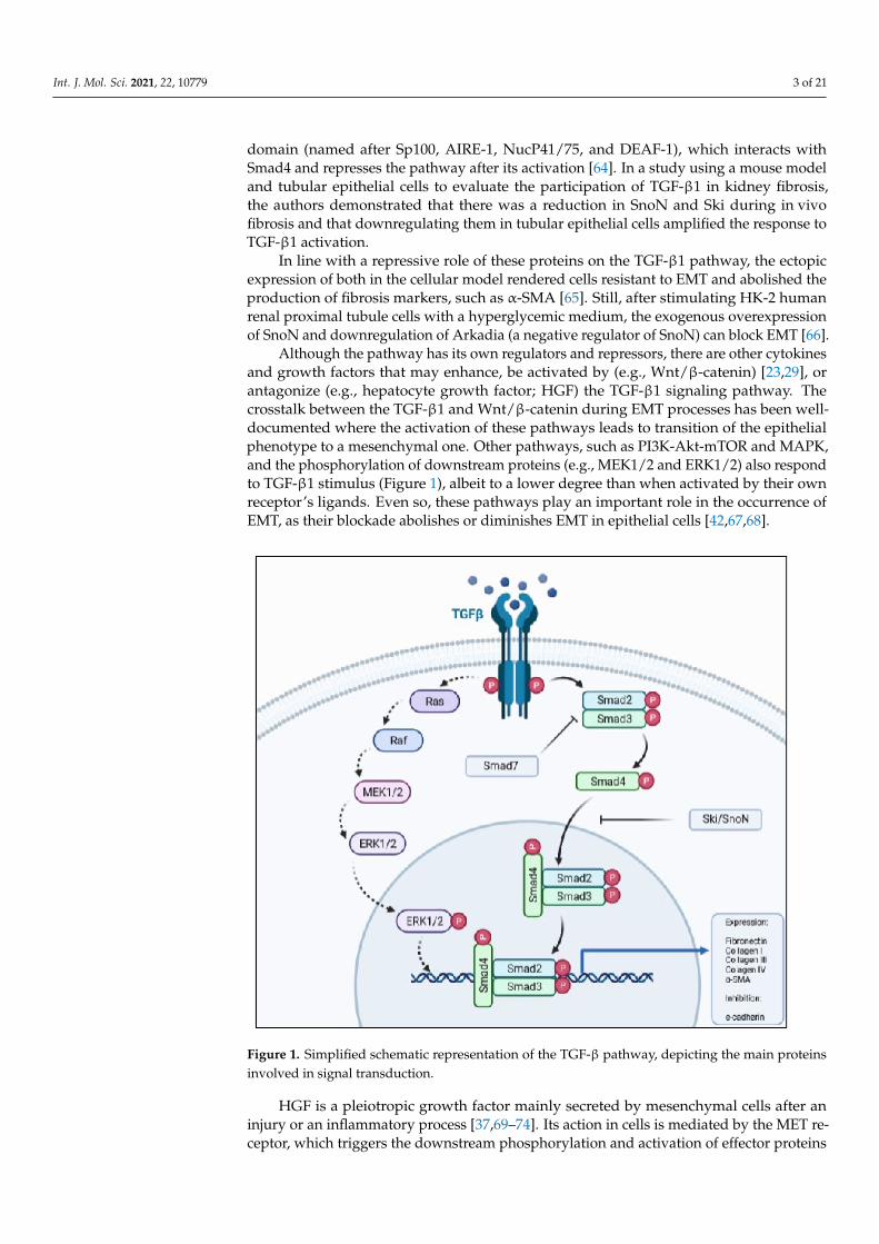
Although the pathway has its own regulators and repressors, there are other cytokinesand growth factors that may enhance, be activated by (e.g., Wnt/β-catenin) [23,29], orantagonize (e.g., hepatocyte growth factor; HGF) the TGF-β1 signaling pathway. Thecrosstalk between the TGF-β1 and Wnt/β-catenin during EMT processes has been well-documented where the activation of these pathways leads to transition of the epithelialphenotype to a mesenchymal one. Other pathways, such as PI3K-Akt-mTOR and MAPK,and the phosphorylation of downstream proteins (e.g., MEK1/2 and ERK1/2) also respondto TGF-β1 stimulus (Figure 1), albeit to a lower degree than when activated by their ownreceptor’s ligands. Even so, these pathways play an important role in the occurrence ofEMT, as their blockade abolishes or diminishes EMT in epithelial cells [42,67,68].
Figure 1. Simplified schematic representation of the TGF-β pathway, depicting the main proteinsinvolved in signal transduction.
HGF is a pleiotropic growth factor mainly secreted by mesenchymal cells after aninjury or an inflammatory process [37,69–74]. Its action in cells is mediated by the MET re-ceptor, which triggers the downstream phosphorylation and activation of effector proteins

Int. J. Mol. Sci. 2021, 22, 10779 4 of 21
(e.g., ERK), leading to the expression of target genes resulting in the production of metallo-proteases 2 and 9 [75], SnoN, and Ski [64–66] and inducing myofibroblast apoptosis [75].
The exogenous administration of HGF in different animal models of fibrosis leadsto less prominent or abolishes organ fibrosis in chronic kidney disease [34–36], unilateralureteral obstruction [38,76], and diabetic nephropathy [40]. Beyond its evident role inantagonizing TGF-β1 and its antifibrotic activity, the anti-inflammatory potential of HGFmay also contribute to the final effects in vivo and in vitro, as renal inflammation directlycontributes to progression of chronic kidney disease [71,77–80]. This is further discussed inanother section.
2.2. Wnt/β-Catenin Signaling Pathway
Another apparently simple but complex pathway involved in renal fibrosis is theWnt/β-catenin pathway. It is simple, in that it has a few actors from its activation untilthe outcome, which are a Wnt-protein ligand, a frizzled receptor (and its co-receptors),and dephosphorylated β-catenin (see Figure 2). On the other hand, it is complex as itmay activate and be controlled by many other pathways, including signaling by TGF-β1 [27,28,30,81–83].
Figure 2. Simplified schematic representation of the Wnt/β-catenin pathway, depicting the mainproteins involved in signal transduction.
Until the past decade, studies on the Wnt signaling pathway focused mainly on cancerand its pharmacological treatment, and embryonic development. Colorectal cancer has beendirectly associated with mutations on important Wnt positive- and negative-regulators, andmutations on the main actors of the pathway cause abnormalities in embryo development,such as a lack of wings in Drosophila melanogaster and a lack of anterior cerebellum in

Int. J. Mol. Sci. 2021, 22, 10779 5 of 21
mice [84]. Deregulation of this pathway has recently been associated with fibrosis, as acause or consequence of tissue damage and pro-fibrotic stimulus [26,28,30,85–87].
In total, 19 Wnt ligands, which are glycoproteins that bind to the frizzled receptorsto trigger activation, have been described [26,88]. The pathway comprises scaffold andadaptor proteins that mediate the interaction between the receptors and effector proteins.After Wnt ligand binding to the frizzled receptor, LDL-related receptor proteins 5 and 6(LRP5/6) are phosphorylated and recruit Dishevelled proteins, which are responsible forinhibiting the destruction complex (Glycogen synthase kinase 3β, GSK-3β; Casein kinaseIα, CKIα; Axin; and Adenomatosis polyposis coli, APC).
Then, stable dephosphorylated β-catenin is released from the complex, enters thenucleus, and interacts with LEF (lymphoid enhancer factor) and T-cell factor (TCF), leadingto the expression of target genes of the pathway [84]. As with other signaling pathways,the Wnt pathway has its own regulators, thus, avoiding over-activation and pathologicaloutcomes. One of the most important Wnt modulators is the Dickkopf family (DKK) ofproteins [29,86,89], which binds to LRP5/6 co-receptors and prevents the binding of Wntligands to the Frizzled receptor, thus, blocking the pathway [90].
In a mouse model of kidney fibrosis caused by UUO, only three Wnt genes (Wnt5b,Wnt8b and Wnt9b) are not upregulated, with a similar expression in sham mice. The other16 Wnt genes are upregulated, following different patterns throughout the course of theexperiment. Some of them are overexpressed within the first week, reach a peak, and thenexpression levels decline over the next seven days; others are upregulated within the firstweek and expression levels are sustained throughout the 14-day experiment; and, in others,the expression levels increase progressively during the entire experiment. There is alsoan increase in cytoplasmic and nuclear β-catenin during the course of the assay, furtherreinforcing that the canonical pathway is truly activated, with a final response of ECMproduction and kidney fibrosis evident at day 14 after UUO [85].
Klotho is a membrane bound protein that has been implicated as a negative regulatorof the Wnt/β-catenin pathway. In HK-2 cells, Klotho inhibition by si-RNA leads to a moreevident EMT after TGF-β1 stimulation, showing that there is a crosstalk between bothpathways. In an in vivo model of chronic allograft dysfunction, Klotho is downregulatedby 24 weeks after transplantation [91]. Reduced Klotho expression was also reportedin the folic acid model of fibrosis [92]. Non-canonical activation of this pathway occursindependently from Klotho activity since the MMP-7 activation of β-catenin is abrogatedby an inhibitor of transcription of target genes, but not by Klotho [93].
2.3. C3a/C3aR Signaling Pathway
Complement system proteins play important roles in renal fibrosis. As part of thebloodstream’s innate immune system, the effectors and regulators of the classical, alter-native, and lectin pathways of the complement system are mostly produced and secretedby the liver directly into the circulation, acting against pathogens and other threats inthe bloodstream [94–102]. As other cells and organs have been described as sources ofcomplement system proteins as well, additional functions have been ascribed to this system.Here, we highlight the importance of intracellular and secreted complement activation indisease conditions, with an emphasis on its consequences for the development of kidneyfibrosis [92,103,104].
Classical pathway activation is mediated by the binding of the C1 complex (C1q/C1r/C1s) to immunoglobulins that are bound to target cell membranes. The lectin pathwayis activated through the recognition of specific carbohydrates located on the pathogensurface and, finally, the alternative pathway, which is constantly activated at low levelsand allows for the generation of C3b. All three pathways converge to a common, terminalpathway, leading to the assembly of the membrane attack complex (MAC), a membranepore responsible for killing pathogens and/or target cells [95–97,99,100]. Cleavage of thecentral complement protein C3 generates the small fragments C3a and C3b, which becomepart of the C5 convertases that cleave C5 into C5a and C5b. Complement C6 and C7 bound

Int. J. Mol. Sci. 2021, 22, 10779 6 of 21
to C5b form a complex that inserts into the plasma membrane, which acts as a scaffold forthe assembly of C8 and multiple C9 molecules, culminating in MAC formation [101].
In a model of kidney fibrosis induced by UUO C57Bl/6, mice knockout for C1qAand C3 genes have an obvious increase in the production and deposition of ECM proteins,augmented production of α-SMA, reduction of E-cadherin, and increased production andsecretion of TGF-β1 from ex vivo pericytes, compared to sham mice. In mice depletedof C1q (C1q−/−), an increase in C1s/C1r proteases and diminished levels of C3 duringkidney fibrosis were observed. The depletion of C1r in another mouse model causes anevident reduction in C3 production and activation. These findings indicate the relationshipbetween C1r/C1s and C3 production and activation in the fibrotic kidney [92,104].
During complement activation, fragments derived from C3 and C5—known as C3aand C5a or anaphylatoxins—are generated. They bind to their respective receptors, leadingto a pro-inflammatory response, such as NLRP3 activation by C5a/C5aR2 and the produc-tion of IL-1β and IFN-γ by TCD4+ cells [105]. C3aR is expressed in renal epithelial cells,and its activation is related to the production of the pro-inflammatory cytokines IL-1β, IL-6,IL-8, and TNF-α by immune cells; these cytokines may exert deleterious functions duringrenal damage, leading to kidney fibrosis [106].
There is growing evidence regarding the participation of C3a and C5a in the occurrenceand progression of kidney fibrosis. HK-2 cells express markers of EMT and overproduceECM molecules when cultured for 72 hours with C3a or C5a. Cells display strong stain-ing for α-SMA and lose positive staining for E-cadherin in immunocytochemistry afterincubation with both complement anaphylatoxins [31].
In a study of kidney transplantation from mice lacking the CR1-related gene/proteiny (Crry; a membrane-associated complement regulatory protein that limits C3 activation)and C3 (C3−/−) to C3aR-, C5aR-, or C3aR/C5aR-deficient mice, the C3aR−/− mice showbetter renal function, less inflammatory cell infiltration, tubular dilatation, and atrophy,compared to C5aR−/−, C3aR/C5aR−/−, and WT transplanted mice. Interestingly, C5aR-and C3aR/C5aR-deficient mice show signs of renal failure, indicating that C3aR but notC5aR is involved in the poor outcome during renal complement activation. These findingshighlight the involvement of the C3a/C3aR pathway in kidney inflammation, leading tosubsequent kidney fibrosis [107].
2.4. TGF-β1 versus HGF Balance
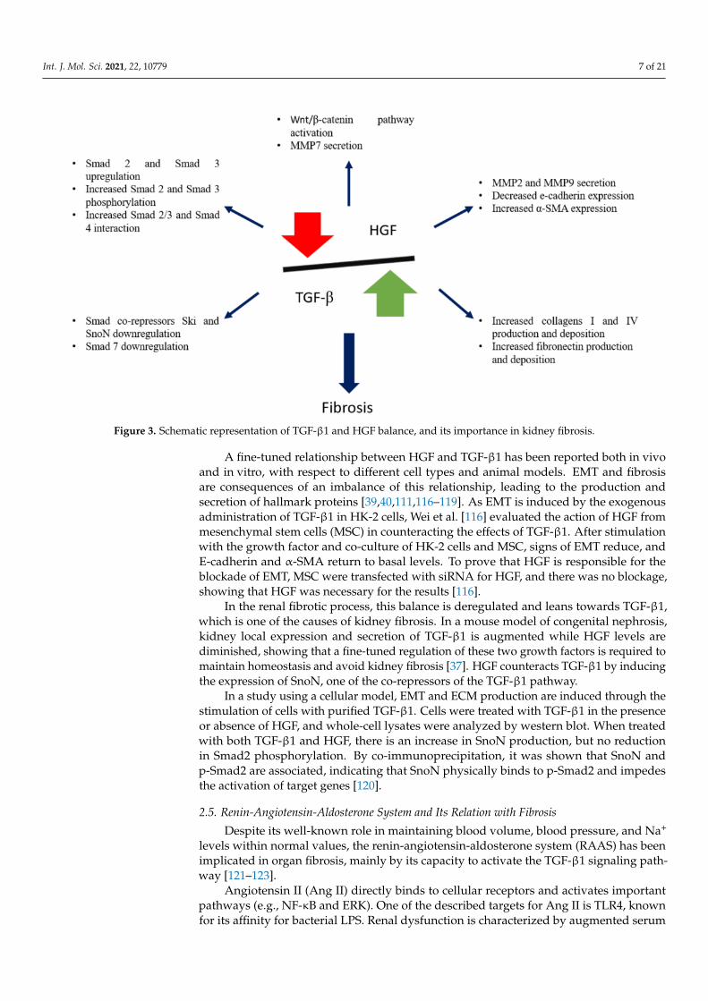
There is a close relationship and a fine balance between TGF-β1 and the multi-functional cytokine HGF (Figure 3), and the crosstalk between these pathways will befurther discussed.
HGF is a pleiotropic growth factor that promotes diverse cell responses, such asmitosis, motility, and morphogenesis, as well as wound healing, tissue regeneration,tumorigenesis, and invasion. The primary structure of the protein, first studied as ahepatocyte mitogenic protein in cell cultures, was deduced more than 30 years ago [108,109].The pathway has only one ligand—the HGF molecule—and only one receptor—the MET.
Although some bacteria are able to subvert and bind the receptor to enter the cell,there are no other endogenous ligands for MET, except for HGF itself [74,110]. HGF iscomposed of an alpha and a beta chain and needs to be cleaved to its active form by serineproteases, mostly by the HGF-activator. HGF is produced and secreted by mesenchymalcells [37,69,70,74,111]. MET is a tyrosine kinase receptor (TKR), which becomes phospho-rylated in multiple tyrosine residues after binding HGF, and then recruits proteins fromdifferent pathways (e.g., PI3K and Akt), which exert their roles in the activated cells (i.e.,suppression of cell death by expression of anti-apoptotic Bcl-xl, inhibition of Fas-FasLbinding, and inhibition of caspase3-mediated apoptosis) [112–115]. Other roles of theHGF–MET pathways are dictated by the activation of RAS/ERK pathways, which regulatecell proliferation and motility [111].
Int. J. Mol. Sci. 2021, 22, 10779 7 of 21
Figure 3. Schematic representation of TGF-β1 and HGF balance, and its importance in kidney fibrosis.
A fine-tuned relationship between HGF and TGF-β1 has been reported both in vivoand in vitro, with respect to different cell types and animal models. EMT and fibrosisare consequences of an imbalance of this relationship, leading to the production andsecretion of hallmark proteins [39,40,111,116–119]. As EMT is induced by the exogenousadministration of TGF-β1 in HK-2 cells, Wei et al. [116] evaluated the action of HGF frommesenchymal stem cells (MSC) in counteracting the effects of TGF-β1. After stimulationwith the growth factor and co-culture of HK-2 cells and MSC, signs of EMT reduce, andE-cadherin and α-SMA return to basal levels. To prove that HGF is responsible for theblockade of EMT, MSC were transfected with siRNA for HGF, and there was no blockage,showing that HGF was necessary for the results [116].
In the renal fibrotic process, this balance is deregulated and leans towards TGF-β1,which is one of the causes of kidney fibrosis. In a mouse model of congenital nephrosis,kidney local expression and secretion of TGF-β1 is augmented while HGF levels arediminished, showing that a fine-tuned regulation of these two growth factors is required tomaintain homeostasis and avoid kidney fibrosis [37]. HGF counteracts TGF-β1 by inducingthe expression of SnoN, one of the co-repressors of the TGF-β1 pathway.
In a study using a cellular model, EMT and ECM production are induced through thestimulation of cells with purified TGF-β1. Cells were treated with TGF-β1 in the presenceor absence of HGF, and whole-cell lysates were analyzed by western blot. When treatedwith both TGF-β1 and HGF, there is an increase in SnoN production, but no reductionin Smad2 phosphorylation. By co-immunoprecipitation, it was shown that SnoN andp-Smad2 are associated, indicating that SnoN physically binds to p-Smad2 and impedesthe activation of target genes [120].
2.5. Renin-Angiotensin-Aldosterone System and Its Relation with Fibrosis
Despite its well-known role in maintaining blood volume, blood pressure, and Na+
levels within normal values, the renin-angiotensin-aldosterone system (RAAS) has beenimplicated in organ fibrosis, mainly by its capacity to activate the TGF-β1 signaling path-way [121–123].
Angiotensin II (Ang II) directly binds to cellular receptors and activates importantpathways (e.g., NF-κB and ERK). One of the described targets for Ang II is TLR4, knownfor its affinity for bacterial LPS. Renal dysfunction is characterized by augmented serum
Int. J. Mol. Sci. 2021, 22, 10779 8 of 21
creatinine, albuminuria, and blood urea nitrogen, and kidney fibrosis hallmarks includeincreased collagens I/IV deposition and elevated levels of TGF-β1 and MMP9. Knockingdown MD2—a TLR4 accessory protein that mediates binding of the ligand to its receptor—or blocking it with a small inhibitor molecule leads to a decrease in renal dysfunctionand kidney fibrosis seen in wild type mice after the subcutaneous injection of Ang II. Italso affects cytokine and chemokine production mediated by NF-κB and ERK pathways.Of note, all the effects observed upon Ang II administration are MD2 dependent, thus,indicating that Ang II signals using the TLR4/MD2 complex [123].
The binding of Ang II to its type-1 receptor (AT1R) is apparently involved in theoccurrence of EMT in HK-2 cells induced by high glucose medium, and mediated by theeffector proteins mTOR and p70S6K. Silencing the receptor rescues the cells from EMT,characterized by overexpression of E-cadherin, diminished levels of α-SMA, and reducedexpression of EMT core transcription factors [124]. Conversely, activation of the pathwayby AT2R has the opposite effects and protects cells from TGF-β1 activation by inhibitingTGFβRII, and consequently, preventing EMT [125]
Ang II is capable of inducing secretion, and activating the latent form of TGF-β1, thus,having a pro-fibrotic role [126]. Ang II activates Smad2 and Smad3, which will enter thenucleus and display the same effects already described upon activation by TFG-β1 bindingto its receptor, such as an enhanced production of the TGF-β1 molecule, leading to positivefeedback [122].
Although this pathway is well-described and associated to renal fibrosis in differentpathological contexts [123–127], the involvement of RAAS in kidney fibrosis due to chronicinfection by pathogenic Leptospira, as deep as it was possible for us to search, has not beenaddressed, but may be a possible subject of investigation regarding the sequelae fromleptospiral infection.
2.6. EMT, a Hallmark of Kidney Fibrosis
As research involving EMT has grown in the past 20 years, it has become almostmandatory to elaborate guidelines to help researchers in formulating projects and todevelop concepts that could aid in eliminating controversies regarding terms associated toEMT. In June 2020, an extensive committee, on behalf of The EMT International Association,published the Guidelines and definitions for research on epithelial–mesenchymal transition [128].
The epithelial to mesenchymal transition is a commonly described phenomenon thattakes place during three major events: embryogenesis, cancer, and fibrosis. It is describedas a process that occurs after specific stimuli in epithelial cells, leading to the acquisition ofmesenchymal phenotype markers and a loss of epithelial markers. Although it may seemto be a static and end-point event, there is a spectrum regarding epithelial to mesenchymalphenotype markers, and, in fibrosis, cells mostly undergo an incomplete EMT, retainingcharacteristics that resemble both epithelial and mesenchymal phenotypes [128].
Although still controversial, there is evidence that the partial EMT of tubular cellsparticipates in renal fibrosis indirectly, as EMT leads to an increased production of cy-tokines and chemokines, such as TGF-β1, that stimulate interstitial fibroblasts to producemore ECM proteins and, as such, attract more immune cells and enhance local inflamma-tion [129–132].
In vitro and in vivo studies have also described the occurrence of EMT or partial EMTin tubular epithelial cells. The expression of mesenchymal markers (e.g., α-SMA), loss ofepithelial markers (e.g., E-cadherin), increased production of ECM proteins (e.g., fibronectinand collagens I, III, and IV), and over- and down-regulation of specific genes (e.g., Zeb1 andZeb2, Snai1, and Snai2) are indicative of this process, pointing to the active participation ofepithelial cells in the occurrence and progression of kidney fibrosis [23,116,132–134].
Although necessary during renal embryogenesis, Snai1 and Snai2 are completelyinactive during adulthood and are only reactivated during fibrosis. Tubular epithelialcells from mice subjected to UUO show an increased expression of Snai1, while there isan evident and important increase in interstitial fibrosis concomitant with a reduction in
Int. J. Mol. Sci. 2021, 22, 10779 9 of 21
the expression of epithelial markers, such as cadherin 1. In Snai1fl/fl mice, fibrosis, andmesenchymal markers are clearly less evident, and epithelial markers are still perceived intubular epithelial cells, demonstrating that Snai1 is necessary for partial EMT in those cells.
The contribution of tubular epithelial cells to the generation of myofibroblasts (ECM-producing cells) in kidney fibrosis is a controversial issue among EMT researchers. Usinga mouse model expressing the Tomato fluorescent protein and UUO to induce fibrosis, itbecame clear that tubular cells undergo partial EMT, but do not delaminate and detachfrom the basement membrane to invade the interstitium, as less than 1% of interstitial cellswere tdTomato+ 7 days after obstruction [130].
Mice lacking Twist1 or Snail1 genes have 48.2% and 64.3% lesser tubular epithelial cells,respectively, that underwent EMT after UUO compared to wild type mice. In addition,those mice displayed less collagen deposition and better renal function. No signs of tubularepithelial cell detachment are seen both in wild type and knockout mice, indicating thatthese cells underwent partial EMT and that they are needed to induce α-SMA+ myofibrob-lasts. EMT in kidney fibrosis is associated with a lower expression of important solutetransporters, such as aquaporin 1 and Na+/K+-ATPase, and suppressing the expression ofcore EMT-transcription factors may explain the better renal function in Twist1 or Snail1 KOmice [135].
EMT is under strict control in adult humans and animals [128,130,131,136]. One of theregulators that helps keep EMT under control are micro-RNAs (miRNAs), especially thefamily of miRNAs known as miR-200, which downregulate the expression of Zeb1 and Zeb2.Both transcription factors are overexpressed during fibrosis and EMT, and downregulatethe expression of E-cadherin [137–139]. In a rat tubular epithelial cell line model, EMT isinduced by TGF-β1, while miR-200a is downregulated in a Smad2-dependent way. WhenmiR-200a is overexpressed, cells are protected from TGF-β1-induced EMT, indicating thatSmad2 is necessary and controls the expression of miR-200a [138]. With that in mind, theuse of miR-200 family precursors as a possibility for kidney fibrosis treatment and/orretardation of fibrosis development has been tested, with some good results [140].
3. Acute Leptospirosis and the Development of Kidney Fibrosis
During infection, pathogenic Leptospira disseminate through the bloodstream andreach target organs, especially the kidneys, liver, and lungs [141]. The kidneys are thencolonized by the bacteria and elicit an immunological response, initially based on therecruitment of neutrophils and further mediated by macrophages. Meanwhile, leptospiresare still found in the blood in the first three days after infection, where neutrophils try toeliminate them by NETosis and other mechanisms.
Neutrophil-depleted mice have a higher leptospiral burden in the kidneys 15 dayspost-infection, which demonstrates that neutrophils help to eliminate Leptospira from theblood within the first few days [142,143]. Although both cell types are phagocytic and playimportant roles in the innate immune response to invading pathogens, Leptospira has theability to subvert and scape phagocytosis, invade renal convoluted tubules, and persist inthis niche [144–148].
The transition of acute kidney injury (AKI) caused by leptospiral infection to chronicinfection and, consequently, kidney fibrosis is still not completely understood, and manyresearch gaps remain open. This is also applied to the transition from AKI to CKD ofnon-infectious cause [149]. Continuous stimuli, the activation of inflammatory pathways,and the participation of innate immune cells in the development of chronic leptospiro-sis and kidney fibrosis are the main underlying causes that have been considered sofar [25,142,143,149–151].
The sustained stimulus caused by the presence of Leptospira in the convoluted tubulesis responsible for the overactivation of inflammatory pathways, triggering pro-fibroticsignals. One month after infection, cellular infiltration in the kidneys is characterizedby CD3-positive T-cells and CD11b-positive macrophages/monocytes, but no more neu-trophils are observed [152]. In a rat model of gentamicin-induced acute kidney injury, the

Int. J. Mol. Sci. 2021, 22, 10779 10 of 21
most prevalent cell infiltrate in kidneys were pro-inflammatory M1 macrophages withinthe first day after injury; whereas, at day 30 after injury, M2 macrophages accounted for45% of the total cell infiltrate [149].
The total healing of the kidney, with no signs of tubular necrosis or glomerularsclerosis, can also be observed. Activation of the NF-κB and NLRP3/IL-1β pathwaysare implicated in the recrudescence of signs in rat kidneys 180 days after injury, witha low-grade inflammation occurring together with activation of Angiotensin II, as wellas collagen and fibronectin deposition in the interstitium. These findings indicate thatsustained inflammatory stimulus may lead to kidney fibrosis after acute injury [149].
As described above, the complement is another important system activated duringleptospirosis. Fluid-phase or membrane-associated negative complement regulatory pro-teins avoid overactivation of this system and, consequently, tissue damage [98,101,105].One of the membrane-associated complement regulators is the decay-accelerating factor 1(Daf1), responsible for inhibiting the assembly and accelerating the disassembly of C3 andC5 convertases. In a mouse model of leptospirosis infection, Daf1−/− mice have higherbacterial loads, greater susceptibility to infection, acute renal lesions, and more evidentkidney fibrosis 90 days post-infection, with more tubulointerstitial collagen depositionthan wild-type littermates. These findings point to a role of Daf1 in controlling the bacterialburden and inflammation during the acute phase, thus, helping to reduce chronic lesionsand fibrosis [150].
Cytotoxicity mediated by nitric oxide (NO) is one of the mechanisms used by macrophagesto control leptospiral infection. Use of the TLR2/NOD2 agonist CL429 increases NO pro-duction by mice peritoneal and bone marrow-derived macrophages when exposed to L.interrogans serovars Manilae str. L495, Copenhageni str. Fiocruz L1-130, and Icterohaemor-raghiae str. Verdun [153]. The NO increase is correlated with a lower number of liveLeptospira in cell culture and, as such, is associated with bacterial killing. Inducible nitricoxide synthase (iNOS) expression—and, consequently, NO production—is also associatedwith kidney fibrosis as the disease transitions from an acute to chronic state in C57BL/6Jmice [152].
Of note, the initial lesion and the type of cellular infiltrate play roles in the diseaseprogression and, thus, contribute to the evolution to chronic and fibrotic Leptospira-relatedkidney disease [142,150,152]. The roles of macrophages and galectin-3 in the survivalrate and clinical course of the disease, acute interstitial nephritis, and development ofchronic infection and kidney fibrosis in C57BL/6 mice infected by L. interrogans sorovarCopenhageni str. L1-130 have been investigated [142]. Although galectin-3 plays a crucialrole in controlling bacterial burden during the acute phase, fibrosis and chronic disease isonly correlated with the initial bacterial burden, being directly related to the developmentand extent of kidney fibrosis, leading to the activation and enrichment of fibrosis-relatedpathway genes (e.g., iNOS, TGF-β1, Wnt, and integrin) [25,142,152].
In brief, acute leptospirosis is characterized mainly by inflammatory and immuneresponses mediated by cells, but it triggers the initial activation of the above-mentionedpathways, such as TGF-β1 and Wnt/β-catenin. Their sustained and imbalanced activationwill then promote major effects on the occurrence of EMT and kidney fibrosis duringinfection [25,132,149].
4. Kidney Fibrosis and Chronic Leptospirosis
Chronic leptospiral infection leads to kidney fibrosis, but the underlying mechanismsand pathways involved are still poorly elucidated. In the first part of this review, thepotential pathways related to renal fibrosis were described. Thus far, the involvement ofthe TGF-β1 and Wnt/β-catenin pathways have been associated with fibrosis resultingfrom chronic leptospiral infection; however, the picture is certainly more complex, andwe must keep in mind that other pathways may contribute to fibrosis caused by thisspirochete [153–155]. In this section, we will address the current knowledge on the findingsrelated to renal fibrosis caused by Leptospira infection.

Int. J. Mol. Sci. 2021, 22, 10779 11 of 21
Hamsters and guinea pigs are considered good animal models for acute and se-vere leptospirosis, as both animals die within the first 5–10 days after Leptospira inocula-tion [141,156,157]. On the other hand, rats and mice are suitable models to study carrierstatus and chronic disease. Although mice do not present signs of the disease, and theirlesions are considered mild to moderate, good models of chronic leptospirosis in those ani-mals have been developed, while some underlying mechanisms involved in the persistenceof Leptospira and fibrosis induction have begun to be elucidated [142,150,152,158].
Using wild-type and different knockout mouse lineages to understand which path-ways may be activated during chronic leptospiral infection, Fanton d´Andon et al. [152]demonstrated that renal fibrosis in chronic mouse infection can be partially attributed tonitric oxide production but in a TLR- and NLR-independent manner. Furthermore, acuteinflammation and T-cell infiltration do not contribute directly to the extent of renal fibrosis.
Chronic leptospiral infection has been associated to fibrosis in many different modelsof mouse infection [142,150,152,159]. Both wild-type and decay-accelerating factor 1-deficient mice (DAFKO) developed fibrosis at 90 days post-infection, where collagendeposition is observed away from lymphocyte infiltration. In contrast to what has beenpreviously described [152], the authors found a relationship between inflammation andinterstitial fibrosis in both wild-type and DAFKO mice, but with more evident collagendeposition in the interstitium of DAFKO mice, compared to the wild-type [150].
Leptospira outer membrane proteins induce the accumulation of ECM proteins in renalepithelial cells through activation of the TGF-β1/Smad3 pathway, thus, contributing tothe evolution of fibrosis associated to chronic infection [132]. Therefore, TGF-β1 presentsa profibrotic action and is involved in the mechanisms of renal fibrosis during chronicleptospirosis. According to Tian et al. [155], outer membrane proteins from L. santarosaiserovar Shermani enhance the secretion of collagen types I and IV by HK-2 cells, and theprocess is mediated by the TGF-β1 pathway.
Bone marrow derived-macrophages can transdifferentiate into myofibroblasts—cellsthat are involved in the secretion of α-SMA. This transition is coordinated by the TGF-β1/Smad3 pathway, in a process known as the macrophage–myofibroblast transition(MMT). In chimeric mice that had their bone marrow depleted by radiation and reconsti-tuted by exogenous GFP-expressing C57BL/6 bone marrow, GFP-positive myofibroblastswere observed in the kidneys after UUO.
To ascertain that the MMT occurs, orchestrated by TGF-β1/Smad3, C57BL/6 micewere irradiated and reconstituted with bone marrow from GFP+Smad3+/+ andGFP+Smad3−/−. Mice lacking Smad3 did not present bone marrow-derived myofibrob-lasts after UUO surgery, thus, corroborating the involvement of TGF-β1/Smad3 in theMMT within the kidney [160].
To understand which pathways are involved in chronic infection and kidney fibrosiscaused by L. interrogans serovar Copenhageni str. L1-130 and the role of leptospiral infectionin progression of CKD, Chou et al. [25] performed a mouse kidney transcriptomic analysisand detected increased gene expression of TGF-β1, Wnt, and integrin-β—crucial players inimportant fibrosis-related pathways. In addition, when mice are submitted to a nephrotoxicdiet with 0.1 or 0.2% of adenine, those pathways are further enriched.
Comparing orthologous genes from mice and humans with leptospiral infection plusnephrotoxic stimulus and CKDu, respectively, there is an overlap of enriched genes. Thesefindings provide support for the hypothesis that Leptospira infection is associated withCKD progression and may be an underlying cause of CKDu. Furthermore, as both Wntand TGF-β1 pathways are enhanced in this model, it is suggested that they contribute tothe progression of kidney fibrosis [25].
Further evidence of a fibrotic process triggered by infection with leptospires comesfrom recent in vitro and in vivo findings from our group. HK-2 cells infected with L.interrogans serovar Manilae str. L495 were shown to produce greater amounts of fibronectinand collagen type IV, compared to non-infected cells. Morphological alterations, such asspindle shape, loss of cell–cell contact, cell grouping, and abundant ECM production, were
Int. J. Mol. Sci. 2021, 22, 10779 12 of 21
also evident in infected HK-2 cells. Cellular and molecular mechanisms underlying thisEMT process are under investigation.
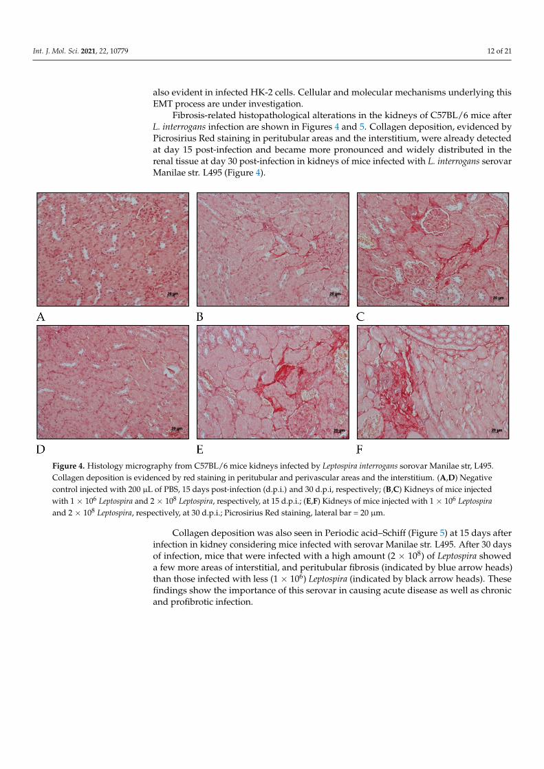
Fibrosis-related histopathological alterations in the kidneys of C57BL/6 mice afterL. interrogans infection are shown in Figures 4 and 5. Collagen deposition, evidenced byPicrosirius Red staining in peritubular areas and the interstitium, were already detectedat day 15 post-infection and became more pronounced and widely distributed in therenal tissue at day 30 post-infection in kidneys of mice infected with L. interrogans serovarManilae str. L495 (Figure 4).
Figure 4. Histology micrography from C57BL/6 mice kidneys infected by Leptospira interrogans sorovar Manilae str, L495.Collagen deposition is evidenced by red staining in peritubular and perivascular areas and the interstitium. (A,D) Negativecontrol injected with 200 µL of PBS, 15 days post-infection (d.p.i.) and 30 d.p.i, respectively; (B,C) Kidneys of mice injectedwith 1 × 10⁶ Leptospira and 2 × 10⁸ Leptospira, respectively, at 15 d.p.i.; (E,F) Kidneys of mice injected with 1 × 10⁶ Leptospiraand 2 × 10⁸ Leptospira, respectively, at 30 d.p.i.; Picrosirius Red staining, lateral bar = 20 µm.
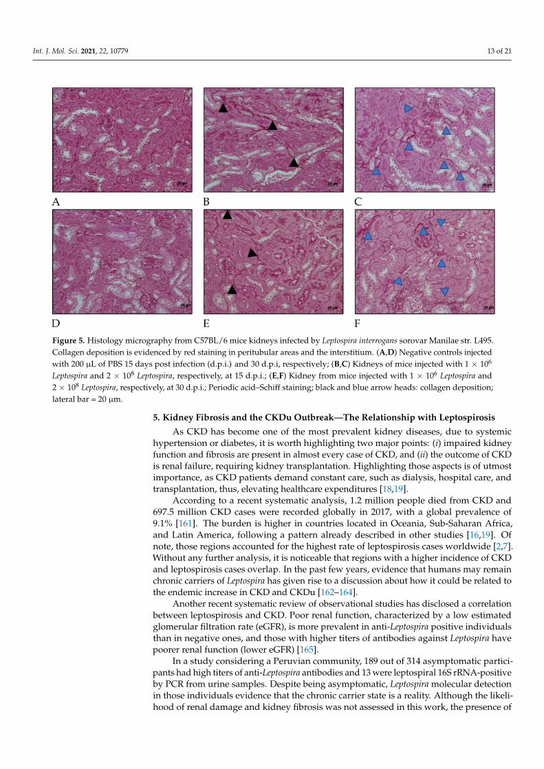
Collagen deposition was also seen in Periodic acid–Schiff (Figure 5) at 15 days afterinfection in kidney considering mice infected with serovar Manilae str. L495. After 30 daysof infection, mice that were infected with a high amount (2 × 10⁸) of Leptospira showeda few more areas of interstitial, and peritubular fibrosis (indicated by blue arrow heads)than those infected with less (1 × 10⁶) Leptospira (indicated by black arrow heads). Thesefindings show the importance of this serovar in causing acute disease as well as chronicand profibrotic infection.
Int. J. Mol. Sci. 2021, 22, 10779 13 of 21
Figure 5. Histology micrography from C57BL/6 mice kidneys infected by Leptospira interrogans sorovar Manilae str. L495.Collagen deposition is evidenced by red staining in peritubular areas and the interstitium. (A,D) Negative controls injectedwith 200 µL of PBS 15 days post infection (d.p.i.) and 30 d.p.i, respectively; (B,C) Kidneys of mice injected with 1 × 10⁶
Leptospira and 2 × 10⁸ Leptospira, respectively, at 15 d.p.i.; (E,F) Kidney from mice injected with 1 × 10⁶ Leptospira and2 × 108 Leptospira, respectively, at 30 d.p.i.; Periodic acid–Schiff staining; black and blue arrow heads: collagen deposition;lateral bar = 20 µm.
5. Kidney Fibrosis and the CKDu Outbreak—The Relationship with Leptospirosis
As CKD has become one of the most prevalent kidney diseases, due to systemichypertension or diabetes, it is worth highlighting two major points: (i) impaired kidneyfunction and fibrosis are present in almost every case of CKD, and (ii) the outcome of CKDis renal failure, requiring kidney transplantation. Highlighting those aspects is of utmostimportance, as CKD patients demand constant care, such as dialysis, hospital care, andtransplantation, thus, elevating healthcare expenditures [18,19].
According to a recent systematic analysis, 1.2 million people died from CKD and697.5 million CKD cases were recorded globally in 2017, with a global prevalence of9.1% [161]. The burden is higher in countries located in Oceania, Sub-Saharan Africa,and Latin America, following a pattern already described in other studies [16,19]. Ofnote, those regions accounted for the highest rate of leptospirosis cases worldwide [2,7].Without any further analysis, it is noticeable that regions with a higher incidence of CKDand leptospirosis cases overlap. In the past few years, evidence that humans may remainchronic carriers of Leptospira has given rise to a discussion about how it could be related tothe endemic increase in CKD and CKDu [162–164].
Another recent systematic review of observational studies has disclosed a correlationbetween leptospirosis and CKD. Poor renal function, characterized by a low estimatedglomerular filtration rate (eGFR), is more prevalent in anti-Leptospira positive individualsthan in negative ones, and those with higher titers of antibodies against Leptospira havepoorer renal function (lower eGFR) [165].
In a study considering a Peruvian community, 189 out of 314 asymptomatic partici-pants had high titers of anti-Leptospira antibodies and 13 were leptospiral 16S rRNA-positiveby PCR from urine samples. Despite being asymptomatic, Leptospira molecular detectionin those individuals evidence that the chronic carrier state is a reality. Although the likeli-hood of renal damage and kidney fibrosis was not assessed in this work, the presence of

Int. J. Mol. Sci. 2021, 22, 10779 14 of 21
Leptospira in the kidneys may be a risk factor for the development of CKD [162]. In a studyof canine leptospirosis, a relevant and close correlation between CKD and leptospirosis hasbeen reported; furthermore, those dogs with CKD and leptospirosis are more frequentlyassociated with Leptospira shedding [166].
Mesoamerican nephropathy, a type of CKDu, has brought to light evidence thatchronic leptospirosis could be a cause of CKD. This condition is highly prevalent inmale sugarcane workers that are in close contact with flooded areas at least during aperiod of the year. It is proposed that leptospirosis may be one of the causes (but not theonly) of Mesoamerican nephropathy, and that exposure to other potentially nephrotoxicconditions or substances may influence the occurrence of the disease [167]. Recently, astudy conducted in a mouse model of chronic leptospirosis, followed by toxic exposureto alimentary adenine, demonstrated that renal lesions and fibrosis are more severe andprevalent in mice primarily infected by Leptospira, showing that chronic leptospirosis maybe a risk factor for the development of CKD [25].
As the evidence that pathways described herein (i.e., TGF-β1 and Wnt/β-catenin)are directly associated to fibrosis and the progression of CKD in humans and in micepreviously infected by L. interrogans becomes clearer, it is questionable whether chronic andasymptomatic leptospiral infection is related to CKDu, as well as whether it contributes tothe progression of CKD to end-stage kidney disease.
6. Gaps and Perspectives
CKD/CKDu are important health problems that directly impact the quality of life andhealth systems all over the world. Despite recent advances in nephrology and conscienti-zation campaigns, the searches for medical assistance and treatment are still insufficient.Assuming that leptospirosis may play a role in the development of CKD/CKDu, and consid-ering the overlap of both conditions in Asia, Central and South America, and Sub-SaharanAfrica, studies into the associated pathophysiology, cellular communication pathways, andtreatment strategies to avoid or control the fibrotic process are needed.
As discussed throughout the text, kidney fibrosis and, consequently, CKD, as a se-quela of Leptospira infection, is a well-documented manifestation of the disease; however,studies on the mechanisms associated with the pathophysiology of leptospiral renal fibro-sis are still scarce and deserve to be deepened. Deciphering the cellular communicationpathways in Leptospira-induced kidney fibrosis may shed light on the specific actors in-volved in this process. The supposed role of TGF-β1 in fibrosis induced by leptospiralcomponents, such as LipL32 or outer membrane proteins in human kidney epithelial cells,has been evaluated [134], but no studies have been conducted using live Leptospira incellular models. Furthermore, animal models of infection have focused mainly on grossand histopathological alterations, and additional studies addressing which pathways arealtered during chronic leptospiral infection will help in preventing and treating importantleptospirosis sequelae.
Author Contributions: Conceptualization, L.G.P. and A.S.B.; Writing—editing and reviewing, L.G.P.and A.S.B.; Supervision, A.S.B.; Funding acquisition, A.S.B. All authors have read and agreed to thepublished version of the manuscript.
Funding: We thank the Fundação de Amparo à Pesquisa do Estado de São Paulo (FAPESP) for thefunding, process number 2021/01122-9. This study was financed in part by the Coordenação deAperfeiçoamento Pessoal de Nível Superior–Brasil (CAPES)–Finance code 001.
Institutional Review Board Statement: The study was conducted according to the guidelines of theDeclaration of Helsinki, and approved by the Institutional Ethics Committee of Butantan Instituteprotocol number 9421030620, approved at 2020/06/03.
Informed Consent Statement: Not applicable.
Data Availability Statement: The data presented in the article are available in the article.
Conflicts of Interest: The authors declare no conflict of interest.
Int. J. Mol. Sci. 2021, 22, 10779 15 of 21
References1. Adler, B. (Ed.); Leptospira and Leptospirosis; Current Topics in Microbiology and Immunology; Springer: Berlin/Heidelberg,
Germany, 2015; Volume 387, ISBN 978-3-662-45058-1.2. Bharti, A.R.; Nally, J.E.; Ricaldi, J.N.; Matthias, M.A.; Diaz, M.M.; Lovett, M.A.; Levett, P.N.; Gilman, R.H.; Willig, M.R.; Gotuzzo,
E.; et al. Leptospirosis: A Zoonotic Disease of Global Importance. Lancet Infect. Dis. 2003, 3, 757–771. [CrossRef]3. Levett, P.N. Leptospirosis. Clin. Microbiol. Rev. 2001, 14, 296–326. [CrossRef] [PubMed]4. Picardeau, M. Virulence of the Zoonotic Agent of Leptospirosis: Still Terra Incognita? Nat. Rev. Microbiol. 2017, 15, 297–307.
[CrossRef] [PubMed]5. Mohammed, H.; Nozha, C.; Hakim, K.; Abdelaziz, F.; Rekia, B. LEPTOSPIRA: Morphology, Classification and Pathogenesis. J
Bacteriol. Parasitol. 2011, 2, 6–9. [CrossRef]6. Cassadou, S.; Rosine, J.; Flamand, C.; Escher, M.; Ledrans, M.; Bourhy, P.; Picardeau, M.; Quénel, P. Underestimation of
Leptospirosis Incidence in the French West Indies. PLoS Negl. Trop. Dis. 2016, 10, e0004668. [CrossRef] [PubMed]7. Costa, F.; Hagan, J.E.; Calcagno, J.; Kane, M.; Torgerson, P.; Martinez-Silveira, M.S.; Stein, C.; Abela-Ridder, B.; Ko, A.I. Global
Morbidity and Mortality of Leptospirosis: A Systematic Review. PLoS Negl. Trop. Dis. 2015, 9, e0003898. [CrossRef] [PubMed]8. Karpagam, K.B.; Ganesh, B. Leptospirosis: A Neglected Tropical Zoonotic Infection of Public Health Importance—An Updated
Review. Eur. J. Clin. Microbiol. Infect. Dis. 2020, 39, 835–846. [CrossRef]9. Picardeau, M. Diagnosis and Epidemiology of Leptospirosis. Med. Mal. Infect. 2013, 43, 1–9. [CrossRef]10. Allan, K.J.; Biggs, H.M.; Halliday, J.E.B.; Kazwala, R.R.; Maro, V.P.; Cleaveland, S.; Crump, J.A. Epidemiology of Leptospirosis in
Africa: A Systematic Review of a Neglected Zoonosis and a Paradigm for ‘One Health’ in Africa. PLoS Negl. Trop. Dis. 2015,9, e0003899. [CrossRef] [PubMed]
11. Marteli, A.N.; Genro, L.V.; Diament, D.; Guasselli, L.A. Análise Espacial Da Leptospirose No Brasil. Saúde Em Debate 2020, 44,805–817. [CrossRef]
12. Dias, J.P.; Teixeira, M.G.; Costa, M.C.N.; Mendes, C.M.C.; Guimarães, P.; Reis, M.G.; Ko, A.; Barreto, M.L. Factors Associated withLeptospira Sp Infection in a Large Urban Center in Northeastern Brazil. Rev. Soc. Bras. Med. Trop. 2007, 40, 499–504. [CrossRef]
13. Yang, H.Y.; Hung, C.C.; Liu, S.H.; Guo, Y.G.; Chen, Y.C.; Ko, Y.C.; Huang, C.T.; Chou, L.F.; Tian, Y.C.; Chang, M.Y.; et al.Overlooked Risk for Chronic Kidney Disease after Leptospiral Infection: A Population-Based Survey and Epidemiological CohortEvidence. PLoS Negl. Trop. Dis. 2015, 9, e0004105. [CrossRef] [PubMed]
14. Gracie, R.; Barcellos, C.; Magalhães, M.; Souza-Santos, R.; Guimarães Barrocas, P.R. Geographical Scale Effects on the Analysis ofLeptospirosis Determinants. Int. J. Environ. Res. Public Health 2014, 11, 10366–10383. [CrossRef] [PubMed]
15. Reis, R.B.; Ribeiro, G.S.; Felzemburgh, R.D.M.; Santana, F.S.; Mohr, S.; Melendez, A.X.T.O.; Queiroz, A.; Santos, A.C.; Ravines,R.R.; Tassinari, W.S.; et al. Impact of Environment and Social Gradient on Leptospira Infection in Urban Slums. PLoS Negl. Trop.Dis. 2008, 2, 11–18. [CrossRef] [PubMed]
16. Yang, C.W. Leptospirosis Renal Disease: Emerging Culprit of Chronic Kidney Disease Unknown Etiology. Nephron 2018, 138,129–136. [CrossRef]
17. Bülow, R.D.; Boor, P. Extracellular Matrix in Kidney Fibrosis: More Than Just a Scaffold. J. Histochem. Cytochem. 2019, 67, 643–661.[CrossRef] [PubMed]
18. Djudjaj, S.; Boor, P. Cellular and Molecular Mechanisms of Kidney Fibrosis. Mol. Asp. Med. 2019, 65, 16–36. [CrossRef]19. Jha, V.; Garcia-Garcia, G.; Iseki, K.; Li, Z.; Naicker, S.; Plattner, B.; Saran, R.; Wang, A.Y.M.; Yang, C.W. Chronic Kidney Disease:
Global Dimension and Perspectives. Lancet 2013, 382, 260–272. [CrossRef]20. Liu, Y. Renal Fibrosis: New Insights into the Pathogenesis and Therapeutics. Kidney Int. 2006, 69, 213–217. [CrossRef]21. Xu, B.H.; Sheng, J.; You, Y.K.; Huang, X.R.; Ma, R.C.W.; Wang, Q.; Lan, H.Y. Deletion of Smad3 Prevents Renal Fibrosis and
Inflammation in Type 2 Diabetic Nephropathy. Metab. Clin. Exp. 2020, 103, 154013. [CrossRef]22. Budi, E.H.; Duan, D.; Derynck, R. Transforming Growth Factor-β Receptors and Smads: Regulatory Complexity and Functional
Versatility. Trends Cell Biol. 2017, 27, 658–672. [CrossRef]23. Guo, L.; Peng, W.; Tao, J.; Lan, Z.; Hei, H.; Tian, L.; Pan, W.; Wang, L.; Zhang, X. Hydrogen Sulfide Inhibits Transforming Growth
Factor-B1-Induced EMT via Wnt/Catenin Pathway. PLoS ONE 2016, 11, e0147018. [CrossRef]24. Chung, S.; Overstreet, J.M.; Li, Y.; Wang, Y.; Niu, A.; Wang, S.; Fan, X.; Sasaki, K.; Jin, G.; Khodo, S.N.; et al. TGF-β Promotes
Fibrosis after Severe Acute Kidney Injury by Enhancing Renal Macrophage Infiltration. JCI Insight 2018, 3, e123563. [CrossRef]25. Chou, L.-F.; Chen, T.; Yang, H.-Y.; Tian, Y.-C.; Chang, M.-Y.; Hung, C.-C.; Hsu, S.-H.; Tsai, C.-Y.; Ko, Y.-C.; Yang, C.-W.
Transcriptomic Signatures of Exacerbated Progression in Leptospirosis Subclinical Chronic Kidney Disease with SecondaryNephrotoxic Injury. Am. J. Physiol.-Ren. Physiol. 2021, 320, F1001–F1018. [CrossRef]
26. Feng, Y.; Ren, J.; Gui, Y.; Wei, W.; Shu, B.; Lu, Q.; Xue, X.; Sun, X. Wnt/b-Catenin—Promoted Macrophage Alternative ActivationContributes to Kidney Fibrosis. J. Am. Soc. Nephrol. 2018, 29, 182–193. [CrossRef]
27. Huelsken, J.; Behrens, J. The Wnt Signalling Pathway. J. Cell Sci. 2002, 115, 3977–3978. [CrossRef]28. Zuo, Y.; Liu, Y. New Insights into the Role and Mechanism of Wnt/β-Catenin Signalling in Kidney Fibrosis. Nephrology 2018, 23,
38–43. [CrossRef] [PubMed]29. Wang, D.; Dai, C.; Li, Y.; Liu, Y. Canonical Wnt/B-Catenin Signaling Mediates Transforming Growth Factor-B1-Driven Podocyte
Injury and Proteinuria. Kidney Int. 2011, 80, 1159–1169. [CrossRef] [PubMed]

Int. J. Mol. Sci. 2021, 22, 10779 16 of 21
30. Tan, R.J.; Zhou, D.; Zhou, L.; Liu, Y. Wnt/β-Catenin Signaling and Kidney Fibrosis. Kidney Int. Suppl. 2014, 4, 84–90. [CrossRef][PubMed]
31. Liu, F.; Gou, R.; Huang, J.; Fu, P.; Chen, F.; Fan, W.X.; Huang, Y.Q.; Zang, L.; Wu, M.; Qui, H.Y.; et al. Effect of AnaphylatoxinC3a, C5a on the Tubular Epithelial-Myofibroblast Transdifferentiation in Vitro. Chin. Med. J. 2011, 124, 4039–4045. [CrossRef][PubMed]
32. Li, T.T.; Zhang, X.H.; Jing, J.F.; Li, X.; Yang, X.Q.; Zhu, F.H.; Tang, W.; Zuo, J.P. Artemisinin Analogue SM934 Ameliorates theProteinuria and Renal Fibrosis in Rat Experimental Membranous Nephropathy. Acta Pharmacol. Sin. 2015, 36, 188–199. [CrossRef]
33. Han, R.; Hu, S.; Qin, W.; Shi, J.; Hou, Q.; Wang, X.; Xu, X.; Zhang, M.; Zeng, C.; Liu, Z.; et al. C3a and SuPAR Drive Versican V1Expression in Tubular Cells of Focal Segmental Glomerulosclerosis. JCI Insight 2019, 4, e122912. [CrossRef] [PubMed]
34. Dworkin, L.D.; Gong, R.; Tolbert, E.; Centracchio, J.; Yano, N.; Zanabli, A.R.; Esparza, A.; Rifai, A. Hepatocyte Growth FactorAmeliorates Progression of Interstitial Fibrosis in Rats with Established Renal Injury. Kidney Int. 2004, 65, 409–419. [CrossRef]
35. Gong, R.; Rifai, A.; Tolbert, E.M.; Centracchio, J.N.; Dworkin, L.D. Hepatocyte Growth Factor Modulates Matrix Metallopro-teinases and Plasminogen Activator/Plasmin Proteolytic Pathways in Progressive Renal Interstitial Fibrosis. J. Am. Soc. Nephrol.2003, 14, 3047–3060. [CrossRef] [PubMed]
36. Inoue, T.; Okada, H.; Kobayashi, T.; Watanabe, Y.; Kanno, Y.; Kopp, J.B.; Nishida, T.; Takigawa, M.; Ueno, M.; Nakamura, T.; et al.Hepatocyte Growth Factor Counteracts Transforming Growth Factor-Beta1, through Attenuation of Connective Tissue GrowthFactor Induction, and Prevents Renal Fibrogenesis in 5/6 Nephrectomized Mice. FASEB J. Off. Publ. Fed. Am. Soc. Exp. Biol. 2003,17, 268–270. [CrossRef]
37. Mizuno, S.; Matsumoto, K.; Kurosawa, T.; Mizuno-Horikawa, Y.; Nakamura, T. Reciprocal Balance of Hepatocyte Growth Factorand Transforming Growth Factor-B1 in Renal Fibrosis in Mice. Kidney Int. 2000, 57, 937–948. [CrossRef] [PubMed]
38. Mizuno, S.; Matsumoto, K.; Nakamura, T. Hepatocyte Growth Factor Suppresses Interstitial Fibrosis in a Mouse Model ofObstructive Nephropathy. Kidney Int. 2001, 59, 1304–1314. [CrossRef]
39. Xu, J.; Yu, T.T.; Zhang, K.; Li, M.; Shi, H.J.; Meng, X.J.; Zhu, L.S.; Zhu, L.K. HGF Alleviates Renal Interstitial Fibrosis via Inhibitingthe Tgf-B1/Smad Pathway. Eur. Rev. Med. Pharmacol. Sci. 2018, 22, 7621–7627. [CrossRef]
40. Mizuno, S.; Nakamura, T. Suppressions of Chronic Glomerular Injuries and TGF-B1 Production by HGF in Attenuation of MurineDiabetic Nephropathy. Am. J. Physiol.-Ren. Physiol. 2004, 286, 134–143. [CrossRef]
41. Hu, H.H.; Chen, D.Q.; Wang, Y.N.; Feng, Y.L.; Cao, G.; Vaziri, N.D.; Zhao, Y.Y. New Insights into TGF-β/Smad Signaling in TissueFibrosis. Chem.-Biol. Interact. 2018, 292, 76–83. [CrossRef]
42. Katsuno, Y.; Derynck, R. Epithelial Plasticity, Epithelial-Mesenchymal Transition, and the TGF-β Family. Dev. Cell 2021, 56,726–746. [CrossRef] [PubMed]
43. Biernacka, A.; Dobaczewski, M.; Frangogiannis, N.G. TGF-β Signaling in Fibrosis. Growth Factors 2011, 29, 196–202. [CrossRef]44. Lan, H.Y. Diverse Roles of TGF-β/Smads in Renal Fibrosis and Inflammation. Int. J. Biol. Sci. 2011, 7, 1056–1067. [CrossRef]
[PubMed]45. Batlle, E.; Massagué, J. Transforming Growth Factor-β Signaling in Immunity and Cancer. Immunity 2019, 50, 924–940. [CrossRef]
[PubMed]46. Meng, X.M.; Tang, P.M.K.; Li, J.; Lan, H.Y. TGF-ß/Smad Signaling in Renal Fibrosis. Front. Physiol. 2015, 6, 82. [CrossRef]
[PubMed]47. Liu, S.; De Boeck, M.; Van Dam, H.; Ten Dijke, P. Regulation of the TGF-β Pathway by Deubiquitinases in Cancer. Int. J. Biochem.
Cell Biol. 2016, 76, 135–145. [CrossRef]48. Wicks, S.J.; Haros, K.; Maillard, M.; Song, L.; Cohen, R.E.; Ten Dijke, P.; Chantry, A. The Deubiquitinating Enzyme UCH37
Interacts with Smads and Regulates TGF-β Signalling. Oncogene 2005, 24, 8080–8084. [CrossRef]49. Kang, J.S.; Saunier, E.F.; Akhurst, R.J.; Derynck, R. The Type I TGF-β Receptor Is Covalently Modified and Regulated by
Sumoylation. Nat. Cell Biol. 2008, 10, 654–664. [CrossRef]50. Rotin, D.; Kumar, S. Physiological Functions of the HECT Family of Ubiquitin Ligases. Nat. Rev. Mol. Cell Biol. 2009, 10, 398–409.
[CrossRef]51. Zuo, W.; Huang, F.; Chiang, Y.J.; Li, M.; Du, J.; Ding, Y.; Zhang, T.; Lee, H.W.; Jeong, L.S.; Chen, Y.; et al. C-Cbl-Mediated
Neddylation Antagonizes Ubiquitination and Degradation of the TGF-β Type II Receptor. Mol. Cell 2013, 49, 499–510. [CrossRef]52. Alisson-Silva, F.; Freire-de-Lima, L.; Donadio, J.L.; Lucena, M.C.; Penha, L.; Sá-Diniz, J.N.; Dias, W.B.; Todeschini, A.R. Increase
of O-Glycosylated Oncofetal Fibronectin in High Glucose-Induced Epithelial-Mesenchymal Transition of Cultured HumanEpithelial Cells. PLoS ONE 2013, 8, e60471. [CrossRef]
53. Bektas, M.; Rubenstein, D.S. The Role of Intracellular Protein O-Glycosylation in Cell Adhesion and Disease. J. Biomed. Res. 2011,25, 227–236. [CrossRef]
54. Liu, C.; Xu, P.; Lamouille, S.; Xu, J.; Derynck, R. TACE-Mediated Ectodomain Shedding of the Type I TGF-β Receptor Downregu-lates TGF-β Signaling. Mol. Cell 2009, 35, 26–36. [CrossRef]
55. Munger, J.S.; Sheppard, D. Cross Talk among TGF-β Signaling Pathways, Integrins, and the Extracellular Matrix. Cold SpringHarb. Perspect. Biol. 2011, 3, a005017. [CrossRef] [PubMed]
56. Robertson, I.B.; Rifkin, D.B. Regulation of the Bioavailability of TGF-β and TGF-β-Related Proteins. Cold Spring Harb. Perspect.Biol. 2016, 8, a021907. [CrossRef]

Int. J. Mol. Sci. 2021, 22, 10779 17 of 21
57. Jenkins, G. The Role of Proteases in Transforming Growth Factor-β Activation. Int. J. Biochem. Cell Biol. 2008, 40, 1068–1078.[CrossRef]
58. Ma, J.; Sanchez-Duffhues, G.; Goumans, M.J.; ten Dijke, P. TGF-β-Induced Endothelial to Mesenchymal Transition in Disease andTissue Engineering. Front. Cell Dev. Biol. 2020, 8, 260. [CrossRef] [PubMed]
59. Walton, K.L.; Johnson, K.E.; Harrison, C.A. Targeting TGF-β Mediated SMAD Signaling for the Prevention of Fibrosis. Front.Pharmacol. 2017, 8. [CrossRef]
60. Li, J.H.; Zhu, H.J.; Huang, X.R.; Lai, K.N.; Johnson, R.J.; Lan, H.Y. Smad7 Inhibits Fibrotic Effect of TGF-β on Renal TubularEpithelial Cells by Blocking Smad2 Activation. J. Am. Soc. Nephrol. 2002, 13, 1464–1472. [CrossRef] [PubMed]
61. Nakao, A.; Afrakhte, M.; Nakayama, T.; Christian, J.L.; Heuchel, R.; Itoh, S.; Kawabata, M.; Heldin, N.-E.; Heldin, C.-H.Identification of Smad7, a TGFβ-inducible antagonist of TGF-β signalling. Nature 1997, 389, 631–635. [CrossRef]
62. De Ceuninck van Capelle, C.; Spit, M.; ten Dijke, P. Current Perspectives on Inhibitory SMAD7 in Health and Disease. Crit. Rev.Biochem. Mol. Biol. 2020, 55, 691–715. [CrossRef] [PubMed]
63. Chung, A.C.K.; Huang, X.R.; Zhou, L.; Heuchel, R.; Lai, K.N.; Lan, H.Y. Disruption of the Smad7 Gene Promotes Renal Fibrosisand Inflammation in Unilateral Ureteral Obstruction (UUO) in Mice. Nephrol. Dial. Transplant. 2009, 24, 1443–1454. [CrossRef]
64. Deheuninck, J.; Luo, K. Ski and SnoN, Potent Negative Regulators of TGF-β Signaling. Cell Res. 2009, 19, 47–57. [CrossRef][PubMed]
65. Yang, J.; Zhang, X.; Li, Y.; Liu, Y. Downregulation of Smad Transcriptional Corepressors SnoN and Ski in the Fibrotic Kidney: AnAmplification Mechanism for TGF-B1 Signaling. J. Am. Soc. Nephrol. 2003, 14, 3167–3177. [CrossRef] [PubMed]
66. Liu, L.; Shi, M.; Wang, Y.; Zhang, C.; Su, B.; Xiao, Y.; Guo, B. SnoN Upregulation Ameliorates Renal Fibrosis in DiabeticNephropathy. PLoS ONE 2017, 12, e0174471. [CrossRef]
67. Lamouille, S.; Derynck, R. Cell Size and Invasion in TGF-β–Induced Epithelial to Mesenchymal Transition Is Regulated byActivation of the MTOR Pathway. J. Cell Biol. 2007, 178, 437–451. [CrossRef]
68. Lamouille, S. Molecular Mechanisms of Epithelial–Mesenchymal Transition. Nat. Rev. Mol. Cell Biol. 2014, 15, 178–196. [CrossRef]69. Molnarfi, N.; Benkhoucha, M.; Funakoshi, H.; Nakamura, T.; Lalive, P.H. Hepatocyte Growth Factor: A Regulator of Inflammation
and Autoimmunity. Autoimmun. Rev. 2015, 14, 293–303. [CrossRef]70. Nakamura, T.; Sakai, K.; Nakamura, T.; Matsumoto, K. Hepatocyte Growth Factor Twenty Years on: Much More than a Growth
Factor. J. Gastroenterol. Hepatol. 2011, 26, 188–202. [CrossRef]71. Gong, R.; Rifai, A.; Ge, Y.; Chen, S.; Dworkin, L.D. Hepatocyte Growth Factor Suppresses Proinflammatory NFκB Activation
through GSK3β Inactivation in Renal Tubular Epithelial Cells. J. Biol. Chem. 2008, 283, 7401–7410. [CrossRef] [PubMed]72. Coudriet, G.M.; He, J.; Trucco, M.; Mars, W.M.; Piganelli, J.D. Hepatocyte Growth Factor Modulates Interleukin-6 Production in
Bone Marrow Derived Macrophages: Implications for Inflammatory Mediated Diseases. PLoS ONE 2010, 5, e15384. [CrossRef]73. Ito, W.; Takeda, M.; Ueki, S.; Tanigai, T.; Kayaba, H.; Chihara, J. Effect of the Hepatocyte Growth Factor on Allergic Inflammatory
Cells. Int. Arch. Allergy Immunol. 2010, 152, 96–100. [CrossRef]74. Imamura, R.; Matsumoto, K. Hepatocyte Growth Factor in Physiology and Infectious Diseases. Cytokine 2017, 98, 97–106.
[CrossRef]75. Iekushi, K.; Taniyama, Y.; Azuma, J.; Sanada, F.; Kusunoki, H.; Yokoi, T.; Koibuchi, N.; Okayama, K.; Rakugi, H.; Morishita, R.
Hepatocyte Growth Factor Attenuates Renal FIbrosis through TGF-B1 Suppression by Apoptosis of Myofibroblasts. J. Hypertens.2010, 28, 2454–2461. [CrossRef] [PubMed]
76. Yang, J.; Liu, Y. Blockage of Tubular Epithelial to Myofibroblast Transition by Hepatocyte Growth Factor Prevents Renal InterstitialFibrosis. J. Am. Soc. Nephrol. 2002, 13, 96–107. [CrossRef] [PubMed]
77. Gong, R.; Rifai, A.; Tolbert, E.M.; Biswas, P.; Centracchio, J.N.; Dworkin, L.D. Hepatocyte Growth Factor Ameliorates RenalInterstitial Inflammation in Rat Remnant Kidney by Modulating Tubular Expression of Macrophage Chemoattractant Protein-1and RANTES. J. Am. Soc. Nephrol. 2004, 15, 2868–2881. [CrossRef] [PubMed]
78. Gong, R.; Rifai, A.; Dworkin, L.D. Anti-Inflammatory Effect of Hepatocyte Growth Factor in Chronic Kidney Disease: Targetingthe Inflamed Vascular Endothelium. J. Am. Soc. Nephrol. 2006, 17, 2464–2473. [CrossRef] [PubMed]
79. Gong, R.; Rifai, A.; Dworkin, L.D. Hepatocyte Growth Factor Suppresses Acute Renal Inflammation by Inhibition of EndothelialE-Selectin. Kidney Int. 2006, 69, 1166–1174. [CrossRef]
80. Daha, M.R.; van Kooten, C. Is the Proximal Tubular Cell a Proinflammatory Cell? Nephrol. Dial. Transplant. 2000, 15, 41–43.[CrossRef]
81. Feng, Y.; Liang, Y.; Ren, J.; Dai, C. Canonical Wnt Signaling Promotes Macrophage Proliferation during Kidney Fibrosis. KidneyDis. 2018, 4, 95–103. [CrossRef] [PubMed]
82. Silva-García, O.; Valdez-Alarcón, J.J.; Baizabal-Aguirre, V.M. The Wnt/β-Catenin Signaling Pathway Controls the InflammatoryResponse in Infections Caused by Pathogenic Bacteria. Mediat. Inflamm. 2014, 2014, 310183. [CrossRef]
83. Schaale, K.; Neumann, J.; Schneider, D.; Ehlers, S.; Reiling, N. Wnt Signaling in Macrophages: Augmenting and InhibitingMycobacteria-Induced Inflammatory Responses. Eur. J. Cell Biol. 2011, 90, 553–559. [CrossRef] [PubMed]
84. Klaus, A.; Birchmeier, W. Wnt Signalling and Its Impact on Development and Cancer. Nat. Rev. Cancer 2008, 8, 387–398. [CrossRef][PubMed]
85. He, W.; Dai, C.; Li, Y.; Zeng, G.; Monga, S.P.; Liu, Y. Wnt/β-Catenin Signaling Promotes Renal Interstitial Fibrosis. J. Am. Soc.Nephrol. 2009, 20, 765–776. [CrossRef] [PubMed]

Int. J. Mol. Sci. 2021, 22, 10779 18 of 21
86. Pyo, M.C. Ochratoxin A Induces Epithelial-to-Mesenchymal Transition and Renal Fibrosis through TGF-β/Smad2/3 andWnt1/β-Catenin Signaling Pathways in Vitro and in Vivo. Arch. Toxicol. 2020, 94, 3329–3342. [CrossRef]
87. Zhou, D.; Fu, H.; Zhang, L.; Zhang, K.; Min, Y.; Xiao, L.; Lin, L.; Bastacky, S.I.; Liu, Y. Tubule-Derived Wnts Are Required forFibroblast Activation and Kidney Fibrosis. J. Am. Soc. Nephrol. 2017, 28, 2322–2336. [CrossRef]
88. Routledge, D.; Scholpp, S. Mechanisms of Intercellular Wnt Transport. Development 2019, 146, dev176073. [CrossRef]89. Niehrs, C. Function and Biological Roles of the Dickkopf Family of Wnt Modulators. Oncogene 2006, 25, 7469–7481. [CrossRef]90. Semënov, M.V.; Tamai, K.; Brott, B.K.; Kühl, M.; Sokol, S.; He, X. Head Inducer Dickkopf-1 Is a Ligand for Wnt Coreceptor LRP6.
Curr. Biol. 2001, 11, 951–961. [CrossRef]91. Li, X.; Lu, P.; Shao, X.-F.; Jiang, T.; Liu, F.; Li, G. Klotho Regulates Epithelial-to-Mesenchymal Transition In Vitro via Wnt/β-Catenin
Pathway and Attenuates Chronic Allograft Dysfunction in a Rat Renal Transplant Model. Ann. Transpl. 2021, 26, e930066-1.[CrossRef]
92. Xavier, S.; Sahu, R.K.; Bontha, S.V.; Mas, V.; Taylor, R.P.; Megyesi, J.; Thielens, N.M.; Portilla, D. Complement C1r Serine ProteaseContributes to Kidney Fibrosis. Am. J. Physiol.-Ren. Physiol. 2019, 317, F1293–F1304. [CrossRef]
93. Zhou, D.; Tian, Y.; Sun, L.; Zhou, L.; Xiao, L.; Tan, R.J.; Tian, J.; Fu, H.; Hou, F.F.; Liu, Y. Matrix Metalloproteinase-7 Is a UrinaryBiomarker and Pathogenic Mediator of Kidney Fibrosis. J. Am. Soc. Nephrol. 2017, 28, 598–611. [CrossRef]
94. De Castro, Í.A.; Bavia, L.; Fraga, T.R.; Amano, M.T. Role of Murine Complement Component C5 in Acute in Vivo Infection byPathogenic Leptospira Interrogans. Front. Cell. Infect. Microbiol. 2018, 8, 63. [CrossRef]
95. Fraga, T.R.; Courrol, S.; Castiblanco-valencia, M.M.; Hirata, I.Y.; Vasconcellos, S.A.; Juliano, L.; Barbosa, A.S.; Isaac, L. ImmuneEvasion by Pathogenic Leptospira Strains: The Secretion of Proteases That Directly Cleave Complement Proteins. J. Infect. Dis.2014, 209, 876–886. [CrossRef]
96. Barbosa, A.S.; Isaac, L. Strategies Used by Leptospira Spirochetes to Evade the Host Complement System. FEBS Lett. 2020, 594,2633–2644. [CrossRef] [PubMed]
97. Heesterbeek, D.A.C.; Angelier, M.L.; Harrison, R.A.; Rooijakkers, S.H.M. Complement and Bacterial Infections: From MolecularMechanisms to Therapeutic Applications. J. Innate Immun. 2018, 10, 455–464. [CrossRef]
98. Carroll, M.C.; Isenman, D.E. Regulation of Humoral Immunity by Complement. Immunity 2012, 37, 199–207. [CrossRef] [PubMed]99. Ermert, D.; Ram, S.; Laabei, M. The Hijackers Guide to Escaping Complement: Lessons Learned from Pathogens. Mol. Immunol.
2019, 114, 49–61. [CrossRef]100. Merle, N.S.; Church, S.E.; Fremeaux-Bacchi, V.; Roumenina, L.T. Complement System Part I €“ Molecular Mechanisms of
Activation and Regulation. Front. Immunol. 2015, 6, 262. [CrossRef]101. Ricklin, D.; Hajishengallis, G.; Yang, K.; Lambris, J.D. Complement: A Key System for Immune Surveillance and Homeostasis.
Nat. Immunol 2010, 11, 785–797. [CrossRef]102. Markiewski, M.M.; Lambris, J.D. The Role of Complement in Inflammatory Diseases From Behind the Scenes into the Spotlight.
Am. J. Pathol. 2007, 171, 715–727. [CrossRef]103. Portilla, D.; Xavier, S. Role of Intracellular Complement Activation in Kidney Fibrosis. Br. J. Pharmacol. 2021, 178, 2880–2891.
[CrossRef]104. Xavier, S.; Sahu, R.K.; Landes, S.G.; Yu, J.; Taylor, R.P.; Ayyadevara, S.; Megyesi, J.; Stallcup, W.B.; Duffield, J.S.; Reis, E.S.; et al.
Pericytes and Immune Cells Contribute to Complement Activation in Tubulointerstitial Fibrosis. Am. J. Physiol.-Ren. Physiol.2017, 312, F516–F532. [CrossRef]
105. Hajishengallis, G.; Reis, E.S.; Mastellos, D.C.; Ricklin, D.; Lambris, J.D. Novel Mechanisms and Functions of Complement. Nat.Immunol. 2017, 18, 1288–1298. [CrossRef]
106. Danobeitia, J.S.; Djamali, A.; Fernandez, L.A. The Role of Complement in the Pathogenesis of Renal Ischemia-Reperfusion Injuryand Fibrosis. Fibrogenesis Tissue Repair 2014, 7, 16. [CrossRef]
107. Bao, L.; Wang, Y.; Haas, M.; Quigg, R.J. Distinct Roles for C3a and C5a in Complement-Induced Tubulointerstitial Injury. KidneyInt. 2011, 80, 524–534. [CrossRef]
108. Nakamura, T.; Nishizawa, T.; Hagiya, M.; Seki, T.; Shimonishi, M.; Sugimura, A.; Tashiro, K.; Shimizu, S. Molecular Cloning andExpression of Human Hepatocyte Growth Factor. Nature 1989, 342, 440–443. [CrossRef]
109. Miyazawa, K.; Tsubouchi, H.; Naka, D.; Takahashi, K.; Okigaki, M.; Arakaki, N.; Nakayama, H.; Hirono, S.; Sakiyama, O.;Takahashi, K.; et al. Molecular Cloning and Sequence Analysis of CDNA for Human Hepatocyte Growth Factor. Biochem. Biophys.Res. Commun. 1989, 163, 967–973. [CrossRef]
110. Shen, Y.; Naujokas, M.; Park, M.; Ireton, K. InlB-Dependent Internalization of Listeria Is Mediated by the Met Receptor TyrosineKinase. Cell 2000, 103, 501–510. [CrossRef]
111. Ilangumaran, S.; Villalobos-Hernandez, A.; Bobbala, D.; Ramanathan, S. The Hepatocyte Growth Factor (HGF)–MET ReceptorTyrosine Kinase Signaling Pathway: Diverse Roles in Modulating Immune Cell Functions. Cytokine 2016, 82, 125–139. [CrossRef]
112. Liu, Y. Hepatocyte Growth Factor Promotes Renal Epithelial Cell Survival by Dual Mechanisms. Am. J. Physiol.-Ren. Physiol. 1999,277, F624–F633. [CrossRef]
113. Ma, J.; Zou, C.; Guo, L.; Seneviratne, D.S.; Tan, X.; Kwon, Y.-K.; An, J.; Bowser, R.; DeFrances, M.C.; Zarnegar, R. Novel DeathDefying Domain in Met Entraps the Active Site of Caspase-3 and Blocks Apoptosis in Hepatocytes: Ma et Al. Hepatology 2014, 59,2010–2021. [CrossRef]

Int. J. Mol. Sci. 2021, 22, 10779 19 of 21
114. García-Ocaña, A.; Takane, K.K.; Reddy, V.T.; Lopez-Talavera, J.-C.; Vasavada, R.C.; Stewart, A.F. Adenovirus-Mediated HepatocyteGrowth Factor Expression in Mouse Islets Improves Pancreatic Islet Transplant Performance and Reduces Beta Cell Death. J. Biol.Chem. 2003, 278, 343–351. [CrossRef]
115. Wang, X.; DeFrances, M.C.; Dai, Y.; Pediaditakis, P.; Johnson, C.; Bell, A.; Michalopoulos, G.K.; Zarnegar, R. A Mechanism of CellSurvival: Sequestration of Fas by the HGF Receptor Met. Mol. Cell 2002, 9, 411–421. [CrossRef]
116. Wei, J.-J.; Tang, L.; Chen, L.-L.; Xie, Z.-H.; Ren, Y.; Qi, H.-G.; Lou, J.; Weng, G.-B.; Zhang, S.-W. Mesenchymal Stem Cells AttenuatesTGF-B1-Induced EMT by Increasing HGF Expression in HK-2 Cells. Iran. J. Public Health 2021, 50, 908–918. [CrossRef] [PubMed]
117. Esposito, C.; Parrilla, B.; Cornacchia, F.; Grosjean, F.; Serpieri, N.; Valentino, R.; Villa, L.; Arra, M.; Esposito, V.; Canton, A.D. TheAntifibrogenic Effect of Hepatocyte Growth Factor (HGF) on Renal Tubular (HK-2) Cells Is Depende. Growth Factors 2009, 27,173–180. [CrossRef]
118. Kaibori, M.; Inoue, T.; Oda, M.; Naka, D.; Kawaguchi, T.; Kitamura, N.; Miyazawa, K.; Kwon, A.-H.; Kamiyama, Y.; Okumura, T.Exogenously Administered HGF Activator Augments Liver Regeneration through the Production of Biologically Active HGF.Biochem. Biophys. Res. Commun. 2002, 290, 475–481. [CrossRef] [PubMed]
119. Li, J.; Liu, X.; Sha, M.; Li, Y. The Balance between HGF and TGF-B1 Acts as a Switch in the Tissue Remodeling of ChronicRhinosinusitis. Int. J. Clin. Exp. Pathol. 2019, 12, 933. [PubMed]
120. Yang, J.; Dai, C.; Liu, Y. A Novel Mechanism by Which Hepatocyte Growth Factor Blocks Tubular Epithelial to MesenchymalTransition. J. Am. Soc. Nephrol. 2005, 16, 68–78. [CrossRef]
121. Lijnen, P.; Petrov, V. Induction of Cardiac Fibrosis by Aldosterone. J. Mol. Cell. Cardiol. 2000, 32, 865–879. [CrossRef]122. AlQudah, M.; Hale, T.M.; Czubryt, M.P. Targeting the Renin-Angiotensin-Aldosterone System in Fibrosis. Matrix Biol. 2020,
91–92, 92–108. [CrossRef] [PubMed]123. Xu, Z.; Li, W.; Han, J.; Zou, C.; Huang, W.; Yu, W.; Shan, X.; Lum, H.; Li, X.; Liang, G. Angiotensin II Induces Kidney Inflammatory
Injury and Fibrosis through Binding to Myeloid Differentiation Protein-2 (MD2). Sci. Rep. 2017, 7, 44911. [CrossRef]124. Gong, Q. Silencing of Angiotensin II Type-1 Receptor Inhibits High Glucose-Induced Epithelial-Mesenchymal Transition in
Human Renal Proximal Tubular Epithelial Cells via Inactivation of MTOR/P70S6K Signaling Pathway. Biochem. Biophys. Res.Commun. 2016, 469, 183–188. [CrossRef]
125. Guo, H.-L.; Liao, X.-H.; Liu, Q.; Zhang, L. Angiotensin II Type 2 Receptor Decreases Transforming Growth Factor-β Type IIReceptor Expression and Function in Human Renal Proximal Tubule Cells. PLoS ONE 2016, 11, e0148696. [CrossRef]
126. Kagami, S.; Border, W.A.; Miller, D.E.; Noble, N.A. Angiotensin II Stimulates Extracellular Matrix Protein Synthesis throughInduction of Transforming Growth Factor-Beta Expression in Rat Glomerular Mesangial Cells. J. Clin. Investig. 1994, 93, 2431–2437.[CrossRef] [PubMed]
127. Balakumar, P. A Potential Role of the Renin-Angiotensin-Aldosterone System in Epithelial-to-Mesenchymal Transition-InducedRenal Abnormalities_ Mechanisms and Therapeutic Implications. Pharmacol. Res. 2019, 146, 104314. [CrossRef]
128. Yang, J.; Antin, P.; Berx, G.; Blanpain, C.; Brabletz, T.; Bronner, M.; Campbell, K.; Cano, A.; Casanova, J.; Christofori, G.; et al.Guidelines and Definitions for Research on Epithelial–Mesenchymal Transition. Nat. Rev. Mol. Cell Biol. 2020, 21, 341–352.[CrossRef]
129. LeBleu, V.S.; Taduri, G.; O’Connell, J.; Teng, Y.; Cooke, V.G.; Woda, C.; Sugimoto, H.; Kalluri, R. Origin and Function ofMyofibroblasts in Kidney Fibrosis. Nat. Med. 2013, 19, 1047–1053. [CrossRef] [PubMed]
130. Grande, M.T.; Sánchez-Laorden, B.; López-Blau, C.; De Frutos, C.A.; Boutet, A.; Arévalo, M.; Rowe, R.G.; Weiss, S.J.; López-Novoa,J.M.; Nieto, M.A. Snail1-Induced Partial Epithelial-to-Mesenchymal Transition Drives Renal Fibrosis in Mice and Can Be Targetedto Reverse Established Disease. Nat. Med. 2015, 21, 989–997. [CrossRef]
131. Nieto, M.A.; Huang, R.Y.-J.; Jackson, R.A.; Thiery, J.P. EMT: 2016. Cell 2016, 166, 21–45. [CrossRef]132. Tian, Y.C.; Chen, Y.C.; Hung, C.C.; Chang, C.T.; Wu, M.S.; Phillips, A.O.; Yang, C.W. Leptospiral Outer Membrane Protein Induces
Extracellular Matrix Accumulation through a TGF-B1/Smad-Dependent Pathway. J. Am. Soc. Nephrol. 2006, 17, 2792–2798.[CrossRef]
133. Hosper, N.A. Epithelial-to-Mesenchymal Transition in Fibrosis_ Collagen Type I Expression Is Highly Upregulated after EMT,but Does Not Contribute to Collagen Deposition. Exp. Cell Res. 2013, 319, 3000–3009. [CrossRef]
134. You, Y. FKN Facilitates HK-2 Cell EMT and Tubulointerstitial Lesions via the Wnt/β-Catenin Pathway in a Murine Model ofLupus Nephritis. Front. Immunol. 2019, 10, 14.
135. Lovisa, S.; LeBleu, V.S.; Tampe, B.; Sugimoto, H.; Vadnagara, K.; Carstens, J.L.; Wu, C.-C.; Hagos, Y.; Burckhardt, B.C.; Pentcheva-Hoang, T.; et al. Epithelial-to-Mesenchymal Transition Induces Cell Cycle Arrest and Parenchymal Damage in Renal Fibrosis.Nat. Med. 2015, 21, 998–1009. [CrossRef] [PubMed]
136. Xiong, M.; Jiang, L.; Zhou, Y.; Qiu, W.; Fang, L.; Tan, R.; Wen, P.; Yang, J. The MiR-200 Family Regulates TGF-B1-InducedRenal Tubular Epithelial to Mesenchymal Transition through Smad Pathway by Targeting ZEB1 and ZEB2 Expression. Am. J.Physiol.-Ren. Physiol. 2012, 302, F369–F379. [CrossRef] [PubMed]
137. Gregory, P.A.; Bert, A.G.; Paterson, E.L.; Barry, S.C.; Tsykin, A.; Farshid, G.; Vadas, M.A.; Khew-Goodall, Y.; Goodall, G.J. TheMiR-200 Family and MiR-205 Regulate Epithelial to Mesenchymal Transition by Targeting ZEB1 and SIP1. Nat. Cell Biol. 2008, 10,593–601. [CrossRef] [PubMed]

Int. J. Mol. Sci. 2021, 22, 10779 20 of 21
138. Korpal, M.; Lee, E.S.; Hu, G.; Kang, Y. The MiR-200 Family Inhibits Epithelial-Mesenchymal Transition and Cancer Cell Migrationby Direct Targeting of E-Cadherin Transcriptional Repressors ZEB1 and ZEB2. J. Biol. Chem. 2008, 283, 14910–14914. [CrossRef][PubMed]
139. Park, S.-M.; Gaur, A.B.; Lengyel, E.; Peter, M.E. The MiR-200 Family Determines the Epithelial Phenotype of Cancer Cells byTargeting the E-Cadherin Repressors ZEB1 and ZEB2. Genes Dev. 2008, 22, 894–907. [CrossRef] [PubMed]
140. Oba, S.; Kumano, S.; Suzuki, E.; Nishimatsu, H.; Takahashi, M.; Takamori, H.; Kasuya, M.; Ogawa, Y.; Sato, K.; Kimura, K.; et al.MiR-200b Precursor Can Ameliorate Renal Tubulointerstitial Fibrosis. PLoS ONE 2010, 5, e13614. [CrossRef]
141. Seguro, A.C.; Andrade, L. Pathophysiology of Leptospirosis. Shock 2013, 39, 17–23. [CrossRef]142. Ferrer, M.F.; Scharrig, E.; Charo, N.; Rípodas, A.L.; Drut, R.; Silva, E.A.C.; Nagel, A.; Nally, J.E.; De Oca, D.P.M.; Schattner, M.; et al.
Macrophages and Galectin 3 Control Bacterial Burden in Acute and Subacute Murine Leptospirosis That Determines ChronicKidney Fibrosis. Front. Cell. Infect. Microbiol. 2018, 8, 384. [CrossRef]
143. Scharrig, E.; Carestia, A.; Ferrer, M.F.; Cédola, M.; Pretre, G.; Drut, R.; Picardeau, M.; Schattner, M.; Gómez, R.M. NeutrophilExtracellular Traps Are Involved in the Innate Immune Response to Infection with Leptospira. PLoS Negl. Trop. Dis. 2015,9, e0003927. [CrossRef]
144. Toma, C.; Okura, N.; Takayama, C.; Suzuki, T. Characteristic Features of Intracellular Pathogenic Leptospira in Infected MurineMacrophages. Cell. Microbiol. 2011, 13, 1783–1792. [CrossRef]
145. Yamaguchi, T.; Higa, N.; Okura, N.; Matsumoto, A.; Hermawan, I.; Yamashiro, T.; Suzuki, T.; Toma, C. Characterizing Interactionsof Leptospira Interrogans with Proximal Renal Tubule Epithelial Cells. BMC Microbiol. 2018, 18, 1–11. [CrossRef]
146. Nair, N.; Guedes, M.S.; Hajjar, A.M.; Werts, C.; Gomes-Solecki, M. Role of TLR4 in Persistent Leptospira Interrogans Infection: AComparative In Vivo Study in Mice. Front. Immunol. 2021, 11, 572999. [CrossRef]
147. Hume, D.A.; Irvine, K.M.; Pridans, C. The Mononuclear Phagocyte System: The Relationship between Monocytes andMacrophages. Trends Immunol. 2019, 40, 98–112. [CrossRef]
148. Murray, P.J. Macrophage Polarization. Annu. Rev. Physiol. 2017, 79, 541–566. [CrossRef]149. Albino, A.H.; Zambom, F.F.F.; Foresto-Neto, O.; Oliveira, K.C.; Ávila, V.F.; Arias, S.C.A.; Seguro, A.C.; Malheiros, D.M.A.C.;
Camara, N.O.S.; Fujihara, C.K.; et al. Renal Inflammation and Innate Immune Activation Underlie the Transition FromGentamicin-Induced Acute Kidney Injury to Renal Fibrosis. Front. Physiol. 2021, 12, 606392. [CrossRef]
150. Ferrer, M.F.; Scharrig, E.; Alberdi, L.; Cedola, M.; Pretre, G.; Drut, R.; Song, W.C.; Gomez, R.M. Decay-Accelerating Factor 1Deficiency Exacerbates Leptospiral-Induced Murine Chronic Nephritis and Renal Fibrosis. PLoS ONE 2014, 9, e102860. [CrossRef]
151. Santecchia, I.; Vernel-Pauillac, F.; Rasid, O.; Quintin, J.; Gomes-Solecki, M.; Boneca, I.G.; Werts, C. Innate Immune Memorythrough TLR2 and NOD2 Contributes to the Control of Leptospira Interrogans Infection. PLoS Pathog. 2019, 15, e1007811.[CrossRef]
152. Fanton d’Andon, M.; Quellard, N.; Fernandez, B.; Ratet, G.; Lacroix-Lamandé, S.; Vandewalle, A.; Boneca, I.G.; Goujon, J.M.;Werts, C. Leptospira Interrogans Induces Fibrosis in the Mouse Kidney through Inos-Dependent, TLR- and NLR-IndependentSignaling Pathways. PLoS Negl. Trop. Dis. 2014, 8, 11. [CrossRef]
153. Hung, C.C.; Chang, C.T.; Tian, Y.C.; Wu, M.S.; Yu, C.C.; Pan, M.J.; Vandewalle, A.; Yang, C.W. Leptospiral Membrane ProteinsStimulate Pro-Inflammatory Chemokines Secretion by Renal Tubule Epithelial Cells through Toll-like Receptor 2 and P38 MitogenActivated Protein Kinase. Nephrol. Dial. Transplant. 2006, 21, 898–910. [CrossRef] [PubMed]
154. Tian, Y.-C.; Hung, C.-C.; Li, Y.-J.; Chen, Y.-C.; Chang, M.-Y.; Yen, T.-H.; Hsu, H.-H.; Wu, M.-S.; Phillips, A.; Yang, C.-W. LeptospiraSantorosai Serovar Shermani Detergent Extract Induces an Increase in Fibronectin Production through a Toll-Like Receptor2-Mediated Pathway. Infect. Immun. 2011, 79, 9. [CrossRef]
155. Yang, C.-W.; Wu, M.-S.; Pan, M.-J.; Hsieh, W.-J.; Vandewalle, A.; Huang, C.-C. The Leptospira Outer Membrane Protein LipL32Induces Tubulointerstitial Nephritis-Mediated Gene Expression in Mouse Proximal Tubule Cells. JASN 2002, 13, 2037–2045.[CrossRef] [PubMed]
156. Richer, L.; Potula, H.H.; Melo, R.; Vieira, A.; Gomes-Solecki, M. Mouse Model for Sublethal Leptospira Interrogans Infection.Infect. Immun. 2015, 83, 4693–4700. [CrossRef]
157. Gomes-Solecki, M.; Santecchia, I.; Werts, C. Animal Models of Leptospirosis: Of Mice and Hamsters. Front. Immunol. 2017, 8, 58.[CrossRef]
158. Werts, C. Murine Models for Leptospirosis Kidney Disease. Transl. Res. Biomed. 2019, 7, 65–75. [CrossRef]159. Chassin, C.; Picardeau, M.; Goujon, J.-M.; Bourhy, P.; Quellard, N.; Darche, S.; Badell, E.; d’Andon, M.F.; Winter, N.; Lacroix-
Lamandé, S.; et al. TLR4- and TLR2-Mediated B Cell Responses Control the Clearance of the Bacterial Pathogen, LeptospiraInterrogans. J. Immunol. 2009, 183, 2669–2677. [CrossRef] [PubMed]
160. Wang, S.; Meng, X.; Ng, Y.; Ma, F.Y.; Zhou, S.; Zhang, Y.; Yang, C.; Huang, X.; Xiao, J.; Wang, Y.; et al. TGF-β/Smad3 SignallingRegulates the Transition of Bone Marrow- Derived Macrophages into Myofibroblasts during Tissue Fibrosis. Oncotarget 2015,7, 8809. [CrossRef]
161. Bikbov, B.; Purcell, C.A.; Levey, A.S.; Smith, M.; Abdoli, A.; Abebe, M.; Adebayo, O.M.; Afarideh, M.; Agarwal, S.K.; Agudelo-Botero, M.; et al. Global, Regional, and National Burden of Chronic Kidney Disease, 1990–2017: A Systematic Analysis for theGlobal Burden of Disease Study 2017. Lancet 2020, 395, 709–733. [CrossRef]
162. Ganoza, C.A.; Matthias, M.A.; Saito, M.; Cespedes, M.; Gotuzzo, E.; Vinetz, J.M. Asymptomatic Renal Colonization of Humans inthe Peruvian Amazon by Leptospira. PLoS Negl. Trop. Dis. 2010, 4, e612. [CrossRef]

Int. J. Mol. Sci. 2021, 22, 10779 21 of 21
163. Herath, N.J.; Kularatne, S.A.M.; Weerakoon, K.G.A.D.; Wazil, A.; Subasinghe, N.; Ratnatunga, N.V.I. Long Term Outcome ofAcute Kidney Injury Due to Leptospirosis? A Longitudinal Study in Sri Lanka. BMC Res. Notes 2014, 7, 1–4. [CrossRef] [PubMed]
164. Monahan, A.M.; Callanan, J.J.; Nally, J.E. Review Paper: Host-Pathogen Interactions in the Kidney during Chronic Leptospirosis.Vet. Pathol. 2009, 46, 792–799. [CrossRef]
165. Carrillo-Larco, R.M.; Altez-Fernandez, C.; Acevedo-Rodriguez, J.G.; Ortiz-Acha, K.; Ugarte-Gil, C. Leptospirosis as a Risk Factorfor Chronic Kidney Disease: A Systematic Review of Observational Studies. PLoS Negl. Trop. Dis. 2019, 13, e0007458. [CrossRef][PubMed]
166. Sant’Anna, R.; Vieira, A.S.; Oliveira, J.; Lilenbaum, W. Asymptomatic Leptospiral Infection Is Associated with Canine ChronicKidney Disease. Comp. Immunol. Microbiol. Infect. Dis. 2019, 62, 64–67. [CrossRef] [PubMed]
167. Correa-Rotter, R.; Wesseling, C.; Johnson, R.J. CKD of Unknown Origin in Central America: The Case for a MesoamericanNephropathy. Am. J. Kidney Dis. 2014, 63, 506–520. [CrossRef]