CELLULAR MECHANISMS OF RENAL SECRETION. A ...
28
CELLULAR MECHANISMS OF RENAL SECRETION. A STUDY BY THE EXTRAVITAL METHOD I. THE STRUCTURAL PHASE O17 THE SECRETORY MECHANISM* Bx JEAN R. OLIVER, M.D., AND EDNA MORRIS LUND (From the Department of Pathology of the Long Island College of Medicine, The Hoagland Laboratory, Brooklyn) PLATES 28 TO 31 (Received for publication, October 24, 1932) It is evident from a survey of the voluminous literature of renal activity that a state of confusion exists in our knowledge concerning the secretory activity of the renal cells. In illustration of this fact we may call attention to an example of the conflict in conclusions and theories that prevails and examine briefly the reasons for its existence. If one considers the relatively simple problem of the correlation between mor- phological alterations in the cells, specifically in the mitochondrial apparatus, and functional activity, a great discrepancy of opinion is found, ranging from the belief of such investigators as Kolster (1), Policard (2) and Oliver (3) that such changes may be correlated with varying functional states, to that of others, such as Cushny (4) and Emge (5) who are skeptical of any relation whatsoever between these two aspects of cell activity. General opinion is therefore quite properly unsettled. Certain reasons for this condition are not far to seek in the light of present knowledge. 1. The basic difficulty has been that we have had no exact definitive knowledge of what part, if any, secretion may play in the elimination of substances into urine. Experiments have had to be designed on a hypothetical basis and have therefore proceeded not from the known but from the assumed. As an example of this, an increase in the volume of urine due to the intravenous injection of salt solution has been assumed to produce an increase of secretory activity. It is not surprising that the sought for structural changes in the renal epithelium were indefinite, when one considers that we know now that the increased urine eliminated under such conditions is best accounted for by the activity of other parts of the renal unit than the tubule. * This investigation has been made with the assistance of a grant from the Josiah Macy, Jr., Foundation. 435 Downloaded from http://rupress.org/jem/article-pdf/57/3/435/1179784/435.pdf by guest on 18 April 2022
-
Upload
khangminh22 -
Category
Documents
-
view
0 -
download
0
Transcript of CELLULAR MECHANISMS OF RENAL SECRETION. A ...

C E L L U L A R M E C H A N I S M S OF R E N A L S E C R E T I O N . A S T U D Y BY T H E E X T R A V I T A L M E T H O D
I. THE STRUCTURAL PHASE O17 THE SECRETORY MECHANISM*
Bx JEAN R. OLIVER, M.D., AND EDNA MORRIS LUND
(From the Department of Pathology of the Long Island College of Medicine, The Hoagland Laboratory, Brooklyn)
PLATES 28 TO 31
(Received for publication, October 24, 1932)
I t is evident f rom a su rvey of the voluminous l i tera ture of renal
ac t iv i ty t ha t a s ta te of confusion exists in our knowledge concerning the secretory ac t iv i ty of the renal cells. In i l lustrat ion of this fact we m a y call a t t en t ion to an example of the conflict in conclusions and theories t h a t prevai ls and examine briefly the reasons for its existence.
If one considers the relatively simple problem of the correlation between mor- phological alterations in the cells, specifically in the mitochondrial apparatus, and functional activity, a great discrepancy of opinion is found, ranging from the belief of such investigators as Kolster (1), Policard (2) and Oliver (3) that such changes may be correlated with varying functional states, to that of others, such as Cushny (4) and Emge (5) who are skeptical of any relation whatsoever between these two aspects of cell activity. General opinion is therefore quite properly unsettled.
Certain reasons for this condition are not far to seek in the light of present knowledge.
1. The basic difficulty has been that we have had no exact definitive knowledge of what part, if any, secretion may play in the elimination of substances into urine. Experiments have had to be designed on a hypothetical basis and have therefore proceeded not from the known but from the assumed. As an example of this, an increase in the volume of urine due to the intravenous injection of salt solution has been assumed to produce an increase of secretory activity. I t is not surprising that the sought for structural changes in the renal epithelium were indefinite, when one considers that we know now that the increased urine eliminated under such conditions is best accounted for by the activity of other parts of the renal unit than the tubule.
* This investigation has been made with the assistance of a grant from the Josiah Macy, Jr., Foundation.
435
Dow
nloaded from http://rupress.org/jem
/article-pdf/57/3/435/1179784/435.pdf by guest on 18 April 2022

436 CELLULAR MECHANISMS OF RENAL SECRETION. I
2. Another confusing factor whose importance has been recognized only in recent years is the part played by absorptive processes in the function of the renal cells, yet the operation of such processes would make it impossible to decide how much of any observed structural change might be due to the intake or to the out- put of material into the urine.
3. Another almost insurmountable difficulty has been always present in that the kidney to be examined in the experiment was already in a certain functional state whose possible effects had to be distinguished from those supposedly produced by the experimental procedure. Various means were taken to put the kidney at rest before the experiment, but these could in their turn only be based on the hypothe- tical assumption as to what normally produced secretory activity. Moreover, it is quite possible that some of these procedures, such as fasting, may have pro- duced structural alterations in themselves, with a resulting further confusion in the experimental results.
4. The experiments of the past were all conducted on the living animal and in many cases considerable time elapsed during their course. I t will be easily recog- nized that with such a responsible organ as the kidney the animal itself acts as a sort of independent and not necessarily cooperative agent in the procedure and the part it plays may well defeat the entire purpose of the experiment since the in- vestigator is easily deceived by a change in its conditions that have thus arisen unknown to him.
5. Dependence in all the investigations of the past has been placed almost solely ou morphological evidence. Granules were found in rows near rods and there- fore were assumed to have arisen from the rods. I t is obvious that the only accu- rate statement of such a conclusion is that it looks as if they might have done so.
T h a t the writers believe these severe crit icisms of previous investi- gat ions have arisen f rom the unsa t i s fac tory s ta te of knowledge of the times, will be evident when it is pointed out t ha t all of them m a y be directed toward certain of the invest igat ions of the senior au thor (3).
Bu t ve ry considerable advances have been made, especially those which have come from the in t roduct ion of new methods , no tab ly direct visual examinat ion of the k idney as devised b y Richards (6) and per- fusion as adap ted to the frog's k idney b y Hoeber (7). The investi- ga tor is now able to again examine the prob lem in a new light.
The following pages describe an a t t e m p t made with wha t m a y be considered another new method. This me thod combines a morpho- logical and a physiological examinat ion of the ac t iv i ty of the isolated perfused kidney, using histological methods for the former purpose and a modificat ion of Hoeber ' s me thod for the lat ter . As a result bo th the s t ruc tura l and functional aspects of the response of the tissues to
Dow
nloaded from http://rupress.org/jem
/article-pdf/57/3/435/1179784/435.pdf by guest on 18 April 2022

JEAN R. OLIVER AND EDNA MORRIS LUND 437
either physiological or pathological stimuli can be observed and cor- related simultaneously. The application of the method to some of the problems of experimental nephritis has already been described and the term "extravital" suggested to describe it (8).
Another aid to our immediate problem has been found in the dis- covery that the dye neutral red is eliminated by the isolated perfused kidney almost entirely through the tubular epithelium and not in any significant amount through the glomeruli (9). Similar findings had been reported independently by Scheminzky (10) and recently the oc- currence of its tubular elimination has been supported by the findings of Richards (11) that in the living frog the degree of its glomerular filtration is not adequate to account for its appearance in the urine.
The possible use of the extravital method in the problem under dis- cussion for a study of the definitely proven secretion of neutral red is evident. By means of such a combination we can be certain that the tubule cells of a kidney eliminating neutral red are actually secreting, and we can also be certain that the dye found in the cell bodies has come from the blood vessel and not been absorbed from the lumen of the tubule. Absorptive process alone may also be produced by the new method in the functioning organ without any secretory activity, and the histological pictures then compared. The extravital method also avoids a further difficulty we have mentioned since the state of the kidneys before the experiment can be determined either by ex- amination of one kidney before the perfusion is started or a portion of the kidney that is to be perfused may be examined before or at any stage of the experiment. So, too, any modification of the activity of the kidney on the part of the animal is prevented by the isolation of the organ. The nervous system is eliminated and all of the constitu- ents of the fluid with which the cells are working are accurately known and these can be modified as desired as the experiment proceeds. The urine is collected almost simultaneously with the activity of the cells, rather than in long periods after its formation. And, finally, it is possible to examine the problem, placing little weight on a subjective interpretation of morphological evidence.
Technique Histological Methods.--The following methods have been used for the morpho-
logical part of the work. For a general picture of the state of the tissues Zenker's,
Dow
nloaded from http://rupress.org/jem
/article-pdf/57/3/435/1179784/435.pdf by guest on 18 April 2022

438 CELLULAR M'ECHANISMS OF RENAL SECRETION. I
Bouin's and 10 per cent neutral formalin in 0.9 per cent salt solution were used and as a stain Delafield's hematoxylin and eosin. For the preservation of the mito- chondria and other granular structures of the cells Kolster's method of fixation was found most satisfactory. Bensley's fluid in our experience was less certain in its results. For staining the mitochondria and granules a method which is essen- tially the original Altmann procedure was used; this was supplemented by the Bens- ley method. For special purposes the Gram-Weigert stain was also used on tissues fixed by all the methods described above, and formol-Zenker fixation for the per- manent fixation of neutral red in the tissues and subsequent embedding in paraffin and preparation of permanent sections (12).
Beside the common methods of fixation and staining other morphological proce- dures were used. The simplest of these was the examination of the fresh tissues. Small bits of tissue from the kidney were teased on a slide in Locke's solution and then crushed between cover-glass and slide with a twisting motion. Another valuable procedure in the morphological examination of certain problems was supravital staining with neutral red and Janus green. Vital staining of the kid- neys with neutral red was also employed. The details of the use of these dyes will be given later.
Another method of staining the living tissue may be termed "extravital stain- ing." The dye, either neutral red or Janus green, was added to the perfusion fluid after the normal function of the kidneys had been established and the ceils of the tubules then stained themselves while actually functioning, a form of staining which can be called neither supravital nor vital staining.
Material from all the various vitally stained tissues was examined in the fresh condition as described above. Preparations from all were also fixed in formol- Zenker's and Kolster's fluid and permanent sections made from paraffin-embedded tissues.
The Perfusion Method.--The details of the method of perfusion of the isolated kidneys have been previously given (9). I t is based on Hoeber's (7) modification of the Barkan, Broemser and Hahn (13) technique by which isotonic Locke's solu- tion containing 0.025 per cent sugar and a small amount of glycocol maintained at a pH of 7.5 is perfused through the kidneys from separate containers by both the renal arteries and the renal-portal veins. The urine is collected from each kidney in a cannula. The urine formed by the procedure, when successful, is sugar-free, its electrolyte content is less than one-half that of the perfusion fluid and the rate of its excretion is comparable to that of the formation of urine by the living frog. If urea or dyes, such as phenol red or neutral red, are added to the perfusion fluid they are concentrated in the urine. The methods of determination of the constituents of the urine in the experimentation to be described were as fol- lows: Benedict's method for sugar, electrolyte content by the Christiansen ionom- eter, the results being expressed as an equivalent per cent of NaC1, and dye con- tent by the usual colorimetric methods.
As a bas i s for t h e c o n s i d e r a t i o n of t he e x p e r i m e n t a l f ind ings we m u s t
f i rs t d e s c r i b e t he a p p e a r a n c e s t h a t a re f o u n d in t he f u n c t i o n i n g r ena l
Dow
nloaded from http://rupress.org/jem
/article-pdf/57/3/435/1179784/435.pdf by guest on 18 April 2022
J'F.AN R. OLIVER AND EDNA MORRIS LUND 439
cells of the l iv ing frog. T h e descr ip t ion t h a t follows is essent ial ly the
same as t h a t of Po l i ca rd (2).
The Histological Characteristics of the Renal Cells of the Livir~g Frog
Since the epi thel ial cells v a r y in the i r s t r uc tu r e in different segments
of the tubu le the appea rances seen in each are g iven separa te ly .
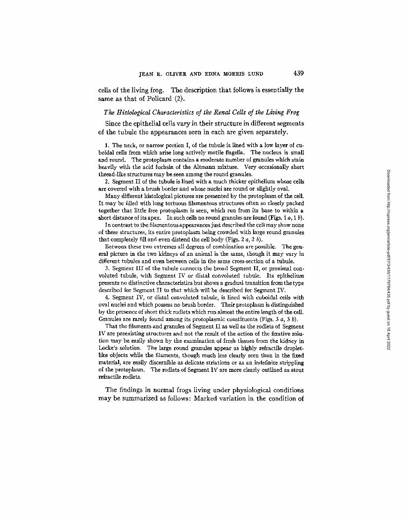
1. The neck, or narrow portion I, of the tubule is lined with a low layer of cu- boidal cells from which arise long actively motile flagella. The nucleus is small and round. The protoplasm contains a moderate number of granules which stain heavily with the acid fuchsin of the Altmann mixture. Very occasionally short thread-like structures may be seen among the round granules.
2. Segment I I of the tubule is lined with a much thicker epithelium whose cells are covered with a brush border and whose nuclei are round or slightly oval.
Many different histological pictures are presented by the protoplasm of the cell. I t may be filled with long tortuous filamentous structures often so closely packed together that little free protoplasm is seen, which run from its base to within a short distance of its apex. In such cells no round granules are found (Figs. 1 a, 1 b).
In contrast to the filamentous appearances just described the cell may show none of these structures, its entire protoplasm being crowded with large round granules that completely fill and even distend the cell body (Figs. 2 a, 2 b).
Between these two extremes all degrees of combination are possible. The gen- eral picture in the two kidneys of an animal is the same, though it may vary in different tubules and even between cells in the same cross-section of a tubule.
3. Segment I I I of the tubule connects the broad Segment II, or proximal con- voluted tubule, with Segment IV or distal convoluted tubule. Its epithelium presents no distinctive characteristics but shows a gradual transition from the type described for Segment I I to that which will be described for Segment IV.
4. Segment IV, or distal convoluted tubule, is lined with cuboidal cells with oval nuclei and which possess no brush border. Their protoplasm is distinguished by the presence of short thick rodlets which run almost the entire length of the cell. Granules are rarely found among its protoplasmic constituents (Figs. 3 a, 3 b).
That the filaments and granules of Segment I I as well as the rodlets of Segment IV are preexisting structures and not the result of the action of the fixative solu- tion may be easily shown by the examination of fresh tissues from the kidney in Locke's solution. The large round granules appear as highly refractile droplet- like objects while the filaments, though much less clearly seen than in the fixed material, are easily discernible as delicate striations or as an indefinite strippling of the protoplasm. The rod_lets of Segment IV are more clearly outlined as stout refracfile rodlets.
T h e findings in n o r m a l frogs l iv ing unde r phys io logica l condi t ions
m a y be s u m m a r i z e d as follows: M a r k e d va r i a t ion in the condi t ion of
Dow
nloaded from http://rupress.org/jem
/article-pdf/57/3/435/1179784/435.pdf by guest on 18 April 2022

440 CELLULAR MECHANISMS OF RENAL SECRETION. I
the protoplasmic structures is seen only in Segment I I of the tubule. The difference in the appearance of these cells is due to a change in the relative amounts of filamentous and granular material present in the cells. Our problem now is to connect if possible this s t ructural variat ion with some variat ion in the functional act iv i ty of the cells.
The first step in such an a t t empt is to determine if the changes are concomitant with some functional state. Two such states mus t be considered; first, absorptive act ivi ty, which includes the absorption of the normal const i tuents of the glomerular filtrate, such as water, sugar and salt; and secondly, secretion, as m a y be evidenced b y the elimination of neutra l red by the cells.
The Absence of Effect of Certain Absorptive Processes upon the Fila- mentous and Granular Material of the Cell
I t is possible by means of the extravi ta l method to cause a k idney to function so tha t no secretory processes are involved in its ac t iv i ty though absorpt ive processes are act ively at work. If the k idney is perfused with a Locke 's solution which contains only simple salts and sugar, such a condition is established, for abundant evidence has shown tha t these substances are el iminated as a fil trate from the plasma by the glomeruli in about the same concentra t ion as exists in
the plasma or perfusion fluid.
A frog was pithed and prepared for perfusion in the usual way except that the left kidney was removed in such a manner as to leave all the cut vessels ligated. It was then fixed in the usual reagents. The other kidney was now perfused for l i hours with Locke's solution. Table I shows the functional findings. It will be noticed that from the urine about two-thirds of the total salt content and all of the sugar of the perfusion fluid had been removed. Other tests would have shown that water as well had been absorbed.
When the histological appearance of the right perfused kidney by the Kolster- Altmann method was compared to that of the left or unperfused organ, no differ- ences in the granulofilamentous material were found. In the broad Segment II of both the unperfused (Figs. 4 a, 4 b) and perfused (Figs. 5 a, 5 b) organ the ceils were filled with long tortuous filaments which extended from the basal membrane to the apex of the cell. Few granular bodies could be found. The ceils of the Segment IV were filled with their short rodlets in sections from both kidneys (Fig. 5 b). The neck of the tubule and the glomeruli showed no significant differences except for the presence of red blood cells in the capillary loops of the tufts of the unper- fused kidney.
Dow
nloaded from http://rupress.org/jem
/article-pdf/57/3/435/1179784/435.pdf by guest on 18 April 2022

J E A N R. OLIVER AND EDNA MORRIS LUND 441
This experiment shows the typical findings in kidneys whose granulofilamentous material of Segment I I was in a filamentous state before the perfusion began. In repetitions of the experiment various conditions were found in the kidney removed before perfusion. In some there were no filamentous structures at all in the cells of Segment II, and these were filled and distended with large round granules. Yet the rodlets of Segment I I were still visible and these ceils contained few or no gran- ules. In other experiments combinations of filaments and granules in varying amount were seen in Segment I I before perfusion In all these cases the unper- fused kidney showed exactly the same arrangement of the stainable material, whether filamentous or granular, in the cells of Segment II.
TABLE I
Absorptive Processes Only in the Kidney
Salt inper- Time Arterial f low Venous f low Urine volume fusion fluid Sugar
10:45-11:00 11:00--11:15 11:15-11:30 11:30-11:45 11:45-12:00
co. per kr.
680 720 760 76O 720
cc. per hr.
880 800 840 840 8OO
co. per hr.
3.6* 5.6 4.8 6.0 5.2
per cerlt
27 39 38 4O 38
* Calculated as output of two kidneys.
I t is reasonable to infer two impor t an t facts f rom these exper iments . First , t h a t the m e t hod of perfusion does not in itself affect the histo-
logical appearance of the f i lamentous granular mater ia l . And second, t h a t the processes of absorpt ion, in as far a t least as they concern the i m p o r t a n t u r ina ry cons t i tuents of salts, wa te r and sugar, can be elimi- na t ed as a possible cause of any morphological changes t h a t m a y be found in the granulof i lamentous s t ruc tures of the cells of Segment I I .
The Effect of a Secretory Process on the Granulofilamentous Material of the Renal Cells
The evidence which indicates t h a t neut ra l red is secreted b y the tubule ceils of the k idney has been previous ly ment ioned (9-11). In the following exper iment the effect of this secretory process on the pro top lasmic const i tuents of these cells is demons t ra ted .
The frog was prepared for perfusion and a normal urine was formed as shown in Table II. After one sample had been collected the left kidney was removed and
Dow
nloaded from http://rupress.org/jem
/article-pdf/57/3/435/1179784/435.pdf by guest on 18 April 2022

442 C E L L U L A R M E C H A N I S M S O1 • R E N A L S E C R E T I O N . I
fixed in the usual solutions, and neutral red in a concentration of 1.25 nag. per 100 cc. was added to the Locke's solution in thebottle supplying the tubular apparatus of the right kidney through the renal-portal venous system. There began an elimination of the dye which reached a rate of 1.0 mg. per hour in the third period. The right kidney was removed after two more periods and fixed. Sections of each kidney were stained by the Altmann method. The contrasting pictures are shown in Figs. 6 a, 6 b and 7 a, 7 b.
The left kidney which had been perfused with clear Locke's solution, and which had therefore no material to secrete, showed the filamentous appearance in the cells of the broad Segment I I that has been previously described. Long tortuous threads filled the protoplasm and only very rarely could a definitely round granular object be found (Figs. 6 a, 6 b). The remaining divisions of the tubules showed the appearance that has been previously given.
TABLE II
Secretion of Neutral Red by lhe Kidney
Time
10:45-11:00
Arterial flow
cc. per hr.
40O
1.25 mg. neutral red in 100 11:30-11:45 440 11:45-12:00 560 12:00-12:15 440 12:15-12:30 440
Venous flow Urine Neutral red Salt in per- Sugar volume fusion fluid
cc. per hr. cc. per hr. rag. per hr. per cent
600 6.0 -- 40 0 Left kidney removed
cc. Locke's solution to tubules of remaining kidney 500 7.2* 0.63* 40 [ 0 500 8.0 1.00 40 / 0 500 6.8 0.85 40 0 600 6.4 0.72 45 0
* Calculated as output of two kidneys
An entirely different picture was found, however, in the kidney which had been perfused with neutral red and which had secreted this substance into the urine. The dye by gross examination was seen to have evenly permeated the organ and sections of it showed a marked change from the appearances noted in the non- secreting kidney. In every cell of practically every Segment I I no filaments could be found whatsoever, for the protoplasmic body was now distended with large round granules. In many instances they were crowded so closely together as to leave no intervening substance visible between them and so closely surrounded the nucleus as to obscure it (Figs. 7 a, 7 b). Yet in those sections where this granular appearance in Segment I I was most extreme the cells of Division IV were still filled by their short thick rodlets (Figs. 8 a, 8 b). The portions of the neck and of Division I I I contiguous to Segment I I showed a greater or lesser similarity to it.
As in the previous series of experiments with clear Locke's solution so in the tests in which neutral red was secreted, animals were found whose left kidney, perfused only by the dye-free fluid, showed a greater or lesser degree of the granu-
Dow
nloaded from http://rupress.org/jem
/article-pdf/57/3/435/1179784/435.pdf by guest on 18 April 2022

J E A N 1~. OLIVER AND EDNA MORRIS LUND 443
lar state in Segment II of its tubule instead of the purely filamentous appearance. The functional activity of these kidneys under the perfusion differed in no way from that of the preceding experiment. The appearances in Segment II of the non- secreting and secreting kidney were, however, found less dissimilar than those described in the preceding experiment. If the cells of the first kidney, non- secreting, were filled with granules and contained no filaments, then the second, secreting, was identical in appearance. If, however, some filaments were present in the first kidney, a dissimilarity was found, for the cells of the second kidney which had secreted the dye contained none of these structures but were filled with gran- ules alone. As an example, the appearance of Segment II is shown in Figs. 9 a, 9b, 10a, 10bandl0c.
The experiments with neutral red just described allow the definite conclusion that in contrast with the negative effect of absorptive processes there occurs concomitantly with the secretion of neutral red a change in the histological appearance of the cells of Segment II that is characterized by the disappearance of filaments and the ap- pearance of granules. If the cells are already in the granular form before the neutral red is given, then no further change occurs in the protoplasmic structures with the secretion of the dye. Secretion of the dye is therefore accompanied by structural changes in the renal cells that are identical with those noted as a part of the vital activities of the living animal and secretory activity occurs only with the histo- logical picture of granule formation.
With the evidence at hand we are justified in stating that the change from the filamentous to the granular state is concomitant with secretory activity. Can a more intimate relation between the morphological and functional aspects of cell response be demonstrated so that we can definitely conclude that the functional activity is determined by the structural change? There remain also several questions of detail. Of the protoplasmic structures of the renal cell, what is mitochondrial substance and what is not ? Are the granules and filaments independent structures or are they related in their constituent material or by a common origin? Are there various sorts of "granules" involved in the histological picture? Are the granules solid substance or are they of the liquid nature and therefore best considered vacuoles? All these questions will be examined directly and objectively by the method of extravital staining and the findings compared with those obtained by the other vital staining and fixed tissue methods.
Dow
nloaded from http://rupress.org/jem
/article-pdf/57/3/435/1179784/435.pdf by guest on 18 April 2022

444 CELLULAR MECHANISMS O~" RENAL SECRETION. I
The Changes Produced in the Isolated Organ by Extravital Staining with Neutral Red
Since an i m p o r t a n t p a r t of the p resen t s t u d y is a compar i son of the
findings b y different m e t h o d s of s ta ining, ma te r i a l f r o m the same ex-
pe r iments descr ibed previous ly , in which the appea rance d e m o n s t r a t e d
b y the use of the A l t m a n n m e t h o d has been noted , was also examined
as examples of ex t rav i t a l s taining.
In the experiment of Table I I the remaining kidney at the end of the perfusion was found to be evenly stained by the neutral red which it had been secreting. The organ was a deep mahogany red in color, striped by the faintly pinki.~h bands of its connective tissue and vessels. I t was not swollen nor edematous. Small bits taken from various portions of the organ were teased apart and crashed with a drop of clear Locke's solution under a cover-glass. In such specimens the dye was contained in its greatest amount in the ceils of Segment II. From here the distribution continued, depending on the degree of staining, towards the neck of the tubule and downwards towards Segment IV with its rodlet cells. The former might contain a considerable amount of dye; the latter was free except for such diffuse staining as may be seen in any tissue that has been long in contact with a dye. The glomeruli contained no dye except for an occasional isolated cell of the clasmatocyte group within the tuft whose irregular protoplasm was filled with dye vacuoles.
The dye, in whatever epithelial cells were examined, was in the form of round granules. In slightly filled cells these were scattered indiscriminately through the protoplasm but in densely filled cells so packed the protoplasmic body as to com- pletely obscure its detail. The nucleus lay as a clear round or oval area in the center of these clusters of granules and was unstained (Figs. 11, 12).
The dye may also be studied in paraffin sections from tissues fixed in formol- Zenker's and the same observations made.
The term "granule" has been used to describe the round deeply staining struc- tures chiefly because that term seemed most appropriate for the description of appearances in the fixed tissues stained by the Altmann method. To most, the word "granule" holds the connotation of solidity and the term "vacuole" might therefore seem more appropriate for the appearances noted in the extravital preparation. That they are not vacuoles in the sense that they are cavities in the protoplasm filled with free liquid dye is shown, however, by the fact that when the cell is crushed they remain intact and float through the surrounding Locke's solution in which the cell is suspended. They remain discrete indefinitely and show little tendency to coalesce, their appearance being that of small droplets of red-stained semifluid material. For we may be sure that they are not solid sub- stance by means of a change similar to one described in vitally stained animals by Policard (2) that occurs in them as the preparation stands. This consists in the
Dow
nloaded from http://rupress.org/jem
/article-pdf/57/3/435/1179784/435.pdf by guest on 18 April 2022

J E A N R. OLIVER AND EDNA MOP._RIS LUND 445
development within the granule of a smaller granule, a minute point-like or comma- shaped object which has all the appearance of condensed dye that has precipitated out of the dye content of the larger granule. The smaUer object is usuany affected by Brownian movement and may be seen twisting and turning within the larger granule, colliding with its inner surface and rebounding into the center again. This active movement proves that the substance of the granule proper must be at least semifluid, so, with our previous determination that its substance is immiscible with water and from what we know of the sohbility of neutral red, the assumption that the granule is a droplet of lipoid material becomes extremely plausible. In one sense, therefore, the structure is a vacuole but in order to maintain the concept that fits the appearance in the fixed tissues so well and which has been used almost entirely by those who have previously studied such material we shall use the terms, "granule, .... vacuole" and "droplet" as synonyms.
The question now presents itself as to the exact relation of these extravitally stained vacuoles and the large round granules which have been described in the Altmann preparation of fixed tissue from the extravitally stained kidney. Since we know from the examination of fresh tissues that the granules of the fixed tissues are preexisting ob- jects a glance at Figs. 7a, 7b and 11, 12, which are from the same kidney stained by the two methods, will show that the extravitally stained objects and the Altmann-stained granules are certainly the same forma- tions. Apart from the similarity of their appearance there would not be space for two preexisting different sets of such structures to exist side by side within the limited room of the cell body. The granule of the extravitally stained kidney can therefore be considered a semi- fluid lipoid droplet deeply stained with neutral red which when fixed is preserved and stainable by the fuchsin of the Altmann method. I t would seem reasonable to extend this conclusion to include in the same category the unstained granules seen in cells of the living animal which resemble identically by the Altmann method the objects seen in the extravitally stained neutral red experiment. Supporting evi- dence of this identity will appear later.
In the description of the kidney stained extravitally with neutral red there has been no mention of filaments within the cells. I t is true that these structures have disappeared, at least in their long thread- like form, with the occurrence of secretory activity and the develop- ment of the granules but, as previously described for the Altmann preparations, portions of them always remain in the lower part of the
Dow
nloaded from http://rupress.org/jem
/article-pdf/57/3/435/1179784/435.pdf by guest on 18 April 2022

446 C E L L U L A R M E C H A N I S M S OF RENAL S E C R E T I O N . I
cell , c l u s t e r ing a r o u n d t h e nuc l eus where t h e y a p p e a r as s h o r t b a c i l l a r y
o r v ib r io - l i ke ob jec t s . I n n e u t r a l r ed e x t r a v i t a l p r e p a r a t i o n s t h e y
a r e n e v e r seen, for he re a l l t he s t a i n e d o b j e c t s a re t h e d e f i n i t e l y r o u n d
l a rge g r a n u l o v a c u o l e s . T h a t these t h r e a d - l i k e s t r u c t u r e s a re i n d e e d
p r e s e n t a n d t h a t t h e y too m a y be s t a i n e d e x t r a v i t a l l y is shown, how-
ever , in t he n e x t ser ies of e x p e r i m e n t s .
The Changes Produced in the Isolated Organ by Extravital Staining with Janus Green
The method of extravital staining with Janus green was identical with that used with neutral red. The perfusion was begun with clear Locke's solution and after one or two samples were examined to determine if a normal condition of kidney
TABLE III
Action of Janus Green on the Kidney
Time A r ~ a l flow Venous flow UHne D • Salt inper- volume Y fusion fluid
co. per hr. co. per hr. co. per hT. [ m . per hr. ] p ~ cent I I
10:00-10:15 400 500 6.0 [ -- I 45 1/500,O00Janus green in Locke's to tubules of remaining kidney
10:30-10:45 400 450 6.0 0 45 10:45-11:00 450 400 8.0 0 40 11:00-11:15 500 400 10.0 0 45 11:15-11:30 450 450 7.2 0 45
Sugar
0 0 0
Wr.
function had been established, the dye was added to the fluid in the bottle that supplied the tubular circulation. Janus green is a definitely toxic substance, so that a dilution of 1/500,000 was used. Another advantage of this dilution whose importance will be emphasized later is the specificity of its staining reaction. But even with such a solution the kidneys showed evidences of damage after 2 or 3 hours of perfusion so that the definitive experiments were limited to 1 hour or less.
The results of such extmvital staining with Janus green are shown in Table I I I . I t will be observed that there was no serious disturbance in the function of the organ, but the dye was not excreted. An explanation of this lack of secretion will be considered later.
In the gross, the kidneys were unchanged except for their dark slate-green color. When examined in fresh crushed specimens many of the cells of Segment I I appeared similar, save for color, to those from the neutral red kidney. Other cells contained the large green round gmnulovacuoles but when examined with the oil immersion a striking difference from the earlier preparation with neutral
Dow
nloaded from http://rupress.org/jem
/article-pdf/57/3/435/1179784/435.pdf by guest on 18 April 2022

JEAN R. OLIVER AND EDNA MORRIS LUND 447
red was noted. For besides the large round green granules there were found in those parts of the cell where the protoplasm was not occupied by them fine thread- like dark green bacillary objects (Fig. 13). Another difference from the neutral red staining was noted in Segment IV. In the Janus green extravital preparations the short thick rodlets were definitely stained distinct green. There were, however no large green vacuolar formations in this division of the tubule.
The staining of the thread-like bodies with Janus green together with their reaction with bichromate and their appearance when stained by the Altmann method leaves no doubt that these objects can be con- sidered mitochondria in the strict and modem sense of the word. The same is true for the rodlets of Division IV. That these filamentous mitochondria have disappeared in great part from the cell bodies of Segment II of kidneys actively secreting neutral red and that they only persist in those portions of the protoplasm that are not occupied by the large droplet formations is worthy of especial note.
The nature of the large droplets in the Janus green extravital prepa- rations is less certain, however, for they have all the appearance, save color, of the objects seen in the kidney stained extravitally with neutral red, and mitochondria do not stain with this dye. Our next problem must be, therefore, to determine whether these structures in the Janus green preparations are indeed the same objects seen in the neutral red preparations.
All the morphological similarities we have mentioned are strong pre- sumptive evidence that the large round green bodies of Janus green extravital staining are identical to the red ones of neutral red extra- vital staining. Still one might demur to such a conclusion with the suggestion that in the former case the droplets have arisen as a result of some specific or toxic action of the Janus green. The extravital method allows us, however, to answer this objection in a decisive manner for it can be shown conclusively that the two appearances are the same object stained by one or the other dye.
The Changes Produced in the Isolated Kidney by Combined Extravital Staining with Janus Green and Neutral Red
Since neutral red is a more rapidly acting dye than Janus green the latter was perfused first to the tubule of the kidney in a dilution of 1/400,000 in the manner previously described. Table IV shows the results. For 1 hour a urine normal except for the appearance of faint traces of sugar was obtained. The bottle c o n -
Dow
nloaded from http://rupress.org/jem
/article-pdf/57/3/435/1179784/435.pdf by guest on 18 April 2022
448 CELLULAR MECtIAINrlSMS OF RENAL SECRETION. I
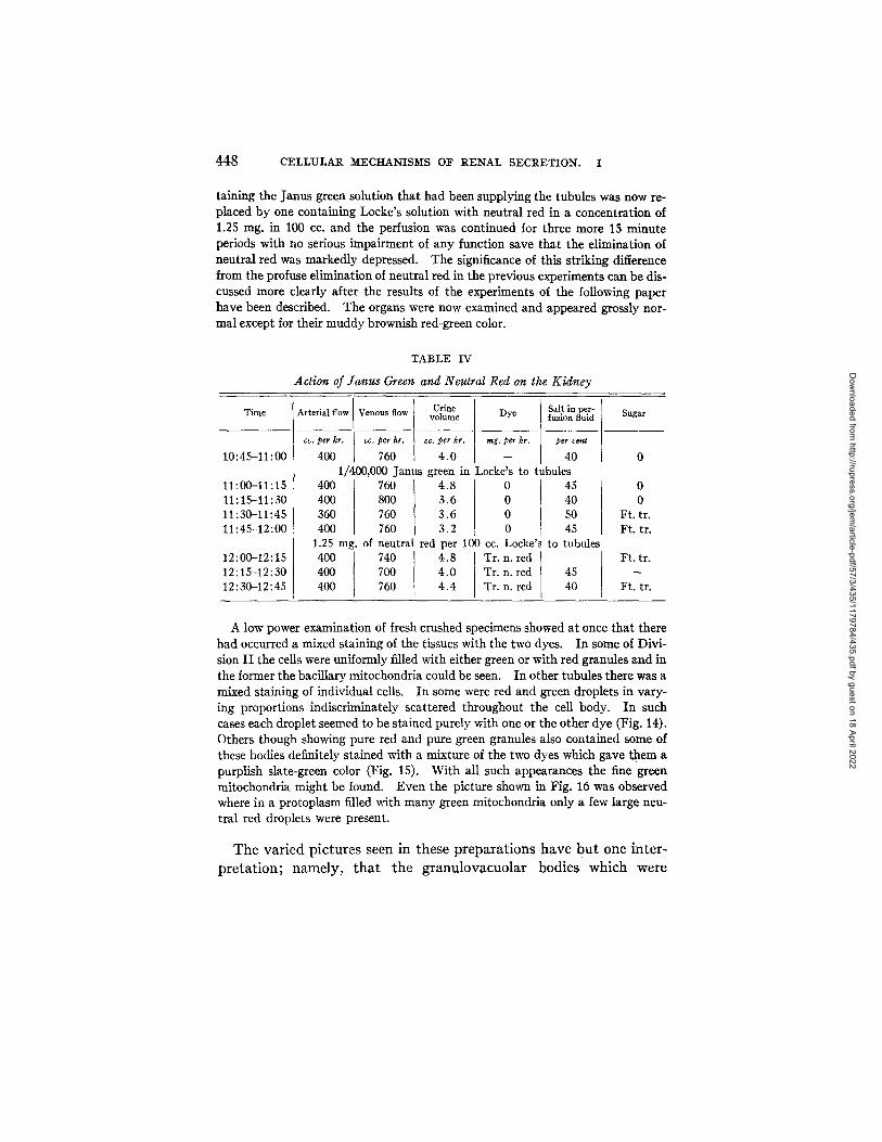
raining the Janus green solution that had been supplying the tubules was now re- placed by one containing Locke's solution with neutral red in a concentration of 1.25 mg. in 100 cc. and the perfusion was continued for three more 15 minute periods with no serious impairment of any function save that the elimination of neutral red was markedly depressed. The significance of this striking difference from the profuse elimination of neutral red in the previous experiments can be dis- cussed more clearly after the results of the experiments of the following paper have been described. The organs were now examined and appeared grossly nor- mal except for their muddy brownish red-green color.
TABLE IV
Ac t ion o f J a n u s Green and Neu t ra l Red on the K i d n e y
Time Sugar
10:45-11:00
11:00-11:15 11:15-11:30 11:30-11:45 11:45-12:00
12:00-12:15 12:15-12:30 12:30-12:45
Arterial flow Venous flow Urine Salt in per- volume Dye fusion fluid
cc. per hr. co. per hr. cc. per hr. rag. per hr. per cent
400 760 4.0 -- 40 1/400,000 Janus green in Locke's to tubules
400 400 360 400
1.25 mg. of 400 400 400
760 4.8 800 3.6 76O 3.6 760 3.2 neutral red per 740 4.8 700 4.0 760 4.4
0 45 0 40 0 5O 0 45
100 cc. Locke's to tubules Tr. n. red Tr. n. red 45 Tr. n. red 40
0 0
Ft. tr. Ft. tr.
Ft. tr.
Ft. tr.
A low power examination of fresh crushed specimens showed at once that there had occurred a mixed staining of the tissues with the two dyes. In some of Divi- sion I I the ceils were uniformly filled with either green or with red granules and in the former the bacillary mitochondria could be seen. In other tubules there was a mixed staining of individual cells. In some were red and green droplets in vary- ing proportions indiscriminately scattered throughout the cell body. In such cases each droplet seemed to be stained purely with one or the other dye (Fig. 14). Others though showing pure red and pure green granules also contained some of these bodies definitely stained with a mixture of the two dyes which gave them a purplish slate-green color (Fig. 15). With all such appearances the fine green mitochondria might be found. Even the picture shown in Fig. 16 was observed where in a protoplasm filled with many green mitochondria only a few large neu- tral red droplets were present.
T h e va r i ed p ic tu res seen in these p r e p a r a t i o n s have b u t one in t e r -
p r e t a t i o n ; name ly , t h a t the g r a n u l o v a c u o l a r bodies which were
Dow
nloaded from http://rupress.org/jem
/article-pdf/57/3/435/1179784/435.pdf by guest on 18 April 2022

JEAN R. OLIVER AND EDNA MORRIS LUND 449
stained indiscriminately by either Janus green or neutral red or by both in mixture are in fact the same structures. I t is a fact of con- siderable significance as will appear later that these structures stain so readily in a very dilute solution of Janus green.
Our experiments have demonstrated one definite difference in the reaction of the mitochondria and the granulovacuolar bodies, since the former never stain with neutral red. Another reaction has been found which indicates a difference in the constitution, either physical or chemical, of the two structures, and that is their reaction to the Gram stain. In all specimens, whether from animals living under natural conditions, from kidneys perfused with plain Locke's or from those extravitally stained with either neutral red or Janus green, the granulo- vacuolar bodies retain the Gram stain (Figs. 23-25). Such Gram positivity, however, is never present in the mitochondria, either in the fine bacillary forms and long filaments of Segment II or in the short rodlets of Segment IV (Fig. 26).
Another difference between the vacuoles and the mitochondria that can be demonstrated by means of the Gram-Weigert stain is the fact that the former do not need to be bichromatized to resist solution in alcohol as do the mitochondria, and also that they resist the action of acetic acid in the fixing solution, a substance which completely de- stroys all evidence of mitochondrial structures (Figs. 23, 24).
Again the question may arise if we are dealing with the same granulo- vacuoles in these sections stained with Gram as in the preparations stained by the extravital method.
A Gram stain done on tissues after such treatment in which the neutral red has been preserved by fixation in formol-Zenker's solution shows cells containing blue vacuoles, red vacuoles, a mixture of the two and granules whose color is a purplish combination of red and blue (Fig. 17).
As stated, it is our plan to bring to bear as many different methods as are applicable to the problem in order to confirm our contention that the extravital method not only produces no artifacts in the tissues that are being examined by it, but that under its conditions processes of the same nature may develop as actually occur during life. For these reasons supravital and vital staining with the two dyes was done and the results compared with those of extravital staining. I t is of
Dow
nloaded from http://rupress.org/jem
/article-pdf/57/3/435/1179784/435.pdf by guest on 18 April 2022

450 CELLULAR MECHANISMS OF RENAL SECRETION. I
course impossible to pe r fo rm e labora te tests wi th these less cont ro l lable
me thods . E n o u g h of the sal ient fea tures of the p rev ious exper iments
can be obse rved wi th t h e m howeve r to afford conv ic t ion t h a t the proc-
esses t h a t occur in the hand l ing of the dye are s imilar u n d e r the con-
d i t ions t h a t ob t a in in all th ree me thods , vi tal , sup rav i t a l and ext ra-
vi tal . T h e f indings t h a t we give are in a s u m m a r y form.
Appearance of the Supravitally Stained Renal Epithelium
Supravital Staining with Neutral Red.--Bits of fresh kidney tissue from killed animals were teased and agitated in a concentration of 0.25 rag. neutral red in 100 cc. Locke's solution. At intervals small portions were removed and examined in fresh crushed smears with Locke's solution. The appearances in the cells can be summarized by the statement that they are identical with those we have described as a result of the extravital method. A few minutes after being placed in the dye solution red granules appeared in the cells of Segment I I of the tubule which increased in number with time, so that in a short period, whose length de- pended apparently chiefly on the speed with which the dye could permeate the teased tissues, the protoplasm of the cells was filled with large round droplets that surrounded the unstained nucleus (Fig. 18). There was no staining of the mito- chondrla nor were the rodlets of Division IV colored. This latter portion of the tubule was also free of the larger round granulovacuoles. In the vacuoles, espe- cially in the older preparations, the same small motile granule within the larger one was seen. In other words one cannot distinguish a fresh crushed specimen of such a preparation under the microscope from one from the previously described extravitally stained kidney.
Supravital Staining with Janus Green.--Bits of kidney tissue placed in Locke's solution containing a 1/500,000 concentration of Janus green showed in the course of a few minutes dye within the protoplasm of their cells. The poor penetration, however, of Janus green as compared to neutral red was particularly noticeable in the supravital preparations. Only the cells of the tubules on the periphery of the tissue masses were stained and these irregularly. The protoplasm of the cells of Segment I I was filled with the fine bacillary mitochondria and among these were scattered large round green droplets in greater or less number. As time passed the latter increased in number while the mitochondria decreased (Fig. 19). In the vacuoles there developed the small motile granule and the rodlets of Division IV of the tubule were stained a definite green. In specimens that had stood for some time evidences of damage appeared. These consisted of irregularities in size and shape in both the mitochondria and the droplets and a greenish discolora- tion of the nucleus associated with pycnosis.
Combined supravital staining with the two dyes was also done and the appear- ances noted were entirely the same as in the combined extravital experiment. The double staining was accomplished by staining the tissues in mixtures of the
Dow
nloaded from http://rupress.org/jem
/article-pdf/57/3/435/1179784/435.pdf by guest on 18 April 2022

~EAN R. OLIVER AND EDNA MORRIS LUND 451
two dyes or successively in one and then in the other. The results depended on the difference in the relative permeability of the two dyes and by varying the pro- cedure all the previously described pictures of extravital staining were produced. Some cells contained only large droplets of red, others only green droplets, while still others showed scattered granules of both colors and even granules whose color was a purplish green mixture of the two dyes (Fig. 20). Wherever the Janus green had entered the cell the mitochondria might be found in a greater or less amount depending on the number of droplets of one color or the other that were present. Perhaps the most striking figures were those in which red droplets were scattered through a protoplasm that still contained a considerable amount of green mito- chondrial material (Fig. 21).
I t is ev iden t f rom these exper iments t h a t the processes invo lved in
ex t r av i t a l a nd sup rav i t a l s t a in ing are essent ia l ly similar. I t r ema ins
to de t e rmine if t he same i d e n t i t y ob ta ins in the case of isola ted t issues
a nd of the processes t h a t occur in the l iv ing an ima l secre t ing the dye .
Com pa r i sons were m a d e wi th v i t a l s t a in ing of l iv ing frogs.
The Appearances Noted in Vital Staining of Living Animals with Neutral Red
10 co. of a 0.25 per cent solution of neutral red in Locke's was injected into the dorsal lymph sac of a living frog. The injection was repeated at hourly intervals three times and the animal killed 1 hour after the last injection. All of the fluid was absorbed. The tissues of the animal were now definitely pink, the coloration being especially noticeable beneath the skin and in the nucleus and oviducts. The urine in the bladder was pink. The kidneys were more or less heavily stained, some having the dark mahogany-red appearance described in the extravital ex- periment. The microscopical appearance of fresh crushed preparations from them was also identical to those of the extra- and supravital preparations. The cells of Segment I I contained typical droplets, some in few numbers while others were completely filled and distended by them. In some kidneys whose cells con- tained only a few red granules there could be seen uncolored droplets. As the preparation stood the condensed motile particle of dye appeared within the vac- uole. In cells which contained only a few vacuoles, it was possible to observe in the protoplasm free of these structures an indefinite unstained stippling whose de- tail it was impossible to resolve. When such vitally stained neutral red material was restained supravitally with Janus green, it was found that the stippling had represented uncolored mitochondria (Fig. 22) and as the supravital staining con- tinued, typical large round vacuoles of Janus green appeared.
T h e exper iments d e m o n s t r a t e the same s t ruc tu res and processes in t he cells of l iv ing an imals secre t ing the d y e t h a t were obse rved in the
Dow
nloaded from http://rupress.org/jem
/article-pdf/57/3/435/1179784/435.pdf by guest on 18 April 2022

452 CELLULAR MECHANISMS O~F RENAL SECRETION. I
extravital experiments. I t follows that in the present instances the results of vital, extra- and supravital staining were essentially similar in their nature.
DISCUSSION
A summary can now be made of the structural changes that accom- pany secretion in the cells of Segment II and their significance can be appraised.
The following facts have been established. 1. Filaments and granulovacuoles are preformed protoplasmic
elements of the renal cell. 2. During the normal life of the animal the cells of Segment II show
a wide variation in the relative amounts of these two protoplasmic structures. In any given cell there is a converse relation between the amount of filamentous and granular material.
3. The principal absorptive processes that occur in the formation of the urine are not accompanied by variations in these structures.
4. The extravital secretion of neutral red which has been shown to be identical in its processes with vital secretion, is accompanied by a disappearance of filaments and the formation of granulovacuoles. If the cells originally contain only the latter, no histological alteration accompanies the secretion of the dye.
5. The granulovacuolar structures stain with neutral red during the secretion of the dye. This can be demonstrated either extravitally or vitally.
6. The filamentous structures never stain with neutral red. 7. The filaments do stain with Janus green. They also require bi-
chromatization for their preservation and are soluble in weak acetic acid.
8. The granulovacuolar material also stains with Janus green even from weak concentrations of the dye but does not require bichromati- zation and is not soluble in dilute acetic acid.
9. The filaments are Gram-negative, the granulovacuolar struc- tures Gram-positive.
That the structural changes observed as concomitant with the secre- tion of the dye play a part in the actual secretory process seems certain when one considers that the dye is concentrated within the cell in the
Dow
nloaded from http://rupress.org/jem
/article-pdf/57/3/435/1179784/435.pdf by guest on 18 April 2022

JEAN R. OLIVER AND EDNA MORRIS LLrND 453
vacuolar structures during the course of the secretion. The signifi- cance of this concentration is examined in an accompanying paper. That the filaments are mitochondrial is also obvious. The question remaining for determination is that of the nature of the vacuoles and of what relation they bear to the mitochondria. From a consideration of the facts noted above, two interpretations are possible.
1. The filaments and granules are entirely independent structures that bear no relation to each other. On this hypothesis the mitochon- dria play no r61e in the secretion of the dye. But if such is assumed one is at once confronted with the difficult admission that highly di- luted Janus green is not so specific a stain as a long series of investi- gations would indicate. There also remains without explanation the mysterious disappearance during the secretory process of the filaments which one must assume have vanished leaving no trace while at the same time an equally unexplained appearance de novo of granulo- vacuoles has occurred, derived not from the mitochondria, but from an unknown substance which has the generally recognized character- istic staining reaction of mitochondria.
2. The granulovacuoles are derived from the mitochondrial fila- ments by alteration both in their form and constituent substance. The change in substance is of such nature that neutral red now stains them, leaving unaffected, however, the original staining reaction of this altered mitochondrial material to Janus green.
The second interpretation seems to us much the more satisfactory. I t explains, for example, the converse occurrence of filaments and granules which is so constantly observed; the reaction of the vacuole to dilute Janus green as a remainder of the characteristics of the original mitochondrial substance; it indicates what becomes of the substance of the disappearing mitochondria and designates from what source the material of the originating granulovacuoles is derived. By it the mitochondria are made essential factors in the secretion of the dye, since the change which occurs in them is the source of the final mechanism by which the dye is concentrated within the cell body. The exact nature of this alteration which accompanies the change of the mitochondrial substance into vacuolar substance is not definitely shown by the experiments but the altered reactions of the latter to dyes and fixatives suggests that a splitting of the protein-lipoid corn-
Dow
nloaded from http://rupress.org/jem
/article-pdf/57/3/435/1179784/435.pdf by guest on 18 April 2022

454 CELLULAR MECHANISMS OF RENAL SECRETION. I
plex of the mitochondrial material has occurred, with a consequent liberation of its constituent substances, so that each, now no longer bound and inert, reacts in its characteristic manner. The freed lipoid takes up neutral red while the protein element, insoluble in alcohol without bichromatization, reacts with the Gram stain like fibrin.
One might visualize the course of events in the secretory process already described somewhat as follows. Neutral red enters the cell from the blood vessel. Owing to its presence and to factors as yet undetermined the mitochondrial filaments disintegrate. One can think of many forces that might cause such a result, such as changes produced by the presence of the dye in interface surface tensions, osmotic pressure or diffusion currents. From the material of the dis- integrated filaments and as a result of constituent alterations, vacuoles are formed in which the dye being readily soluble is concentrated. Whatever vacuoles may be present from previous processes of secretion are also saturated with the dye. And here the description of the process of elimination must, for the time being, end, for no evidence whatsoever has been obtained as to how the dye passes from the vac- uoles into the lumen of the tubule. This question will be examined in the succeeding article and the relation of our findings to those of previous observers will be discussed.
CONCLUSIONS
1. The secretion of neutral red reproduces those variations which are observed in the mitochondrial apparatus of the renal tubule cells of animals living under native conditions. The tubular absorptive processes concerned with water, salts and sugars do not produce these effects.
2. The changes in the mitochondria consist of both structural and constituent alterations. These have been shown to be not merely phenomena concomitant with secretion, but a determining factor in one part of this process; namely, in the concentration of the dye within the cell.
BIBLIOGRAPHY
1. Kolster, R., Beitr. path. Anat. u. allg. Path., 1911, 51, 209. 2. Policard, A., Arch. anat. micr., 1910, 12, 177. 3. Oliver, J., J. Exp. Med., 1916, 23, 301.
Dow
nloaded from http://rupress.org/jem
/article-pdf/57/3/435/1179784/435.pdf by guest on 18 April 2022

JEAN R. OLIVER AND EDNA MORRIS LUND 455
4. Cushny, A. R., The secretion of the urine, London, Longmans, Green and Co., 2nd edition, 1926.
5. Emge, L. A., Stanford Univ. Pub., Univ. Series, Med. Sc., No. 2, 1921, 1. 6. Richards, A. N., Methods and results of direct investigation of the function of
the kidney, The Williams & Wilkins Co., Baltimore, 1929. 7. Hoeber, R., Klin-therap. Woch., 1927, 6, 671. 8. 0liver, J., and Smith, P., J. Exp. Med., 1931, 58, 785. 9. Oliver, J., and Shevky, E., ]. Exp. Med., 1929, 50, 15.
10. Scheminzky, F., Arch. ges. Physiol., 1929, 221,641. I1. Richards, A. N., Arch. Path., 1932, 13, 847. 12. Forkner, C. E., J. Exp. Med., 1930, 52, 379. 13. Barkan, C., Broemser, P., and Hahn, A., Z. Biol., 1922, 74, 1.
EXPLANATION OF PLATES
PLATE 28
Magnification of photographs X 525; of drawings X 680. FlOS. 1 a to 3 a, and 1 b to 3 b, tubules from two animals living under native
conditions, Kolster-Altmann procedure. Figs. 1 a and 1 b show Segment I I of the tubule. The cells are filled with long filamentous structures. Figs. 2 a and 2 b show Segment I I from another animal. Its cells are greatly swollen and filled with large round granules. Figs. 3 a and 3 b, from the same specimen as Nos. 2 a and 2 b, show sections of Segment IV. The flat cells are filled with heavy rod- lets. They contain no granules.
FIGs. 4 a, 4 b and 5 a, 5 b. The negative effect of absorptive processes on the filaments of Segment II. Experiment of Table I. Kolster-Altmann procedure. Figs. 4 a and 4 b show the cells of the kidney removed before the perfusion was started, filled with long filaments and containing no large round granules. Figs. 5 a and 5 b show the other kidney of the animal after 1,1 hours perfusion with plain Locke's solution. Under these conditions absorption by the tubule cells of water, salts and sugar occurred, but no secretion of any substances. The cells show no alteration from their previous condition. In the right part of Fig. 5 b Segment IV is seen with its unaltered rodlets.
PLATE 29
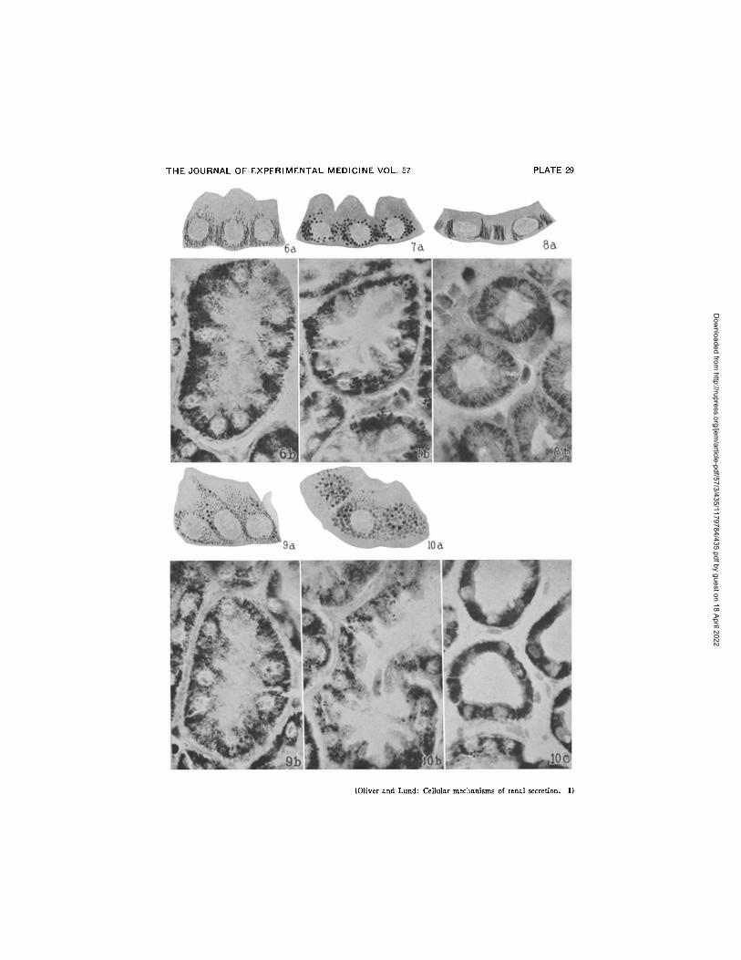
Magnification of photographs × 525; of drawings X 680. FIGs. 6 a, 6 b, 7 a, 7 b, 8 a and 8 b. The changes produced by the secretion
of neutral red on the filaments of Segment II. Experiment of Table II. Kolster- Altmann procedure. Figs. 6 a and 6 b show the cells of Segment I I of the kidney removed after the preliminary perfusion with plain Locke's solution that contained no secretable substances. They are filled with long filaments and contain no granules. Figs. 7 a and 7 b show Segment I I from the other kidney that was perfused with neutral red and which secreted this substance in considerable
Dow
nloaded from http://rupress.org/jem
/article-pdf/57/3/435/1179784/435.pdf by guest on 18 April 2022

456 CELLULAR MECHANISMS OF RENAL SECRETION. I
amount. The filaments are replaced by large granulovacuoles and no filaments are visible. Figs. 8 a and 8 b show the unaltered rodlets of Segment IV from the same kidney.
FIGs. 9 a, 9 b, 10 a, 10 b and 10 c. Effect of the secretion of neutral red on cells of Segment I I which originally contained a mixture of filaments and granules. Experiment of page 443. Kolster-Altmann procedure. Figs. 9 a and 9 b show the condition of the cells of Segment I I of the kidney removed before the secretion of neutral red. Many filaments and a certain number of granules are seen. Figs. 10 a and 10 b show similar cells from the other kidney which had been perfused with and which had secreted neutral red. The filaments have entirely disappeared and the cells are filled with large round granules. Fig. 10 c shows the rodlets of Segment IV unchanged.
PLATE 30
All figures, except No. 17, are of fresh preparations of unfixed tissue suspended in Locke's solution.
FIG. 11. Extravital staining with neutral red. Experiment of Table I. A por- tion of Segment I I whose cells have retained their normal position in the tubule. They are filled with granulovacuoles of neutral red. The nuclei are unstained. Magnification X 390.
FIG. 12. Extravital staining with neutral red. An isolated cell from Segment I I of the same kidney. Clustered around the nucleus are granulovacuoles of neu- tral red. Compare the appearance with that of the same tissues after the Kolster- Altmann procedure (Figs. 7a and 7b) and after Bouin fixation and Gram-Weigert staining (Fig. 23) and Zenker fixation and Gram-Weigert staining (Fig. 24). Magnification × 1200.
FIG. 13. Extravital staining with Janus green. Unfixed cell from Segment I I of the kidney of the experiment of Table I I I . The large granulovacuoles of Janus green are seen as well as the fine bacillary thread-like mitochondrla. Note that in this and the following figures mitochondria occur only in parts of the cell free of granulovacuoles. Magnification × 1200.
FIG. 14. Extravital staining with both neutral red and Janus green. Experi- ment of Table IV. There is an indiscriminate mixing of red and green granules throughout the protoplasm of the cell with a few scattered mitochondria. Magni- fication × 1200.
FIG. 15. The same specimen and procedure. Another cell from Segment I I shows red and green vacuoles as well as some which are stained with a mixture of the two dyes. This mixture is represented for purposes of reproduction as a stipple whereas in fact the admixture of dyes produces an even greenish brown tone. Magnification X 1900.
FIG. 16. The same specimen and procedure. Many fine bacillary forms of mitochondria stained with Janus green are visible. All of the granulovacuolar bodies are stained with neutral red. Magnification X 1200.
Dow
nloaded from http://rupress.org/jem
/article-pdf/57/3/435/1179784/435.pdf by guest on 18 April 2022

JEAN R. OLIVER AND EDNA MORRIS LUND 457
FIG. 17. Extravital staining with neutral red, fixation in formol-Zenker's solu- tion and counterstaining by the Gram-Weigert method. Experiment of Table I. A cell from Segment II . Some of the granules have retained their neutral red color, some are heavily stained by the methyl violet, represented as black, while others show a mixture of the two dyes. These latter present an even purplish red tone that has been represented by a coarse stipple. Magnification × 1900.
Fro. 18. Supravital staining with neutral red. A cell from Segment I I of the kidney of an untreated animal supravitally stained. The protoplasm is filled with deeply stained red granulovacuoles, and the picture is identical to that ob- tained by extravital staining. Compare Fig. 12. Magnification × 1200.
Fro. 19. Supravital staining with Janus green. A similar cell from the same kidney illustrated in Fig. 18, supravitally stained. Many green granulovacuolar bodies and a few fine mitochondria are visible. Compare with Fig. 13, an extra- vitally stained cell. Magnification × 1200.
Fro. 20. Supravital staining with both neutral red and Janus green. Cell of Segment I I from same material supravitally stained in Locke's solution contain- ing 1/100,000 neutral red and 1/500,000 Janus green. A mixture of red and green vacuoles is clustered about the nucleus. For similarity to the results of combined extravital staining of the two dyes compare with Fig. 14. Magnification × 1200.
Fro. 21. Supravital staining with neutral red and Janus green. Same material and method. Fine green mitochondria are visible with areas of clustered neutral red granulovacuolar bodies. The corresponding appearance by the combined ex- travital method is seen in Fig. 16. Magnification × 1200.
Fro. 22. Vital staining with neutral red, with subsequent supravital staining with Janus green. A cell from Segment I I of the kidney of an animal vitally stained with neutral red. Material from this kidney was then supravitally stained in Locke's solution containing 1/500,000 Janus green. The vital granulovacuolar bodies of neutral red alternate with areas where the persisting mitochondria have been supravitally stained by the Janus green. The effect is identical with com- bined extravital (Fig. 16) and combined supravital staining (Fig. 21). Magnifi- cation X 1200.
PLATE 31
FIG. 23. Segment I I of the tubule from the kidney secreting neutral red in experiment of Table II. Bouin fixation, Gram-Weigert stain and no counterstain. The cells are filled with granulovacuoles that have resisted the action of acetic acid and retained methyl violet. Compare with Figs. 7 a and 7 b where the same structures are stained by the Koister-Altmann procedure. Magnification X 525.
FIG. 24. From the same kidney. Zenker fixation, Gram-Weigert stain. The same Gram-positive granules fill the cells. Magnification X 525.
FIG. 25. Segment I I of a normal animal living under native conditions, Kolster fixation, Gram-Weigert stain. The cells are filled with Gram-positive granulo-
Dow
nloaded from http://rupress.org/jem
/article-pdf/57/3/435/1179784/435.pdf by guest on 18 April 2022

458 CELLULAR MECHANISMS OF RENAL SECRETION. I
vacuoles. The same specimen with the Altmann stain is shown in Figs. 2 a, 2 b. Magnification X 525.
FIo. 26. Segment I I of another normal untreated animal. Kolster fixation, Gram-Weigert stain. No Gram-positive granules are visible, as the cells in this instance are filled with mitochondrial filaments. Compare Figs. 1 a and 1 b, a section from the same block stained by the Altmann method. Magnification X 525.
Dow
nloaded from http://rupress.org/jem
/article-pdf/57/3/435/1179784/435.pdf by guest on 18 April 2022

THE JOURNAL OF EXPERIMENTAL MEDICINE VOL. 57 PLATE 28
(Oliver and Lund: Cellular mechanisms of renal secretion. I)
Dow
nloaded from http://rupress.org/jem
/article-pdf/57/3/435/1179784/435.pdf by guest on 18 April 2022
THE JOURNAL OF EXPERIMENTAL MEDICINE VOL. 57 PLATE 29
(Oliver and Lund: Cellular mechanisms of renal secretion. I)
Dow
nloaded from http://rupress.org/jem
/article-pdf/57/3/435/1179784/435.pdf by guest on 18 April 2022

THE JOURNAL OF EXPERIMENTAL MEDICINE VOL. 57 PLATE 30
(Oliver and Lund: Cellular mechanisms of renal secretion, l)
Dow
nloaded from http://rupress.org/jem
/article-pdf/57/3/435/1179784/435.pdf by guest on 18 April 2022

THE JOURNAL OF EXPERIMENTAL MEDICINE VOL. 57 PLATE 31
(Oliver and Lund: Cellular mechanisms of renal secretion. I)
Dow
nloaded from http://rupress.org/jem
/article-pdf/57/3/435/1179784/435.pdf by guest on 18 April 2022