Multiple Mechanisms for Anti-Fibrotic Functions of Statins on Radiotherapy Induced Fibrosis
8
Journal of Cancer Research Updates, 2014, 3, 000-000 1 ISSN: 1929-2260 / E-ISSN: 1929-2279/14 © 2014 Lifescience Global Multiple Mechanisms for Anti-Fibrotic Functions of Statins on Radiotherapy Induced Fibrosis Chao Li 1 , Wei Li 2 , Lathika Mohanraj 1 , Qing Cai 1 , Mitchell S. Anscher 3 and Youngman Oh 1,2,* 1 Department of Pathology, Virginia Commonwealth University, School of Medicine, Richmond, VA 23298, USA 2 Biocure Pharma LLC, Richmond, VA 23298, USA 3 Department of Radiation Oncology, Virginia Commonwealth University, School of Medicine, Richmond, VA 23298, USA Abstract: Radiotherapy-induced fibrosis (RTIF) presents a challenge in radiotherapy for cancer patients. Although numerous studies have attempted to elucidate the mechanisms leading to RTIF, the pathogenesis of RTIF at the cellular and molecular level is still incompletely described. One key component involved in the post-radiation injury is the pleuripotent cytokine transforming growth factor (TGF)-. TGF- signaling pathway has been under intensive investigation about its critical role in radiation-induced fibroproliferative disease. Connective tissue growth factor (CTGF), also known as insulin-like growth factor binding protein-related protein 2 (IGFBP-rP2) is a potent regulator of fibroblast proliferation, cell adhesion, and stimulation of extracellular matrix production. CTGF is known as a major downstream mediator of the chronic fibrotic effects of TGF-. Here we have demonstrated that irradiation and TGF- induced CTGF, subsequently upregulates fibrotic factors such as fibronectin and type IV collagen. Furthermore, as HMG-CoA reductase inhibitors, statins inhibit expressions of CTGF and downstream fibrotic proteins in both normal human fetal fibroblasts (HFL-1) and human dermal fibroblasts (HDF) on TGF- treatment or irradiation. Our study also demonstrates that simvastatin not only suppressed TGF--induced fibrosis through inhibition of CTGF production but also CTGF-induced fibrosis. We further show that simvastatin may act in a TGF--independent manner by inhibiting Rho kinase pathway. Taken together, these data suggest that radiotherapy may upregulate CTGF expression in a TGF--dependent and - independent manner, thereby enhancing expression of profibrotic factors and inducing lung fibrosis. Keywords: CTGF, Statins, Fibrosis, TGF-, Radiation, Rho/ROCK pathway. INTRODUCTION Radiation induced lung injury is the main dose- limiting factor in patients with lung cancer who receive radiation treatment. Based on the onset, radiation induced lung injury can be categorized into two phases: acute radiation pneumonitis (early stage) and pulmonary fibrosis late stage toxicity [1, 2]. Radiotherapy-induced fibrosis (RTIF) is confined to the irradiated area of the lung with a complex process of repair following activation of fibroblasts and local release of pro-fibrotic factors such as transforming growth factor (TGF)- [3], connective tissue growth factor (CTGF) [4] and platelet derived growth factor (PDGF) [1]. CTGF is a mediator, downstream of TGF ß1 and these two cytokines act together as co-factors in fibrogenesis [5]. Targeting the TGF- signaling pathway represents a rewarding treatment to reduce radiation-induced fibrosis [3]. However, Anscher et al. have shown that blocking the effects of TGF- with an anti-TGF- antibody does not completely eliminate RTIF in the rat model [6]. CTGF, also known as insulin- *Address correspondence to this author at the 1101 East Marshall Street, Richmond, VA 23298-0662, USA; Tel: 804-827-1324; Fax: 804-828-9749; E-mail: [email protected] like growth factor binding protein-related protein 2 (IGFBP-rP2) and a member of the CCN family, is a secreted matricellular protein that has multiple effects on development, cellular differentiation, homeostasis and fibroproliferative diseases as well as certain types of cancer [4, 5, 7]. CTGF facilitates cell proliferation, extracellular matrix deposition such as fibronectin and collagen synthesis, angiogenesis, wound repair and phenotype change from fibroblast differentiation into myofibroblast [5, 8]. Further studies revealed that CTGF can also be induced in a TGF--independent manner and contribute to the development of fibrosis in fibroblasts [5, 7]. This presents CTGF as a potential anti-fibrotic target that leads to the suppression of fibrotic proteins and inhibits fibrotic response in many fibrotic diseases [9, 10]. Simvastatin, one of HMG-CoA reductase inhibitors, originally applied in the treatment of cardiovascular diseases, has been demonstrated to attenuate or inhibit RTIF in vitro and in vivo systems [11, 12]. This study focuses on determining if pro- fibrotic markers such as fibronectin and collagen IV can be induced further downstream in the TGF- pathway by inducing CTGF alone as a TGF- independent effect and if simvastatin is capable of inhibiting fibrosis by targeting more than one site in the pathway in a TGF- dependent and an independent manner. There
Transcript of Multiple Mechanisms for Anti-Fibrotic Functions of Statins on Radiotherapy Induced Fibrosis

Journal of Cancer Research Updates, 2014, 3, 000-000 1
ISSN: 1929-2260 / E-ISSN: 1929-2279/14 © 2014 Lifescience Global
Multiple Mechanisms for Anti-Fibrotic Functions of Statins on Radiotherapy Induced Fibrosis
Chao Li1, Wei Li2, Lathika Mohanraj1, Qing Cai1, Mitchell S. Anscher3 and Youngman Oh1,2,*
1Department of Pathology, Virginia Commonwealth University, School of Medicine, Richmond, VA 23298,
USA
2Biocure Pharma LLC, Richmond, VA 23298, USA
3Department of Radiation Oncology, Virginia Commonwealth University, School of Medicine, Richmond, VA
23298, USA
Abstract: Radiotherapy-induced fibrosis (RTIF) presents a challenge in radiotherapy for cancer patients. Although numerous studies have attempted to elucidate the mechanisms leading to RTIF, the pathogenesis of RTIF at the cellular
and molecular level is still incompletely described. One key component involved in the post-radiation injury is the pleuripotent cytokine transforming growth factor (TGF)- . TGF- signaling pathway has been under intensive investigation about its critical role in radiation-induced fibroproliferative disease. Connective tissue growth factor (CTGF),
also known as insulin-like growth factor binding protein-related protein 2 (IGFBP-rP2) is a potent regulator of fibroblast proliferation, cell adhesion, and stimulation of extracellular matrix production. CTGF is known as a major downstream mediator of the chronic fibrotic effects of TGF- . Here we have demonstrated that irradiation and TGF- induced CTGF,
subsequently upregulates fibrotic factors such as fibronectin and type IV collagen. Furthermore, as HMG-CoA reductase inhibitors, statins inhibit expressions of CTGF and downstream fibrotic proteins in both normal human fetal fibroblasts (HFL-1) and human dermal fibroblasts (HDF) on TGF- treatment or irradiation. Our study also demonstrates that
simvastatin not only suppressed TGF- -induced fibrosis through inhibition of CTGF production but also CTGF-induced fibrosis. We further show that simvastatin may act in a TGF- -independent manner by inhibiting Rho kinase pathway. Taken together, these data suggest that radiotherapy may upregulate CTGF expression in a TGF- -dependent and -
independent manner, thereby enhancing expression of profibrotic factors and inducing lung fibrosis.
Keywords: CTGF, Statins, Fibrosis, TGF- , Radiation, Rho/ROCK pathway.
INTRODUCTION
Radiation induced lung injury is the main dose-
limiting factor in patients with lung cancer who receive
radiation treatment. Based on the onset, radiation
induced lung injury can be categorized into two phases:
acute radiation pneumonitis (early stage) and
pulmonary fibrosis late stage toxicity [1, 2].
Radiotherapy-induced fibrosis (RTIF) is confined to the
irradiated area of the lung with a complex process of
repair following activation of fibroblasts and local
release of pro-fibrotic factors such as transforming
growth factor (TGF)- [3], connective tissue growth
factor (CTGF) [4] and platelet derived growth factor
(PDGF) [1]. CTGF is a mediator, downstream of TGF
ß1 and these two cytokines act together as co-factors
in fibrogenesis [5]. Targeting the TGF- signaling
pathway represents a rewarding treatment to reduce
radiation-induced fibrosis [3]. However, Anscher et al.
have shown that blocking the effects of TGF- with an
anti-TGF- antibody does not completely eliminate
RTIF in the rat model [6]. CTGF, also known as insulin-
*Address correspondence to this author at the 1101 East Marshall Street, Richmond, VA 23298-0662, USA; Tel: 804-827-1324; Fax: 804-828-9749; E-mail: [email protected]
like growth factor binding protein-related protein 2
(IGFBP-rP2) and a member of the CCN family, is a
secreted matricellular protein that has multiple effects
on development, cellular differentiation, homeostasis
and fibroproliferative diseases as well as certain types
of cancer [4, 5, 7]. CTGF facilitates cell proliferation,
extracellular matrix deposition such as fibronectin and
collagen synthesis, angiogenesis, wound repair and
phenotype change from fibroblast differentiation into
myofibroblast [5, 8]. Further studies revealed that
CTGF can also be induced in a TGF- -independent
manner and contribute to the development of fibrosis in
fibroblasts [5, 7]. This presents CTGF as a potential
anti-fibrotic target that leads to the suppression of
fibrotic proteins and inhibits fibrotic response in many
fibrotic diseases [9, 10]. Simvastatin, one of HMG-CoA
reductase inhibitors, originally applied in the treatment
of cardiovascular diseases, has been demonstrated to
attenuate or inhibit RTIF in vitro and in vivo systems
[11, 12]. This study focuses on determining if pro-
fibrotic markers such as fibronectin and collagen IV can
be induced further downstream in the TGF- pathway
by inducing CTGF alone as a TGF- independent
effect and if simvastatin is capable of inhibiting fibrosis
by targeting more than one site in the pathway in a
TGF- dependent and an independent manner. There

2 Journal of Cancer Research Updates, 2014 Vol. 3, No. 1 Li et al.
are reports that suggest that in addition to the TGF-
pathway, another pathway that has been studied with
regards to fibrosis is the Rho/ROCK pathway. Rho-
proteins belong to a family of small GTPases that are
responsible for a wide range of cellular functions and
these functions largely depend on the activation of their
effectors downstream, Rho kinase (ROCK) [13].
Inhibition of Rho-kinase (ROCK) has been shown to
mediate LPS mediated induction of CTGF in renal
mesengial cells [14]. This led us to investigate the
effect of statins on CTGF-induced pro-fibrotic factors
with respect to the Rho/ROCK pathway in RTIF.
RESULTS
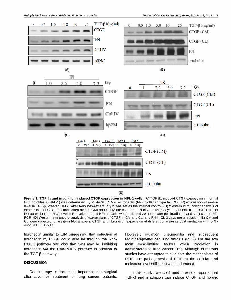
TGF- and Irradiation Induce CTGF and its Downstream Pro-Fibrotic Proteins in Human Lung Fibroblasts
CTGF has been shown as a major downstream
fibrotic factor of TGF- -induced fibrosis. To confirm the
effect of TGF- and irradiation on induction of CTGF
and downstream fibrotic targets such as fibronectin and
collagen type IV (Col IV), HFL-1 cells were treated with
TGF- or irradiated and both mRNA and protein levels
were determined. The levels of CTGF, fibronectin and
Col IV increased in a dose- dependent manner, on
TGF- treatment and irradiation, at both mRNA (Figure
1A and 1C) and protein levels (Figure 1B and 1D). A
time-dependent increase in the expression levels of
CTGF and fibronectin was observed when the cells
were irradiated with 5Gy (Figure 1E). We also
performed experiments in human dermal fibroblasts
(HDF) and observed similar results (SI-1A, 1B). These
data indicate that CTGF and related pro-fibrotic
downstream proteins can be induced by TGF- and
radiation in a dose- and time-dependent manner.
Statins Attenuate Effects of TGF- and Radiation on Expression of CTGF and Downstream Targets
Simvastatin (SIM), a HMG-CoA reductase inhibitor,
has been shown to have anti-fibrotic activity [11]. We
observed that SIM inhibited TGF- -induced CTGF and
downstream targets at both mRNA and protein levels in
dose-dependent manner (Figure 2A(i) and 2A(ii)).
Similar effect of SIM was also observed in HFL-1 post-
irradiation (Figure 2B(i) and 2B(ii)). To further explore
effects of other potential HMG-CoA inhibitors on
radiation-induced CTGF expression, HFL-1 were
incubated with pravastatin (PRA), mevinolin (MVO),
mevastatin (MVS) and SR12813, in addition to SIM
(Figure 2C). More potential anti-fibrotic effect was
observed in the cells treated with SIM, MVO and MVS
other than with PRA and SR 12813 compared with
control. We also tested effect of SIM on TGF- - and
irradiation induced CTGF and pro-fibrotic factors in
HDFs and found that SIM inhibits CTGF, fibronectin
and ColIV in HDFs also (SI-1C,1D). Together these
data suggest that statins may be used for mitigation
and treatment of RTIF through inhibition of CTGF and
downstream pro-fibrotic proteins expression.
TGF- Independent Signaling Pathway may be Involved in RTIF
To investigate if the induction of CTGF expression
in TGF- - treated or irradiated HFL-1 cells is primarily
via the TGF- pathway, these cells were treated with
TGF- neutralizing antibody. The neutralizing antibody
completely abolished CTGF expression in TGF-
treated cells (Figure 3A), but the decrease was not
significant in the irradiated cells (Figure 3B). On
treating TGF- - treated or irradiated HFL-1 cells with
SIM, the inhibitory effect on CTGF was more potent in
both the cells. Our observation is in line with Anscher et
al. report that blocking TGF- 1 function cannot
completely prevent fibrogenesis caused by high-dose
radiation in a rat model using TGF- 1 neutralizing
antibody [6]. These data suggest that radiation-induced
CTGF upregulation occurs partially through TGF- -
independent signaling pathway and that SIM may
inhibit CTGF upregulation occurring via the TGF- -
independent pathway as well.
SIM Inhibits CTGF Induced Pro-Fibrotic Markers in HFL-1 Cells
We and others have established that TGF- -
induced CTGF and fibrotic markers can be inhibited by
SIM. We next investigated if SIM can act further
downstream, i.e. if it can inhibit CTGF-induced
fibronectin. In order to investigate this, HFL-1 cells
were infected with adenoviral plasmids containing
CTGF cDNA sequence, with or without SIM treatment.
Figure 4 shows that SIM also inhibits CTGF-induced
fibronectin at both mRNA (Figure 4A) and protein
(Figure 4B) levels while it could not suppress ectopic
expression of CTGF.
SIM Inhibits TGF- - and CTGF- Induced Pro-Fibrotic Markers through the Rho-ROCK Pathway
To determine the pathway in which CTGF-induced
pro-fibrotic markers are inhibited, HFL-1 cells were
treated with either SIM or ROCK inhibitor (Y27632),
followed by treatment with CTGF. Figure 4C and 4D
show that Y-27632 also inhibits CTGF-induced
Multiple Mechanisms for Anti-Fibrotic Functions of Statins Journal of Cancer Research Updates, 2014 Vol. 3, No. 1 3
(A) (B)
(C) (D)
(E)
Figure 1: TGF- 1 and irradiation-induced CTGF expression in HFL-1 cells. (A) TGF- 1 induced CTGF expression in normal lung fibroblasts (HFL-1) was determined by RT-PCR. CTGF, Fibronectin (FN), Collagen type IV (COL IV) expression at mRNA level in TGF- 1-treated HFL-1 after 6-hour-treatment. h 2M was set as the internal control. (B) Western immunoblot analysis of expressions of CTGF in conditioned media (CM) and cell lysate (CL), and FN in CL after 3 days’ treatment. (C) CTGF, FN, Col IV expression at mRNA level in Radiation-treated HFL-1. Cells were collected 20 hours later postirradiation and subjected to RT-PCR. (D) Western immunoblot analysis of expressions of CTGF in CM and CL, and FN in CL 3 days postirradiation. (E) CM and CL were collected for western blot analysis. CTGF and fibronectin expression at different time points post irradiation with 5 Gy dose in HFL-1 cells.
fibronectin similar to SIM suggesting that induction of
fibronectin by CTGF could also be through the Rho-
ROCK pathway and also that SIM may be inhibiting
fibronectin via the Rho-ROCK pathway in addition to
the TGF- pathway.
DISCUSSION
Radiotherapy is the most important non-surgical
alternative for treatment of lung cancer patients.
However, radiation pneumonitis and subsequent
radiotherapy-induced lung fibrosis (RTIF) are the two
main dose-limiting factors when irradiation is
administered to lung cancer [15]. Although numerous
studies have attempted to elucidate the mechanisms of
RTIF, the pathogenesis of RTIF at the cellular and
molecular level still is not well understood.
In this study, we confirmed previous reports that
TGF- and irradiation can induce CTGF and fibrotic

4 Journal of Cancer Research Updates, 2014 Vol. 3, No. 1 Li et al.
(A(i)) (A(ii))
(B(i)) (B(ii))
(C)
Figure 2: SIM suppressed TGF- 1 and irradiation-induced expression of CTGF and downstream factors. (A) Inhibitory effect of SIM on TGF- 1 -induced expression of CTGF and pro-fibrotic factors in HFL-1 at the mRNA (i) and protein (ii) levels. Cells were treated with the indicated amount of SIM in presence of 10 ng/ml TGF- 1 for 20 hours (RT-PCR) and 3 days (Western blot) in the medium. (B) Inhibitory effect of SIM on irradiation induced expression of CTGF and pro-fibrotic factors in HFL-1 cells at the mRNA (i) and protein (ii) levels. Cells were treated with the indicated amount of SIM for 6 hours before subjecting to 10 Gy radiation and incubated for 20 hours (RT-PCR) in the medium whereas 5 Gy radiation and incubated for 3 days in the serum free medium (Western blot). (C) Inhibitory effect of Statins on radiation-induced CTGF expression in HFL-1. Cells were treated with the indicated amount of Statins for 6 hours before subjected to 5 Gy radiation and incubated for 3 days in serum free medium. Cell lysate were collected for Western blot analysis. SR12813, a HMG-CoA inhibitor, which is not a statin, but a cholesterol lowering agent was also used in this experiment.
proteins further downstream, such as fibronectin and
collagen type IV in a dose- and time- dependent
manner (Figure 1) [16]. We further showed that statins,
a clinically approved class of HMG-CoA reductase
inhibitor, inhibits the expression of TGF- and
irradiation induced-CTGF and subsequent targets in
HFL-1 (Figure 2), however irradiation induced CTGF
upregulation cannot be completely abrogated by TGF-
neutralizing antibody. This suggests the involvement of
a TGF- -independent signaling pathway in RTIF
(Figure 3). An interesting finding is that downstream in
the TGF- pathway, overexpression of CTGF alone,
without any treatment with TGF- can also induce the
levels of pro-fibrotic factors and this induction can be
inhibited by administration of SIM in HFL-1 cells (Figure
4). This suggests that statins may inhibit RTIF at
multiple levels including inhibition of TGF- -induced
CTGF production as well as CTGF-enhanced fibrotic
proteins expression. However it is still possible that
statins may also utilize other pathways to suppress
RTIF.

Multiple Mechanisms for Anti-Fibrotic Functions of Statins Journal of Cancer Research Updates, 2014 Vol. 3, No. 1 5
(A)
(B)
Figure 3: TGF- 1-independent signaling pathway may be involved in radiation-induced CTGF upregulation. (A) Blocking of TGF- 1-induced CTGF expression in HFL-1 cells by TGF- 1 neutralizing antibody (8 μg/ml). Proteins levels of CTGF expression in CM and CL were confirmed by Western lot. (B) Radiation-induced CTGF upregulation cannot be completely inhibited by TGF- 1 neutralizing antibody. HFL-1 cells were pre-treated with TGF- 1 neutralizing antibody 16 μg/ml for 6 hours prior to irradiation. Densitometric analysis of CTGF bands after normalization to -tubulin is also shown graphically.
Previous studies reported that increasing serum
CTGF expression was observed in patients with
systemic sclerosis and associated with the extent of
skin sclerosis and the severity of pulmonary fibrosis
[17]. Lopes et al. in their study show that analogues of
small heat shock proteins decrease the expression of
CTGF and collagen type I induced by TGF- in human
dermal keloid fibroblasts and that has a potential of
preventing excessive tissue scarring [18]. Statins have
also shown to be effective for the treatment of systemic
sclerosis and digital ulcers [19]. We in our study have
shown that TGF- and irradiation induced CTGF,
fibronectin and ColIV can be inhibited by SIM in a dose
dependent manner in dermal fibroblasts as well (Figure
SI-1).
Statins have also shown anti-fibrotic functions in a
variety of mammalian cell lines or tissues through
interference with the Rho/ROCK/CCN2/-ECM cascade
[11]. The correlation between Rho/ROCK pathway and
fibrogenic signaling pathway has gained more
attentions in recent years. Reports show that inhibition
of Rho activation by statins (inhibition of Rho
isoprenylation) or ROCK kinase inhibitor decreased
CTGF expression and subsequent extracellular matrix
deposition in vitro [12]. Our in vitro data indicated that
anti-fibrotic action of SIM not only blocked TGF- - or
irradiation- induced CTGF expression but repressed
CTGF induced upregulation of pro-fibrotic factors as
well. Data presented in Figure 3B imply that there is a
pathway in addition to the TGF- pathway that is
involved in the induction of CTGF. Indeed, our results
in Figure 4 demonstrate that suggesting specific
inhibition of Rho/ROCK and TGF- signaling pathway
may provide a synergistic anti-fibrotic therapy for
irradiation-induced fibrosis.

6 Journal of Cancer Research Updates, 2014 Vol. 3, No. 1 Li et al.
(A) (B)
(C) (D)
Figure 4: SIM inhibits CTGF- induced pro-fibrotic markers through the Rho-ROCK pathway. HFL-1 cells were pretreated with SIM 6 hours prior to adenoviral infection with CTGF vector (MOI 500). Cells were incubated for 24 hours and 48 hours for mRNA (A) and protein expression (B) respectively post-infection. HFL-1 cells were treated with SIM for 6 hours and 50 M ROCK inhibitor (Y-27632) for 3 hours before treatment with TGF- or CTGF adenovirus and were incubated for 20 hrs and 48 hrs respectively for mRNA (C) and protein expression levels (D).
Taken together, our study indicated that CTGF
plays a critical role for RTIF and serves as a potential
fibrotic marker for evaluation of the extent of fibrosis
post-irradiation and the response to the drug treatment.
Statins inhibit RTIF in normal fibroblasts at multiple
levels including inhibition of CTGF through TGF- and
the Rho/ROCK signaling pathway. Our data provide
that CTGF can be a potential anti-fibrotic target to
develop successful modalities for optimal radiotherapy
in the clinic.
MATERIALS AND METHODS
Cell Culture, TGF- and Radiation Treatment
Normal human fetal lung fibroblasts (HFL-1) (CCL-
153) and human dermal fibroblasts (HDF) (PCS-201-
012) were purchased from the American Type Culture
Collection and cultured in Ham's F12K medium with
10%FBS (GIBCO, 11765) and fibroblast basal media
supplemented with fibroblast growth kit respectively.
After reaching 75-80% confluence, the medium was
changed to serum free medium (SFM) for irradiation
with different dose 1, 2.5, 5, 7.5 and 10 Gy by using 137
Cesium. Cells were treated with 5-10 ng/ml TGF-ß1
(Sigma, T7039) to stimulate CTGF production. Cells
were incubated for 3 days and subjected to Western
blot.
Treatment with Statins
Cells were plated into 35mm plates and the
following day, the cells were washed with PBS and the

Multiple Mechanisms for Anti-Fibrotic Functions of Statins Journal of Cancer Research Updates, 2014 Vol. 3, No. 1 7
media was changed to SFM. Cells were treated with
the indicated amount of Statins for 6 hours before
subjecting them to 5Gy irradiation and then incubated
for 3 days in SFM.
RT-PCR
Total RNA was extracted from cells, 20 hours post
treatment and 1 μg of purified total RNA was used for
RT-PCR using the ThermoScript RT-PCR System
(Invitrogen). The sequences of the forward and reverse
primers were used as follows: CTGF, fwd5’-CTGGTCC
AGACCACAGAGTG-3’, rev5’-CGGTATGTCTTCATGC
TGGT-3’;COL-IV,fwd5’-AGCAAGGCAACAGAGGA-
CTT-3’, rev 5’-GATCTGGGTGGAAGGTGACT-3’;FN,
fwd,5’-GACTGGAGCTGGAGACATGA-3’,rev5’-GTGAT
GATGGTGGACTGCTC-3’; 2M fwd, 5-GTGCTCGCG
CTCTCTCT-3’; rev,5-CGGCAGGCATACTCATCTTT-
3’.The CTGF PCR product is 242 bp in length, COL-
138bp, FN-203bp, and h 2M-278bp. PCR products
were run on a 1% agarose gel and visualized by
ethidium bromide staining.
Western Blot Analysis
Cell lysates were harvested in HBSST lysis buffer.
The primary antibodies selectively recognized CTGF
(sc-14939), Collagen type IV (sc-11360), Fibronectin
(sc-9068), were purchased from Santa Cruz
Biotechnology, Inc and -tubulin (T9026) from Sigma-
Aldrich Inc. Primary antibodies except -tubulin
(1:4,000) were diluted at 1:600 and incubated at 4 °C
overnight; and corresponding secondary antibodies at
1:6,000 and incubated at room temperature 1 hour.
Antibodies were detected by the enhanced
chemiluminescence (PerkinElmer Life Sciences Inc).
Adenoviral Constructs
We used the AdEasy system (Quantum
Biotechnologies) to generate Ad:CTGF as described
previously [20].
TGF- 1 Neutralizing Antibody Treatment
TGF- 1 was purchased from Sigma-Aldrich, and
TGF- 1-neutralizing antibody was purchased from
R&D systems (Minneapolis). After achieving 75-80%
confluence in 6-well plate, the medium was changed to
SFM for treatment. Cells were exposed for 72 hours in
each individual well to TGF- 1 (10ng/ml), TGF- 1
(10ng/ml) plus TGF- 1-neutralizing antibody (16μg/ml)
(preincubated and shaken together at room
temperature for 30 min in 1.5 ml eppendorf tube
containing 400 μl SFM before the addition to the cells).
Cells were subjected to 5 Gy irradiation after addition of
the preincubated complex. Conditioned media and cell
lysate were then collected for Western Blot.
Treatment with ROCK-Inhibitor
Cells were plated into 12 well plates and the
following day, the cells were washed with PBS and the
media was changed to SFM. Cells were treated with
the indicated amount of ROCK-inhibitor (Y-27632)
(Sigma-Aldrich) for 3 hours before subjecting them to
TGF- treatment or Ad: CTGF infection. The cells were
further incubated for 24 hours (RT-PCR) and for 48
hours (western blot) in SFM.
REFERENCES
[1] Tsoutsou PG, Koukourakis MI. Radiation pneumonitis and
fibrosis: mechanisms underlying its pathogenesis and implications for future research. Int J Radiat Oncol Biol Phys 2006; 66: 1281-93. http://dx.doi.org/10.1016/j.ijrobp.2006.08.058
[2] Anscher MS, Chen L, Rabbani Z, et al. Recent progress in defining mechanisms and potential targets for prevention of normal tissue injury after radiation therapy. Int J Radiat Oncol
Biol Phys 2005; 62: 255-9. http://dx.doi.org/10.1016/j.ijrobp.2005.01.040
[3] Zhao L, Sheldon K, Chen M, et al. The predictive role of plasma TGF-beta1 during radiation therapy for radiation-induced lung toxicity deserves further study in patients with
non-small cell lung cancer. Lung Cancer 2008; 59: 232-9. http://dx.doi.org/10.1016/j.lungcan.2007.08.010
[4] Cicha I, Goppelt-Struebe M. Connective tissue growth factor: context-dependent functions and mechanisms of regulation.
Biofactors 2009; 35: 200-8. http://dx.doi.org/10.1002/biof.30
[5] Leask A, Abraham DJ. The role of connective tissue growth factor, a multifunctional matricellular protein, in fibroblast biology. Biochemistry and Cell Biology = Biochimie et
Biologie Cellulaire 2003; 81: 355-63. http://dx.doi.org/10.1139/o03-069
[6] Anscher MS, Thrasher B, Rabbani Z, et al. Antitransforming growth factor-beta antibody 1D11 ameliorates normal tissue
damage caused by high-dose radiation. Int J Radiat Oncol Biol Phys 2006; 65: 876-81. http://dx.doi.org/10.1016/j.ijrobp.2006.02.051
[7] Brigstock DR. The connective tissue growth factor/cysteine-rich 61/nephroblastoma overexpressed (CCN) family. Endocr Rev 1999; 20: 189-206.
[8] Haydont V, Mathe D, Bourgier C, et al. Induction of CTGF by TGF-beta1 in normal and radiation enteritis human smooth muscle cells: Smad/Rho balance and therapeutic
perspectives. Radiother Oncol: J Eur Soc Therapeut Radiol Oncol 2005; 76: 219-25. http://dx.doi.org/10.1016/j.radonc.2005.06.029
[9] Li G, Xie Q, Shi Y, et al. Inhibition of connective tissue growth factor by siRNA prevents liver fibrosis in rats. J Gene
Med 2006; 8: 889-900. http://dx.doi.org/10.1002/jgm.894
[10] Brigstock DR. Strategies for blocking the fibrogenic actions of connective tissue growth factor (CCN2): From
pharmacological inhibition in vitro to targeted siRNA therapy in vivo. J Cell Commun Signal 2009; 3: 5-18. http://dx.doi.org/10.1007/s12079-009-0043-9

8 Journal of Cancer Research Updates, 2014 Vol. 3, No. 1 Li et al.
[11] Watts KL, Sampson EM, Schultz GS, Spiteri MA. Simvastatin
inhibits growth factor expression and modulates profibrogenic markers in lung fibroblasts. Am J Respirat Cell Mol Biol 2005; 32: 290-300. http://dx.doi.org/10.1165/rcmb.2004-0127OC
[12] Haydont V, Bourgier C, Pocard M, et al. Pravastatin Inhibits
the Rho/CCN2/extracellular matrix cascade in human fibrosis explants and improves radiation-induced intestinal fibrosis in rats. Clin Cancer Res: Official J Am Assoc Cancer Res 2007;
13: 5331-40. http://dx.doi.org/10.1158/1078-0432.CCR-07-0625
[13] Riento K, Ridley AJ. Rocks: multifunctional kinases in cell behaviour. Nat Rev Mol Cell Biol 2003; 4: 446-56. http://dx.doi.org/10.1038/nrm1128
[14] Hahn A, Heusinger-Ribeiro J, Lanz T, et al. Induction of
connective tissue growth factor by activation of heptahelical receptors. Modulation by Rho proteins and the actin cytoskeleton. J Biol Chem 2000; 275: 37429-35. http://dx.doi.org/10.1074/jbc.M000976200
[15] Prise KM, O'Sullivan JM. Radiation-induced bystander signalling in cancer therapy. Nat Rev Cancer 2009; 9: 351-60. http://dx.doi.org/10.1038/nrc2603
[16] Goppelt-Struebe M, Hahn A, Iwanciw D, et al. Regulation of
connective tissue growth factor (ccn2; ctgf) gene expression
in human mesangial cells: modulation by HMG CoA reductase inhibitors (statins). Mol Pathol: MP 2001; 54: 176-9. http://dx.doi.org/10.1136/mp.54.3.176
[17] Sato S, Nagaoka T, Hasegawa M, et al. Serum levels of
connective tissue growth factor are elevated in patients with systemic sclerosis: association with extent of skin sclerosis and severity of pulmonary fibrosis. J Rheumatol 2000; 27: 149-54.
[18] Lopes LB, Furnish EJ, Komalavilas P, et al. Cell permeant peptide analogues of the small heat shock protein, HSP20, reduce TGF-beta1-induced CTGF expression in keloid
fibroblasts. J Investig Dermatol 2009; 129: 590-8. http://dx.doi.org/10.1038/jid.2008.264
[19] Abou-Raya A, Abou-Raya S, Helmii M. Statins: potentially useful in therapy of systemic sclerosis-related Raynaud's phenomenon and digital ulcers. J Rheumatol 2008; 35: 1801-8.
[20] Haberberger TC, Kupfer K, Murphy JE. Profiling of genes which are differentially expressed in mouse liver in response to adenoviral vectors and delivered genes. Gene Therapy
2000; 7: 903-9. http://dx.doi.org/10.1038/sj.gt.3301181