Radiobiological Research For Improving Tumor Radiotherapy -An Indian Perspective*
17
Def Sci J, Vol40, No 4, October 1990, pp 401-417. Radiobiological Research For Improving Tumor Radiotherapy -An Indian Perspective* Viney Jain Znstitute of Nuclear Medicine and Allied Sciences, Delhi-II0 054 ABSTRACT Radiation-induced damage to normal tissues within the non- target volume is a major limitation of tumor radiotherapy. Physical methods to obtain superior spatial dose distributions use sophisticated technology and are expensive. Large scale applications of these technologies in a developing country like India; with a large number of cancer patients, poor’ instrumental facilities and inadequate infrastructure face several problems. Radiobiological research aiming at developing simple, inexpensive and effective methods to increase the differential response between tumor and normal tissues should be, therefore, strengthened. Biological end-points are determined not only by the molecular lesions produced due to the absorption of the radiation energy but also by the cellular repair processes, which become operative in response to lesions in the living system. Therefore, enhancement of repair processes in the normal tissues and inhibition of the same in tumors should considerably improve the therapeutic index of radiation treatment. A combination of agents which can su$abIy alter the spectrum of important molecular lesions with modifiers of cellular repair could be an effective strategy. Initial ezperiments using halopyrimidines to increase repairable DNA lesions produced by sparsely ionizing radiations in combination with 2-deoxy-D-glucose to modulate differentially the repair and fixation processes in the tumor and normal tissues have provided promising results. Further research work is warranted since this strategy appears to have great potential for improving tumor radiotherapy. Received 7 November 19% ‘Dedicated to the memory of Brig. S.K. Mazumdar 401
-
Upload
writerightindia -
Category
Documents
-
view
2 -
download
0
Transcript of Radiobiological Research For Improving Tumor Radiotherapy -An Indian Perspective*

Def Sci J, Vol40, No 4, October 1990, pp 401-417.
Radiobiological Research For Improving Tumor Radiotherapy-An Indian Perspective*
Viney JainZnstitute of Nuclear Medicine and Allied Sciences, Delhi-II0 054
ABSTRACT
Radiation-induced damage to normal t issues within the non- targetvolume is a major limitation of tumor radiotherapy. Physical methodsto obtain superior spatial dose distributions use sophisticatedtechnology and are expensive. Large scale applications of thesetechnologies in a developing country like India; with a large numberof cancer patients, poor’ instrumental facilities and inadequateinfrastructure face several problems. Radiobiological research aimingat developing simple, inexpensive and effective methods to increasethe differential response between tumor and normal t issues should be,therefore, strengthened.
Biological end-points are determined not only by the molecularlesions produced due to the absorption of the radiat ion energy but alsoby the cellular repair processes, which become operative in responseto lesions in the living system. Therefore, enhancement of repairprocesses in the normal tissues and inhibition of the same in tumorsshould considerably improve the therapeutic index of radiationtreatment. A combination of agents which can su$abIy alter thespectrum of important molecular lesions with modifiers of cellularrepair could be an effective strategy. Initial ezperiments usinghalopyrimidines to increase repairable DNA lesions produced bysparsely ionizing radiat ions in combinat ion with 2-deoxy-D-glucose tomodulate differentially the repair and fixation processes in the tumorand normal t issues have provided promising results . Further researchwork is warranted since this strategy appears to have great potentialfor improving tumor radiotherapy.
Received 7 November 19%‘Dedicated to the memory of Brig. S.K. Mazumdar
401
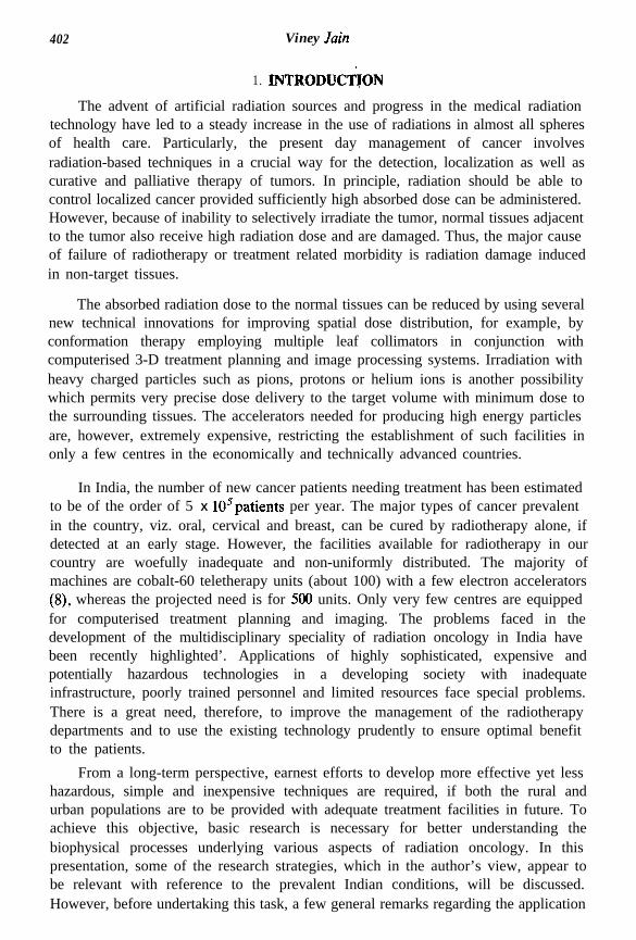
402 Viney Jain
1. INTRODUCilON
The advent of artificial radiation sources and progress in the medical radiationtechnology have led to a steady increase in the use of radiations in almost all spheresof health care. Particularly, the present day management of cancer involvesradiation-based techniques in a crucial way for the detection, localization as well ascurative and palliative therapy of tumors. In principle, radiation should be able tocontrol localized cancer provided sufficiently high absorbed dose can be administered.However, because of inability to selectively irradiate the tumor, normal tissues adjacentto the tumor also receive high radiation dose and are damaged. Thus, the major causeof failure of radiotherapy or treatment related morbidity is radiation damage inducedin non-target tissues.
The absorbed radiation dose to the normal tissues can be reduced by using severalnew technical innovations for improving spatial dose distribution, for example, byconformation therapy employing multiple leaf collimators in conjunction withcomputerised 3-D treatment planning and image processing systems. Irradiation withheavy charged particles such as pions, protons or helium ions is another possibilitywhich permits very precise dose delivery to the target volume with minimum dose tothe surrounding tissues. The accelerators needed for producing high energy particlesare, however, extremely expensive, restricting the establishment of such facilities inonly a few centres in the economically and technically advanced countries.
In India, the number of new cancer patients needing treatment has been estimatedto be of the order of 5 X 10⁵patients per year. The major types of cancer prevalentin the country, viz. oral, cervical and breast, can be cured by radiotherapy alone, ifdetected at an early stage. However, the facilities available for radiotherapy in ourcountry are woefully inadequate and non-uniformly distributed. The majority ofmachines are cobalt-60 teletherapy units (about 100) with a few electron accelerators(8), whereas the projected need is for 500 units. Only very few centres are equippedfor computerised treatment planning and imaging. The problems faced in thedevelopment of the multidisciplinary speciality of radiation oncology in India havebeen recently highlighted’. Applications of highly sophisticated, expensive andpotentially hazardous technologies in a developing society with inadequateinfrastructure, poorly trained personnel and limited resources face special problems.There is a great need, therefore, to improve the management of the radiotherapydepartments and to use the existing technology prudently to ensure optimal benefitto the patients.
From a long-term perspective, earnest efforts to develop more effective yet lesshazardous, simple and inexpensive techniques are required, if both the rural andurban populations are to be provided with adequate treatment facilities in future. Toachieve this objective, basic research is necessary for better understanding thebiophysical processes underlying various aspects of radiation oncology. In thispresentation, some of the research strategies, which in the author’s view, appear tobe relevant with reference to the prevalent Indian conditions, will be discussed.However, before undertaking this task, a few general remarks regarding the application

Radiomodifiers for Improving Cancer Therapy 403
of science and technology for social development are presented in order to providean appropriate framework for further discussions and action.
2. SCIENCE, TECHNOLOGY AND SOCIAL DEVELOPMENT
The multiple and complex relationships between research in basic sciences,development of technology and its applications for the benefit of the society are shownin Fig. 1. Research in basic sciences systematically attempts to determine truth, therebyleading to generation of new knowledge. Advances in knowledge are necessary forthe development of new technology and industry. New technology, if used withresponsibility, can be exploited for the benefit of the society.
Translation of advances in knowledge into new technology and industrialdevelopment in a reasonably short time span requires cooperation and coordinationbetween a large number of individuals, institutions, organisations and industry. Thisdifficult task can be accomplished only within a flexible and progressive system whichencourages easy (with minimum bureaucratic and administrative delay) multiplelinkages between research institutes, universities, industry and consumerorganisations.
In the absence of any worthwhile development of indegenous technology in thefield of radiation medicine in India, we have to depend almost entirely on the transferof technology from the advanced countries. The successful acceptance and optimaluse of any new technology by the society depends, however, upon a number of factors
TECHNOLOGY
a. -b . -c. -d. -
f
v v
BASICSCIENCES
A ‘9
1INSTRUMENTS
4 ANDMETHdDS
A
L
V
TECHNICAL* PERSONNEL
I-I I
ADVANCES DEVELOPMENT SOCIO- ECONOMICIN - OF - BENIFITS
KNOWLEDGE TECHNOLOGY
a F INANCIAL AND MATERIAL RESOURCES C. INFRASTRUCTURE
b.‘ HUMAN RESOURCES d. SOCIO- CULTURAL ATTITUDESAND VALUES
Figure 1. Relationships between science, technology and social development.

404 Viney bin
including industrial infrastructure, socio-economic and cultural backgrounds besidesconsiderations of technical merits (as compared with the existing technologies) andassociated hazards and risks. In order to objectively *evaluate the suitability of thenew technology, a total cost-benefit analysis should be carried out. Such an analysiswould be helpful in optimizing the use of the technology. Optimization requires thatthe total cost-benefit ratio, 0,, for application in a given situation be minimised. Thetotal cost includes not only the economic costs involved in the application of thetechnology, but also the cost of the associated hazards and risks.
Adoption of the technology for regular use will be determined by the index ofaffordability, which may be represented by a critical threshold value 0c dependentupon the resources of the society. For a wide acceptance of the technology 0= >> 0,.
The calculation of the cost of the hazards, risks and benefits is complex, since itmay also depend upon the value systems of the society. Attempts to estimate 0rvalues under different conditions for alternate technological approaches are necessaryto objectively compare the alternatives and to help the planners and decision makers.
3. OPTIMIZING USE OF RADIATIONS IN CANCER MANAGEMENT
Within this framework, the present and future prospects of different strategiesfor optimizing applications of radiation in cancer management in a developing countrylike India should be examined. It is desirable to develop methods to reduce 0,. Thiscould be attempted in a number of ways by (i) reducing the cost of required equipments,(ii) using the existing technology prudently, and (iii) carrying out research to developbetter and cheaper technology suited to the Indian environment.
3.1 Reduction in the Economic Cost of the Equipment and Prucedures
Cost of most equipment depends upon the industry and market forces. In ourcountry, the field of biomedical instrumentation, has not developed as an industry.Consequently, the users have to rely entirely on imports. The foreign companies quotespecial prices for India, which are higher, by at least 20-50 per cent than the localprice. Furthermore, for subsequent maintenance and servicing, the foreign companiesor their Indian agents charge exorbitant rates, In view of the increasing demand ofthe sophisticated equipment in the field of radiation medicine, it is suggested thatadequate financial and human resources be mobilised to encourage inclegenousresearch, development and manufacture of these equipment suited to the localenvironment and needs.
3.2 Prudent Use of the Existing Technology
The attitudes and behaviour of health care professionals determine to a greatextent the use or misuse of the technology. Through proper education and training,it must be ascertained that the medical practitioners:
(a) avoid using the technology when not necessary;(b) avoid using the more expensive techniques, when the same information and
benefit can be obtained through simpler and cheaper techniques;

Radiomodifiers for Improving Cancer Therapy 405
(c) use the technique properly and ensure proper quality control to derive themaximum benefit; and
(d) give due consideration to the potential hazards of the teehnology andmethods employed and take appropriate precautions to minim& thehazards.
3.3 Radiobiological Research for Improving Tumor Radiotherapy
The goal of radiobiological research is to understand the effects of radiations onthe biological systems and use this knowledge to increase the differential between theradiation responses of the tumor and normal tissues so that a high probability of curewithout any significant morbidity at a given radiation dose can be achieved (Fig. 2).
Considerable work to develop radioprotectors for normal tissues andradiosensitizers for the tumor has been going on throughout the world for more thanfour decades. A large number of physical, chemical and biological agents have beenscreened (for example, see Uma Devi2 and Singh3 for recent reviews). Inspite ofsignificant research effort, the success of these radiomodifiers in improvingradiotherapy has been rather limited so far. Enormous complexity of the problem,gaps in the basic understanding of the processes determining the radiation responsesof the living systems coupled with the large inter- and intra-tumoral heterogeneitydisplayed by human cancers (even of the same type) could be some of the reasonsfor the slow progress. A critical review of the literature, however, reveals that a lackof systematic, integrated and holistic approach and greater reliance on hit and trialmethodology with little emphasis on understanding the basic processes, could also bepartially responsible for the sub-optimal results. Recent progress in molecular geneticsand development of powerful non-invasive techniques to study structures and functionsin the intact organism appear promising to adopt a new approach and to fokulate
P R E S E N T FUTURE
D/Gy D/ Gy
PC : PROBABILITY OF TUMOR CONTROL
Pm: PROBABILITY OF TREATMENT RELATED MORBIDITY

406 Viney Jain
a coordinated research programme to achieve the above mentioned desirable yetelusive goal. The rationale of this approach is briefly presented in the following.
3.3.1 Radiation-Induced Biological Damage and Strategies to Improve TumorRadiotherapy
The biological damage induced by ionizing radiations evolves through a complexnetwork of processes occurring at various levels of organisation and extending overa large time scale (Table 1). For simplicity, we may identify two important stagescomprising (i) physico-chemical processes leading to the production of lesions in thebiomolecules, and (ii) biological processes representing the responses of the livingsystem to the molecular lesions. Both these phases can be suitably influenced in anumber of ways to optimize tumor radiotherapy.
(a) Induction of biomolecular lesions
Absorption of radiation in biological tissues gives rise to excitations and ionizationsof atoms and molecules resulting in the formation of highly reactive ion radicals andfree radicals which may interact with each other and with other molecules. Some ofthese reactions may result in the induction of relatively long-lived lesions in importantbiomolecules. All types of biomolecules including nucleic acids, proteins, lipids andcarbohydrates have been shown to be damaged by ionizing radiations. It is nowgenerally agreed that DNA is the most important target for radiation-induced ceildeath while damage to molecules in biomembranes may also play a role under certainconditions. The most important DNA lesion responsible for cell death is the doublestrand break (DSB), which is readily repairable in mammalian cells4Y5.
The spectrum of molecular lesions induced depends upon the quality of radiation,microenvironment, structures and conformations of the molecules involved.
The quantity and pattern of energy depositions are primarily determined by theradiation quality and the irradiation technique. Passage of radiation through matterresults in stochastically distributed energy depositions along the tracks of the ionizingparticles. Biological damage, however, is influenced by the density of energy depositionalong the particle track. Two small energy depositions, produced by chance in a shortdistance, may act in a biological macromolecule as one large energy deposition leadingto a more severe, h-repairable lesion in the molecule than would be expected by singlesmall energy depositions (repairable lesions). Therefore, densely ionizing radiations(neutrons and heavy ions) are biologically more effective than sparsely ionizingradiations (photons, electrons). Use of heavy charged particles offers many advantagesbecause of their superior depth-dose distribution showing a narrow peak towards theend of their range (Bragg peak) with high relative biological efficiency (RBE) andsmall lateral scattering. However, production of these particles is prohibitivelyexpensive, as mentioned already in the introduction.
During irradiation with sparsely ionizing radiations, such as fast electrons orgamma rays, indirect action for producing macromolecular lesions may becomeimportant. Since 743-80 per cent of biological mass consists of water, reactions withradiolysis products of water, (*OH, H, eq, etc) in particular, free radicals and active
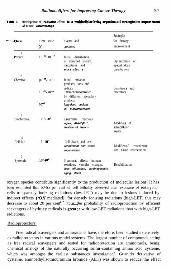
Radiomodifiers for Improving Cancer Therapy 407
Tabte 1. Development of radiatton effects fa a mulm tiving orgdsm and atmtegks for tmprevementof tumor radiothe~py
‘“-b--PJhase Time scale
6)
Events andprocesses
Strategiesfor therapyimprovement
IPhysical
2Chemical
3Biochemical
4Cellular
5Systemic
Initial distributionof absorbed energy,ionizations andexcitat ions
Optimization ofspatial dosedistributions
IO-“-@’
lo-’
lo-“-104
104-10’
loR-10”’
Initial radiationproducts, ions andradicals,interactionscontrolledby diffusion, secondaryproducts,long-lived lesionsin macromolecules
Sensitizers andprotectors
Enzymatic reactions.repair, misrepair.fixation of lesions
Modifiers ofintracellularrepair
Cell death, and lossrecruitment and tissueregeneration
Modifiersof recruitmentand tissue regeneration
Hormonal effects, immunereactions, vascular changes, Rehabilitationloss offunction, carcinogenesis,aging, death
oxygen species contribute significantly to the production of molecular lesions. It hasbeen estimated that 60-65 per cent of cell lethality observed after exposure of eukaryoticcells to sparsely ionizing radiations (low-LET) may be due to lesions induced byindirect effects (.OH mediated); for densely ionizing radiations (high-LET) this maydecrease to about 20 per cent‘*’ Thus the probability of radioprotection by efficient.scavengers of hydroxy radicals is great& with low-LET radiations than with high-LETradiations.
Radioprotectors
Free radical scavengers and antioxidants have, therefore, been studied extensivelyas radioprotectors in various model systems. The largest number of compounds actingas free radical scavengers and tested for radioprotection are aminothiols, beingchemical analogs of the naturally occurring sulfur-containing amino acid cysteine,which was amongst the earliest substances investigated’. Guanido derivative ofcysteine, aminoethylisothiouronium bromide (AET) was shown to reduce the effect

408 Viney Jain
of a radiation dose by a factor of 2.1 in mice’, however, it was,found unsuitable forhuman use because of high toxicity. The toxicity of AET can be reduced by combiningwith other thiol compounds“pi A combination of AET with 5hydroxytryptophan.(5HI’) appears tobe promising; it has been shown to give better protectionless toxic”.
Synthesis of a phosphorylated aminothiol known as WR-2721 by the Walter ReedArmy Institute’s research programme was a significant advance12. WR-2721 has beenreported to show a dose reduction factor (DRF) of 2.7 in mice when injectedintraperitoneally 30 minutes before irradiation. WR-2721 has been studied widelyduring the last two decades and has been shown to protect different normal tissuesto various extents wiih generally low protection to tumors13. For this reason, clinicaltrials on human patients have been initiated inspite of some of the side effects observedin animal as well as human studies 14,15 Research is in progress to develop less toxic.analogs or to combine WR-2721 at subtoxic doses with other compounds which mayact additively or synergistically. For example, experiments on mice suggest thatcombination of low doses of WR-2721 with administration of 2-mecaptopropionylglycine (MPG) could be effective in providing significant protection without serioustoxicity16.
Radiosensitizers
A number of studies have also been made to develop radiosensitizers for tumorcells. Amongst a large number of compounds investigated, clinical trials have beencarried out using hyperbaric oxygen, electron affinic agents, in particularnitroheterocyclics metronidazole and misonidazole, to sensitize hypoxic cells.
The use of hyperbaric oxygen is very cumbersome. Results of clinical trials withmetro- and misonidazoles have shown serious neurotoxicity with little therapeuticbenefiti7. Attempts are now being made to develop less toxic analogs.
Since, biomembranes, besides DNA, could also be important critical targets forradiation-induced biological damage*8*19,several drugs and chemicals which are knownto act specifically on membranes, have been investigated; a few have been shown tomodify radiation effects. Phenothiazines, in particular trimeprazime (TMZ) andchlorpromazine (CPZ) , have shown siginificant radiosensitization of hypoxic tumorcells and protection of euoxic cells”. .Experiments on animal models have also shownpromising results. The mechanisms of action remain to be elucidated. Since thesedrugs are already being used in clinical practice as tranquilizers, theirpharmacokineticsare well known and clinical trials can be initiated.
DNA base analogs such as halopyrimidines have been used to increase theradiation sensitivity of proliferating tumor cells. Halogenated pyrimidine analogs like5-bromo-deoxyuridine (BrdU) and 23 iododeoxyuridine (IdU) can be incorporatedin DNA in place of thymidine in the proliferating cells21. It has been shown that theincorporation of these analogs into the DNA, increases the radiation-induced DNAstrand breaks, chromosome aberrations, micronuclei and cell deathhusm. The increasein DNA strand breaks is supposed to occur via the production of highly reactive
Radiomodifiers for improving Cancer Therapy 409
uracilyl radical-due to the interaction of radiation-produced hydrated electron withBrdUz5. The uracilyl radical in DNA abstracts a hydrogen atom from the nearbydeoxyribose in the same or the opposite strand, resulting in the formation of a singlestrand DNA break.
Clinical trials using halopyrimidines were initiated in,USA and Japan in the earlysixties26,27 but were abondoned because of problems of drug delivery andsevere normaltissue toxicity. For significant radiosensitization, it is essential that sufficient amountof halopyrimidines be incorporated in the tumor cell DNA. This requires the drug tobe administered for a long duration, since the cell-cycle times in most human tumorsare long. Using continuous intravenous infusions-over long periods of time, the clinicaltrials have been started again by the National Cancer Institute, USA. The initialresults do not show any major improvement in case of treatment of glioblastomapatients but the results in a few cases of unresectable sarcomas are veryencouraging .28,29 In these studies also, systemic toxicity due to myelosuppressionappear to be the limiting factor. Attempts are being made to increase the incorporationsof halopyrimidines in the tumor and minimising in the normal proliferating tissues.
(6) Biological response modifiers
As an adaptive response to attack by cytotoxic agents, the biological systemshave developed various repair pathways acting at different organisational levels. Theserepair pathways attempt to reverse the molecular lesions and ‘the tissue injury torestore the original state. Modification of the repair pathways can profoundly influencethe biological effects of radiation. Thus, an inhibition of the repair processes in the
*tumor can increase the radiation-induced cell-killing and prevent the regrowth of thetumor: Enhancement of the repair processes in the normal tissues; on the other hand,can reduce the radiation-induced morbidity. Studies to understand the regulation andcontrol of repair Qathways will be helpful in developing differential modifiers ofradiation response suitable for improving tumor radiotherapy. This will be discussedin some detail in the remaining part of the present communication.
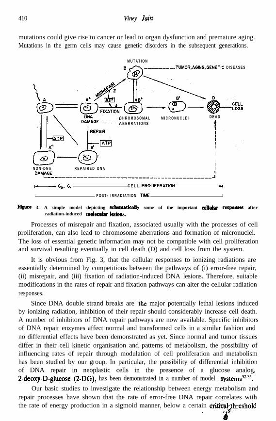
Intracellular repair
There is considerable experimental evidence to suggest that DNA, being thecarrier of genetic information, forms a prominent target for radiation-induced celldeath. We may, therefore, classify the molecular damage into DNA damage (A*)and non-DNA lesions (A”). Following the formation of molecular lesions, differentcellular responses resulting in various biological consequences (Fig. 3) are possible.
(a) If the molecular lesions are completely reversed (error-free repair), thedamaged cells can return back to the original viable state (A).
(b) If the DNA lesions are transformed into irrepairable forms (fixation) orsome errors are introduced during repair (misrepair), alterations and lossof genetic information will result.
If the errors introduced during repair are not detrimental to cell proliferation,mutated cell populations with altered functions (B) may be generated. Somatic cell
410 Viney Jain
mutations could give rise to cancer or lead to organ dysfunction and premature aging.Mutations in the germ cells may cause genetic disorders in the subsequent generations.
M U T A T I O N--------..TUMOR,AGING.GENETlC D I S E A S E S
\o
C H R O M O S O M A L M I C R O N U C L E I D E A D
A B E R R A T I O N S
I
I
N O N - D N A R E P A I R E D D N A iDAMfGE-------_------------------------------------- _J
-Go, G, C E L L PROLIFERATION-
I P O S T - I R R A D I A T I O N TIME I
m 3. A simple model depicting s&ematicaUy some of the important celhdar responses afterradiation-induced mokcuhleBions.
Processes of misrepair and fixation, associated usually with the processes of cellproliferation, can also lead to chromosome aberrations and formation of micronuclei.The loss of essential genetic information may not be compatible with cell proliferationand survival resulting eventually in cell death (D) and cell loss from the system.
It is obvious from Fig. 3, that the cellular responses to ionizing radiations areessentially determined by competitions between the pathways of (i) error-free repair,(ii) misrepair, and (iii) fixation of radiation-induced DNA lesions. Therefore, suitablemodifications in the rates of repair and fixation pathways can alter the cellular radiationresponses.
Since DNA double strand breaks are thi major potentially lethal lesions inducedby ionizing radiation, inhibition of their repair should considerably increase cell death.A number of inhibitors of DNA repair pathways are now available. Specific inhibitorsof DNA repair enzymes affect normal and transformed cells in a similar fashion andno differential effects have been demonstrated as yet. Since normal and tumor tissuesdiffer in their cell kinetic organisation and patterns of metabolism, the possibility ofinfluencing rates of repair through modulation of cell proliferation and metabolismhas been studied by our group. In particular, the possibility of differential inhibitionof DNA repair in neoplastic cells in the presence of a glucose analog,2-deoxy-D-glucose (2-DG) , has been demonstrated in a number of model systems32”5.
Our basic studies to investigate the relationship between energy metabolism andrepair processes have shown that the rate of error-free DNA repair correlates withthe rate of energy production in a sigmoid manner, below a certain &t&&threshold
’

Radiomodifiers for Improving Cancer Therapy 411
value of the rate of energy production, DNA repair is completely inhibited. Rates ofmisrepair and fixation of DNA lesions, which are associated with the processes ofcellular proliferation, can also be modified by the energy supply, but quantitativelyin a different way than the rates of error-free DNA repair. The differences lead tothe existence of cross-over points, which determine the direction of modification ofthe cellular radiation response under conditions of energy deficiency. Thus, byappropriate modifications of the energy supply, a reduction or an enhancement ofradiation injury to the system can be achieved.
2-DG is an inhibitor of glucose transport and glycolysis and therefore, its presencedrastically reduces the supply of metabolic energy in the tumor cells which dependto a large extent on the glycolytic pathway for the production of adenosine-triphosphate (ATP), the cellular energy currency. This state of energy deficiencyinhibits the rate of DNA repair in tumors. The normal cells, on the other hand, derivemost of the energy through the respiratory pathway and therefore, their energy supplyis only marginally affected by 2-DG, which is sufficient to reduce the rate of cellproliferation and fixation of lesions, but DNA repair is not reduced significantly underthese conditions. Consequently, in the presence of 2-DG in normal proliferating cells,for example bone marrow, the radiation damage is reduced considerably if 2-DG isadministered immediately before or after irradiation36.
These results suggest that 2-DG is a unique agent which can increase theradiation-induced damage to tumor cells and reduce the damage to normal cells.Therefore, to investigate the feasibility of using this antimetabolite in humans, clinicaltrials on brain tumor patients have been initiated3’.
Intercellular repair and tissue regeneration
Superimposed upon the above mentioned repair pathways, additional mechanismsof repair operate at the tissue level to compensate for the cell loss. A tissue is acomplex dynamic system composed of various types of cells in different physiologicalstates. From the point of view of cell kinetics, these cells can be broadly classifiedinto two categories, viz., (i) cycling cells, engaged in proliferative activity (P), and(ii) non-cycling cells (Q and D), as shown in Fig.4. The non-cycling cells can be eitherpermanently or temporarily out of cefS;cycle and may be blocked in Go-, Gi- orG,-phases of the cell cycle (Q). Cells %ich .are permanently non-cycling are thedifferentiated mature functional cells (F) and der&ells ‘@)&ells, which are out ofcell cycle (Q) but retain the capability to re-enter the cycle (stem cells) comprise thepotential proliferative pool.
Living tissue is a dynamic system; new cells are being constantly produced whilesome cells are being lost. A non-growing tissue of an adult organism is generally inthe steady state-the rate of cell birth and cell loss are constant and equal.
Non-cycling + cycling transitions and their regulations through negative feedbackcontrols provide adaptability to return back to the steady state after perturbations.For example, upon irradiation (Fig. 4(b)), many cells may be killed resulting in anincrease in the cell loss. To compensate for the cell loss, the rate of cell proliferation

412 Viney Jain
TUMOR
%hb r------ - - - - - - - - - - - - - - - - - 1
2-DG
slC r------ -‘-‘-“-“‘-“-7
Figure 4. AmuddsbuwingtbeeffectaafradMon and 2-DG on cellular dynamics, (a) undisturbed state,(b) perbubatiun by imadiah, and (c) efhts of2-DG in combinatien with radiation. ‘-
should increase so that tissue can be repopulated. This can be done in two ways: (i)by reducing the cell cycle time, and (ii) by stimulating the-non-cycling cells to re-enterthe cell cycle (recruitment). Both these phenomena have been observed to occur intumors and normal tissues and represent important mechanisms of intercellular ortissue repair.
The tissue regeneration capacity depends upon the number of surviving stem cellsand the successful completion of the recruitment process. The recruitment processdepends upon complex interactions of various biochemical reactions involved in thesignal transduction by small molecules such as cytokinines, growth stimulating factorsand receptor mediated mechanisms on the cellular membranes. Recent work hasshown that certain immunomodulators such as glucan (8-1,3 polyglucose isolated fromthe yeast, Sacchuomyces cerevesiae) and interleukin-1 (IL-l) can significantly enhancethe recruitment of stem cells in the bone marrow of whole-body irradiated mice,thereby considerably increasing their survival38*39 These findings are of great interest.for protection against radiation and treatment of radiation injuries.

Radiomodifiers for Improving Cancer Therapy 413
The effects of these modulators on tumors remain yet to be investigated. Ourpreliminary studies suggest that 2-DG could differentially inhibit the. recruitmentprocess in cell populations exhibiting high rates of glycolysis40. Thus, the increase inradiation induced tumor regression in mice could be partially explained on this basis41as shown schematically in ,Fig. 4(c). Further work to study this possibility needs to
. be carried out.
The above considerations suggest that it may be possible to develop agents whichcan stimulate the regeneration of normal tissues after radiation injury and inhibit therepopulation of tumors preventing thereby their regrowth after radiotherapy.
Combination of different strategies
The approaches discussed above are not mutually exclusive and for optimal resultscombination of different strategies should be attempted. A combination of free radicalscavengers and radiosensitizers with biological response modifiers could give additiveor synergistic effects. Initial attempts in this direction are quite encouraging. Thus acombination of WR-2721 given before irradiation with soluble glucan administered
. post-irradiation has shown greater than additive effect42. Similar results have beenreported for the combination of WR-2721 with IL-143.
The radiosensitizing effects af halopyrimidines have been observed to be enhancedin tumor cells in the presence of 2-DG* and reduced in normal bone marrow cellsof mice45*46; Therefore, the differential between radiation-induced cell-killing in tumorsand normal tissues can be significantly enhanced by combining halopyrimidines and2-DG administration with radiation treatment.
A number of new combinations are being studied.
4. FUTURE PROSPECTS
The above discussion shows that the major problems encountered~with the useof present day radioprotectors and radiosensitizers in clinical radiotherapy concernthe toxicity of these agents and inadequate differential effects between the tumor andthe normal tissues.
The new approach. of combining radioprotectors and radiosensitixers withbiological response modifiers appears to be promising. The search for an effectiveand non-toxic combination of radiomodifiers will be greatly facilitated by a betterunderstanding of the processes involved in the biological responses of tumors andnormal tissues to radiation induced molecular lesions. Repair, of DNA damage andthe recruitment of stem cells in the cell-cycle are of particular interest in this respect.Using techniques of molecular biology, it should be possible to identify the genes(DNA sequences) which code for the various repair enzymes and to study the regulationand control of their expression. The knowledge thus gained will be helpful in developingmethods to modulate the contents and activities of repair enzymes appropriately toachieve enhancement of repair in normal and inhibition of repair in tumor cells. Useof 2-DG to increase the differential effects of cytotoxic agents between tumors andnormal tissues is also a step in this direction, though it exploits the differences in the
414 Viney Jain
cellular energy and nucleotide metabolism. Further research work to study the exactmechanisms involved and to carry out clinical trials is warranted.
Non-invasive imaging techniques should be exploited to study and monitor theradiation responses of various tissues in the intact organism. The problems connectedwith tumor heterogeneity and development of radiation resistance could be betterinvestigated using the non-invasive methods. Improved understanding of tumor biologywill be crucial in the development of more effective and simpler therapeutic regimens.
Thus, radiobiological research *may eventually achieve the goal depicted inFig. 2 by developing suitable modifiers of radiation response. If this endeavour issuccessful, it may be possible to treat local and metastatic malignant disease withwhole-body irradiation without,any serious toxic effects on normal tissues and withoutthe need for expensive high technology machines. Needless to say that such adevelopment will eminently suit the Indian environment and would go a long waytowards the goal of providing health care to all by the year 2000.
REFERENCES
1. Krishnan Nair, M., Development of radiotherapy in India--prospects andproblems, In Recent Advances in Radiation Oncology, Jain, V., Goel, H. andPohlit, W. (Eds), (Indian Council of Medical Research, New Delhi), 1990, pp.173-183.
2. Uma Devi, P., Normal tissue protection in radiation oncology, In RecentAdvances in Radiation Oncology, Jain, V., Goel, H. and Pohlit, W. (Eds),(Indian Council of Medical Research, New Delhi), 1990, pp. 79-89.
3 . Singh, B.B., Radiation sensitizers for tumor therapy, In Recent Advances inRadiation Oncology, Jain, V., Goel, H. and Pohlit, W. (Eds), (Indian Councilof Medical Research, New Delhi), 1990, pp. 67-77.
4 . Dugle, D,.L., Gillespie, C.J. & Chapman, J.D., DNA strand breaks, repair andsurvival in x-irradiated mammalian cells, Proc. Nut. Acad. Sci. USA, 73 (1976),809-812.
5 . Blocher, D. & Pohlit, W., DNA double strand breaks in Ehrlich ascites tumorcells at low doses of x-rays-II: Can cell death be attributed, to double strandbreaks? Znt. J. Radiat. Biol., 42 (1982), 329-338.
6. Roots, R., Chaterjee, A., Chang, P. , Lommel, L & Blakely, E.A.,Characterization of hydroxyl radical-induced damage after sparsely and denselyionizing radiation, Znt. J. Radiat. Biol., 47 (1985), 157-166.
7 . Frankenberg, D., Frankerberg-Schwager, M. & Harbich, R., The contributionof OH* in densely ionizing electron track ends or particle tracks to the inductionof DNA double breaks, Radiut. Prot. DOS., 31 (1990), 249-252.
8 . Patt, H.M., Tyree, E.B., Straube, R.L. & Smith, D.E., Cysteine protectionagainst x-irradiation, Science, 110 (1949), 213-214.
9. Doherty, D.G. & B u m e l t , W . T . , Protective effect ofS-/I-aminoethylisothiouronium Br. HBr and related compounds againstx-irradiation death in mice, Proc. Sot. Exp. Biol. Med., 89 (1955), 312-314.

1 0 .
11.
12 .
13 .
14 .
15.
16 .
17 .
18 .
19 .
20.
21.
22.
23.
Radiomodifiers for Zmproving Cancer Therapy 415
Ma&in, J.R., Mattelin, G., Fridman-Manduzio, A. & Van der Pa&n, J.,Reduction of ‘short- and long-term radiation lethality by mixtures of chemicalradioprotectors, Radiat. Res., 35 (1968)) 26-48.Ghose, A., Ganguly, S.K. & Kaur,J., Protection with combinations ofhydroxytryptophan and some thiol compounds against whole-body gammairradiation, Znt. J. Radiat. Biol., 44 (1983), 175-181.
Piper, J.R., Stringfellow, C.R., Jr, Elliot, R.D. & Johnston, T.P. ,S-2-(omega-aminoalkylamino) ethyl dihydrogen phosphorothioates and relatedcompounds as potential antiradiation agents, J. Med. Chem., 12 (1969), 236-243.Yuhas, J.M. & Storer, J.B., Differential chemoprotection of normal andmalignant tissues, J. Natl. ‘Cancer Inst., 42 (1969) 331-335.
Yuhas, J.M., Spellman, J.M. & Culo, F., The role of WR-2721 in radiotherapyand/or chemotherapy, Cancer Clin. Trial., 3 (1980), 211-216.Turrisi, A.T., Kligerman, M.M., Glover, D. J. et al., Experience with phase Itrials of WR-2721 preceding radiation therapy, In Radioprotectors andAnticarcinogens, Nygaard, O.F. and Simic, M.G. (Eds), (Academic Press, NewYork), 1983, pp. 681-694.Thomas, B. & Uma Devi, P., Chromosome protection by WR-2721 andMPG-single and combination treatments, Strahlentherapie und Onkologie, 163(1987)) 807-810.Wasserman, T.H., Clinical trials of nitronidaxoles in cancer treatment :experience in the USA, In New Chemo and Radiosensitising Drugs, Breccia,A. and Fowler, J.F. (Eds), (L.O.Sarabeo, Italy), 1985, pp. 171-183.
Alper, T., Low oxygen enhancement ratios for radiosensitive bacterial strainsand probable interaction of two types of primary lesions, Nature, 217 (1968),862-863. .
Shenoy, M.A., Sir@, B.B. & Gopal-Ayengar, A.R., Iodine incorporation incell constituents during sensitization to radiation by iodoacetic acid, Science, 160(1968), 999.Singh, B.B., Chemical radiosensitization : a new strategy for radiotherapy, InRecent Trends in Radiobiological Research, Uma Devi, P. (Ed), (ScientificPublishers, Jodhpur), 1990, pp. 125-139.
Zamenhof, S. & Griboff, G., Incorporation of halogenated pyrimidines into thedeoxyribonucleic acid of Bacterium coli and its bacteriophages, Nature, 174(1954), 307-308.Djordjevic B. & Szybalski, W., Genetics of human cell line-III: incorporationof 5-bromo and 5-iodo-deoxyuridine into the deoxyribonucleic acid of humancells and its effect on radiation sensitivity, J. Exp. Med., 112 (1960), 509-531.
Dewey, W.C. & Humphrey, R.M., Increase in radiosensitivity to ionizingradiation related to replacement of thymidine in mammalian cells with5-bromodeoxvuridine. Radiat. Res.. 26 (19651. 538553.
416
24.
25.
26.
27.
28:
29.
30.
31.
32.
33.
34.
35.
36.
37.
Viney lain
Dahehey, LE., Thompson, L,H. & Carrona, A.V., DNA strand breaksassociated with halogenated pyrimidines incorporation, Mutut. Res., 131 (1984))129-136.
Zimbrick, J.D., Ward, J.F. & Meyers, L.S., Studies on the chemical basis ofcellular radiosensitization by 5- bromouracil and thymine, Int. J. R&at. Biol.,16 (1969) 502-523.
Bagshaw, M.A., Doggett; R.L.S., Smith, K.C., Kaplan, H.S & Nelsen, T.S.,Intra-arterial 5-bromodeoxy-uridine and x-ray therapy, Am. J. Roentgenology,99 (1967),. 886-894.Hoshino, T. & Sano, K., Radiosensitization of malignant brain tumors withbromouridine.(thymidine analog), Actu Rudiologicu, 8 (1969), 15-26.
Jackson, D., Kinsella, T.) Rowland, J. s Wright, D., Katz, D., Main., D., Collins,J., Komblith, P. & Glatstein, E., Halogenated pyrimidines as radiosensitizersin the treatment of glioblastoma multiforme, Am. J. Clin. Oncology (CCT), 10(1987) 4 3 7 - 4 4 3 .
Kinsella, T.J. & Glatstein E., Clinical experience. with intravenousradiosensitizers in unresectable sarcomas, Cancer, 59 (1987), 908-915.Collins, A. & Johnson, R.T., The inhibition of DNA repair, Adv.. Rudiut. Biol.,11 (1984), 72-131.Collins, A., Cellular responses to ionizing radiation : effects of interrupting DNA
’repair with chemical agents, Znt. J. Rudiat. Biol., 51 (1987), 971-983.Jain, V.K. & Pohlit, W., Influence of energy metabolism on-the repair of x-raydamage in living cells-III: effects of 2-deoxy-D-glucose on liquid-holdingreactivation in y&t, Biophysik, 10 (1973), 137-142.Jain, V.K., Purohit, S.C. & Pohlit, W., Optimization of cancer therapy-I:Inhibition of repair of x-ray induced potentially lethal damage by-+2-deoxy-D-glucose in ehrlich ascites tumor cells, Indian J. Exp. Biol.,, 15 (B’ri),711-713.
Jain, V.K., Porschen, W. & Feinendegen, L.E., Optimization of cancertherapy-II: effects of combinbing 2-deoxy-D-glucose treatment withgammairradiation on Sarcoma-180, Indiy J. {xp. B&Z., 15.(1977), h4%8.
Jain, V.K., Kalia, V.K., Sharma, RI, Maharalan, V. & Menon, M., Effect of2-deoxy-D-glucose on glycolysis, proliferation kinetics and radiation response ofhuman cancer cells, Znt. J. Radiut. O~coZ. BioZ. Phys., 11 (1985), 943-950.
Jain, V.K., Kalia, V.K., Gopinath, P.M., Naqvi, S. & Kucheria, K.,Optimization of cancer therapy-III: effects of combining 2-deoxy-D-glucosetreatment with gamma irradiation on normal mice, Indian J. Exp. Biol., 17(1987), 1 3 2 0 - 1 3 2 5 .
Mohanti, B.K., Anantha, N., Ravichandran, R., Sharma, A.K., Pattabhiraman,V., Devaru, S,. , Chandramouli., Joshi, N.B., Dwarkanath, B.S. & Jain, V.,Improving cancer radiotherapy with 2-DG : phase II clinical trials on humancerebral gliomas, ZZI Recent Advances in Radiation Oncology, Jain, V., Goel,
Radiomodifiers for Improving Cancer Therapy 4 1 7
138.
39.
40.
41.
42.
43.
44.
45.
46.
H. and Pohlit, W. (Eds), (Indian Council of Medical Research, New Delhi),1990, p. 341.
Patchen, M.L. & MacVitte, T.J., Stimulated hemopoiesis and enhanced survivalfollowing glucan treatment in sublethally and lethally irradiated mice, Znt. J.Immunopharmacol., 7 (1985)) 923-932.Neta, R., Sztein, M.B., Oppenheim, J.J., Gillis, S. & Douches, S.D., The intivo effects of interleukin 1-I: bone marrow cells are induced to cycle afteradministration of interleukin 1, J. Zmmunol., 139 (1987), 1861-1866.Jain, V.K., Gupta, I., Lata, K. Verma, A & Kalia, V.K., Modulation of cellularrepair and proliferation by energy supply, In Proceedings of the Symposium onCellular Control Mechanisms, (BARC, Bombay), 1982, pp. 135-146.Bhaumik, K., Mukhopadhyaya, R., Dwarakanath, B.S. & Jain, V.K.,Optimization of cancer therapy : development of a cell kinetic model for tumorradiotherapy, NZMHANS Journal, 2 (1984), 129-140.Patchen, M.L., MacVitte, T.J. & Jackson, W.E., Postirradiation glucanadministration enhances the radioprotective effects of WR-2721, Radiation Res.,117 (1989), 59-69.Weiss, J.F., Kumar, K.S., Walden, T.L., Neta, R., Landauer, M.R. & Clark,
E.P., Advances in radioprotection through the use of combined agent regimens,Znt. J. Radiat. &of., 57 (1990), 709-722.Kalia, V.K. & Jain, V.K., 2-deoxy-D-glucose induced enhancement of radiationdamage in 5-bromo-2-deoxy-uridine sensitized mammalian cells, Zndian J. Med.Res., 85 (1987), 580-583.Jain, V., Kalia, V.K., Dwarakanath, B.S. & Singh, S.P., Modification of repairprocesses for improving radiotherapy, In Recent Advances in RadiationOncology, Jain, V., Goel, H. and Pohlit, W. (Eds), (Indian Council of MedicalResearch, New Delhi), 1990, pp. 103-121.
Singh, S.P. & Jam, V., Modulation of radiosensitization and radio-protectionby 2-deoxy-D-glucose in mouse bone marrow, Ln Recent Advances in RadiationOncology, Jain, V., Goel, H. and Pohlit, W. (Eds), (Indian Council of MedicalResearch, New Delhi), 1990. p. 338.