NADPH-oxidase and a Hydrogen Peroxide-Sensitive K+ Channel May Function as an Oxygen Sensor Complex...
6
Proc. Natl. Acad. Sci. USA Vol. 93, pp. 13182–13187, November 1996 Medical Sciences NADPH-oxidase and a hydrogen peroxide-sensitive K 1 channel may function as an oxygen sensor complex in airway chemoreceptors and small cell lung carcinoma cell lines (pulmonary neuroepithelial bodiesyoxygen sensingyreactive oxygen intermediatesynonisotopic in situ hybridization) DASHOU WANG*, CHARLOTTE YOUNGSON*, VERONICA WONG*, HERMAN YEGER*, MARY C. DINAUER ² , ELEAZAR VEGA-SAENZ DE MIERA ‡ ,BERNARDO RUDY ‡ , AND ERNEST CUTZ* § *Department of Pathology, The Research Institute, The Hospital for Sick Children and University of Toronto, 555 University Avenue, Toronto, ON, Canada M5G 1X8; ² Department of Pediatrics (HematologyyOncology), Herman B. Wells Center for Pediatric Research, James Whitcomb Riley Hospital for Children, 702 Barnhill Drive, Indiana University Medical Center, Indianapolis, IN 46202; and ‡ Department of Physiology and Neuroscience, New York University Medical Center, School of Medicine, 550 First Avenue, New York, NY 10016 Communicated by Ewald R. Weibel, Universita ¨t Bern, Bern, Switzerland, August 14, 1996 (received for review January 25, 1996) ABSTRACT Pulmonary neuroepithelial bodies (NEB) are widely distributed throughout the airway mucosa of human and animal lungs. Based on the observation that NEB cells have a candidate oxygen sensor enzyme complex (NADPH oxidase) and an oxygen-sensitive K 1 current, it has been suggested that NEB may function as airway chemoreceptors. Here we report that mRNAs for both the hydrogen peroxide sensitive voltage gated potassium channel subunit (KH 2 O 2 ) KV3.3a and membrane components of NADPH oxidase (gp91 phox and p22 phox ) are coexpressed in the NEB cells of fetal rabbit and neonatal human lungs. Using a microfluorometry and dihydrorhodamine 123 as a probe to assess H 2 O 2 generation, NEB cells exhibited oxidase activity under basal conditions. The oxidase in NEB cells was significantly stimulated by exposure to phorbol esther (0.1 mM) and inhibited by diphenyliodonium (5 mM). Studies using whole- cell voltage clamp showed that the K 1 current of cultured fetal rabbit NEB cells exhibited inactivating properties similar to KV3.3a transcripts expressed in Xenopus oocyte model. Exposure of NEB cells to hydrogen peroxide (H 2 O 2 , the dismuted by- product of the oxidase) under normoxia resulted in an increase of the outward K 1 current indicating that H 2 O 2 could be the transmitter modulating the O 2 -sensitive K 1 channel. Expressed mRNAs or orresponding protein products for the NADPH oxi- dase membrane cytochrome b as well as mRNA encoding KV3.3a were identified in small cell lung carcinoma cell lines. The studies presented here provide strong evidence for an oxidase-O 2 sen- sitive potassium channel molecular complex operating as an O 2 sensor in NEB cells, which function as chemoreceptors in airways and in NEB related tumors. Such a complex may represent an evolutionary conserved biochemical link for a membrane bound O 2 -signaling mechanism proposed for other cells and life forms. Pulmonary neuroepithelial bodies (NEB) are innervated clus- ters of amine and peptide containing cells located within the airway mucosa, particularly at airway bifurcations (1, 2). Although the physiologic function of NEB is presently un- known, Lauweryns and Cokeleare (1), over 25 years ago, proposed that their role to be that of hypoxia sensitive airway sensors. The notion that NEB represent airway chemoreceptors was strengthened recently by the demonstration that they trans- duce a hypoxic stimulus via an oxygen sensing mechanism (3) similar, if not identical, to that of arterial chemoreceptors, carotid bodies (4, 5). Using a whole-cell patch clamp technique we have shown that NEB cells isolated from fetal rabbit lungs exhibit membrane properties of excitable cells since they possess voltage activated K 1 , Na 1 , and Ca 21 currents (3). Upon exposure to hypoxia (pO 2 25–30 mmHg) there was reversible reduction in K 1 current without effects on Na 1 or Ca 21 currents. By immunohistochemistry using specific anti- body against gp91 phox of the phagocyte NADPH oxidase, we have localized this glycoprotein to the NEB cell membrane. In subsequent immunohistochemical studies other components of the oxidase (p22 phox , p47 phox , p67 phox , and Rac 2) were also shown to be present in NEB cells of rabbit fetal lung as well as in glomus cells of rat carotid bodies (6). The above studies have provided strong evidence for the presence in NEB cells of an O 2 sensing mechanism consisting of an O 2 sensor protein complex (NADPH oxidase) coupled to O 2 sensitive K 1 channel as proposed by Lopez-Barneo et al. (4, 5). In their ‘‘membrane model,’’ hypoxia affects the function of the oxidase via a decrease in the availability of substrate (O 2 ) resulting in reduced production of O 2 reactive intermediates including H 2 O 2 , which is likely used as the second messenger molecule to interact with O 2 -sensitive K 1 channel. Closure of the O 2 sensitive K 1 channel in turn initiates depolarization of NEB cell membrane leading to opening of voltage sensitive Ca 21 channels. The Ca 21 influx could trigger the release of neurotransmitters or effect spike duration andyor frequency. Experimental data for hypoxia induced release of serotonin (5-HT; 5-hydroxytryptamine) from NEB cells has, in fact, been obtained both in vivo (7) and in vitro (8). The candidate for the O 2 sensor, the NADPH oxidase, is a multisubunit complex that generates superoxide (O 2 2 ) in the one-electron reduction of O 2 using electrons supplied by NADPH (9–11). The NADPH oxidase is expressed at high levels in neutrophils and macrophages, where superoxide is the precursor to hydrogen peroxide and other reactive oxidants that are used to kill bacteria and fungi (9). The oxidase consists of two membrane proteins (gp91 phox and p22 phox ) that together form a b-type cytochrome and two cystolic peptides (p47 phox and p67 phox ) (10). A cytosolic small GTPase, Rac, also appears to be required for oxidase activity (11). In neutrophils, the oxidase is activated by assembly of cytosolic with membrane components (11, 12). Heritable defects of either gp91 phox , p22 phox , p47 phox , or p67 phox are the basis of chronic granulo- matous disease, a disorder of white cell function characterized by recurrent, severe bacterial and fungal infections (11, 12). The genes encoding the different NADPH oxidase compo- nents have been cloned and respective cDNA probes gener- ated, thus allowing studies on the expression and localization of different mRNA transcripts in cells and tissues (11). The publication costs of this article were defrayed in part by page charge payment. This article must therefore be hereby marked ‘‘advertisement’’ in accordance with 18 U.S.C. §1734 solely to indicate this fact. Abbreviations: NEB, neuroepithelial body(ies); KV, K 1 channels; SCLC, small cell lung carcinoma; NISH, nonisotopic in situ hybridization; DHR, dihydrorhodamine; DPI, diphenyline iodonium; PMA, phorbol 12- myristate 13-acetate; 5-HT, 5-hydroxytryptamine or serotonin. § To whom reprint requests should be addressed. 13182
-
Upload
independent -
Category
Documents
-
view
0 -
download
0
Transcript of NADPH-oxidase and a Hydrogen Peroxide-Sensitive K+ Channel May Function as an Oxygen Sensor Complex...
Proc. Natl. Acad. Sci. USAVol. 93, pp. 13182–13187, November 1996Medical Sciences
NADPH-oxidase and a hydrogen peroxide-sensitive K1 channelmay function as an oxygen sensor complex in airwaychemoreceptors and small cell lung carcinoma cell lines
(pulmonary neuroepithelial bodiesyoxygen sensingyreactive oxygen intermediatesynonisotopic in situ hybridization)
DASHOU WANG*, CHARLOTTE YOUNGSON*, VERONICA WONG*, HERMAN YEGER*, MARY C. DINAUER†,ELEAZAR VEGA-SAENZ DE MIERA‡, BERNARDO RUDY‡, AND ERNEST CUTZ*§
*Department of Pathology, The Research Institute, The Hospital for Sick Children and University of Toronto, 555 University Avenue, Toronto, ON, CanadaM5G 1X8; †Department of Pediatrics (HematologyyOncology), Herman B. Wells Center for Pediatric Research, James Whitcomb Riley Hospital for Children,702 Barnhill Drive, Indiana University Medical Center, Indianapolis, IN 46202; and ‡Department of Physiology and Neuroscience, New York University MedicalCenter, School of Medicine, 550 First Avenue, New York, NY 10016
Communicated by Ewald R. Weibel, Universitat Bern, Bern, Switzerland, August 14, 1996 (received for review January 25, 1996)
ABSTRACT Pulmonary neuroepithelial bodies (NEB) arewidely distributed throughout the airway mucosa of human andanimal lungs. Based on the observation that NEB cells have acandidate oxygen sensor enzyme complex (NADPH oxidase) andan oxygen-sensitive K1 current, it has been suggested that NEBmay function as airway chemoreceptors. Here we report thatmRNAs for both the hydrogen peroxide sensitive voltage gatedpotassium channel subunit (KH2O2) KV3.3a and membranecomponents of NADPH oxidase (gp91phox and p22phox) arecoexpressed in the NEB cells of fetal rabbit and neonatal humanlungs. Using a microfluorometry and dihydrorhodamine 123 asa probe to assess H2O2 generation, NEB cells exhibited oxidaseactivity under basal conditions. The oxidase in NEB cells wassignificantly stimulated by exposure to phorbol esther (0.1 mM)and inhibited by diphenyliodonium (5mM). Studies using whole-cell voltage clamp showed that the K1 current of cultured fetalrabbit NEB cells exhibited inactivating properties similar toKV3.3a transcripts expressed in Xenopus oocytemodel. Exposureof NEB cells to hydrogen peroxide (H2O2, the dismuted by-product of the oxidase) under normoxia resulted in an increaseof the outward K1 current indicating that H2O2 could be thetransmitter modulating the O2-sensitive K1 channel. ExpressedmRNAs or orresponding protein products for the NADPH oxi-dasemembrane cytochrome b as well asmRNA encodingKV3.3awere identified in small cell lung carcinoma cell lines. The studiespresented here provide strong evidence for an oxidase-O2 sen-sitive potassium channel molecular complex operating as an O2sensor inNEBcells, which function as chemoreceptors in airwaysand in NEB related tumors. Such a complex may represent anevolutionary conserved biochemical link for a membrane boundO2-signaling mechanism proposed for other cells and life forms.
Pulmonary neuroepithelial bodies (NEB) are innervated clus-ters of amine and peptide containing cells located within theairway mucosa, particularly at airway bifurcations (1, 2).Although the physiologic function of NEB is presently un-known, Lauweryns and Cokeleare (1), over 25 years ago,proposed that their role to be that of hypoxia sensitive airwaysensors.The notion that NEB represent airway chemoreceptors was
strengthened recently by the demonstration that they trans-duce a hypoxic stimulus via an oxygen sensing mechanism (3)similar, if not identical, to that of arterial chemoreceptors,carotid bodies (4, 5). Using a whole-cell patch clamp techniquewe have shown that NEB cells isolated from fetal rabbit lungsexhibit membrane properties of excitable cells since theypossess voltage activated K1, Na1, and Ca21 currents (3).
Upon exposure to hypoxia (pO2 25–30 mmHg) there wasreversible reduction in K1 current without effects on Na1 orCa21 currents. By immunohistochemistry using specific anti-body against gp91phox of the phagocyte NADPH oxidase, wehave localized this glycoprotein to the NEB cell membrane. Insubsequent immunohistochemical studies other componentsof the oxidase (p22phox, p47phox, p67phox, and Rac 2) were alsoshown to be present in NEB cells of rabbit fetal lung as well asin glomus cells of rat carotid bodies (6).The above studies have provided strong evidence for the
presence in NEB cells of an O2 sensing mechanism consistingof an O2 sensor protein complex (NADPH oxidase) coupled toO2 sensitive K1 channel as proposed by Lopez-Barneo et al. (4,5). In their ‘‘membrane model,’’ hypoxia affects the function ofthe oxidase via a decrease in the availability of substrate (O2)resulting in reduced production of O2 reactive intermediatesincluding H2O2, which is likely used as the second messengermolecule to interact with O2-sensitive K1 channel. Closure ofthe O2 sensitive K1 channel in turn initiates depolarization ofNEB cell membrane leading to opening of voltage sensitiveCa21 channels. The Ca21 influx could trigger the release ofneurotransmitters or effect spike duration andyor frequency.Experimental data for hypoxia induced release of serotonin(5-HT; 5-hydroxytryptamine) fromNEB cells has, in fact, beenobtained both in vivo (7) and in vitro (8).The candidate for the O2 sensor, the NADPH oxidase, is a
multisubunit complex that generates superoxide (O22) in theone-electron reduction of O2 using electrons supplied byNADPH (9–11). The NADPH oxidase is expressed at highlevels in neutrophils and macrophages, where superoxide is theprecursor to hydrogen peroxide and other reactive oxidantsthat are used to kill bacteria and fungi (9). The oxidase consistsof two membrane proteins (gp91phox and p22phox) that togetherform a b-type cytochrome and two cystolic peptides (p47phoxand p67phox) (10). A cytosolic small GTPase, Rac, also appearsto be required for oxidase activity (11). In neutrophils, theoxidase is activated by assembly of cytosolic with membranecomponents (11, 12). Heritable defects of either gp91phox,p22phox, p47phox, or p67phox are the basis of chronic granulo-matous disease, a disorder of white cell function characterizedby recurrent, severe bacterial and fungal infections (11, 12).The genes encoding the different NADPH oxidase compo-nents have been cloned and respective cDNA probes gener-ated, thus allowing studies on the expression and localizationof different mRNA transcripts in cells and tissues (11).
The publication costs of this article were defrayed in part by page chargepayment. This article must therefore be hereby marked ‘‘advertisement’’ inaccordance with 18 U.S.C. §1734 solely to indicate this fact.
Abbreviations: NEB, neuroepithelial body(ies); KV, K1 channels; SCLC,small cell lung carcinoma; NISH, nonisotopic in situ hybridization; DHR,dihydrorhodamine; DPI, diphenyline iodonium; PMA, phorbol 12-myristate 13-acetate; 5-HT, 5-hydroxytryptamine or serotonin.§To whom reprint requests should be addressed.
13182
The evidence for H2O2 modulation of K1 channels (O2-sensitive K1 channel) is derived from studies of cloned voltageactivated K1 channels (KV) previously referred to as Shakerfamily of K1 channels (13). Out of several mRNA transcriptsexpressed in Xenopus oocyte system only three (KV3.3, KV3.4,and KV1.4) have shown inhibition of K1 channel inactivationupon external application of H2O2 (13). Initial studies on tissueand cell distribution of these H2O2 sensitive K1 channeltranscripts showed localization in the brain, lung and a smallamount in the kidneys (14).In the present study, we have used nonisotopic in situ hybrid-
ization (NISH) andNorthern blot analysis to localize and identifymRNAs encoding the different components of NADPH oxidaseand H2O2-sensitive K1 channel (KV3.3a) in NEB of rabbit andhuman lung. Molecular probes for different components ofNADPH oxidase and KV3.3a were also used to screen small celllung carcinoma (SCLC) cell lines phenotypically related to NEBin normal lung. The expression of oxidase components in SCLCcell lines at protein level was analyzed by immunohistochemistry.We also used whole-cell patch clamp technique to further char-acterize inactivating properties of the native O2 sensitive K1
channel in NEB cells and the effects of H2O2 on this K1 channelfunction. Additional evidence for a functioning oxidase in NEBcells is provided by studies using microflourometry with dihy-drorhodamine (DHR) 123 as a probe for H2O2 generation.
MATERIALS AND METHODSTissues and RNA Preparation. Lung tissue samples were
obtained from 26-day gestation fetal rabbits. The human lungsamples were obtained at autopsy less than 10 hr after death froma newborn infant who died of cerebral aneurysm. The lung tissueswere fixed in 10% buffered formalin and embedded in paraffinand 5-mm sections placed on to sialinated slides. Snap frozen lungtissues from rabbit fetuses and autopsy sample of neonatal humanlung were used for Northern blot analysis. Total RNAs wereextracted by the method of Chomczynski (15). Human mononu-clear RNA was prepared as described (16).Cell Cultures. The SCLC cell lines NCI-H69, NCI-H128,
and NCI-H146 were obtained from American Type CultureCollection; NCI-H727, a carcinoid line, was a gift from AdiGazdar (National Cancer Institute, Bethesda,MD); NCI-H345small cell carcinoma cell line, a high expressor of bombesinreceptor, was a gift from Frank Cuttitta (National CancerInstitute, Rockville, MD); and H69V, an adherent version ofthe parental line NCI-H69, was a gift from Ian Freshney,(University of Glasgow). The cell lines were maintained inculture media and conditions as reported (17).The Preparation of cRNA Probes for NADPH Oxidase and
KH2O2 Channel. The full-length cDNAs for human NADPHoxidase gp91phox and p22phox were cloned in to vector Blue-script KS1 and pGEM9 respectively (16). gp91phox was linear-ized with BamHI and p22phox with SstI and transcribed withRNA polymerase T3 and SP6 to produce gp91phox and p22phoxantisense RNA probes respectively. The rat KH2O2 Channelsubunit KV3.3a (18) cRNA probe was generated from '1-kbcDNA (NcoIyEcoRI cloned in Bluescript KS1) encoding the39 region of the rat KH2O2 channel subunit KV3.3a. KV3.3awas linearized with NcoI and transcribed with RNA polymer-ase T7 to produce KV3.3a antisense RNA probe. The digoxi-genin-labeled cRNA probes were prepared as described (19).NISH. In situ hybridization was performed with digoxigenin-
labeled antisense and sense RNA probes as previously de-scribed (19). Briefly, formalin-fixed paraffin embedded sec-tions of rabbit fetal lung (26 days gestation) and humannewborn lung were dewaxed and treated with protease VIII tounmask mRNA signal (19). The tumor cell lines grown onLab-Tech culture slides were fixed in 4% buffered parafor-maldehyde for 1 min, rinsed and dehydrated in gradient ethnolbefore processing for NISH. Proteinase treatment was notrequired. The protocol for single NISH using DIG-labeled
cRNA probes for human gp91phox and p22phox or rat KV3.3awas similar to that described previously (19). Detection ofsignal was achieved with Dig Nucleic Acid detection kit(Boehringer Mannheim) giving a dark purple color. In someexperiments, lung sections were first processed for immuno-histochemistry to identify NEB (see below), followed by NISH.Northern Blot Analysis. Total RNAs were subjected to elec-
trophoresis through 0.8% formaldehyde agarose gel and trans-ferred to Nytran (Schleicher & Schuell) membrane by standardmethods (20). The membranes were hybridized with 32P-labeledhuman gp91phox and rat KV3.3a subunit antisense RNA probes,respectively. Hybridizations were carried out in a solution of 53SSC, 5 mM NaPO4 (pH 6.5), 0.1% SDS, 1 mM EDTA, 0.05%BSA, 0.05% Ficoll, 0.05% polyvinylpyrrolidone, and 200 mgymldenatured salmon sperm DNA for 18 hr at 608C. Membraneswere washed at room temperature twice for 15 min each time in23 SSCy0.2% SDS followed by 2 3 30 min wash in 0.23 SSCy0.1% SDS and exposed for seven days at 2708C to KodakX-Omat film with an intensifying screen, with the exception oftotal mononuclear cell RNA, which was exposed for only 18 hr.Immunohistochemistry. To cross-identify the cells expressing
mRNA signal for NADPH oxidase components andyor KH2O2channel we used immunohistochemistry on the same or adjacentsection to that used for NISH. The anti-5-HT mAb (Sera-Lab,CrawleyDown, Sussex,U.K.) (for rabbit lung) and antibombesinygastrin releasing peptide polyclonal antibody (Incstar, Stillwater,MI) (for human lung) were used to localize NEB cells in the lung.We used 1:100 dilution of the anti-5-HT antibody with avidin-biotin complex detection method and 1:500 dilution of theantibombesin antibody with a peroxidase-antiperoxidase com-plex method to visualize NEB cells as described (2, 21).For localization of various components of NADPH oxidase
in tumor cell lines monoclonal and polyclonal antibodies wereused according to methods and procedures reported for NEBcells (6). Tumor cells grown on Lab-Tech culture slides(GIBCO) or frozen sections of cell pellets were fixed in 100%ethanol or in 2% paraformaldehyde and immunostained usingindirect immunofluorescence method. The following primaryantibodies directed against different components of NADPHoxidase were used; mAb against gp91phox (gift from A. Ver-hoeven, University of Amsterdam), affinity purified rabbitpolyclonal antibody against gp91phox, and rabbit polyclonalantibody against p22phox (gift from J. T. Curnutte, Genentech).In addition to standard immunofluorescencemicroscopy, laser
confocal microscopy was performed using a Leitz (TCS 4D)confocal microscope with argon Krypton laser (excitation at 488nm for fluorescein isothiocyantate and 567 nm for rhodamine)and pinhole of 40 mm. Signals were analyzed using Scanware(Leica).Electrophysiology. Whole cell currents were recorded from
primary cultures of rabbit fetal NEB. Cultures of NEB wereprepared according to a method previously reported (3). In brief,airway epithelium was dissociated frommicrodissected bronchialtrees using 0.2% collagenase (type IV, Sigma). Epithelial frag-ments were plated on collagen-coated coverslips and maintainedat 378C in 5% CO2 for 3–7 days in MEM or F12 medium withsupplements (3). In living cell culture, NEB clusters were iden-tified using vital dye neutral red (0.02 mgyml for 30–40 min at378C) which is selectively taken up by NEB cells (3).Whole cell currents in cultured NEB cells were recorded with
an Axopatch IB amplifier (Axon Instruments, Burlingame, CA)and a probe equipped with a IGV headstage feedback resistor.Typical external recording solutions contained either 140 mMNaCl, 3 mM KCl, 1 mM MgCl2, 1.5 mM CaCl2, 5 mM glucose,and 10 mMHepes (solution A), or 140 mM choline chloride, 1.5mM CoCl2, 3 mMKCl, 1 mMMg Cl, 5 mM glucose, and 10 mMHepes (solution B). The pH of both solutions was 7.3.Fluorescence Assay for H2O2 Generation Using DHR 123 as
a Probe. For measurement of H2O2 production by NEB cellsas a reflection of functional activity of the oxidase we used
Medical Sciences: Wang et al. Proc. Natl. Acad. Sci. USA 93 (1996) 13183

reagents and methods similar to those reported by Cross et al.(22) for rat carotid body.DHR 123 (Molecular Probes) was dissolved in dimethyl
sulfoxide, aliquoted in dark colored tubes and stored undernitrogen gas at 808C. The DHR stock solution was diluted inHepes-buffered physiological saline solution to a final workingsolution concentration of 10 mM. Phorbol 12-myristate 13-acetate (PMA, Sigma), which stimulates oxidase activity, anddiphenylene iodonium (DPI; Toronto Research Chemicals,Downsview, ON, Canada), an inhibitor of oxidase (23), werekept as a 1 mM and 5 mM stock solution in dimethyl sulfoxideand used in a final working dilution of 0.1 mm and 5 mM.The primary cultures of rabbit fetal lung cells were supravi-
taly stained with neutral red before each experiment to identifyNEB cells in a living state (3). Cell cultures were incubatedwith DHR working solution either alone or in the presence ofPMA or DPI for 20 min at 378C and rinsed three times in freshsaline solution before viewing. A phase contrast photograph ofeach microscopic field was taken during the experiment. Aftercompletion of experiment the cultures were fixed in 10%neutral buffered formalin and immunostained for 5-HT. Inmost cases, the cells analyzed were rephotographed to ensurethat NEB cells were correctly identified.The changes in fluorescence intensity of single cells in the
presence of DHR were assessed by microfluorometry using aNikon inverted microscope fitted with a CCD camera with epi-fluorescence illuminator (fluorescein, blue excitation filter, BP470–490; Nikon). Cells were exposed to UV light for 3 seconds inthe presence of a neutral density filter. Fluorescence intensititeswere calculated using National Institutes of Health IMAGE soft-ware, version 1.57. We obtained signals by tracing around NEBcells of interest as well as representative nonfluorescent back-ground cells.
RESULTSIn rabbit fetal lungs, abundant gp91phox mRNAwas specificallylocalized by NISH to NEB cells (Fig. 1A). To confirm theidentity of NEB, some sections were first immunostained for5-HT, marker of NEB (Fig. 1B), followed by NISH on the samesections. Colocalization of 5-HT and mRNA signals for gp91phox was confirmed in the same NEB cells (Fig. 1C). Toconfirm specificity of hybridization reaction, some sectionswere first immunostained for 5-HT followed by NISH usingsense probe for gp91phox (Fig. 1D). There was minimal back-ground staining of adjacent lung cells but no signal wasdetected in NEB cells (Fig. 1D). In a serial section adjacent tothat of Fig. 1D, strong positive signal was localized in NEB cells
FIG. 1. NISH for gp91phox and p22phox subunits of the NADPHoxidase (O2-binding protein) components and KH2O2 channel KV3.3ain pulmonary NEB of 26 day gestation fetal rabbit lung. (A) NISHusing the NADPH oxidase subunit gp91phox RNA antisense probeshows specific signal (purple) localized in NEB cells. The section wascounter-stained with safranin O. (3400.) (B) 5-HT-immunoreactiveNEB cells located within airway mucosa at bronchial bifurcation(arrowhead). (3400.) (C) NISH on the same section as in B using theNADPHoxidase subunit gp91phox RNA antisense probe shows specificmRNA signal (purple blue) localized in the same NEB cells. (3400.)(D) Immunostaining for 5-HT (light brown) combined with NISHusing the NADPH oxidase subunit gp91phox RNA sense probe (neg-ative control). There is slight focal background staining of lunginterstitial cells but no mRNA signal is present in NEB cells indicating
specificity of the procedure. (3400.) (E) Immunostaining for 5-HTfollowed by NISH with the NADPH oxidase subunit gp91phox RNAantisense probe on a serial section next to D. (3400.) (F and G) Thesame tissue and procedures as B and C using the NADPH oxidasesubunit p22phox RNA antisense probe. Strong positive signal forp22phox mRNA (G) (purple color) is localized in 5-HT positive NEBcells (brown color) (F). Immunostaining for 5-HT followed by (H)NISH for KV3.3a using RNA antisense probe (I) showing specificmRNA signal (arrowhead) localized in the same 5-HT immunopositiveNEB cells. (3250.) Immunostaining of NEB cells for 5-HT in H hasbeen purposefully under developed, just enough to allow identificationof NEB cells without possible interference of chromogen used inNISH. (J) Bombesin immunostaining on formalin fixed paraffinembedded sections of neonatal human lung followed by NISH usingthe KV3.3a sense RNA probe (negative control). Two bombesinimmunoreactive foci (light brown) corresponding to NEB cells withinairway epithelium. Absence of mRNA signal indicates specificity of theNISH reaction. (K) Bombesin immunostaining followed by NISH withthe KV3.3a RNA antisense probe on a serial section next to J showingspecific expression of the K1 channel mRNA in NEB cells. (L)Bombesin immunostaining followed by NISH with the NADPHsubunit gp91phox RNA antisense probe on a serial section next to Kdemonstrating correlation with the K1 channel mRNA signal in thesame NEB cells. (Nomarski interference contrast, 3400.)
13184 Medical Sciences: Wang et al. Proc. Natl. Acad. Sci. USA 93 (1996)
when antisense probe for gp91phox was used (Fig. 1E). Similarresults for NISH were obtained with antisense probes forp22phox (Fig. 1 F and G). NISH experiments using RNAantisense probes for KV3.3a showed specific localization ofmRNA signal in NEB cells (Fig. 1 H and J).In sections of human neonatal lung, NEB were identified by
positive staining with antibody against bombesin, a peptideexpressed in NEB cells of human lung (Fig. 1K). When using asense probe for KV3.3a, no signal was detected in NEB cells (Fig.1K). However, in a serial section probed with antisense, KV3.3aprobe, strong signal was localized in NEB cells (Fig. 1L). In aserial section next to that of Fig. 1L, probed with antisense probefor gp91phox positive signal was localized to the same NEB clusterconfirming coexpression of mRNA’s encoding gp91phox andKV3.3a in the same NEB cells (Fig. 1M). Expression of mRNAfor p22phox was also localized with NEB of human lung and wassimilar to that shown for the rabbit lung (data not shown).NISH in various SCLC cells lines grown in Lab-Tech chambers
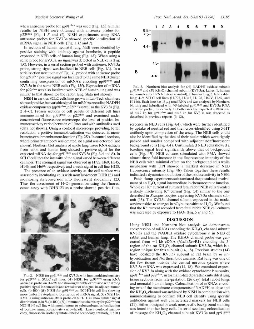
showed positive but variable signal formRNAs encodingNADPHoxidase components (gp91phox, p22phox) as well as theKV3.3a (Fig.2 A–C). Frozen sections of cell pellets of different cell linesimmunostained for gp91phox or p22phox and examined underconventional fluorescence microscope, the level of positive im-munoreactivity varied between cell lines and with antibodies used(data not shown). Using a confocal microscope providing betterresolution, a positive immunolocalization was detected in mem-branous or submembranous location (Fig. 2D). In control sections,where primary antibody was omitted, no signal was detected (notshown). Northern blot analysis of whole lung tissue RNA extractsfrom rabbit and human lung showed a positive signal for theexpectedmRNA size for gp91phox and KV3.3a (Fig. 3A andB). InSCLC cell lines the intensity of the signal varied between differentcell lines. The strongest signal was observed in H727, H69, H345,H146, and H69V respectively and a weak signal in cell line H128.The presence of an oxidase activity at the cell surface was
assessed by incubating cells with nonfluorescent DHR123 andmonitoring its conversion into fluorescent rhodamine 123.Thus the assessment of H2O2 generation using the fluores-cence assay with DHR123 as a probe showed positive fluo-
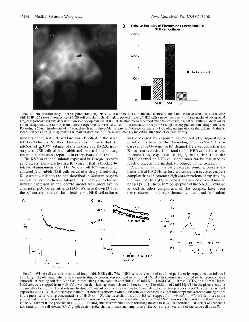
rescence in NEB cells (Fig. 4A), which were further identifiedby uptake of neutral red and then cross-identified using 5-HTantibody upon completion of the assay. The NEB cells couldalso be identified by the size of their nuclei which were tightlypacked and smaller compared with adjacent nonfluorescentbackground cells (Fig. 4A). Unstimulated NEB cells showed abaseline signal level significantly above that of backgroundcells (Fig. 4B). NEB cultures stimulated with PMA showedalmost three-fold increase in the fluorescence intensity of theNEB cells with minimal effect on the background cells whileincubation with DPI showed a marked decrease in NEBfluorescence intensity (Fig. 4B) Taken together these resultsindicated a dynamic modulation of the oxidase activity in NEB.Patch-clamp experiments substantiated the postulated involve-
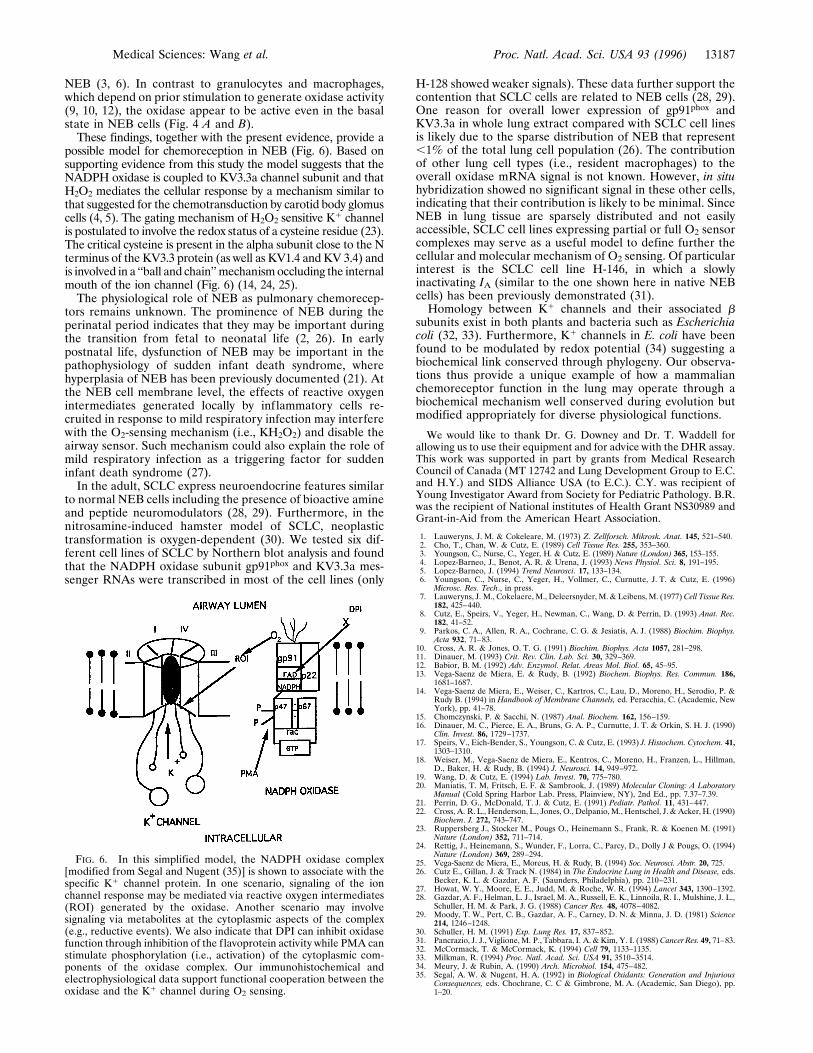
ment of an H2O2 signal intermediate in chemoreceptor function.Whole cell K1 current of cultured fetal rabbit NEB cells revealeda slowly inactivating K1 current (Fig. 5A) similar to the onedescribed in Xenopus oocytes expressing KV3.3a channels sub-unit (13). The KV3.3a channel subunit expressed in the modelwas insensitive to changes in pO2 but sensitive toH2O2.We foundthat the K1 current recorded from fetal rabbit NEB cell cultureswas increased by exposure to H2O2 (Fig. 5 B and C).
DISCUSSIONUsing NISH and Northern blot analysis we demonstratecoexpression of mRNAs encoding the KH2O2 channel subunitKV3.3a and the NADPH oxidase cytochrome b in NEB ofrabbit and human lung. The KH2O2 channel probe was gen-erated from '1 kb cDNA (NcoIyEcoRI) encoding the 39region of the rat KH2O2 channel subunit KV3.3a, which is aregion unique for this subunit (14, 18). Previous studies (14)have localized the KV3.3a subunit in rat brain by in situhybridization and Northern blot analysis. Rat lung was one ofthe few tissues outside the central nervous system whereKV3.3a mRNA was expressed (14, 18). We examined expres-sion of KV3.3a along with the oxidase cytochrome b subunits,gp91phox and p22phox, in formalin-fixed paraffin embedded lungtissue sections from late-gestation (26 day) fetal rabbit lungsand neonatal human lungs. Colocalization of mRNAs encod-ing two of the membrane components of NADPH oxidase andthe KH2O2 channel was achieved by NISH in combination withimmunostaining to confirm NEB cell identity using specificantibodies against well characterized markers for NEB cells(21). Either no signal or weak nonspecific background stainingwas found in other lung cells. In serial sections, colocalizationof message for KH2O2 channel subunit KV3.3a and gp91phox
FIG. 2. NISH for gp91phox and KV3.3a with immunohistochemistryfor p22phox in SCLC cell lines. (A) NISH for gp91phox using RNAantisense probe onH-69V line showing variable expression with strongpositive signal in some cells and a weaker or no signal in adjacent tumorcells. (3400.) (B) NISH for gp91phox on NCI-H146 cell line showingmore uniform cytoplasmic localization of mRNA signal. (C) NISH forKV3.3a using antisense RNA probe on NCI-H146 show similar signaldistribution as in B. (3400.) (D) Immunohistochemistry for p22phox onNCI-H146 cell line with membranous or submembranous localizationof positive immunoreactivity (arrowhead). (Laser confocal micros-copy, f luorescein isothiocyantate-labeled secondary antibody, 3800.)
FIG. 3. Northern blot analysis for (A) NADPH oxidase subunitgp91phox and (B) KH2O2 channel subunit (KV3.3a). Lanes: 1, humanmononuclear cell RNA extract (control); 2, human lung; 3, fetal rabbitlung; 4–9, SCLC cell lines (H-727, H-345, H-128, H69V, H-69, andH-146). Each lane has 15 mg total RNA and was analyzed by Northernblotting and hybridized with 32P-labeled gp91phox and KV3.3a RNAantisense probe, respectively. In both cases the expected mRNA sizeof '4.7 kb for gp91phox and '4.8 kb for KV3.3a was detected asdescribed in previous reports (9, 12).
Medical Sciences: Wang et al. Proc. Natl. Acad. Sci. USA 93 (1996) 13185
subunits of the NADPH oxidase was identified in the sameNEB cell clusters. Northern blot analysis indicated that themRNAs of gp91phox subunit of the oxidase and KV3.3a tran-scripts in NEB cells of fetal rabbit and neonatal human lungmatched in size those reported in other tissues (16, 18).The KV3.3a channel subunit expressed in Xenopus oocytes
generates a slowly inactivating K1 current that is blocked bytetraethylammonium (13, 14). Whole cell K1 currents ofcultured fetal rabbit NEB cells revealed a slowly inactivatingK1 current similar to the one described in Xenopus oocytesexpressing KV3.3a channel subunit (13). The KV3.3a channelsubunit expressed in the oocyte model was insensitive tochanges in pO2, but sensitive to H2O2. We have shown (3) thatthe K1 current recorded form fetal rabbit NEB cell cultures
was decreased by exposure to reduced pO2 suggesting apossible link between the O2-binding protein (NADPH oxi-dase) and the O2-sensitive K1 channel. Here we report that theK1 current recorded from fetal rabbit NEB cell cultures wasincreased by exposure to H2O2 indicating that theKH2O2channel on NEB cell membranes can be regulated byreactive oxygen intermediates produced by the oxidase.A potential candidate for an oxygen sensor protein is the
heme-linkedNADPHoxidase, a membrane associated enzymecomplex that can generate high concentrations of superoxide,the precursor to H2O2, as occurs in granulocytes and macro-phages (9, 10). The p91phox polypeptide of the NADPHoxidaseas well as other components of this complex have beendemonstrated immunocytochemically in cultured fetal rabbit
FIG. 4. Fluorescence assay for H2O2 generation using DHR 123 as a probe. (A) Unstimulated culture of rabbit fetal NEB cells 20 min after loadingwith DHR 123 shows fluorescence of NEB cell cytoplasm. Small, tightly packed nuclei of NEB cells (arrow) contrast with large nuclei of backgroundlung cells (arrowhead) with dark nonfluorescent cytoplasm. (31000.) (B) Relative intensity of rhodamine fluorescence in NEB cell cultures. Mean valuesfor all background cells (n5 8) from different experiments. Baseline values for unstimulated NEB (n5 8) is significantly greater than background cells.Following a 20-min incubation with PMA, there is up to three-fold increase in fluorescence intensity indicating upregulation of the oxidase. A similarincubation with DPI (n 5 4) resulted in marked decrease in fluorescence intensity indicating inhibition of oxidase activity.
FIG. 5. Whole-cell currents in cultured fetal rabbit NEB cells. When NEB cells were exposed to a brief period of hyperpolarization followedby a longer depolarizing pulse, a slowly inactivating IA current was revealed (n 5 15). (A) NEB cells shown was recorded in the presence of anextracellular bathing solution A and an intracellular pipette solution containing: 140 mM KCl, 1 mM CaCl2, 10 mM EGTA, and 10 mM Hepes.NEB cells were stepped from 290 mV to various depolarizing potentials for 3–4 sec (n 5 8). The addition of 2 mMMgATP in the pipette solutiondid not alter the results. The slowly inactivating K1 current observed was similar to the one described in Xenopus oocytes KV3.3a channel subunitexpressing cells (13). (B) An increase in the K1 current was observed when NEB cells were exposed to either brief or prolonged depolarizing pulsesin the presence of varying concentrations of H2O2 (n 5 3). The trace shown is of a NEB cell stepped from 290 mV to 170 mV for 4 sec in thepresence of extracellular solution B. This solution was used to eliminate any contribution of Ca21 and Na1 currents. There was a resultant increasein the K1 current in the presence of H2O2 (0.1–1.6 mM) that was reversible upon returning the cell to H2O2-free solution. This effect was repeatedtwo times on the cell shown. (C) A graph depicting the change in maximal amplitude of the K1 current over time of the same cell as in B.
13186 Medical Sciences: Wang et al. Proc. Natl. Acad. Sci. USA 93 (1996)
NEB (3, 6). In contrast to granulocytes and macrophages,which depend on prior stimulation to generate oxidase activity(9, 10, 12), the oxidase appear to be active even in the basalstate in NEB cells (Fig. 4 A and B).These findings, together with the present evidence, provide a
possible model for chemoreception in NEB (Fig. 6). Based onsupporting evidence from this study the model suggests that theNADPH oxidase is coupled to KV3.3a channel subunit and thatH2O2 mediates the cellular response by a mechanism similar tothat suggested for the chemotransduction by carotid body glomuscells (4, 5). The gating mechanism of H2O2 sensitive K1 channelis postulated to involve the redox status of a cysteine residue (23).The critical cysteine is present in the alpha subunit close to the Nterminus of the KV3.3 protein (as well as KV1.4 and KV 3.4) andis involved in a ‘‘ball and chain’’mechanismoccluding the internalmouth of the ion channel (Fig. 6) (14, 24, 25).The physiological role of NEB as pulmonary chemorecep-
tors remains unknown. The prominence of NEB during theperinatal period indicates that they may be important duringthe transition from fetal to neonatal life (2, 26). In earlypostnatal life, dysfunction of NEB may be important in thepathophysiology of sudden infant death syndrome, wherehyperplasia of NEB has been previously documented (21). Atthe NEB cell membrane level, the effects of reactive oxygenintermediates generated locally by inflammatory cells re-cruited in response to mild respiratory infection may interferewith the O2-sensing mechanism (i.e., KH2O2) and disable theairway sensor. Such mechanism could also explain the role ofmild respiratory infection as a triggering factor for suddeninfant death syndrome (27).In the adult, SCLC express neuroendocrine features similar
to normal NEB cells including the presence of bioactive amineand peptide neuromodulators (28, 29). Furthermore, in thenitrosamine-induced hamster model of SCLC, neoplastictransformation is oxygen-dependent (30). We tested six dif-ferent cell lines of SCLC by Northern blot analysis and foundthat the NADPH oxidase subunit gp91phox and KV3.3a mes-senger RNAs were transcribed in most of the cell lines (only
H-128 showed weaker signals). These data further support thecontention that SCLC cells are related to NEB cells (28, 29).One reason for overall lower expression of gp91phox andKV3.3a in whole lung extract compared with SCLC cell linesis likely due to the sparse distribution of NEB that represent,1% of the total lung cell population (26). The contributionof other lung cell types (i.e., resident macrophages) to theoverall oxidase mRNA signal is not known. However, in situhybridization showed no significant signal in these other cells,indicating that their contribution is likely to be minimal. SinceNEB in lung tissue are sparsely distributed and not easilyaccessible, SCLC cell lines expressing partial or full O2 sensorcomplexes may serve as a useful model to define further thecellular and molecular mechanism of O2 sensing. Of particularinterest is the SCLC cell line H-146, in which a slowlyinactivating IA (similar to the one shown here in native NEBcells) has been previously demonstrated (31).Homology between K1 channels and their associated b
subunits exist in both plants and bacteria such as Escherichiacoli (32, 33). Furthermore, K1 channels in E. coli have beenfound to be modulated by redox potential (34) suggesting abiochemical link conserved through phylogeny. Our observa-tions thus provide a unique example of how a mammalianchemoreceptor function in the lung may operate through abiochemical mechanism well conserved during evolution butmodified appropriately for diverse physiological functions.
We would like to thank Dr. G. Downey and Dr. T. Waddell forallowing us to use their equipment and for advice with the DHR assay.This work was supported in part by grants from Medical ResearchCouncil of Canada (MT 12742 and Lung Development Group to E.C.and H.Y.) and SIDS Alliance USA (to E.C.). C.Y. was recipient ofYoung Investigator Award from Society for Pediatric Pathology. B.R.was the recipient of National institutes of Health Grant NS30989 andGrant-in-Aid from the American Heart Association.
1. Lauweryns, J. M. & Cokeleare, M. (1973) Z. Zellforsch. Mikrosk. Anat. 145, 521–540.2. Cho, T., Chan, W. & Cutz, E. (1989) Cell Tissue Res. 255, 353–360.3. Youngson, C., Nurse, C., Yeger, H. & Cutz, E. (1989) Nature (London) 365, 153–155.4. Lopez-Barneo, J., Benot, A. R. & Urena, J. (1993) News Physiol. Sci. 8, 191–195.5. Lopez-Barneo, J. (1994) Trend Neurosci. 17, 133–134.6. Youngson, C., Nurse, C., Yeger, H., Vollmer, C., Curnutte, J. T. & Cutz, E. (1996)
Microsc. Res. Tech., in press.7. Lauweryns, J. M., Cokelaere, M., Deleersnyder, M. & Leibens, M. (1977) Cell Tissue Res.
182, 425–440.8. Cutz, E., Speirs, V., Yeger, H., Newman, C., Wang, D. & Perrin, D. (1993) Anat. Rec.
182, 41–52.9. Parkos, C. A., Allen, R. A., Cochrane, C. G. & Jesiatis, A. J. (1988) Biochim. Biophys.
Acta 932, 71–83.10. Cross, A. R. & Jones, O. T. G. (1991) Biochim. Biophys. Acta 1057, 281–298.11. Dinauer, M. (1993) Crit. Rev. Clin. Lab. Sci. 30, 329–369.12. Babior, B. M. (1992) Adv. Enzymol. Relat. Areas Mol. Biol. 65, 45–95.13. Vega-Saenz de Miera, E. & Rudy, B. (1992) Biochem. Biophys. Res. Commun. 186,
1681–1687.14. Vega-Saenz de Miera, E., Weiser, C., Kartros, C., Lau, D., Moreno, H., Serodio, P. &
Rudy B. (1994) in Handbook of Membrane Channels, ed. Peracchia, C. (Academic, NewYork), pp. 41–78.
15. Chomczynski, P. & Sacchi, N. (1987) Anal. Biochem. 162, 156–159.16. Dinauer, M. C., Pierce, E. A., Bruns, G. A. P., Curnutte, J. T. & Orkin, S. H. J. (1990)
Clin. Invest. 86, 1729–1737.17. Speirs, V., Eich-Bender, S., Youngson, C. & Cutz, E. (1993) J. Histochem. Cytochem. 41,
1303–1310.18. Weiser, M., Vega-Saenz de Miera, E., Kentros, C., Moreno, H., Franzen, L., Hillman,
D., Baker, H. & Rudy, B. (1994) J. Neurosci. 14, 949–972.19. Wang, D. & Cutz, E. (1994) Lab. Invest. 70, 775–780.20. Maniatis, T. M, Fritsch, E. F. & Sambrook, J. (1989) Molecular Cloning: A Laboratory
Manual (Cold Spring Harbor Lab. Press, Plainview, NY), 2nd Ed., pp. 7.37–7.39.21. Perrin, D. G., McDonald, T. J. & Cutz, E. (1991) Pediatr. Pathol. 11, 431–447.22. Cross, A. R. L., Henderson, L., Jones, O., Delpanio, M., Hentschel, J. &Acker, H. (1990)
Biochem. J. 272, 743–747.23. Ruppersberg J., Stocker M., Pougs O., Heinemann S., Frank, R. & Koenen M. (1991)
Nature (London) 352, 711–714.24. Rettig, J., Heinemann, S., Wunder, F., Lorra, C., Parcy, D., Dolly J & Pougs, O. (1994)
Nature (London) 369, 289–294.25. Vega-Saenz de Miera, E., Moreus, H. & Rudy, B. (1994) Soc. Neurosci. Abstr. 20, 725.26. Cutz E., Gillan, J. & Track N. (1984) in The Endocrine Lung in Health and Disease, eds.
Becker, K. L. & Gazdar, A. F. (Saunders, Philadelphia), pp. 210–231.27. Howat, W. Y., Moore, E. E., Judd, M. & Roche, W. R. (1994) Lancet 343, 1390–1392.28. Gazdar, A. F., Helman, L. J., Israel, M. A., Russell, E. K., Linnoila, R. I., Mulshine, J. L.,
Schuller, H. M. & Park, J. G. (1988) Cancer Res. 48, 4078–4082.29. Moody, T. W., Pert, C. B., Gazdar, A. F., Carney, D. N. & Minna, J. D. (1981) Science
214, 1246–1248.30. Schuller, H. M. (1991) Exp. Lung Res. 17, 837–852.31. Pancrazio, J. J., Viglione,M. P., Tabbara, I. A. &Kim, Y. I. (1988)Cancer Res. 49, 71–83.32. McCormack, T. & McCormack, K. (1994) Cell 79, 1133–1135.33. Milkman, R. (1994) Proc. Natl. Acad. Sci. USA 91, 3510–3514.34. Meury, J. & Rubin, A. (1990) Arch. Microbiol. 154, 475–482.35. Segal, A. W. & Nugent, H. A. (1992) in Biological Oxidants: Generation and Injurious
Consequences, eds. Chochrane, C. C & Gimbrone, M. A. (Academic, San Diego), pp.1–20.
FIG. 6. In this simplified model, the NADPH oxidase complex[modified from Segal and Nugent (35)] is shown to associate with thespecific K1 channel protein. In one scenario, signaling of the ionchannel response may be mediated via reactive oxygen intermediates(ROI) generated by the oxidase. Another scenario may involvesignaling via metabolites at the cytoplasmic aspects of the complex(e.g., reductive events). We also indicate that DPI can inhibit oxidasefunction through inhibition of the flavoprotein activity while PMA canstimulate phosphorylation (i.e., activation) of the cytoplasmic com-ponents of the oxidase complex. Our immunohistochemical andelectrophysiological data support functional cooperation between theoxidase and the K1 channel during O2 sensing.
Medical Sciences: Wang et al. Proc. Natl. Acad. Sci. USA 93 (1996) 13187