NADPH Oxidase-Derived Overproduction of Reactive Oxygen Species Impairs Postischemic...
10
Vascular Biology, Atherosclerosis and Endothelium Biology NADPH Oxidase-Derived Overproduction of Reactive Oxygen Species Impairs Postischemic Neovascularization in Mice with Type 1 Diabetes Te ´ ni G Ebrahimian,* Christophe Heymes, † Dong You,* Olivier Blanc-Brude,* Barend Mees, ‡ Ludovic Waeckel,* Micheline Duriez,* Jose ´ Vilar,* Ralph P. Brandes, § Bernard I. Levy,* Ajay M. Shah, † and Jean-Se ´ bastien Silvestre* From the Cardiovascular Research Center,* INSERM Lariboisie `re U689, Universite ´ Paris, Paris, France; the Cardiovascular Division, † King’s College, London, United Kingdom; the Departments of Vascular Surgery and Cell Biology and Genetics, ‡ Erasmus University Medical Center, Rotterdam, The Netherlands; and the Institut fur Kardiovaskulare Physiologie, § Universitat Frankfurt, Frankfurt, Germany We hypothesized that diabetes-induced oxidative stress may affect postischemic neovascularization. The response to unilateral femoral artery ligation was studied in wild-type or gp91 phox -deficient control or type 1 diabetic mice or in animals treated with the anti-oxidant N-acetyl-L-cysteine (NAC) or with in vivo electrotransfer of a plasmid encoding dominant-neg- ative Rac1 (50 g) for 21 days. Postischemic neovas- cularization was reduced in diabetic mice in associa- tion with down-regulated vascular endothelial growth factor-A protein levels. In diabetic animals vascular endothelial growth factor levels and postischemic neovascularization were restored to nondiabetic lev- els by the scavenging of reactive oxygen species (ROS) by NAC administration or the inhibition of ROS gen- eration by gp91 phox deficiency or by administration of dominant-negative Rac1. Finally , diabetes reduced the ability of adherent bone marrow-derived mono- nuclear cells (BM-MNCs) to differentiate into endothe- lial progenitor cells. Treatment with NAC (3 mmol/L) , apocynin (200 mol/L) , or the p38MAPK inhibitor LY333351 (10 mol/L) up-regulated the number of endothelial progenitor cell colonies derived from di- abetic BM-MNCs by 1.5-, 1.6-, and 1.5-fold, respec- tively (P < 0.05). In the ischemic hindlimb model , injection of diabetic BM-MNCs isolated from NAC- treated or gp91 phox -deficient diabetic mice increased neovascularization by 1.5-fold greater than un- treated diabetic BM-MNCs (P < 0.05). Thus , inhibition of NADPH oxidase-derived ROS overproduction im- proves the angiogenic and vasculogenic processes and restores postischemic neovascularization in type 1 diabetic mice. (Am J Pathol 2006, 169:719 –728; DOI: 10.2353/ajpath.2006.060042) Cardiovascular complications are the leading cause of morbidity and mortality in patients with diabetes mellitus. In particular, diabetes is associated with a poor outcome after vascular occlusion. This can be attributed in part to impaired neovascularization. Three principal events, ie, vasculogenesis, true angiogenesis, and collateral growth, contribute to postnatal vessel growth and each may be affected by diabetes. Indeed, there are a number of equally tenable hypotheses regarding the mechanisms underlying alterations in blood vessel growth in diabetes, including a reduction in vascular endothelial growth fac- tor-A (VEGF-A) signaling, changes in inflammation-re- lated pathways, 1,2 and accumulation of advanced glyca- tion end products. 3 Postnatal vasculogenesis can also be affected because the proangiogenic effects of bone mar- row mononuclear cells (BM-MNCs) and endothelial pro- genitor cells (EPCs) are reduced in diabetic mice and patients with either type 1 or 2 diabetes. 4–6 Each hypoth- esis might be a different aspect of common underlying pathogenic mechanisms that remain to be defined. Supported by the British Heart Foundation (Chair of Cardiology at King’s College London to A.M.S.); Agence Nationale de la Recherche (young investigator grant JC05-45445 and 2005 Cardiovascular, Obesity, and Diabetes grant ANR-05-PCOD-028-01 to J.-S.S.); The Netherlands Orga- nization for Health Research and Development (Agiko stipend 920-03-291 to B.M.); the Prof. Michae ¨ l-van Vloten Fund; and the European Commis- sion (INSERM U689 is partner of the European Vascular Genomics Network, a Network of Excellence granted by contract no. LSHM- CT-2003-503254). Accepted for publication May 2, 2006. Address reprint requests to Jean-Sebastien Silvestre, U689-INSERM, Ho ˆ pital Lariboisie ` re, 41 Blvd de la Chapelle, 75475 Paris Cedex 10, France. E-mail: [email protected]. The American Journal of Pathology, Vol. 169, No. 2, August 2006 Copyright © American Society for Investigative Pathology DOI: 10.2353/ajpath.2006.060042 719
-
Upload
independent -
Category
Documents
-
view
3 -
download
0
Transcript of NADPH Oxidase-Derived Overproduction of Reactive Oxygen Species Impairs Postischemic...
Vascular Biology, Atherosclerosis and Endothelium Biology
NADPH Oxidase-Derived Overproduction ofReactive Oxygen Species Impairs PostischemicNeovascularization in Mice with Type 1 Diabetes
Teni G Ebrahimian,* Christophe Heymes,†
Dong You,* Olivier Blanc-Brude,* Barend Mees,‡
Ludovic Waeckel,* Micheline Duriez,* Jose Vilar,*Ralph P. Brandes,§ Bernard I. Levy,*Ajay M. Shah,† and Jean-Sebastien Silvestre*From the Cardiovascular Research Center,* INSERM Lariboisiere
U689, Universite Paris, Paris, France; the Cardiovascular
Division,† King’s College, London, United Kingdom; the
Departments of Vascular Surgery and Cell Biology and Genetics,‡
Erasmus University Medical Center, Rotterdam, The Netherlands;
and the Institut fur Kardiovaskulare Physiologie,§ Universitat
Frankfurt, Frankfurt, Germany
We hypothesized that diabetes-induced oxidativestress may affect postischemic neovascularization.The response to unilateral femoral artery ligation wasstudied in wild-type or gp91phox-deficient control ortype 1 diabetic mice or in animals treated with theanti-oxidant N-acetyl-L-cysteine (NAC) or with in vivoelectrotransfer of a plasmid encoding dominant-neg-ative Rac1 (50 �g) for 21 days. Postischemic neovas-cularization was reduced in diabetic mice in associa-tion with down-regulated vascular endothelial growthfactor-A protein levels. In diabetic animals vascularendothelial growth factor levels and postischemicneovascularization were restored to nondiabetic lev-els by the scavenging of reactive oxygen species (ROS)by NAC administration or the inhibition of ROS gen-eration by gp91phox deficiency or by administrationof dominant-negative Rac1. Finally, diabetes reducedthe ability of adherent bone marrow-derived mono-nuclear cells (BM-MNCs) to differentiate into endothe-lial progenitor cells. Treatment with NAC (3 mmol/L),apocynin (200 �mol/L), or the p38MAPK inhibitorLY333351 (10 �mol/L) up-regulated the number ofendothelial progenitor cell colonies derived from di-abetic BM-MNCs by 1.5-, 1.6- , and 1.5-fold, respec-tively (P < 0.05). In the ischemic hindlimb model,injection of diabetic BM-MNCs isolated from NAC-treated or gp91phox-deficient diabetic mice increasedneovascularization by �1.5-fold greater than un-
treated diabetic BM-MNCs (P < 0.05). Thus, inhibitionof NADPH oxidase-derived ROS overproduction im-proves the angiogenic and vasculogenic processesand restores postischemic neovascularization in type1 diabetic mice. (Am J Pathol 2006, 169:719–728; DOI:
10.2353/ajpath.2006.060042)
Cardiovascular complications are the leading cause ofmorbidity and mortality in patients with diabetes mellitus.In particular, diabetes is associated with a poor outcomeafter vascular occlusion. This can be attributed in partto impaired neovascularization. Three principal events,ie, vasculogenesis, true angiogenesis, and collateralgrowth, contribute to postnatal vessel growth and eachmay be affected by diabetes. Indeed, there are a numberof equally tenable hypotheses regarding the mechanismsunderlying alterations in blood vessel growth in diabetes,including a reduction in vascular endothelial growth fac-tor-A (VEGF-A) signaling, changes in inflammation-re-lated pathways,1,2 and accumulation of advanced glyca-tion end products.3 Postnatal vasculogenesis can also beaffected because the proangiogenic effects of bone mar-row mononuclear cells (BM-MNCs) and endothelial pro-genitor cells (EPCs) are reduced in diabetic mice andpatients with either type 1 or 2 diabetes.4–6 Each hypoth-esis might be a different aspect of common underlyingpathogenic mechanisms that remain to be defined.
Supported by the British Heart Foundation (Chair of Cardiology at King’sCollege London to A.M.S.); Agence Nationale de la Recherche (younginvestigator grant JC05-45445 and 2005 Cardiovascular, Obesity, andDiabetes grant ANR-05-PCOD-028-01 to J.-S.S.); The Netherlands Orga-nization for Health Research and Development (Agiko stipend 920-03-291to B.M.); the Prof. Michael-van Vloten Fund; and the European Commis-sion (INSERM U689 is partner of the European Vascular GenomicsNetwork, a Network of Excellence granted by contract no. LSHM-CT-2003-503254).
Accepted for publication May 2, 2006.
Address reprint requests to Jean-Sebastien Silvestre, U689-INSERM,Hopital Lariboisiere, 41 Blvd de la Chapelle, 75475 Paris Cedex 10,France. E-mail: [email protected].
The American Journal of Pathology, Vol. 169, No. 2, August 2006
Copyright © American Society for Investigative Pathology
DOI: 10.2353/ajpath.2006.060042
719
Reactive oxygen species (ROS) including superoxide,hydrogen peroxide, hydroxyl radical, and reactive nitro-gen species, such as nitric oxide and peroxynitrite, arebiologically active species that are increasingly recog-nized to play major roles in vascular biology throughredox signaling.7–9 Each of these species derives fromspecific enzymatic or chemical reactions. A predominantsource of ROS in the normal vessel is thought to be afamily of membrane-associated NADPH oxidases.7,8,10
The NADPH oxidase, as found in neutrophils and endo-thelial cells,7,8,11 consists of a membrane-localized cyto-chrome b558 comprised of two subunits, gp91phox (orNox 2) and p22phox, as well as cytosolic componentsp47phox and p67phox. The small GTPase Rac1 is alsonecessary for full NADPH oxidase activity. ROS havebeen suggested as important mediators of angiogenesis.H2O2 stimulates cell migration and proliferation in endo-thelial cells,12 and ROS directly modulate VEGF-A ex-pression and vascular smooth muscle cell proliferation.13
Previous reports also suggest that both the gp91phox-containing NADPH oxidase and Rac1 play a major role inVEGF-A-induced endothelial cell proliferation.14 Similarly,mice lacking gp91phox displayed impaired postischemicneovascularization.15 Taken together, these observationssuggest that both gp91phox and Rac1 play a significantrole in NADPH oxidase-induced angiogenesis.
Diabetes is associated with increased NADPH oxidaseactivation leading to ROS overproduction.16,17 A link be-tween ROS and diabetes has been evidenced in exper-imental models of type 1 and 2 diabetes, as well as inhuman patients with type 2 diabetes.7,8,16,17 Further-more, increased ROS production may contribute to dia-betes-induced vascular complications. In support of thisview, ROS overproduction has been shown to participatein endothelial dysfunction in both rodent models andpatients with type 2 diabetes.16,18
The aim of our study was to analyze the role of diabe-tes-induced ROS overproduction in postischemic neo-vascularization in a mouse model of unilateral hindlimbischemia. We hypothesized that oxidative stress is one ofthe key events that triggers pathways leading to vascularcomplications in diabetes. In particular, we sought toinvestigate the involvement of NADPH oxidase-derivedROS in diabetes-induced alterations of VEGF-A expres-sion and postnatal vasculogenesis.
Materials and Methods
Experimental Protocol
All of the experiments were performed in accordancewith the European Community guidelines for the care anduse of laboratory animals (no. 07430). To induce diabe-tes, C57BL/6 wild-type mice and C57BL/6 gp91phox-de-ficient mice were injected intraperitoneally with 60 mg/kgstreptozotocin in sodium citrate buffer (0.05 mol/L, pH4.5) daily for 5 days. Three days after the fifth injection,blood glucose levels were measured. Mice with glucoselevels �300 mg/dl were excluded from the study, aspreviously described.3,5 In control groups, mice were
injected intraperitoneally with sodium citrate buffer (0.05mol/L, pH 4.5). After 2 months of monitored diabetes,mice underwent surgical ligation of the proximal part ofthe right femoral artery, just above the origin of the cir-comflexa femoris lateralis, as previously described.19,20
Diabetic and nondiabetic mice were then treated with orwithout the anti-oxidant N-acetyl-L-cysteine (NAC; 200mg/kg/day, i.p.) and in vivo electrotransfer of a pCAGexpression plasmid encoding for dominant-negative mu-tant Rac1 (N17Rac1, 50 �g injected in the gastrocnemiusmuscle). An additional group of mice also received 50 �gof empty pCAG expression plasmid as a negative con-trol. To further analyze the role of VEGF-A up-regulation inNAC-induced restoration of neovascularization, NAC-treated diabetic mice also received an intraperitonealinjection of neutralizing antibody directed against VEGFreceptor 2 (anti-Flk-1, 100 �g/day, clone DC101; ImCloneSystems Inc., New York, NY21).
Quantification of Neovascularization in theHindlimb
Three weeks after the onset of ischemia, vessel densitywas evaluated by three different methods, as previouslydescribed19,20: 1) high-definition microangiography us-ing barium sulfate (1 g/ml) injected in the abdominalaorta, followed by image acquisition with a digital X-raytransducer and computerized quantification of vesseldensity expressed as a percentage of pixels per imageoccupied by vessels in the quantification area; 2) assess-ment of capillary density in gastrocnemius muscle byimmunostaining with a rabbit polyclonal antibody di-rected against total fibronectin (dilution 1/50; ChemiconInt., Temecula, CA) and morphometric quantification us-ing Histolab software (Microvisions, Paris, France); and3) laser Doppler perfusion imaging to assess in vivo tissuefoot perfusion.
Determination of Protein Expression
Gastrocnemius muscles from ischemic and nonischemichindlimbs were thawed and homogenized in 300 �l ofbuffer (200 mmol/L sucrose, 20 mmol/L HEPES, pH 7.4)with protease inhibitors. Protein extracts from BM-MNCswere obtained by lysing cells in 200 �l of buffer (20%sodium dodecyl sulfate, 100 mmol/L sodium orthovana-date, 0.5 mol/L Tris, pH 7.4) with protease inhibitors.Proteins were separated in 12% denaturing sodium do-decyl sulfate-polyacrylamide gel electrophoresis andblotted onto nitrocellulose sheets (Hybond ECL; Amer-sham, Orsay, France). Antibodies against VEGF-A (1:2000, Santa Cruz Biotechnology, Santa Cruz, CA),gp91phox (1:500, BD Transduction, Franklin Lakes, NJ),p47phox (1:2000, BD Transduction), p67phox (1:2000, BDTransduction), Rac1 (1:1000, Cell Signaling, Danvers,MA), phospho-p38 mitogen-activated protein kinase(p38MAPK) (1:1000, Cell Signaling) and pan-p38MAPK(1:1000, Santa Cruz Biotechnology) were used for immu-noblotting. As a protein loading control, membranes werestripped, incubated with a goat polyclonal antibody di-
720 Ebrahimian et alAJP August 2006, Vol. 169, No. 2
rected against total actin (dilution 1/1000, Santa CruzBiotechnology), and specific chemiluminescent signalwas detected as previously described.19,20
Proangiogenic Effect of BM-MNCs
BM-MNCs were obtained by flushing tibia and femur ofwild-type and gp91phox-deficient control and diabeticmice treated with or without NAC. Low-density mononu-clear cells were then isolated by centrifugation on a Ficollgradient, as previously described.5 Five hours after hind-limb ischemia, control animals received intravenous in-jections of 1 � 106 BM-MNCs. Animals were euthanizedat day 10 after ischemia, and the neovascularization re-action was assessed as described above.
BM-MNC Differentiation into EPCs
BM-MNCs (1.5 � 106) per ml were plated on 11-mmcell-culture dishes coated with rat plasma vitronectin(Sigma, St. Quentin Fallavier, France) and gelatin (0.1%)and maintained in endothelial basal medium (EGM2; Bio-Whittaker, Walkersville, MD). BM-MNCs were treated withor without apocynin (200 �mol/L), NAC (3 mmol/L), or ap38MAPK inhibitor (LY333351, 10 �mol/L) for 7 days.Nonadherent cells were then removed and adherent cellsunderwent im-munochemical analysis. To detect the up-take of 1,1�-dioctadecyl-3,3,3�,3�-tetramethylindocarbo-cyanine-labeled acetylated low-density lipoprotein(AcLDL-Dil), cells were incubated in medium containingAcLDL-Dil (Tebu, Le Perray en Yvelines, France) at 37°Cfor 1 hour. Cells were then fixed with 2% paraformalde-hyde and incubated with fluorescein isothiocyanate-labeled BS-1 lectin (Sigma). Dual-positive staining forboth AcLDL-Dil and BS-1 lectin characterized EPCs. EPCnumbers were counted and expressed in cells per well,as previously described.5 Five replicates were countedfor each experimental condition. Three independent in-vestigators evaluated the number of EPCs per well bycounting three randomly selected high-power fields un-der epifluorescence microscopy. Results are expressedas percentages of total cell numbers.
BM-MNC Apoptosis Measurement
BM-MNCs were incubated with tumor necrosis factor-�(10 ng/ml) plus cycloheximide (10 �mol/L) or C2-cer-amide (100 �mol/L) for 8 hours. BM-MNCs were fixedwith paraformaldehyde for 12 hours at 4°C, stainedwith 4,6-diamidino-2-phenylindole (Calbiochem, Paris,France) for 30 minutes for examination of nuclear mor-phology by fluorescence microscopy. Total and apopto-tic nuclei showing condensed chromatin and fragmentednuclear bodies (karyorrhexis, pyknosis) were counted infive random fields per treatment, totaling a minimum of150 cells in each of three independent experiments. Re-sults are presented as percentage of apoptotic cells.Alternatively, cell death was assessed by propidium io-dide DNA staining and fluorescence-activated cell sort-ing analysis. Fractions of the cell population in the apo-
ptotic G0/G1 and G2/M cell cycle phases were evaluatedand expressed in percentages.
Immunohistochemistry
Frozen tissue sections (7 �m) were incubated with rabbitpolyclonal anti-nitrotyrosine antibody (dilution of 1/50) toevaluate 3-nitrotyrosine formation as a biomarker for invivo nitration. To account for variables including hetero-geneity of ROS levels, three different sections were per-formed at three different levels of gastrocnemius musclefor each animal. In addition, we compared the number ofnitrotyrosine-specific stainings between the three differ-ent levels for each animal. Using one-way analysis ofvariance, we did not observe any statistical differences inthe experimental group. Immunostains were visualizedby fluorescence microscopy and then analyzed in ran-domly chosen fields (five fields per section) of a definedarea with Histolab software.
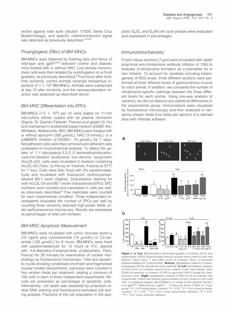
Figure 1. a: Top: Representative photomicrographs of ischemic (Isch) andnonischemic (NIsch) gastrocnemius muscle sections from control (Cont) anddiabetic (Diab) mice, 7 days after onset of ischemia. There is increasedimmunostaining for 3-nitrotyrosine. Bottom: Quantitative analysis of immu-nostaining with the anti-nitrotyrosine antibody. b: Left: Quantitative analysisof ROS levels in ischemic muscle from control (Cont) and diabetic mice(Diab) in presence or absence of DPI or apocynin (APO) during the lumi-nescence assay. Right: Quantitative analysis of ROS levels in ischemic andnonischemic control and diabetic gastrocnemius of mice treated in vivo withNAC or dominant-negative Rac1(dnRac1) or empty pCAG plasmid (pCAG)or in gp91phox-deficient mice (gp91�/�). Values are mean � SEM, n � 6 pergroup. *P � 0.05 nonischemic controls; †P � 0.05, ††P � 0.01 versus ischemiccontrols; �P � 0.05, ��P � 0.01 versus nonischemic diabetics; #P � 0.05,##P � 0.01 versus ischemic diabetics.
Diabetes and Angiogenesis 721AJP August 2006, Vol. 169, No. 2

Luminescence Assay
Tissue and cellular ROS levels, reflecting a balance be-tween oxidant production and removal by endogenousantioxidants, were also quantified using L-012 as de-scribed recently.22,23 Gastrocnemius muscle and BM-MNCs were lysed in 50 mmol/L Tris buffer (pH 7.5)containing protease inhibitors (Boehringer-Mannheim,Mannheim, Germany) and centrifuged at 10,000 � g for15 minutes at 4°C. Supernatants were then incubatedwith L-012 100 �mol/L (Wako, Neuss, Germany) with orwithout diphenylene iodonium (DPI; 30 �mol/L) or apoc-ynin (1 mmol/L). The same part of the gastrocnemiusmuscle was used in the different treated groups. Lumi-nescence was counted (Topcount NXT; Perkin Elmer)during 20 seconds after a 10-minute interval, allowing forthe plates to become dark-adapted.
Statistical Analysis
Results were expressed as mean � SEM. One-way anal-ysis of variance was used to compare each parameter.Comparisons between groups were performed usingpost hoc Bonferroni t-test when the analysis of variancetest was statistically significant. P � 0.05 was consideredsignificant.
Results
Changes in ROS Levels and NADPH OxidaseSubunit Protein Expression
In control animals, 7 days of ischemia raised 3-nitroty-rosine-positive staining in hindlimb muscle (Figure 1a).These results were substantiated by L-012 luminescencedemonstrating that ischemia enhanced ROS levels incontrol animals (Figure 1b). Treatment with DPI or apoc-ynin impaired ischemia-induced increase in ROS levels(P � 0.01 and P � 0.05 for DPI and apocynin treatment,respectively; Figure 1b). Although the experiments withDPI and apocynin are suggestive, we used gp91phox-deficient mice to obtain more definitive evidence andshowed that ischemia-induced up-regulation of ROS lev-els was abrogated in this setting (Figure 1b). Taken to-gether, these results suggest that ischemia activatesNADPH oxidase-dependent ROS synthesis. In diabeticmice, ROS accumulation in ischemic tissue was furtherup-regulated by 1.8-fold compared to control animals
Figure 2. Representative Western blot and quantitative evaluation of NADPHoxidase subunit protein levels in ischemic and nonischemic tissues fromcontrol (Cont) and diabetic (Diab) animals, expressed as mean � SEM, n �6 per group. *P � 0.05, **P � 0.01, ***P � 0.001 versus nonischemic controls;†P � 0.05 versus ischemic controls.
Figure 3. Quantitative evaluation of microangiography (a), capillary density(b), and foot perfusion (c) in control and diabetic animals, 21 days afterischemia. Cont, untreated control animals; Cont NAC, control animaltreated with NAC; Cont gp91�/�, control animal lacking gp91phox; Diab,untreated diabetic animals; Diab NAC, diabetic animal treated with NAC;Diab gp91�/�, diabetic animal lacking gp91phox; Diab dnRac1, diabeticanimal treated with plasmid encoding for Rac1 dominant-negative; Diab pCAG, diabetic animal treated with empty plasmid; Diab NAC anti-Flk-1,diabetic animal co-treated with NAC and neutralizing antibody directedagainst VEGF receptor type 2, Flk-1. *P � 0.05, **P � 0.01 versus untreatedwild-type controls; †P � 0.05, ††P � 0.01 versus diabetic wild-type animals;�P � 0.05 versus diabetic animals treated with NAC.
722 Ebrahimian et alAJP August 2006, Vol. 169, No. 2

(P � 0.01 versus ischemic control; Figure 1b). Treatmentwith NAC and dominant-negative Rac-1 or gp91phox de-ficiency abrogated ischemia- and diabetes-induced in-creases in ROS (Figure 1, a and b).
The increase in ROS was associated with variations inprotein levels of NADPH oxidase subunits. In controlanimals, gp91phox and p67phox protein levels were up-regulated by 2.9-fold and 1.7-fold, respectively, whencompared to nonischemic controls (P � 0.01 and P �0.05, respectively). p47phox and Rac1 protein levels alsotended to be increased, but this did not reach statisticalsignificance (Figure 2). In diabetic mice, gp91phox andRac1 protein levels were further increased by 1.4- and1.3-fold in diabetic ischemic hindlimbs compared to isch-emic controls (P � 0.05; Figure 2). In contrast, p47phox
and p67phox protein content was decreased in diabeticischemic muscles in reference to ischemic control ani-mals (P � 0.05; Figure 2). Taken together, these resultsindicate an up-regulation of gp91phox and Rac1 proteincontent and increased ROS levels in diabetic ischemicmuscles.
NADPH Oxidase Blockade RestoredPostischemic Neovascularization in DiabeticMice
We next assessed the role of ROS and NADPH oxidaseactivity in postischemic vessel growth 21 days after isch-emia. In control animals, neovascularization was im-paired by NAC administration, or the absence ofgp91phox, as revealed by the 40% decrease in angio-graphic scores, the 43% decrease in capillary density,and the 40% reduction in foot Doppler perfusion scores(Figure 3). In diabetic animals, angiography scores, cap-
illary density, and foot perfusion were decreased by 40%,60%, and 43%, respectively, compared to control ani-mals (Figure 3). The scavenging of ROS by NAC admin-istration or the inhibition of ROS generation by gp91phox
deficiency or administration of dominant-negative Rac1restored postischemic neovascularization in diabetic an-imals to control levels (Figure 3). Hence, reduction inROS levels restored vessel growth in diabetic ischemicleg whereas it impaired neovascularization in controlischemic tissue.
Figure 4. Representative Western blot (a) and quantitative evaluation ofVEGF-A protein levels in ischemic and nonischemic tissues (b), 21 days afterischemia. Cont, untreated control animals; Cont NAC, control animaltreated with NAC; Cont gp91�/�, control animal lacking gp91phox; Diab,untreated diabetic animals; Diab NAC, diabetic animal treated with NAC;Diab dnRac1, diabetic animal treated with plasmid encoding for Rac1dominant-negative; Diab gp91�/�, diabetic animal lacking gp91phox. Valuesare mean � SEM, n � 6 per group. *P � 0.05, ***P � 0.001 versus untreatedwild-type controls; †P � 0.05, ††P � 0.01 versus diabetic wild-type animals.
Figure 5. Representative Western blot (a) and quantitative evaluation (b) ofNADPH oxidase subunit protein levels in control and diabetic BM-MNCs.Values are mean � SEM, n � 6 per group. *P � 0.05, **P � 0.01 versuscontrol BM-MNCs. c: Quantitative evaluation of BM-MNC-derived ROS usingluminescence assay. Values are mean � SEM, n � 5 per group. *P � 0.05,**P � 0.01 versus control BM-MNCs, †P � 0.05, ††P � 0.01 versus diabeticBM-MNCs.
Diabetes and Angiogenesis 723AJP August 2006, Vol. 169, No. 2

724 Ebrahimian et alAJP August 2006, Vol. 169, No. 2

NADPH Oxidase Inhibition Restored VEGF-AProtein Expression in Diabetic Mice
We sought to determine the molecular and cellular path-ways affected by diabetes-induced oxidative stress.VEGF-A signaling represents a critical rate-limiting stepin both physiological and pathological angiogenesis. Incontrol mice, VEGF-A protein levels were decreased by25% in NAC-treated animals and gp91phox-deficient micecompared to control mice (P � 0.05; Figure 4). In dia-betic mice, VEGF-A levels were reduced by 60% in isch-emic tissues compared to ischemic control hindlimbs(P � 0.001; Figure 4). Interestingly, VEGF-A protein con-tents were significantly increased by 2.5-, 2.0-, and 2.7-fold in diabetic animals treated with NAC or dominant-negative Rac1 and in gp91phox-deficient diabetic mice,respectively, compared to untreated diabetic mice (Fig-ure 4). In addition, co-treatment with anti-Flk-1 abrogatedNAC-related effects suggesting that VEGF-A up-regula-tion participates in the restoration of vessel growth inNAC-treated diabetic mice (Figure 3).
NADPH Oxidase Inhibition Restored PostnatalVasculogenesis in Diabetic Animals
We finally evaluated the effects of diabetes-induced in-creased ROS on the ability of BM-MNCs to stimulateneovascularization. Protein expression of NADPH oxi-dase subunits was detected in control BM-MNCs. Theirlevels were up-regulated in diabetic versus control BM-MNCs (Figure 5a). ROS levels were also increased by2.3-fold in diabetic BM-MNCs compared to control BM-MNCs (P � 0.01; Figure 5b). Treatment with NAC ordeficiency in gp91phox reduced ROS levels in both con-trol and diabetic cells.
NADPH Oxidase Inhibition Restored theProangiogenic Potential of Diabetic BM-MNCs
The proangiogenic potential of BM-MNCs isolated fromcontrol mice treated with NAC or gp91phox-deficient micewas lower than that observed with BM-MNCs isolatedfrom control mice. However, this difference did not reachstatistical significance (Figure 6). In contrast, transplan-tation of BM-MNCs isolated from diabetic mice treatedwith NAC improved the angiography score by 1.6-fold,capillary numbers by 1.4-fold, and foot perfusion by 1.5-fold greater than BM-MNCs isolated from untreated dia-betic animals (Figure 6). Similarly, injection of BM-MNCsisolated from gp91phox-deficient diabetic mice improvedthe angiography score by 1.7-fold, capillary numbers by1.4-fold, and foot perfusion by 1.6-fold, compared toBM-MNCs isolated from untreated diabetic animals(Figure 6).
NADPH Oxidase Inhibition Restored BM-MNCDifferentiation into Endothelial Progenitor Cellsin Vitro
We could not detect any significant difference in percent-ages of apoptotic cells in control and diabetic animals,suggesting that major alterations in BM-MNC apoptosiswere not a significant cause of diabetes-induced BM-MNC dysfunction (Figure 7). Treatment of BM-MNCs iso-lated from control animals with NAC and apocynin re-duced the number of DilLDL/BS1 lectin double-positivecells (Figure 8a) suggesting that low levels of ROS mayfacilitate BM-MNC differentiation into EPCs. The ability ofdiabetic BM-MNCs to differentiate into EPCs was mark-edly reduced, as revealed by the 66% reduction in num-
Figure 6. Representative photomicrographs and quantitative evaluation of microangiography (a), capillary density (b, capillary appears in white, arrowsindicating representative examples of fibronectin-positive capillaries), and foot perfusion (c), 10 days after ischemia and cell transplantation. In this set ofexperiments, control animals received BM-MNCs from control mice (Cont), control mice treated with NAC (Cont NAC), gp91phox-deficient control mice (Contgp91�/�), diabetic mice (diab), diabetic mice treated with NAC (diab NAC), gp91phox-deficient diabetic mice (diab gp91phox�/�). *P � 0.05 versus controlanimals receiving control BM-MNCs. †P � 0.05, ††P � 0.01 versus control animals receiving diabetic BM-MNCs.
Figure 7. a: Quantification (top) and representative photomicrographs(bottom) of control and diabetic BM-MNC apoptosis after incubation withtumor necrosis factor-� (10 ng/ml) plus cyclohexamide (CHX) or C2-cer-amide for 8 hours. BM-MNCs were fixed and stained with 4,6-diamidino-2-phenylindole. Arrows indicate apoptotic nuclei showing condensed chro-matin or fragmented nuclear bodies. b: Quantification of control and diabeticBM-MNC apoptosis assessed by propidium iodide DNA staining and fluores-cence-activated cell sorting analysis. Fractions of the cell population in theapoptotic G0/G1 and G2/M cell cycle phases were evaluated and expressedin percentages.
Diabetes and Angiogenesis 725AJP August 2006, Vol. 169, No. 2
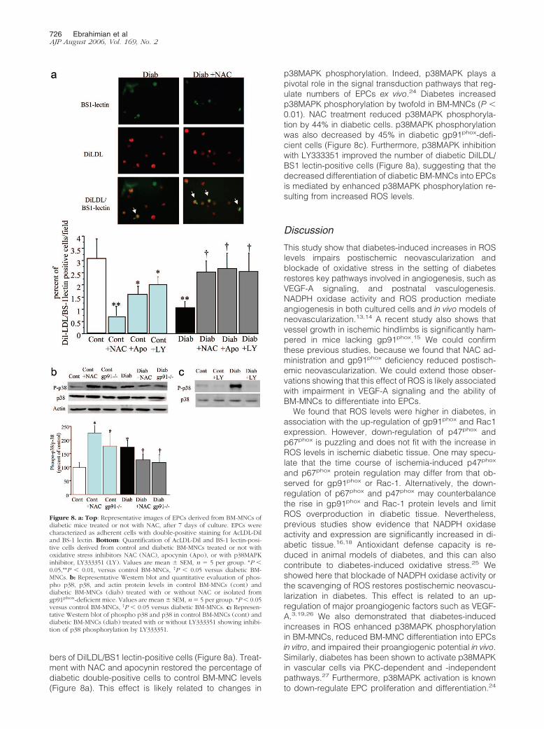
bers of DilLDL/BS1 lectin-positive cells (Figure 8a). Treat-ment with NAC and apocynin restored the percentage ofdiabetic double-positive cells to control BM-MNC levels(Figure 8a). This effect is likely related to changes in
p38MAPK phosphorylation. Indeed, p38MAPK plays apivotal role in the signal transduction pathways that reg-ulate numbers of EPCs ex vivo.24 Diabetes increasedp38MAPK phosphorylation by twofold in BM-MNCs (P �0.01). NAC treatment reduced p38MAPK phosphoryla-tion by 44% in diabetic cells. p38MAPK phosphorylationwas also decreased by 45% in diabetic gp91phox-defi-cient cells (Figure 8c). Furthermore, p38MAPK inhibitionwith LY333351 improved the number of diabetic DilLDL/BS1 lectin-positive cells (Figure 8a), suggesting that thedecreased differentiation of diabetic BM-MNCs into EPCsis mediated by enhanced p38MAPK phosphorylation re-sulting from increased ROS levels.
Discussion
This study show that diabetes-induced increases in ROSlevels impairs postischemic neovascularization andblockade of oxidative stress in the setting of diabetesrestores key pathways involved in angiogenesis, such asVEGF-A signaling, and postnatal vasculogenesis.NADPH oxidase activity and ROS production mediateangiogenesis in both cultured cells and in vivo models ofneovascularization.13,14 A recent study also shows thatvessel growth in ischemic hindlimbs is significantly ham-pered in mice lacking gp91phox.15 We could confirmthese previous studies, because we found that NAC ad-ministration and gp91phox deficiency reduced postisch-emic neovascularization. We could extend those obser-vations showing that this effect of ROS is likely associatedwith impairment in VEGF-A signaling and the ability ofBM-MNCs to differentiate into EPCs.
We found that ROS levels were higher in diabetes, inassociation with the up-regulation of gp91phox and Rac1expression. However, down-regulation of p47phox andp67phox is puzzling and does not fit with the increase inROS levels in ischemic diabetic tissue. One may specu-late that the time course of ischemia-induced p47phox
and p67phox protein regulation may differ from that ob-served for gp91phox or Rac-1. Alternatively, the down-regulation of p67phox and p47phox may counterbalancethe rise in gp91phox and Rac-1 protein levels and limitROS overproduction in diabetic tissue. Nevertheless,previous studies show evidence that NADPH oxidaseactivity and expression are significantly increased in di-abetic tissue.16,18 Antioxidant defense capacity is re-duced in animal models of diabetes, and this can alsocontribute to diabetes-induced oxidative stress.25 Weshowed here that blockade of NADPH oxidase activity orthe scavenging of ROS restores postischemic neovascu-larization in diabetes. This effect is related to an up-regulation of major proangiogenic factors such as VEGF-A.3,19,26 We also demonstrated that diabetes-inducedincreases in ROS enhanced p38MAPK phosphorylationin BM-MNCs, reduced BM-MNC differentiation into EPCsin vitro, and impaired their proangiogenic potential in vivo.Similarly, diabetes has been shown to activate p38MAPKin vascular cells via PKC-dependent and -independentpathways.27 Furthermore, p38MAPK activation is knownto down-regulate EPC proliferation and differentiation.24
Figure 8. a: Top: Representative images of EPCs derived from BM-MNCs ofdiabetic mice treated or not with NAC, after 7 days of culture. EPCs werecharacterized as adherent cells with double-positive staining for AcLDL-Diland BS-1 lectin. Bottom: Quantification of AcLDL-Dil and BS-1 lectin-posi-tive cells derived from control and diabetic BM-MNCs treated or not withoxidative stress inhibitors NAC (NAC), apocynin (Apo), or with p38MAPKinhibitor, LY333351 (LY). Values are mean � SEM, n � 5 per group. *P �0.05,**P � 0.01, versus control BM-MNCs, †P � 0.05 versus diabetic BM-MNCs. b: Representative Western blot and quantitative evaluation of phos-pho p38, p38, and actin protein levels in control BM-MNCs (cont) anddiabetic BM-MNCs (diab) treated with or without NAC or isolated fromgp91phox-deficient mice. Values are mean � SEM, n � 5 per group. *P � 0.05versus control BM-MNCs, †P � 0.05 versus diabetic BM-MNCs. c: Represen-tative Western blot of phospho p38 and p38 in control BM-MNCs (cont) anddiabetic BM-MNCs (diab) treated with or without LY333351 showing inhibi-tion of p38 phosphorylation by LY333351.
726 Ebrahimian et alAJP August 2006, Vol. 169, No. 2

Taken together, our results suggest that increased oxi-dative stress constitutes an underlying pathogenic mech-anism that affects both angiogenesis and vasculogenesisin the setting of diabetes. In addition, this study supportsthe concept that the effects of ROS on vascular functiondepend critically on the amounts present. In low amountsunder physiological conditions, ROS can act as intracel-lular second messengers, modulating proangiogenicpathways such as VEGF-A signaling and postnatal vas-culogenesis. Conversely, higher amounts of ROS canbear negatively on vessel growth.7,8,28,29 Nevertheless,because gp91phox and Rac-1 may have multiple roles innonphagocytic cells, one cannot exclude the possibilitythat gp91phox and Rac-1 may modulate neovasculariza-tion through ROS-independent related pathways.
Because ROS are involved in numerous signalingpathways, the mechanisms by which they may affectneovascularization are potentially diverse. A consider-able body of evidence implicates oxidative stress as animportant pathogenic element in diabetic endothelialdysfunction.30 The chronic treatment of diabetic animalswith NAC improved or normalized endothelium-depen-dent responses.31 Alternatively, increased superoxideproduction by NADPH oxidase likely reduces NO bioac-tivity by scavenging or through uncoupling of endothelialnitric oxide synthase and may also lead to the formationof other signaling species such as peroxynitrite.16,18 Inkeeping with this, we found increased 3-nitrotyrosinestaining in ischemic muscles, consistent with increasedlevels of nitrating species. Changes in NO signalingmight contribute to alterations in postischemic neovascu-larization because NO is known to mediate BM-MNCmobilization and differentiation, as well as basal neovas-cularization reaction.32,33
In conclusion, this study reports for the first time thatdiabetes-induced increases in NADPH oxidase-derivedROS reduces postischemic neovascularization throughthe inhibition of proangiogenic pathways and impairedpostnatal vasculogenesis. Our results also highlight theconcept that inhibition of this oxidative stress might be apotential therapeutic avenue to promote neovasculariza-tion in the setting of diabetes.
Acknowledgment
We thank Dr. F. Jaisser (INSERM U36) for the kind gift ofdominant-negative Rac-1.
References
1. Rivard A, Silver M, Chen D, Kearney M, Magner M, Annex B, PetersK, Isner JM: Rescue of diabetes-related impairment of angiogenesisby intramuscular gene therapy with adeno-VEGF. Am J Pathol 1999,154:355–363
2. Waltenberger J, Lange J, Kranz A: Vascular endothelial growth factor-A-induced chemotaxis of monocytes is attenuated in patients withdiabetes mellitus: a potential predictor for the individual capacity todevelop collaterals. Circulation 2000, 102:185–190
3. Tamarat R, Silvestre JS, Huijberts M, Benessiano J, Ebrahimian TG,Duriez M, Wautier MP, Wautier JL, Levy BI: Blockade of advancedglycation end-product formation restores ischemia-induced angio-
genesis in diabetic mice. Proc Natl Acad Sci USA 2003,100:8555–8560
4. Tepper OM, Galiano RD, Capla JM, Kalka C, Gagne PJ, JacobowitzGR, Levine JP, Gurtner GC: Human endothelial progenitor cells fromtype 2 diabetics exhibit impaired proliferation, adhesion, and incor-poration into vascular structures. Circulation 2002, 106:2781–2786
5. Tamarat R, Silvestre JS, Le Ricousse-Roussanne S, Barateau V,Lecomte-Raclet L, Clergue M, Duriez M, Tobelem G, Levy BI: Impair-ment in ischemia-induced neovascularization in diabetes: bone mar-row mononuclear cell dysfunction and therapeutic potential of pla-centa growth factor treatment. Am J Pathol 2004, 164:457–466
6. Loomans CJ, de Koning EJ, Staal FJ, Rookmaaker MB, Verseyden C,de Boer HC, Verhaar MC, Braam B, Rabelink TJ, van Zonneveld AJ:Endothelial progenitor cell dysfunction: a novel concept in the patho-genesis of vascular complications of type 1 diabetes. Diabetes 2004,53:195–199
7. Griendling KK, FitzGerald GA: Oxidative stress and cardiovascularinjury: part I: basic mechanisms and in vivo monitoring of ROS.Circulation 2003, 108:1912–1916
8. Griendling KK, FitzGerald GA: Oxidative stress and cardiovascularinjury: part II: animal and human studies. Circulation 2003,108:2034–2040
9. Li JM, Shah AM: Endothelial cell superoxide generation: regulationand relevance for cardiovascular pathophysiology. Am J Physiol2004, 287:R1014–R1030
10. Lambeth JD: NOX enzymes and the biology of reactive oxygen. NatRev Immunol 2004, 4:181–189
11. Bayraktutan U, Blayney L, Shah AM: Molecular characterization andlocalization of the NAD(P)H oxidase components gp91-phox andp22-phox in endothelial cells. Arterioscler Thromb Vasc Biol 2000,20:1903–1911
12. Yasuda M, Ohzeki Y, Shimizu S, Naito S, Ohtsuru A, Yamamoto T,Kuroiwa Y: Stimulation of in vitro angiogenesis by hydrogen per-oxide and the relation with ETS-1 in endothelial cells. Life Sci 1999,64:249 –258
13. Ruef J, Hu ZY, Yin LY, Wu Y, Hanson SR, Kelly AB, Harker LA, RaoGN, Runge MS, Patterson C: Induction of vascular endothelial growthfactor in balloon-injured baboon arteries. A novel role for reactiveoxygen species in atherosclerosis. Circ Res 1997, 81:24–33
14. Ushio-Fukai M, Tang Y, Fukai T, Dikalov SI, Ma Y, Fujimoto M,Quinn MT, Pagano PJ, Johnson C, Alexander RW: Novel role ofgp91(phox)-containing NAD(P)H oxidase in vascular endothelialgrowth factor-induced signaling and angiogenesis. Circ Res 2002,91:1160 –1167
15. Tojo T, Ushio-Fukai M, Yamaoka-Tojo M, Ikeda S, Patrushev N, Alex-ander RW: Role of gp91phox (Nox2)-containing NAD(P)H oxidase inangiogenesis in response to hindlimb ischemia. Circulation 2005,111:2347–2355
16. Guzik TJ, Mussa S, Gastaldi D, Sadowski J, Ratnatunga C, Pillai R,Channon KM: Mechanisms of increased vascular superoxide produc-tion in human diabetes mellitus: role of NAD(P)H oxidase and endo-thelial nitric oxide synthase. Circulation 2002, 105:1656s–1662s
17. Baynes JW, Thorpe SR: Role of oxidative stress in diabeticcomplications: a new perspective on an old paradigm. Diabetes1999, 48:1–9
18. Hink U, Li H, Mollnau H, Oelze M, Matheis E, Hartmann M, SkatchkovM, Thaiss F, Stahl RA, Warnholtz A, Meinertz T, Griendling K, HarrisonDG, Forstermann U, Munzel T: Mechanisms underlying endothelialdysfunction in diabetes mellitus. Circ Res 2001, 88:E14–E22
19. Silvestre JS, Mallat Z, Tamarat R, Duriez M, Tedgui A, Levy BI:Regulation of matrix metalloproteinase activity in ischemic tissue byinterleukin-10: role in ischemia-induced angiogenesis. Circ Res 2001,89:259–264
20. Ebrahimian TG, Tamarat R, Clergue M, Duriez M, Levy BI, SilvestreJS: Dual effect of angiotensin-converting enzyme inhibition on angio-genesis in type 1 diabetic mice. Arterioscler Thromb Vasc Biol 2005,25:65–70
21. Luttun A, Tjwa M, Moons L, Wu Y, Angelillo-Scherrer A, Liao F,Nagy JA, Hooper A, Priller J, De Klerck B, Compernolle V, Daci E,Bohlen P, Dewerchin M, Herbert JM, Fava R, Matthys P, CarmelietG, Collen D, Dvorak HF, Hicklin DJ, Carmeliet P: Revascularizationof ischemic tissues by PlGF treatment, and inhibition of tumorangiogenesis, arthritis and atherosclerosis by anti-Flt1. Nat Med2002, 8:831– 840
Diabetes and Angiogenesis 727AJP August 2006, Vol. 169, No. 2
22. Daiber A, August M, Baldus S, Wendt M, Oelze M, Sydow K, Kle-schyov AL, Munzel T: Measurement of NAD(P)H oxidase-derivedsuperoxide with the luminol analogue L-012. Free Radic Biol Med2004, 36:101–111
23. Castier Y, Brandes RP, Leseche G, Tedgui A, Lehoux S: p47phox-dependent NADPH oxidase regulates flow-induced vascular remod-eling. Circ Res 2005, 97:533–540
24. Seeger FH, Haendeler J, Walter DH, Rochwalsky U, Reinhold J,Urbich C, Rossig L, Corbaz A, Chvatchko Y, Zeiher AM, Dimmeler S:p38 mitogen-activated protein kinase downregulates endothelial pro-genitor cells. Circulation 2005, 111:1184–1191
25. Wohaieb SA, Godin DV: Alterations in free radical tissue-defensemechanisms in streptozocin-induced diabetes in rat. Effects of insulintreatment. Diabetes 1987, 36:1014–1018
26. Carmeliet P: Angiogenesis in health and disease. Nat Med 2003,9:653–660
27. Igarashi M, Wakasaki H, Takahara N, Ishii H, Jiang ZY, Yamauchi T,Kuboki K, Meier M, Rhodes CJ, King GL: Glucose or diabetes acti-vates p38 mitogen-activated protein kinase via different pathways.J Clin Invest 1999, 103:185–195
28. Niwa K, Inanami O, Yamamori T, Ohta T, Hamasu T, Karino T, Ku-
wabara M: Roles of protein kinase C delta in the accumulation of P53and the induction of apoptosis in H2O2-treated bovine endothelialcells. Free Radic Res 2002, 36:1147–1153
29. Deshpande NN, Sorescu D, Seshiah P, Ushio-Fukai M, Akers M, YinQ, Griendling KK: Mechanism of hydrogen peroxide-induced cellcycle arrest in vascular smooth muscle. Antioxid Redox Signal 2002,4:845–854
30. De Vriese AS, Verbeuren TJ, Van de Voorde J, Lameire NH, Van-houtte PM: Endothelial dysfunction in diabetes. Br J Pharmacol 2000,130:963–974
31. Pieper GM, Siebeneich W: Oral administration of the antioxidant,N-acetylcysteine, abrogates diabetes-induced endothelial dysfunc-tion. J Cardiovasc Pharmacol 1998, 32:101–105
32. Murohara T, Asahara T, Silver M, Bauters C, Masuda H, Kalka C,Kearney M, Chen D, Symes JF, Fishman MC, Huang PL, Isner JM:Nitric oxide synthase modulates angiogenesis in response to tissueischemia. J Clin Invest 1998, 101:2567–2578
33. Aicher A, Heeschen C, Mildner-Rihm C, Urbich C, Ihling C, Technau-Ihling K, Zeiher AM, Dimmeler S: Essential role of endothelial nitricoxide synthase for mobilization of stem and progenitor cells. Nat Med2003, 9:1370–1376
728 Ebrahimian et alAJP August 2006, Vol. 169, No. 2